![Research Paper Disease-specific ... - Journal of Cancer · Lung cancer is the leading cause of cancer-death for men and the second cause of cancer-death for women worldwide [1]. In](https://static.fdocument.org/doc/165x107/5ec819717980846d715bda4b/research-paper-disease-specific-journal-of-cancer-lung-cancer-is-the-leading.jpg)
EL ESCAPE INMUNOLOGICO DEL CANCER · EL ESCAPE INMUNOLOGICO DEL CANCER IMPLICACIONES EN...
55
EL ESCAPE INMUNOLOGICO DEL CANCER IMPLICACIONES EN INVESTIGACION BASICA Y EN MEDICINA CLINICA Federico Garrido Hospital Universitario Virgen de las Nieves Facultad de Medicina GRANADA
Transcript of EL ESCAPE INMUNOLOGICO DEL CANCER · EL ESCAPE INMUNOLOGICO DEL CANCER IMPLICACIONES EN...
EL ESCAPE INMUNOLOGICO DEL CANCER
IMPLICACIONES EN INVESTIGACION BASICA Y EN
MEDICINA CLINICA
Federico GarridoHospital Universitario Virgen de las Nieves
Facultad de MedicinaGRANADA
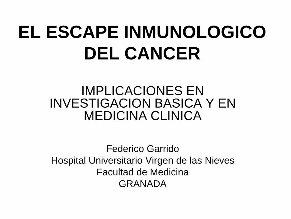
Malignant Transformation
Normal Cell
Tumor Cell
DNA mutations
Oncogenic activation
Radiation Virus Chemicals“Generation of New Antigens”
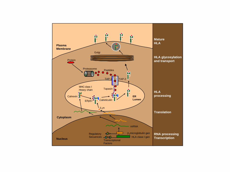
HLA clase I genβ2microglobulin gen
mRNA
TranscriptionalFactors
RegulatorySecuences
Nucleus
Cytoplasm
Plasma Membrane
ERLumen
Golgi
Protein
Proteasome Peptides
TAP-1 TAP-2
Calnexin
MHC class IHeavy chain
β2m
CalreticulinERp57
TapasinHLAprocessing
MatureHLA
HLA glycosylationand transport
Translation
RNA processingTranscription
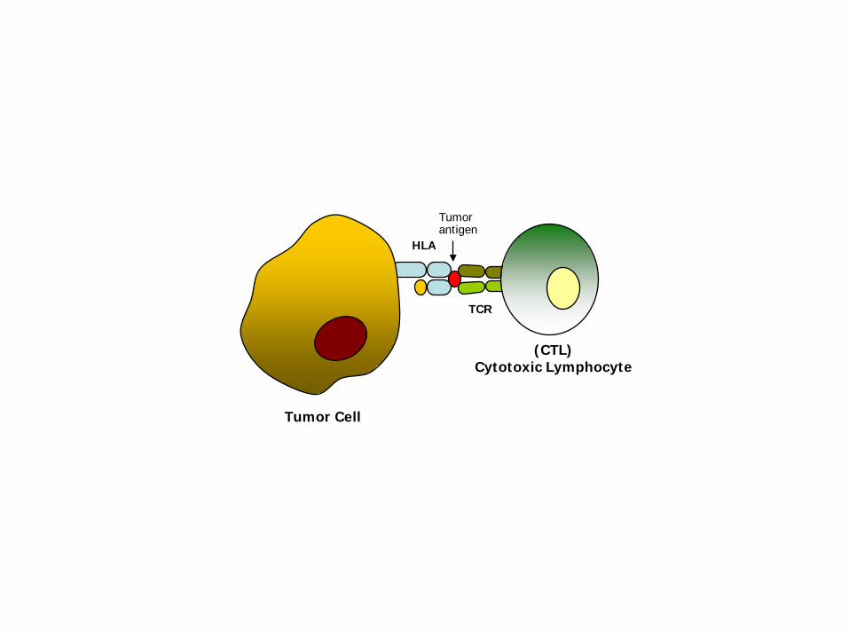
Tumor Cell
(CTL)Cytotoxic Lymphocyte
HLA
TCR
Tumor antigen

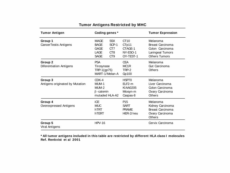
Tumor Antigen Coding genes * Tumor Expression
Group 1CancerTestis Antigens
SSXSCP-1CT7CT8 CT9
MAGEBAGEGAGELAGESAGE
CT10CTp11CTAGE-1NY-ESO-1OY-TEST-1
MelanomaBreast Carcinoma Colon Carcinoma Laringeal TumorsOthers Tumors
Group 2Diferentiation Antigens
PSATirosynaseTRP-1(gp75)MART-1/Melan-A
CEA MC1RTRP-2Gp100
MelanomaGut Carcinoma Others
Group 3Antigens originated by Mutation
CDK-4MUM-1MUM-2β−cateninmutaded HLA-A2
HSP70ELF2-mKIAA0205 Miosyn-mCaspas-8
MelanomaLiver Carcinoma Colon Carcinoma Ovary Carcinoma Others
Group 4Overexpressed Antigens
iCEMUChTRThTERT
P15 SART PRAMEHER-2/neu
MelanomaKidney Carcinoma Breast Carcinoma Ovary Carcinoma Others
Group 5Viral Antigens
HPV-16 Cervix Carcinoma
Tumor Antigens Restricted by MHC
*All tumor antigens included in this table are restricted by different HLA class I molecules Ref. Renkvist et al 2001
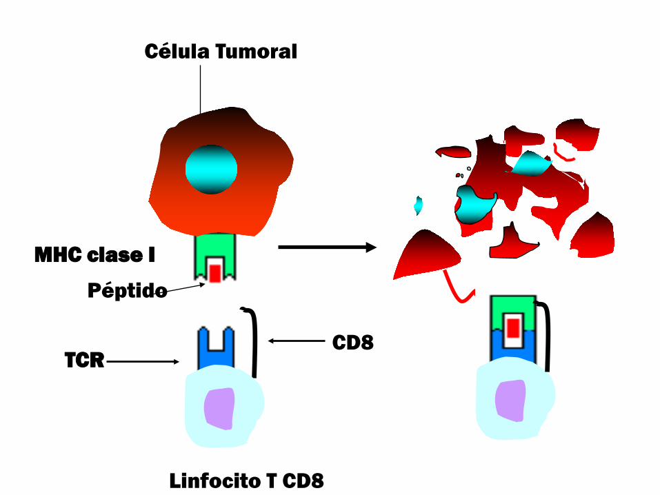
MHC clase I
Péptido
Célula Tumoral
Linfocito T CD8
TCRCD8
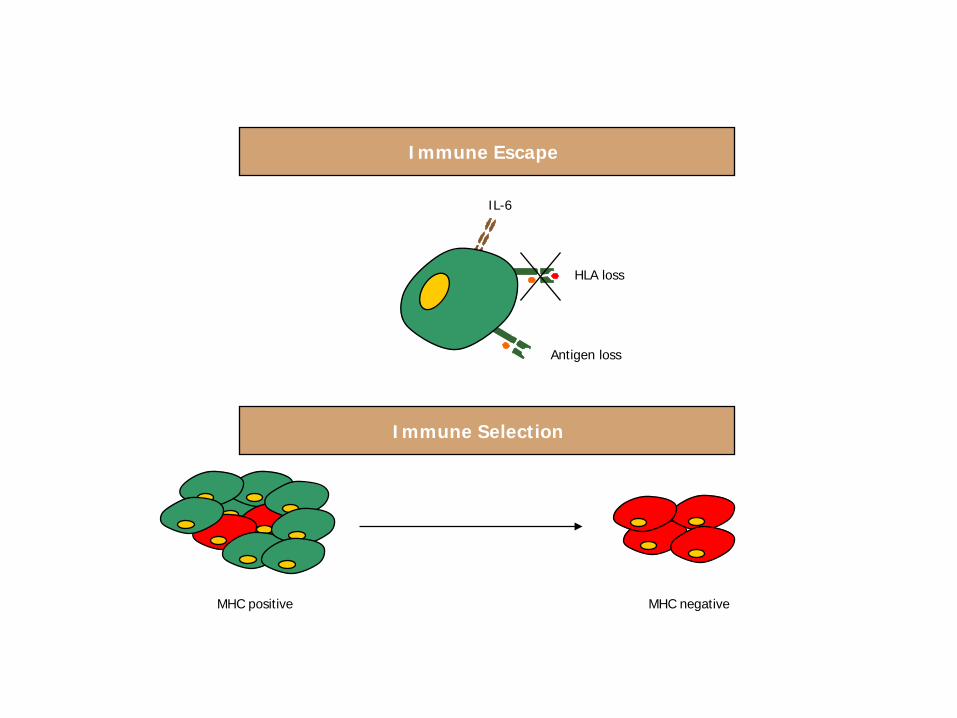
Immune Escape
Immune Selection
IL-6
HLA loss
Antigen loss
MHC positive MHC negative
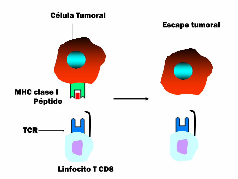
MHC clase IPéptido
Célula TumoralEscape tumoral
Linfocito T CD8
TCR
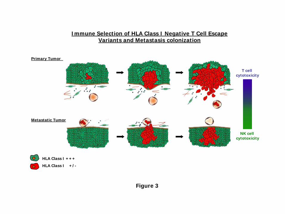
Immune Selection of HLA Class I Negative T Cell EscapeVariants and Metastasis colonization
Primary Tumor
Metastatic Tumor
HLA Class I +++
HLA Class I +/-
T cell cytotoxicity
NK cell cytotoxicity
Figure 3
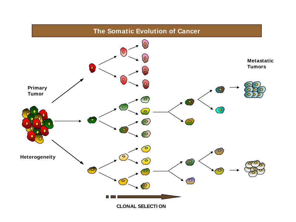
The Somatic Evolution of Cancer
CLONAL SELECTION
Heterogeneity
Primary Tumor
MetastaticTumors
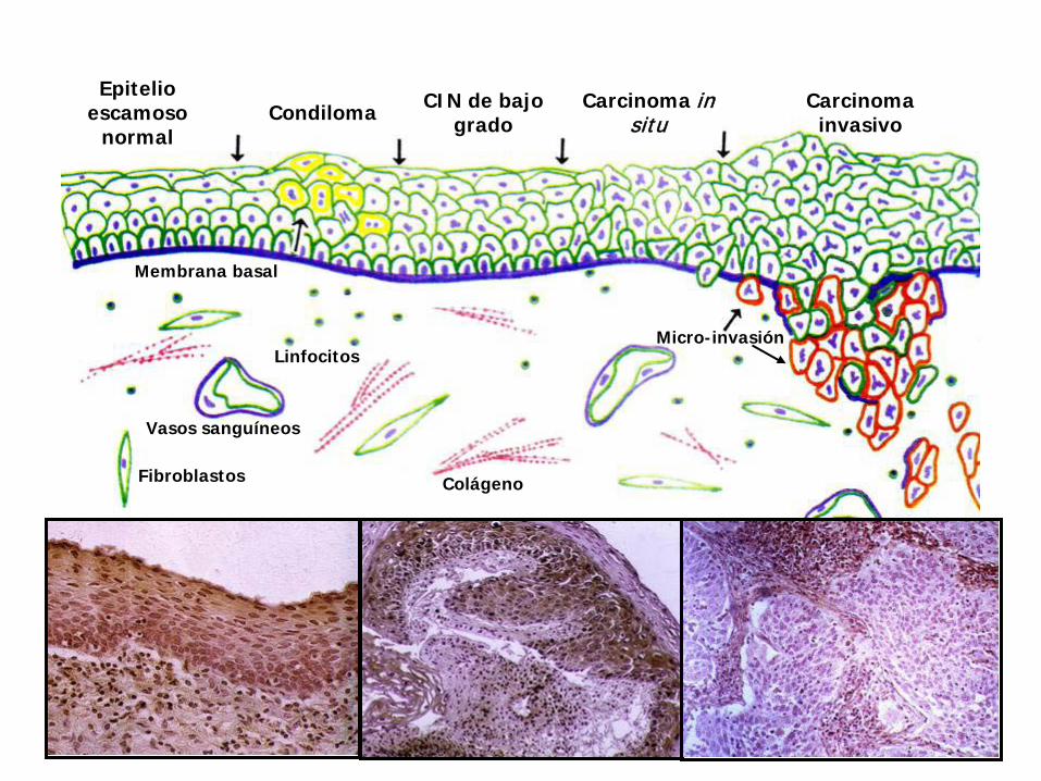
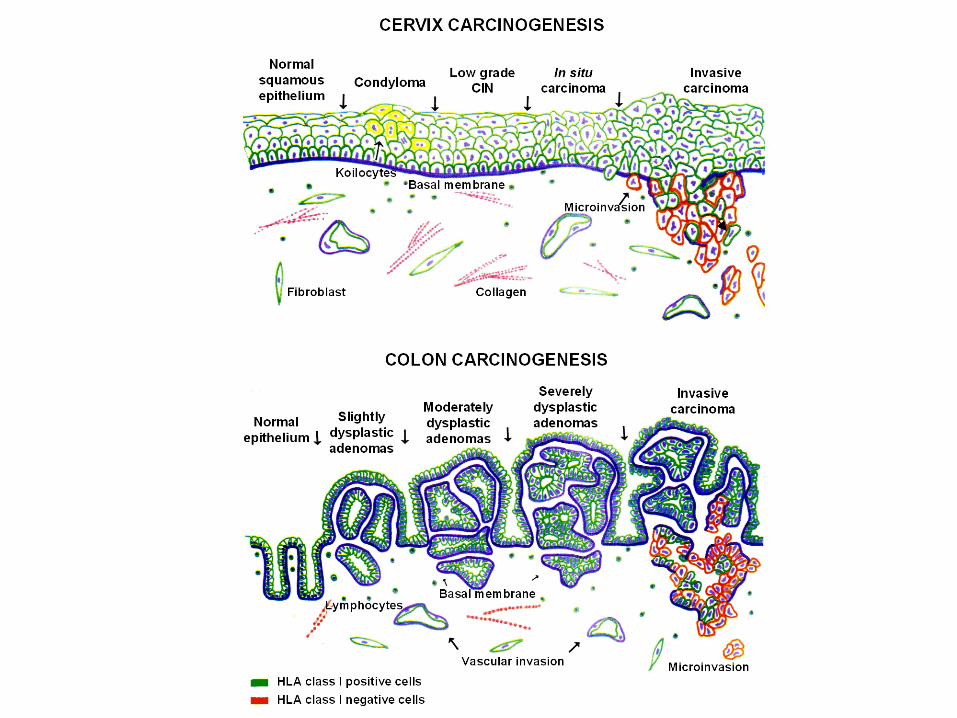
Epitelio escamoso
normalCondiloma CIN de bajo
gradoCarcinoma in
situCarcinoma
invasivo
Membrana basal
Micro-invasiónLinfocitos
ColágenoFibroblastos
Vasos sanguíneos
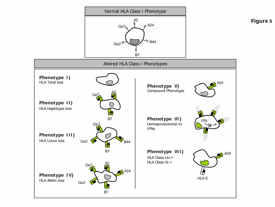
Normal HLA Class I Phenotype
Altered HLA Class I Phenotypes
A2
B7
A24Cw7
Cw2
B44
Cw7
Cw2
B7
B7
Cw7A2
Phenotype I)HLA Total loss
HLA Haplotype lossPhenotype II)
HLA Locus lossPhenotype III)
HLA Allelic lossPhenotype IV)
Compound PhenotypePhenotype V)
A24
Unresponsiveness toIFNs
Phenotype VI)
HLA Class Ia+/-HLA Class Ib +
Phenotype VII)
Cw2
Cw7
A2A24
B44
B7
HLA E
IFNX
A24
Figure 5
PHENOTYPE Nº I
HLA TOTAL LOSS
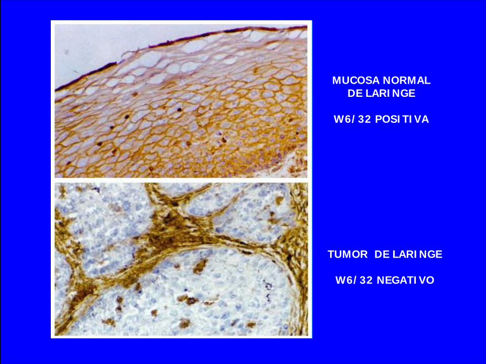
MUCOSA NORMAL DE LARINGE
W6/32 POSITIVA
TUMOR DE LARINGE
W6/32 NEGATIVO
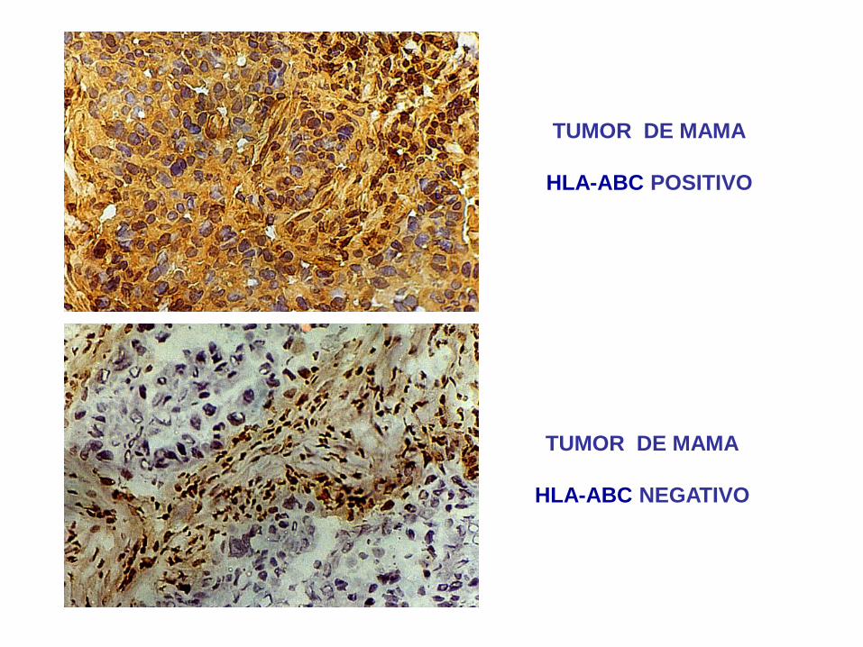
TUMOR DE MAMA
HLA-ABC POSITIVO
TUMOR DE MAMA
HLA-ABC NEGATIVO
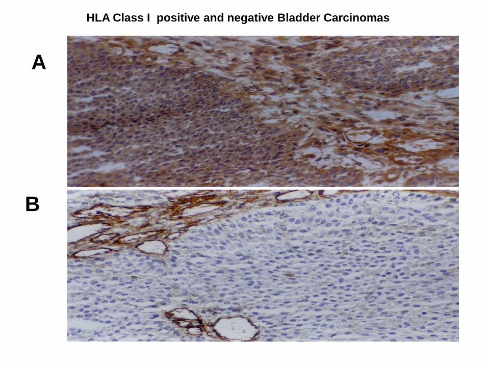
A
B
HLA Class I positive and negative Bladder Carcinomas
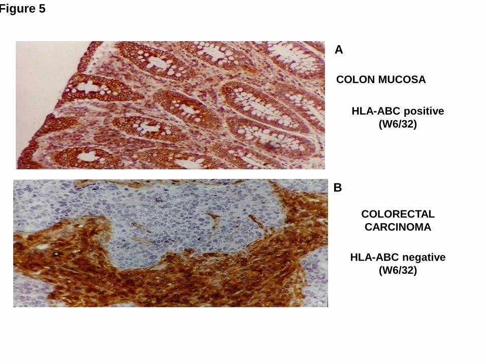
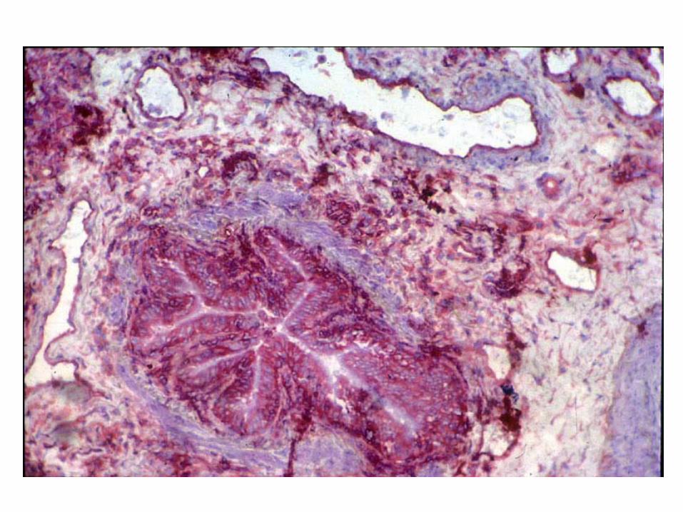
COLORECTAL CARCINOMA
HLA-ABC positive(W6/32)
HLA-ABC negative(W6/32)
COLON MUCOSA
Figure 5
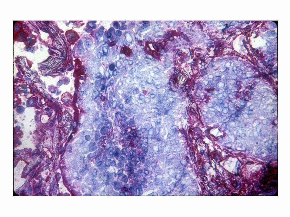
A
B
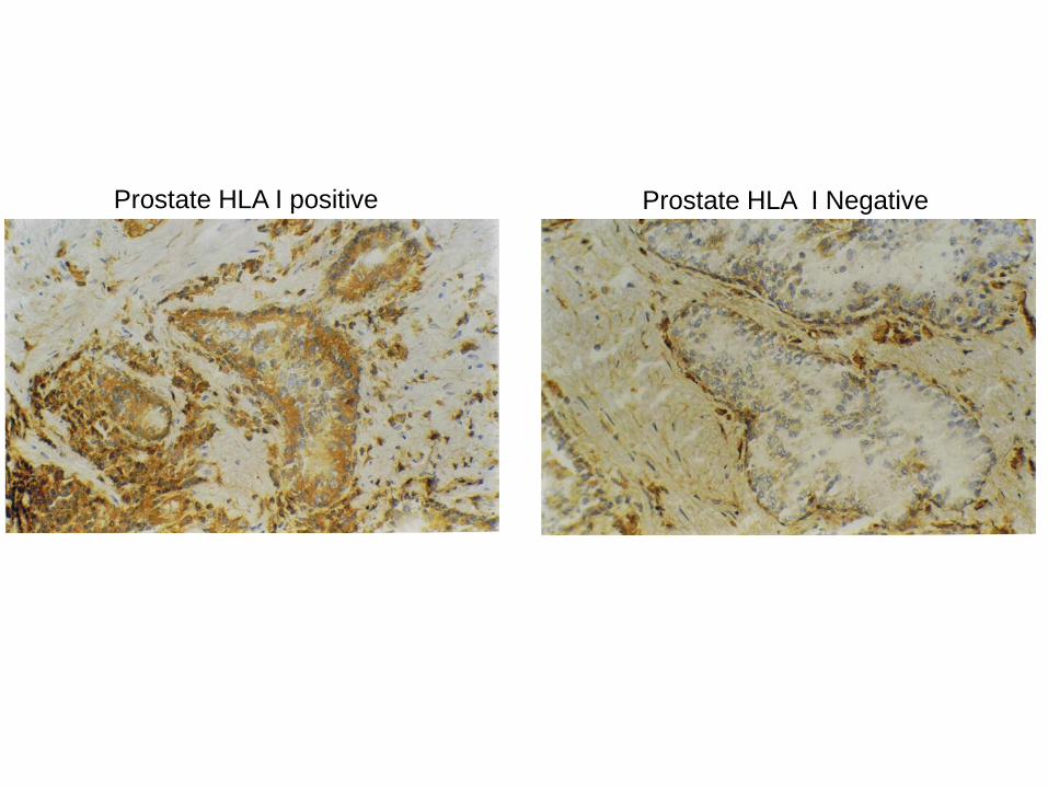
Prostate HLA I positive Prostate HLA I Negative
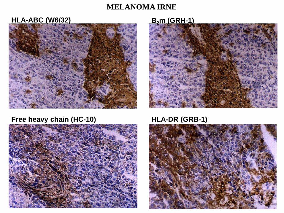
HLA-ABC (W6/32) B2m (GRH-1)
Free heavy chain (HC-10) HLA-DR (GRB-1)
MELANOMA IRNE
PHENOTYPE Nº II
HLA HAPLOTYPE LOSS
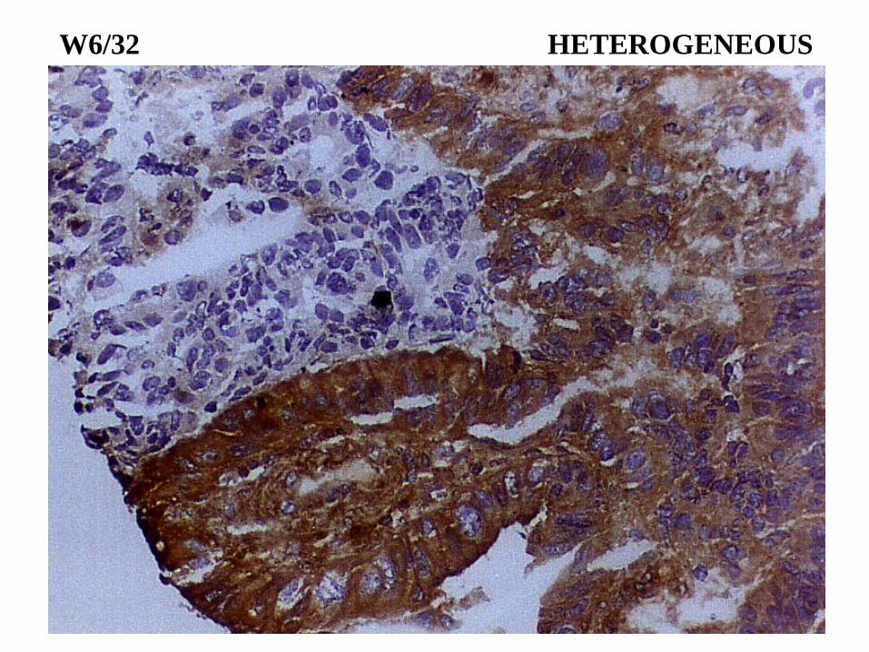
W6/32 HETEROGENEOUS
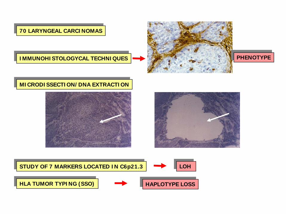
70 LARYNGEAL CARCINOMAS
IMMUNOHISTOLOGYCAL TECHNIQUES
MICRODISSECTION/DNA EXTRACTION
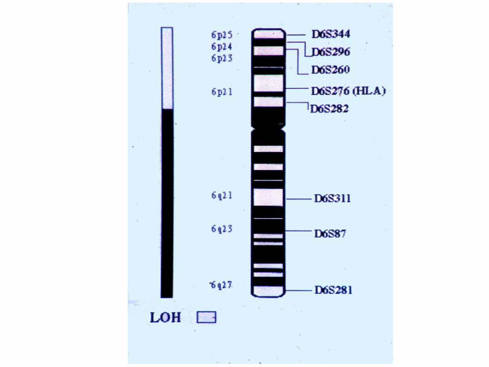
STUDY OF 7 MARKERS LOCATED IN C6p21.3
HLA TUMOR TYPING (SSO)
PHENOTYPE
LOH
HAPLOTYPE LOSS
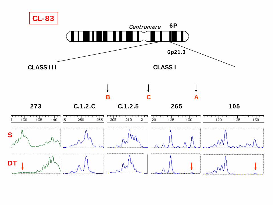
CL-836P
CLASS III CLASS I
6p21.3
Centromere
B C A105265C.1.2.C273 C.1.2.5
DT
S
PHENOTYPE Nº IV
HLA ALLELIC LOSS
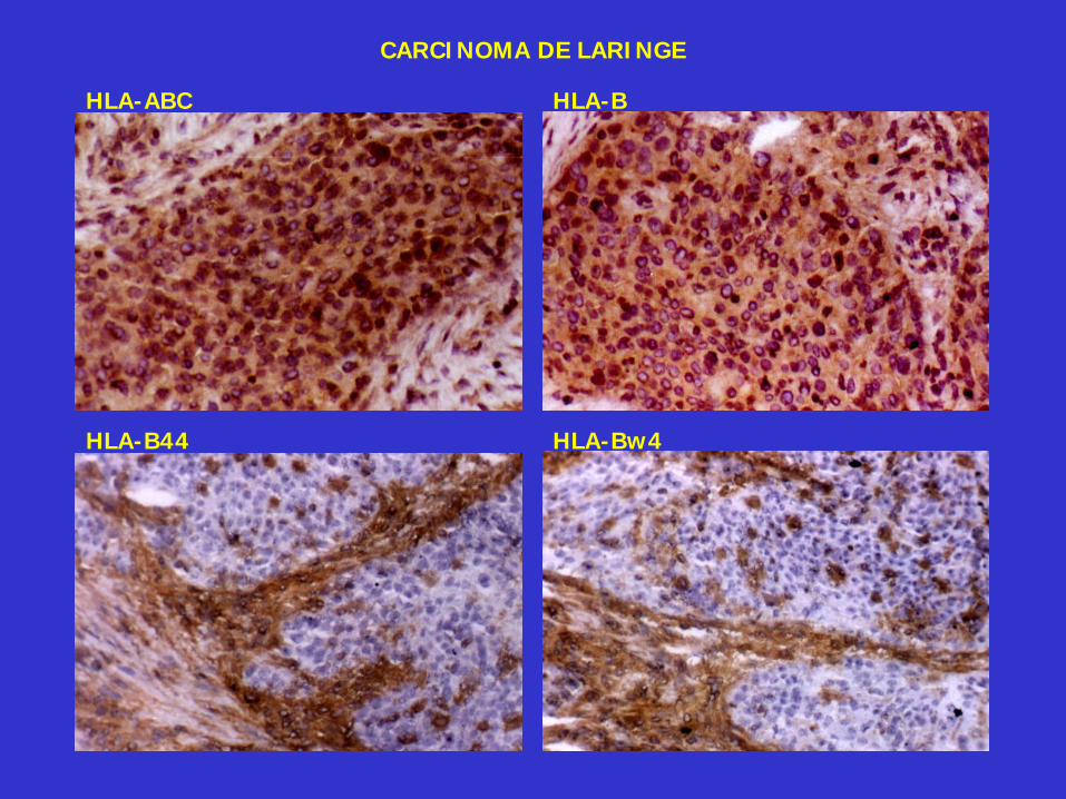
HLA-BHLA-ABC
HLA-B44 HLA-Bw4
CARCINOMA DE LARINGE
HLA EXPRESSION AND CANCER
IMMUNOTHERAPY
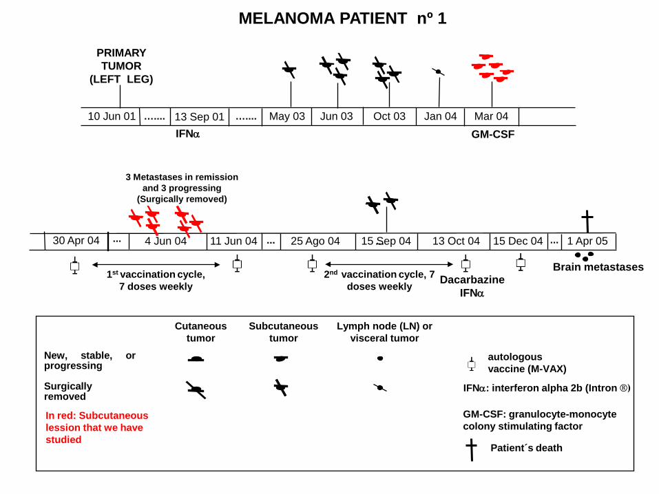
New, stable, orprogressing
Surgicallyremoved
3 Metastases in remissionand 3 progressing
(Surgically removed)
autologousvaccine (M-VAX)
...
IFNα
PRIMARY TUMOR
(LEFT LEG)
MELANOMA PATIENT nº 1
10 Jun 01 ….... 13 Sep 01GM-CSF
May 03 Oct 03 Jan 04 Mar 04
4 Jun 04 11 Jun 04 15 Sep 04 15 Dec 04
…....
13 Oct 04
Jun 03
Cutaneoustumor
Subcutaneoustumor
Lymph node (LN) or visceral tumor
In red: Subcutaneous lession that we have studied
Brain metastases
IFNα: interferon alpha 2b (Intron ®)
GM-CSF: granulocyte-monocyte colony stimulating factor
Patient´s death
1 Apr 0525 Ago 04 ......
DacarbazineIFNα
30 Apr 04
1st vaccination cycle, 7 doses weekly
...
2nd vaccination cycle, 7 doses weekly
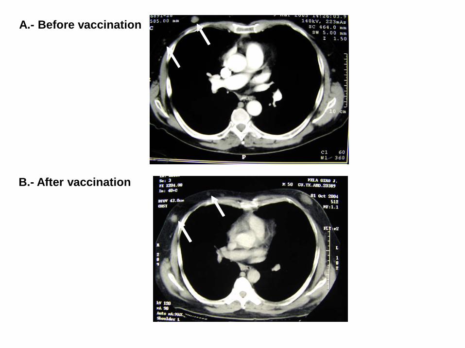
A.- Before vaccination
B.- After vaccination
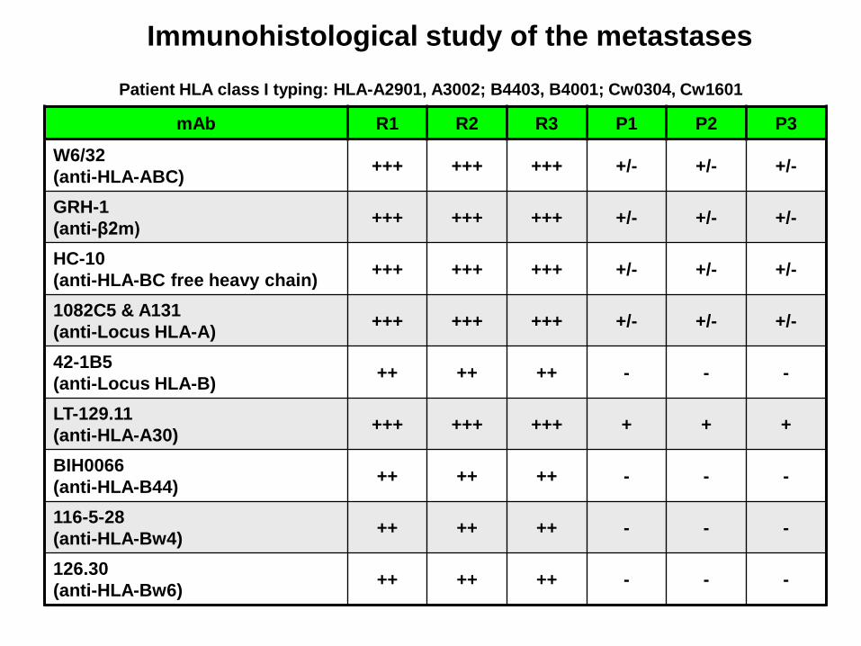
mAb R1 R2 R3 P1 P2 P3
W6/32(anti-HLA-ABC) +++ +++ +++ +/- +/- +/-
GRH-1(anti-β2m) +++ +++ +++ +/- +/- +/-
HC-10(anti-HLA-BC free heavy chain) +++ +++ +++ +/- +/- +/-
1082C5 & A131(anti-Locus HLA-A) +++ +++ +++ +/- +/- +/-
42-1B5(anti-Locus HLA-B) ++ ++ ++ - - -
LT-129.11(anti-HLA-A30) +++ +++ +++ + + +
BIH0066(anti-HLA-B44) ++ ++ ++ - - -
116-5-28(anti-HLA-Bw4) ++ ++ ++ - - -
126.30(anti-HLA-Bw6) ++ ++ ++ - - -
Immunohistological study of the metastasesPatient HLA class I typing: HLA-A2901, A3002; B4403, B4001; Cw0304, Cw1601
0
20
40
60
80
100
120
R1 R2 R3 P1 P2 P3
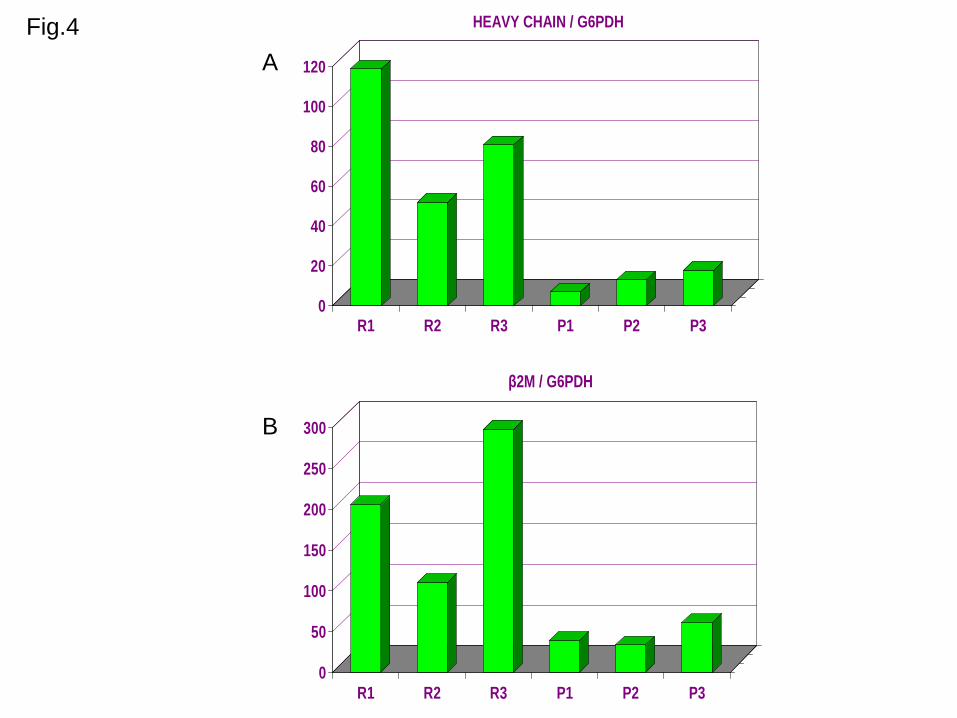
HEAVY CHAIN / G6PDH
0
50
100
150
200
250
300
R1 R2 R3 P1 P2 P3
β2M / G6PDH
Fig.4A
B
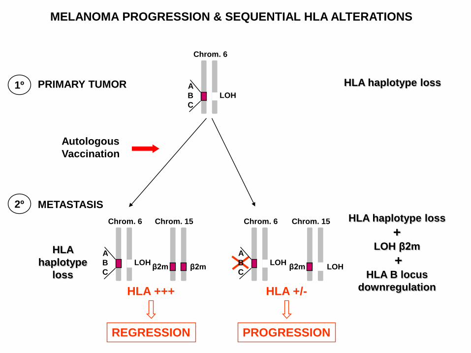
MELANOMA PROGRESSION & SEQUENTIAL HLA ALTERATIONS
PRIMARY TUMOR
REGRESSION PROGRESSION
HLA +/-
ABC
Chrom. 6
LOH
HLA +++
ABC
Chrom. 6
LOH
Chrom. 15
β2m β2mABC
Chrom. 6
LOH
Chrom. 15
β2m LOH
1º
METASTASIS2º
HLA haplotype loss
HLA haplotype loss+
LOH β2m+
HLA B locus downregulation
Autologous Vaccination
HLA haplotype
loss
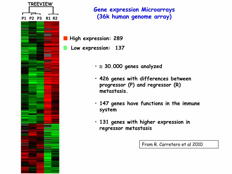
High expression: 289
Low expression: 137
P1 P2 P3 R1 R2
TREEVIEWGene expression Microarrays (36k human genome array)
• ≅ 30.000 genes analyzed
• 426 genes with differences between progressor (P) and regressor (R) metastasis.
• 147 genes have functions in the immune system
• 131 genes with higher expression in regressor metastasis
From R. Carretero et al 2010
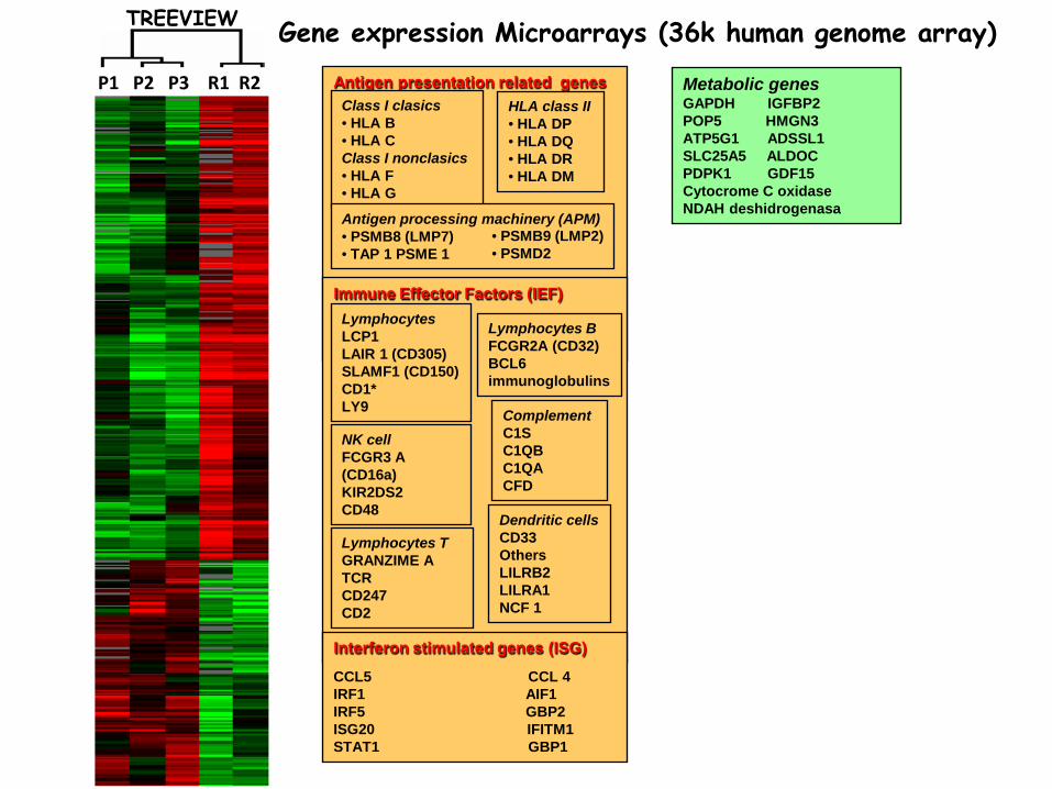
Metabolic genesGAPDH IGFBP2 POP5 HMGN3 ATP5G1 ADSSL1 SLC25A5 ALDOC PDPK1 GDF15 Cytocrome C oxidaseNDAH deshidrogenasa
P1 P2 P3 R1 R2
TREEVIEW Gene expression Microarrays (36k human genome array)
Antigen presentation related genesHLA class II• HLA DP • HLA DQ • HLA DR • HLA DM
Class I clasics • HLA B • HLA C Class I nonclasics• HLA F • HLA G
Antigen processing machinery (APM)• PSMB8 (LMP7)• TAP 1 PSME 1
Immune Effector Factors (IEF)
Complement C1S C1QB C1QA CFD
LymphocytesLCP1LAIR 1 (CD305) SLAMF1 (CD150)CD1*LY9
Lymphocytes BFCGR2A (CD32)BCL6immunoglobulins
NK cellFCGR3 A (CD16a) KIR2DS2CD48
Lymphocytes TGRANZIME ATCRCD247CD2
Dendritic cellsCD33OthersLILRB2LILRA1NCF 1
Interferon stimulated genes (ISG)
CCL5 CCL 4IRF1 AIF1IRF5 GBP2ISG20 IFITM1STAT1 GBP1
• PSMB9 (LMP2)• PSMD2
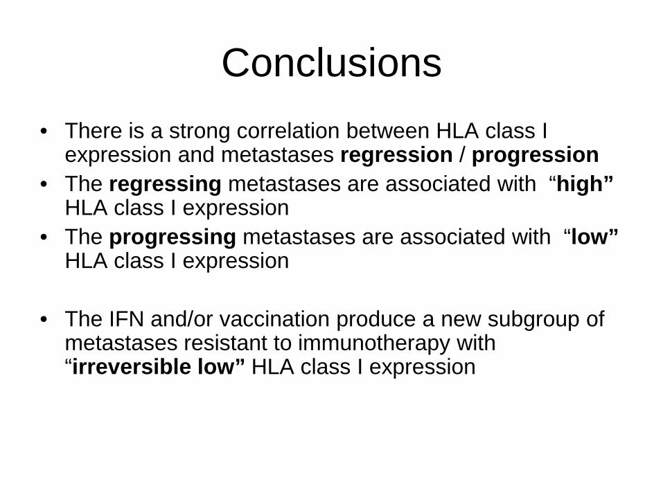
Conclusions• There is a strong correlation between HLA class I
expression and metastases regression / progression• The regressing metastases are associated with “high”
HLA class I expression• The progressing metastases are associated with “low”
HLA class I expression
• The IFN and/or vaccination produce a new subgroup of metastases resistant to immunotherapy with “irreversible low” HLA class I expression
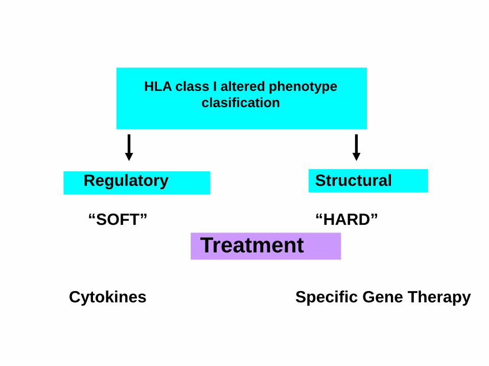
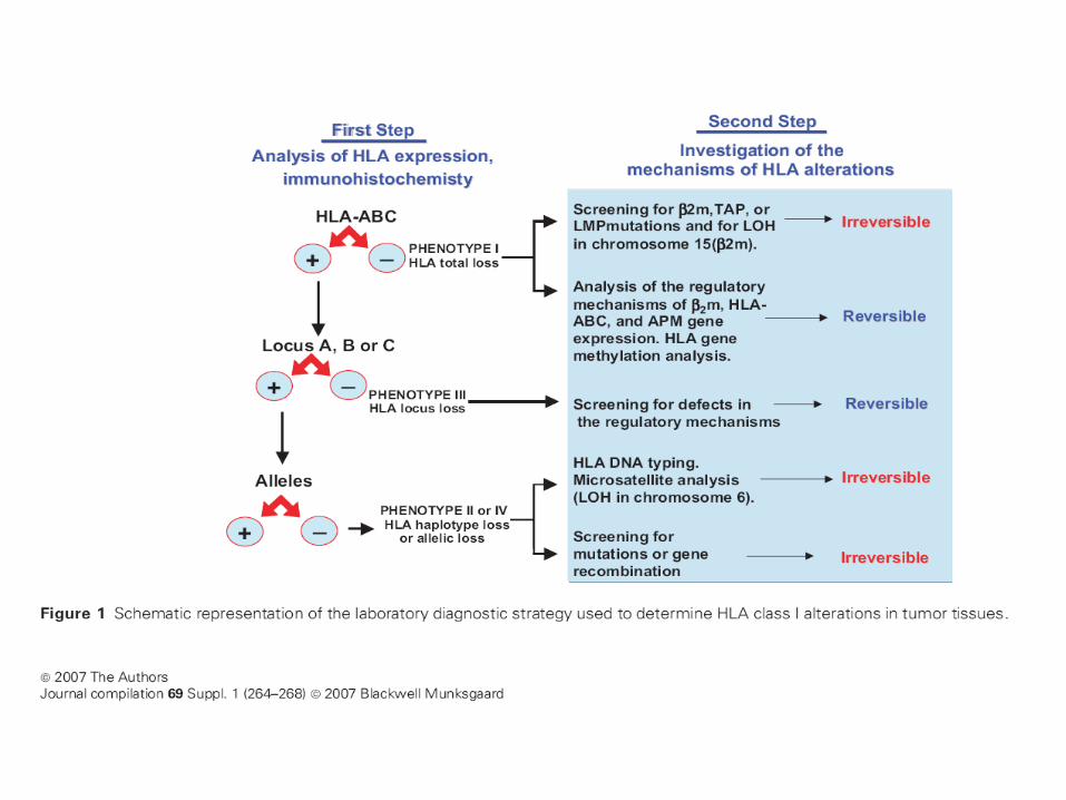
HLA class I altered phenotype clasification
Treatment
Regulatory
“SOFT”
Structural
“HARD”
Cytokines Specific Gene Therapy
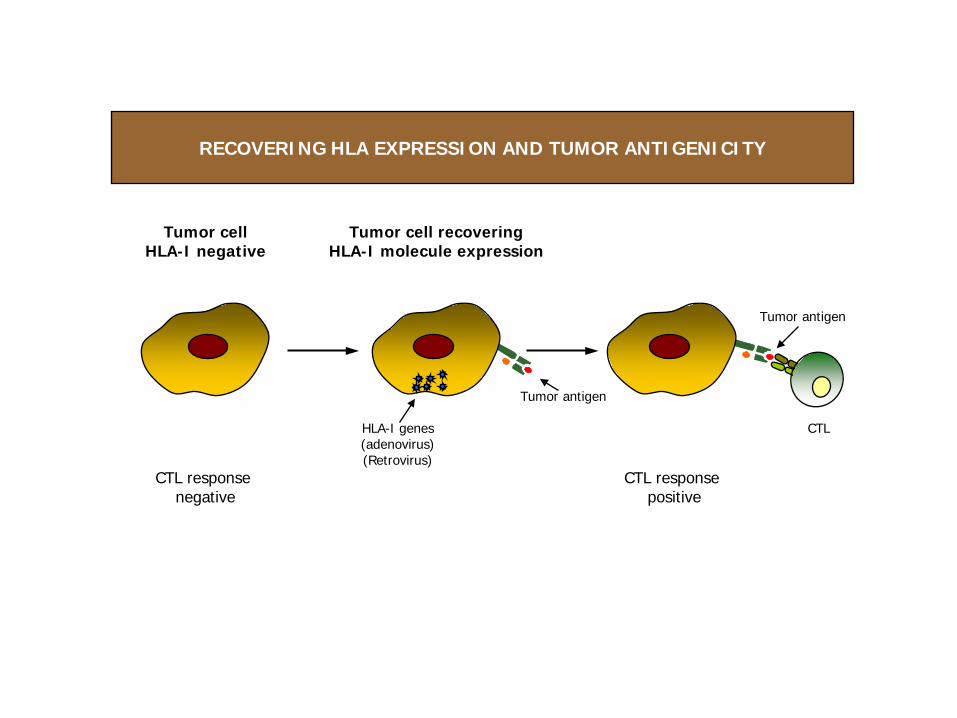
Tumor cellHLA-I negative
Tumor cell recoveringHLA-I molecule expression
CTL response negative
HLA-I genes(adenovirus)(Retrovirus)
CTL response positive
CTL
Tumor antigen
Tumor antigen
RECOVERING HLA EXPRESSION AND TUMOR ANTIGENICITY
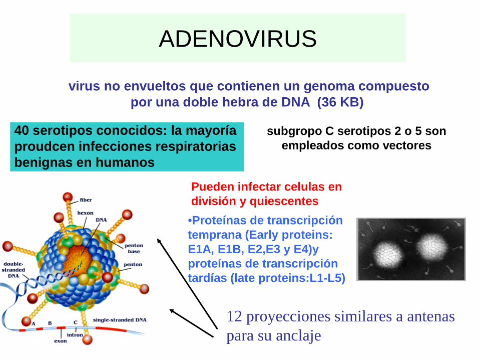
ADENOVIRUS
virus no envueltos que contienen un genoma compuesto por una doble hebra de DNA (36 KB)
40 serotipos conocidos: la mayoría proudcen infecciones respiratorias benignas en humanos
subgropo C serotipos 2 o 5 son empleados como vectores
Pueden infectar celulas en división y quiescentes
12 proyecciones similares a antenas para su anclaje
•Proteínas de transcripción temprana (Early proteins: E1A, E1B, E2,E3 y E4)y proteínas de transcripción tardías (late proteins:L1-L5)
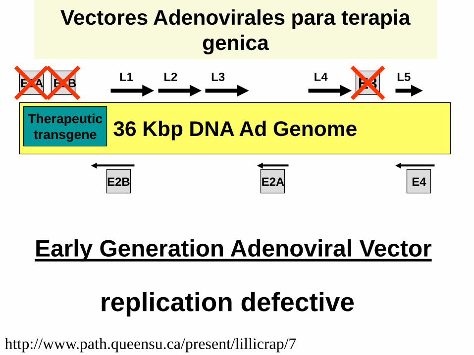
36 Kbp DNA Ad Genome
E1A E3E1B
E2A E4E2B
L1 L2 L4L3 L5
Therapeutictransgene
Early Generation Adenoviral Vector
replication defectivehttp://www.path.queensu.ca/present/lillicrap/7
Vectores Adenovirales para terapia genica
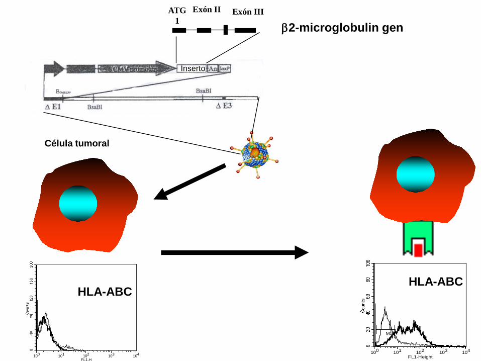
Inserto
100 101 102 103 104FL1-H
ATG1
Exón II Exón III
β2-microglobulin gen
100 101 102 103 104FL1-Height
M1
HLA-ABCHLA-ABC
Célula tumoral
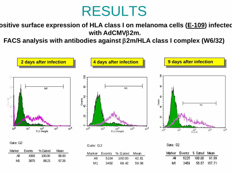
RESULTS
100 101 102 103 104FL1-Height
M1
Marker Events % Gated MeanAll 5104 100.00 42.81M1 3492 68.42 59.58
Gate: G2
2 days after infection
Marker Events % Gated MeanAll 4393 100.00 86.93M1 3875 88.21 97.26
Gate: G2
4 days after infection
100 101 102 103 104FL1-H
M1
9 days after infection
ositive surface expression of HLA class I on melanoma cells (E-109) infected with AdCMVβ2m.
FACS analysis with antibodies against β2m/HLA class I complex (W6/32)
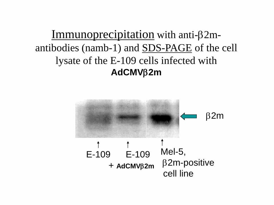
Immunoprecipitation with anti-β2m-antibodies (namb-1) and SDS-PAGE of the cell
lysate of the E-109 cells infected with AdCMVβ2m
E-109 E-109 + AdCMVβ2m
Mel-5, β2m-positivecell line
β2m
DEPT. OF ANALISIS CLINICOS E INMUNOLOGIAHospital Virgen Nieves
GRANADA SPAIN
Dr. Francisco Ruiz-Cabello•Dra. Pilar Jimenez•Julia Canton•Dra.Isabel Maleno•Monica Bernall
Dr. Angel Miguel García Lora• Cristina Garrido Lopez• Irene Romero•Dr. Ignacio Algarra. Univ. Jaen•Dra Antonia Collado
Dra. Teresa Cabrera• Rafael Carretero• Pablo Saenz Lopez•Dr.Jose Maria Romero
MOLECULAR MECHANISMSHLA GENOTYPINGLOH STUDIESIMMUNOHISTOLOGY
MICRODISSECTION
EXPERIMENTAL MOUSE MODELS
GENE THERAPY
Dra.Natalia Apsiauri•Dra.Rosa Mendez•Ana del Campo•Javier Carretero


![Cancer Biology 2019;9(1) · 2019. 2. 6. · Oxidant / antioxidant parameters in breast cancer patients and its relation to VEGF, TGF-β or Foxp3 factors. Cancer Biology 2019;9(1):5-17].](https://static.fdocument.org/doc/165x107/60fa2d04f21a9b206b77c605/cancer-biology-201991-2019-2-6-oxidant-antioxidant-parameters-in-breast.jpg)


![MicroRNA-505 functions as a tumor suppressor in ... · nant tumors, including osteosarcoma, hepatic cancer, prostate cancer and breast cancer [20, 22, 26, 32, 33]. Recent studies](https://static.fdocument.org/doc/165x107/5f024f927e708231d403a367/microrna-505-functions-as-a-tumor-suppressor-in-nant-tumors-including-osteosarcoma.jpg)