Effects of Delta Np73 beta on cisplatin treatment in colon...
28
Effects of Delta Np73 beta on cisplatin treatment in colon cancer cells Jasmine Loof, Daniella Pfeifer, Zhen-Yu Ding, Xiao-Feng Sun and Hong Zhang Linköping University Post Print N.B.: When citing this work, cite the original article. This is the authors’ version of the following article: Jasmine Loof, Daniella Pfeifer, Zhen-Yu Ding, Xiao-Feng Sun and Hong Zhang, Effects of Delta Np73 beta on cisplatin treatment in colon cancer cells, 2012, Molecular Carcinogenesis, (51), 8, 628-635. which has been published in final form at: http://dx.doi.org/10.1002/mc.20835 Copyright: Wiley-Blackwell http://eu.wiley.com/WileyCDA/Brand/id-35.html Postprint available at: Linköping University Electronic Press http://urn.kb.se/resolve?urn=urn:nbn:se:liu:diva-79660
Transcript of Effects of Delta Np73 beta on cisplatin treatment in colon...
Effects of Delta Np73 beta on cisplatin
treatment in colon cancer cells
Jasmine Loof, Daniella Pfeifer, Zhen-Yu Ding, Xiao-Feng Sun and Hong Zhang
Linköping University Post Print
N.B.: When citing this work, cite the original article.
This is the authors’ version of the following article:
Jasmine Loof, Daniella Pfeifer, Zhen-Yu Ding, Xiao-Feng Sun and Hong Zhang, Effects of
Delta Np73 beta on cisplatin treatment in colon cancer cells, 2012, Molecular Carcinogenesis,
(51), 8, 628-635.
which has been published in final form at:
http://dx.doi.org/10.1002/mc.20835
Copyright: Wiley-Blackwell
http://eu.wiley.com/WileyCDA/Brand/id-35.html
Postprint available at: Linköping University Electronic Press
http://urn.kb.se/resolve?urn=urn:nbn:se:liu:diva-79660
Lööf et al
1
Effects of ΔNp73β on Cisplatin Treatment in Colon Cancer Cells
Jasmine Lööf1, Daniella Pfeifer
2, Zhenyu Ding
2, Xiao-Feng Sun
2 and Hong Zhang
1
1Division of Tumor Biology, Systems Biology Research Centre, University of
Skövde, Sweden
2Division of Oncology, Department of Clinical and Experimental Medicine, Faculty
of Health Sciences, Linköping University, Sweden
Corresponding authors: Xiao-Feng Sun, Division of Oncology, Department of
Clinical and Experimental Medicine, Faculty of Health Science, Linköping
University, SE-58185 Linköping, Sweden. Tel: +46-10-1032066, Fax: +46-10-
1033090. E-mail: [email protected];
Hong Zhang, Division of Tumor Biology, Systems Biology Research Centre,
University of Skövde, SE-54128 Skövde, Sweden, Tel: +46-500-448482, Fax: +46-
500-448499, E-mail: [email protected]
Lööf et al
2
Grant support: This study was supported by grants from the Swedish Cancer
Foundation, Swedish Research Council and the Health Research Council in the
South-East of Sweden.
Abbreviated title: p73 in colon cancer cells
Keywords: Cell death; HCT116 cells; HT29 cells; p73 protein; p53
Lööf et al
3
ABSTRACT
p73 can activate transcription of p53-responsive genes, thereby inhibiting cell
growth. An alternative promoter in the TP73 gene gives rise to an N- terminally
truncated isoform of p73, ∆Np73, which lacks the transactivation domain of the full
length TAp73 protein. TAp73 is considered pro-apoptotic, and ∆Np73 anti-
apoptotic. In this study, we overexpressed ∆Np73β in p53 wild type and p53 mutant
colon cancer cell lines and further exposed the cells to cancer therapeutic drug
cisplatin. The results showed that cisplatin decreased the protein expression levels of
∆Np73β in a dose- dependent manner, and both TAp73 and p53 were upregulated
after cisplatin treatment. Further, clonogenic potential and cell viability were
decreased, and apoptotic cells increased, in p53 mutant and in p53 wild type cells.
Cellular viability was significantly higher in ∆Np73β-cells than mock-transfected
cells. However, ∆Np73β overexpression did not affect the cellular susceptibility to
cisplatin. In conclusion, the overexpression of ∆Np73β increases viability in p53
wild type and p53 mutant colon cancer cells, and cisplatin induces the degradation
of ∆Np73β in a dose-dependent manner.
Lööf et al
4
INTRODUCTION
p73 protein is encoded by the TP73 gene located to chromosome 1p36, a region
frequently deleted in various cancers [1]. The structure and function of the p73
protein are homologous to p53, and it activates the transcription of p53-responsive
genes and inhibits cell growth in a p53-like manner by inducing apoptosis [2]. It
has been speculated that p73, like p53, is a tumor suppressor. However, initial
genetic studies showed that p73-deficient mice do not develop spontaneous tumors
[3]. Inactivation of p53 has been shown in the majority of human malignancies [4],
while p73 mutations are rare in primary tumors [5]. Overexpression of p73 has been
found in various tumor types, such as colorectal, breast, ovarian and lung cancers
[6-9], suggesting that p73 plays an oncogenic role in tumorigenesis. Further,
overexpression of the p73 protein has been associated with a poor prognosis in
several types of cancers [6,10].
TP73 gene gives rise to several different mRNAs, and thus a number of different
isoforms of the protein differing both N-and C- terminally. There are two N-terminal
isoforms of p73, the full length proapoptotic variant of the protein, TAp73, and the
N-terminally truncated form, ∆Np73. The ∆Np73 isoform arises through the use of
an alternative promoter located in intron 3 [3,11]. There are also several C-terminal
splice variants of the protein termed α, β, etc [1,12-14].
The TAp73 isoform is homologous to p53; harboring the N-terminal transactivating
(TA) domain responsible for gene activation [2], while the ∆-isoforms lack the TA
domain [3]. ∆Np73 shares the DNA-binding and oligomerisation domains with
TAp73 and inhibits p53 and TAp73, either by competing for DNA-binding sites or
by oligomerizing with the full length proteins [14-15]. Interestingly, the alternative
Lööf et al
5
promoter in intron 3 contains a p53/TAp73 responsive element, and p53, TAp73 and
∆Np73 regulate each other through a negative feedback loop where ∆Np73 displays
dominant negative behavior [11,16,17]. This indicates that the oncogenic properties
of p73 are attributed to the ∆-isoforms. ∆Np73 is involved in development of the
brain in mouse, where it protects the neuronal cells from apoptosis [18].
Furthermore, knock-out mice lacking the TA-isoforms of p73 are prone to
developing tumors [19]. This supports the idea that full length p73 has tumor
suppressor functions which are balanced by the anti-apoptotic ∆-isoforms, and the
ratio of the different isoforms determines p73 functions. Further complicating the
scenario is the differential behavior of the C-terminal isoforms. The anti-apoptotic
role of ΔNp73 β is not clearly established, as results from studies of the β-isoform
seem ambiguous [20-21].
Cisplatin (cis-diammine-dichloro-platinum) is widely used as DNA-damaging drug
in cancer therapy. It interacts with DNA, resulting in the formation of DNA adducts,
primarily intrastrand crosslinks [22]. Subsequently, it induces DNA damage
recognition proteins to signal to downstream effectors such as p53, resulting in cell
cycle arrest and apoptosis. It is well-known that p53 plays an important role in
response to DNA-damaging agents, with p53-/-
cells being resistant to drug-induced
apoptosis [23]. Also, p73 is activated by DNA damaging drugs, including
cisplatin [24-25], and silencing of p73 with siRNA results in drug resistance in
various cancer cells [24,26]. In this study, we attempted to clarify the mechanisms
behind the effects of ∆Np73β on cisplatin treatment in colon cancer cells.
Lööf et al
6
MATERIALS AND METHODS
Cell Culture and Transfection
Human colon carcinoma cell lines, HCT116 with wild type p53 (HCT116p53+/+
) and
truncated p53 missing 40 amino acid residues (HCT116p53-/-
) [27], as well as HT29
were cultivated in McCoy’s 5A medium (Sigma-Aldrich, St. Louis, MO)
supplemented with 10% FBS (GIBCO, Invitrogen, Carlsbad, CA), 1.5 mM L-
glutamine (GIBCO) and 1X PEST (GIBCO) at 37oC in a 5% CO2 incubator. The
HCT116p53-/-
cells are considered functionally p53 negative. The cells (1.5×10⁴
cells/cm2) were seeded in 6-, 12-, or 96- well plates under standard cell culture
conditions as described above for 24 h. HCT116 cells were then transfected with a
pCMV6-XL5 vector (OriGene, Rockville, MD) containing transfection-ready
cDNA for ∆Np73β ( NM_001126241.1, OriGene) using transfection reagent
FuGENE® 6 (Roche Diagnostics Corporation, Indianapolis, IN) according to the
manufacturer’s instructions. HT29 cells were transfected using the calcium
precipitation method. Briefly, plasmid DNA was mixed with HEPES buffer and
calcium chloride, and added with equal volume of 2 times HeBs buffer in a
dropwise fashion. The mixture was incubated for 20 min at room temperature and
added to the culture medium. The pCMV6-XL5 vector lacking the cDNA insert was
used as a negative control. The cells were then treated with various concentrations of
cisplatin (Sigma-Aldrich) 24 h after transfection.
Colony Forming Assay
HCT116 cells and HT29 were transfected in six-well plates as described above.
After 24 h cells were trypsinized with TrypLE (Invitrogen, Carlsbad, CA) and
Lööf et al
7
1×10³ cells were seeded in six-well plates with or without cisplatin (0.6 µM). The
cells were grown under standard cell culture condition for seven days, fixed in 4%
buffered formaldehyde and stained with 5% Giemsa. The cell colonies were counted
and a survival fraction between cisplatin treated and control cells was calculated.
XTT Viability Assay
Cellular viability was determined at 48 and 72 h after the transfection using
TACS™ XTT assay in 96-well plates according to the manufacturer's instructions
(R&D Systems, Minneapolis, MN). The assay is based on the cleavage of the yellow
tetrazolium salt XTT (2,3-Bis(2-methoxy-4-nitro-5-sulfophenyl)-2H-tetrazolium-5-
carbox-anilide) into a soluble orange formazan dye. This reaction is attributed
mainly to the succinate-tetrazolium reductase system in the mitochondria of
metabolically active cells. The absorbance measured at 450 nm is proportional to the
number of viable cells.
Western Blot
The cells were lysed in RIPA buffer (150 mM NaCl, 2% Triton, 0.1% SDS, 50 mM
Tris pH 8.0) containing 1% Protease Inhibitor Cocktail (Sigma) at 48 and 72 h after
the transfection. Protein concentrations were determined by BCA Protein Assay
(Pierce, Woburn, MA). The samples (20 µg protein) were subjected to
electrophoresis (35 min, 200V) on precast Criterion Tris-HCl gels, 4-15% (Bio-
Rad, Hercules, CA) and electrotransferred on to polyvinylidene difluoride
membranes (Amersham Biosciences/GE healthcare, Piscataway, NJ). The
membranes were incubated with mouse monoclonal antibodies against ∆Np73
Lööf et al
8
(1:500), TAp73 (1:500) and p53 (1:1000) (Abcam, Cambridge, MA) at 4oC over
night, then with a secondary HRP-conjugated goat-anti-mouse antibody (1:1000,
DAKO Cytomation, Glostrup, Denmark) at room temperature for 1 h. The proteins
were detected using the Amersham ECL Plus Western Blot detection system
(Amersham biosciences/GE Healthcare) and visualized in a Fujifilm LAS-1000 CCD
camera (Fujifilm, Tokyo, Japan). Equal loading of protein samples was verified
using a primary polyclonal rabbit anti-β-actin antibody (1:1000, Cell Signalling
Technology, Danvers, MA) and a secondary polyclonal goat anti-rabbit antibody
(1:2000, DAKO).
M30-Apoptosense ELISA and DAPI Assay
Apoptosis was quantitatively detected by using M30-Apoptosense® ELISA kit
(Peviva, Bromma, Sweden) at 72 h after the transfection according to the
manufacturer’s instructions. Absorbance was measured at 450 nm and normalized
against total protein concentration in each sample.
The plates with cells were centrifuged to spin down apoptotic cells. Cells were
trypsinized, resuspended in PBS and centrifuged on to glass slides using a Shandon
Cytospin® 2 Cytocentrifuge (Thermo Scientific, Waltham, MA), then fixed with
4% buffered formaldehyde and mounted with VECTASHIELD® HardSet™
Mounting Medium containing DAPI (Vector Laboratories, Burlingame, CA). The
cells were visualized in a UV-light fluorescence microscope, and apoptotic cells
were examined.

Lööf et al
9
Statistical Analysis
All experiments were performed in triplicates on three different occasions. Student´s
t-test was performed to evaluate statistical significance. Data are represented as
means ± standard error of mean (SEM). Two-sided p-values below 0.05 were
considered as statistically significant.
Lööf et al
10
RESULTS
Colony Forming Assay
Transfected cells were treated with 0.6 µM cisplatin for 7 d, and cell colonies were
counted (Figure 1). Cisplatin markedly reduced the number of colonies, and a
quota between cisplatin treated samples and controls, the survival fraction, was
calculated. ∆Np73β-transfected HCT116p53+/+
cells had a survival fraction of
0.53±0.08 and mock-transfected cells had a fraction of 0.44±0.1. The survival
fractions were not significantly different (p=0.13). The survival fraction for
∆Np73β-transfected HCT116p53-/-
cells was 0.49±0.07 and for mock-transfected
cells 0.43±0.06. The survival fractions did not significantly differ (p=0.07). HT29
cells were highly resistant to cisplatin, as shown by results both from our
experiments and other reports [28-29]. The survival fraction for ∆Np73β-
transfected HT29 cells was 0.95±0.20 and for mock-transfected cells 0.91±0.22.
There was no significant difference (p=0.80).
Cellular Viability
Cellular viability was examined using the XTT assay after the treatment with
increasing concentrations of cisplatin (0-60 µM). The HCT116 cells were found to
respond to 10-20 µM of cisplatin, therefore these concentrations were chosen for
subsequent experiments.
Cellular viability was determined at 48 and 72 h after transfection. ∆Np73β-
transfected HCT116p53+/+
cells were significantly more viable than mock-
transfected cells at 48 h after the transfection (p=0.005, Figure 2A). The cellular
viability of HCT116p53+/+
cells was significantly decreased after treatment with 20
Lööf et al
11
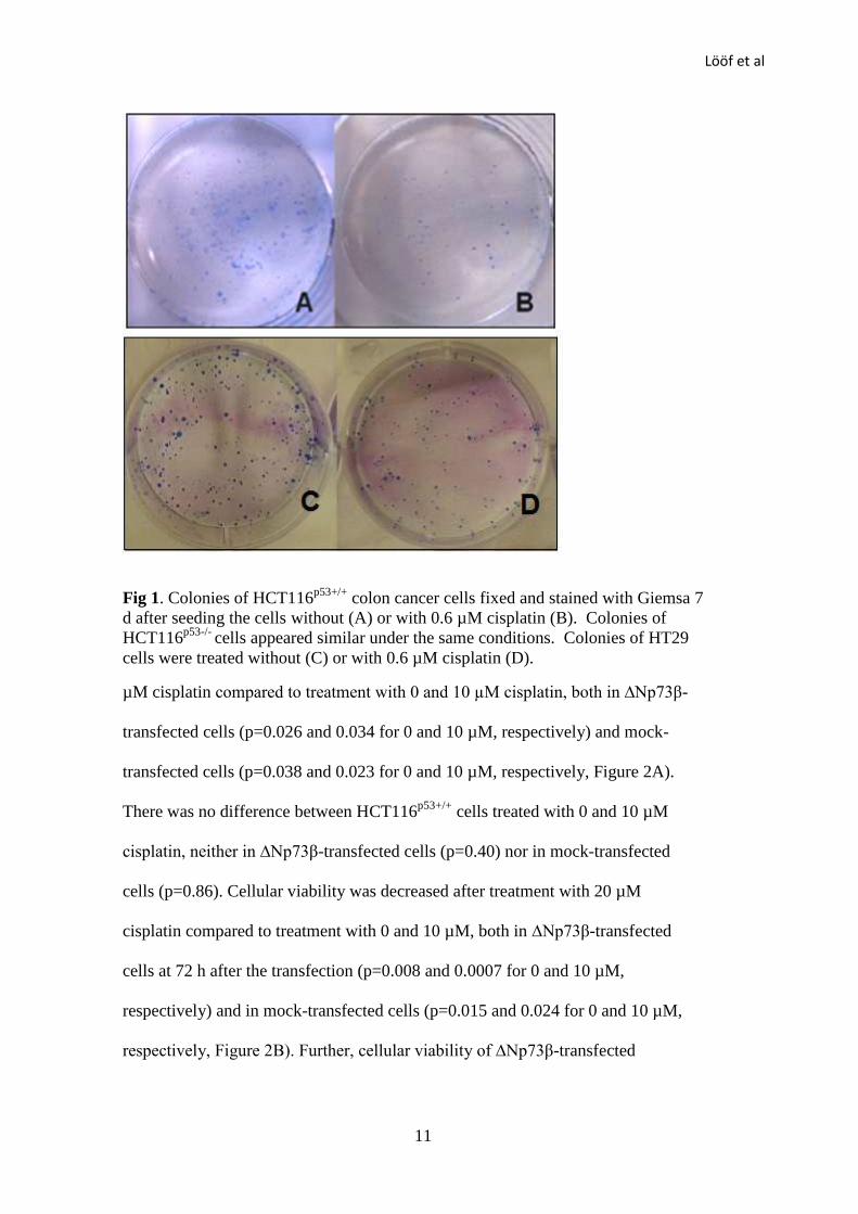
Fig 1. Colonies of HCT116p53+/+
colon cancer cells fixed and stained with Giemsa 7
d after seeding the cells without (A) or with 0.6 µM cisplatin (B). Colonies of
HCT116p53-/-
cells appeared similar under the same conditions. Colonies of HT29
cells were treated without (C) or with 0.6 µM cisplatin (D).
µM cisplatin compared to treatment with 0 and 10 µM cisplatin, both in ∆Np73β-
transfected cells (p=0.026 and 0.034 for 0 and 10 µM, respectively) and mock-
transfected cells (p=0.038 and 0.023 for 0 and 10 µM, respectively, Figure 2A).
There was no difference between HCT116p53+/+
cells treated with 0 and 10 µM
cisplatin, neither in ∆Np73β-transfected cells (p=0.40) nor in mock-transfected
cells (p=0.86). Cellular viability was decreased after treatment with 20 µM
cisplatin compared to treatment with 0 and 10 µM, both in ∆Np73β-transfected
cells at 72 h after the transfection (p=0.008 and 0.0007 for 0 and 10 µM,
respectively) and in mock-transfected cells (p=0.015 and 0.024 for 0 and 10 µM,
respectively, Figure 2B). Further, cellular viability of ∆Np73β-transfected
Lööf et al
12
HCT116p53+/+
cells were significantly higher than mock-transfected cells treated
with 20 µM cisplatin (p=0.006, Figure 2B).
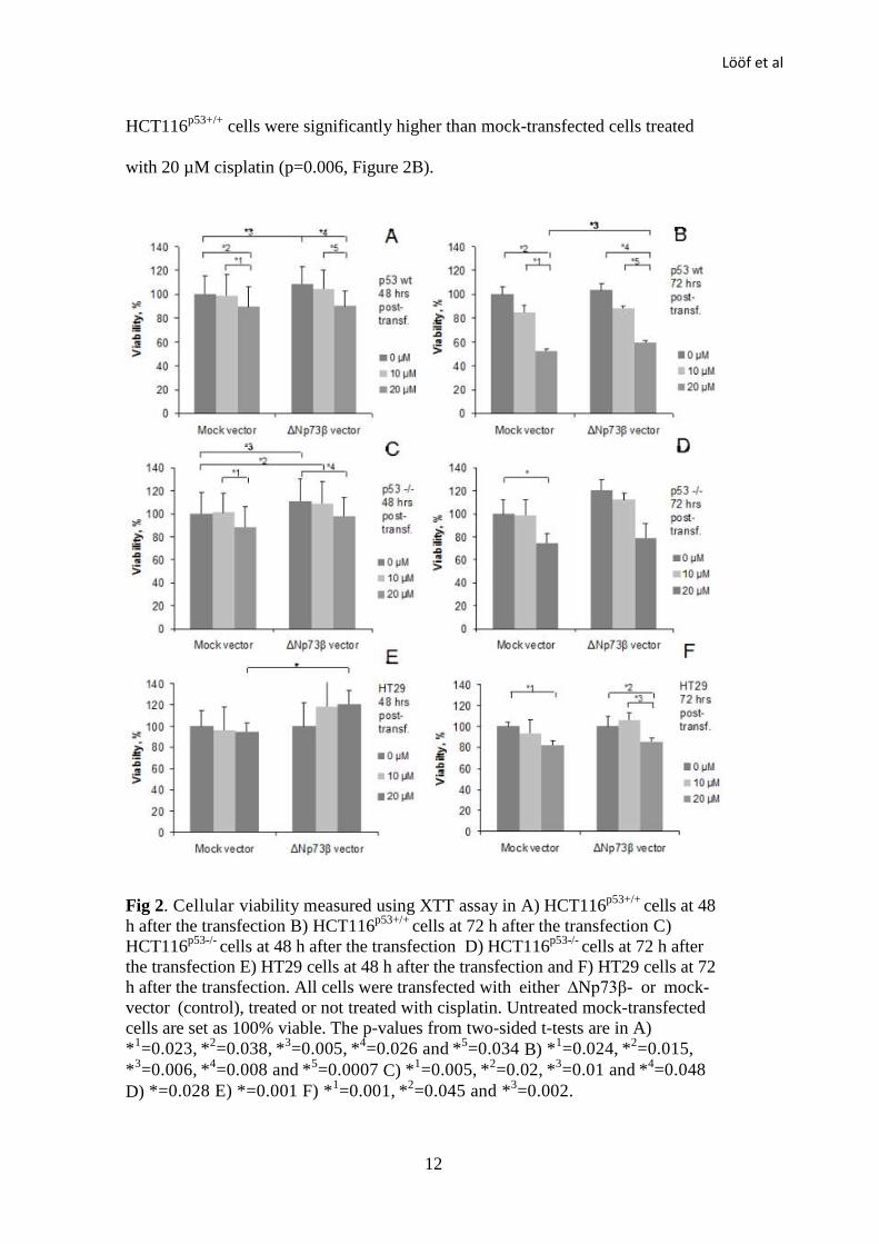
Fig 2. Cellular viability measured using XTT assay in A) HCT116p53+/+
cells at 48
h after the transfection B) HCT116p53+/+
cells at 72 h after the transfection C)
HCT116p53-/-
cells at 48 h after the transfection D) HCT116p53-/-
cells at 72 h after
the transfection E) HT29 cells at 48 h after the transfection and F) HT29 cells at 72
h after the transfection. All cells were transfected with either ∆Np73β- or mock-
vector (control), treated or not treated with cisplatin. Untreated mock-transfected
cells are set as 100% viable. The p-values from two-sided t-tests are in A)
*1=0.023,
*
2=0.038,
*
3=0.005,
*
4=0.026 and
*
5=0.034 B) *
1=0.024,
*
2=0.015,
*3=0.006,
*
4=0.008 and
*
5=0.0007 C) *
1=0.005,
*
2=0.02,
*
3=0.01 and
*
4=0.048
D) *=0.028 E) *=0.001 F) *1=0.001,
*
2=0.045 and *
3=0.002.
Lööf et al
13
∆Np73β-transfected HCT116p53-/-
cells were significantly more viable than mock-
transfected cells at 48 h after transfection (p=0.01, Figure 2C). The viability of
∆Np73β- transfected cells treated with 10 µM cisplatin was also higher than
untreated mock-transfected cells (p=0.02, Figure 2C). The treatment with 20 µM
cisplatin decreased cellular viability in ∆Np73β-transfected HCT116p53-/-
,
compared to no cisplatin treatment (p=0.048, Figure 2C). In mock-transfected
HCT116p53-/-
cellular viability was lower after the treatment with 20 µM cisplatin
compared to treatment with 10 µM cisplatin (p=0.005, Figure 2C). ∆Np73β-
transfected HCT116p53-/-
cells were more viable than mock-transfected cells both
with 0 and 10 µM cisplatin at 72 h after transfection, however, the difference was
not significant (p=0.076 and 0.34 for 0 and 10 µM, respectively, Figure 2D). In
mock-transfected cells viability was decreased after the treatment with 20 µM
cisplatin as compared to the control (p=0.028, Figure 2D). A similar trend was
found in the ∆Np73β-transfected cells (p=0.09, Figure 2D).
HT29 cells were treated with the same concentrations of cisplatin. At 48 h after
transfection, there was no difference between 0 and 10 µM cisplatin, neither in
∆Np73β-transfected cells (p=0.35) nor in mock-transfected cells (p=0.76). Cellular
viability remained similar with treatment of 20 µM cisplatin compared to treatment
with 0 or 10 µM, both in ∆Np73β-transfected cells (p=0.10 and 0.84 for 0 and 10
µM, respectively) and in mock-transfected cells (p=0.47 and 0.85 for 0 and 10 µM,
respectively, Figure 2E). ∆Np73β- transfected HT29 cells were more viable than
mock-transfected cells at the concentration of 20 µM (p=0.001). A similar trend was
seen at 10µM (p=0.055). At 72 h after transfection, cellular viability was decreased
after treatment with 20 µM cisplatin compared to no treatment, both in ∆Np73β-
Lööf et al
14
transfected cells (p=0.001) and mock-transfected cells (p=0.045 Figure 2F). There
was a significant decrease in viability with 20 µM cisplatin compared to 10 µM in
∆Np73β-transfected cells (p=0.002) but not in mock-transfected cells (p=0.13).
Again, there was a trend that ∆Np73β- transfected cells were more viable than the
mock-transfected cells at the concentration of 10 µM (p=0.074) but not at 20 µM
(p=0.28).
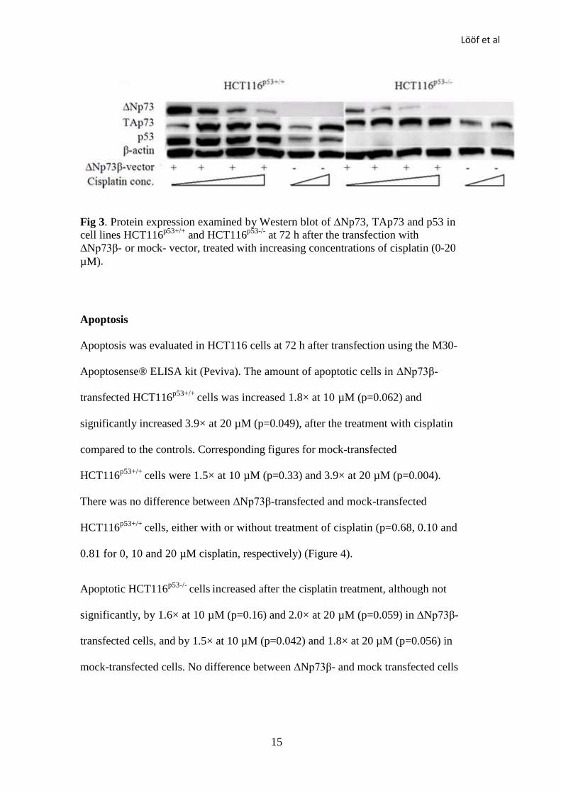
Protein Expression
Expression of ∆Np73, TAp73 and p53 proteins in HCT116p53+/+
and HCT116p53-/-
cells was determined by Western blot at 48 and 72 h after the transfection (Figure
3). The expression of ∆Np73 was markedly increased after ∆Np73β-transfection.
Levels of the ∆Np73β protein in both HCT116p53+/+
and HCT116p53-/-
cells were
decreased in a dose-dependent manner after cisplatin treatment. TAp73 expression
was increased in both ∆Np73β- and mock–transfected HCT116p53+/+
and
HCT116p53-/-
cells after the treatment with cisplatin. In HCT116p53+/+
p53
expression was increased in response to cisplatin. p53 expression was also
increased in ∆Np73β overexpressing cells, without the cisplatin treatment. The
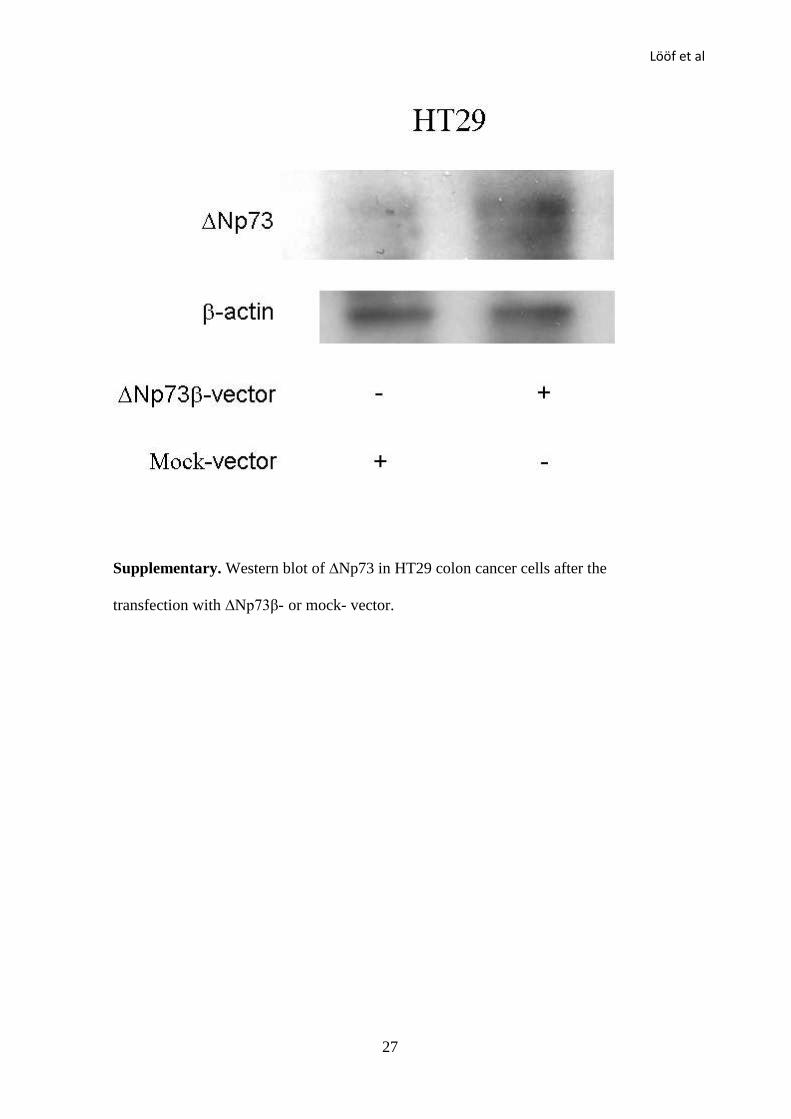
HT29 cells were transfected using the calcium phosphate transfection protocol as
other transfection reagents were ineffective. An increase in the ∆Np73 protein
levels could be observed after transfection (supplementary data).
Lööf et al
15
Fig 3. Protein expression examined by Western blot of ∆Np73, TAp73 and p53 in
cell lines HCT116p53+/+
and HCT116p53-/-
at 72 h after the transfection with
∆Np73β- or mock- vector, treated with increasing concentrations of cisplatin (0-20
µM).
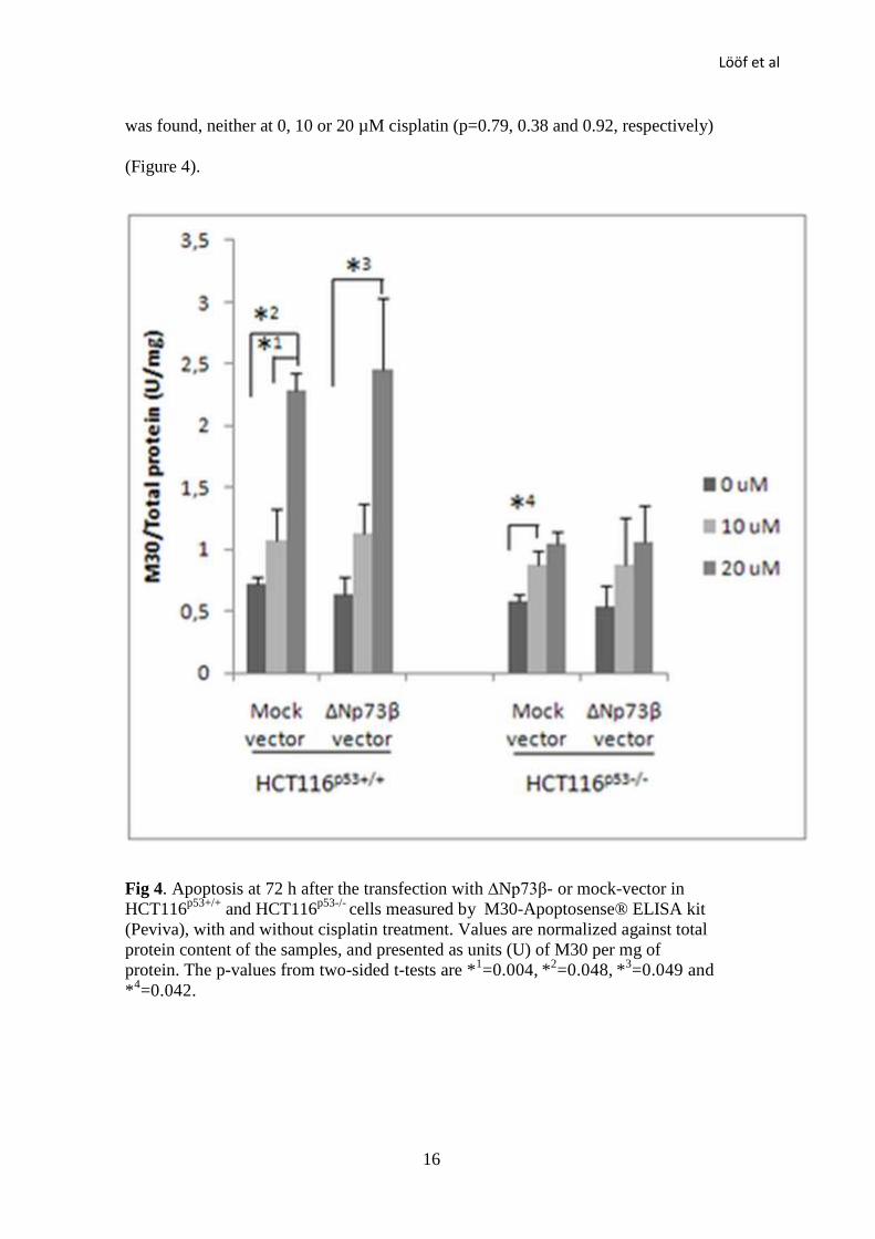
Apoptosis
Apoptosis was evaluated in HCT116 cells at 72 h after transfection using the M30-
Apoptosense® ELISA kit (Peviva). The amount of apoptotic cells in ∆Np73β-
transfected HCT116p53+/+
cells was increased 1.8× at 10 µM (p=0.062) and
significantly increased 3.9× at 20 µM (p=0.049), after the treatment with cisplatin
compared to the controls. Corresponding figures for mock-transfected
HCT116p53+/+
cells were 1.5× at 10 µM (p=0.33) and 3.9× at 20 µM (p=0.004).
There was no difference between ∆Np73β-transfected and mock-transfected
HCT116p53+/+
cells, either with or without treatment of cisplatin (p=0.68, 0.10 and
0.81 for 0, 10 and 20 µM cisplatin, respectively) (Figure 4).
Apoptotic HCT116p53-/-
cells increased after the cisplatin treatment, although not
significantly, by 1.6× at 10 µM (p=0.16) and 2.0× at 20 µM (p=0.059) in ∆Np73β-
transfected cells, and by 1.5× at 10 µM (p=0.042) and 1.8× at 20 µM (p=0.056) in
mock-transfected cells. No difference between ∆Np73β- and mock transfected cells
Lööf et al
16
was found, neither at 0, 10 or 20 µM cisplatin (p=0.79, 0.38 and 0.92, respectively)
(Figure 4).
Fig 4. Apoptosis at 72 h after the transfection with ∆Np73β- or mock-vector in
HCT116p53+/+
and HCT116p53-/-
cells measured by M30-Apoptosense® ELISA kit
(Peviva), with and without cisplatin treatment. Values are normalized against total
protein content of the samples, and presented as units (U) of M30 per mg of
protein. The p-values from two-sided t-tests are *1=0.004,
*
2=0.048,
*
3=0.049 and
*4=0.042.
Lööf et al
17
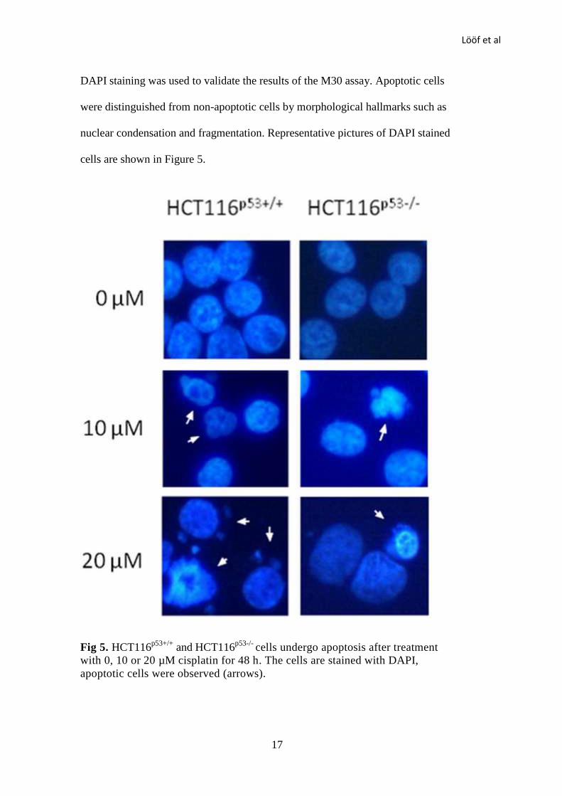
DAPI staining was used to validate the results of the M30 assay. Apoptotic cells
were distinguished from non-apoptotic cells by morphological hallmarks such as
nuclear condensation and fragmentation. Representative pictures of DAPI stained
cells are shown in Figure 5.
Fig 5. HCT116p53+/+
and HCT116p53-/-
cells undergo apoptosis after treatment
with 0, 10 or 20 µM cisplatin for 48 h. The cells are stained with DAPI,
apoptotic cells were observed (arrows).
Lööf et al
18
DISCUSSION
In this study, we investigated the effects of ∆Np73β on colon cancer cells with
respect of cytotoxicity of cisplatin. HCT116p53+/+
, HCT116p53-/-
and HT29 cells,
were transfected with a ∆Np73β expression vector and further treated with
cisplatin, then colony formation potential, cellular viability, apoptosis, and protein
expression were examined. We found that overexpression of ∆Np73β significantly
increased cellular viability in all the three cell lines. There were no significant
differences between ∆Np73β- and mock-transfected cells regarding apoptosis in
HCT116p53+/+
and HCT116p53-/-
cell lines, indicating that the differences in cellular
viability do not seem to be primarily due to increased apoptosis. A reduction in cell
cycle arrest could possibly explain the difference were due to decreased cell cycle
arrest rather than apoptosis.
Since ∆Np73β was degraded in a dose dependent manner in response to the
cisplatin, any resistance to cisplatin treatment conferred by ∆Np73β should be
minor at the concentrations used in the cellular viability and apoptosis assays. A
relatively low concentration of cisplatin was used in the colony forming assay, and
the survival fractions slightly increased in ∆Np73β-transfected cells compared to
mock-transfected cells, indicating resistance to cisplatin treatment.
It has been shown in sarcoma cell line SAOS-2 that ∆Np73 is rapidly degraded in a
proteasome dependent manner in response to DNA-damage [30]. ∆Np73
expression is induced by p53 and TAp73 [11], constituting a regulatory feedback
mechanism. The degradation of ∆Np73 may be a safety mechanism, allowing cell
cycle arrest and apoptosis to occur properly in damaged cells without inhibition
from the anti-apoptotic ∆Np73. In contrast, cisplatin up-regulates ∆Np73 in SH-
Lööf et al
19
SY5Y neuroblastoma cells [17,31]. The signaling pathways leading to DNA
damage-induced degradation of ∆Np73 could be disturbed in some tumor types,
providing a mechanism for resistance to various DNA- damaging treatments.
p53 contributes to cisplatin treatment by inducing cell cycle arrest and apoptosis
[22], and we found the protein levels of p53 to be increased after the cisplatin
treatment. Further, HCT116p53-/-
cells were less sensitive to the cisplatin treatment
than HCT116p53+/+
cells, confirming the involvement of p53 in cisplatin treatment.
We also found an increase in p53 protein in cells overexpressing ∆Np73β, even
without the cisplatin treatment. This could be due to blocking of the mdm2
promoter; ∆Np73β has been previously shown to reduce the transcriptional
activation of mdm2 [32]. Mdm2 promotes the ubiquitin-mediated degradation of
p53 [33], and high levels of ∆Np73β may, therefore, result in reduced degradation
of p53.
In addition, we found tha t expression of TAp73 protein was increased after the
cisplatin treatment. Inhibition of TAp73 has been shown to increase the resistance
to the cisplatin treatment [24], suggesting that TAp73 is involved in cisplatin-
induced cell cycle arrest and apoptosis. On the contrary, it has been reported that
p73 is not induced in response to cisplatin in the HCT116 cell line as a result of
deficient mismatch repair [25]. Cisplatin increases the half-life of p73 by
activating the tyrosine kinase c-abl in mismatch- repair-proficient cells [25].
However, p73 has been stabilized by the transcription factor c-Jun in response to
cisplatin, in cells defective in c-abl binding and phosphorylation [34], suggesting
alternative pathways for p73 induction independent of c-abl and mismatch-repair
signaling pathway.
Lööf et al
20
Upregulation of ∆Np73 in tumors may disturb apoptotic signaling pathway by
interfering with p53 and TAp73, and high expression of ∆Np73 has been
correlated with a worse outcome of patients [35-36] and resistance to DNA-
damaging drugs [37-38]. Recent studies have shown that overexpression of ∆Np73
protects cells from apoptosis, also when treated with cisplatin, by inhibiting p53
target genes such as p21 [30], while downregulation of ∆Np73 increases apoptosis
[39]. However, the effect of the ∆Np73β isoform remains indistinct. Unlike
∆Np73α, ∆Np73β possessed the capability of activating some p53-responsive
genes, thereby inducing both cell cycle arrest and apoptosis [20]. Resulting from
the use of alternative intron 3 promoter, the ∆Np73 isoforms contain 13 unique
residues at the N-terminus shown to contain an activation domain [20]. The
∆Np73α and β isoforms are identical apart from the C-terminal where the β-
isoform lacks exon 13 [1], suggesting that the C-terminal of α-isoform represses
transcription. Both ∆Np73α and β have been shown to inhibit the transcription of
p53 responsive genes, although the α-isoform is much more potent than the β-
isoform which shows some intrinsic transactivation capacity [16]. Also in TAp73,
the α-isoform has lower transactivation ability than the β-isoform [40].
In conclusion, we demonstrate that cisplatin decreased clonogenic potential and
cellular viability while increasing apoptosis. These effects were more
pronounced in HCT116p53+/+
than HCT116p53-/-
, confirming the involvement of p53
in the cisplatin treatment. Both p53 and TAp73 were upregulated in cisplatin
treated HCT116 cells. Overexpression of ∆Np73β increased cellular viability in
HT29 cells as well as both HCT116p53+/+
and HCT116p53-/-
. Resistance to cisplatin
treatment increased slightly in ∆Np73β overexpressing HCT116p53+/+
and

Lööf et al
21
HCT116p53-/-
cells, indicating that both p53 and TAp73 were involved in cisplatin
treatment.
Lööf et al
22
REFERENCES
1. Kaghad M, Bonnet H, Yang A et al. Monoallelically expressed gene related to
p53 at 1p36, a region frequently deleted in neuroblastoma and other human
cancers. Cell 1997;90:809-819.
2. Jost CA, Marin MC, Kaelin WG, Jr. p73 is a simian [correction of human]
p53-related protein that can induce apoptosis. Nature 1997;389:191-194.
3. Yang A, Walker N, Bronson R et al. p73-deficient mice have neurological,
pheromonal and inflammatory defects but lack spontaneous tumours. Nature
2000;404:99-103.
4. Levine AJ. p53, the cellular gatekeeper for growth and division. Cell
1997;88:323-331.
5. Stiewe T, Putzer BM. Role of p73 in malignancy: tumor suppressor or
oncogene? Cell Death Differ 2002;9:237-245.
6. Sun XF. p73 overexpression is a prognostic factor in patients with colorectal
adenocarcinoma. Clin Cancer Res 2002;8:165-170.
7. Zaika AI, Kovalev S, Marchenko ND, Moll UM. Overexpression of the wild
type p73 gene in breast cancer tissues and cell lines. Cancer Res
1999;59:3257-3263.
8. Chen CL, Ip SM, Cheng D, Wong LC, Ngan HY. P73 gene expression in
ovarian cancer tissues and cell lines. Clin Cancer Res 2000;6:3910-3915.
9. Tokuchi Y, Hashimoto T, Kobayashi Y et al. The expression of p73 is
increased in lung cancer, independent of p53 gene alteration. Br J Cancer
1999;80:1623-1629.
Lööf et al
23
10. Tannapfel A, Wasner M, Krause K et al. Expression of p73 and its relation to
histopathology and prognosis in hepatocellular carcinoma. J Natl Cancer Inst
1999;91:1154-1158.
11. Grob TJ, Novak U, Maisse C et al. Human delta Np73 regulates a dominant
negative feedback loop for TAp73 and p53. Cell Death Differ 2001;8:1213-
1223.
12. De Laurenzi V, Costanzo A, Barcaroli D et al. Two new p73 splice variants,
gamma and delta, with different transcriptional activity. J Exp Med
1998;188:1763-1768.
13. De Laurenzi VD, Catani MV, Terrinoni A et al. Additional complexity in p73:
induction by mitogens in lymphoid cells and identification of two new splicing
variants epsilon and zeta. Cell Death Differ 1999;6:389-390.
14. Ishimoto O, Kawahara C, Enjo K, Obinata M, Nukiwa T, Ikawa S. Possible
oncogenic potential of DeltaNp73: a newly identified isoform of human p73.
Cancer Res 2002;62:636-641.
15. Stiewe T, Zimmermann S, Frilling A, Esche H, Putzer BM. Transactivation-
deficient DeltaTA-p73 acts as an oncogene. Cancer Res 2002;62:3598-3602.
16. Kartasheva NN, Contente A, Lenz-Stoppler C, Roth J, Dobbelstein M. p53
induces the expression of its antagonist p73 Delta N, establishing an
autoregulatory feedback loop. Oncogene 2002;21:4715-4727.
17. Nakagawa T, Takahashi M, Ozaki T et al. Autoinhibitory regulation of p73 by
Delta Np73 to modulate cell survival and death through a p73-specific target
element within the Delta Np73 promoter. Mol Cell Biol 2002;22:2575-2585.
Lööf et al
24
18. Pozniak CD, Radinovic S, Yang A, McKeon F, Kaplan DR, Miller FD. An
anti-apoptotic role for the p53 family member, p73, during developmental
neuron death. Science 2000;289:304-306.
19. Tomasini R, Tsuchihara K, Wilhelm M et al. TAp73 knockout shows genomic
instability with infertility and tumor suppressor functions. Genes Dev
2008;22:2677-2691.
20. Liu G, Nozell S, Xiao H, Chen X. DeltaNp73beta is active in transactivation
and growth suppression. Mol Cell Biol 2004;24:487-501.
21. Marrazzo E, Marchini S, Tavecchio M et al. The expression of the
DeltaNp73beta isoform of p73 leads to tetraploidy. Eur J Cancer 2009;45:443-
453.
22. Siddik ZH. Cisplatin: mode of cytotoxic action and molecular basis of
resistance. Oncogene 2003;22:7265-7279.
23. Johnstone RW, Ruefli AA, Lowe SW. Apoptosis: a link between cancer
genetics and chemotherapy. Cell 2002;108:153-164.
24. Irwin MS, Kondo K, Marin MC, Cheng LS, Hahn WC, Kaelin WG, Jr.
Chemosensitivity linked to p73 function. Cancer Cell 2003;3:403-410.
25. Gong JG, Costanzo A, Yang HQ et al. The tyrosine kinase c-Abl regulates p73
in apoptotic response to cisplatin-induced DNA damage. Nature
1999;399:806-809.
26. Vayssade M, Haddada H, Faridoni-Laurens L et al. P73 functionally replaces
p53 in Adriamycin-treated, p53-deficient breast cancer cells. Int J Cancer
2005;116:860-869.
27. Bunz F, Dutriaux A, Lengauer C et al. Requirement for p53 and p21 to sustain
G2 arrest after DNA damage. Science 1998;282:1497-1501.
Lööf et al
25
28. Fernández de Mattos S, Villalonga P, Clardy J, et al. FOXO3a mediates the
cytotoxic effects of cisplatin in colon cancer cells. Mol Cancer Ther
2008;7:3237-3246.
29. Gambi N, Tramontano F, Quesada P. Poly(ADPR)polymerase inhibition and
apoptosis induction in cDDP-treated human carcinoma cell lines. Biochem
Pharmacol 2008;75:2356-2363.
30. Maisse C, Munarriz E, Barcaroli D, Melino G, De Laurenzi V. DNA damage
induces the rapid and selective degradation of the DeltaNp73 isoform,
allowing apoptosis to occur. Cell Death Differ 2004;11:685-687.
31. Million K, Horvilleur E, Goldschneider D et al. Differential regulation of p73
variants in response to cisplatin treatment in SH-SY5Y neuroblastoma cells.
Int J Oncol 2006;29:147-154.
32. Haupt Y, Maya R, Kazaz A, Oren M. Mdm2 promotes the rapid degradation of
p53. Nature 1997;387:296-299.
33. Nakagawa T, Takahashi M, Ozaki T et al. Negative autoregulation of p73 and
p53 by DeltaNp73 in regulating differentiation and survival of human
neuroblastoma cells. Cancer Lett 2003;197:105-109.
34. Toh WH, Siddique MM, Boominathan L, Lin KW, Sabapathy K. c-Jun
regulates the stability and activity of the p53 homologue, p73. J Biol Chem
2004;279:44713-44722.
35. Muller M, Schilling T, Sayan AE et al. TAp73/Delta Np73 influences
apoptotic response, chemosensitivity and prognosis in hepatocellular
carcinoma. Cell Death Differ 2005;12:1564-1577.
Lööf et al
26
36. Casciano I, Mazzocco K, Boni L et al. Expression of DeltaNp73 is a molecular
marker for adverse outcome in neuroblastoma patients. Cell Death Differ
2002;9:246-251.
37. Concin N, Hofstetter G, Berger A et al. Clinical relevance of dominant-
negative p73 isoforms for responsiveness to chemotherapy and survival in
ovarian cancer: evidence for a crucial p53-p73 cross-talk in vivo. Clin Cancer
Res 2005;11:8372-8383.
38. Meier M, den Boer ML, Meijerink JP et al. Differential expression of p73
isoforms in relation to drug resistance in childhood T-lineage acute
lymphoblastic leukaemia. Leukemia 2006;20:1377-1384.
39. Simoes-Wust AP, Sigrist B, Belyanskaya L, Hopkins Donaldson S, Stahel RA,
Zangemeister-Wittke U. DeltaNp73 antisense activates PUMA and induces
apoptosis in neuroblastoma cells. J Neurooncol 2005;72:29-34.
40. Dobbelstein M, Wienzek S, Konig C, Roth J. Inactivation of the p53-
homologue p73 by the mdm2-oncoprotein. Oncogene 1999;18:2101-2106.
Lööf et al
27
Supplementary. Western blot of ∆Np73 in HT29 colon cancer cells after the
transfection with ∆Np73β- or mock- vector.

![Genistein induces apoptosis of colon cancer cells by ...€¦ · pathway [3]. In this study, we demonstrated that GEN can inhibite proliferation and induce apoptosis of colon cancer](https://static.fdocument.org/doc/165x107/6091035508039222da437990/genistein-induces-apoptosis-of-colon-cancer-cells-by-pathway-3-in-this-study.jpg)