Effect of Genetic Variation on the Tryptic Hydrolysis of ... · hydrolysis of the intact protein...
10
J. Dairy Sci. 87:4023–4032 American Dairy Science Association, 2004. Effect of Genetic Variation on the Tryptic Hydrolysis of Bovine β-Lactoglobulin A, B, and C L. K. Creamer, 1 H. C. Nilsson, 2 M. A. Paulsson, 2 C. J. Coker, 1 J. P. Hill, 1 and R. Jime ´ nez-Flores 3 1 Fonterra Research Centre, P.O. Box 11 029, Palmerston North, New Zealand 2 Department of Food Engineering, Lund University, SE-22100, Lund, Sweden 3 Dairy Products Technology Center, California Polytechnic State University, San Luis Obispo 93407 ABSTRACT The structure, stability, and hydrolysis characteris- tics of β-lactoglobulin (LG) A are different from those of either β-LG B or β-LG C. Purified samples of these proteins were hydrolyzed with trypsin and the rates of loss of native monomeric β-LG structure were measured using sodium dodecyl sulfate PAGE. At the same time, the appearance of many individual peptides were iden- tified and followed in time by HPLC, measuring their concentration as a function of solution pH, temperature, protein concentration, and added urea or palmitate. The identity of the peptides was confirmed by liquid chromatography-mass spectrometry. This semiquanti- tative exploration showed that the rate of hydrolysis was in the order β-LG A > β-LG B > β-LG C under most circumstances, and that 12 of the 18 trypsin-susceptible bonds were cleaved at very similar rates that were gov- erned by the variant type. Consequently, the rate of hydrolysis of the intact protein was related to the over- all structural stability of the individual proteins and the accessibility of certain peptide bonds to the enzyme. The hydrolysis of mixtures of 2 or more variants or of denatured β-LG gave more heterogeneous peptide mixtures. (Key words: β-lactoglobulin, tryptic hydrolysis, ge- netic variant, kinetics) Abbreviation key: DTT = dithiothreitol, E:S = en- zyme:substrate, RP = reversed-phase, TFA = trifluo- roacetic acid. INTRODUCTION Whey protein concentrates are used as food ingredi- ents because of their functional properties as well as their nutritional properties. The major protein of bovine Received June 15, 2004. Accepted July 22, 2004. Corresponding author: R. Jime ´nez-Flores; e-mail: rjimenez@ calpoly.edu. 4023 whey is β-LG, which is responsible for many of the functional properties of whey protein products (Mulvih- ill and Kinsella, 1987). In some instances, these func- tionalities can be enhanced by heat treatments or by partial hydrolysis to mixtures of large peptides (Tur- geon et al., 1992; Chen et al., 1994; Huang et al., 1996; Caessens et al., 1999a, 1999b). Additionally, the hydro- lyzates themselves are used as ingredients for pro- cessed foods. Bovine β-LG occurs naturally in a number of geneti- cally different variants, which differ from one another by substitutions of one or more amino acids within the protein sequence. The most common variants are known as β-LG A and β-LG B; additionally, β-LG C occurs in milk in both New Zealand (Hill et al., 1996) and Australia (McLean et al., 1984) at moderate fre- quencies (Farrell et al., 2004). It has been reported that β-LG A is hydrolyzed by papain (Schmidt and van Markwijk, 1993), chymotrypsin (Motion and Hill, 1994; Van Willige and FitzGerald, 1995), V8 protease (Caes- sens et al., 1999b), plasmin (Caessens et al., 1999a), and trypsin (Monnot and Yon, 1964; Huang et al., 1994a, 1994b; Motion and Hill, 1994; Van Willige and FitzGerald, 1995; Caessens et al., 1999b) more rapidly than is β-LG B, and a preliminary note indicates that β-LG C is hydrolyzed more slowly than β-LG B by tryp- sin and chymotrypsin (Motion and Hill, 1994; Hill et al., 1996). Although there have been many studies of the func- tional properties of β-LG hydrolyzates, only a few have looked at differences between the A and B variants, and none has examined β-LG C. Huang et al. (1994b) compared the size and the rate of formation of peptides of β-LG A and β-LG B and found that the initial peptide release was different for the 2 variants. They also found that there was a difference in size distribution of the peptides of β-LG A and β-LG B. The hydrolysis of β-LG has been studied using a range of techniques. These include the increase in N- terminal amino groups (Monnot, 1964; Van Willige and FitzGerald, 1995); SDS-PAGE, which can show the rate
Transcript of Effect of Genetic Variation on the Tryptic Hydrolysis of ... · hydrolysis of the intact protein...
J. Dairy Sci. 87:4023–4032 American Dairy Science Association, 2004.
Effect of Genetic Variation on the Tryptic Hydrolysisof Bovine β-Lactoglobulin A, B, and C
L. K. Creamer,1 H. C. Nilsson,2 M. A. Paulsson,2 C. J. Coker,1
J. P. Hill,1 and R. Jimenez-Flores31Fonterra Research Centre, P.O. Box 11 029, Palmerston North, New Zealand2Department of Food Engineering, Lund University, SE-22100, Lund, Sweden3Dairy Products Technology Center, California Polytechnic State University,San Luis Obispo 93407
ABSTRACT
The structure, stability, and hydrolysis characteris-tics of β-lactoglobulin (LG) A are different from thoseof either β-LG B or β-LG C. Purified samples of theseproteins were hydrolyzed with trypsin and the rates ofloss of native monomeric β-LG structure were measuredusing sodium dodecyl sulfate PAGE. At the same time,the appearance of many individual peptides were iden-tified and followed in time by HPLC, measuring theirconcentration as a function of solution pH, temperature,protein concentration, and added urea or palmitate.The identity of the peptides was confirmed by liquidchromatography-mass spectrometry. This semiquanti-tative exploration showed that the rate of hydrolysiswas in the order β-LG A > β-LG B > β-LG C under mostcircumstances, and that 12 of the 18 trypsin-susceptiblebonds were cleaved at very similar rates that were gov-erned by the variant type. Consequently, the rate ofhydrolysis of the intact protein was related to the over-all structural stability of the individual proteins andthe accessibility of certain peptide bonds to the enzyme.The hydrolysis of mixtures of 2 or more variants or ofdenatured β-LG gave more heterogeneous peptidemixtures.(Key words: β-lactoglobulin, tryptic hydrolysis, ge-netic variant, kinetics)
Abbreviation key: DTT = dithiothreitol, E:S = en-zyme:substrate, RP = reversed-phase, TFA = trifluo-roacetic acid.
INTRODUCTION
Whey protein concentrates are used as food ingredi-ents because of their functional properties as well astheir nutritional properties. The major protein of bovine
Received June 15, 2004.Accepted July 22, 2004.Corresponding author: R. Jimenez-Flores; e-mail: rjimenez@
calpoly.edu.
4023
whey is β-LG, which is responsible for many of thefunctional properties of whey protein products (Mulvih-ill and Kinsella, 1987). In some instances, these func-tionalities can be enhanced by heat treatments or bypartial hydrolysis to mixtures of large peptides (Tur-geon et al., 1992; Chen et al., 1994; Huang et al., 1996;Caessens et al., 1999a, 1999b). Additionally, the hydro-lyzates themselves are used as ingredients for pro-cessed foods.
Bovine β-LG occurs naturally in a number of geneti-cally different variants, which differ from one anotherby substitutions of one or more amino acids within theprotein sequence. The most common variants areknown as β-LG A and β-LG B; additionally, β-LG Coccurs in milk in both New Zealand (Hill et al., 1996)and Australia (McLean et al., 1984) at moderate fre-quencies (Farrell et al., 2004). It has been reportedthat β-LG A is hydrolyzed by papain (Schmidt and vanMarkwijk, 1993), chymotrypsin (Motion and Hill, 1994;Van Willige and FitzGerald, 1995), V8 protease (Caes-sens et al., 1999b), plasmin (Caessens et al., 1999a),and trypsin (Monnot and Yon, 1964; Huang et al.,1994a, 1994b; Motion and Hill, 1994; Van Willige andFitzGerald, 1995; Caessens et al., 1999b) more rapidlythan is β-LG B, and a preliminary note indicates thatβ-LG C is hydrolyzed more slowly than β-LG B by tryp-sin and chymotrypsin (Motion and Hill, 1994; Hill etal., 1996).
Although there have been many studies of the func-tional properties of β-LG hydrolyzates, only a few havelooked at differences between the A and B variants,and none has examined β-LG C. Huang et al. (1994b)compared the size and the rate of formation of peptidesof β-LG A and β-LG B and found that the initial peptiderelease was different for the 2 variants. They also foundthat there was a difference in size distribution of thepeptides of β-LG A and β-LG B.
The hydrolysis of β-LG has been studied using arange of techniques. These include the increase in N-terminal amino groups (Monnot, 1964; Van Willige andFitzGerald, 1995); SDS-PAGE, which can show the rate
CREAMER ET AL.4024
of loss of the intact protein (Schmidt and van Markwijk,1993) and can indicate the existence of large peptides(Huang et al., 1994a, 1996); size-exclusion HPLC inacetonitrile solution, which can show the reduction insize of groups of peptides (Hill et al., 1996); and re-versed-phase (RP)-HPLC, which can be used to sepa-rate most of the peptides in the hydrolyzate from oneanother (Dalgalarrondo et al., 1992; Dufour et al., 1994;Otte et al., 1997). When RP-HPLC is coupled with massspectrometry, the identity of the peaks can often beascertained without resorting to traditional sequencingmethods (Caessens et al., 1999a; Chevalier et al., 2002;Imre et al., 2003; Schilling et al., 2003).
In the present study, we used SDS-PAGE of reducedsamples to show the effects of temperature, protein con-centration, and pH on the relative rates of loss of β-LGA, β-LG B, and β-LG C. We also used RP-HPLC witha C2-C18 column to show the differences in the peptidepatterns given by the 3 variant proteins.
MATERIALS AND METHODS
Materials
β-Lactoglobulin A, B, and C were prepared as de-scribed earlier (Manderson et al., 1998). 1-L-Tosylam-ide-2-phenylethyl chloromethyl ketone-treated trypsin(Cat. No. T-1426), DL-dithiothreitol (DTT) (Cat. No. D-6032), and 2-mercaptoethanol were obtained fromSigma Chemical Co. (St. Louis, MO). The electrophore-sis chemicals were obtained from Bio-Rad Laboratories(Hercules, CA). All other chemicals were from BDHLaboratory Supplies (Poole, England) and were at leastAnalaR grade.
Protein Hydrolysis
Samples (3 mL) of purified β-LG A, B, and C, at aconcentration of 1.4% (wt/vol), were mixed with 3 mLof 100 mM Tris-HCl buffer, pH 7.5, 8.1, 8.4, and 8.7,which lowered the pH values to 7.24, 7.85, 8.31, and8.58, respectively, and 5 mL of the mixture was putinto a plastic tube and cooled to 20°C or warmed to 37,47, or 57 ± 0.1°C in a temperature-controlled waterbath. A 350-µL aliquot of 0.1% (wt/vol) trypsin solutionwas added at an enzyme:substrate (E:S) ratio of 1:100(wt/wt) for the first 3 pH trials and an E:S ratio of 1:200(wt/wt) for the last trial (pH 8.58) to give a final β-LGconcentration of 6.5 mg/mL. Before addition of enzymeand at various times after addition of enzyme (2.5, 4,6, 9, 13, 20, 30, 45, 65, 100, and 150 min), 2 samplesof 200 µL were taken for SDS-PAGE and RP-HPLCanalysis, respectively. For the high concentration sam-ples, the protein solution was mixed 1:1 with double-strength buffer and concentrated to 5% of its original
Journal of Dairy Science Vol. 87, No. 12, 2004
volume by using a stirred ultrafiltration cell fitted witha YM 10 DIAFLO ultrafiltration membrane (Amicon,Inc., Beverly, MA) to obtain solutions with protein con-centrations of more than 130 mg/mL. These solutionswere subsequently diluted and their pH was deter-mined; they were then mixed with enzyme solution togive β-LG concentrations of 122 mg/mL in the reac-tion mixture.
The samples for SDS-PAGE analysis were mixedwith 1.0 mL of SDS sample buffer containing 0.13 mgDTT/mL, boiled for 4 min, and then stored at 4°C untilanalyzed. The samples for RP-HPLC analysis weremixed with 200 µL of phosphate buffer (26 mM sodiumphosphate/68 mM NaCl buffer adjusted to pH 6.0 andcontaining 6.54 mg of DTT/mL), boiled for 4 min, andthen mixed 1:1 with 200 µL of a 5% acetonitrile, 0.1%trifluoroacetic acid (TFA) solution and stored at 4°Cuntil analyzed.
SDS-PAGE
The preparation of the gels, loading of samples, elec-trophoresis, gel staining, destaining, scanning, andband quantitation have been described previously(Manderson et al., 1998). The reaction rates were esti-mated using Microsoft Excel 97 by regressing the loga-rithm of band intensity vs. reaction time. The calcula-tions included only band intensity results that were>10% of the band intensity of the zero-time sample.
RP-HPLC
The hydrolyzed protein samples were chromato-graphed at 0.7 mL/min on 2 Pharmacia PepRPC HR 5/5 FPLC columns, which contained 120 A pore-size beadscoated with C2–C18 phase (held at 25°C with a Timber-line TL-50 controller), connected in series using a Wa-ters 600E system controller and a Waters 700 SatelliteWisp autoinjector. The column effluent was monitoredwith a Hewlett-Packard 1040A multiwavelength detec-tion system (Hewlett-Packard Co., Camas, WA), andthe data were fed to a ChemStation for processing. Theeluting solvent sequence was 5% acetonitrile in 0.1%TFA for 5 min, followed by a linear gradient from 5%acetonitrile in 0.1% TFA to 60% acetonitrile in 0.1%TFA over 70 min, followed by 60% acetonitrile in 0.1%TFA for 15 min, followed by 5% acetonitrile in 0.1%TFA for 15 min.
Liquid Chromatography-Mass Spectrometry
The peptides were separated by RP-HPLC on a Wa-ters HPLC system (Waters Associates, Millipore Corp.,Waters Chromatography Division, MA) Alliance 2690
TRYPTIC HYDROLYSIS OF β-LG VARIANTS 4025
Separations Module, which comprised a Waters 486MS Tunable Absorbance Detector and a Waters 996Photodiode Array Detector. Signals at 205 and 280 nmwere monitored. The same HPLC columns and methodpreviously described were used in the Waters system.The software used was Waters Millennium version3.05.01 within a Windows operating system.
The eluting peptides were characterized by massspectrometry using a Perkin Elmer Sciex (Thornhill,ON, Canada) Triple Quadrupole API 300 LC/MS/MSsystem. The eluent stream from the Waters HPLC sys-tem was split such that approximately 10 to 15 µL/minwas introduced into the mass spectrometer using anelectrospray Perkin Elmer Sciex API Ion Spray inletinterface. Ions were generated and focused using a posi-tive ion spray voltage of 5600 V with detection betweenm/z values of 200 and 3000 amu. Parameters for opera-tion of the mass spectrometer were step size, 0.2 amu;dwell time, 0.5 ms; scan time, 7 s; and 25 V. The RNGand IQ2 voltages were ramped from 140 to 280 V andfrom −15 to −90 V, respectively. The mass spectrometerwas calibrated using a propylene glycol standard asoutlined in the Perkin Elmer Sciex API 300 manual,using MassChrom 1.0 software. The data were analyzedusing BioMultiView 1.3.1 (Perkin Elmer Sciex) and thereference mass library used was Peptide Map 2.2 (Per-kin Elmer Sciex).
RESULTS
Loss of Intact Protein using SDS-PAGE
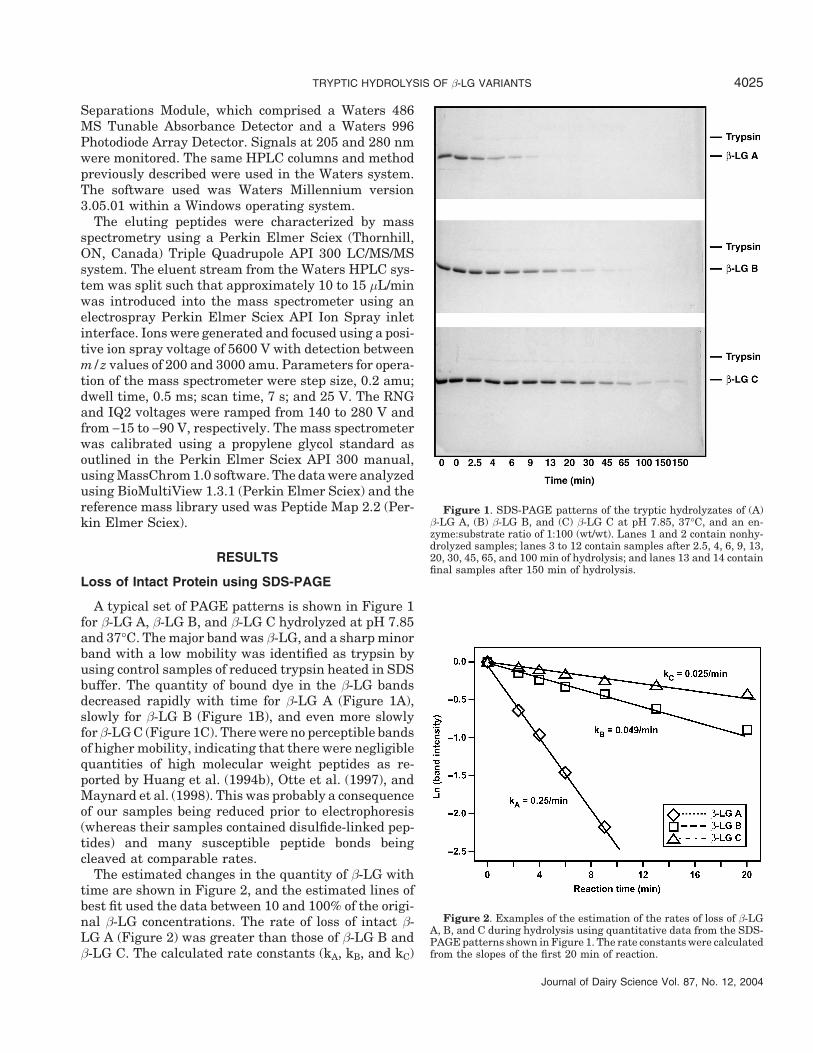
A typical set of PAGE patterns is shown in Figure 1for β-LG A, β-LG B, and β-LG C hydrolyzed at pH 7.85and 37°C. The major band was β-LG, and a sharp minorband with a low mobility was identified as trypsin byusing control samples of reduced trypsin heated in SDSbuffer. The quantity of bound dye in the β-LG bandsdecreased rapidly with time for β-LG A (Figure 1A),slowly for β-LG B (Figure 1B), and even more slowlyfor β-LG C (Figure 1C). There were no perceptible bandsof higher mobility, indicating that there were negligiblequantities of high molecular weight peptides as re-ported by Huang et al. (1994b), Otte et al. (1997), andMaynard et al. (1998). This was probably a consequenceof our samples being reduced prior to electrophoresis(whereas their samples contained disulfide-linked pep-tides) and many susceptible peptide bonds beingcleaved at comparable rates.
The estimated changes in the quantity of β-LG withtime are shown in Figure 2, and the estimated lines ofbest fit used the data between 10 and 100% of the origi-nal β-LG concentrations. The rate of loss of intact β-LG A (Figure 2) was greater than those of β-LG B andβ-LG C. The calculated rate constants (kA, kB, and kC)
Journal of Dairy Science Vol. 87, No. 12, 2004
Figure 1. SDS-PAGE patterns of the tryptic hydrolyzates of (A)β-LG A, (B) β-LG B, and (C) β-LG C at pH 7.85, 37°C, and an en-zyme:substrate ratio of 1:100 (wt/wt). Lanes 1 and 2 contain nonhy-drolyzed samples; lanes 3 to 12 contain samples after 2.5, 4, 6, 9, 13,20, 30, 45, 65, and 100 min of hydrolysis; and lanes 13 and 14 containfinal samples after 150 min of hydrolysis.
Figure 2. Examples of the estimation of the rates of loss of β-LGA, B, and C during hydrolysis using quantitative data from the SDS-PAGE patterns shown in Figure 1. The rate constants were calculatedfrom the slopes of the first 20 min of reaction.
CREAMER ET AL.4026
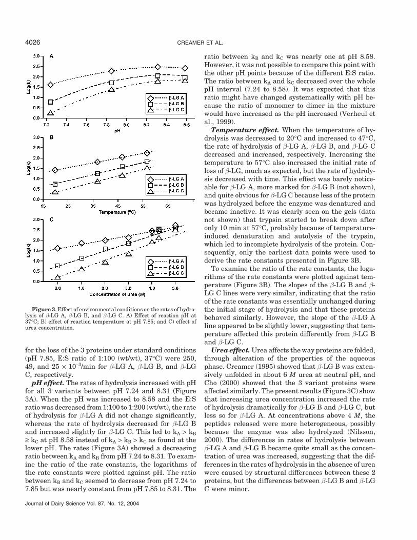
Figure 3. Effect of environmental conditions on the rates of hydro-lysis of β-LG A, β-LG B, and β-LG C. A) Effect of reaction pH at37°C; B) effect of reaction temperature at pH 7.85; and C) effect ofurea concentration.
for the loss of the 3 proteins under standard conditions(pH 7.85, E:S ratio of 1:100 (wt/wt), 37°C) were 250,49, and 25 × 10−3/min for β-LG A, β-LG B, and β-LGC, respectively.
pH effect. The rates of hydrolysis increased with pHfor all 3 variants between pH 7.24 and 8.31 (Figure3A). When the pH was increased to 8.58 and the E:Sratio was decreased from 1:100 to 1:200 (wt/wt), the rateof hydrolysis for β-LG A did not change significantly,whereas the rate of hydrolysis decreased for β-LG Band increased slightly for β-LG C. This led to kA > kB≥ kC at pH 8.58 instead of kA > kB > kC as found at thelower pH. The rates (Figure 3A) showed a decreasingratio between kA and kB from pH 7.24 to 8.31. To exam-ine the ratio of the rate constants, the logarithms ofthe rate constants were plotted against pH. The ratiobetween kB and kC seemed to decrease from pH 7.24 to7.85 but was nearly constant from pH 7.85 to 8.31. The
Journal of Dairy Science Vol. 87, No. 12, 2004
ratio between kB and kC was nearly one at pH 8.58.However, it was not possible to compare this point withthe other pH points because of the different E:S ratio.The ratio between kA and kC decreased over the wholepH interval (7.24 to 8.58). It was expected that thisratio might have changed systematically with pH be-cause the ratio of monomer to dimer in the mixturewould have increased as the pH increased (Verheul etal., 1999).
Temperature effect. When the temperature of hy-drolysis was decreased to 20°C and increased to 47°C,the rate of hydrolysis of β-LG A, β-LG B, and β-LG Cdecreased and increased, respectively. Increasing thetemperature to 57°C also increased the initial rate ofloss of β-LG, much as expected, but the rate of hydroly-sis decreased with time. This effect was barely notice-able for β-LG A, more marked for β-LG B (not shown),and quite obvious for β-LG C because less of the proteinwas hydrolyzed before the enzyme was denatured andbecame inactive. It was clearly seen on the gels (datanot shown) that trypsin started to break down afteronly 10 min at 57°C, probably because of temperature-induced denaturation and autolysis of the trypsin,which led to incomplete hydrolysis of the protein. Con-sequently, only the earliest data points were used toderive the rate constants presented in Figure 3B.
To examine the ratio of the rate constants, the loga-rithms of the rate constants were plotted against tem-perature (Figure 3B). The slopes of the β-LG B and β-LG C lines were very similar, indicating that the ratioof the rate constants was essentially unchanged duringthe initial stage of hydrolysis and that these proteinsbehaved similarly. However, the slope of the β-LG Aline appeared to be slightly lower, suggesting that tem-perature affected this protein differently from β-LG Band β-LG C.
Urea effect. Urea affects the way proteins are folded,through alteration of the properties of the aqueousphase. Creamer (1995) showed that β-LG B was exten-sively unfolded in about 6 M urea at neutral pH, andCho (2000) showed that the 3 variant proteins wereaffected similarly. The present results (Figure 3C) showthat increasing urea concentration increased the rateof hydrolysis dramatically for β-LG B and β-LG C, butless so for β-LG A. At concentrations above 4 M, thepeptides released were more heterogeneous, possiblybecause the enzyme was also hydrolyzed (Nilsson,2000). The differences in rates of hydrolysis betweenβ-LG A and β-LG B became quite small as the concen-tration of urea was increased, suggesting that the dif-ferences in the rates of hydrolysis in the absence of ureawere caused by structural differences between these 2proteins, but the differences between β-LG B and β-LGC were minor.
TRYPTIC HYDROLYSIS OF β-LG VARIANTS 4027
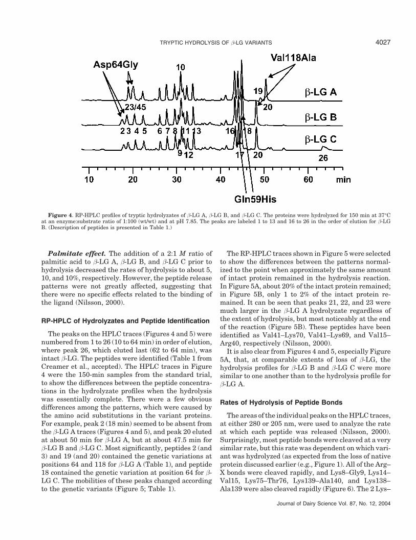
Figure 4. RP-HPLC profiles of tryptic hydrolyzates of β-LG A, β-LG B, and β-LG C. The proteins were hydrolyzed for 150 min at 37°Cat an enzyme:substrate ratio of 1:100 (wt/wt) and at pH 7.85. The peaks are labeled 1 to 13 and 16 to 26 in the order of elution for β-LGB. (Description of peptides is presented in Table 1.)
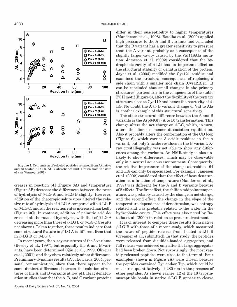
Palmitate effect. The addition of a 2:1 M ratio ofpalmitic acid to β-LG A, β-LG B, and β-LG C prior tohydrolysis decreased the rates of hydrolysis to about 5,10, and 10%, respectively. However, the peptide releasepatterns were not greatly affected, suggesting thatthere were no specific effects related to the binding ofthe ligand (Nilsson, 2000).
RP-HPLC of Hydrolyzates and Peptide Identification
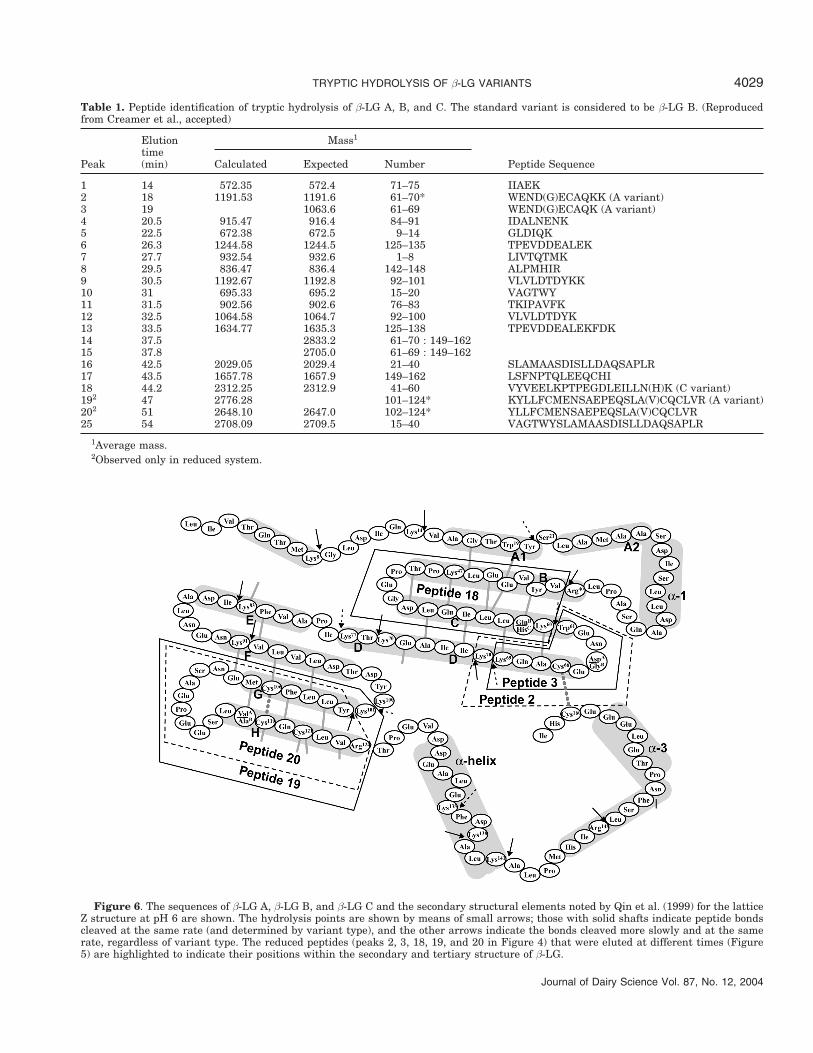
The peaks on the HPLC traces (Figures 4 and 5) werenumbered from 1 to 26 (10 to 64 min) in order of elution,where peak 26, which eluted last (62 to 64 min), wasintact β-LG. The peptides were identified (Table 1 fromCreamer et al., accepted). The HPLC traces in Figure4 were the 150-min samples from the standard trial,to show the differences between the peptide concentra-tions in the hydrolyzate profiles when the hydrolysiswas essentially complete. There were a few obviousdifferences among the patterns, which were caused bythe amino acid substitutions in the variant proteins.For example, peak 2 (18 min) seemed to be absent fromthe β-LG A traces (Figures 4 and 5), and peak 20 elutedat about 50 min for β-LG A, but at about 47.5 min forβ-LG B and β-LG C. Most significantly, peptides 2 (and3) and 19 (and 20) contained the genetic variations atpositions 64 and 118 for β-LG A (Table 1), and peptide18 contained the genetic variation at position 64 for β-LG C. The mobilities of these peaks changed accordingto the genetic variants (Figure 5; Table 1).
Journal of Dairy Science Vol. 87, No. 12, 2004
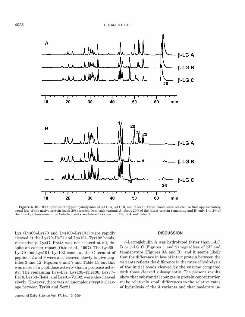
The RP-HPLC traces shown in Figure 5 were selectedto show the differences between the patterns normal-ized to the point when approximately the same amountof intact protein remained in the hydrolysis reaction.In Figure 5A, about 20% of the intact protein remained;in Figure 5B, only 1 to 2% of the intact protein re-mained. It can be seen that peaks 21, 22, and 23 weremuch larger in the β-LG A hydrolyzate regardless ofthe extent of hydrolysis, but most noticeably at the endof the reaction (Figure 5B). These peptides have beenidentified as Val41–Lys70, Val41–Lys69, and Val15–Arg40, respectively (Nilsson, 2000).
It is also clear from Figures 4 and 5, especially Figure5A, that, at comparable extents of loss of β-LG, thehydrolysis profiles for β-LG B and β-LG C were moresimilar to one another than to the hydrolysis profile forβ-LG A.
Rates of Hydrolysis of Peptide Bonds
The areas of the individual peaks on the HPLC traces,at either 280 or 205 nm, were used to analyze the rateat which each peptide was released (Nilsson, 2000).Surprisingly, most peptide bonds were cleaved at a verysimilar rate, but this rate was dependent on which vari-ant was hydrolyzed (as expected from the loss of nativeprotein discussed earlier (e.g., Figure 1). All of the Arg–X bonds were cleaved rapidly, and Lys8–Gly9, Lys14–Val15, Lys75–Thr76, Lys139–Ala140, and Lys138–Ala139 were also cleaved rapidly (Figure 6). The 2 Lys–
CREAMER ET AL.4028
Figure 5. RP-HPLC profiles of tryptic hydrolyzates of β-LG A, β-LG B, and β-LG C. These traces were selected so that approximatelyequal loss of the native protein (peak 26) occurred from each variant: A) about 20% of the intact protein remaining and B) only 1 to 2% ofthe intact protein remaining. Selected peaks are labeled as shown in Figure 4 and Table 1.
Lys (Lys69–Lys70 and Lys100–Lys101) were rapidlycleaved at the Lys70–Ile71 and Lys101–Tyr102 bonds,respectively. Lys47–Pro48 was not cleaved at all, de-spite an earlier report (Otte et al., 1997). The Lys69–Lys70 and Lys101–Lys102 bonds at the C-termini ofpeptides 2 and 9 were also cleaved slowly to give pep-tides 3 and 12 (Figures 6 and 7 and Table 1), but thiswas more of a peptidase activity than a protease activ-ity. The remaining Lys–Lys, Lys135–Phe136, Lys77–Ile78, Lys83–Ile84, and Lys91–Val92, were also cleavedslowly. However, there was an anomalous tryptic cleav-age between Tyr20 and Ser21.
Journal of Dairy Science Vol. 87, No. 12, 2004
DISCUSSION
β-Lactoglobulin A was hydrolyzed faster than β-LGB or β-LG C (Figures 1 and 2) regardless of pH andtemperature (Figures 3A and B), and it seems likelythat the difference in loss of intact protein between thevariants reflects the difference in the rates of hydrolysisof the initial bonds cleaved by the enzyme comparedwith those cleaved subsequently. The present resultsshow that substantial changes in protein concentrationmake relatively small differences to the relative ratesof hydrolysis of the 3 variants and that moderate in-
TRYPTIC HYDROLYSIS OF β-LG VARIANTS 4029
Table 1. Peptide identification of tryptic hydrolysis of β-LG A, B, and C. The standard variant is considered to be β-LG B. (Reproducedfrom Creamer et al., accepted)
Elution Mass1
timePeak (min) Calculated Expected Number Peptide Sequence
1 14 572.35 572.4 71–75 IIAEK2 18 1191.53 1191.6 61–70* WEND(G)ECAQKK (A variant)3 19 1063.6 61–69 WEND(G)ECAQK (A variant)4 20.5 915.47 916.4 84–91 IDALNENK5 22.5 672.38 672.5 9–14 GLDIQK6 26.3 1244.58 1244.5 125–135 TPEVDDEALEK7 27.7 932.54 932.6 1–8 LIVTQTMK8 29.5 836.47 836.4 142–148 ALPMHIR9 30.5 1192.67 1192.8 92–101 VLVLDTDYKK10 31 695.33 695.2 15–20 VAGTWY11 31.5 902.56 902.6 76–83 TKIPAVFK12 32.5 1064.58 1064.7 92–100 VLVLDTDYK13 33.5 1634.77 1635.3 125–138 TPEVDDEALEKFDK14 37.5 2833.2 61–70 : 149–16215 37.8 2705.0 61–69 : 149–16216 42.5 2029.05 2029.4 21–40 SLAMAASDISLLDAQSAPLR17 43.5 1657.78 1657.9 149–162 LSFNPTQLEEQCHI18 44.2 2312.25 2312.9 41–60 VYVEELKPTPEGDLEILLN(H)K (C variant)192 47 2776.28 101–124* KYLLFCMENSAEPEQSLA(V)CQCLVR (A variant)202 51 2648.10 2647.0 102–124* YLLFCMENSAEPEQSLA(V)CQCLVR25 54 2708.09 2709.5 15–40 VAGTWYSLAMAASDISLLDAQSAPLR
1Average mass.2Observed only in reduced system.
Figure 6. The sequences of β-LG A, β-LG B, and β-LG C and the secondary structural elements noted by Qin et al. (1999) for the latticeZ structure at pH 6 are shown. The hydrolysis points are shown by means of small arrows; those with solid shafts indicate peptide bondscleaved at the same rate (and determined by variant type), and the other arrows indicate the bonds cleaved more slowly and at the samerate, regardless of variant type. The reduced peptides (peaks 2, 3, 18, 19, and 20 in Figure 4) that were eluted at different times (Figure5) are highlighted to indicate their positions within the secondary and tertiary structure of β-LG.
Journal of Dairy Science Vol. 87, No. 12, 2004
CREAMER ET AL.4030
Figure 7. Comparison of selected peptides released from A) nativeand B) heated β-LG B. AU = absorbance unit. Drawn from the dataof van Wanroij (2001).
creases in reaction pH (Figure 3A) and temperature(Figure 3B) decrease the differences between the ratesof hydrolysis of β-LG A and β-LG B slightly. However,addition of the chaotropic solute urea altered the rela-tive rate of hydrolysis of β-LG A compared with β-LG Bor β-LG C, and all the reaction rates increased markedly(Figure 3C). In contrast, addition of palmitic acid de-creased all the rates of hydrolysis, with that of β-LG Adecreasing more than those of β-LG B or β-LG C (resultsnot shown). Taken together, these results indicate thatsome structural feature in β-LG A is different from thatin β-LG B or β-LG C.
In recent years, the x-ray structures of the 3 variants(Bewley et al., 1997), but especially the A and B vari-ants, have been determined (Qin et al., 1999; Oliveiraet al., 2001), and they show relatively minor differences.Preliminary dynamics results (P. J. Edwards, 2004, per-sonal communication) show that there appear to besome distinct differences between the solution struc-tures of the A and B variants at low pH. Heat denatur-ation studies show that the A, B, and C variant proteins
Journal of Dairy Science Vol. 87, No. 12, 2004
differ in their susceptibility to higher temperatures(Manderson et al., 1998). Botelho et al. (2000) appliedhigh pressures to the A and B variants and concludedthat the B variant has a greater sensitivity to pressurethan the A variant, probably as a consequence of theslightly larger cavity caused by the Val118Ala muta-tion. Jameson et al. (2002) considered that the hy-drophobic cavity of β-LG has an important effect onthe structural stability or denaturation of the protein.Jayat et al. (2004) modified the Cys121 residue andexamined the structural consequences of replacing aside chain with a smaller side chain (Cys121Ser). Itcan be concluded that small changes in the primarystructures, particularly in the components of the stableFGH motif (Figure 6), affect the flexibility of the tertiarystructure close to Cys119 and hence the reactivity of β-LG. No doubt the A to B variant change of Val to Alais another example of this structural sensitivity.
The other structural difference between the A and Bvariants is the Asp64Gly (A to B) transformation. Thischange alters the net charge on β-LG, which, in turn,alters the dimer–monomer dissociation equilibrium.Also it probably alters the conformation of the CD loop(Figure 6), which carries 3 acidic residues in the Avariant, but only 2 acidic residues in the B variant. X-ray crystallography was not able to show any differ-ences among the variants. An NMR study is also un-likely to show differences, which may be observableonly in a neutral aqueous environment. Consequently,the relative importance of the change at residues 64and 118 can only be speculated. For example, Jamesonet al. (2002) considered that the effect of heat denatur-ation as a function of temperature (Manderson et al.,1997) was different for the A and B variants becauseof 2 effects. The first effect, the shift in midpoint temper-ature, was probably caused by the change in net charge,and the second effect, the change in the slope of thetemperature dependence of denaturation, was entropyrelated and was probably related to the water in thehydrophobic cavity. This effect was also noted by Bo-telho et al. (2000) in relation to pressure treatments.
It is of interest to compare these results from nativeβ-LG B with those of a recent study, which measuredthe rates of peptide release from heated β-LG B(Creamer et al., submitted). In that study, the peptideswere released from disulfide-bonded aggregates, andfull release was achieved only after the large aggregateshad been broken down. Not surprisingly, the most rap-idly released peptides were close to the termini. Fourexamples (shown in Figure 7A) were chosen becausethe peptides contained Tyr and/or Trp, which could bemeasured quantitatively at 280 nm in the presence ofother peptides. As shown earlier, 12 of the 18 trypsin-susceptible bonds in native β-LG B appear to cleave
TRYPTIC HYDROLYSIS OF β-LG VARIANTS 4031
simultaneously. This is confirmed by the data shownin Figure 7A for the first 7 to 10 min (the peptide ofpeak 25 has a higher absorbance than any of the otherpeptides and thus appears to be released faster), butthis is not the case for heated β-LG B.
The first example (peak 25) shows a very rapid re-lease and a relatively slower disappearance for theheated sample (Figure 7B). The rapid release occurspartly because Arg is a preferred site for trypsin, andpossibly because this region is no longer constrainedby the native structure of the protein. Peak 9 (Val91–Lys101) is also released more rapidly than in the nativeprotein, whereas peak 2 appears to be released a littlemore slowly than in the native protein.
A different behavior is observed in the increase andsubsequent decrease (by cleavage at Lys8–Gly9, Lys14–Val15, and Tyr20–Ser21) of the N-terminal peptideLeu15–Arg40 (peak 25). Both this peptide and Val92–Lys101 (peak 9) are released more slowly from nativeβ-LG B (Figure 7A) than the heated protein (Figure7B). It is also more rapidly hydrolyzed to smaller prod-ucts. This very rapid release shows that the Arg40–Val41 bond is less available to the enzyme in the nativeprotein than in the heated β-LG B, possibly becausethis bond is within a β-sheet and needs to be free ofthe constraints of the native structure.
The appearance of peaks 2 and 3 (Trp61–Lys70 andTrp61–Lys69, respectively) is initially at similar ratesfor both the heated and native proteins, showing thatthe Lys70–Ile71 and Lys69–Lys70 bonds are initiallycleaved at comparable rates. However, cleavage ofLys70 from the peptide of peak 2, an aminopeptidaseactivity, is slower.
CONCLUSIONS
A number of trypsin-sensitive peptide bonds are hy-drolyzed more rapidly in β-LG A than in β-LG B or β-LG C, but many others are cleaved at the same ratefor all variants. Higher temperature, addition of urea,and higher pH all increased the rate of reaction, butpalmitic acid decreased the rate of reaction markedly.All of these treatments decreased the rate of hydrolysisof β-LG A relative to β-LG B but did not change therelative rates of hydrolysis of β-LG B and β-LG C. Iden-tification of tryptic peptides allowed detailed compari-son of the rates of hydrolysis between the β-LG geneticvariants. Twelve bonds were cleaved significantly fasterthan the other 6 bonds, and the difference in the ratesof cleavage was greater for β-LG A than for β-LG B orβ-LG C. It is probable that the overall stabilities ofthe 3 variants influence the differences in the rates ofcleavage and that these stabilities are affected by thedifferences in the solution structures of the 3 variants.
Journal of Dairy Science Vol. 87, No. 12, 2004
ACKNOWLEDGMENTS
We thank Anna Whitlock’s foundation, Makarna Lin-deqvist Foundation, Svenska Livsmedelstekniska fore-ningen, Lund University, the Foundation for Research,Science and Technology (Contracts DRI-801 andDRIX0201), the California State University Agricul-tural Research Initiative, and Dairy Management Inc.for the financial support that made this project possible.We also thank Mike Boland, Rose Motion, Darren En-gelbretson, Carmen Norris, Garnett Davy, and NicolaWallace for valuable assistance with this study.
REFERENCES
Bewley, M. C., B. Y. Qin, G. B. Jameson, L. Sawyer, and E. N. Baker.1997. Bovine β-lactoglobulin and its variants: A three-dimen-sional structural perspective. Pages 100–109 in InternationalDairy Federation Special Issue 9702. Milk Protein Polymorphism.International Dairy Federation, Brussels, Belgium.
Botelho, M. M., V. L. Valente-Mesquita, K. M. G. Oliveira, I. Polikar-pov, and S. T. Ferreira. 2000. Pressure denaturation of β-lacto-globulin. Different stabilities of isoforms A and B, and an investi-gation of the Tanford transition. Eur. J. Biochem. 267:2235–2241.
Caessens, P., W. F. Daamen, H. Gruppen, S. Visser, and A. G. J.Voragen. 1999a. β-lactoglobulin hydrolysis. 2. Peptide identifica-tion, SH/SS exchange, and functional properties of hydrolysatefractions formed by the action of plasmin. J. Agric. Food Chem.47:2980–2990.
Caessens, P. W., S. Visser, H. Gruppen, and A. G. J. Voragen. 1999b.β-Lactoglobulin hydrolysis. 1. Peptide composition and functionalproperties of hydrolysates obtained by the action of plasmin, tryp-sin, and Staphylococcus aureus V8 protease. J. Agric. Food Chem.47:2973–2979.
Chen, S. X., H. E. Swaisgood, and E. A. Foegeding. 1994. Gelationof β-lactoglobulin treated with limited proteolysis by immobilizedtrypsin. J. Agric. Food Chem. 42:234–239.
Chevalier, F., J. M. Chobert, M. Dalgalarrondo, Y. Choiset, and T.Haertle. 2002. Maillard glycation of β-lactoglobulin induces con-formation changes. Nahrung-Food 46:58–63.
Cho, Y.-H. 2000. Effect of bound ligands and κ-casein on the denatur-ation of bovine β-lactoglobulin. Ph.D. Thesis, Massey University,Palmerston North, New Zealand.
Creamer, L. K. 1995. Effect of sodium dodecyl sulfate and palmiticacid on the equilibrium unfolding of bovine β-lactoglobulin. Bio-chemistry 34:7170–7176.
Creamer, L. K., A. Bienvenue, H. Nilsson, M. Paulsson, M. Van Wan-roij, E. K. Lowe, S. G. Anema, M. J. Boland, and R. Jimenez-Flores. Heat-induced redistribution of disulfide bonds in milkpeptides: Part 1: Bovine β-lactoglobulin. J. Agric. Food Chem (ac-cepted).
Dalgalarrondo, M., E. Dufour, C. Bertrandharb, J. M. Chobert, andT. Haertle. 1992. Purification and characterization of 2 porcineβ-lactoglobulin variants by NaCl salting-out and reversed phase-HPLC. Lait 72:35–42.
Dufour, E., M. Dalgalarrondo, and T. Haertle. 1994. Limited proteoly-sis of β-lactoglobulin using thermolysin. Effects of calcium on theoutcome of proteolysis. Int. J. Biol. Macromol. 16:37–41.
Farrell, H. M. J., R. Jimenez-Flores, G. T. Bleck, E. M. Brown, J. E.Butler, L. K. Creamer, C. L. Hicks, C. M. Hollar, K. F. Ng-Kwai-Hang, and H. E. Swaisgood. 2004. Nomenclature of the proteinsof cows’ milk - sixth revision. J. Dairy Sci. 87:1641–1674.
Hill, J. P., M. J. Boland, L. K. Creamer, S. G. Anema, D. E. Otter,G. R. Paterson, R. Lowe, R. L. Motion, and W. C. Thresher. 1996.Effect of the bovine β-lactoglobulin phenotype on the propertiesof β-lactoglobulin, milk composition, and dairy products. Pages281–294 in Macromolecular Interactions in Food Technology.
CREAMER ET AL.4032
ACS Symposium Series 650. American Chemical Society, Wash-ington, DC.
Huang, X. L., G. L. Catignani, and H. E. Swaisgood. 1994a. Compari-son of the size and rate formation of peptides released by limitedproteolysis of β-lactoglobulins A and B with immobilized trypsin.J. Agric. Food Chem. 42:1281–1284.
Huang, X. L., G. L. Catignani, and H. E. Swaisgood. 1994b. Relativestructural stabilities of β-lactoglobulins A and B as determined byproteolytic susceptibility and differential scanning calorimetry. J.Agric. Food Chem. 42:1276–1280.
Huang, X. L., G. L. Catignani, and H. E. Swaisgood. 1996. Improvedemulsifying properties of β-barrel domain peptides obtained bymembrane-fractionation of a limited tryptic hydrolysate of β-lac-toglobulin. J. Agric. Food Chem. 44:3437–3443.
Imre, T., F. Zsila, and P. T. Szabo. 2003. Electrospray mass spectro-metric investigation of the binding of cis-parinaric acid to bovineβ-lactoglobulin and study of the ligand-binding site of the proteinusing limited proteolysis. Rapid Commun. Mass Spectrom.17:2464–2470.
Jameson, G. B., J. J. Adams, and L. K. Creamer. 2002. Flexibility,functionality and hydrophobicity of bovine β-lactoglobulin. Int.Dairy J. 12:319–329.
Jayat, D., J. C. Gaudin, J. M. Chobert, T. V. Burova, C. Holt, I.McNae, L. Sawyer, and T. Haertle. 2004. A recombinant C121Smutant of bovine β-lactoglobulin is more susceptible to pepticdigestion and to denaturation by reducing agents and heating.Biochemistry 43:6312–6321.
Manderson, G. A., M. J. Hardman, and L. K. Creamer. 1997. Spectro-scopic examination of the heat-induced changes in β-lactoglobulinA, B and C. Pages 204–211 in International Dairy FederationSpecial Issue 9702. Milk Protein Polymorphism. InternationalDairy Federation, Brussels, Belgium.
Manderson, G. A., M. J. Hardman, and L. K. Creamer. 1998. Effectof heat treatment on the conformation and aggregation of β-lacto-globulin A, B, and C. J. Agric. Food Chem. 46:5052–5061.
Maynard, F., A. Weingand, J. Hau, and R. Jost. 1998. Effect of high-pressure treatment on the tryptic hydrolysis of bovine β-lactoglob-ulin AB. Int. Dairy J. 8:125–133.
McLean, D. M., E. R. B. Graham, R. W. Ponzoni, and H. A. McKenzie.1984. Effects of milk protein genetic variants on milk yield andcomposition. J. Dairy Res. 51:531–546.
Monnot, M. 1964. Etude cinetique de l’hydrolyse tryptique des lactog-lobulines A et B. Biochim. Biophys. Acta 93:31–39.
Journal of Dairy Science Vol. 87, No. 12, 2004
Monnot, M., and J. Yon. 1964. Etude cinetique de l’hydrolyse tryp-tique des lactoglobulines A et B en fonction du pH. Biochim.Biophys. Acta 92:105–110.
Motion, R. L., and J. P. Hill. 1994. A comparison of the tryptic andchymotryptic hydrolysis of β-lactoglobulin A, B and C variants.Page 122 in 24th Int. Dairy Congr., Melbourne, Australia, 18–22 September 1994.
Mulvihill, D. M., and J. E. Kinsella. 1987. Gelation characteristicsof whey proteins and β-lactoglobulin. Food Technol. 41:102–111.
Nilsson, H. 2000. Hydrolysis of β-lactoglobulin A, B and C with tryp-sin. Thesis, Lund University, Lund, Sweden.
Oliveira, K. M. G., V. L. Valente-Mesquita, M. M. Botelho, L. Sawyer,S. T. Ferreira, and I. Polikarpov. 2001. Crystal structures of bo-vine β-lactoglobulin in the orthorhombic space group C222(1).Structural differences between genetic variants A and B andfeatures of the Tanford transition. Eur. J. Biochem. 268:477–483.
Otte, J., M. Zakora, K. B. Qvist, C. E. Olsen, and V. Barkholt. 1997.Hydrolysis of bovine β-lactoglobulin by various proteases andidentification of selected peptides. Int. Dairy J. 7:835–848.
Qin, B. Y., M. C. Bewley, L. K. Creamer, E. N. Baker, and G. B.Jameson. 1999. Functional implications of structural differencesbetween variants A and B of bovine β-lactoglobulin. Protein Sci.8:75–83.
Schilling, B., R. H. Row, B. W. Gibson, X. Guo, and M. M. Young.2003. MS2Assign, automated assignment and nomenclature oftandem mass spectra of chemically crosslinked peptides. J. Am.Soc. Mass Spectrom. 14:834–850.
Schmidt, D. G., and B. W. van Markwijk. 1993. Enzymatic hydrolysisof whey proteins. Influence of heat treatment of α-lactalbuminand β-lactoglobulin on their proteolysis by pepsin and papain.Neth. Milk Dairy J. 47:15–22.
Turgeon, S. L., S. F. Gauthier, D. Molle, and J. Leonil. 1992. Interfa-cial properties of tryptic peptides of β-lactoglobulin. J. Agric. FoodChem. 40:669–675.
van Wanroij, M. 2001. Hydrolysis of heated β-lactoglobulin with tryp-sin. M.S. Thesis, Wageningen University, The Netherlands.
Van Willige, R. W. G., and R. J. FitzGerald. 1995. Tryptic and chymo-tryptic hydrolysis of β-lactoglobulin A, B and AB at ambient andhigh pressure. Milchwissenschaft 50:183–186.
Verheul, M., J. S. Pedersen, S. Roefs, and K. G. de Kruif. 1999.Association behavior of native β-lactoglobulin. Biopolymers49:11–20.