E · Doc. #: SOP340533 Revision: E Effective Date: 1/13/2015 . Change History . ... • Load biopsy...
27
DCTD Standard Operating Procedures (SOP) Title: Image Capture of Tumor Biopsy Slides from γH2AX Immunofluorescence Assay Page 1 of 27 Doc. #: SOP340533 Revision: E Effective Date: 1/13/2015 National Clinical Target Validation Laboratory Applied/Developmental Research Directorate, Leidos Biomedical Research, Inc. Frederick National Laboratory for Cancer Research Technical Reviewer: William H. Yutzy Date: NCTVL Approval: Jiuping Ji Date: IQC Approval: Katherine V. Ferry-Galow Date: LHTP Approval: Ralph E. Parchment Date: DCTD OD Approval: Joseph E. Tomaszewski Date: Please check for revision status at http://dctd.cancer.gov/ResearchResources/ResearchResources-biomarkers.htm and be sure to use the current version. 1/14/2015
Transcript of E · Doc. #: SOP340533 Revision: E Effective Date: 1/13/2015 . Change History . ... • Load biopsy...
DCTD Standard Operating Procedures (SOP) Title: Image Capture of Tumor Biopsy Slides from γH2AX Immunofluorescence Assay Page 1 of 27
Doc. #: SOP340533 Revision: E Effective Date: 1/13/2015
National Clinical Target Validation Laboratory
Applied/Developmental Research Directorate, Leidos Biomedical Research, Inc.
Frederick National Laboratory for Cancer Research
Technical Reviewer: William H. Yutzy Date:
NCTVL Approval: Jiuping Ji Date:
IQC Approval: Katherine V. Ferry-Galow Date:
LHTP Approval: Ralph E. Parchment Date:
DCTD OD Approval: Joseph E. Tomaszewski Date:
Please check for revision status at
http://dctd.cancer.gov/ResearchResources/ResearchResources-biomarkers.htm
and be sure to use the current version.
1/14/2015
Jiuping Ji
Group
Jiuping Ji
Pencil
ferrygalowkv
Pencil
JTomaszewski
Stamp
DCTD Standard Operating Procedures (SOP) Title: Image Capture of Tumor Biopsy Slides from γH2AX Immunofluorescence Assay Page 2 of 27
Doc. #: SOP340533 Revision: E Effective Date: 1/13/2015
Change History
Revision Approval Date Description Originator Approval
E 1/13/2015 Removed minimum image per slide requirement, changed to minimum images per slide set. Updated Appendix 4 to reflect changes to SOP340522 for IFA slide-use designation and handling.
KFG, YAE KFG
D 9/22/2013 New calibrator/control slide set-up used. Image capture order for calibrator/control slide updated. Data field requirements during image capture updated.
KFG, YAE JJ
C 4/27/2010 Updates to macro scripts including version numbers, macro toolbar and capture menu. New macro scripts require the use of Image-Pro 7.0 or higher.
WHY JJ
B 12/29/2010
Update SOP following in-house assay runs of patient samples. Critical Reagents identified and detailed handling provided. Image capture and data analyses section split into separate SOP340534.
WHY JJ
A 2/10/2010
Add appendices, including Batch Record, update calibrator/control slides, add quality control section, and add sample data report. Separate γH2AX slide staining (SOP340523) and image capture and quantitation (SOP340533) SOPs.
YAE JJ
-- 10/22/2008 New Document WHY JJ
DCTD Standard Operating Procedures (SOP) Title: Image Capture of Tumor Biopsy Slides from γH2AX Immunofluorescence Assay Page 3 of 27
Doc. #: SOP340533 Revision: E Effective Date: 1/13/2015
TABLE OF CONTENTS
OVERVIEW OF IMMUNOFLUORESCENCE ASSAY FOR BIOPSIES ...............................................................4
1.0 PURPOSE .....................................................................................................................................................5
2.0 SCOPE...........................................................................................................................................................5
3.0 ABBREVIATIONS .......................................................................................................................................5
4.0 INTRODUCTION .........................................................................................................................................5
5.0 ROLES AND RESPONSIBILITIES .............................................................................................................6
6.0 MATERIALS AND EQUIPMENT REQUIRED .........................................................................................7
7.0 OPERATING PROCEDURES .....................................................................................................................8
APPENDIX 1: BATCH RECORD ..........................................................................................................................15
APPENDIX 2: UPDATING QIMAGING DRIVER ...............................................................................................19
APPENDIX 3: MACRO INSTALLATION AND MICROSCOPE SET UP ..........................................................20
APPENDIX 4: CALIBRATOR/CONTROL SLIDES AND CLINICAL SLIDES..................................................22
APPENDIX 5: FIELD SELECTION FOR IMAGING ............................................................................................24
APPENDIX 6: IMAGE CAPTURE EXAMPLES ...................................................................................................26
DCTD Standard Operating Procedures (SOP) Title: Image Capture of Tumor Biopsy Slides from γH2AX Immunofluorescence Assay Page 4 of 27
Doc. #: SOP340533 Revision: E Effective Date: 1/13/2015
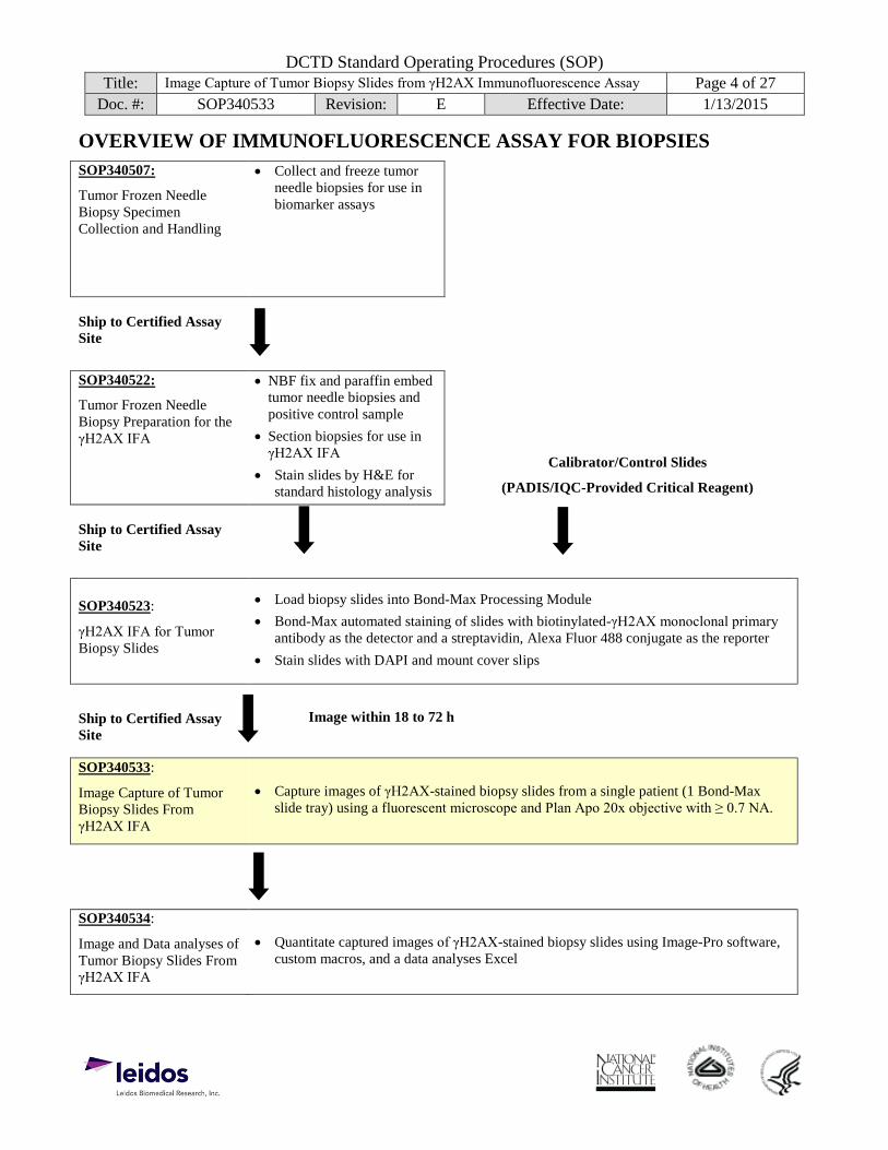
OVERVIEW OF IMMUNOFLUORESCENCE ASSAY FOR BIOPSIES SOP340507:
Tumor Frozen Needle Biopsy Specimen Collection and Handling
• Collect and freeze tumorneedle biopsies for use inbiomarker assays
Ship to Certified Assay Site
SOP340522:
Tumor Frozen Needle Biopsy Preparation for the γH2AX IFA
• NBF fix and paraffin embedtumor needle biopsies andpositive control sample
• Section biopsies for use inγH2AX IFA
• Stain slides by H&E forstandard histology analysis
Calibrator/Control Slides
(PADIS/IQC-Provided Critical Reagent)
Ship to Certified Assay Site
SOP340523:
γH2AX IFA for Tumor Biopsy Slides
• Load biopsy slides into Bond-Max Processing Module• Bond-Max automated staining of slides with biotinylated-γH2AX monoclonal primary
antibody as the detector and a streptavidin, Alexa Fluor 488 conjugate as the reporter• Stain slides with DAPI and mount cover slips
Ship to Certified Assay Site
Image within 18 to 72 h
SOP340533:
Image Capture of Tumor Biopsy Slides From γH2AX IFA
• Capture images of γH2AX-stained biopsy slides from a single patient (1 Bond-Maxslide tray) using a fluorescent microscope and Plan Apo 20x objective with ≥ 0.7 NA.
SOP340534:
Image and Data analyses of Tumor Biopsy Slides From γH2AX IFA
• Quantitate captured images of γH2AX-stained biopsy slides using Image-Pro software,custom macros, and a data analyses Excel
DCTD Standard Operating Procedures (SOP) Title: Image Capture of Tumor Biopsy Slides from γH2AX Immunofluorescence Assay Page 5 of 27
Doc. #: SOP340533 Revision: E Effective Date: 1/13/2015 1.0 PURPOSE
Standardize an immunohistochemical method for detecting and quantifying histone H2AX phosphorylated at serine 139 (γH2AX) staining in formalin-fixed, paraffin-embedded human tissue biopsies for pharmacodynamic studies of chemotherapeutic DNA-damaging agents.
2.0 SCOPE
This procedure applies to all personnel involved in the image capture of slides stained using the γH2AX Immunofluorescence Assay (IFA) for Tumor Biopsy Slides (SOP340523). This SOP outlines the recommended procedure for image capture of γH2AX-stained, paraffin-embedded tumor biopsy sections. The goal of the SOP and associated training is to ensure consistency of γH2AX measurement between clinical sites.
3.0 ABBREVIATIONS
Ab = Antibody CalCon = Calibrator/Control CCD = Charge Coupled Device DAPI = 4',6-Diamidino-2-Phenylindole DCTD = Division of Cancer Treatment and Diagnosis DIH = Digital Imaging Head FITC = Fluorescein Isothiocyanate γH2AX = Histone H2AX Phosphorylated at Serine 139 H&E = Hemotoxylin and Eosin ID = Identification/Identifier IFA = Immunofluorescence Assay LHTP = Laboratory of Human Toxicology & Pharmacology NA = Numerical Aperture NBF = Neutral Buffered Formalin NCTVL = National Clinical Target Validation Laboratory %NAP = Percent Nuclear Area Positive for γH2AX QC = Quality Control SOP = Standard Operating Procedure Strp488 = Alexa Fluor 488-Streptavidin Conjugate
4.0 INTRODUCTION
The γH2AX IFA is an immunohistochemistry-based staining assay developed to quantify the nuclear DNA damage marker, histone γH2AX. The assay uses a biotinylated-γH2AX monoclonal antibody as the detector and an Alexa Fluor 488-streptavidin conjugate (Strp488) as the reporter for immunostaining.
DCTD Standard Operating Procedures (SOP) Title: Image Capture of Tumor Biopsy Slides from γH2AX Immunofluorescence Assay Page 6 of 27
Doc. #: SOP340533 Revision: E Effective Date: 1/13/2015 5.0 ROLES AND RESPONSIBILITIES
Laboratory Director/Supervisor The Laboratory Director/Supervisor, directs laboratory operations, supervises technical personnel and reporting of findings, and is responsible for the proper performance of all laboratory procedures. The Laboratory Director/Supervisor oversees the personnel who follow the SOPs within the laboratory and is responsible for ensuring the personnel are certified and have sufficient experience to handle clinical samples.
Certified Assay Operator A Certified Assay Operator may be a Laboratory Technician/ Technologist, Research Associate, or Laboratory Scientist who has been certified through DCTD training on this SOP. The Certified Assay Operator works under the guidance of the Laboratory Director/Supervisor. This person performs laboratory procedures and examinations in accordance with the current SOP(s), as well as any other procedures conducted by a laboratory, including maintaining equipment and records and performing quality assurance activities related to performance.
5.1 It is the responsibility of the Laboratory Director/Supervisor to ensure that all personnel have documented training and qualification on this SOP prior to the actual handling and processing of samples from clinical trial patients. The Laboratory Director/Supervisor is responsible for ensuring the Certified Assay Operator running the SOP has sufficient experience to handle and analyze clinical samples.
5.2 The Certified Assay Operator for this SOP should be well versed and comfortable with light microscopy techniques.
5.3 Digital versions of the Image Information Table in the Batch Record (Appendix 1, Sections 2) can be created for logging sample information as long as all column information exactly matches the table in the Batch Record. If any portion of the Batch Record is recorded in a digital copy, cross through that section, state “See attached sheet,” and initial. A copy of the completed, digital table must be printed and attached to the Batch Record in order to maintain a complete audit trail.
5.4 The Certified Assay Operator responsible for conducting the assay is to follow this SOP and complete the required tasks and associated documentation. The Batch Record (Appendix 1) must be completed in real-time for each experimental run, with each page dated and initialed, and placed with the clinical sample information.
5.5 All responsible personnel are to check the DCTD Biomarkers Web site (http://dctd.cancer.gov/ResearchResources/ResearchResources-biomarkers.htm) to verify that the most recent version of the SOP for the assay is being used.
DCTD Standard Operating Procedures (SOP) Title: Image Capture of Tumor Biopsy Slides from γH2AX Immunofluorescence Assay Page 7 of 27
Doc. #: SOP340533 Revision: E Effective Date: 1/13/2015 6.0 MATERIALS AND EQUIPMENT REQUIRED
6.1 Kimwipes (e.g., Fischer Scientific, Cat#: 06-666A)
6.2 Bond-Max slide tray containing one patient’s clinical sample slides and calibrator/control slides processed according to SOP340523 6.2.1 A copy of the Slide Information Table from the Batch Record of SOP340523 for all
slides being processed (SOP340523; Appendix 1, Section 3) 6.2.2 A copy of the patient sample information from the Batch Record of SOP340522 for the
biopsy samples (SOP340522; Appendix 1, Sections 2 and 3)
6.3 QImaging Retiga-2000R fast 1394 mono-cooled camera (QImaging, Model#: RET-2000R-F-M-12-C)
6.4 Leica Microscope (SOP as written references this set up) 6.4.1 Leica DM5000B microscope with 1X C mount adaptor (Leica, Cat#: 541510) 6.4.2 EL6000 external light source with Liquid Light Guide and OEM bulbs (Leica, Cat#:
11504116). Must have known usage hours for light source and light guide; these hours must be within the manufacturer’s specifications
6.4.3 Leica Plan Apo 20x/0.70 numerical aperture (NA) objective, PH2 6.4.4 DAPI (blue channel, Chroma A4 filter, BP 360/40) and FITC (green channel, Chroma L5
filter, BP 480/40) filter sets
6.5 Nikon Microscope 6.5.1 Nikon Eclipse 80i or 90i (Nikon Instruments)
DIH-E or DIH-M digital imaging head 1X C-mount CCD camera coupler
6.5.2 Nikon Intensilight, Model: C-HGFl (manual; Nikon Instruments) 6.5.3 Nikon CFI Plan Apo DM 20x/0.75 NA objective, PH2 (Nikon Instruments) 6.5.4 DAPI (blue channel, UV-2E/C) and FITC (green channel, B-2E/C) filter cubes (Nikon
Instruments)
6.6 PC (Parallels for Mac not supported) 6.7 Image-Pro 7.0 or higher (lower versions of Image-Pro are not supported and may not work with
the macro) with updated drivers for the QImaging camera (see Appendix 2; Media Cybernetics) 6.8 Microsoft Excel 2003, 2007, or 2010 6.9 The following files will be provided to DCTD Certified Assay Operators during the training
course: 6.9.1 A self-extracting zip file entitled “gH2AX qIFA macro v091611.exe” will include
required files for image capture and analysis including the macro files gH2AXCaptureControl_ v011211.bas, ListandExcelSorter_for_gH2AXImageAssay.bas, and qIFA_gH2AX_ v072007.ipm).
Alternate microscope systems using 20x objective with at least a 0.70 NA may be used, but must meet the following performance specifications: (1) image capture time for each field for each calibrator must be less than or equal to 1 min, and (2) each calibrator must return a %NAP within the acceptable ranges as defined in SOP340534.
DCTD Standard Operating Procedures (SOP) Title: Image Capture of Tumor Biopsy Slides from γH2AX Immunofluorescence Assay Page 8 of 27
Doc. #: SOP340533 Revision: E Effective Date: 1/13/2015 7.0 OPERATING PROCEDURES
7.1 Image capture should be completed for a single slide tray from the Bond-Max Processing Module tray (up to 8 clinical slides from a single patient and 2 calibrator/control [CalCon] slides). Images captured using this SOP will be analyzed in SOP340534, batched by slide tray.
7.1.1 Record the name and certification number of the Certified Assay Operator performing the image capture, the facility running this SOP, the Patient ID, and the clinical protocol number in the Batch Record (Appendix 1).
7.1.2 Attach a COPY of the Slide Information Table from the Batch Record of SOP340523 for the patient slides being processed in this SOP and record the date the slide tray was processed on the Bond-Max System and the slide tray number in the Batch Record (Appendix 1). Cross out any slides that are not imaged in this SOP.
7.1.3 Patient sample information from the Shipping Manifest of SOP340507 (attached to SOP340522) will be needed to fill in dose level information during image capture.
7.2 Install the Image-Pro software on the PC associated with the microscope; this software will use the custom-designed macros for this assay to perform image capture.
7.2.1 Macro installation instructions can be found in Appendix 3, Section 1A; the first time the macro is run the instructions in Appendix 3, Section 1B should be followed before proceeding.
7.2.2 Auto-white balance, dark frame subtraction, and background correction should be established the first time the Image-Pro software is run on a PC; see Appendix 3, Section 2 for details.
7.2.3 If using software other than Image-Pro, the image capture module for quantitative immunofluorescence can be achieved using manual or automated capture methods that meet a set of minimum specifications as outlined in Appendix 3, Section 3.
7.3 Protocol for Image Capture
7.3.1 Turn on the Leica DM5000B microscope, light source, and the QImaging Retiga-2000R camera. Allow lamp to warm up for 10 min before use. Important: An objective with at least a 0.70 NA is required for optimal resolution during image capture.
7.3.1.1 Record the total usage hours to date for the EL6000 Light Source and Liquid Light Guide in the Batch Record (Appendix 1, Section 1). Calculate and record the current rating for both and ensure they are within the manufacturer’s specifications.
7.3.1.2 Record the time the EL6000 Light Source and Liquid Light Guide are turned on in the Batch Record (Appendix 1, Section 1).
7.3.2 Open the Image-Pro software and in the macro toolbar that pops up select the Initialize Macro button; the macro Capture Menu window will open.
7.3.2.1 If the macro toolbar does not pop up when Image-Pro software is opened, return to SOP Step 7.2.1 and the follow instructions for first time use of the macro.
DCTD Standard Operating Procedures (SOP) Title: Image Capture of Tumor Biopsy Slides from γH2AX Immunofluorescence Assay Page 9 of 27
Doc. #: SOP340533 Revision: E Effective Date: 1/13/2015
7.3.3 For each assay run, the macro will create a folder using the entry in the “Protocol” field of the Capture Menu (SOP Step 7.3.7). This folder, which will be referred to as the Header Folder from this point on, will be placed in the location selected for the “Image Directory” field on the Capture Menu (default C:\gH2AX qIFA\IpWin Source Files\Scripts\Projects\). All *.tif images will be saved in the Header Folder.
7.3.3.1 In order to ensure images captured for the same Protocol are not overwritten we suggest entering “CTEP#_PatinetID_Date_Increment” in the “Protocol” field of the Capture Menu (e.g., CTEP1234_20101024_1).
7.3.3.2 Record the name of the Header Folder in the Batch Record (Appendix 1, Section 2).
7.3.4 The following order for image capture will ensure data are analyzed, sorted and graphed correctly in SOP340534. Capture images for BOTH calibrator/control (CalCon) slides first, in the section order below, followed by the clinical samples.
Slide Capture Order
Bond Tray Position Slide Description Section Capture Order
1 Position 1 CalCon*
Cal-Low (A) Cal-Mid (B) Cal-High (C) Positive Control (D) Negative Control (E)
2 Last Position CalCon*
Cal-Low (A) Cal-Mid (B) Cal-High (C) Positive Control (D) Negative Control (E)
3-10 Position 2-N Clinical Samples Post-dose Biopsy Pre-dose Biopsy Positive Control (testes)
*See Appendix 4 for CalCon and clinical slide set up.
7.3.5 As images are captured, complete the Image Information Table in the Batch Record (Appendix 1, Section 2) with the Bond Slide ID Number for each slide, in the order it is captured, along with the corresponding Bond-Max slide tray position; add notes for individual slides or images as needed.
DCTD Standard Operating Procedures (SOP) Title: Image Capture of Tumor Biopsy Slides from γH2AX Immunofluorescence Assay Page 10 of 27
Doc. #: SOP340533 Revision: E Effective Date: 1/13/2015
7.3.6 Image Requirements Per Tissue Section
For the calibrator and control tissue sections, a minimum of 3 (more is highlyrecommended) analyzable images should be captured for each tissue section on aslide; avoid necrotic areas, large gaps in the tissue, or areas where tissue has foldedonto itself.
If < 3 analyzable images can be captured from a calibrator or control tissue section,note in the Image Information Table (Appendix 1, Section 2) the reason for theinability to capture 3 analyzable images.
For the clinical biopsy tissue section, a minimum of 8 non-overlapping images perslide set is required; it is highly recommended that more are captured when possible.Each positive control on the clinical biopsy slide should have a minimum of 3analyzable images captured.
If no analyzable images can be captured from a clinical biopsy tissue section on anindividual slide, note in the Image Information Table (Appendix 1, Section 2) thereason for the inability to capture images.
IMPORTANT: If a tissue section has no analyzable fields, capture a single background image for that section to ensure the data are sorted and graphed correctly in SOP340534.
DCTD Standard Operating Procedures (SOP) Title: Image Capture of Tumor Biopsy Slides from γH2AX Immunofluorescence Assay Page 11 of 27
Doc. #: SOP340533 Revision: E Effective Date: 1/13/2015
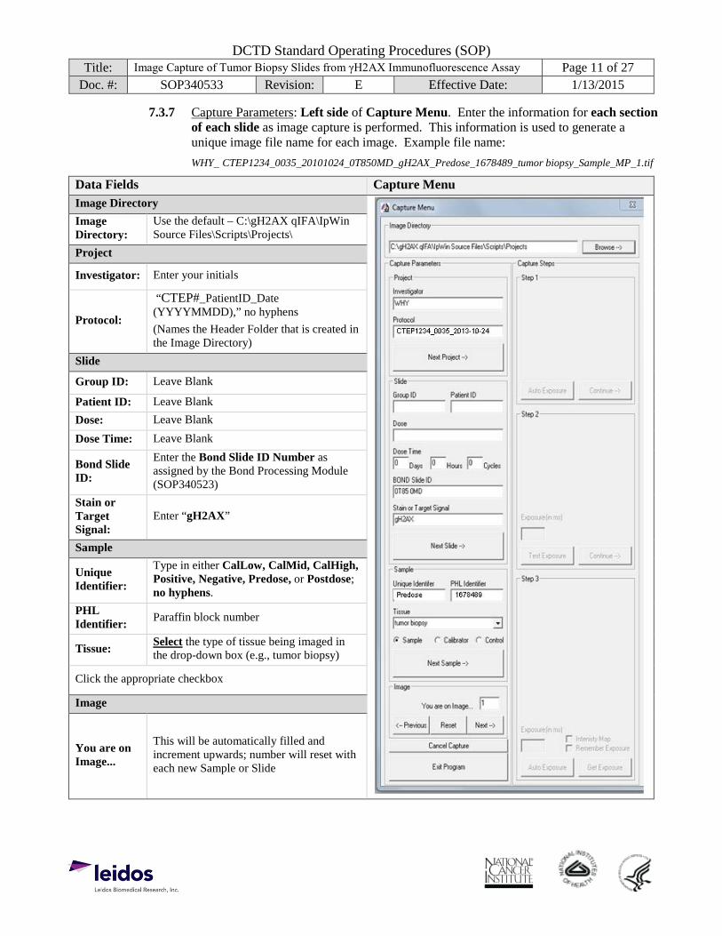
7.3.7 Capture Parameters: Left side of Capture Menu. Enter the information for each section of each slide as image capture is performed. This information is used to generate a unique image file name for each image. Example file name: WHY_ CTEP1234_0035_20101024_0T850MD_gH2AX_Predose_1678489_tumor biopsy_Sample_MP_1.tif
Data Fields Capture Menu Image Directory Image Directory:
Use the default – C:\gH2AX qIFA\IpWin Source Files\Scripts\Projects\
Project
Investigator: Enter your initials
Protocol:
“CTEP#_PatientID_Date (YYYYMMDD),” no hyphens (Names the Header Folder that is created in the Image Directory)
Slide
Group ID: Leave Blank
Patient ID: Leave Blank Dose: Leave Blank Dose Time: Leave Blank
Bond Slide ID:
Enter the Bond Slide ID Number as assigned by the Bond Processing Module (SOP340523)
Stain or Target Signal:
Enter “gH2AX”
Sample
Unique Identifier:
Type in either CalLow, CalMid, CalHigh, Positive, Negative, Predose, or Postdose; no hyphens.
PHL Identifier: Paraffin block number
Tissue: Select the type of tissue being imaged in the drop-down box (e.g., tumor biopsy)
Click the appropriate checkbox
Image
You are on Image...
This will be automatically filled and increment upwards; number will reset with each new Sample or Slide
DCTD Standard Operating Procedures (SOP) Title: Image Capture of Tumor Biopsy Slides from γH2AX Immunofluorescence Assay Page 12 of 27
Doc. #: SOP340533 Revision: E Effective Date: 1/13/2015
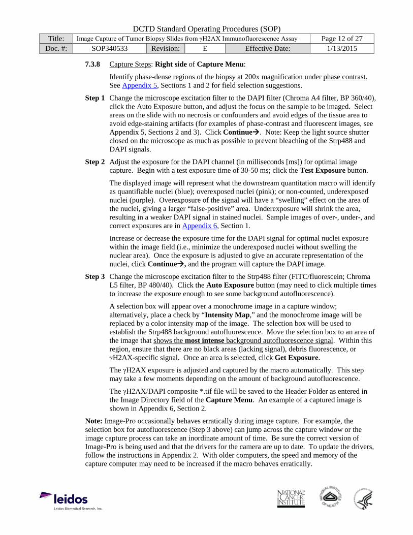
7.3.8 Capture Steps: Right side of Capture Menu:
Identify phase-dense regions of the biopsy at 200x magnification under phase contrast. See Appendix 5, Sections 1 and 2 for field selection suggestions.
Step 1 Change the microscope excitation filter to the DAPI filter (Chroma A4 filter, BP 360/40), click the Auto Exposure button, and adjust the focus on the sample to be imaged. Select areas on the slide with no necrosis or confounders and avoid edges of the tissue area to avoid edge-staining artifacts (for examples of phase-contrast and fluorescent images, see Appendix 5, Sections 2 and 3). Click Continue. Note: Keep the light source shutter closed on the microscope as much as possible to prevent bleaching of the Strp488 and DAPI signals.
Step 2 Adjust the exposure for the DAPI channel (in milliseconds [ms]) for optimal image capture. Begin with a test exposure time of 30-50 ms; click the Test Exposure button.
The displayed image will represent what the downstream quantitation macro will identify as quantifiable nuclei (blue); overexposed nuclei (pink); or non-counted, underexposed nuclei (purple). Overexposure of the signal will have a “swelling” effect on the area of the nuclei, giving a larger “false-positive” area. Underexposure will shrink the area, resulting in a weaker DAPI signal in stained nuclei. Sample images of over-, under-, and correct exposures are in Appendix 6, Section 1.
Increase or decrease the exposure time for the DAPI signal for optimal nuclei exposure within the image field (i.e., minimize the underexposed nuclei without swelling the nuclear area). Once the exposure is adjusted to give an accurate representation of the nuclei, click Continue, and the program will capture the DAPI image.
Step 3 Change the microscope excitation filter to the Strp488 filter (FITC/fluorescein; Chroma L5 filter, BP 480/40). Click the Auto Exposure button (may need to click multiple times to increase the exposure enough to see some background autofluorescence).
A selection box will appear over a monochrome image in a capture window; alternatively, place a check by “Intensity Map,” and the monochrome image will be replaced by a color intensity map of the image. The selection box will be used to establish the Strp488 background autofluorescence. Move the selection box to an area of the image that shows the most intense background autofluorescence signal. Within this region, ensure that there are no black areas (lacking signal), debris fluorescence, or γH2AX-specific signal. Once an area is selected, click Get Exposure.
The γH2AX exposure is adjusted and captured by the macro automatically. This step may take a few moments depending on the amount of background autofluorescence.
The γH2AX/DAPI composite *.tif file will be saved to the Header Folder as entered in the Image Directory field of the Capture Menu. An example of a captured image is shown in Appendix 6, Section 2.
Note: Image-Pro occasionally behaves erratically during image capture. For example, the selection box for autofluorescence (Step 3 above) can jump across the capture window or the image capture process can take an inordinate amount of time. Be sure the correct version of Image-Pro is being used and that the drivers for the camera are up to date. To update the drivers, follow the instructions in Appendix 2. With older computers, the speed and memory of the capture computer may need to be increased if the macro behaves erratically.
DCTD Standard Operating Procedures (SOP) Title: Image Capture of Tumor Biopsy Slides from γH2AX Immunofluorescence Assay Page 13 of 27
Doc. #: SOP340533 Revision: E Effective Date: 1/13/2015
7.3.9 Next Image, Section, or Slide:
Important: On the Capture Menu, the Next, Next Sample, or Next Slide button must be selected between each image captured (see next step). Changing the entry fields in the Capture Menu without clicking one of these buttons will cause the data in the data analyses Excel workbook to be mis-grouped/-sorted.
IMPORTANT: If a tissue section has no analyzable fields, capture a single background image for that section to ensure the data are sorted and graphed correctly in SOP340534.
7.3.9.1 Capture ≥ 3 different images per tissue section for each calibrator and control. Capture ≥ 8 different images per slide set for the biopsy tissue and ≥ 3 analyzable images for the positive control on the clinical slide. See Appendix 4 for CalCon and clinical slide section orientation.
7.3.9.2 Next Image, Same Section, Same Slide:
Click the Next button at the bottom of the screen in the “Image” section; the Capture Menu window will cycle to the next image number for that tissue section. Repeat image capture as outlined in SOP Step 7.3.8.
7.3.9.3 Next Image, Different Section, Same Slide:
Click Next Sample; the Capture Menu window will reset the image number to 1. The Bond Slide ID Number will remain the same.
As needed, update fields on the left side of the Capture Menu (SOP Step 7.3.7) for the new section being analyzed. Be sure to update the fields to reflect which section on the slide is being imaged, i.e., positive control (testes), pre-dose, or post-dose biopsy, or specific CalCon section. Repeat image capture as outlined in SOP Step 7.3.8.
7.3.9.4 Next Image, Different Slide:
Click Next Slide; the Capture Menu window will reset to image number 1.
Change the Bond Slide ID Number. As needed, update fields on the left side of the Capture Menu (SOP Step 7.3.7) for the new slide and section being analyzed. Repeat image capture as outlined in SOP Step 7.3.8.
7.3.10 Once all images have been captured, click Exit Program at the bottom of the Capture Menu.
7.3.11 Record the time the light source and guide are turned off and total hours used in this assay in the Batch Record (Appendix 1, Section 1).
7.4 Proceed to SOP340534 for image and data analyses.
7.5 Review and finalize the Batch Record (Appendix 1) and obtain required signatures. Document ANY and ALL deviations from this SOP in the Batch Record (Appendix 1, Section 3).
7.6 The Laboratory Director/Supervisor should review the Batch Record and sample reports and sign the Batch Record affirming the data contained within the reports are correct (Appendix 1, Section 4).

DCTD Standard Operating Procedures (SOP) Title: Image Capture of Tumor Biopsy Slides from γH2AX Immunofluorescence Assay Page 14 of 27
Doc. #: SOP340533 Revision: E Effective Date: 1/13/2015
THIS PAGE LEFT BLANK ON PURPOSE
DCTD Standard Operating Procedures (SOP) Title: Image Capture of Tumor Biopsy Slides from γH2AX Immunofluorescence Assay Page 15 of 27
Doc. #: SOP340533 Revision: E Effective Date: 1/13/2015
APPENDIX 1: BATCH RECORD The Batch Record should contain information for one slide tray, and therefore a single patient’s batched clinical slides. Record times using military time (24-h designation); for example, specify 16:15 to indicate 4:15 PM.
Patient ID:
CTEP/Clinical Protocol Number:
Date Slide Tray Processed in Bond-Max:
Slide Tray Number:
Certified Assay Operator:
Certification Number:
Facility/Laboratory Running Image Capture:
Attach a copy of the Slide Information Table from SOP340523 for the slides being processed in this SOP.
1. Light Source and Guide Usage Hours
The known usage hours for the Light Source and Light Guide must be within the manufacturer’s specifications. The Liquid Light Guide should be replaced at the same time as the Light Source.
Before Assay After Assay
Total Usage Hours
Current Rating* Time
Turned On Time
Turned Off Hours Used in this Assay Manuf. max. hours
Total usage hours = x
Light Source h = : : h
* Exposure time is a function of both the light source and light guide. Recommendations for tracking the current rating and minimum amount of illumination needed to proceed with the SOP are provided during the training course.
BATCH RECORD: INITIALS DATE:
DCTD Standard Operating Procedures (SOP) Title: Image Capture of Tumor Biopsy Slides from γH2AX Immunofluorescence Assay Page 16 of 27
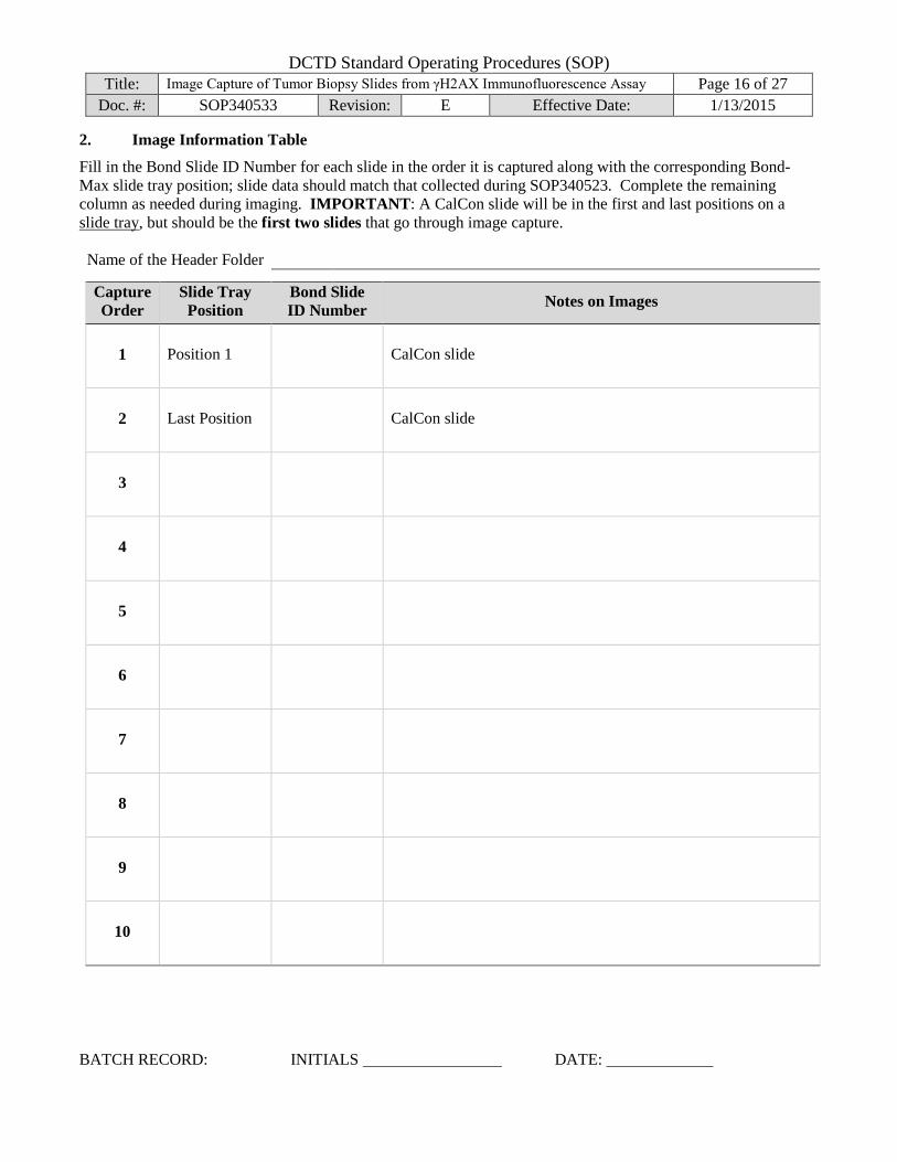
Doc. #: SOP340533 Revision: E Effective Date: 1/13/2015 2. Image Information Table
Fill in the Bond Slide ID Number for each slide in the order it is captured along with the corresponding Bond-Max slide tray position; slide data should match that collected during SOP340523. Complete the remaining column as needed during imaging. IMPORTANT: A CalCon slide will be in the first and last positions on a slide tray, but should be the first two slides that go through image capture.
Name of the Header Folder
Capture Order
Slide Tray Position
Bond Slide ID Number Notes on Images
1 Position 1 CalCon slide
2 Last Position CalCon slide
3
4
5
6
7
8
9
10
BATCH RECORD: INITIALS DATE:

DCTD Standard Operating Procedures (SOP) Title: Image Capture of Tumor Biopsy Slides from γH2AX Immunofluorescence Assay Page 17 of 27
Doc. #: SOP340533 Revision: E Effective Date: 1/13/2015 3. Notes, including any deviations from the SOP:
4. Laboratory Director/Supervisor Review of Batch Record
Laboratory Director/Supervisor: (PRINT)
(SIGN)
Date:
BATCH RECORD: INITIALS DATE:

DCTD Standard Operating Procedures (SOP) Title: Image Capture of Tumor Biopsy Slides from γH2AX Immunofluorescence Assay Page 18 of 27
Doc. #: SOP340533 Revision: E Effective Date: 1/13/2015
THIS PAGE LEFT BLANK ON PURPOSE
BATCH RECORD: INITIALS DATE:

DCTD Standard Operating Procedures (SOP) Title: Image Capture of Tumor Biopsy Slides from γH2AX Immunofluorescence Assay Page 19 of 27
Doc. #: SOP340533 Revision: E Effective Date: 1/13/2015
APPENDIX 2: UPDATING QIMAGING DRIVER The most current version of the QImaging Family Driver can be downloaded at
http://support.mediacy.com/drivers.asp
1. Installation:
A. Download the driver *.zip file to a temporary location.
B. Extract/unzip the contents.
C. Double-click (run) the *.exe installer and follow the prompts.
The installer does 3 things:
a. Copies the Image-Pro Capture Driver and Capture Module into the Image-Pro directory. If you have more than one Media Cybernetics application installed on this computer, make sure to select the appropriate directories when prompted.
b. Installs low-level libraries (QCAM) provided by the QImaging Corporation. This includes the Windows drivers (.sys, .inf) necessary to operate the camera.
c. Installs and loads a third-party 1394 IEEE (Firewire) stack provided by Thesycon.
2. Sometimes the last step (c.) does not complete properly.
If this is the case, the camera will not be recognized and the software will behave as if no camera is attached. To correct this issue, the Thesycon driver must be manually loaded. To accomplish this, complete the following steps:
A. Unplug the camera from the computer. B. Open the Control Panel (Start Menu) and select “System.” On the System Properties dialog,
select the “Hardware” tab, then “Device Manager.” C. Locate the “IEEE 1394 Bus host controllers” hive and expand by clicking on the + icon. If it says
anything other than “T1394…” (for example, “Texas Instruments OHCI Compliant IEEE….,” the Thesycon stack is not loaded.
D. Right click on the listing and select “Update Driver…” If the Windows Update screen appears, select “No, not at this time.”
E. Depending on the Operating System, you will have different options: In Windows XP, select “Advanced” then “Don’t search, I will choose the driver…” In Windows Vista, select “Browse my computer for driver software,” then “Let me pick from a list of device drivers on this computer.”
F. On the next dialog, a list of Firewire stacks will be found. It will typically list a generic “OHCI Compliant IEEE 1394 Host Controller” and the default “Texas Instruments….” There should also be an option for “T1394…”—this is the Thesycon stack. Select this option and press Next.
G. When Windows has successfully updated the driver, close this dialog and plug in the camera. H. If you are presented with the “Found New Hardware” wizard, follow the prompts to complete the
installation. Otherwise, a new hive will appear in Device Manager displaying a generic QImaging camera entry or “Imaging Device.” You are now ready to use the camera in Image-Pro.
DCTD Standard Operating Procedures (SOP) Title: Image Capture of Tumor Biopsy Slides from γH2AX Immunofluorescence Assay Page 20 of 27
Doc. #: SOP340533 Revision: E Effective Date: 1/13/2015
APPENDIX 3: MACRO INSTALLATION AND MICROSCOPE SET UP 1. Installing the Image Capture and Data analyses Macro
A. Installation Instructions
a. Double-click (run) the self-extracting zip file entitled “gH2AX qIFA macro v091611.exe” and follow the prompts
b. The macro scripts are automatically saved on the hard drive under C:\gH2AX qIFA\. Note: Do not create this folder ahead of time or change the name of the folder; it will prevent proper loading of the files.
B. First Time the Macro is Run
Before the macro is used for data analyses, the user needs to activate the macro toolbar on startup and initialize the macro.
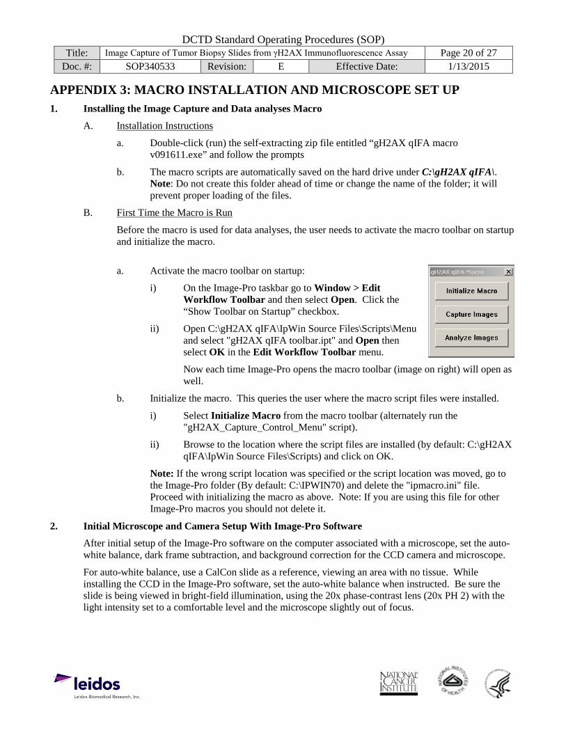
a. Activate the macro toolbar on startup:
i) On the Image-Pro taskbar go to Window > Edit Workflow Toolbar and then select Open. Click the “Show Toolbar on Startup” checkbox.
ii) Open C:\gH2AX qIFA\IpWin Source Files\Scripts\Menu and select "gH2AX qIFA toolbar.ipt" and Open then select OK in the Edit Workflow Toolbar menu.
Now each time Image-Pro opens the macro toolbar (image on right) will open as well.
b. Initialize the macro. This queries the user where the macro script files were installed.
i) Select Initialize Macro from the macro toolbar (alternately run the "gH2AX_Capture_Control_Menu" script).
ii) Browse to the location where the script files are installed (by default: C:\gH2AX qIFA\IpWin Source Files\Scripts) and click on OK.
Note: If the wrong script location was specified or the script location was moved, go to the Image-Pro folder (By default: C:\IPWIN70) and delete the "ipmacro.ini" file. Proceed with initializing the macro as above. Note: If you are using this file for other Image-Pro macros you should not delete it.
2. Initial Microscope and Camera Setup With Image-Pro Software
After initial setup of the Image-Pro software on the computer associated with a microscope, set the auto-white balance, dark frame subtraction, and background correction for the CCD camera and microscope.
For auto-white balance, use a CalCon slide as a reference, viewing an area with no tissue. While installing the CCD in the Image-Pro software, set the auto-white balance when instructed. Be sure the slide is being viewed in bright-field illumination, using the 20x phase-contrast lens (20x PH 2) with the light intensity set to a comfortable level and the microscope slightly out of focus.
DCTD Standard Operating Procedures (SOP) Title: Image Capture of Tumor Biopsy Slides from γH2AX Immunofluorescence Assay Page 21 of 27
Doc. #: SOP340533 Revision: E Effective Date: 1/13/2015 3. Image Capture Macro Specifications
The image capture macro is responsible for acquiring images in a consistent manner for the image analysis macro. Consistency between each image capture is controlled by adjusting the exposure based on background intensity. Below are recommendations to achieve this using either a manual or automated capture method.
It should be noted that development of image capture and analysis macro scripts requires familiarity of both the image analysis software being used as well as basic macro programming knowledge to ensure the required specifications for the SOP are met.
1.) Using a 20x objective with at least a 0.70 NA, capture separate 12 bit images in the DAPI and FITC channels.
2.) Specifications for DAPI Channel Image A. DAPI intensity across all nuclei is typically even throughout a field of view and only
requires a visual representation of an acceptable exposure. For the γH2AX IFA, target nuclei should have DAPI intensity values within a predefined "Optimal Exposure" range. a. For 12 bit images, the following DAPI fluorescence intensity ranges are
recommended when creating an image capture macro: i) Glass (no DAPI stained nuclei): 0-401 ii) Over-exposed: 2506-4095 iii) Optimal exposure: 820-2505 iv) Under-exposed: 402-819
B. Once the DAPI fluorescence intensity has been adjusted within the optimal exposure range and no intact nuclei fall into the under-exposed range, the DAPI channel image for that field can be captured. Be sure to minimize nuclei that fall into the over-exposed range; over-exposed nuclei are acceptable if the signal is restricted to the center of the nucleus.
3.) Specifications for FITC Channel Image
A. For the γH2AX IFA, FITC channel acquisition is done using step-wise exposures until a mean intensity threshold (MIT) of 500 is achieved on a known area of background autofluorescence. The image capture macro should use the following specifications to achieve this. a. Draw a region of interest (ROI) box on a known area of background
autofluorescence and measure the mean intensity. b. The exposure should then be adjusted until the MIT = 500. This can be achieved
by manually selecting exposures until the target threshold is met or by using an exposure increment routine as follows: i) If Target(MIT) - Current(MIT) ≤ 20; increment exposure by 1, ii) Else if Target(MIT) - Current(MIT) ≤ 60; increment exposure by 2, iii) Else if Target(MIT) - Current(MIT) ≤ 200; increment exposure by 5, iv) Else if Target(MIT) - Current(MIT) ≤ Target(MIT); increment exposure
by 10. c. Acquire the FITC channel once the MIT exposure is reached.
4.) Pseudo-color the monochrome images (DAPI = blue, FITC = green) and combine into an 8 bit TIFF image.
DCTD Standard Operating Procedures (SOP) Title: Image Capture of Tumor Biopsy Slides from γH2AX Immunofluorescence Assay Page 22 of 27
Doc. #: SOP340533 Revision: E Effective Date: 1/13/2015
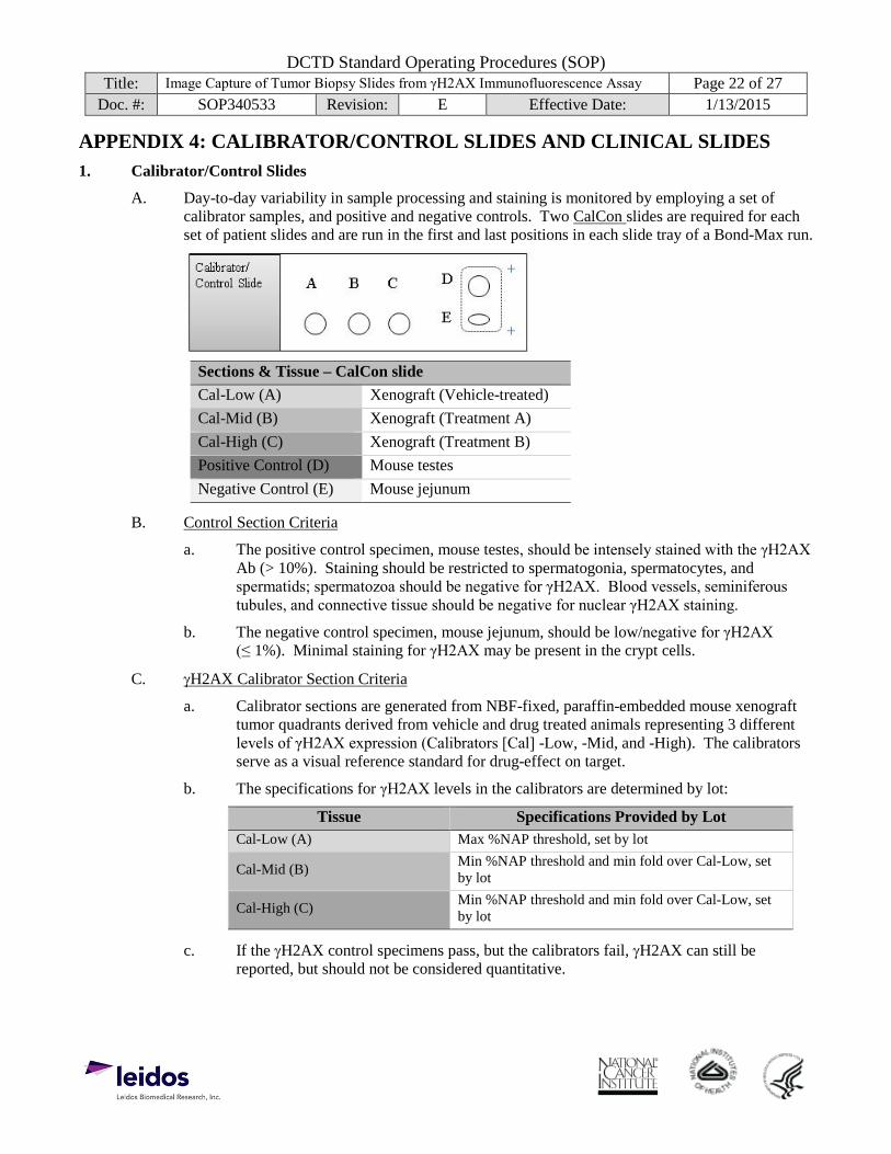
APPENDIX 4: CALIBRATOR/CONTROL SLIDES AND CLINICAL SLIDES 1. Calibrator/Control Slides
A. Day-to-day variability in sample processing and staining is monitored by employing a set of calibrator samples, and positive and negative controls. Two CalCon slides are required for each set of patient slides and are run in the first and last positions in each slide tray of a Bond-Max run.
Sections & Tissue – CalCon slide Cal-Low (A) Xenograft (Vehicle-treated) Cal-Mid (B) Xenograft (Treatment A) Cal-High (C) Xenograft (Treatment B) Positive Control (D) Mouse testes Negative Control (E) Mouse jejunum
B. Control Section Criteria
a. The positive control specimen, mouse testes, should be intensely stained with the γH2AX Ab (> 10%). Staining should be restricted to spermatogonia, spermatocytes, and spermatids; spermatozoa should be negative for γH2AX. Blood vessels, seminiferous tubules, and connective tissue should be negative for nuclear γH2AX staining.
b. The negative control specimen, mouse jejunum, should be low/negative for γH2AX (≤ 1%). Minimal staining for γH2AX may be present in the crypt cells.
C. γH2AX Calibrator Section Criteria
a. Calibrator sections are generated from NBF-fixed, paraffin-embedded mouse xenograft tumor quadrants derived from vehicle and drug treated animals representing 3 different levels of γH2AX expression (Calibrators [Cal] -Low, -Mid, and -High). The calibrators serve as a visual reference standard for drug-effect on target.
b. The specifications for γH2AX levels in the calibrators are determined by lot:
Tissue Specifications Provided by Lot Cal-Low (A) Max %NAP threshold, set by lot
Cal-Mid (B) Min %NAP threshold and min fold over Cal-Low, set by lot
Cal-High (C) Min %NAP threshold and min fold over Cal-Low, set by lot
c. If the γH2AX control specimens pass, but the calibrators fail, γH2AX can still be reported, but should not be considered quantitative.
DCTD Standard Operating Procedures (SOP) Title: Image Capture of Tumor Biopsy Slides from γH2AX Immunofluorescence Assay Page 23 of 27
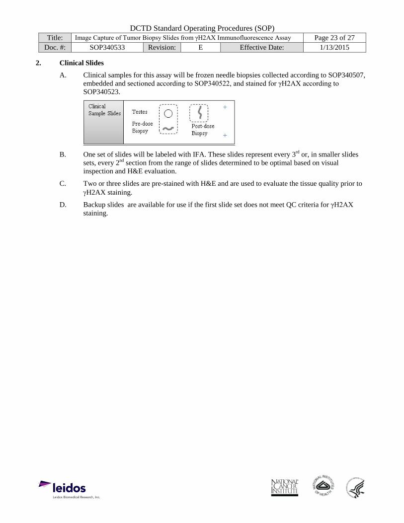
Doc. #: SOP340533 Revision: E Effective Date: 1/13/2015 2. Clinical Slides
A. Clinical samples for this assay will be frozen needle biopsies collected according to SOP340507, embedded and sectioned according to SOP340522, and stained for γH2AX according to SOP340523.
B. One set of slides will be labeled with IFA. These slides represent every 3rd or, in smaller slides sets, every 2nd section from the range of slides determined to be optimal based on visual inspection and H&E evaluation.
C. Two or three slides are pre-stained with H&E and are used to evaluate the tissue quality prior to γH2AX staining.
D. Backup slides are available for use if the first slide set does not meet QC criteria for γH2AX staining.
DCTD Standard Operating Procedures (SOP) Title: Image Capture of Tumor Biopsy Slides from γH2AX Immunofluorescence Assay Page 24 of 27
Doc. #: SOP340533 Revision: E Effective Date: 1/13/2015
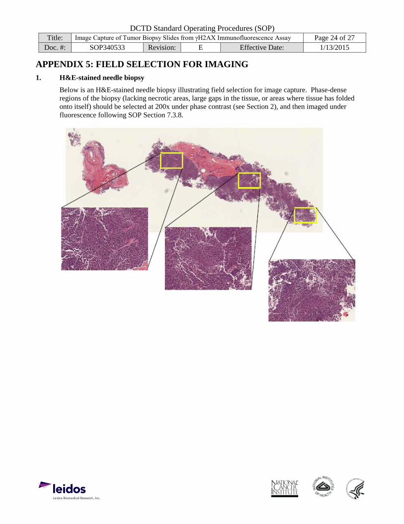
APPENDIX 5: FIELD SELECTION FOR IMAGING 1. H&E-stained needle biopsy
Below is an H&E-stained needle biopsy illustrating field selection for image capture. Phase-dense regions of the biopsy (lacking necrotic areas, large gaps in the tissue, or areas where tissue has folded onto itself) should be selected at 200x under phase contrast (see Section 2), and then imaged under fluorescence following SOP Section 7.3.8.
DCTD Standard Operating Procedures (SOP) Title: Image Capture of Tumor Biopsy Slides from γH2AX Immunofluorescence Assay Page 25 of 27
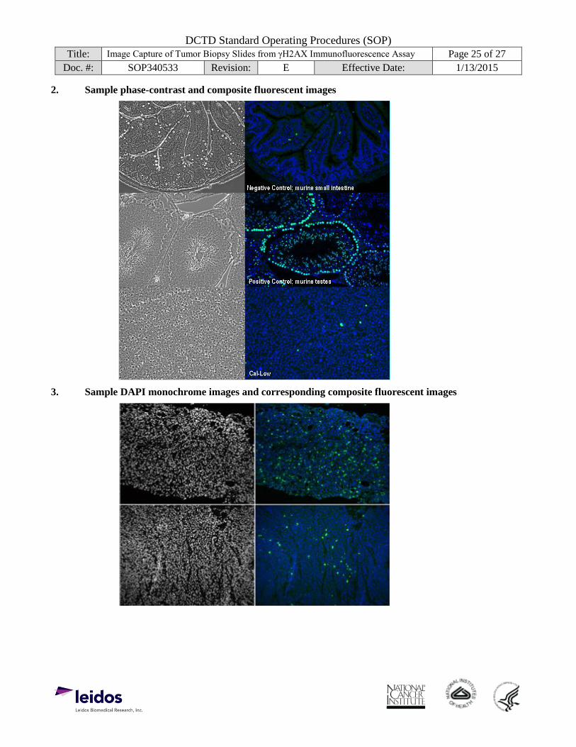
Doc. #: SOP340533 Revision: E Effective Date: 1/13/2015 2. Sample phase-contrast and composite fluorescent images
3. Sample DAPI monochrome images and corresponding composite fluorescent images
DCTD Standard Operating Procedures (SOP) Title: Image Capture of Tumor Biopsy Slides from γH2AX Immunofluorescence Assay Page 26 of 27
Doc. #: SOP340533 Revision: E Effective Date: 1/13/2015
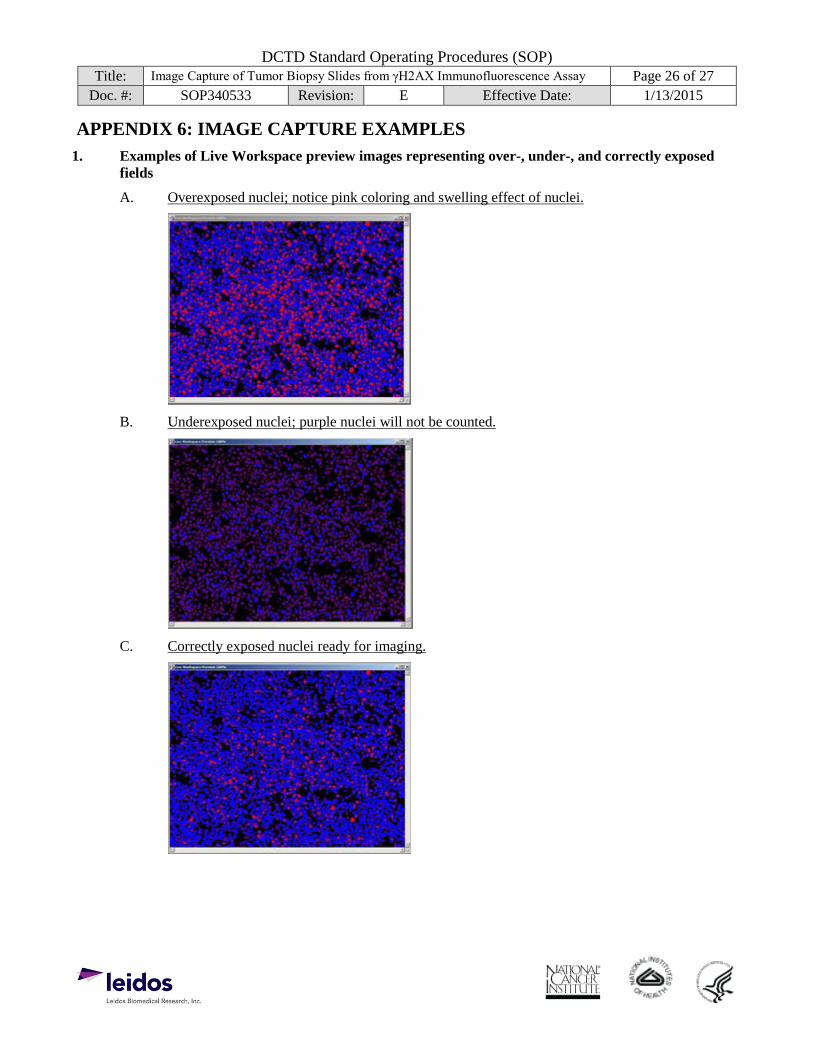
APPENDIX 6: IMAGE CAPTURE EXAMPLES 1. Examples of Live Workspace preview images representing over-, under-, and correctly exposed
fields
A. Overexposed nuclei; notice pink coloring and swelling effect of nuclei.
B. Underexposed nuclei; purple nuclei will not be counted.
C. Correctly exposed nuclei ready for imaging.
DCTD Standard Operating Procedures (SOP) Title: Image Capture of Tumor Biopsy Slides from γH2AX Immunofluorescence Assay Page 27 of 27
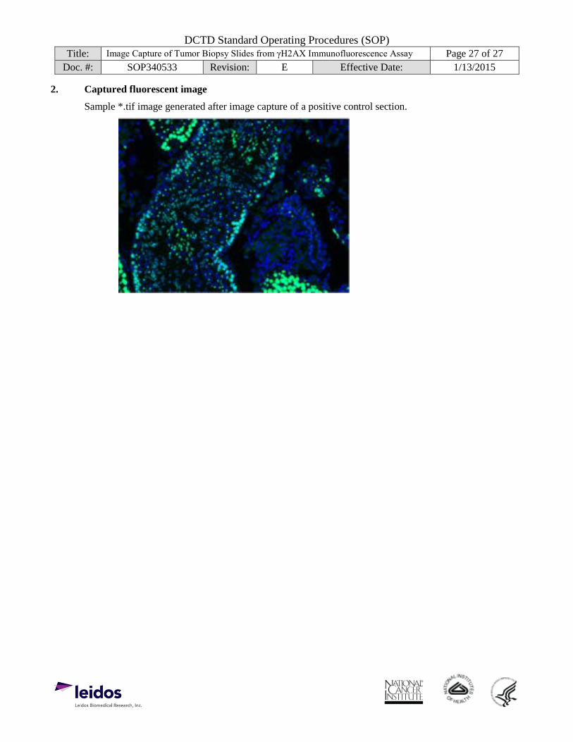
Doc. #: SOP340533 Revision: E Effective Date: 1/13/2015 2. Captured fluorescent image
Sample *.tif image generated after image capture of a positive control section.


![Topic 7 Revision [143 marks]](https://static.fdocument.org/doc/165x107/616a52ca11a7b741a3513f24/topic-7-revision-143-marks.jpg)