DOCKING AND MOLECULAR DYNAMICS SIMULATION STUDIES...
45
DOCKING AND MOLECULAR DYNAMICS SIMULATION STUDIES OF INSULIN-β- CYCLODEXTRIN INTERACTIONS ERMA FATIHA BINTI MUHAMMAD UNIVERSITI SAINS MALAYSIA 2016
Transcript of DOCKING AND MOLECULAR DYNAMICS SIMULATION STUDIES...
DOCKING AND MOLECULAR DYNAMICS
SIMULATION STUDIES OF INSULIN-β-
CYCLODEXTRIN INTERACTIONS
ERMA FATIHA BINTI MUHAMMAD
UNIVERSITI SAINS MALAYSIA
2016
DOCKING AND MOLECULAR DYNAMICS
SIMULATION STUDIES OF INSULIN-β-
CYCLODEXTRIN INTERACTIONS
by
ERMA FATIHA BINTI MUHAMMAD
Thesis submitted in fulfillment of the
requirements for the degree of Master
of Sciences
FEBRUARI 2016
ii
ACKNOWLEDGEMENT
Alhamdulillah. I would like to express the deepest appreciation to my
beloved supervisor, Prof. Dr. Rohana Adnan for her tremendous guidance,
assistance, motivation, love and support as well as for providing me with an excellent
atmosphere for doing research during my study. I sincerely thank all my group
members, Irfan, Dr. Rani, Syed Fariq, Jaga, Leong, Najm, Fitrah, Shikin, and Amneh
who helped me during my study and also for always being there cheering me up and
stood by me through the good and the bad times.
I owe my thanks to the very special person in my life, ibu (Karimah binti
Yaacob) and papa (Muhammad bin Jaafar), for their continuous love, support,
patience and understanding and for their prayers. My special gratitude goes to my
siblings for their continuous support and motivation to continue the study.
Special thanks to the members of computational chemistry group from
Universiti Putra Malaysia, especially, Prof. Dr. Mohd Basyaruddin Abdul Rahman,
Dr. Alif, Dr. Roghayeh, Zalikha, Kak Hana, Zana, and Zhuang for their help and
guidance during my attachment at Universiti Putra Malaysia. Thank you very much
for the technical assistance provided, comments and motivational supports during my
study. I also wish to thanks to all staff in School of Chemical Sciences, especially,
Mr. Razak and Mr. Fairoz for the many kind helps.
Finally, I would like to thank Universiti Sains Malaysia for the financial
support under grant number 1001/PKIMIA/815099. May Allah bless all of you.
iii
TABLE OF CONTENTS
Page
ACKNOWLEDGEMENT ii
TABLE OF CONTENTS iii
LIST OF TABLES vii
LIST OF FIGURES viii
LIST OF ABBREVIATIONS xii
LIST OF SYMBOLS xv
ABSTRAK xvii
ABSTRACT xix
CHAPTER 1 – INTRODUCTION
1.1 Introduction 1
1.2 Problem Statement 5
1.3 Objectives 5
1.4 Thesis Outlines 6
CHAPTER 2 – LITERATURE REVIEW
2.1 Diabetes and Insulin 7
2.1.1 Structure of Insulin 11
2.1.2 Development of Insulin Delivery 15
iv
2.2 Cyclodextrins 17
2.2.1 Structures and Properties of α-, β- and γ-CDs 17
2.2.2 Applications of CDs 20
2.2.3 Mechanism of Protein-CDs Interaction 23
2.3 Molecular Modeling 26
2.3.1 Molecular Docking 26
2.3.1.1 Scoring Function 29
2.3.1.2 Genetic Algorithm 30
2.3.2 Molecular Dynamics Simulation 32
2.3.2.1 Newton‘s Law of Motion 33
2.3.2.2 Statistical Ensemble 34
2.3.2.3 Thermostat 35
2.3.2.4 Force Field 35
2.3.2.5 Periodic Boundary Condition 37
CHAPTER 3 – METHODOLOGY AND COMPUTATIONAL DETAILS
3.1 Technical Details 39
3.2 Geometry Optimization of Single Molecules 40
3.3 Multiple Molecular Docking Study 40
3.4 Molecular Dynamics Simulation 41
3.4.1 Initialization 42
3.4.2 Equilibration 42
3.4.3 Production Run 43
3.4.4 Analysis Techniques 43
v
CHAPTER 4 – RESULTS AND DISCUSSION
4.1 Geometry Optimization of Single Molecules 47
4.1.1 Molecule Structures of Insulin Monomer and Insulin Dimer 47
4.1.2 Molecule Structure of β-CD 48
4.2 Multiple Molecular Docking 52
4.2.1 Binding Free Energies of Insulin Monomer-β-CD and Insulin 53
Dimer-β-CD Formations
4.2.2 Insights into β-CD Binding to Insulin Monomer 54
4.2.3 Insights into β-CD Binding to Insulin Dimer 61
4.3 Molecular Dynamics Simulation 69
4.3.1 Structural Stability of Insulin Monomer and Dimer 69
4.3.1.1 Root Mean Square Deviation 70
4.3.1.2 Radius of Gyration 75
4.3.1.3 Solvent Accessible Surface Area 79
4.3.2 Flexibility and Conformational Changes of Insulin Monomer 85
and Insulin Dimer Structures
4.3.2.1 Root Mean Square Fluctuation of Insulin 85
4.3.2.2 Secondary Structure Content of Insulin 90
4.3.3 Interactions Between β-CDs and Amino Acids Residues at 95
Binding Sites of Insulin Monomer and Dimer
4.3.3.1 Mean Square Displacement of β-CDs 95
4.3.3.2 Hydrogen Bonds Analysis 97
vi
CHAPTER 5 – CONCLUSIONS AND RECOMMENDATIONS
5.1 Conclusions 103
5.2 Recommendations for Future Research 104
REFERENCES 105
APPENDICES 125
Appendix I 125
Appendix II 126
LIST OF PUBLICATIONS AND CONFERENCES 128
vii
LIST OF TABLES Page
Table 2.1 Nobel Prizes for Diabetes Related Research 10
Table 2.2 Physicochemical Properties of α-, β- and γ-CDs 19
Table 2.3 The Summary Applications of CDs 22
Table 2.4 Terminology Used for the Receptor and Ligand 26
in Docking
Table 4.1 Diameter and Height of β-CD Structure Compared to the 49
Literature
Table 4.2 Intramolecular Hydrogen Bonds Formation in Single Molecule 51
of β-CD
Table 4.3 Binding Free Energies of Insulin Monomer-β-CD and 53
Insulin Dimer-β-CD System at Different Ratios
Table 4.4 Hydrogen Bond Formations and Hydrophobic Interactions 58
Between Insulin Monomer and β-CDs Based on LigPlot
Analysis
Table 4.5 Hydrogen Bond Formations and Hydrophobic Interactions 65
Between Insulin Dimer and β-CDs Based on LigPlot Analysis
Table 4.6 The Summary of the Potential Energy for Insulin Systems 70
Investigated
Table 4.7 Average Solvent Accessible Surface Area (SASA) for Insulin 84
Monomer and Dimer in Free and Complex Systems
viii
LIST OF FIGURES Page
Figure 2.1 Structures of insulin (a) monomer (b) dimer 12
Figure 2.2 The numbering scheme for insulin dimer, which
comprises of monomer I and II. Blue lines represent the
disulphide bridges
14
Figure 2.3 Structures of α-, β- and γ-CDs 17
Figure 2.4 The hollow truncated cone structure of CD 18
Figure 2.5 Distribution of the CD relevant abstracts published by
Cyclodextrin News
21
Figure 2.6 A 3D Grid by AutoGrid for the calculation of atomic,
electrostatic, and desolvation energy grids
28
Figure 2.7 Diagram of genetic algorithm applied in AutoDock 31
Figure 2.8 Model of chemical bonds describe by force field 36
Figure 2.9 Schematic representation of PBC 37
Figure 4.1 The crystal versus optimized insulin in dimer form. Blue
ribbon represents the crystal structure of insulin (Smith
et al., 1996), while Red shows the optimized structure of
insulin.
48
Figure 4.2 The (a) top (b) side views structure of β-CD (c)
intramolecular hydrogen bonds formation (Blue lines) in
β-CD structure
50
Figure 4.3 Binding sites I-IV calculated by docking approach with
closer view of the binding interactions of insulin
monomer and β-CD. Key insulin residues are shown in
ball and sticks with A chain in Magenta and B chain in
Blue; β-CDs are representes in sticks
56
Figure 4.4 Hydrogen bonds formation at the insulin monomer-β-
CD complex at (a) binding site I (b) binding site II (c)
binding site III (d) binding site III (e) binding site IV.
Blue lines represent the intermolecular hydrogen bonds
59
ix
Figure 4.5
Hydrophobic interactions of insulin monomer-β-CD
complex at (a) binding site I (b) binding site II (c)
binding site III (d) binding site III (e) binding site IV
61
Figure 4.6
Binding sites I-IV calculated by docking approach with
closer view of the binding interactions of insulin dimer
and β-CD. Key insulin residues are shown in ball and
sticks with A chain in Magenta and B chain in Blue; β-
CDs are representes in sticks
64
Figure 4.7 Hydrogen bonds formation at the insulin dimer-β-CD
complex at (a) binding site I (b) binding site II (c)
binding site III (d) binding site III (e) binding site IV.
Blue lines represent the intermolecular hydrogen bonds
66
Figure 4.8 Hydrophobic interactions of insulin monomer-β-CD
complex at (a) binding site I (b) binding site II (c)
binding site III (d) binding site III (e) binding site IV
67
Figure 4.9 The root mean square deviations (RMSDs) of the free
insulin monomer (Black), insulin monomer-β-CD
complex system (Red), free insulin dimer (Green) and
insulin dimer-β-CD complex system (Blue) in reference
to minimized structure
71
Figure 4.10 The root mean square deviations (RMSDs) of the insulin
monomer in complex system showing A chain (Black)
and B chain (Red)
73
Figure 4.11 The root mean square deviations (RMSDs) of the insulin
dimer in complex system showing A (Black), B (Red),
A‘ (Green), and B‘ (Blue) chains
74
Figure 4.12 Time dependence of the radius of gyration (Rg) for the
Cα atoms of insulin monomer in free and complex
system during 100 ns simulation time
76
Figure 4.13 Time dependence of the radius of gyration (Rg) for the
Cα atoms of insulin dimer in free and complex system
during 100 ns simulation time
78
Figure 4.14 Solvent accessible surface area of insulin monomer in
(a) free (b) complex systems; total SASA is represented
in Green lines, while hydrophobic and hydrophilic
surface are represented in Black and Red, respectively
80
x
Figure 4.15 Solvent accessible surface area of insulin dimer in (a)
free (b) complex systems; total SASA is represented in
Green lines, while hydrophobic and hydrophilic surface
are represented in Black and Red, respectively
81
Figure 4.16 RMSF of insulin monomer in the free and complex
system
85
Figure 4.17 The insulin monomer-β-CD structure showing the
independent folding characteristic of B chain of insulin
was shown in Black circle
87
Figure 4.18 RMSF of insulin dimer in the free and complex system 88
Figure 4.19 The insulin dimer-β-CDs structure showing the
conformational changes during simulation time
89
Figure 4.20 The secondary conformations of (a) α-helix (b) β-sheet
(c) 310 helix (d) π helix (e) random coil
90
Figure 4.21 Percentage of secondary structure of distribution of
insulin monomer in free (Blue) and complex (Red)
during the last 40 ns simulation time
91
Figure 4.22 Percentage of secondary structure of distribution of
insulin dimer in free (Blue) and complex (Red) during
the last 40 ns simulation time
93
Figure 4.23 Secondary structure content of (a) insulin monomer and
(b) insulin dimer in complexes at 60 ns simulation time.
Coil conformation was in Red, α-helices in Blue.
94
Figure 4.24 Mean square displacement (MSD) of β-CDs on insulin
monomer during 100 ns
96
Figure 4.25 Mean square displacement (MSD) of β-CDs on insulin
dimer during 100 ns
97
Figure 4.26 The percentage of occupancy of hydrogen bonding
formation between amino acids residues in insulin
monomer and β-CDs at binding site I (Red), II (Blue),
III (Green), during the last 40 ns simulation time. Only
those with > 2 % occupancy are shown
98
xi
Figure 4.27 The percentage of occupancy of hydrogen bonding
formation between amino acids residues in insulin dimer
and β-CDs at binding site I (Red), II (Blue), III (Green),
during the last 40 ns simulation time. Only those with >
2 % occupancy are shown
100
Figure 4.28 The movement of Ser63 in insulin dimer towards β-CD
throughout simulation time
102
xii
LIST OF ABBREVIATIONS
μVT constant chemical potential, volume and temperature
Ala Alanine
APZ Aripiprazole
Arg Arginine
Asn Asparagine
β-CD beta cyclodextrin
Cys Cysteine
DM Diabetes Mellitus
DNA deoxyribonucleic acid
DSSP Database of Secondary Structure of Protein
ER Endoplasmic Reticulum
FTIR Fourier Transform Infrared
Glu Glutamic acid
Gln Glutamine
Gly Glycine
GROMACS Groningen Machine for Chemical Simulation
His Histidine
HSA human serum albumin
Ile Isoleucine
LGA Lamarckian Genetic Algorithm
MD molecular dynamics
MSD mean square displacement

xiii
Leu Leucine
Lys Lysine
MC Monte Carlo
NMR Nuclear Magnetic Resonance
NPH constant number of molecules, pressure and enthalphy
NPT constant number of molecules, pressure and temperature
NST constant number of molecules, stress and temperature
NVE constant number of molecules, volume and energy
NVT constant number of molecules, volume and temperature
PBC Periodic Boundary Condition
PDB Protein Data Bank
PDB ID identification of entries in Protein Data Bank
Phe Phenylalanine
PMAA Polymethacrylic Acid
PME Particle Mesh Ewald
PM3 Parameterized model number 3
Pro Proline
RAM Random Access Memory
RNA ribonucleic acid
RMSD root mean square deviation
RMSF root mean square fluctuation
Rg radius of gyration
SASA solvent accessible surface area

xiv
Ser Serine
SPC Simple Point Charge
Thr Threonine
Tyr Tyrosine
Val Valine
xv
LIST OF SYMBOLS
∆Gbinding binding free energy
∆Gconf free energy arises from the conformational changes in protein and
ligand
∆Gint free energy of specific protein-ligand interactions
∆Grot free energy loss caused by freezing of the internal rotations
∆Gsolvent free energy contribution with solvent effects
∆Gt/r free energy changes in translational and rotational modes
∆Gvib free energy changes in vibrational modes
ΔSconf loss of conformational entropy upon binding to protein
ϵ depth of potential well
σ finite value of r
uLJ the Lennard-Jonnes potential
ai acceleration of molecule i
Cv total heat capacity of system
Fi force exerted on molecule i
mi mass of molecule i
ms millisecond
nm nanometer
nm2
nanometer square
ns nanosecond
center of mass of N atoms of insulin
ri vector of Cartesian coordinates of the i-th atom
xvi
position of the atom i in the MD simulation
i position of the atom i in the reference structure
reference position of particle i
mean square displacement
T actual temperature
Tτ temperature time constant
T0 desired temperature
U potential energy of the system
V velocities Verlett algorithm
vi velocities of molecule i
xvii
KAJIAN PENDOKKAN DAN SIMULASI DINAMIK MOLEKUL
TERHADAP INTERAKSI INSULIN-β-SIKLODEKSTRIN
ABSTRAK
Interaksi protein-ligan memainkan peranan penting dalam menyediakan
produk farmaseutikal yang baharu. Kajian ini merupakan usaha untuk memahami
struktur dan dinamik kompleks insulin-siklodekstrin sebagai formula insulin oral
yang baharu. Pendokkan dan simulasi dinamik molekul telah dijalankan untuk
mengkaji interaksi antara monomer insulin dan dimer insulin terhadap β-
siklodekstrins (β-CDs). Kajian pendokkan molekul berganda telah dijalankan
menggunakan program Autodock v4.2 untuk menentukan bilangan β-CD yang boleh
terikat pada tapak ikatan insulin selain menentukan konformasi insulin-β-CD yang
paling stabil. Pendokkan molekul dengan 100 struktur rawak menggunakan
konformasi awal nisbah monomer insulin kepada β-CD dan dimer insulin-β-CD 1:1
telah dijalankan dan daripada struktur pendokkan terakhir, β-CD telah ditambah dan
proses diulangi sehingga peningkatan tenaga didapati. Keputusan pendokkan
molekul menunjukkan maksimum empat molekul β-CD boleh terikat kepada struktur
insulin dan nisbah insulin kepada β-CD 1:3 menghasilkan tenaga bebas pengikatan
terendah. Selain pembentukan ikatan hidrogen, keputusan pendokkan menunjukkan
bahawa interaksi hidrofobik memainkan peranan penting dalam menentukan
kestabilan kompleks insulin-β-CD. Simulasi dinamik molekul 100 ns seterusnya
dijalankan untuk membandingkan keputusan yang terhasil daripada kajian
pendokkan molekul. Analisa daripada simulasi dinamik molekul mengesahkan
kestabilan pada kompleks dimer insulin-β-CD 1:3 dengan nilai sisihan punca min
xviii
kuasa dua (RMSD) sebanyak 0.40 ± 0.02 nm selepas 38 ns menunjukkan perbezaan
yang sangat besar berbanding sistem insulin bebas. Peningkatan nilai luas permukaan
boleh dicapai pelarut (SASA) pada kompleks 1:3 insulin dimer-β-CD selaras dengan
interaksi hidrofobik dalam sistem kompleks, oleh yang demikian menambahkan
lipatan dan kestabilan insulin. Profil pergolakan punca min kuasa dua (RMSF)
menunjukkan pengurangan fleksibiliti terhadap residu asid amino dalam insulin
dimer dalam sistem kompleks berbanding sistem insulin bebas. Seterusnya, struktur
dan dinamik asid amino pada setiap tapak ikatan dan kesannya terhadap β-CD turut
dikaji. Keputusan menunjukkan β-CD terikat stabil pada setiap tapak ikatan pada
insulin dimer dengan pergerakan terhad sekitar nilai minima. Selain itu, asid-asid
amino berkutub seperti sistina (Cys62), serina (Ser63) dan glutamina (Gln66) terlibat
dengan kekerapan tertinggi dalam pembentukkan ikatan hidrogen masing-masing
dengan nilai peratusan 74.0 %, 54.3 % dan 51.9 %. Keputusan teori ini menunjukkan
kewujudan interaksi penting antara insulin dan β-CD yang mana boleh
menyumbangkan maklumat penting terhadap formulasi insulin.
xix
DOCKING AND MOLECULAR DYNAMICS SIMULATION STUDIES OF
INSULIN-β-CYCLODEXTRIN INTERACTIONS
ABSTRACT
Protein-ligand interactions play an essential role in the design of new
pharmaceutical products. This study attempts to understand the theoretical basis on
the structure and dynamics of insulin-cyclodextrin complex for new oral insulin
formulation. Docking and molecular dynamics simulations were performed to
explore the interactions between insulin monomer and insulin dimer with β-
cyclodextrins (β-CDs). A multiple molecular docking study was performed using the
Autodock v4.2 program to determine the number of β-CD that can adhere to the
binding sites of insulin as well as to determine the most stable conformations of
insulin to β-CDs. A 100 random structure docking using 1:1 insulin monomer-β-CD
and insulin dimer-β-CD ratio were conducted and from the final docked structure,
additional β-CDs were added and the process were repeated until the energy increase.
Molecular docking results revealed that a maximum of four β-CDs can bind to an
insulin structure with the 1:3 insulin-β-CD ratios having the lowest binding free
energy. A 100 ns molecular dynamics simulation was then conducted to verify the
results obtained by molecular docking. In addition to the hydrogen bonding
formations, the docked conformations showed that hydrophobic interactions played a
crucial role in insulin-β-CD conformational stability. The analysis of the molecular
dynamics simulation confirmed the stability of the 1:3 insulin dimer-β-CD complex
system with root mean square deviation (RMSD) values of 0.40 ± 0.02 nm after 38
ns and showed a large difference compared to the free insulin system. The increase
xx
of solvent accessible surface area (SASA) values in 1:3 insulin dimer-β-CD complex
is consistent with the hydrophobic interactions in the formation complex, therefore
increasing the insulin folding and stability. The root mean square fluctuation (RMSF)
profiles of the 1:3 insulin dimer in complex system also showed a reduced flexibility
of amino acid residues of insulin dimer in complex compared to free insulin.
Furthermore, the structure and dynamics of amino acids in insulin at each binding
site along with its effect on β-CDs were further investigated. The results indicated
that the β-CDs were stably bound to each binding site of the insulin dimer with
limited movement around the mean value. In addition to this, the polar amino acids
of cystein (Cys62), serine (Ser63) and glutamine (Gln66) were involved with the
highest occupancy of hydrogen bonds with 74.0 %, 54.3 % and 51.9 %, respectively.
The theoretical results indicated the presence of significant interactions between
insulin and β-CD, which could provide useful insights into an oral insulin
formulation.
1
CHAPTER 1
INTRODUCTION
1.1 Introduction
During the last decade, approximately 300 million people worldwide were
affected by Diabetes Mellitus (DM) (Williams et al., 2002) with the Asian countries
representing more than 60% of that diabetic population (Ramachandran et al., 2012).
DM is defined as an endocrine disease which is related to the disorders of
carbohydrate metabolism carried about by deficiency in insulin secretion, insulin
resistance or both (Belchetz & Hammond, 2004). There are three types of DM: Type
1, Type 2 and the gestational diabetes that happens only to pregnant women (Carino
& Mathiowitz, 1999).
Type 1 diabetes occurs due to the destruction of pancreatic beta islet cells.
This leads to insulin deficiency and predisposes individuals to diabetic ketoacidosis
(Mohan, 2005). This type of diabetes usually happens in childhood and requires
patients to inject themselves with insulin to reduce glucose levels. Type 2 diabetes
occurs in adults and is caused by the inability of the pancreas to produce insulin and
resist peripheral insulin (Gale, 2001; Skyler, 2004). The development of factors
leading to Type 2 diabetes are due to genetic inheritance, beta cell dysfunction
(Mohan, 2005), the environment of beta cells (Pittas & Greenberg, 2003), insulin
resistance (Saltiel, 2001), obesity (Kahn & Flier, 2000), the role of fatty acids which
inhibit insulin signaling (Shulman, 2000), the role of adipokines (Saltiel, 2001),
improper nutrition (Steyn et al., 2004) and low physical activity in patients (Caro et
al., 1989).
2
In order to prevent these complications, people with Type 2 diabetes are
usually treated with dietary measures, oral medicines and exercises. Insulin
injections are compulsory for Type 1 diabetic patients, however, some Type 2
diabetic patients need insulin injections to control the glucose level in their
blood. Due to the fast development in biotechnology over the past few decades,
variants of insulin from different sources were produced. These include rapid (Shafie
et al., 2014), short (Siebenhofer et al., 2006), intermediate (Norrman et al., 2007),
long (Luzio et al., 2013) and ultralong-acting (Wang et al., 2012) insulin. These
insulin variants must be administered via injections and may result in one or more
disadvantages to patients such as needle phobia, skin bulges, allergic reactions,
common infections and tenseness of long-term usage of injection insulin (Kennedy,
1991; Khafagy et al., 2007). Furthermore, the treatment of injected insulin can lead
to peripheral hyperinsulinemia, which is due to the directly insulin transfer into
general circulation in human body. Hyperinsulinemia is diabetes‘s side effect which
is linked to cancer, hypoglycemia, peripheral hypertension and the development of
atherosclerosis (Nordestgard, 1997). The clinical trials by Pamnani (2008) shows that
a significant percentage of patients failed to achieve lasting glycemic control due to
the patients‘ noncompliance with injectable insulin treatment. Due to this problem,
current research on insulin has focused on the development of an alternative delivery
method to injection. These include buccal/sublingual (Portero et al., 2007), nasal (Yu
et al., 2004), pulmonary (Stephen et al., 2000), ocular (Owens, 2002), transdermal
(Rastogi et al., 2010) as well as oral (Zhang et al., 2013).
Oral insulin administration is the most effective and possible technique to
be used compared to the other routes. This is because insulin will be directly
transferred to the liver and consequently avoids the peripheral hyperinsulinemic side
3
effects to patients caused by injection. Furthermore, the pain caused by injections
and the psychological barriers to daily injections such as needle phobia can be
eliminated via this oral route (Korytkowski, 2002).
However, there are major hurdles to the effective use of orally delivered insulin.
These obstacles include the rapid enzymatic degradation of insulin in the stomach,
inactivation and digestion by proteolytic enzymes in the intestinal lumen, low
penetration rate of insulin through the gastrointestinal membrane as well as
biological and structural stability of the insulin in a body system (Timmy et al., 2002;
Elsayed, 2012). In order to overcome these barriers for successful oral
administration, many research groups are currently working towards developing new
oral insulin formulations. These include the attempt to overcome barriers and
limitations through chemical modification of insulin with various fatty acids (Ashada
et al., 1995), enzyme inhibitors and penetration enhancers (Liu et al., 2003; Miller &
Johnston, 2005) and incorporation of insulin into carriers such as hydrogels
(Nakamura et al., 2014), liposomes (Choudhari & Labhasetwar, 1994), erythrocytes
(Al-achi & Greenwood, 1998), nanospheres (Damge et al., 1997) and nanocubicles
(Chung et al., 2004).
A promising approach used in oral insulin delivery is the encapsulation of
insulin by cyclodextrin (Zhang et al., 2010; Sajeesh et al., 2010; Uehata et al., 2011;
Uehata et al., 2012; Zhang et al., 2012; Zhang et al., 2013). The complexations of
insulin with cyclodextrins offer a unique and effective way to improve the insulin
properties and stability against aggregation, thermal denaturation and degradation
(Sigurjonsdottir et al., 1999; Dong et al., 2002). Previously, studies on the
complexation of insulin with cyclodextrins focused on the interactions of the cationic
4
β-CD polymers with insulin (Huang et al., 2010), the insulin cell-penetrating peptide
co-administrations with hydroxylpropyl-β-CD (Zhang et al., 2010), insulin glargine
with maltosyl-β-CD (Uehata et al., 2012), insulin encapsulated polymethacrylic acid
(PMAA) hydrogel microparticles with methyl-β-CD (Sajeesh & Sharma, 2006),
insulin with hydroxylpropyl-β-CD (Zhang et al., 2009), kinetic degradation of insulin
complexed with methyl-β-CD (Dotsikas & Loukas, 2002) and β-CD grafting
hyperbranched polyglycerols as carriers for insulin (Zhang et al., 2011).
Computer modeling is widely used to understand and predict the properties and
behaviour of systems at the molecular level. Molecular modeling of insulin using
molecular dynamics method has been reported. These studies include the dissociation
of insulin-phenol complex (Kru, 2003), binding of glucose to insulin (Falconi et al.,
2001a; Zoete et al., 2004a), dynamic behaviour of insulin monomer and dimer (Zoete
et al., 2004b), association of glycoprotein structure with B chain human insulin
(Stavrakoudis, 2011), insulin stability on graphene (Liang et al., 2009), flexibility of
B chain human insulin (Legge et al., 2006), structure and stability of insulin dimer
(Falconi et al., 2001b), insulin interaction with boron-nitride and functionalized
graphene nanosheets (Atabay et al., 2014), conformational flexibility of insulin
analogs (Ksenofontova & Stefanov, 2013), simulation of insulin protection by
trehalose (Li et al., 2014) and the aggregation and release rate of insulin (Berhanu &
Masunov, 2012).
5
1.2 Problem Statement
Among the latest development in oral insulin delivery formulation, the
complexation of insulin with cyclodextrin has been found to be advantageous and
encouraging. The experimental data on the complexation of insulin with β-CD on
varying conditions has been investigated in the past few years and support the fact
that complexation with cyclodextrin improve the stability of insulin. However, it is
not really understood on how cyclodextrins interact with insulin which may or may
not lead to insulin conformational change and how these interactions affect the
thermodynamics and structural properties of insulin upon binding to CD. In order to
improve our understanding on the molecular properties insulin-β-CD formation,
theoretical study comprising of quantum mechanics, molecular docking and
molecular dynamics simulation was conducted as part of our attempt to understand
the β-CD behaviour toward insulin monomer and dimer as an oral delivery medium
for insulin.
1.3 Objectives
The objectives of this research are:
1. To determine the binding sites of β-CDs onto insulin monomer and
insulin dimer and the best ratios of β-CDs to insulin using molecular
docking calculation.
2. To determine the structural change and stability of insulin monomer and
insulin dimer following the complexation with β-CDs using molecular
dynamics simulation.
6
3. To identify the type of interactions exists between β-CDs and the amino
acids of insulin involved in the stabilization of the insulin-β-CD
complexes.
1.4 Thesis Outline
This thesis contains five chapters. Chapter 1 provides an overview and the
background of this study. Chapter 2 reviews the structure, properties of insulin as
well as cyclodextrins and the previous study on protein-cyclodextrin systems. The
fundamentals behind computer modeling techniques applied in this study were also
discussed in this chapter. Chapter 3 describes the methodology for the geometry
optimization of single insulin monomer and insulin dimer as well as the β-CD
structure, the multiple molecular docking and finally, the molecular dynamics
simulation of insulin-β-CD conformations in detail. Chapter 4 contains the results
and discussion which are divided into three main sections: geometry optimization,
molecular docking results and molecular dynamics simulation study. Finally, Chapter
5 summarizes the research findings and further recommendation for possible
research related to this study. Appendices associated with the relevant documents
were provided at the end of this thesis.
7
CHAPTER 2
LITERATURE REVIEW
2.1 Diabetes and Insulin
Worldwide, it is expected that over 500 million people will be affected by
DM in 2030 (Williams et al., 2002). DM causes about 5% of deaths each year (Liu et
al., 2010) and this is likely to increase by more than 50% in the next 10 years without
proper medication (http://www.who.int/diabetes/en/). The increasing prevalence of
diabetes may be due to several factors which are population growth, ageing where an
increased life expentancy results in a higher ratio of aged population more prone to
diabetes, urbanisation, increasing obesity, increasing physical inactivity and passive
lifestyles (Sonia & Sharma, 2014).
The word ‗Diabetes‘ comes from the Greek work for ‗pipe-like‘ because
essential nutrients of the body start to pass through the system instead of being
utilised. Meanwhile „Mellitus‟ is the Latin word for ‗honey‘ or ‗sweet‘ (van Diepen,
1996). Scientifically, DM is a chronic metabolic disease, which is caused by insulin
deficiency and an abnormal increase of blood sugar levels in the body (Genuth et al.,
2003). Type 1 DM is most commonly diagnosed in children and adolescents, which
cannot be prevented and thus occurs when the pancreas does not produce insulin at
all or only a little. Meanwhile, Type 2 DM occurs when the pancreas produces too
little glucose. In Type 2 cases, the production of insulin is normal initially, but the
response activated by insulin in the peripheral tissues is blunted (Alexander &
Hunter, 2004). When the production of insulin by the pancreas can no longer
compensate for the peripheral insulin resistance due to β-cell dysfunction, the Type 2
DM and hyperglycemia become overt. This type of DM usually occurs with
8
increasing of age and depends on the lifestyle of the individuals. Type 2 diabetes is
also found as a component of metabolic syndrome, which is characterised by
hypertension, central obesity, hyperlipidemia, and insulin resistance that results in
increased mortality due to cardiovascular incidents. The pervasiveness of both Type
2 diabetes and metabolic syndrome is reaching epidemic proportions, as the onset
average age of both diseases has markedly decreased over the past decades (Horton,
2008).
Currently, there is no practical cure for diabetes. However, it can be
controlled effectively through several ways. Lifestyle change involving a modified
dietary sugar intake and physical exercise (Franz, 1997) should be taken into
consideration. The oral hypoglycaemic agents are administered to treat diabetic
patients when diet and exercise are not enough to achieve the desired glycaemic
control (Krents & Bailey, 2005). There are four classes of hypoglycaemic agents;
insulin secretagogues (Dunning, 1997), insulin sensitizers (Bailey et al., 1992), α-
glucosidase inhibitors (Gerard et al., 1984) and insulin. Insulin secretagogues, insulin
sensitizers and α-glucosidase inhibitors are oral medication treatments for
stimulating endogenous insulin by the pancreas. Meanwhile, insulin is a protein
therapy needed to be administered exogenously (Sonia & Sharma, 2014).
As a protein therapy used to treat diabetes, insulin was widely used to
regulate the level of glucose in the blood system. Approximately 20–30% of all
diabetic patients receive daily insulin injections in order to maintain their glucose
levels (Babu et al., 2008). Human insulin is synthesised from beta-cells of the islets
of Langerhans and is secreted into the bloodstream (Greenspan & Gardner, 2004). It
plays a crucial role in monitoring the metabolic activities of the body, particularly the
9
homeostasis of the blood glucose. Insulin secretion is a regulated process providing a
stable concentration of glucose in blood during eating and fasting (Yeh et al., 2010).
The history of insulin as a protein therapy to diabetes started back to 1922
when it was first used successfully in humans to treat the symptoms of DM (Bliss,
1993). The study of diabetes, including the related aspects of glucose metabolism
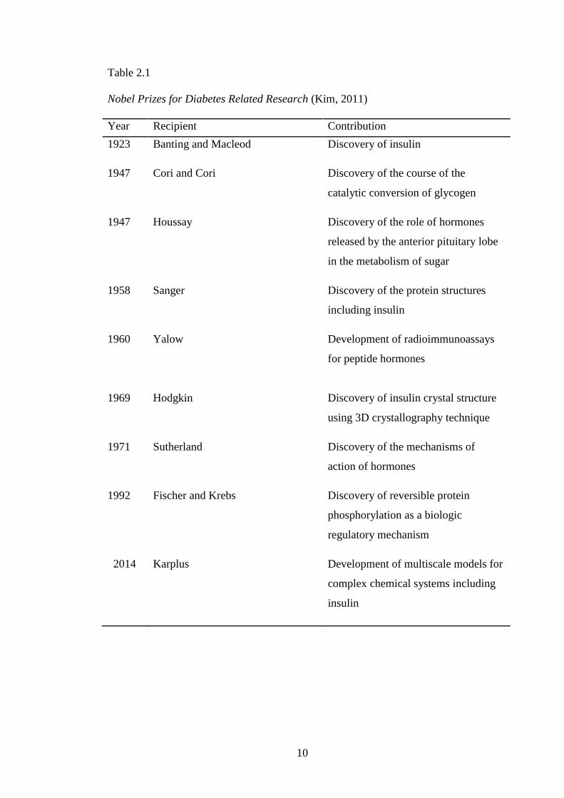
was also a fertile ground for scientific inquiry that 10 scientists received the Nobel
Prize for diabetes-related investigations between 1923 to 2014 as shown in Table 2.1
(Kim, 2011). In 1958, Frederick Sanger was awarded the Nobel Prize for developing
techniques to sequence the amino acids of insulin.
Rosalyn Yalow in 1960 permitted the quantitative measurement of pancreatic
beta-cell function in animals and humans and established the radioimmunoassay as a
powerful tool for measuring proteins, metabolites, and other chemicals present in
very low concentrations. After that, Donald Steiner‘s in 1967 demonstrated two
polypeptide insulin molecules were derived from a single chain precursor proinsulin.
This formation was crucial for the understanding of the biochemistry of insulin and
was also applied to other peptide hormones. The crystal structure of insulin was then
determined using 3D crystallography technique by Dorothy Hodgkin in 1969. Insulin
was the first hormone to be cloned and then produced for therapeutic use by means
of recombinant DNA technology, while providing an unlimited supply of this
important molecule and laid down the foundation for the biotechnology industry
(Ullrich et al., 1977). In the recent development in insulin, Karplus won the Nobel
Prize for developing multiscale models to describe complex biomolecules. Karplus
and co-workers successfully developed methods which combined quantum and
classical mechanics to describe the interaction between glucose and insulin (Zoete et
al., 2004a).
10
Table 2.1
Nobel Prizes for Diabetes Related Research (Kim, 2011)
Year Recipient Contribution
1923 Banting and Macleod Discovery of insulin
1947 Cori and Cori Discovery of the course of the
catalytic conversion of glycogen
1947 Houssay Discovery of the role of hormones
released by the anterior pituitary lobe
in the metabolism of sugar
1958 Sanger Discovery of the protein structures
including insulin
1960 Yalow Development of radioimmunoassays
for peptide hormones
1969 Hodgkin Discovery of insulin crystal structure
using 3D crystallography technique
1971 Sutherland Discovery of the mechanisms of
action of hormones
1992 Fischer and Krebs Discovery of reversible protein
phosphorylation as a biologic
regulatory mechanism
2014 Karplus Development of multiscale models for
complex chemical systems including
insulin
11
2.1.1 Structure of Insulin
Insulin is synthesised in beta cells of the pancreas where the messenger RNA
(mRNA) is translated as a single chain precursor named preproinsulin (Dodson &
Steiner, 1998). The removal of preproinsulin‘s signal peptide during insertion into
the endoplasmic reticulum (ER) generates proinsulin. After that, the proinsulin folds
into a three-dimensional structure, whereby it is transported with the help of vesicles
to Golgi. In this medium, proinsulin is likely self-assembles to form proinsulin
hexamers, in which connecting peptide linking A and B chain, then is cleaved by
enzymes. This promotes towards the formation of zinc assembled of insulin hexamer,
which are capable of forming microcrystals. Finally, hexamers will fall apart into
insulin monomers due to repulsion in certain charged residues at its core (Vashisth,
2010). The final formation of insulin is in monomeric form which is used to stabilise
glucose concentrations in living system.
Insulin monomer is a small globular protein composed of two polypeptide
chains. A Chain consists of the sequence of 21 amino acids, whereas B chain consists
of 30 amino acids. Both chains were stabilised by three disuplhide bonds, whereby,
two disulphide bridges between A chain and B chain were formed between the
residues Cystein7 (Cys7) to Cystein28 (Cys28) and Cystein20 (Cys20) to Cystein40
(Cys40). The other disuphilde bridge was internally formed between residues
Cystein6 (Cys6) to Cystein11 (Cys11) in A chain of insulin monomer. Generally, A
chain contains N-terminal α-helix, turn conformations, whereas the B-chain contains
N-terminal and central α-helix, and C-terminal β-strand (Olsen et al., 1996). At
micromolar concentration, the insulin dimerises and further forms hexamers in the
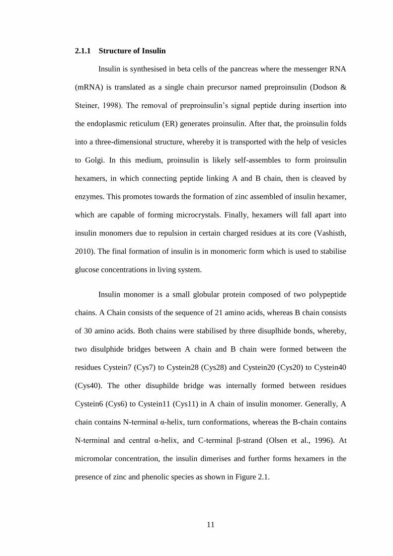
presence of zinc and phenolic species as shown in Figure 2.1.
12
(a) (b)
Figure 2.1. Structures of insulin (a) monomer (b) dimer.
Chain A
Chain B
Chain B’ Chain A
Chain B
Chain A’
13
The insulin dimer was formed when the extended C-terminal of two
monomers are brought together, thus, forming a two-stranded antiparallel β-sheet.
The interactions stabilizing the dimer form of insulin are predominantly non-polar
with β-sheet hydrogen bonds replacing water hydrogen bonds and contributing to
orientate two monomers (Jorgensen et al., 1996). The published high resolution X-
ray structures of insulin suggested as the aggregated species at which several reports
provide the results for monomeric form of insulin (Zhang et al., 2002). Therefore, the
current insulin structure was derived from insulin hexamer and dimer crystal
structures.
Biologically, insulin monomer is active and capable of self-association
forming dimers and hexamers in biosynthesis and during storage of insulin (Dodson
& Steiner, 1998). Insulin monomer is unstable compared to dimer and hexamers and
is therefore the main reason why many researchers used the dimer form. In addition,
insulin monomer forms aggregates and fibrils due to the interactions between the
hydrophobic residues in the monomer and this behaviour are often associated with
the reduction of biological potency of insulin monomer (Dunn, 2005). Insulin dimer
and hexamer are also known to be more resistant to chemical and physical
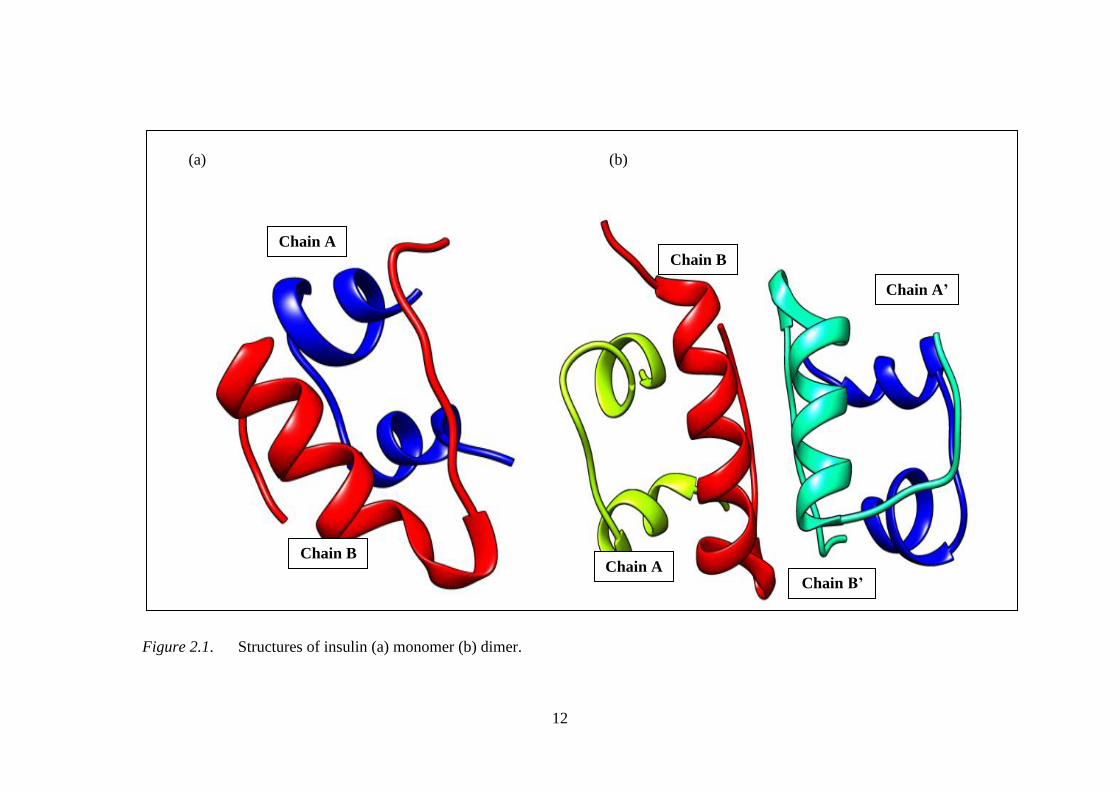
degradation than its monomer form (Loftsson et al., 2005). Insulin monomer with A
and B chains comprises of Gly1 Ala51, while, insulin dimer is made up of two
monomers with chains A, B, A‘ and B‘ consists of Gly1 Ala102 as shown in the
numbering structure of insulin in Figure 2.2.
14
Figure 2.2. The numbering scheme for insulin dimer, which comprises of monomers I and II. Blue lines represent the disuplhide bridges.
A’ Chain
Cys71
Lys101 Phe97 Tyr47 Gln25
Glu4
Asn72
Ala102 Phe96 His26
Gln5
Phe46
B Chain
Gln66
Tyr65
Arg94 Cys7 Gly44
Leu64
Ser63 Cys28
Glu93 Arg43
Cys62 Gly29
Ser30
Thr8
Gly92 Glu42
Gly41
B’ Chain
Cys91 Phe73
Ile61
His31
Ser9
Ile10
Cys40 Val90 Val74
Ser60
Leu32 Cys11
Leu89 Asn75
Thr59
Val39 Val33 Ser12
Tyr88 Gln76
Cys58
Leu38 Glu34 Leu13
Leu87 His77
Cys57
Tyr37 Leu36 Ala35 Tyr14
Ala86 Leu78
Gln56
Gln15
Glu85 Cys79
Glu55
Leu16
Val84 Gly80
Val54 A Chain
Glu17
Leu83 His82 Ser81
Ile53
Asn18 Asn21 Cys20 Tyr19 Gly52
Thr48 Asn24 Pro100 Thr99 Tyr98
Tyr70
Cys6
15
2.1.2 Development of Insulin Delivery
The most crucial treatment strategy for DM focuses on the control of
postprandial blood glucose. Therefore, the goal of exogenous insulin is to mimic the
physiological profile in a non-diabetic person. In the past few decades, various routes
of insulin administration were explored in order to discover a new technique of
insulin administrations that will improve the metabolic effects of insulin. The current
administration of insulin which is via subcutaneous route has various disadvantages,
such as injection site pain, low patient compliance, and occasional hypoglycemia
(Khafagy et al., 2007). Furthermore, insulin administration via subcutaneous
injection does not mirror the normal dynamics of endogenous insulin release,
promoting in failure to achieve lasting glycemic control (Hoffman & Ziv, 1997;
Morishita et al., 2006). Various routes other than subcutaneous, which under
investigation for insulin delivery include oral (Huang et al., 2009; Zhang et al.,
2013), pulmonary (Mastrandrea, 2010), transdermal (Krishnankutty et al., 2009),
nasal (Turker et al., 2004), buccal (Senel & Hincal, 2001), ocular (Lee et al., 2002),
rectal (Ritschel et al., 1988) and vaginal (Choudhury et al., 2011). Among all the
possible routes, oral insulin delivery serves as the most expedient and desired way of
insulin administration due to patient compliance and comfort. Furthermore, an oral
pathway of insulin delivery is expected to follow the physiological route of insulin
secretion (Lewis et al., 1996), which is accompanied by greater hepatic versus
peripheral concentrations (Gordon-Still, 2002).
There are a few other major hurdles to develop oral insulin delivery, such as
the presence of chemical and enzymatic barrier in the gastrointestinal tract that could
degrade the insulin structure (Sonia & Sharma, 2014), the poor permeation of insulin
16
across the intestinal epithelia (Muller, 2011), with low bioavailability (typically less
than 1–2%) (Renukuntla et al., 2013), and stability of insulin which depend on a few
factors such as temperature, pH, solvent, solutes and crystallinity states of the insulin
(Manning et al., 1989). One of other major barrier in order to develop oral delivery
insulin is poor of insulin absorption through the gastrointestinal membrane (Carino &
Mathiowitz, 1999). The high molecular weight of insulin, which is about 6 kDa
could not also penetrate through this route. The insulin absorption is prevented due
to its large molecule size, charge and hydrophilicity (Elsayed, 2012). Oral delivery of
insulin for DM was tried in diabetic induced rats by Roques and co-workers (1992)
who reported one of the major problems is degradation of the insulin due to
proteolytic activity of enzymes in the gastrointestinal tract.
A perfect oral drug delivery system should be capable of maintaining the
purity of insulin molecules until it reaches the absorption site, releasing insulin at the
targeted absorption site and maintaining inside the gastrointestinal tract irrespective
of its transitory constraints (Renukuntla et al., 2013). The crucial parts of oral
delivery insulin should be safe delivery to human body, increased in bioavailability
as well as enhanced insulin absorption. Many research groups focused on
development of a delivery system for insulin oral administration using absorption
enhancers (Onuki et al., 2000; Thanou et al., 2000), enzyme inhibitor (Agarwal et al.,
2000), enteric coatings (Hosny et al., 2002) and nanoparticle delivery (Ezpeleta et al.,
1999). The permeation of nanoparticles still results in poor bioavailability of the
insulin. The use of protease inhibitors in addition to absorption enhancers leads to
improved uptake insulin. Nonetheless, these agents are not specific and could assist
in the uptake of other unwanted protein or peptides in the gastrointestinal tract
17
(Nakamura et al., 2014). Some promising results were also obtained, indicating
clearly that oral administration of insulin mimicking the physiological fate of insulin.
However, most production techniques involve the use of organic solvents, heat or
vigorous agitation potentially harmful to the structure as well as the biological
activity of insulin and were lead to problems of cytotoxicity (Khafagy et al., 2007).
These limitations could be effectively overcomed by insulin encapsulation agent
such as β-CD and derivatives (Ahsan et al., 2003; Sajeesh et al., 2010; Zhang et al.,
2012).
2.2 Cyclodextrins
2.2.1 Structures and Properties of α-, β- and γ-CDs
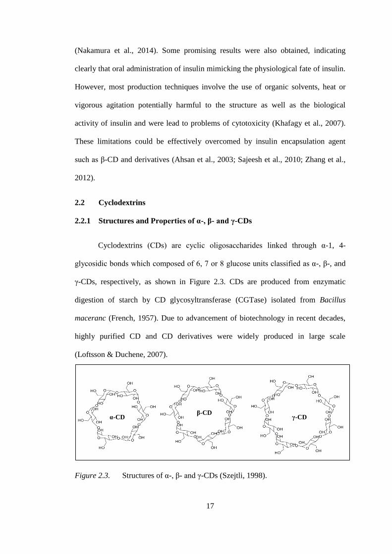
Cyclodextrins (CDs) are cyclic oligosaccharides linked through α-1, 4-
glycosidic bonds which composed of 6, 7 or 8 glucose units classified as α-, β-, and
γ-CDs, respectively, as shown in Figure 2.3. CDs are produced from enzymatic
digestion of starch by CD glycosyltransferase (CGTase) isolated from Bacillus
maceranc (French, 1957). Due to advancement of biotechnology in recent decades,
highly purified CD and CD derivatives were widely produced in large scale
(Loftsson & Duchene, 2007).
Figure 2.3. Structures of α-, β- and γ-CDs (Szejtli, 1998).
α-CD β-CD
γ-CD
18
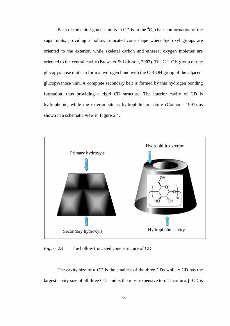
Each of the chiral glucose units in CD is in the 4C1 chair conformation of the
sugar units, providing a hollow truncated cone shape where hydroxyl groups are
oriented to the exterior, while skeletal carbon and ethereal oxygen moieties are
oriented to the central cavity (Brewster & Loftsson, 2007). The C-2-OH group of one
glucopyranose unit can form a hydrogen bond with the C-3-OH group of the adjacent
glucopyranose unit. A complete secondary belt is formed by this hydrogen bonding
formation, thus providing a rigid CD structure. The interior cavity of CD is
hydrophobic, while the exterior site is hydrophilic in nature (Connors, 1997) as
shown in a schematic view in Figure 2.4.
Figure 2.4. The hollow truncated cone structure of CD.
The cavity size of α-CD is the smallest of the three CDs while γ-CD has the
largest cavity size of all three CDs and is the most expensive too. Therefore, β-CD is
Secondary hydroxyls
Primary hydroxyls
Hydrophobic cavity
Hydrophilic exterior
19
most widely used in research and manufacturing due to its cost and suitable cavity
size for most compound molecules (Loftsson & Brewster, 1996; Szejtli, 1998).
Cyclodextrins are soluble in water and insoluble in most organics solvents
although β-CD is the least soluble in water compare to the other native CDs. The
solubility of cyclodextrins in water is unusual and does not seem to be dependent on
the structure of cyclodextrin (Sabadini et al., 2006). α-CD is the most strained torus
structure while γ-CD ring is the least strained among the three native cyclodextrins.
β-CD is 9 and 11 times less soluble in water in comparison to α-CD and γ-CD,
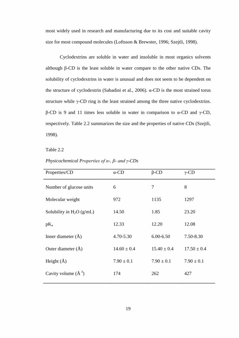
respectively. Table 2.2 summarizes the size and the properties of native CDs (Szejtli,
1998).
Table 2.2
Physicochemical Properties of α-, β- and γ-CDs
Properties/CD α-CD β-CD γ-CD
Number of glucose units 6 7 8
Molecular weight 972 1135 1297
Solubility in H2O (g/mL) 14.50 1.85 23.20
pKa 12.33 12.20 12.08
Inner diameter (Å) 4.70-5.30 6.00-6.50 7.50-8.30
Outer diameter (Å) 14.60 ± 0.4 15.40 ± 0.4 17.50 ± 0.4
Height (Å) 7.90 ± 0.1 7.90 ± 0.1 7.90 ± 0.1
Cavity volume (Å 3
) 174 262 427
20
According to the report summarized by Szejtli (1998), CDs are mostly
nontoxic. One of the few toxicities of CD reported is hemolysis, which is a
phenomenon resulted from interactions of CD with membrane components (Szejtli,
1988; Shen et al., 1998). It is reported that lower concentration of CD protects the
human erythrocytes against osmotic and heat-induced hemolysis. CD is known to
cause the release of cholesterol and phospholipids from cell membrane thus resulting
in cell disruption (Uekama et al., 1981; Shen et al., 1998).
Apart from that, the nonbonding electron pairs of the glycosidic oxygen
bridges of β-CDs are directed toward the inside of the cavity producing high electron
density and also lending on some Lewis base characteristics which is important for
their capability to form host-guest complexes (Szejtli, 1998).
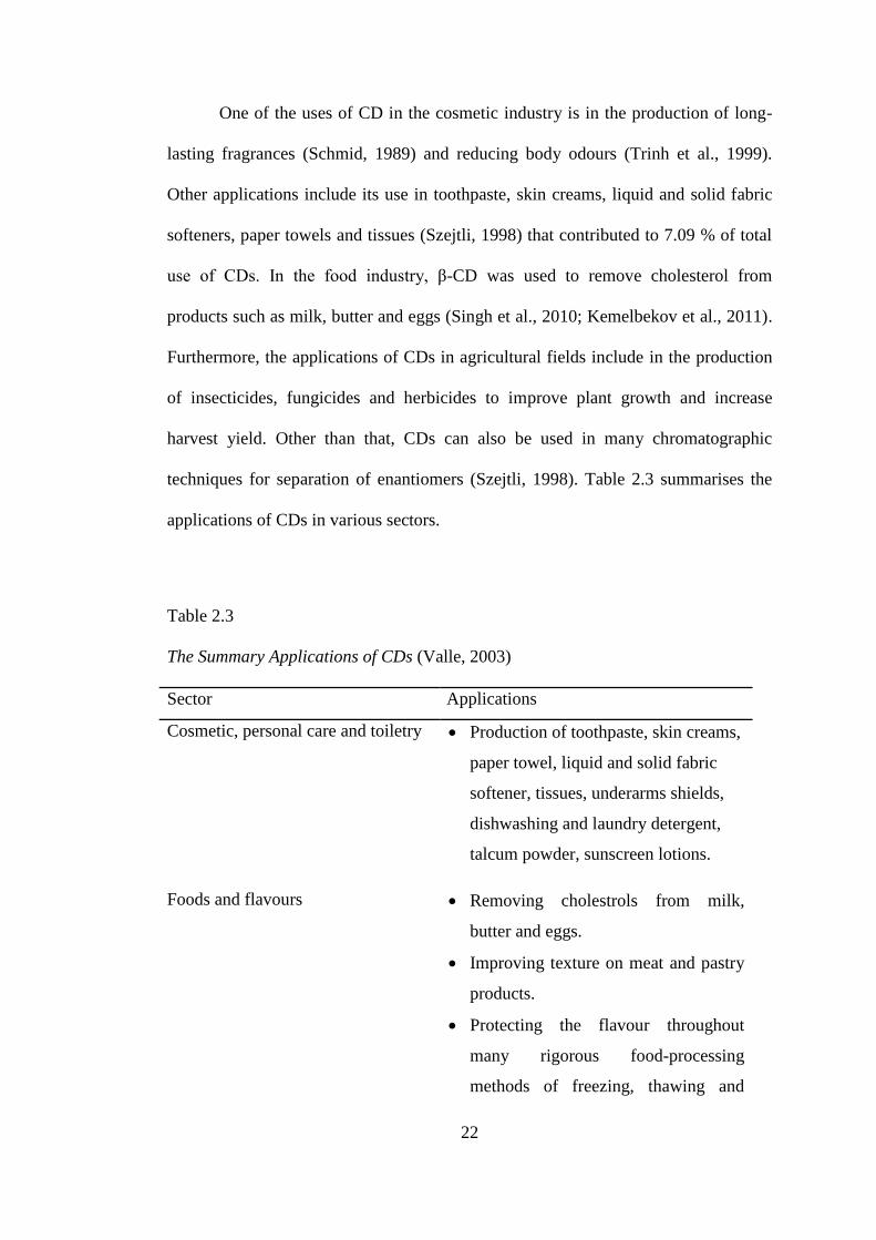
2.2.2 Applications of CDs
The modification of compounds with CDs lead to a large number of
applications in various fields related to pharmaceutical industry, food technology,
analytical chemistry, chemical synthesis and catalyst (Saenger, 1980; Szejtli, 1988;
Wilsom & Verall, 1998) as illustrated in Figure 2.5.

21
Figure 2.5. Distribution of the CD relevant abstracts published by Cyclodextrin
News (Cyclodextrin News, 1996).
CDs provide many beneficial improvements to products including solubility
and stabilisation of the guest compound which provide long-term protection of
colour, odour and flavour of products. Besides, the nontoxicity of CD is also the
main reason for its application in the pharmaceutical and food industry (Omari et al.,
2010; Misiuk & Zalewska, 2011) with more than 40 formulations containing CDs on
the market currently, intended for oral, parenteral, ophthalmic, rectal and dermal
route of application (Gidwani & Vyas, 2015). Barone et al. (1998) and Nasongkla et
al. (2003) reported that β-CD was used to increase bioavailability of poorly soluble
drugs. Light, thermal and oxidative stability of drug molecules can also be improved
through the formation of cyclodextrin complexes (Cwiertnia et al., 1999; Tirucherai
& Mitra, 2003). Other pharmaceutical application of CDs is to reduce dermal
(Uekama et al., 1992), ocular irritation (Loftsson & Stefansson, 1997) and to avoid
adverse drug-ingredient interactions (Redenti et al., 2001).
Chemistry of
CD complexes
21.82%
Pharmaceutical
24.62% Foods/cosmetic
7.09% Pesticides
0.76%
Chemical and
biochemical
processes and
products
10.67%
Analytical
chemistry
19.52%
Chemistry,
enzymology,
biological,
production
of CD
16.06%
22
One of the uses of CD in the cosmetic industry is in the production of long-
lasting fragrances (Schmid, 1989) and reducing body odours (Trinh et al., 1999).
Other applications include its use in toothpaste, skin creams, liquid and solid fabric
softeners, paper towels and tissues (Szejtli, 1998) that contributed to 7.09 % of total
use of CDs. In the food industry, β-CD was used to remove cholesterol from
products such as milk, butter and eggs (Singh et al., 2010; Kemelbekov et al., 2011).
Furthermore, the applications of CDs in agricultural fields include in the production
of insecticides, fungicides and herbicides to improve plant growth and increase
harvest yield. Other than that, CDs can also be used in many chromatographic
techniques for separation of enantiomers (Szejtli, 1998). Table 2.3 summarises the
applications of CDs in various sectors.
Table 2.3
The Summary Applications of CDs (Valle, 2003)
Sector Applications
Cosmetic, personal care and toiletry Production of toothpaste, skin creams,
paper towel, liquid and solid fabric
softener, tissues, underarms shields,
dishwashing and laundry detergent,
talcum powder, sunscreen lotions.
Foods and flavours
Removing cholestrols from milk,
butter and eggs.
Improving texture on meat and pastry
products.
Protecting the flavour throughout
many rigorous food-processing
methods of freezing, thawing and
23
microwaving.
Production of dairy products with low
cholestrols.
Improving elasticity in noodles, pie,
dough and pizza.
Act as the antimicrobial food
preservatives.
Pharmaceuticals Production of mouthwash solution,
nasal drug, eye drop solution.
Reducing the effects of bitter or
irritant tasting and bad smelling drugs.
Agriculture and chemical industries Production of herbicides, insecticides,
fungicides, repellents, pheromones.
Use in separation of isomers and
enantiomers.
Removing or detoxifying waste
materials.
Adhesives, coating and polymers Increasing tackiness and adhesion of
hot melts and adhesive.
2.2.3 Mechanism of Protein-CDs Interaction
Interactions between CD and protein have also given surprising outcomes as
CDs were employed as catalyst to alter some properties of protein (Hamilton et al.,
2000; Mcgarraghy & Darcy, 2000) and is in the improvement on protein solubility,
which could lead to the protein stabilisation in water (Koralewska et al., 2004). This
happened as the hydrophobic cavity of CDs is capable to provide temporary asylum
for hydrophobic parts of protein molecules to dissolve in water. The solubilisation
24
ability of CDs is currently of interest in the development of many formulations to
improve the bioavailability of protein system (Duchene, 1991).
Furthermore, the interactions of CDs with hydrophobic groups on protein
molecules can reduce protein aggregation. This phenomenon was observed in the
dissociation of bovine insulin dimers in the presence of different CDs (Lovatt et al.,
1996). The inhibition of protein aggregation also occurred to bovine insulin
(Dotsikas & Loukas, 2002), recombinant human growth hormone (rh-Gh) (Otzen et
al., 2002) and several other proteins (Sharma & Sharma, 2001; Sigurjonsdottir et al.,
1999). The study of Tavornvipas and co-workers (2004) showed the relationship
between reduction of aggregation and binding constants of CD-derivatives and rh-
GH. In their study, the branched CDs turned out to be the most competent in the
prevention of protein unfolding and aggregation. The same CDs have been also
reported to produce the highest stability constants among other CDs. Other findings
involving the interaction between CD and protein include the binding mechanism
between aripiprazole (APZ) with human serum albumin (HSA) in the presence of
three types of CDs (Yan et al., 2015). The study reported the refolding recovery of
APZ and HSA caused by CD which is specifically involved the interaction between
HSA and CD. Current studies on the interaction between CD and protein such as
phenylalanine dehydrogenase enzyme (Gubica et al., 2015) and mithochondrial
ADP/ATP carrier (AAC) (Rather et al., 2015) have also been reported.
Nonetheless, proteins such as insulin are mostly hydrophilic and too bulky to
be wholly encapsulated into the β-CD cavity. According to Irie and Uekama (1999)
the hydrophobic side chains of insulin peptides penetrate into the β-CD cavity
leading to the formation of non-covalent inclusion complexes. The CDs‘ ability to