DNA and RNA topoisomerase activities of Top3β …DNA and RNA topoisomerase activities of Top3β are...
8
DNA and RNA topoisomerase activities of Top3β are promoted by mediator protein Tudor domain-containing protein 3 Grace Ee-Lu Siaw a,b,c,1,2 , I-Fen Liu c,1 , Po-Yen Lin c , Michael D. Been d,2 , and Tao-shih Hsieh c,d,3 a Molecular Cell Biology, Taiwan International Graduate Program, Academia Sinica, Taipei 115, Taiwan; b Graduate Institute of Life Sciences, National Defense Medical Center, Taipei 114, Taiwan; c Institute of Cellular and Organismic Biology, Academia Sinica, Taipei 115, Taiwan; and d Department of Biochemistry, Duke University Medical Center, Durham, NC 27710 Edited by James M. Berger, The Johns Hopkins University School of Medicine, Baltimore, MD, and approved July 19, 2016 (received for review April 5, 2016) Topoisomerase 3β (Top3β) can associate with the mediator protein Tudor domain-containing protein 3 (TDRD3) to participate in two gene expression processes of transcription and translation. De- spite the apparent importance of TDRD3 in binding with Top3β and directing it to cellular compartments critical for gene expres- sion, the biochemical mechanism of how TDRD3 can affect the func- tions of Top3β is not known. We report here sensitive biochemical assays for the activities of Top3β on DNA and RNA substrates in resolving topological entanglements and for the analysis of TDRD3 functions. TDRD3 stimulates the relaxation activity of Top3β on hypernegatively supercoiled DNA and changes the reaction from a distributive to a processive mode. Both supercoil retention as- says and binding measurement by fluorescence anisotropy reveal a heretofore unknown preference for binding single-stranded nucleic acids over duplex. Whereas TDRD3 has a structure-specific binding preference, it does not discriminate between DNA and RNA. This unique property for binding with nucleic acids can have an important function in serving as a hub to form nucleoprotein complexes on DNA and RNA. To gain insight into the roles of Top3β on RNA metabolism, we designed an assay by annealing two single- stranded RNA circles with complementary sequences. Top3β is ca- pable of converting two such single-stranded RNA circles into a double-stranded RNA circle, and this strand-annealing activity is en- hanced by TDRD3. These results demonstrate that TDRD3 can en- hance the biochemical activities of Top3β on both DNA and RNA substrates, in addition to its function of targeting Top3β to critical sites in subcellular compartments. Tudor domain-containing domain 3 | DNA topoisomerases | RNA topoisomerases | RNA circles | RNA duplex H igher-order structural complexities in nucleic acids can be brought about by the folding and intertwining through inter- and intramolecular base pairing. They are impediments to the transactions of genetic information, including replication, tran- scription, recombination, and translation, which involve the un- winding and rewinding of base-paired regions to access encoded information (1–3). DNA topoisomerases are nature’s tools to resolve the topological entanglements in nucleic acids. They are ubiquitous in nature, first discovered in bacteria in 1971 (4), and in mammalian cells in 1972 (5), and are characterized by a mechanism involving the formation of a covalent protein/DNA adduct to generate a transient and reversible break allowing for topological transformation (6). Based on whether the strand pas- sage is through a protein-mediated single-stranded gate or a dou- ble-stranded gate, the enzymes are classified into type I or type II topoisomerases, respectively. Both types are further classified into A and B families based on their structural and mechanistic features (7). All topoisomerases are essential for the growth and develop- ment of an organism, suggesting that they have critical but distinct functions in all cells. Ever since their discovery, there is a fascination for whether these critical DNA enzymes may play a role in the RNA world. Early experiments involving the knotting of a single-stranded circle in trefoil formation (8) and protein-linked RNA cleavage (9, 10) suggest the biochemical feasibility of this idea. The interest is further boosted by recent experiments demonstrating that hu- man and Drosophila topoisomerase 3β (Top3β) is associated with mRNA in cytoplasm and has RNA topoisomerase activity (11, 12). The deficiency in this enzyme is attributed to one of the causative mutations for schizophrenia and cognitive disorder (11, 12) and results in abnormal synaptic structures in Drosophila (12). How RNA or DNA topoisomerase activity can affect neuro- logical functions remains to be elucidated. However, there are interesting insights to be gained by considering the interacting partners of Top3β. A mediator protein with multiple domains for interprotein interactions, TDRD3 is a key partner for Top3β in both the nucleus and cytoplasm (13). TDRD3 can recognize dimethylated arginine in proteins including inner core histones and RNA polymerase II, thus targeting its partner proteins to the transcriptionally active chromatin, especially at their 5′ end (14, 15). Because the knockdown or deletion of TDRD3 results in R-loop formation near the promoter regions, it is plausible that the absence of Top3β near the transcriptional start site can lead to a buildup of excessive negative supercoiling, thus stabilizing the R-loop structure (13). TDRD3 also associates with the partner Significance Tudor domain-containing protein 3 (TDRD3), a multidomain scaffold protein functions as an epigenetic reader on nuclear chromatin and binds with fragile X mental retardation protein on mRNA. It forms a conserved complex with topoisomerase 3β (Top3β), a type IA topoisomerase, and participates in both transcription and translation. The mechanism of how TDRD3 acts as Top3β’s partner in regulating these cellular processes is unknown. Here, we demonstrated that TDRD3 is able to stim- ulate Top3β’s DNA and RNA topoisomerase catalytic activities, through binding and stabilizing single-stranded regions in DNA and RNA substrates. Because these regions are the preferred site for Top3β, TDRD3 therefore acts as a regulator to provide access of the enzyme to these nucleic acid substrates and to act upon them. Author contributions: G.E.-L.S., I.-F.L., and T.-s.H. designed research; G.E.-L.S., I.-F.L., and P.-Y.L. performed research; G.E.-L.S., I.-F.L., P.-Y.L., M.D.B., and T.-s.H. contributed new reagents/analytic tools; G.E.-L.S., I.-F.L., M.D.B., and T.-s.H. analyzed data; and G.E.-L.S., I.-F.L., M.D.B., and T.-s.H. wrote the paper. The authors declare no conflict of interest. This article is a PNAS Direct Submission. 1 G.E.-L.S. and I.-F.L. contributed equally to this work. 2 To whom correspondence may be addressed. Email: [email protected] or graceelsiaw@ gmail.com. 3 Deceased August 4, 2016. This article contains supporting information online at www.pnas.org/lookup/suppl/doi:10. 1073/pnas.1605517113/-/DCSupplemental. E5544–E5551 | PNAS | Published online August 31, 2016 www.pnas.org/cgi/doi/10.1073/pnas.1605517113 Downloaded by guest on February 27, 2020
Transcript of DNA and RNA topoisomerase activities of Top3β …DNA and RNA topoisomerase activities of Top3β are...
DNA and RNA topoisomerase activities of Top3βare promoted by mediator protein Tudordomain-containing protein 3Grace Ee-Lu Siawa,b,c,1,2, I-Fen Liuc,1, Po-Yen Linc, Michael D. Beend,2, and Tao-shih Hsiehc,d,3
aMolecular Cell Biology, Taiwan International Graduate Program, Academia Sinica, Taipei 115, Taiwan; bGraduate Institute of Life Sciences, NationalDefense Medical Center, Taipei 114, Taiwan; cInstitute of Cellular and Organismic Biology, Academia Sinica, Taipei 115, Taiwan; and dDepartment ofBiochemistry, Duke University Medical Center, Durham, NC 27710
Edited by James M. Berger, The Johns Hopkins University School of Medicine, Baltimore, MD, and approved July 19, 2016 (received for review April 5, 2016)
Topoisomerase 3β (Top3β) can associate with the mediator proteinTudor domain-containing protein 3 (TDRD3) to participate in twogene expression processes of transcription and translation. De-spite the apparent importance of TDRD3 in binding with Top3βand directing it to cellular compartments critical for gene expres-sion, the biochemical mechanism of how TDRD3 can affect the func-tions of Top3β is not known. We report here sensitive biochemicalassays for the activities of Top3β on DNA and RNA substrates inresolving topological entanglements and for the analysis of TDRD3functions. TDRD3 stimulates the relaxation activity of Top3β onhypernegatively supercoiled DNA and changes the reaction froma distributive to a processive mode. Both supercoil retention as-says and binding measurement by fluorescence anisotropy reveala heretofore unknown preference for binding single-strandednucleic acids over duplex. Whereas TDRD3 has a structure-specificbinding preference, it does not discriminate between DNA andRNA. This unique property for binding with nucleic acids can havean important function in serving as a hub to form nucleoproteincomplexes on DNA and RNA. To gain insight into the roles of Top3βon RNAmetabolism, we designed an assay by annealing two single-stranded RNA circles with complementary sequences. Top3β is ca-pable of converting two such single-stranded RNA circles into adouble-stranded RNA circle, and this strand-annealing activity is en-hanced by TDRD3. These results demonstrate that TDRD3 can en-hance the biochemical activities of Top3β on both DNA and RNAsubstrates, in addition to its function of targeting Top3β to criticalsites in subcellular compartments.
Tudor domain-containing domain 3 | DNA topoisomerases |RNA topoisomerases | RNA circles | RNA duplex
Higher-order structural complexities in nucleic acids can bebrought about by the folding and intertwining through inter-
and intramolecular base pairing. They are impediments to thetransactions of genetic information, including replication, tran-scription, recombination, and translation, which involve the un-winding and rewinding of base-paired regions to access encodedinformation (1–3). DNA topoisomerases are nature’s tools toresolve the topological entanglements in nucleic acids. They areubiquitous in nature, first discovered in bacteria in 1971 (4), andin mammalian cells in 1972 (5), and are characterized by amechanism involving the formation of a covalent protein/DNAadduct to generate a transient and reversible break allowing fortopological transformation (6). Based on whether the strand pas-sage is through a protein-mediated single-stranded gate or a dou-ble-stranded gate, the enzymes are classified into type I or type IItopoisomerases, respectively. Both types are further classified intoA and B families based on their structural and mechanistic features(7). All topoisomerases are essential for the growth and develop-ment of an organism, suggesting that they have critical but distinctfunctions in all cells.Ever since their discovery, there is a fascination for whether
these critical DNA enzymes may play a role in the RNA world.
Early experiments involving the knotting of a single-strandedcircle in trefoil formation (8) and protein-linked RNA cleavage(9, 10) suggest the biochemical feasibility of this idea. The interestis further boosted by recent experiments demonstrating that hu-man and Drosophila topoisomerase 3β (Top3β) is associated withmRNA in cytoplasm and has RNA topoisomerase activity (11, 12).The deficiency in this enzyme is attributed to one of the causativemutations for schizophrenia and cognitive disorder (11, 12) andresults in abnormal synaptic structures in Drosophila (12).How RNA or DNA topoisomerase activity can affect neuro-
logical functions remains to be elucidated. However, there areinteresting insights to be gained by considering the interactingpartners of Top3β. A mediator protein with multiple domains forinterprotein interactions, TDRD3 is a key partner for Top3βin both the nucleus and cytoplasm (13). TDRD3 can recognizedimethylated arginine in proteins including inner core histonesand RNA polymerase II, thus targeting its partner proteins to thetranscriptionally active chromatin, especially at their 5′ end (14,15). Because the knockdown or deletion of TDRD3 results inR-loop formation near the promoter regions, it is plausible thatthe absence of Top3β near the transcriptional start site can lead toa buildup of excessive negative supercoiling, thus stabilizing theR-loop structure (13). TDRD3 also associates with the partner
Significance
Tudor domain-containing protein 3 (TDRD3), a multidomainscaffold protein functions as an epigenetic reader on nuclearchromatin and binds with fragile X mental retardation proteinon mRNA. It forms a conserved complex with topoisomerase 3β(Top3β), a type IA topoisomerase, and participates in bothtranscription and translation. The mechanism of how TDRD3acts as Top3β’s partner in regulating these cellular processes isunknown. Here, we demonstrated that TDRD3 is able to stim-ulate Top3β’s DNA and RNA topoisomerase catalytic activities,through binding and stabilizing single-stranded regions in DNAand RNA substrates. Because these regions are the preferredsite for Top3β, TDRD3 therefore acts as a regulator to provideaccess of the enzyme to these nucleic acid substrates and to actupon them.
Author contributions: G.E.-L.S., I.-F.L., and T.-s.H. designed research; G.E.-L.S., I.-F.L., andP.-Y.L. performed research; G.E.-L.S., I.-F.L., P.-Y.L., M.D.B., and T.-s.H. contributed newreagents/analytic tools; G.E.-L.S., I.-F.L., M.D.B., and T.-s.H. analyzed data; and G.E.-L.S., I.-F.L.,M.D.B., and T.-s.H. wrote the paper.
The authors declare no conflict of interest.
This article is a PNAS Direct Submission.1G.E.-L.S. and I.-F.L. contributed equally to this work.2To whom correspondence may be addressed. Email: [email protected] or [email protected].
3Deceased August 4, 2016.
This article contains supporting information online at www.pnas.org/lookup/suppl/doi:10.1073/pnas.1605517113/-/DCSupplemental.
E5544–E5551 | PNAS | Published online August 31, 2016 www.pnas.org/cgi/doi/10.1073/pnas.1605517113
Dow
nloa
ded
by g
uest
on
Feb
ruar
y 27
, 202
0
protein fragile X mental retardation protein (FMRP) in thecytoplasm. Missing FMRP can cause fragile X mental retardation,a major cause of congenital mental disorder (16). FMRP ispreferentially localized in the coding regions of mRNA and playsa role in inhibiting the translation of mRNA relevant to synapticfunctions in neuronal cells (17). It is thus suggested that Top3βmay also have a role in translation through its RNA topoisomeraseactivity in modulating the complex structures of folded mRNA(12). Whereas Top3β can have important functions in two pro-cesses of gene expression in nucleus and cytoplasm through itsinteractions with TDRD3, it is unclear whether TDRD3 hasany additional role in regulating Top3β activity, beyond its tar-geting function of directing Top3β to critical sites in subcellularcompartments.Our results presented here show that TDRD3 can promote
both DNA and RNA topoisomerase activities, likely throughstabilizing the complex of the enzyme bound to the single-stranded regions of the substrate, which is also the site wheretype IA enzymes carry out strand passage reactions. The findingthat TDRD3 has a structure-specific preference for single-stranded nucleic acids, but does not discriminate between DNAand RNA, adds an aspect to the scaffolding function of TDRD3in the assembly of the nucleoprotein complex. A mediator pro-tein that is able to interact with specific proteins and to target tothe preferred structure of nucleic acids can have a versatile roleserving as a hub for assembling stable complexes in chromatin orpolysomes.With an increasing interest in RNA topoisomerase activity, it
is important to develop biochemical assays other than the trefoilformation of circular RNA. Using two single-stranded circularRNA substrates with complementary sequences, we have dem-onstrated that Top3β and other type IA enzymes can mediate theformation of double-stranded circular RNA through the in-termolecular stranded passage activity of these enzymes. Thiscircle-annealing assay poses a less stringent requirement for thesequence and structure of the substrate RNA compared with theknotting assay, and can thus broaden our capability to test the effectof an RNA sequence or structure on RNA topoisomerase ac-tivity. In addition, this versatile biochemical method to preparean interesting class of molecules of circular duplex RNA hasunique potential for investigating the functions of other RNAenzymes, including topoisomerases.
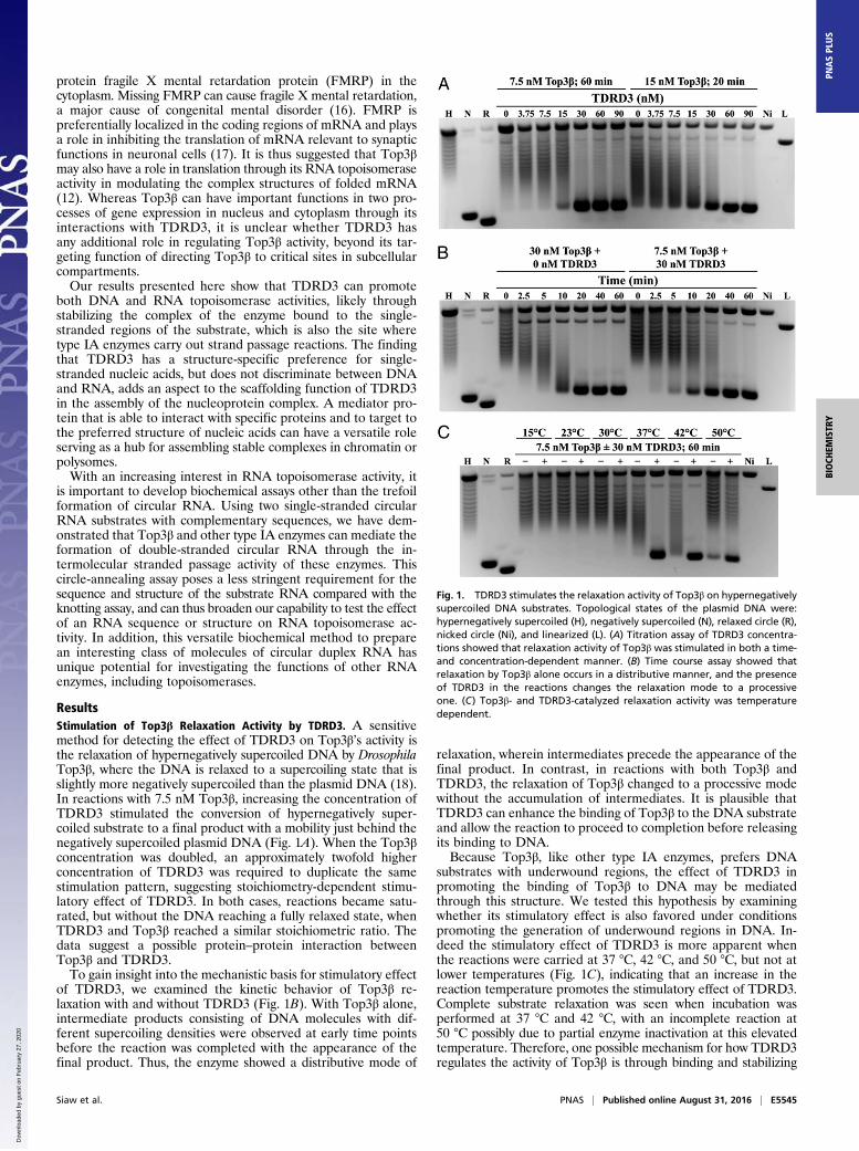
ResultsStimulation of Top3β Relaxation Activity by TDRD3. A sensitivemethod for detecting the effect of TDRD3 on Top3β’s activity isthe relaxation of hypernegatively supercoiled DNA by DrosophilaTop3β, where the DNA is relaxed to a supercoiling state that isslightly more negatively supercoiled than the plasmid DNA (18).In reactions with 7.5 nM Top3β, increasing the concentration ofTDRD3 stimulated the conversion of hypernegatively super-coiled substrate to a final product with a mobility just behind thenegatively supercoiled plasmid DNA (Fig. 1A). When the Top3βconcentration was doubled, an approximately twofold higherconcentration of TDRD3 was required to duplicate the samestimulation pattern, suggesting stoichiometry-dependent stimu-latory effect of TDRD3. In both cases, reactions became satu-rated, but without the DNA reaching a fully relaxed state, whenTDRD3 and Top3β reached a similar stoichiometric ratio. Thedata suggest a possible protein–protein interaction betweenTop3β and TDRD3.To gain insight into the mechanistic basis for stimulatory effect
of TDRD3, we examined the kinetic behavior of Top3β re-laxation with and without TDRD3 (Fig. 1B). With Top3β alone,intermediate products consisting of DNA molecules with dif-ferent supercoiling densities were observed at early time pointsbefore the reaction was completed with the appearance of thefinal product. Thus, the enzyme showed a distributive mode of
relaxation, wherein intermediates precede the appearance of thefinal product. In contrast, in reactions with both Top3β andTDRD3, the relaxation of Top3β changed to a processive modewithout the accumulation of intermediates. It is plausible thatTDRD3 can enhance the binding of Top3β to the DNA substrateand allow the reaction to proceed to completion before releasingits binding to DNA.Because Top3β, like other type IA enzymes, prefers DNA
substrates with underwound regions, the effect of TDRD3 inpromoting the binding of Top3β to DNA may be mediatedthrough this structure. We tested this hypothesis by examiningwhether its stimulatory effect is also favored under conditionspromoting the generation of underwound regions in DNA. In-deed the stimulatory effect of TDRD3 is more apparent whenthe reactions were carried at 37 °C, 42 °C, and 50 °C, but not atlower temperatures (Fig. 1C), indicating that an increase in thereaction temperature promotes the stimulatory effect of TDRD3.Complete substrate relaxation was seen when incubation wasperformed at 37 °C and 42 °C, with an incomplete reaction at50 °C possibly due to partial enzyme inactivation at this elevatedtemperature. Therefore, one possible mechanism for how TDRD3regulates the activity of Top3β is through binding and stabilizing
Fig. 1. TDRD3 stimulates the relaxation activity of Top3β on hypernegativelysupercoiled DNA substrates. Topological states of the plasmid DNA were:hypernegatively supercoiled (H), negatively supercoiled (N), relaxed circle (R),nicked circle (Ni), and linearized (L). (A) Titration assay of TDRD3 concentra-tions showed that relaxation activity of Top3β was stimulated in both a time-and concentration-dependent manner. (B) Time course assay showed thatrelaxation by Top3β alone occurs in a distributive manner, and the presenceof TDRD3 in the reactions changes the relaxation mode to a processiveone. (C) Top3β- and TDRD3-catalyzed relaxation activity was temperaturedependent.
Siaw et al. PNAS | Published online August 31, 2016 | E5545
BIOCH
EMISTR
YPN
ASPL
US
Dow
nloa
ded
by g
uest
on
Feb
ruar
y 27
, 202
0
single-stranded regions in plasmid substrates, and thereby recruitsthe enzyme to these sites.
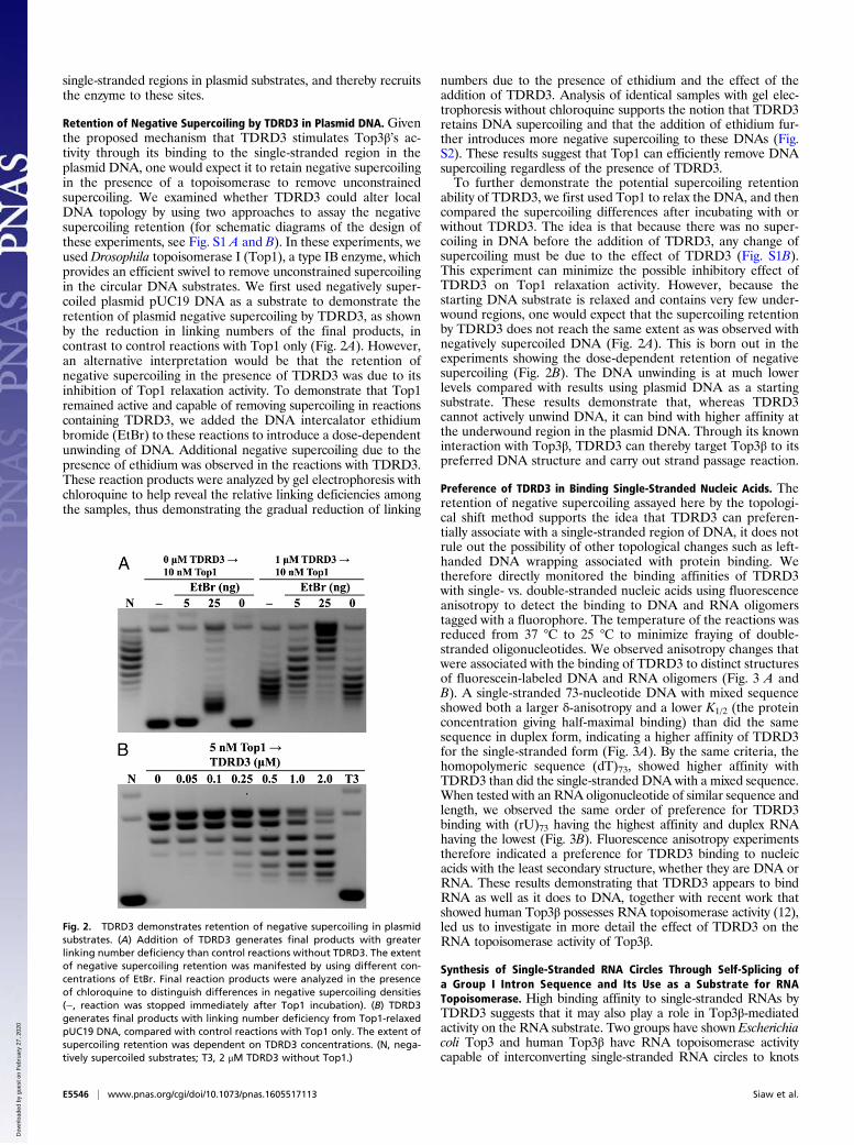
Retention of Negative Supercoiling by TDRD3 in Plasmid DNA. Giventhe proposed mechanism that TDRD3 stimulates Top3β’s ac-tivity through its binding to the single-stranded region in theplasmid DNA, one would expect it to retain negative supercoilingin the presence of a topoisomerase to remove unconstrainedsupercoiling. We examined whether TDRD3 could alter localDNA topology by using two approaches to assay the negativesupercoiling retention (for schematic diagrams of the design ofthese experiments, see Fig. S1 A and B). In these experiments, weused Drosophila topoisomerase I (Top1), a type IB enzyme, whichprovides an efficient swivel to remove unconstrained supercoilingin the circular DNA substrates. We first used negatively super-coiled plasmid pUC19 DNA as a substrate to demonstrate theretention of plasmid negative supercoiling by TDRD3, as shownby the reduction in linking numbers of the final products, incontrast to control reactions with Top1 only (Fig. 2A). However,an alternative interpretation would be that the retention ofnegative supercoiling in the presence of TDRD3 was due to itsinhibition of Top1 relaxation activity. To demonstrate that Top1remained active and capable of removing supercoiling in reactionscontaining TDRD3, we added the DNA intercalator ethidiumbromide (EtBr) to these reactions to introduce a dose-dependentunwinding of DNA. Additional negative supercoiling due to thepresence of ethidium was observed in the reactions with TDRD3.These reaction products were analyzed by gel electrophoresis withchloroquine to help reveal the relative linking deficiencies amongthe samples, thus demonstrating the gradual reduction of linking
numbers due to the presence of ethidium and the effect of theaddition of TDRD3. Analysis of identical samples with gel elec-trophoresis without chloroquine supports the notion that TDRD3retains DNA supercoiling and that the addition of ethidium fur-ther introduces more negative supercoiling to these DNAs (Fig.S2). These results suggest that Top1 can efficiently remove DNAsupercoiling regardless of the presence of TDRD3.To further demonstrate the potential supercoiling retention
ability of TDRD3, we first used Top1 to relax the DNA, and thencompared the supercoiling differences after incubating with orwithout TDRD3. The idea is that because there was no super-coiling in DNA before the addition of TDRD3, any change ofsupercoiling must be due to the effect of TDRD3 (Fig. S1B).This experiment can minimize the possible inhibitory effect ofTDRD3 on Top1 relaxation activity. However, because thestarting DNA substrate is relaxed and contains very few under-wound regions, one would expect that the supercoiling retentionby TDRD3 does not reach the same extent as was observed withnegatively supercoiled DNA (Fig. 2A). This is born out in theexperiments showing the dose-dependent retention of negativesupercoiling (Fig. 2B). The DNA unwinding is at much lowerlevels compared with results using plasmid DNA as a startingsubstrate. These results demonstrate that, whereas TDRD3cannot actively unwind DNA, it can bind with higher affinity atthe underwound region in the plasmid DNA. Through its knowninteraction with Top3β, TDRD3 can thereby target Top3β to itspreferred DNA structure and carry out strand passage reaction.
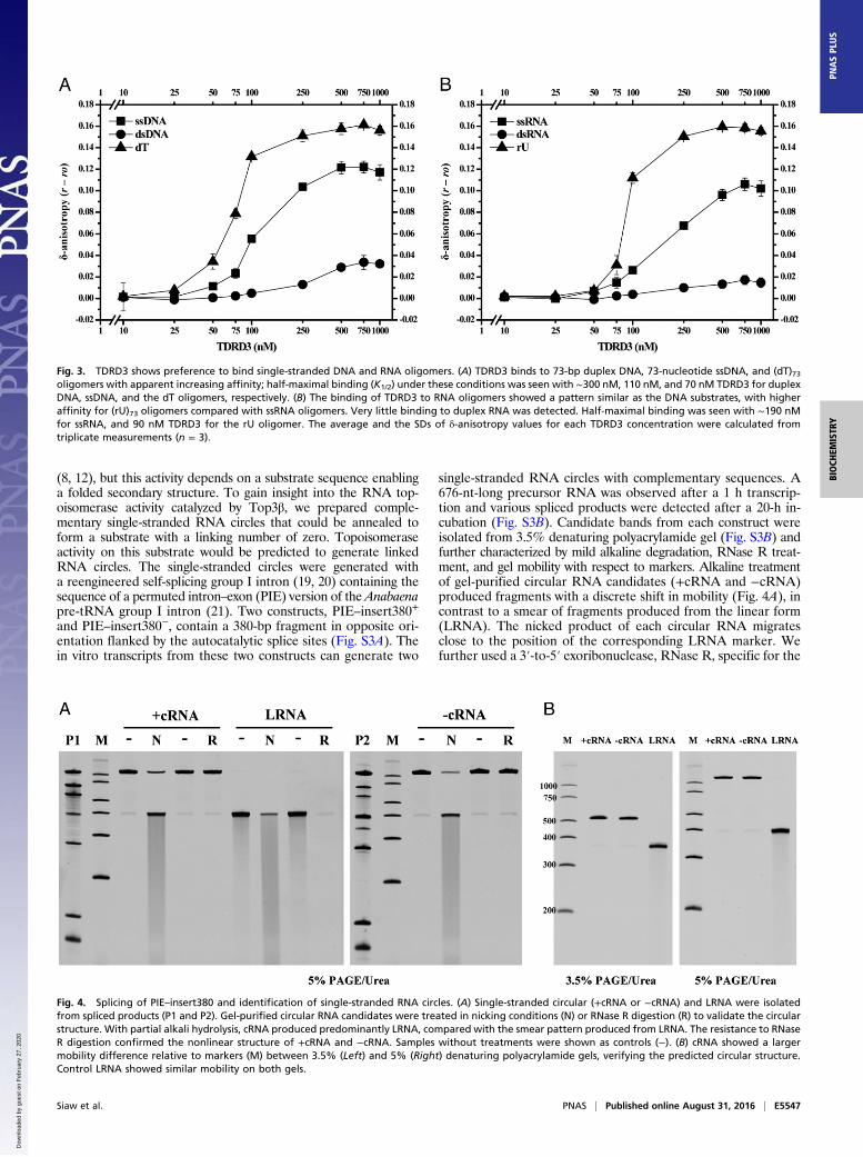
Preference of TDRD3 in Binding Single-Stranded Nucleic Acids. Theretention of negative supercoiling assayed here by the topologi-cal shift method supports the idea that TDRD3 can preferen-tially associate with a single-stranded region of DNA, it does notrule out the possibility of other topological changes such as left-handed DNA wrapping associated with protein binding. Wetherefore directly monitored the binding affinities of TDRD3with single- vs. double-stranded nucleic acids using fluorescenceanisotropy to detect the binding to DNA and RNA oligomerstagged with a fluorophore. The temperature of the reactions wasreduced from 37 °C to 25 °C to minimize fraying of double-stranded oligonucleotides. We observed anisotropy changes thatwere associated with the binding of TDRD3 to distinct structuresof fluorescein-labeled DNA and RNA oligomers (Fig. 3 A andB). A single-stranded 73-nucleotide DNA with mixed sequenceshowed both a larger δ-anisotropy and a lower K1/2 (the proteinconcentration giving half-maximal binding) than did the samesequence in duplex form, indicating a higher affinity of TDRD3for the single-stranded form (Fig. 3A). By the same criteria, thehomopolymeric sequence (dT)73, showed higher affinity withTDRD3 than did the single-stranded DNA with a mixed sequence.When tested with an RNA oligonucleotide of similar sequence andlength, we observed the same order of preference for TDRD3binding with (rU)73 having the highest affinity and duplex RNAhaving the lowest (Fig. 3B). Fluorescence anisotropy experimentstherefore indicated a preference for TDRD3 binding to nucleicacids with the least secondary structure, whether they are DNA orRNA. These results demonstrating that TDRD3 appears to bindRNA as well as it does to DNA, together with recent work thatshowed human Top3β possesses RNA topoisomerase activity (12),led us to investigate in more detail the effect of TDRD3 on theRNA topoisomerase activity of Top3β.
Synthesis of Single-Stranded RNA Circles Through Self-Splicing ofa Group I Intron Sequence and Its Use as a Substrate for RNATopoisomerase. High binding affinity to single-stranded RNAs byTDRD3 suggests that it may also play a role in Top3β-mediatedactivity on the RNA substrate. Two groups have shown Escherichiacoli Top3 and human Top3β have RNA topoisomerase activitycapable of interconverting single-stranded RNA circles to knots
Fig. 2. TDRD3 demonstrates retention of negative supercoiling in plasmidsubstrates. (A) Addition of TDRD3 generates final products with greaterlinking number deficiency than control reactions without TDRD3. The extentof negative supercoiling retention was manifested by using different con-centrations of EtBr. Final reaction products were analyzed in the presenceof chloroquine to distinguish differences in negative supercoiling densities(−, reaction was stopped immediately after Top1 incubation). (B) TDRD3generates final products with linking number deficiency from Top1-relaxedpUC19 DNA, compared with control reactions with Top1 only. The extent ofsupercoiling retention was dependent on TDRD3 concentrations. (N, nega-tively supercoiled substrates; T3, 2 μM TDRD3 without Top1.)
E5546 | www.pnas.org/cgi/doi/10.1073/pnas.1605517113 Siaw et al.
Dow
nloa
ded
by g
uest
on
Feb
ruar
y 27
, 202
0
(8, 12), but this activity depends on a substrate sequence enablinga folded secondary structure. To gain insight into the RNA top-oisomerase activity catalyzed by Top3β, we prepared comple-mentary single-stranded RNA circles that could be annealed toform a substrate with a linking number of zero. Topoisomeraseactivity on this substrate would be predicted to generate linkedRNA circles. The single-stranded circles were generated witha reengineered self-splicing group I intron (19, 20) containing thesequence of a permuted intron–exon (PIE) version of the Anabaenapre-tRNA group I intron (21). Two constructs, PIE–insert380+
and PIE–insert380−, contain a 380-bp fragment in opposite ori-entation flanked by the autocatalytic splice sites (Fig. S3A). Thein vitro transcripts from these two constructs can generate two
single-stranded RNA circles with complementary sequences. A676-nt-long precursor RNA was observed after a 1 h transcrip-tion and various spliced products were detected after a 20-h in-cubation (Fig. S3B). Candidate bands from each construct wereisolated from 3.5% denaturing polyacrylamide gel (Fig. S3B) andfurther characterized by mild alkaline degradation, RNase R treat-ment, and gel mobility with respect to markers. Alkaline treatmentof gel-purified circular RNA candidates (+cRNA and −cRNA)produced fragments with a discrete shift in mobility (Fig. 4A), incontrast to a smear of fragments produced from the linear form(LRNA). The nicked product of each circular RNA migratesclose to the position of the corresponding LRNA marker. Wefurther used a 3′-to-5′ exoribonuclease, RNase R, specific for the
Fig. 3. TDRD3 shows preference to bind single-stranded DNA and RNA oligomers. (A) TDRD3 binds to 73-bp duplex DNA, 73-nucleotide ssDNA, and (dT)73oligomers with apparent increasing affinity; half-maximal binding (K1/2) under these conditions was seen with ∼300 nM, 110 nM, and 70 nM TDRD3 for duplexDNA, ssDNA, and the dT oligomers, respectively. (B) The binding of TDRD3 to RNA oligomers showed a pattern similar as the DNA substrates, with higheraffinity for (rU)73 oligomers compared with ssRNA oligomers. Very little binding to duplex RNA was detected. Half-maximal binding was seen with ∼190 nMfor ssRNA, and 90 nM TDRD3 for the rU oligomer. The average and the SDs of δ-anisotropy values for each TDRD3 concentration were calculated fromtriplicate measurements (n = 3).
Fig. 4. Splicing of PIE–insert380 and identification of single-stranded RNA circles. (A) Single-stranded circular (+cRNA or −cRNA) and LRNA were isolatedfrom spliced products (P1 and P2). Gel-purified circular RNA candidates were treated in nicking conditions (N) or RNase R digestion (R) to validate the circularstructure. With partial alkali hydrolysis, cRNA produced predominantly LRNA, compared with the smear pattern produced from LRNA. The resistance to RNaseR digestion confirmed the nonlinear structure of +cRNA and −cRNA. Samples without treatments were shown as controls (−). (B) cRNA showed a largermobility difference relative to markers (M) between 3.5% (Left) and 5% (Right) denaturing polyacrylamide gels, verifying the predicted circular structure.Control LRNA showed similar mobility on both gels.
Siaw et al. PNAS | Published online August 31, 2016 | E5547
BIOCH
EMISTR
YPN
ASPL
US
Dow
nloa
ded
by g
uest
on
Feb
ruar
y 27
, 202
0
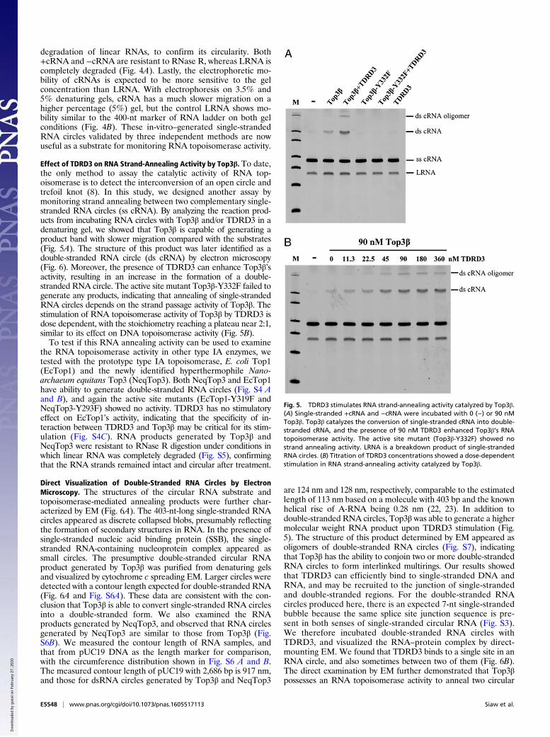
degradation of linear RNAs, to confirm its circularity. Both+cRNA and −cRNA are resistant to RNase R, whereas LRNA iscompletely degraded (Fig. 4A). Lastly, the electrophoretic mo-bility of cRNAs is expected to be more sensitive to the gelconcentration than LRNA. With electrophoresis on 3.5% and5% denaturing gels, cRNA has a much slower migration on ahigher percentage (5%) gel, but the control LRNA shows mo-bility similar to the 400-nt marker of RNA ladder on both gelconditions (Fig. 4B). These in-vitro–generated single-strandedRNA circles validated by three independent methods are nowuseful as a substrate for monitoring RNA topoisomerase activity.
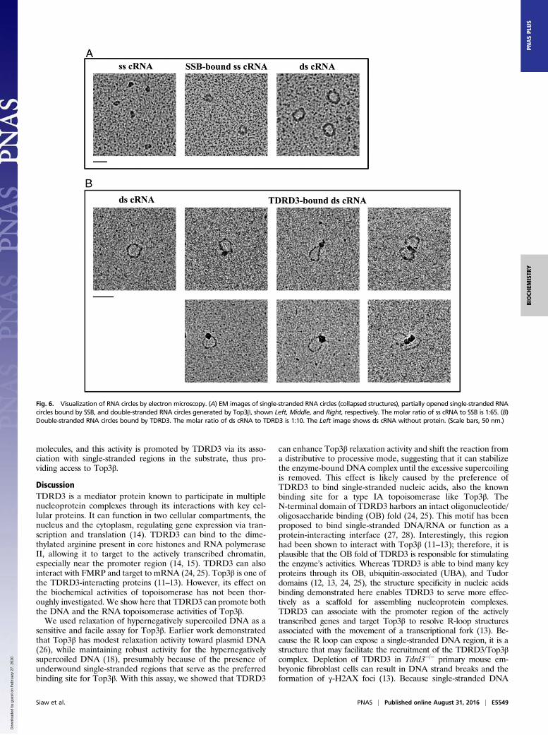
Effect of TDRD3 on RNA Strand-Annealing Activity by Top3β. To date,the only method to assay the catalytic activity of RNA top-oisomerase is to detect the interconversion of an open circle andtrefoil knot (8). In this study, we designed another assay bymonitoring strand annealing between two complementary single-stranded RNA circles (ss cRNA). By analyzing the reaction prod-ucts from incubating RNA circles with Top3β and/or TDRD3 in adenaturing gel, we showed that Top3β is capable of generating aproduct band with slower migration compared with the substrates(Fig. 5A). The structure of this product was later identified as adouble-stranded RNA circle (ds cRNA) by electron microscopy(Fig. 6). Moreover, the presence of TDRD3 can enhance Top3β’sactivity, resulting in an increase in the formation of a double-stranded RNA circle. The active site mutant Top3β-Y332F failed togenerate any products, indicating that annealing of single-strandedRNA circles depends on the strand passage activity of Top3β. Thestimulation of RNA topoisomerase activity of Top3β by TDRD3 isdose dependent, with the stoichiometry reaching a plateau near 2:1,similar to its effect on DNA topoisomerase activity (Fig. 5B).To test if this RNA annealing activity can be used to examine
the RNA topoisomerase activity in other type IA enzymes, wetested with the prototype type IA topoisomerase, E. coli Top1(EcTop1) and the newly identified hyperthermophile Nano-archaeum equitans Top3 (NeqTop3). Both NeqTop3 and EcTop1have ability to generate double-stranded RNA circles (Fig. S4 Aand B), and again the active site mutants (EcTop1-Y319F andNeqTop3-Y293F) showed no activity. TDRD3 has no stimulatoryeffect on EcTop1’s activity, indicating that the specificity of in-teraction between TDRD3 and Top3β may be critical for its stim-ulation (Fig. S4C). RNA products generated by Top3β andNeqTop3 were resistant to RNase R digestion under conditions inwhich linear RNA was completely degraded (Fig. S5), confirmingthat the RNA strands remained intact and circular after treatment.
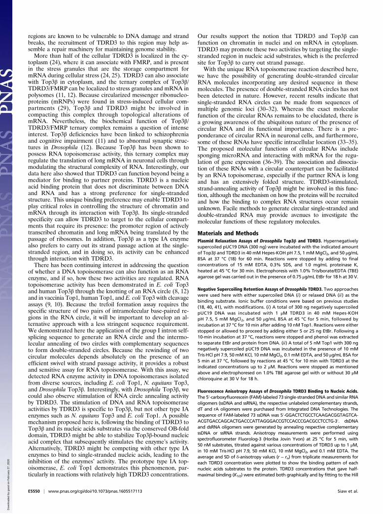
Direct Visualization of Double-Stranded RNA Circles by ElectronMicroscopy. The structures of the circular RNA substrate andtopoisomerase-mediated annealing products were further char-acterized by EM (Fig. 6A). The 403-nt-long single-stranded RNAcircles appeared as discrete collapsed blobs, presumably reflectingthe formation of secondary structures in RNA. In the presence ofsingle-stranded nucleic acid binding protein (SSB), the single-stranded RNA-containing nucleoprotein complex appeared assmall circles. The presumptive double-stranded circular RNAproduct generated by Top3β was purified from denaturing gelsand visualized by cytochrome c spreading EM. Larger circles weredetected with a contour length expected for double-stranded RNA(Fig. 6A and Fig. S6A). These data are consistent with the con-clusion that Top3β is able to convert single-stranded RNA circlesinto a double-stranded form. We also examined the RNAproducts generated by NeqTop3, and observed that RNA circlesgenerated by NeqTop3 are similar to those from Top3β (Fig.S6B). We measured the contour length of RNA samples, andthat from pUC19 DNA as the length marker for comparison,with the circumference distribution shown in Fig. S6 A and B.The measured contour length of pUC19 with 2,686 bp is 917 nm,and those for dsRNA circles generated by Top3β and NeqTop3
are 124 nm and 128 nm, respectively, comparable to the estimatedlength of 113 nm based on a molecule with 403 bp and the knownhelical rise of A-RNA being 0.28 nm (22, 23). In addition todouble-stranded RNA circles, Top3β was able to generate a highermolecular weight RNA product upon TDRD3 stimulation (Fig.5). The structure of this product determined by EM appeared asoligomers of double-stranded RNA circles (Fig. S7), indicatingthat Top3β has the ability to conjoin two or more double-strandedRNA circles to form interlinked multirings. Our results showedthat TDRD3 can efficiently bind to single-stranded DNA andRNA, and may be recruited to the junction of single-strandedand double-stranded regions. For the double-stranded RNAcircles produced here, there is an expected 7-nt single-strandedbubble because the same splice site junction sequence is pre-sent in both senses of single-stranded circular RNA (Fig. S3).We therefore incubated double-stranded RNA circles withTDRD3, and visualized the RNA–protein complex by direct-mounting EM. We found that TDRD3 binds to a single site in anRNA circle, and also sometimes between two of them (Fig. 6B).The direct examination by EM further demonstrated that Top3βpossesses an RNA topoisomerase activity to anneal two circular
Fig. 5. TDRD3 stimulates RNA strand-annealing activity catalyzed by Top3β.(A) Single-stranded +cRNA and −cRNA were incubated with 0 (−) or 90 nMTop3β. Top3β catalyzes the conversion of single-stranded cRNA into double-stranded cRNA, and the presence of 90 nM TDRD3 enhanced Top3β’s RNAtopoisomerase activity. The active site mutant (Top3β-Y332F) showed nostrand annealing activity. LRNA is a breakdown product of single-strandedRNA circles. (B) Titration of TDRD3 concentrations showed a dose-dependentstimulation in RNA strand-annealing activity catalyzed by Top3β.
E5548 | www.pnas.org/cgi/doi/10.1073/pnas.1605517113 Siaw et al.
Dow
nloa
ded
by g
uest
on
Feb
ruar
y 27
, 202
0
molecules, and this activity is promoted by TDRD3 via its asso-ciation with single-stranded regions in the substrate, thus pro-viding access to Top3β.
DiscussionTDRD3 is a mediator protein known to participate in multiplenucleoprotein complexes through its interactions with key cel-lular proteins. It can function in two cellular compartments, thenucleus and the cytoplasm, regulating gene expression via tran-scription and translation (14). TDRD3 can bind to the dime-thylated arginine present in core histones and RNA polymeraseII, allowing it to target to the actively transcribed chromatin,especially near the promoter region (14, 15). TDRD3 can alsointeract with FMRP and target to mRNA (24, 25). Top3β is one ofthe TDRD3-interacting proteins (11–13). However, its effect onthe biochemical activities of topoisomerase has not been thor-oughly investigated. We show here that TDRD3 can promote boththe DNA and the RNA topoisomerase activities of Top3β.We used relaxation of hypernegatively supercoiled DNA as a
sensitive and facile assay for Top3β. Earlier work demonstratedthat Top3β has modest relaxation activity toward plasmid DNA(26), while maintaining robust activity for the hypernegativelysupercoiled DNA (18), presumably because of the presence ofunderwound single-stranded regions that serve as the preferredbinding site for Top3β. With this assay, we showed that TDRD3
can enhance Top3β relaxation activity and shift the reaction froma distributive to processive mode, suggesting that it can stabilizethe enzyme-bound DNA complex until the excessive supercoilingis removed. This effect is likely caused by the preference ofTDRD3 to bind single-stranded nucleic acids, also the knownbinding site for a type IA topoisomerase like Top3β. TheN-terminal domain of TDRD3 harbors an intact oligonucleotide/oligosaccharide binding (OB) fold (24, 25). This motif has beenproposed to bind single-stranded DNA/RNA or function as aprotein-interacting interface (27, 28). Interestingly, this regionhad been shown to interact with Top3β (11–13); therefore, it isplausible that the OB fold of TDRD3 is responsible for stimulatingthe enzyme’s activities. Whereas TDRD3 is able to bind many keyproteins through its OB, ubiquitin-associated (UBA), and Tudordomains (12, 13, 24, 25), the structure specificity in nucleic acidsbinding demonstrated here enables TDRD3 to serve more effec-tively as a scaffold for assembling nucleoprotein complexes.TDRD3 can associate with the promoter region of the activelytranscribed genes and target Top3β to resolve R-loop structuresassociated with the movement of a transcriptional fork (13). Be-cause the R loop can expose a single-stranded DNA region, it is astructure that may facilitate the recruitment of the TDRD3/Top3βcomplex. Depletion of TDRD3 in Tdrd3−/− primary mouse em-bryonic fibroblast cells can result in DNA strand breaks and theformation of γ-H2AX foci (13). Because single-stranded DNA
Fig. 6. Visualization of RNA circles by electron microscopy. (A) EM images of single-stranded RNA circles (collapsed structures), partially opened single-stranded RNAcircles bound by SSB, and double-stranded RNA circles generated by Top3β, shown Left, Middle, and Right, respectively. The molar ratio of ss cRNA to SSB is 1:65. (B)Double-stranded RNA circles bound by TDRD3. The molar ratio of ds cRNA to TDRD3 is 1:10. The Left image shows ds cRNA without protein. (Scale bars, 50 nm.)
Siaw et al. PNAS | Published online August 31, 2016 | E5549
BIOCH
EMISTR
YPN
ASPL
US
Dow
nloa
ded
by g
uest
on
Feb
ruar
y 27
, 202
0
regions are known to be vulnerable to DNA damage and strandbreaks, the recruitment of TDRD3 to this region may help as-semble a repair machinery for maintaining genome stability.More than half of the cellular TDRD3 is localized in the cy-
toplasm (24), where it can associate with FMRP, and is presentin the stress granules that are the storage compartment formRNA during cellular stress (24, 25). TDRD3 can also associatewith Top3β in cytoplasm, and the ternary complex of Top3β/TDRD3/FMRP can be localized to stress granules and mRNA inpolysomes (11, 12). Because circularized messenger ribonucleo-proteins (mRNPs) were found in stress-induced cellular com-partments (29), Top3β and TDRD3 might be involved incompacting this complex through topological alterations ofmRNA. Nevertheless, the biochemical function of Top3β/TDRD3/FMRP ternary complex remains a question of intenseinterest. Top3β deficiencies have been linked to schizophreniaand cognitive impairment (11) and to abnormal synaptic struc-tures in Drosophila (12). Because Top3β has been shown topossess RNA topoisomerase activity, this ternary complex mayregulate the translation of long mRNA in neuronal cells throughmodulating the structural complexity of RNA. Interestingly, ourdata here also showed that TDRD3 can function beyond being amediator for binding to partner proteins. TDRD3 is a nucleicacid binding protein that does not discriminate between DNAand RNA and has a strong preference for single-strandedstructure. This unique binding preference may enable TDRD3 toplay critical roles in controlling the structure of chromatin andmRNA through its interaction with Top3β. Its single-strandedspecificity can allow TDRD3 to target to the cellular compart-ments that require its presence: the promoter region of activelytranscribed chromatin and long mRNA being translated by thepassage of ribosomes. In addition, Top3β as a type IA enzymealso prefers to carry out its strand passage action at the single-stranded region, and in doing so, its activity can be enhancedthrough interaction with TDRD3.There has been continuing interest in addressing the question
of whether a DNA topoisomerase can also function as an RNAenzyme, and if so, how these two activities are regulated. RNAtopoisomerase activity has been demonstrated in E. coli Top3and human Top3β through the knotting of an RNA circle (8, 12)and in vaccinia Top1, human Top1, and E. coli Top3 with cleavageassays (9, 10). Because the trefoil formation assay requires thespecific structure of two pairs of intramolecular base-paired re-gions in the RNA circle, it will be important to develop an al-ternative approach with a less stringent sequence requirement.We demonstrated here the application of the group I intron self-splicing sequence to generate an RNA circle and the intermo-lecular annealing of two circles with complementary sequencesto form double-stranded circles. Because the rewinding of twocircular molecules depends absolutely on the presence of anefficient swivel with strand passage activity, it provides a robustand sensitive assay for RNA topoisomerase. With this assay, wedetected RNA enzyme activity in DNA topoisomerases isolatedfrom diverse sources, including E. coli Top1, N. equitans Top3,and Drosophila Top3β. Interestingly, with Drosophila Top3β, wecould also observe stimulation of RNA circle annealing activityby TDRD3. The stimulation of DNA and RNA topoisomeraseactivities by TDRD3 is specific to Top3β, but not other type IAenzymes such as N. equitans Top3 and E. coli Top1. A possiblemechanism proposed here is, following the binding of TDRD3 toTop3β and its nucleic acids substrates via the conserved OB-folddomain, TDRD3 might be able to stabilize Top3β-bound nucleicacid complex that subsequently stimulates the enzyme’s activity.Alternatively, TDRD3 might be competing with other type IAenzymes to bind to single-stranded nucleic acids, leading to theinhibition of the enzymes’ activity. The prototype type IA top-oisomerase, E. coli Top1 demonstrates this phenomenon, par-ticularly in reactions with relatively high TDRD3 concentrations.
Our results support the notion that TDRD3 and Top3β canfunction on chromatin in nuclei and on mRNA in cytoplasm.TDRD3 may promote these two activities by targeting the single-stranded region in nucleic acid substrates, which is the preferredsite for Top3β to carry out strand passage.With the unique RNA topoisomerase reaction described here,
we have the possibility of generating double-stranded circularRNA molecules incorporating any desired sequence in thesemolecules. The presence of double-stranded RNA circles has notbeen detected in nature. However, recent results indicate thatsingle-stranded RNA circles can be made from sequences ofmultiple genomic loci (30–32). Whereas the exact molecularfunction of the circular RNAs remains to be elucidated, there isa growing awareness of the ubiquitous nature of the presence ofcircular RNA and its functional importance. There is a pre-ponderance of circular RNA in neuronal cells, and furthermore,some of these RNAs have specific intracellular location (33–35).The proposed molecular functions of circular RNAs includesponging microRNA and interacting with mRNA for the regu-lation of gene expression (36–39). The association and dissocia-tion of these RNAs with a circular counterpart can be facilitatedby an RNA topoisomerase, especially if the partner RNA is longand has an extensively folded structure. TDRD3-stimulated,strand-annealing activity of Top3β might be involved in this func-tion, although the mechanism on how the proteins will be recruitedand how the binding to complex RNA structures occur remainunknown. Facile methods to generate circular single-stranded anddouble-stranded RNA may provide avenues to investigate themolecular functions of these regulatory molecules.
Materials and MethodsPlasmid Relaxation Assays of Drosophila Top3β and TDRD3. Hypernegativelysupercoiled pUC19 DNA (300 ng) were incubated with the indicated amountof Top3β and TDRD3 in 40 mMHepes-KOH pH 7.5, 1 mMMgCl2, and 50 μg/mLBSA at 37 °C (18) for 60 min. Reactions were stopped by adding to finalconcentrations of 15 mM EDTA, 0.3% SDS, and 1.0 mg/mL proteinase K,heated at 45 °C for 30 min. Electrophoresis with 1.0% Tris/borate/EDTA (TBE)agarose gel was carried out in the presence of 0.75 μg/mL EtBr for 18 h at 30 V.
Negative Supercoiling Retention Assays of Drosophila TDRD3. Two approacheswere used here with either supercoiled DNA (i) or relaxed DNA (ii) as thebinding substrate. Ionic buffer conditions were based on previous studies(18, 40, 41), with modifications. (i) A total of 300 ng negatively supercoiledpUC19 DNA was incubated with 1 μM TDRD3 in 40 mM Hepes-KOHpH 7.5, 5 mM MgCl2, and 50 μg/mL BSA at 45 °C for 5 min, followed byincubation at 37 °C for 10 min after adding 10 nM Top1. Reactions were eitherstopped or allowed to proceed by adding either 5 or 25 ng EtBr. Following a10-min incubation at 37 °C, reactions were stopped and phenol was extractedto separate EtBr and protein from DNA. (ii) A total of 5 nM Top1 with 300 ngnegatively supercoiled pUC19 DNA was incubated in the presence of 10 mMTris·HCl pH 7.9, 50 mMKCl, 10 mMMgCl2, 0.1 mM EDTA, and 50 μg/mL BSA for5 min at 37 °C, followed by reactions at 45 °C for 10 min with TDRD3 at theindicated concentrations up to 2 μM. Reactions were stopped as mentionedabove and electrophoresed on 1.0% TBE agarose gel with or without 30 μMchloroquine at 30 V for 18 h.
Fluorescence Anisotropy Assays of Drosophila TDRD3 Binding to Nucleic Acids.The 5′-carboxyfluorescein (FAM)-labeled 73 single-stranded DNA and similar RNAoligomers (ssDNA and ssRNA), the respective unlabeled complementary strands,dT and rA oligomers were purchased from Integrated DNA Technologies. Thesequence of FAM-labeled 73 ssDNA was 5′-GGACTCTGCCTCAAGACGGTAGTCA-ACGTGACCAGCACTGACCCATTAGGGACCGTCCACCCGACGCCTCCTG-3′. dsDNAand dsRNA oligomers were generated by annealing respective complementaryssDNA or ssRNA strands. Anisotropy measurements were performed usingspectrofluorometer Fluorolog-3 (Horiba Jovin Yvon) at 25 °C for 5 min, with50 nM substrates, titrated against various concentrations of TDRD3 up to 1 μM,in 10 mM Tris·HCl pH 7.9, 50 mM KCl, 10 mM MgCl2, and 0.1 mM EDTA. Theaverage and SD of δ-anisotropy values (r − ro) from triplicate measurements foreach TDRD3 concentration were plotted to show the binding pattern of eachnucleic acids substrates to the protein. TDRD3 concentrations that gave half-maximal binding (K1/2) were estimated both graphically and by fitting to the Hill
E5550 | www.pnas.org/cgi/doi/10.1073/pnas.1605517113 Siaw et al.
Dow
nloa
ded
by g
uest
on
Feb
ruar
y 27
, 202
0
equation for cooperative binding. Similar values were obtained from bothmethods. The values in the legend to Fig. 3 are obtained from curve fitting.
RNA Strand-Annealing Assays. The assay contained 15 ng of single-stranded+cRNA and –cRNA, 40 mM Hepes-KOH (pH 7.5), 5 mM MgCl2, 50 μg/mL BSA,7.5 mM DTT, and Top3β or TDRD3 with indicated concentrations. Reactionswere performed at 37 °C for 1 h and stopped with 10 mM EDTA, 0.2% SDS,and 1 mg/mL proteinase K at 45 °C for 30 min. For EcTop1 and EcTop1-Y319Fmutant, reactions were performed in 10 mM Tris·HCl (pH 8.0), 50 mM NaCl,100 μg/mL gelatin, and 6 mM MgCl2 at 37 °C for 1 h. Reactions for NeqTop3and NeqTop3-Y293F mutant were incubated at 56 °C for 0.5 h in 10 mMTris·HCl (pH 8.0), 50 mM KCl, 10 mM MgCl2, 0.1 mM EDTA, and 50 μg/mLgelatin. All samples were denatured with formamide in loading buffer (95%formamide, 18 mM EDTA, 0.025% SDS, and xylene cyanol) at 90 °C for 4 minand run on 3% TBE-urea polyacrylamide gels at 150 V for 1 h followed bySYBR Gold staining (Invitrogen).
ElectronMicroscopy.Gel-purified double-stranded RNA circles were examinedby the cytochrome c drop-spreading method (42) and processed for tungsten
shadowing. For visualization of TDRD3 and ds cRNA, purified ds cRNA wereincubated with TDRD3 at a molar ratio of 1:10 in 40 mM Hepes-KOH (pH 7.5)and 1 mM MgCl2 at 37 °C for 20 min, fixed by 0.4% glutaraldehyde at roomtemperature for 5 min, followed by adding a final concentration of 100 mMTris·HCl (pH 7.5) to terminate the fixation. The RNA–protein complex was in-cubated with spermidine buffer (2 mM spermidine HCl, 10mM Tris·HCl (pH 7.5),50 mM NaCl, 75 mM KCl, 2 mM MgCl2, and 0.5 mM CaCl2), mounted directlyon glow-discharged carbon-coated 400-mesh copper grids (EM Sciences), thenwashed with a water/ethanol series, followed by rotary shadowcast withtungsten (43). Grids were imaged with a FEI Tecnai G2 F20 TEM instrument at120 kV and a Gatan US1000 digital camera. To visualize ss cRNA, RNA wasincubated with extreme thermostable SSB (New England Biolabs) at 50 °C for20 min followed by fixation, gel filtration, and direct mounting.
ACKNOWLEDGMENTS. We thank Dr. Yuk-Ching Tse-Dinh (Florida Interna-tional University) for wild-type and mutant (Y319F) EcTop1 proteins andHung-Hsun Shuai for technical assistance. This work was supported byMinistry of Science and Technology Taiwan Grant MOST-104-2321-B-001-012and Academia Sinica intramural funding AS-105-TP-B04 (to T.-s.H.).
1. Wang JC (2009) Untangling the Double Helix. DNA Entanglement and the Action ofthe DNA Topoisomerases (Cold Spring Harbor Lab Press, Cold Spring Harbor, NY).
2. Wang JC (2002) Cellular roles of DNA topoisomerases: A molecular perspective. NatRev Mol Cell Biol 3(6):430–440.
3. Chen SH, Chan NL, Hsieh TS (2013) New mechanistic and functional insights into DNAtopoisomerases. Annu Rev Biochem 82:139–170.
4. Wang JC (1971) Interaction between DNA and an Escherichia coli protein omega.J Mol Biol 55(3):523–533.
5. Champoux JJ, Dulbecco R (1972) An activity from mammalian cells that untwists su-perhelical DNA: A possible swivel for DNA replication (polyoma-ethidium bromide-mouse-embryo cells-dye binding assay). Proc Natl Acad Sci USA 69(1):143–146.
6. Tse YC, Kirkegaard K, Wang JC (1980) Covalent bonds between protein and DNA.Formation of phosphotyrosine linkage between certain DNA topoisomerases andDNA. J Biol Chem 255(12):5560–5565.
7. Schoeffler AJ, Berger JM (2008) DNA topoisomerases: Harnessing and constrainingenergy to govern chromosome topology. Q Rev Biophys 41(1):41–101.
8. Wang H, Di Gate RJ, Seeman NC (1996) An RNA topoisomerase. Proc Natl Acad SciUSA 93(18):9477–9482.
9. Sekiguchi J, Shuman S (1997) Site-specific ribonuclease activity of eukaryotic DNAtopoisomerase I. Mol Cell 1(1):89–97.
10. DiGate RJ, Marians KJ (1992) Escherichia coli topoisomerase III-catalyzed cleavage ofRNA. J Biol Chem 267(29):20532–20535.
11. Stoll G, et al. (2013) Deletion of TOP3β, a component of FMRP-containing mRNPs,contributes to neurodevelopmental disorders. Nat Neurosci 16(9):1228–1237.
12. Xu D, et al. (2013) Top3β is an RNA topoisomerase that works with fragile X syndromeprotein to promote synapse formation. Nat Neurosci 16(9):1238–1247.
13. Yang Y, et al. (2014) Arginine methylation facilitates the recruitment of TOP3B tochromatin to prevent R loop accumulation. Mol Cell 53(3):484–497.
14. Yang Y, et al. (2010) TDRD3 is an effector molecule for arginine-methylated histonemarks. Mol Cell 40(6):1016–1023.
15. Sims RJ, 3rd, et al. (2011) The C-terminal domain of RNA polymerase II is modified bysite-specific methylation. Science 332(6025):99–103.
16. Bhakar AL, Dölen G, Bear MF (2012) The pathophysiology of fragile X (and what itteaches us about synapses). Annu Rev Neurosci 35:417–443.
17. Darnell JC, et al. (2011) FMRP stalls ribosomal translocation on mRNAs linked tosynaptic function and autism. Cell 146(2):247–261.
18. Wilson TM, Chen AD, Hsieh T (2000) Cloning and characterization of Drosophilatopoisomerase IIIbeta. Relaxation of hypernegatively supercoiled DNA. J Biol Chem275(3):1533–1540.
19. Cech TR, Bass BL (1986) Biological catalysis by RNA. Annu Rev Biochem 55:599–629.20. Cech TR (1990) Self-splicing of group I introns. Annu Rev Biochem 59:543–568.21. Puttaraju M, BeenMD (1992) Group I permuted intron-exon (PIE) sequences self-splice
to produce circular exons. Nucleic Acids Res 20(20):5357–5364.22. Arnott S, Hukins DW, Dover SD (1972) Optimised parameters for RNA double-helices.
Biochem Biophys Res Commun 48(6):1392–1399.23. Gast FU, Hagerman PJ (1991) Electrophoretic and hydrodynamic properties of duplex
ribonucleic acid molecules transcribed in vitro: Evidence that A-tracts do not generatecurvature in RNA. Biochemistry 30(17):4268–4277.
24. Goulet I, Boisvenue S, Mokas S, Mazroui R, Côté J (2008) TDRD3, a novel Tudor do-main-containing protein, localizes to cytoplasmic stress granules. Hum Mol Genet17(19):3055–3074.
25. Linder B, et al. (2008) Tdrd3 is a novel stress granule-associated protein interactingwith the Fragile-X syndrome protein FMRP. Hum Mol Genet 17(20):3236–3246.
26. Seki T, Seki M, Onodera R, Katada T, Enomoto T (1998) Cloning of cDNA encoding anovel mouse DNA topoisomerase III (Topo IIIbeta) possessing negatively supercoiledDNA relaxing activity, whose message is highly expressed in the testis. J Biol Chem273(44):28553–28556.
27. Theobald DL, Mitton-Fry RM, Wuttke DS (2003) Nucleic acid recognition by OB-foldproteins. Annu Rev Biophys Biomol Struct 32:115–133.
28. Flynn RL, Zou L (2010) Oligonucleotide/oligosaccharide-binding fold proteins: Agrowing family of genome guardians. Crit Rev Biochem Mol Biol 45(4):266–275.
29. Anderson P, Kedersha N (2009) RNA granules: Post-transcriptional and epigeneticmodulators of gene expression. Nat Rev Mol Cell Biol 10(6):430–436.
30. Salzman J, Gawad C, Wang PL, Lacayo N, Brown PO (2012) Circular RNAs are thepredominant transcript isoform from hundreds of human genes in diverse cell types.PLoS One 7(2):e30733.
31. Jeck WR, et al. (2013) Circular RNAs are abundant, conserved, and associated withALU repeats. RNA 19(2):141–157.
32. Salzman J, Chen RE, Olsen MN, Wang PL, Brown PO (2013) Cell-type specific featuresof circular RNA expression. PLoS Genet 9(9):e1003777.
33. Rybak-Wolf A, et al. (2015) Circular RNAs in the mammalian brain are highly abun-dant, conserved, and dynamically expressed. Mol Cell 58(5):870–885.
34. Westholm JO, et al. (2014) Genome-wide analysis of drosophila circular RNAs revealstheir structural and sequence properties and age-dependent neural accumulation.Cell Reports 9(5):1966–1980.
35. You X, et al. (2015) Neural circular RNAs are derived from synaptic genes and regu-lated by development and plasticity. Nat Neurosci 18(4):603–610.
36. Ashwal-Fluss R, et al. (2014) circRNA biogenesis competes with pre-mRNA splicing.Mol Cell 56(1):55–66.
37. Hansen TB, et al. (2013) Natural RNA circles function as efficient microRNA sponges.Nature 495(7441):384–388.
38. Memczak S, et al. (2013) Circular RNAs are a large class of animal RNAs with regu-latory potency. Nature 495(7441):333–338.
39. Zhang Y, et al. (2013) Circular intronic long noncoding RNAs. Mol Cell 51(6):792–806.40. Wilson-Sali T, Hsieh TS (2002) Generation of double-stranded breaks in hyper-
negatively supercoiled DNA by Drosophila topoisomerase IIIbeta, a type IA enzyme.J Biol Chem 277(30):26865–26871.
41. Wilson-Sali T, Hsieh TS (2002) Preferential cleavage of plasmid-based R-loops andD-loops by Drosophila topoisomerase IIIbeta. Proc Natl Acad Sci USA 99(12):7974–7979.
42. Thresher R, Griffith J (1992) Electron microscopic visualization of DNA and DNA-protein complexes as adjunct to biochemical studies. Methods Enzymol 211:481–490.
43. Griffith JD, Christiansen G (1978) Electron microscope visualization of chromatin andother DNA-protein complexes. Annu Rev Biophys Bioeng 7:19–35.
44. Shaiu WL, Hsieh TS (1998) Targeting to transcriptionally active loci by the hydrophilicN-terminal domain of Drosophila DNA topoisomerase I. Mol Cell Biol 18(7):4358–4367.
45. Lee SH, Siaw GE, Willcox S, Griffith JD, Hsieh TS (2013) Synthesis and dissolution ofhemicatenanes by type IA DNA topoisomerases. Proc Natl Acad Sci USA 110(38):E3587–E3594.
46. Puttaraju M, Beebe JA, Niranjanakumari S, Been MD, Fierke CA (1995) Generationand characterization of circular Bacillus subtilis RNase P RNA; activation by RNase Pprotein. Nucleic Acids Symp Ser (33):92–94.
Siaw et al. PNAS | Published online August 31, 2016 | E5551
BIOCH
EMISTR
YPN
ASPL
US
Dow
nloa
ded
by g
uest
on
Feb
ruar
y 27
, 202
0