Disinfection & Sterilizationdoctor2016.jumedicine.com/wp-content/uploads/sites/6/2018/01/... ·...
19
Transcript of Disinfection & Sterilizationdoctor2016.jumedicine.com/wp-content/uploads/sites/6/2018/01/... ·...
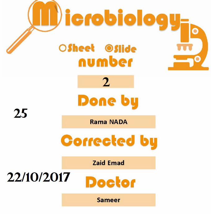

Balantidium coli
It causes balantidiasis or balantidial dysentery, is the largestintestinal protozoan of humans.The trophozoite is a ciliated, oval organism 60 X 45 μm or larger. It has a steady progression and rotation around the long axis motion.Most infections are apparently harmless. However, rarely, the trophozoites invade the large bowel and terminal ileum, causing abscesses & ulcerations.Treatment: Oxytetracycline may be followed by iodoquinol ormetronidazole if necessary.
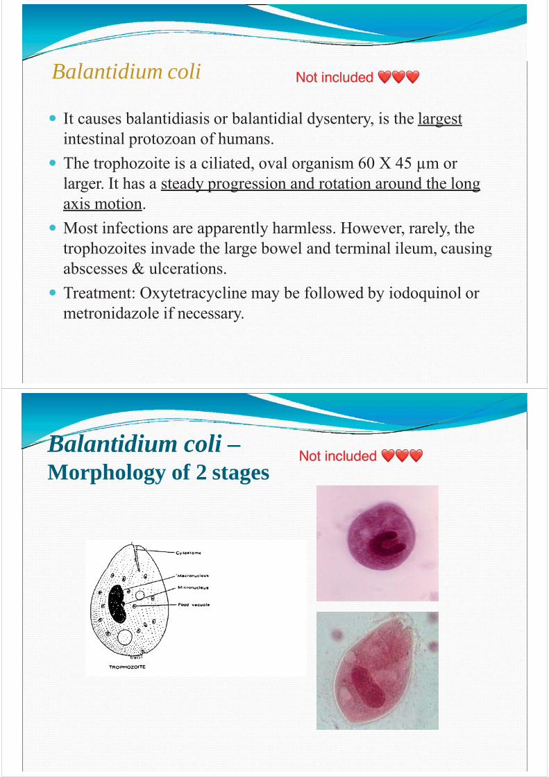
Balantidium coli –Morphology of 2 stages
The Hemoflagellates
The hemoflagellates of humans include the generaTrypanosoma and Leishmania, causing Trypanosomiasis&
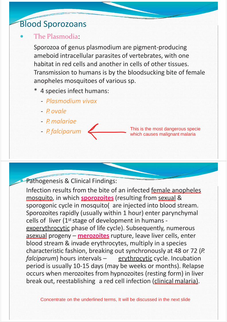
Human trypanosomes:- African: T. brucei rhodesiense & T. bruceigambiense,transmitted by tsetse flies (glossina), causing sleeping sickness.- American: T. (schizotrypanum) cruzi, transmitted by conenose bugs (triatoma), causing Chagas’ disease.
In blood
aren't found in JordanLeishmaniasis respectively.are found in Jordan
You will be asked about underlined syllable of thesetwo names
You must know how the parasite is transmitted and
what is the resulting disease
braziliensis.
Leishmania:
Divided into a number of species infecting humansby sandflies (Phlebotomus) causing:
Affect skin- Cutaneous leishmaniasis (Oriental sore or Baghdad
boil) – L. tropica, L. major, L. mexicana, L. We will be asked aboutunderlined name form these names
- Mucocutaneous or naso-oral leishmaniasis (espundia)caused by L. braziliensis braziliensis.
- Visceral leishmaniasis (kala-azar) – L. donovani.This is another name forvisceral leishmaniasis
Memorize these three categories with underlined examples
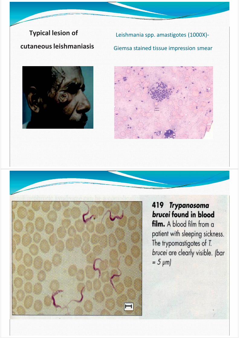
Leishmania spp. amastigotes (1000X)-Typical lesion of
cutaneous leishmaniasis Giemsa stained tissue impression smear
Blood Sporozoans
- P. falciparum
The Plasmodia:Sporozoa of genus plasmodium are pigment-producing ameboid intracellular parasites of vertebrates, with one habitat in red cells and another in cells of other tissues. Transmission to humans is by the bloodsucking bite of female anopheles mosquitoes of various sp.* 4 species infect humans:
- Plasmodium vivax- P. ovale- P. malariae
This is the most dangerous speciewhich causes malignant malaria
Pathogenesis & Clinical Findings:Infection results from the bite of an infected female anopheles mosquito, in which sporozoites (resulting from sexual & sporogonic cycle in mosquito( are injected into blood stream.Sporozoites rapidly (usually within 1 hour) enter parynchymal cells of liver (1st stage of development in humans -exoerythrocytic phase of life cycle). Subsequently, numerous asexual progeny – merozoites rupture, leave liver cells, enter blood stream & invade erythrocytes, multiply in a speciescharacteristic fashion, breaking out synchronously at 48 or 72 (P. falciparum) hours intervals – erythrocytic cycle. Incubation period is usually 10-15 days (may be weeks or months). Relapse occurs when merozoites from hypnozoites (resting form) in liver break out, reestablishing a red cell infection (clinical malaria).
Concentrate on the underlined terms, It will be discussed in the next slide
Additional slide
Malaria parasites infect two types of hosts: Human and female Anopheles mosquitoIt passes in two cycles:1- Asexual cycle in human2- Sexual cycle in female Anopheles mosquito
When “gametocytes” are packed up by a female Anopheles mosquito during a blood meal they start sexual cycle in the mosquito: female and male gametocytes fertilizes and become zygote which developed to cysts, these cysts rupture and release sporozoiteswhich is the infective stage (remember until now these processes take place in the gut of female Anopheles mosquito “sexual cycle”). These sporozoites located in themosquito saliva. So when the female Anopheles mosquito takes a blood meal from
another human , sporozoites are injected with the mosquito saliva to the blood and start the asexual cycle in the human. First of all sporozoites have high affinity to liverso they rapidly enter parenchymal cells of liver.
Continue…..
Additional slide
Sporozoites enter the liver cells and start dividing to produce merozoites which ruptureliver cells and released to blood stream then they are directed toward RBCs. In RBCs
Merozoites are converted to another phases of this parasite, they’ll rupture RBCs releasing these phases to blood stream . The most important phase is gametocyte which picked upby female Anopheles mosquito and repeat the cycle again.
Note: Sporozoites may not follow the reproduction step and stay as hypnozoites in liver Cells (resting sporozoites), they may be activated after a long time (weeks, months or evenyears ) and enter blood stream as merozoites, this process is called “clinical malaria”,
Diagnostic Laboratory Tests:
Treatment:- Chloroquine: drug of choice for susceptible forms during
the acute attack.- Primaquine: eliminates exoerythrocytic forms in the liver
(potentially relapsing malaria), permitting radical cure.
Large amount of blood- Thick blood film: stained with Giemsa’s stain, it
concentrates the parasites & permits detection even of mild infections.
- Thin blood film: stained with Giemsa’s stain is necessary for species differentiation. These species( vivax,..etc) can be recognized
by their shape, such as Gamet- Antigen – capture tests using chromatographic methods to
detect a trophozoite-derived protein (rapid diagnostic test).
Memories underlined words
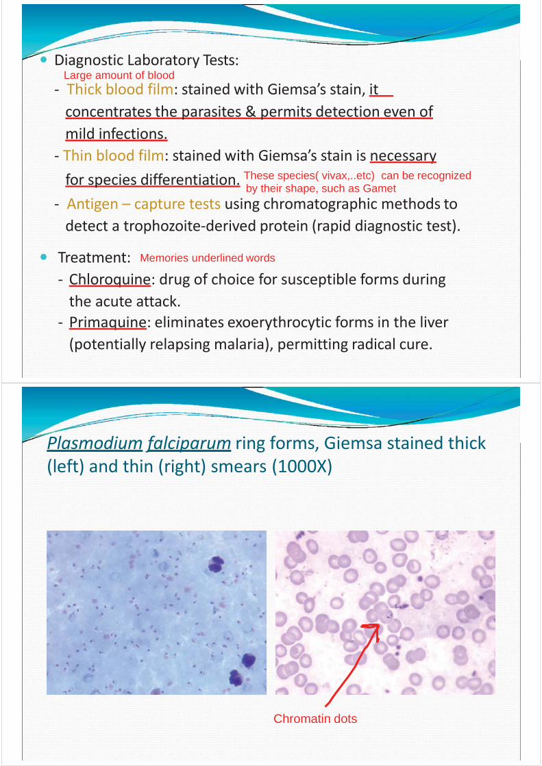
Plasmodium falciparum ring forms, Giemsa stained thick (left) and thin (right) smears (1000X)
Chromatin dots
Cryptosporidium
Cryptosporidium species, typically C. parvum, can infect the intestine in immunocompromised persons. C. parvum is a protozoal infection which causes an acute diarrhea in immunocompromised patients. It has probably been an unrecognized cause of self-limited, mild gastroenteritis and diarrhea in humans.The parasites are minute (2-5 μm) intracellular spheres found in great numbers just under the outer membrane of the cells lining the stomach or intestine.It inhibits the brush border of mucosal epithelial cell of GIT, esp. surface of villi of lower small bowel. The prominent clinical feature is diarrhea (mild & self-limited, 1-2 weeks)
Are found in Jordan
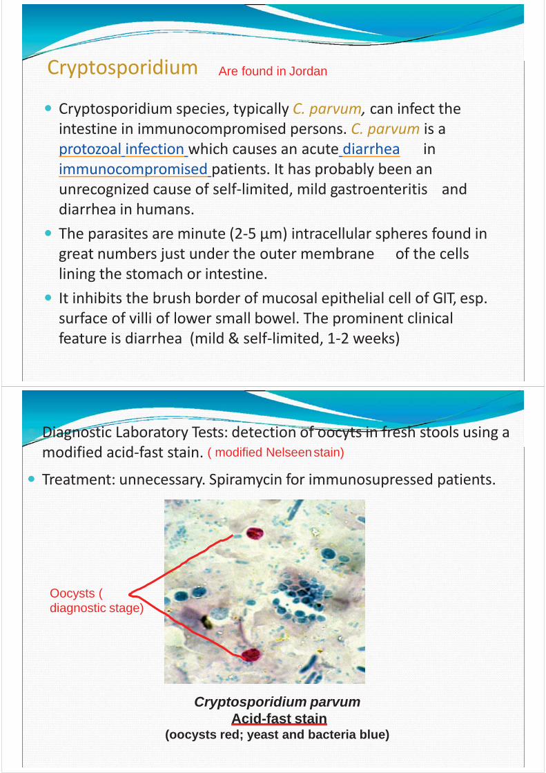
Cryptosporidium parvumAcid-fast stain
(oocysts red; yeast and bacteria blue)
Diagnostic Laboratory Tests: detection of oocyts in fresh stools using a modified acid-fast stain. ( modified Nelseenstain)
Treatment: unnecessary. Spiramycin for immunosupressed patients.
Oocysts ( diagnostic stage)
Toxoplasma gondii
a cat, or-if ingested by humans-can establish an infection inwhich it can reproduces asexually, where it opens and releases sporozoites in the duodenum then invade various cells esp. macrophages where they form trophozoites which spreads
In human
It is a coccidian protozoan of worldwide distribution that infects a wide range of animals and birds but does not appear to cause disease in them.The normal final hosts are strictly the cat and its relatives, the only host of which the oocyst-producing sexual stage of toxoplasma can develop.
In catsWhen oocysts are ingested, can either repeat its sexual cycle in
infection to lymph nodes and other organs.( and in reticularendothelial system)
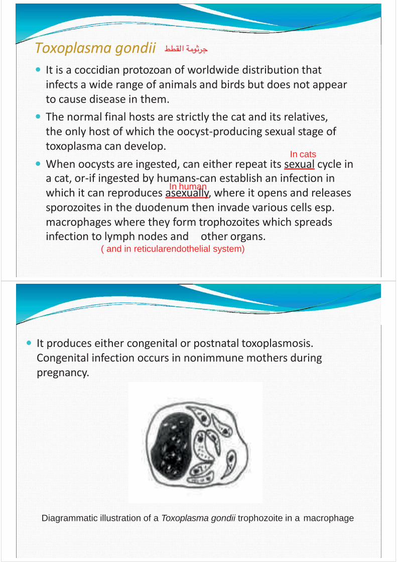
It produces either congenital or postnatal toxoplasmosis. Congenital infection occurs in nonimmune mothers during pregnancy.
Diagrammatic illustration of a Toxoplasma gondii trophozoite in a macrophage
HelminthsMulticellular (metazoa)The Nematodes: (Round worms)-They are small round elongated worms, non-segmented, with a body cavity, have separate sexes, usually don’t need anintermediate host, and don’t multiply in the body because eggs don’t hatch unless they leave the body first.-They can parasitize either intermediate host )worms in the juvenile, larval, or developmental stages) or final / definitive host (worms occur in the adult or sexually reproductive stage).-Infection patterns vary widely. Human intestinal nematodesinfect via food-borne, water-borne, and soil-borne routes.
Female and male Gamet produce fertilized egg which is the diagnostic stage and some time the infective stage, this egg develops to larva which is the infective
stage
Ascaris lumbricoides:
• Infect by eggs that are strongly resistant to desiccation &environmental conditions.
You don't need to memorize the number (just to know that it's large)• Eggs are 75x40 μm, with a thick mamillated brownish shell.• Freshly passed eggs in feces are not infective, they require 2-3
weeks to develop to be embryonated (contain larva).
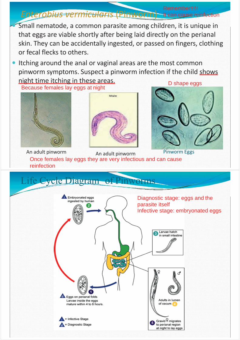
Diagnostic stage: fertilized/ unfertilized eggs and the parasite itself Infective stage: embryonated egg (contains larva)
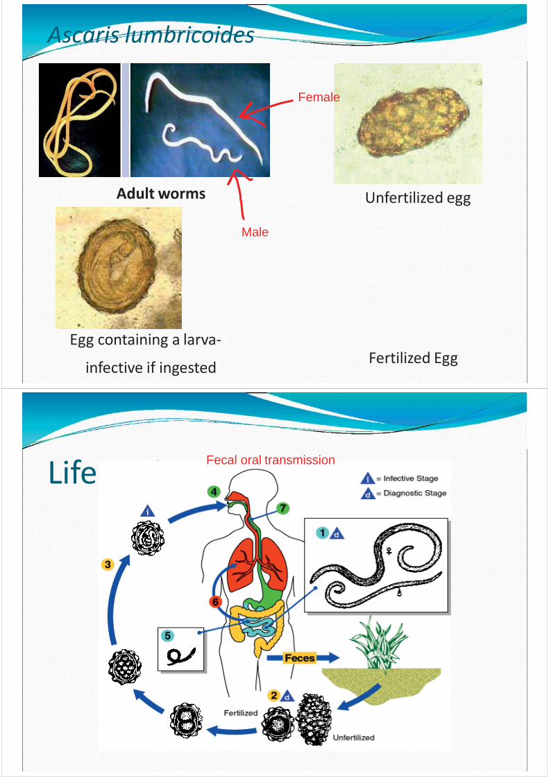
Ascaris lumbricoides
Unfertilized egg
Fertilized EggEgg containing a larva-
infective if ingested
Adult worms
Female
Male
Life Cycle Fecal oral transmission
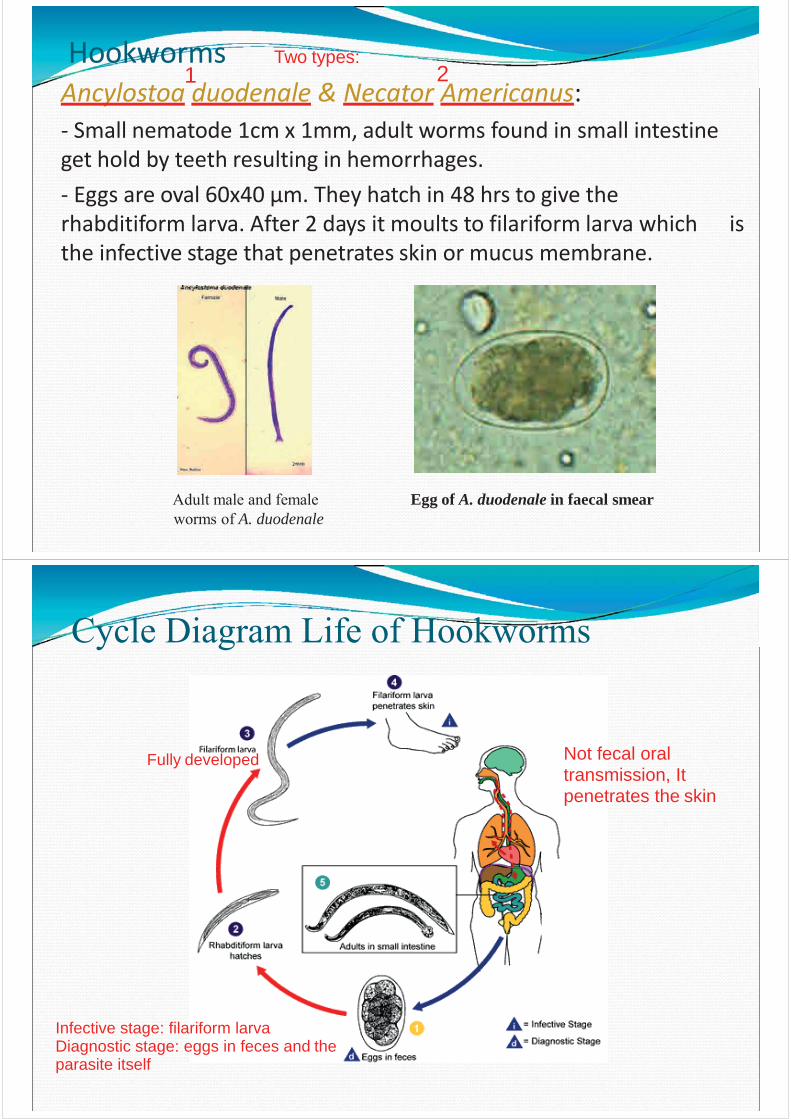
Enterobius vermicularis (Pinworm)Small nematode, a common parasite among children, it is unique in that eggs are viable shortly after being laid directly on the perianal skin. They can be accidentally ingested, or passed on fingers, clothing or fecal flecks to others.Itching around the anal or vaginal areas are the most common pinworm symptoms. Suspect a pinworm infection if the child shows night time itching in these areas.
An adult pinworm Pinworm EggsAn adult pinworm
Because females lay eggs at nightD shape eggs
Once females lay eggs they are very infectious and can causereinfection
Remember!!!!!It can cause reinfection
Life Cycle Diagram of PinwormsDiagnostic stage: eggs and the parasite itselfInfective stage: embryonated eggs
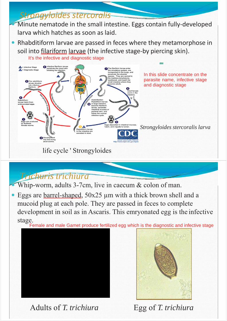
HookwormsAncylostoa duodenale & Necator Americanus:- Small nematode 1cm x 1mm, adult worms found in small intestine get hold by teeth resulting in hemorrhages.- Eggs are oval 60x40 μm. They hatch in 48 hrs to give the rhabditiform larva. After 2 days it moults to filariform larva which is the infective stage that penetrates skin or mucus membrane.
Adult male and female worms of A. duodenale
Egg of A. duodenale in faecal smear
Two types:1 2
Cycle Diagram Life of Hookworms
Not fecal oral transmission, It penetrates the skin
Fully developed
Infective stage: filariform larva Diagnostic stage: eggs in feces and the parasite itself
Strongyloides stercoralis
Strongyloides stercoralis larva
life cycle ' Strongyloides
Minute nematode in the small intestine. Eggs contain fully-developed larva which hatches as soon as laid.Rhabditiform larvae are passed in feces where they metamorphose in soil into filariform larvae (the infective stage-by piercing skin).
It's the infective and diagnostic stage
In this slide concentrate on theparasite name, infective stageand diagnostic stage
Trichuris trichiura
Adults of T. trichiura Egg of T. trichiura
Whip-worm, adults 3-7cm, live in caecum & colon of man.Eggs are barrel-shaped, 50x25 μm with a thick brown shell and a mucoid plug at each pole. They are passed in feces to complete development in soil as in Ascaris. This emryonated egg is the infective stage.
Female and male Gamet produce fertilized egg which is the diagnostic and infective stage
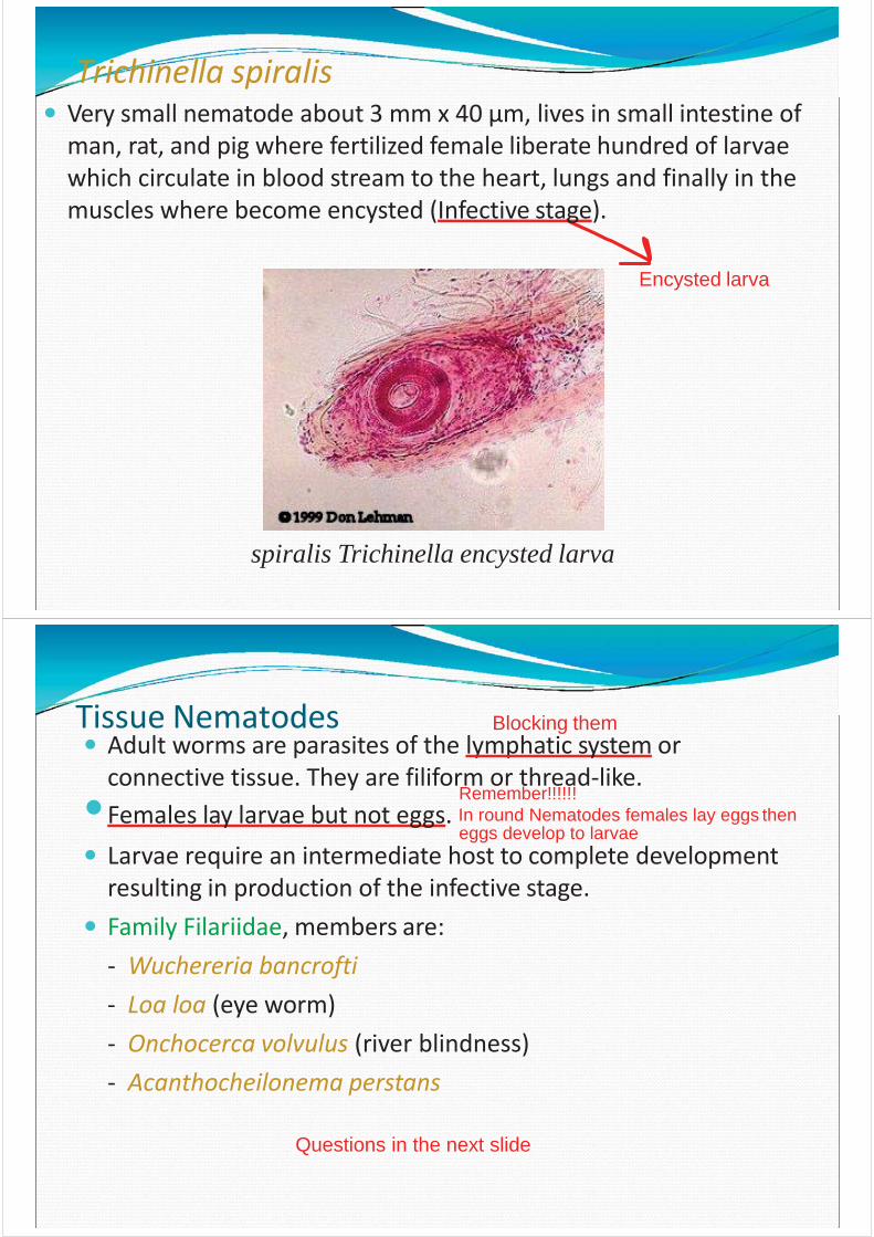
Trichinella spiralis
spiralis Trichinella encysted larva
Very small nematode about 3 mm x 40 μm, lives in small intestine of man, rat, and pig where fertilized female liberate hundred of larvae which circulate in blood stream to the heart, lungs and finally in the muscles where become encysted (Infective stage).
Encysted larva
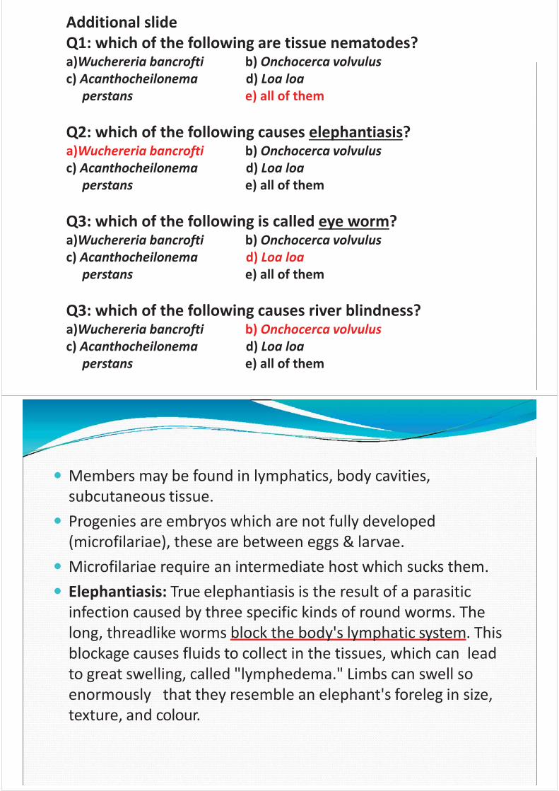
Tissue NematodesAdult worms are parasites of the lymphatic system or connective tissue. They are filiform or thread-like.
Remember!!!!!!Females lay larvae but not eggs. In round Nematodes females lay eggs then
eggs develop to larvaeLarvae require an intermediate host to complete developmentresulting in production of the infective stage.Family Filariidae, members are:- Wuchereria bancrofti- Loa loa (eye worm)- Onchocerca volvulus (river blindness)- Acanthocheilonema perstans
Questions in the next slide
Blocking them
Additional slideQ1: which of the following are tissue nematodes?a)Wuchereria bancrofti b) Onchocerca volvulus c) Acanthocheilonema d) Loa loa
perstans e) all of them
Q2: which of the following causes elephantiasis?a)Wuchereria bancrofti b) Onchocerca volvulus c) Acanthocheilonema d) Loa loa
perstans e) all of them
Q3: which of the following is called eye worm?a)Wuchereria bancrofti b) Onchocerca volvulus c) Acanthocheilonema d) Loa loa
perstans e) all of them
Q3: which of the following causes river blindness?a)Wuchereria bancrofti b) Onchocerca volvulus c) Acanthocheilonema d) Loa loa
perstans e) all of them
Members may be found in lymphatics, body cavities, subcutaneous tissue.Progenies are embryos which are not fully developed (microfilariae), these are between eggs & larvae.Microfilariae require an intermediate host which sucks them.Elephantiasis: True elephantiasis is the result of a parasitic infection caused by three specific kinds of round worms. The long, threadlike worms block the body's lymphatic system. This blockage causes fluids to collect in the tissues, which can lead to great swelling, called "lymphedema." Limbs can swell so enormously that they resemble an elephant's foreleg in size, texture, and colour.
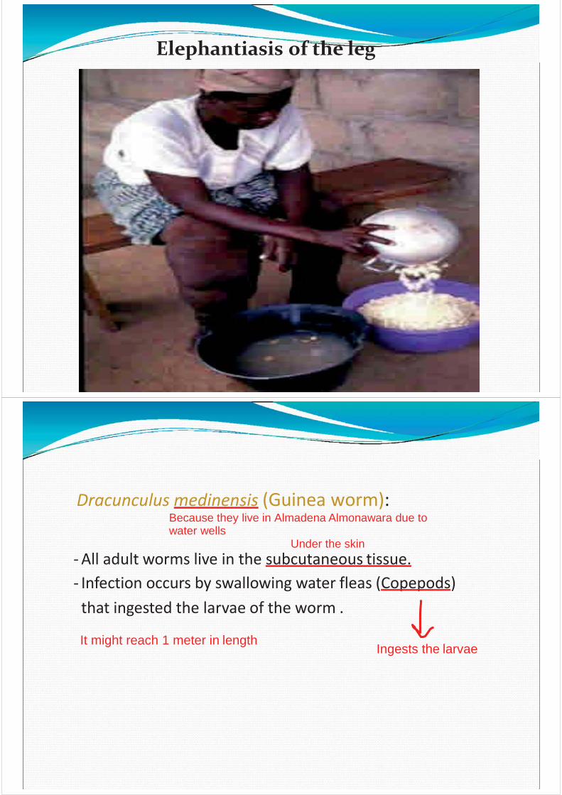
Elephantiasis of the leg
Under the skin- All adult worms live in the subcutaneous tissue.- Infection occurs by swallowing water fleas (Copepods) that ingested the larvae of the worm .
Ingests the larvaeIt might reach 1 meter in length
Dracunculus medinensis (Guinea worm):Because they live in Almadena Almonawara due to water wells
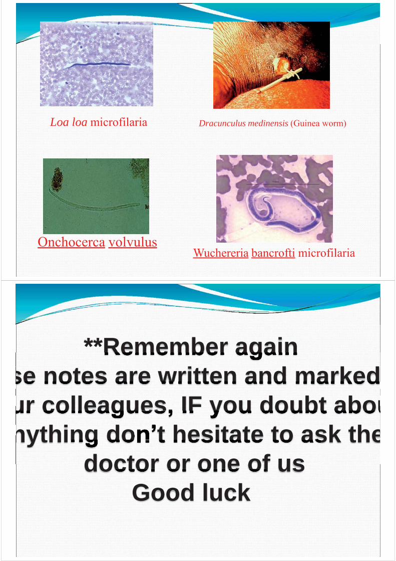
Onchocerca volvulusWuchereria bancrofti microfilaria
Dracunculus medinensis (Guinea worm)Loa loa microfilaria
**Remember againse notes are written and markedur colleagues, IF you doubt abounything don’t hesitate to ask the
doctor or one of usGood luck