DIAGNOSIS OF HEREDITARY SPHEROCYTOSIS©rocytose... · Osmotic fragility test not recommended or in...
33
DIAGNOSIS OF HEREDITARY SPHEROCYTOSIS Elena Lazarova, MD Clinical Biology 24/09/2015
Transcript of DIAGNOSIS OF HEREDITARY SPHEROCYTOSIS©rocytose... · Osmotic fragility test not recommended or in...
DIAGNOSIS OF HEREDITARY SPHEROCYTOSIS
Elena Lazarova, MDClinical Biology
24/09/2015
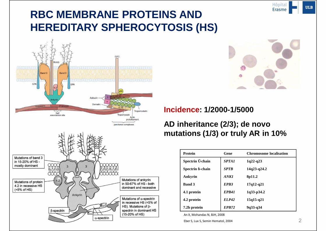
RBC MEMBRANE PROTEINS ANDHEREDITARY SPHEROCYTOSIS (HS)
Protein Gene Chromosome localisation
Spectrin α-chain SPTA1 1q22-q23
Spectrin β-chain SPTB 14q23-q24.2
Ankyrin ANK1 8p11.2
Band 3 EPB3 17q12-q21
4.1 protein EPB41 1q33-p34.2
4.2 protein ELP42 15q15-q21
7.2b protein EPB72 9q33-q34
Incidence: 1/2000-1/5000
AD inheritance (2/3); de novo mutations (1/3) or truly AR in 10%
An X, Mohandas N, BJH, 2008
Eber S, Lux S, Semin Hematol, 2004 2
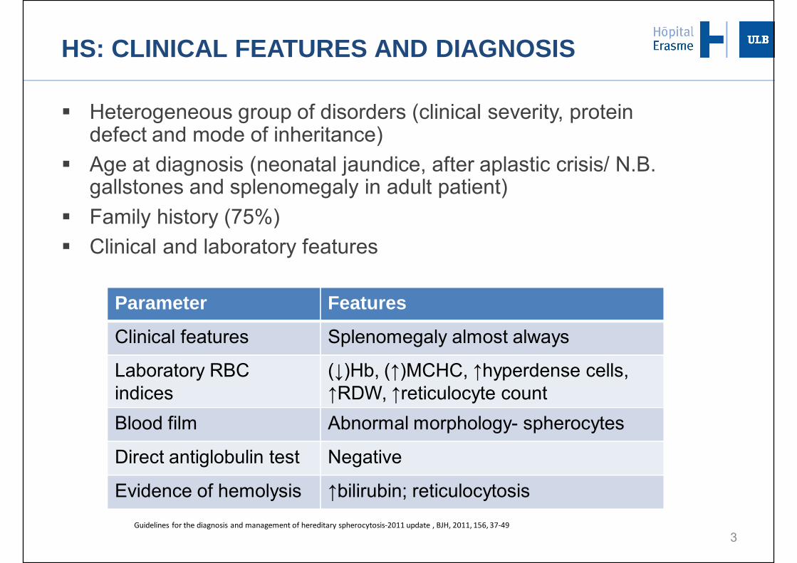
HS: CLINICAL FEATURES AND DIAGNOSIS
§ Heterogeneous group of disorders (clinical severity, protein defect and mode of inheritance)
§ Age at diagnosis (neonatal jaundice, after aplastic crisis/ N.B. gallstones and splenomegaly in adult patient)
§ Family history (75%)§ Clinical and laboratory features
3
Parameter Features
Clinical features Splenomegaly almost always
Laboratory RBC indices
(↓)Hb, (↑)MCHC, ↑hyperdense cells, ↑RDW, ↑reticulocyte count
Blood film Abnormal morphology- spherocytes
Direct antiglobulin test Negative
Evidence of hemolysis ↑bilirubin; reticulocytosisGuidelines for the diagnosis and management of hereditary spherocytosis-2011 update , BJH, 2011, 156, 37-49
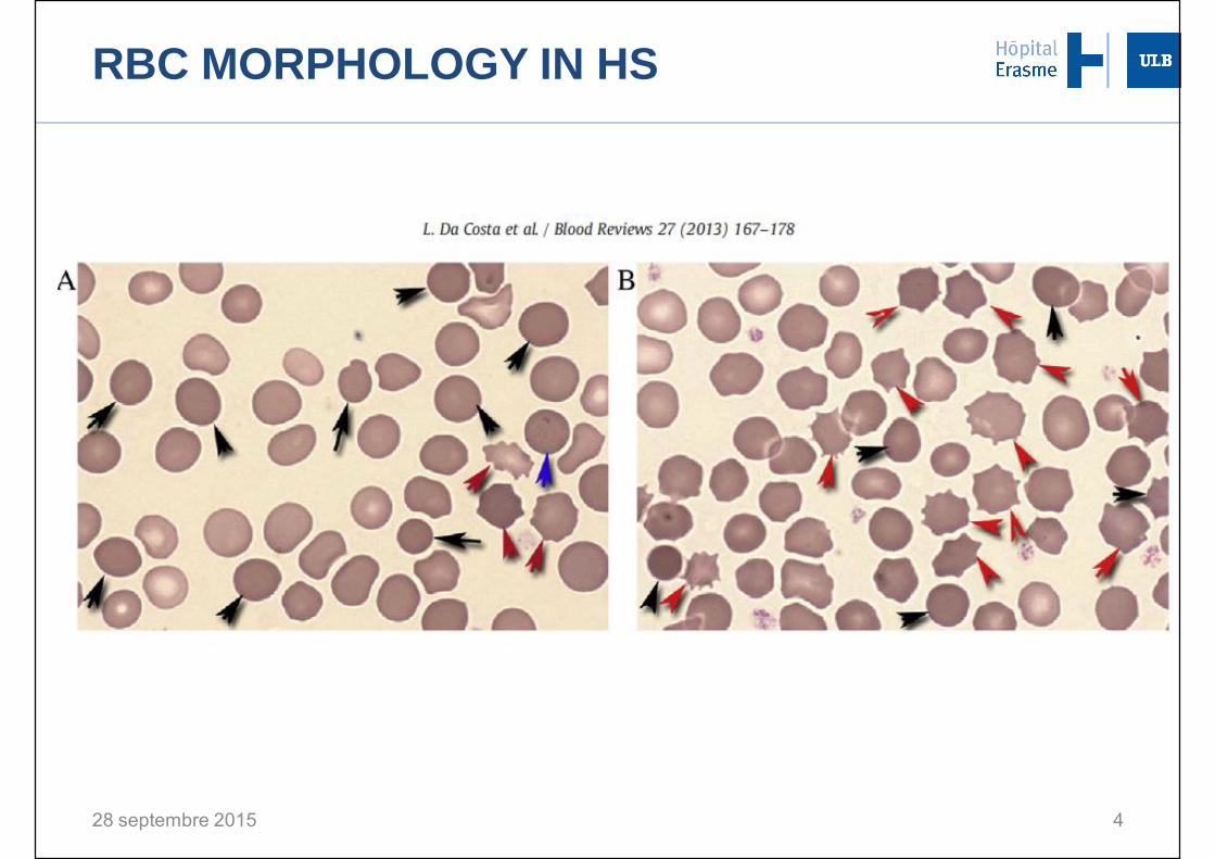
RBC MORPHOLOGY IN HS
28 septembre 2015 4
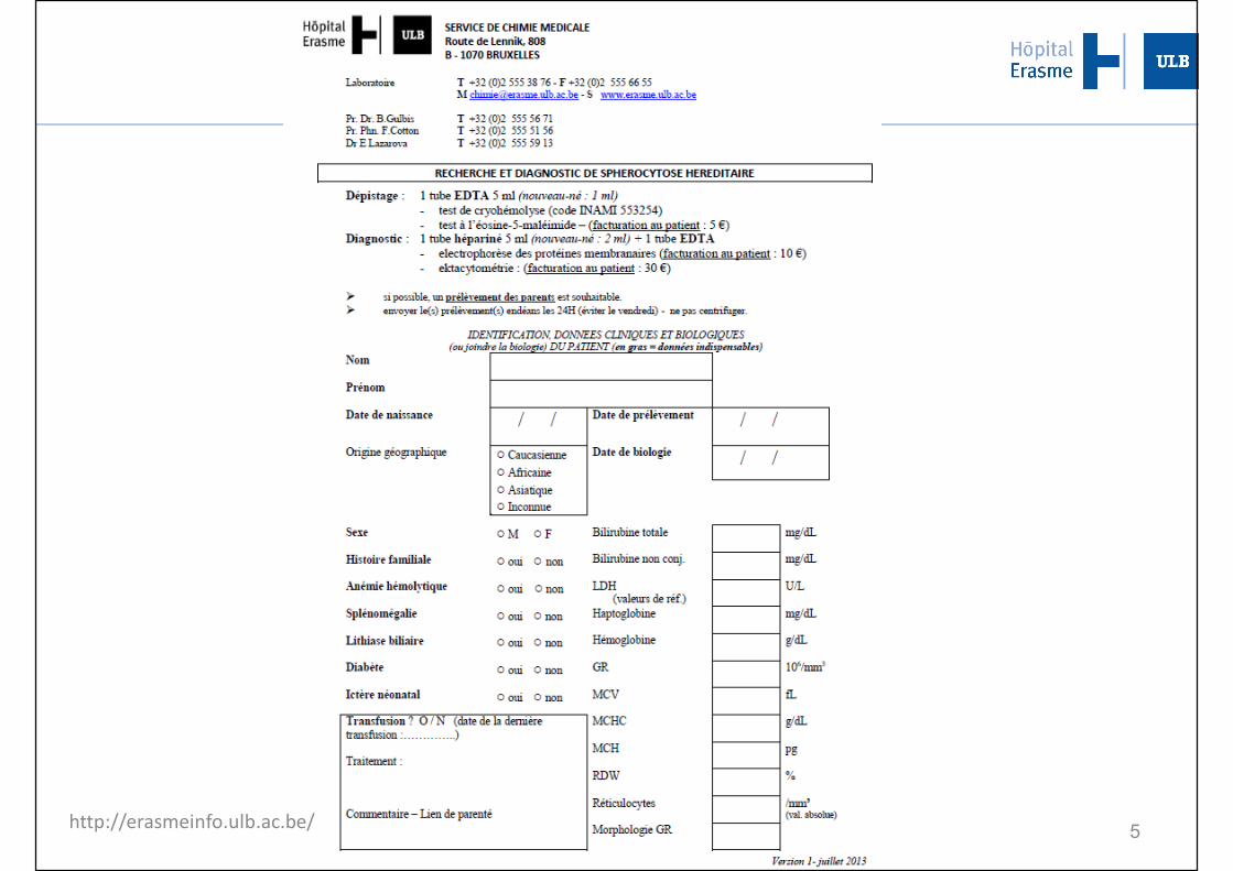
5http://erasmeinfo.ulb.ac.be/
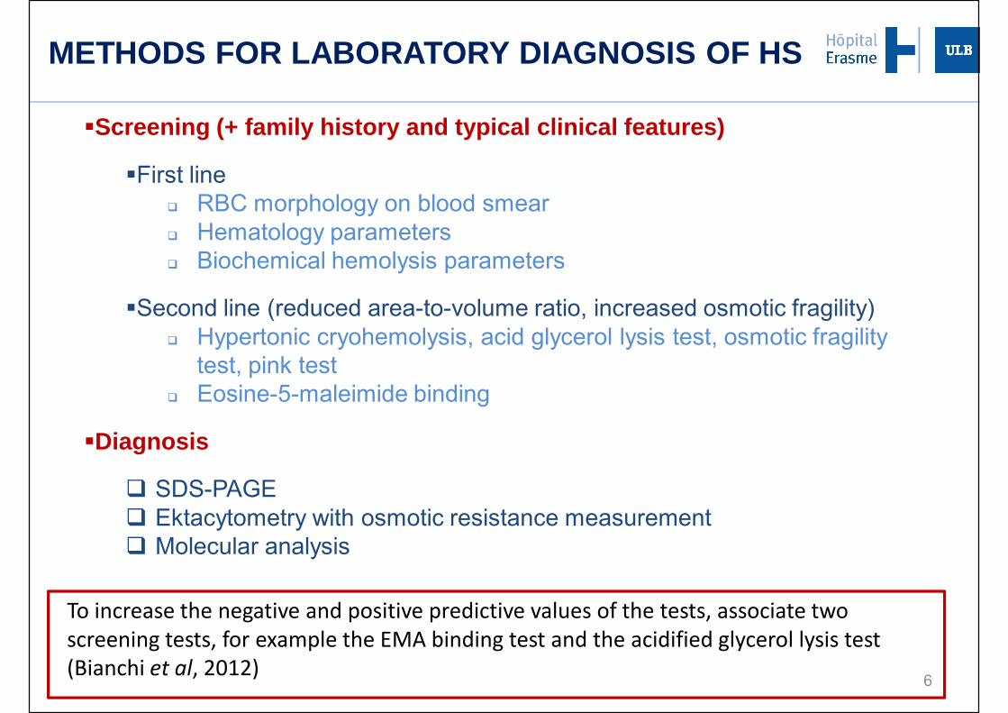
METHODS FOR LABORATORY DIAGNOSIS OF HS
§Screening (+ family history and typical clinical features)
§First lineq RBC morphology on blood smearq Hematology parametersq Biochemical hemolysis parameters
§Second line (reduced area-to-volume ratio, increased osmotic fragility)q Hypertonic cryohemolysis, acid glycerol lysis test, osmotic fragility
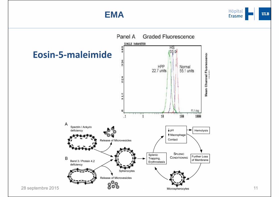
test, pink test q Eosine-5-maleimide binding
§Diagnosis
q SDS-PAGEq Ektacytometry with osmotic resistance measurementq Molecular analysis
6
To increase the negative and positive predictive values of the tests, associate two screening tests, for example the EMA binding test and the acidified glycerol lysis test (Bianchi et al, 2012)
§ European survey on red cell membrane disorders and enzyme defects
ENERCA 2013
7
METHODS FOR LABORATORY DIAGNOSIS OF HS
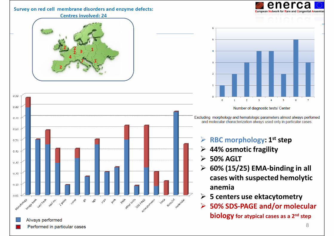
Ø RBC morphology: 1st stepØ 44% osmotic fragilityØ 50% AGLTØ 60% (15/25) EMA-binding in all
cases with suspected hemolyticanemia
Ø 5 centers use ektacytometryØ 50% SDS-PAGE and/or molecular
biology for atypical cases as a 2nd step8
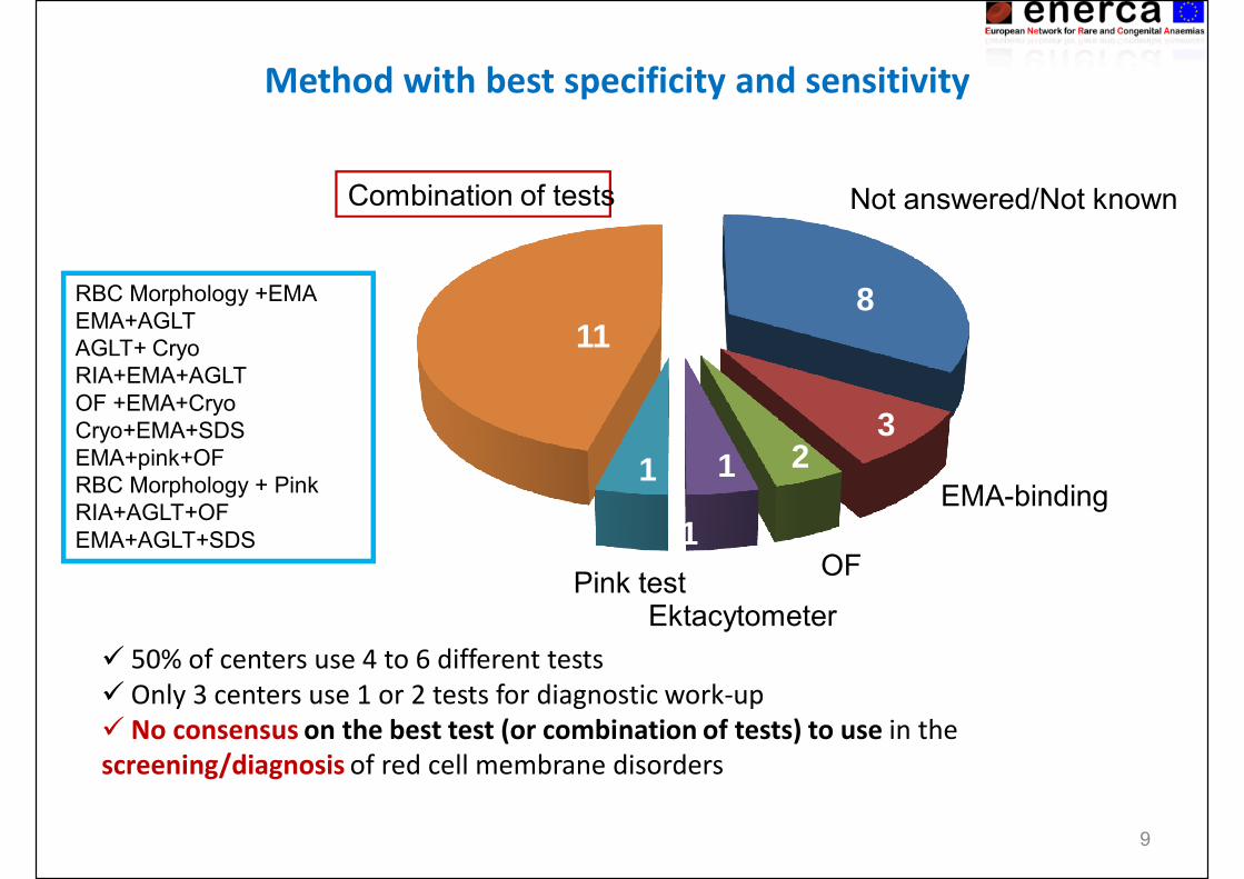
Method with best specificity and sensitivity
Not answered/Not known
EMA-binding
OF
EktacytometerPink test
Combination of tests
RBC Morphology +EMA EMA+AGLTAGLT+ CryoRIA+EMA+AGLTOF +EMA+CryoCryo+EMA+SDSEMA+pink+OFRBC Morphology + PinkRIA+AGLT+OFEMA+AGLT+SDS
8
21
1
1
11
3
ü 50% of centers use 4 to 6 different testsü Only 3 centers use 1 or 2 tests for diagnostic work-upü No consensus on the best test (or combination of tests) to use in the screening/diagnosis of red cell membrane disorders
9
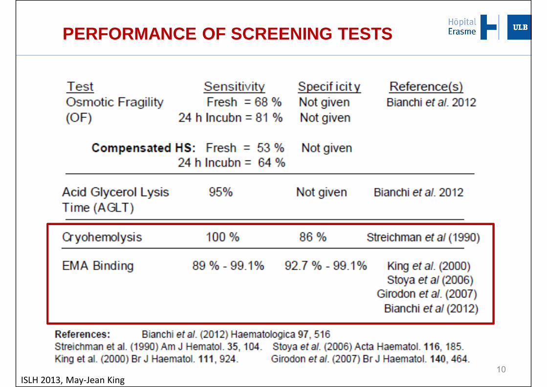
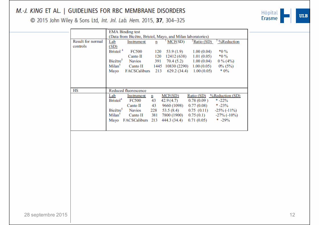
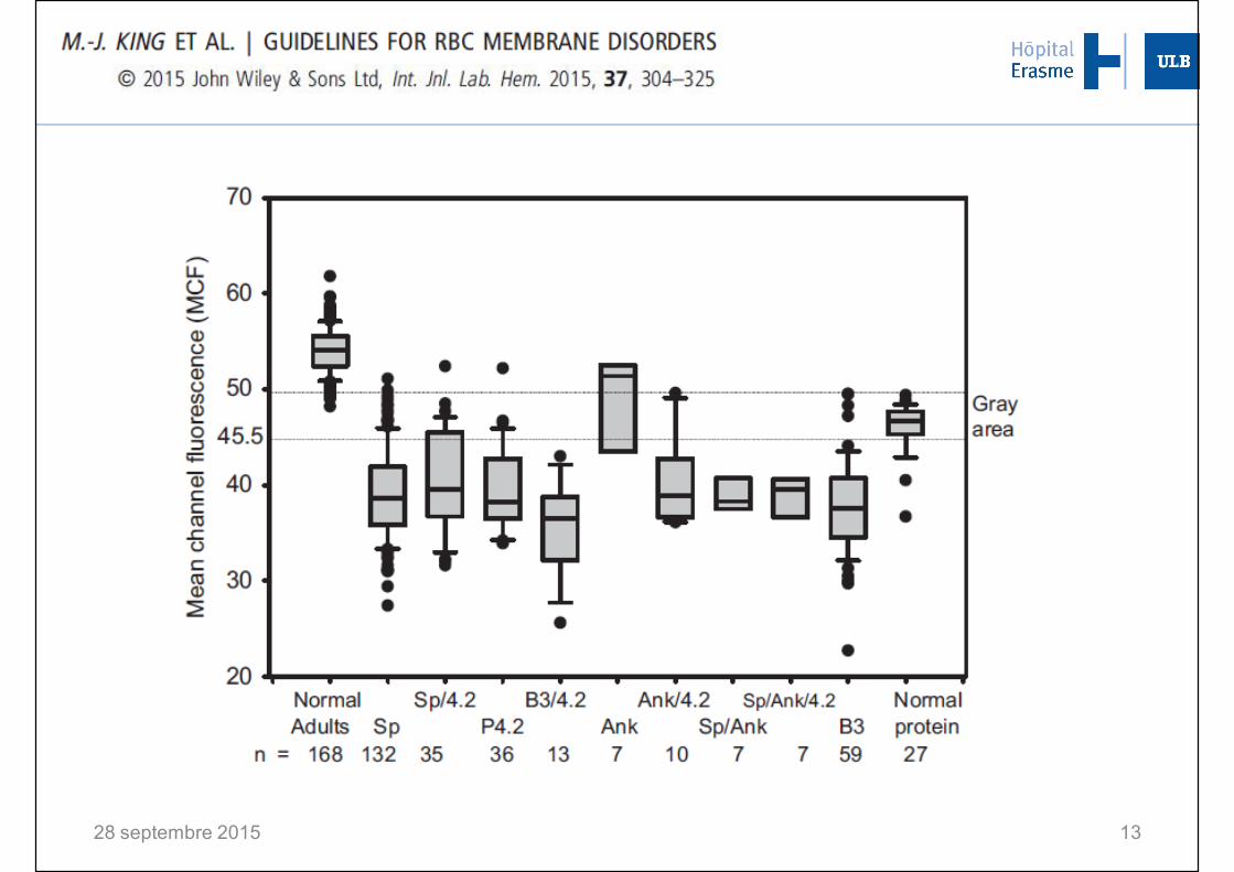
PERFORMANCE OF SCREENING TESTS
ISLH 2013, May-Jean King10
EMA
28 septembre 2015 11
Eosin-5-maleimide
28 septembre 2015 12
28 septembre 2015 13

§ Screening (+ family history and typical clinical features)§ First line
¨ RBC morphology on blood smear
¨ Hematology parameters
¨ Biochemical hemolysis parameters
§ Second line (reduced area-to-volume ratio, increased osmotic fragility)¨ Hypertonic cryohemolysis, acid glycerol lysis test,
osmotic fragility test, pink test ¨ Eosine-5-maleimide binding
14
METHODS FOR LABORATORY DIAGNOSIS OF HS
28 septembre 2015
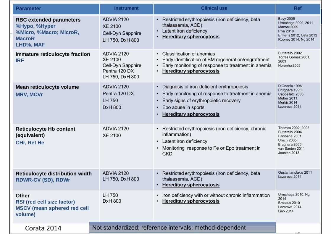
Parameter Instrument Clinical use Ref
RBC extended parameters%Hypo, %Hyper%Micro, %Macro; MicroR, MacroRLHD%, MAF
ADVIA 2120XE 2100 Cell-Dyn Sapphire LH 750, DxH 800
• Restricted erythropoiesis (iron deficiency, beta thalassemia, ACD)
• Latent iron deficiency• Hereditary spherocytosis
Bovy 2005Urrechaga 2009, 2011Maconi 2009Piva 2010Ermens 2012, Osta 2012Rooney 2014, Ng 2014
Immature reticulocyte fractionIRF
ADVIA 2120XE 2100 Cell-Dyn Sapphire Pentra 120 DXLH 750, DxH 800
• Classification of anemias• Early identification of BM regeneration/engraftment• Early monitoring of response to treatment in anemia• Hereditary spherocytosis
Buttarello 2002Torres Gomez 2001, 2003Noronha 2003
Mean reticulocyte volumeMRV, MCVr
ADVIA 2120Pentra 120 DXLH 750DxH 800
• Diagnosis of iron-deficient erythropoiesis• Early monitoring of response to treatment in anemia• Early signs of erythropoietic recovery• Epo abuse in sports• Hereditary spherocytosis
D’Onorfio 1995Brugnara 1998Cappelletti 2006Mullier 2011Morkis 2014Lazarova 2014
Reticulocyte Hb content (equivalent)CHr, Ret He
ADVIA 2120XE 2100
• Restricted erythropoiesis (iron deficiency, chronic inflammation)
• Latent iron deficiency• Monitoring response to Fe or Epo treatment in
CKD
Thomas 2002, 2005Buttarello 2004Fishbane 2001Ullrich 2005Brugnara 2006van Santen 2011Joosten 2013
Reticulocyte distribution widthRDWR-CV (SD), RDWr
ADVIA 2120LH 750, DxH 800
• Restricted erythropoiesis (iron deficiency, beta thalassemia, ACD)
• Hereditary spherocytosis
Oustamanolakis 2011Lazarova 2014
OtherRSf (red cell size factor)MSCV (mean sphered red cellvolume)
LH 750DxH 800
• Iron deficiency with or without chronic inflammation• Hereditary spherocytosis
Urrechaga 2010, Ng2014Broseus 2010Lazarova 2014Liao 2014
Not standardized; reference intervals: method-dependent15
Corata 2014
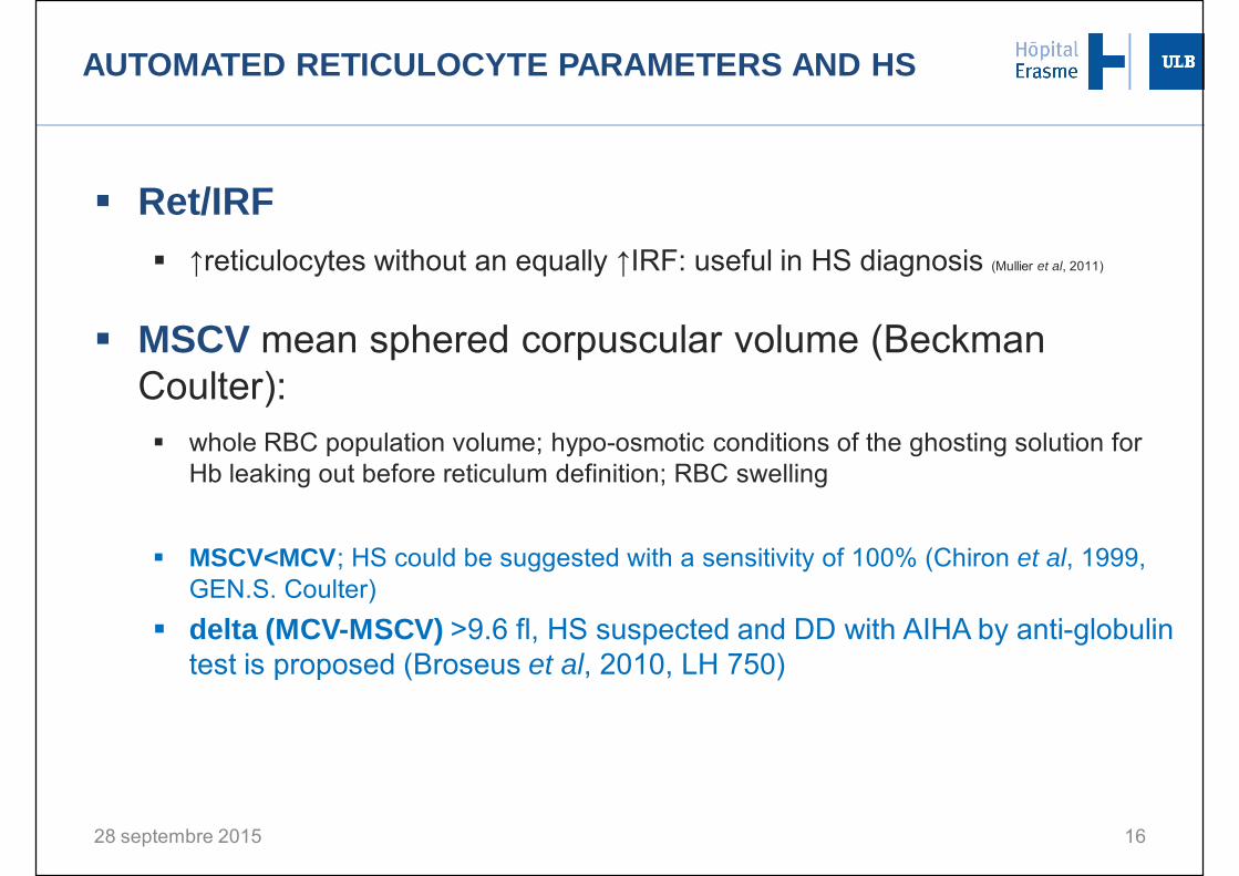
AUTOMATED RETICULOCYTE PARAMETERS AND HS
§ Ret/IRF§ ↑reticulocytes without an equally ↑IRF: useful in HS diagnosis (Mullier et al, 2011)
§ MSCV mean sphered corpuscular volume (Beckman Coulter): § whole RBC population volume; hypo-osmotic conditions of the ghosting solution for
Hb leaking out before reticulum definition; RBC swelling
§ MSCV<MCV; HS could be suggested with a sensitivity of 100% (Chiron et al, 1999, GEN.S. Coulter)
§ delta (MCV-MSCV) >9.6 fl, HS suspected and DD with AIHA by anti-globulin test is proposed (Broseus et al, 2010, LH 750)
28 septembre 2015 16
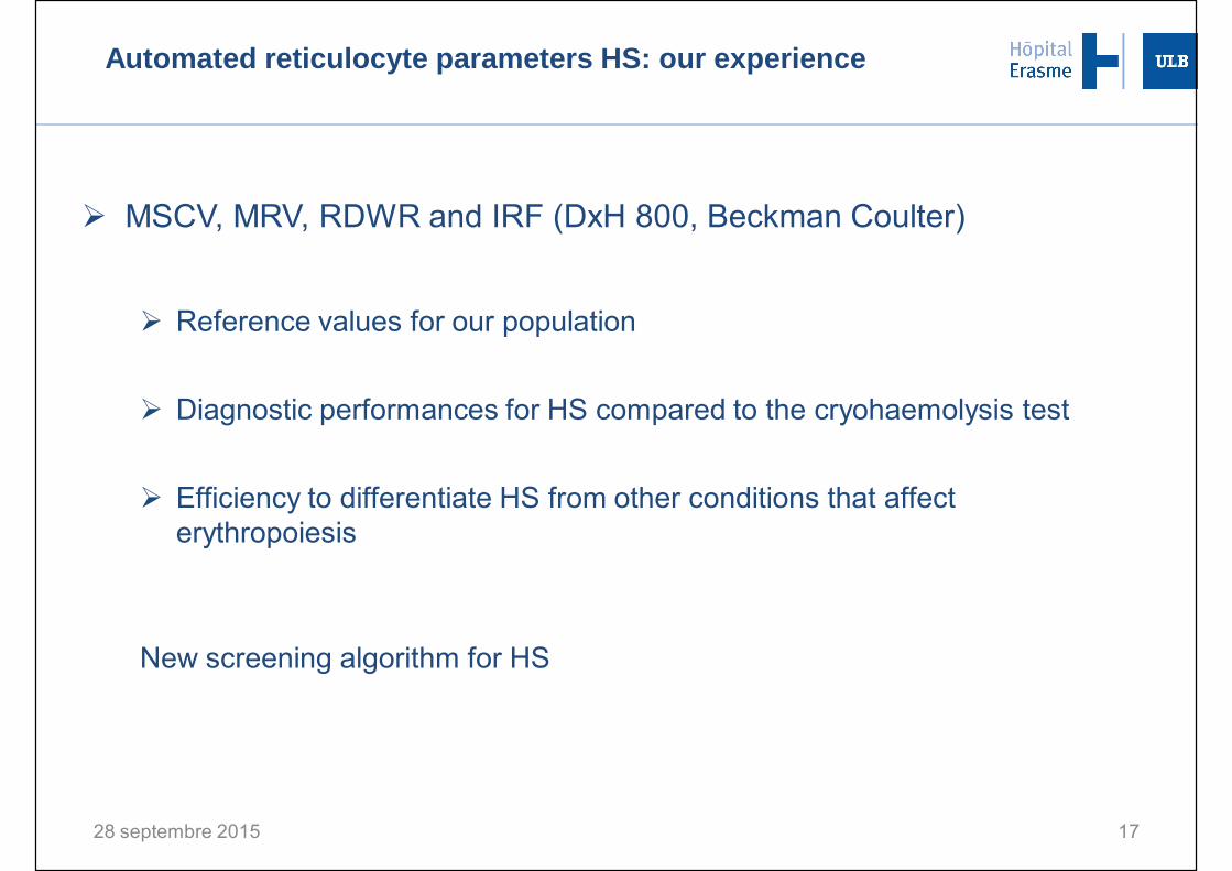
Ø MSCV, MRV, RDWR and IRF (DxH 800, Beckman Coulter)
Ø Reference values for our population
Ø Diagnostic performances for HS compared to the cryohaemolysis test
Ø Efficiency to differentiate HS from other conditions that affect erythropoiesis
New screening algorithm for HS
28 septembre 2015 17
Automated reticulocyte parameters HS: our experience
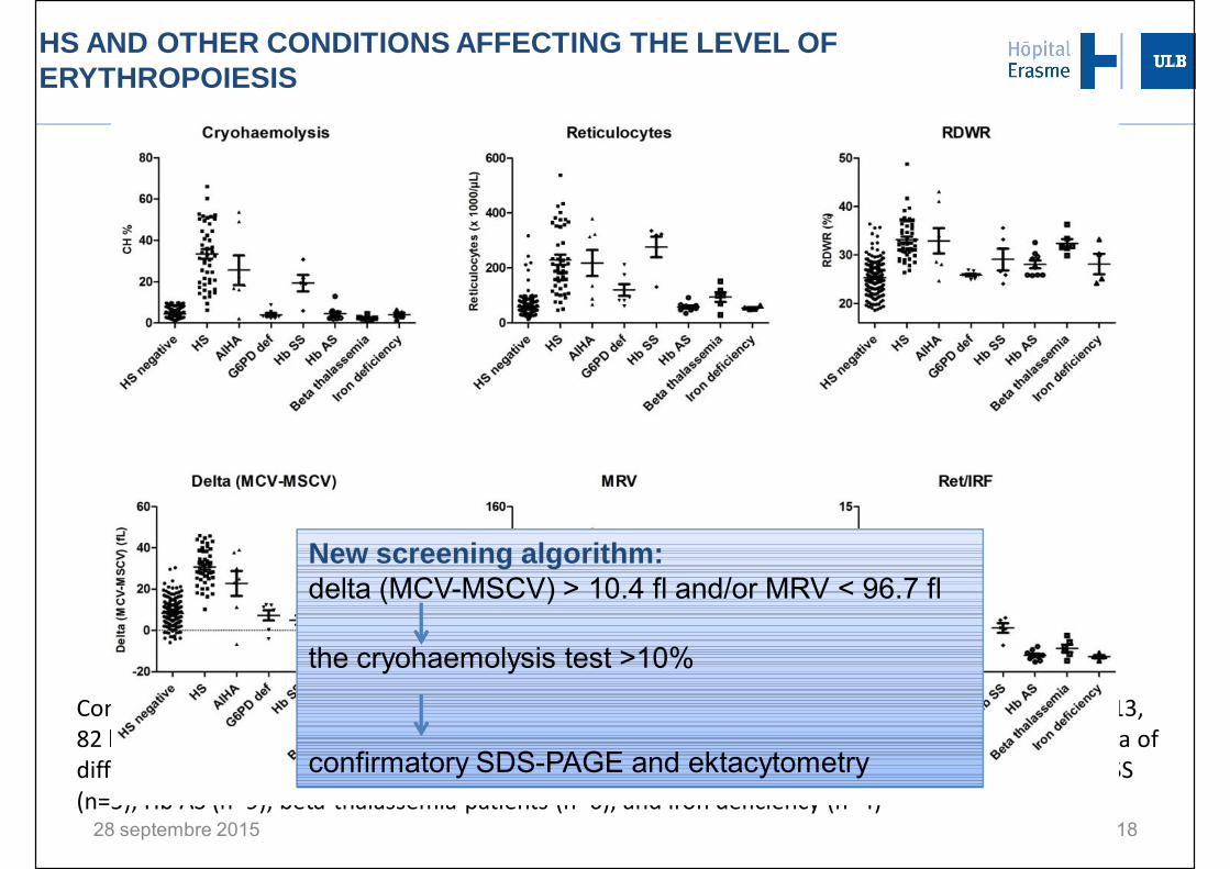
HS AND OTHER CONDITIONS AFFECTING THE LEVEL OF ERYTHROPOIESIS
28 septembre 2015
Comparison of screening tests results in hereditary spherocytosis patients (HS, n=48), controls (n=213, 82 healthy subjects and 131 cryohaemolysis negative without anaemia) and in patients with anaemia of different origins: auto-immune haemolytic anaemia (AIHA, n=7), G6PD deficient patients (n=7), Hb SS (n=5), Hb AS (n=9), beta-thalassemia patients (n=6), and iron deficiency (n=4)
18
New screening algorithm:delta (MCV-MSCV) > 10.4 fl and/or MRV < 96.7 fl
the cryohaemolysis test >10%
confirmatory SDS-PAGE and ektacytometry
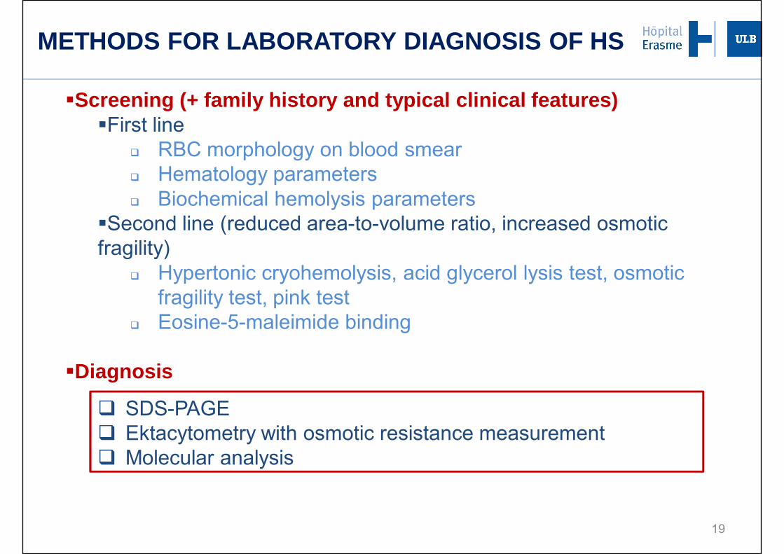
METHODS FOR LABORATORY DIAGNOSIS OF HS
§Screening (+ family history and typical clinical features)§First line
q RBC morphology on blood smearq Hematology parametersq Biochemical hemolysis parameters
§Second line (reduced area-to-volume ratio, increased osmotic fragility)
q Hypertonic cryohemolysis, acid glycerol lysis test, osmotic fragility test, pink test
q Eosine-5-maleimide binding
§Diagnosis
q SDS-PAGEq Ektacytometry with osmotic resistance measurementq Molecular analysis
19
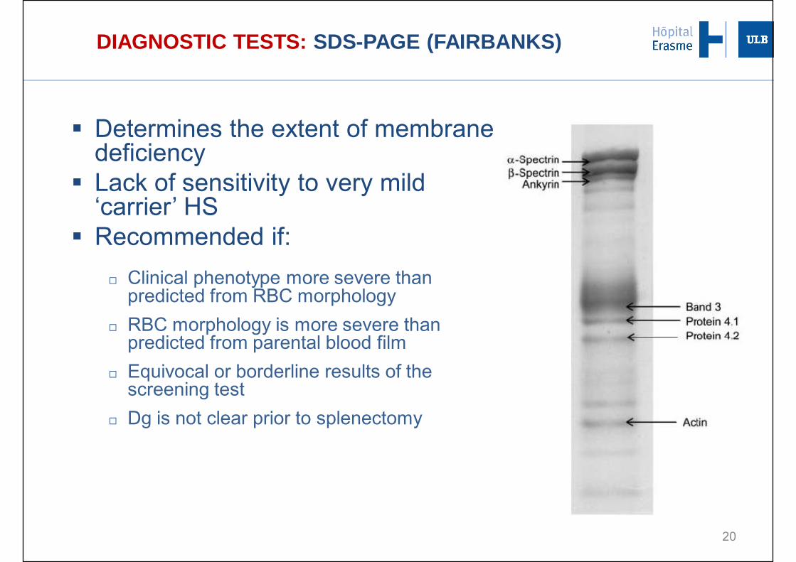
DIAGNOSTIC TESTS: SDS-PAGE (FAIRBANKS)
§ Determines the extent of membrane deficiency§ Lack of sensitivity to very mild
‘carrier’ HS§ Recommended if:
¨ Clinical phenotype more severe than predicted from RBC morphology
¨ RBC morphology is more severe than predicted from parental blood film
¨ Equivocal or borderline results of the screening test
¨ Dg is not clear prior to splenectomy
20
28 septembre 2015 21
28 septembre 2015 22
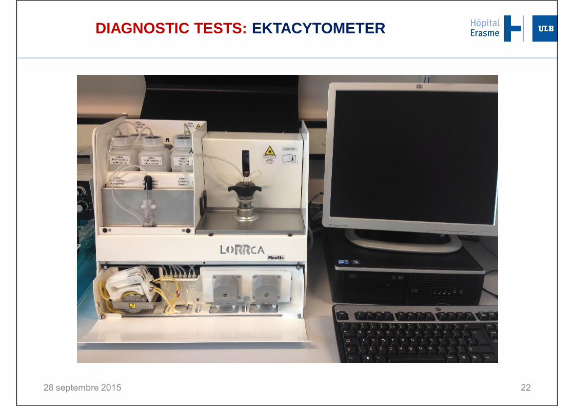
DIAGNOSTIC TESTS: EKTACYTOMETER
23
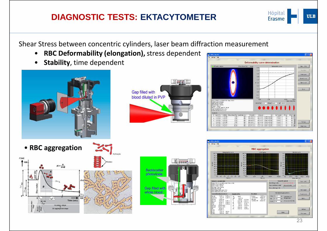
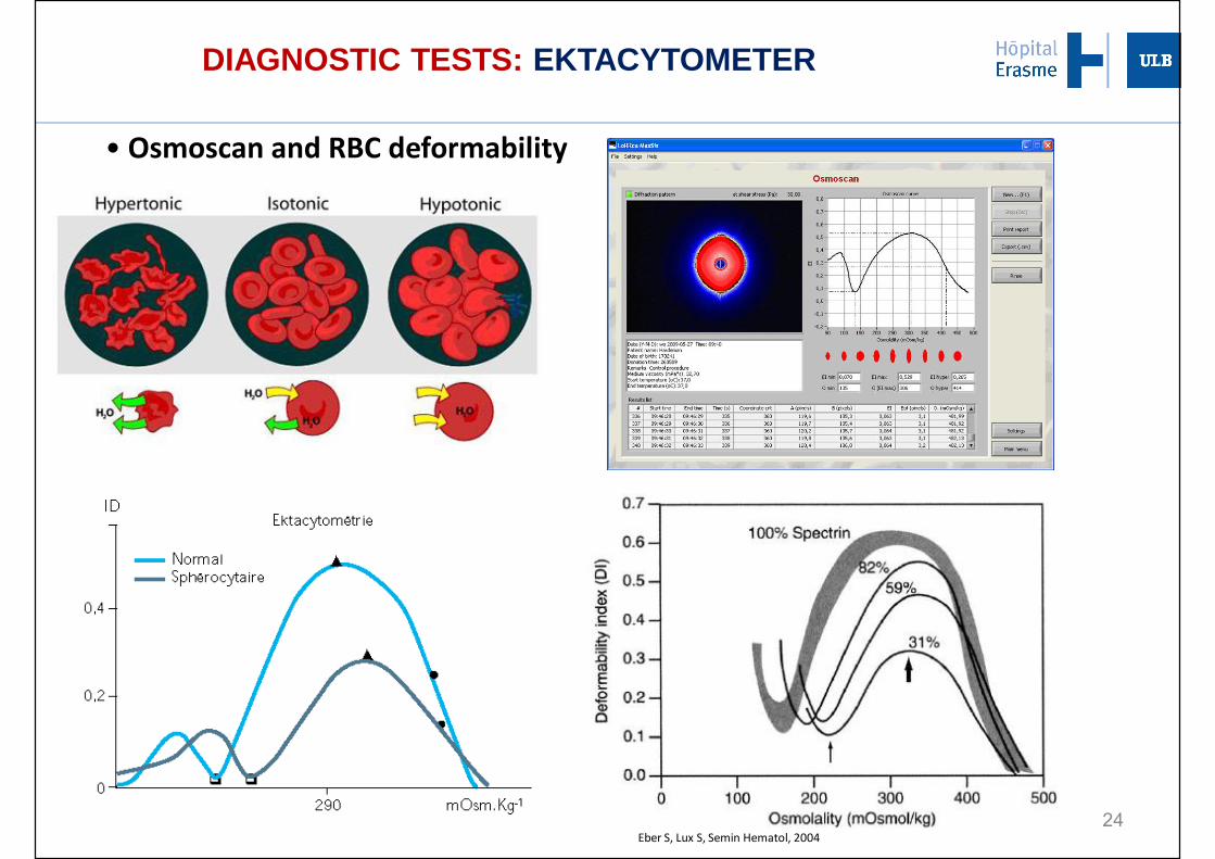
Shear Stress between concentric cylinders, laser beam diffraction measurement• RBC Deformability (elongation), stress dependent• Stability, time dependent
• RBC aggregation
DIAGNOSTIC TESTS: EKTACYTOMETER
H2OH2O
Eber S, Lux S, Semin Hematol, 200424
• Osmoscan and RBC deformability
DIAGNOSTIC TESTS: EKTACYTOMETER
28 septembre 2015 25
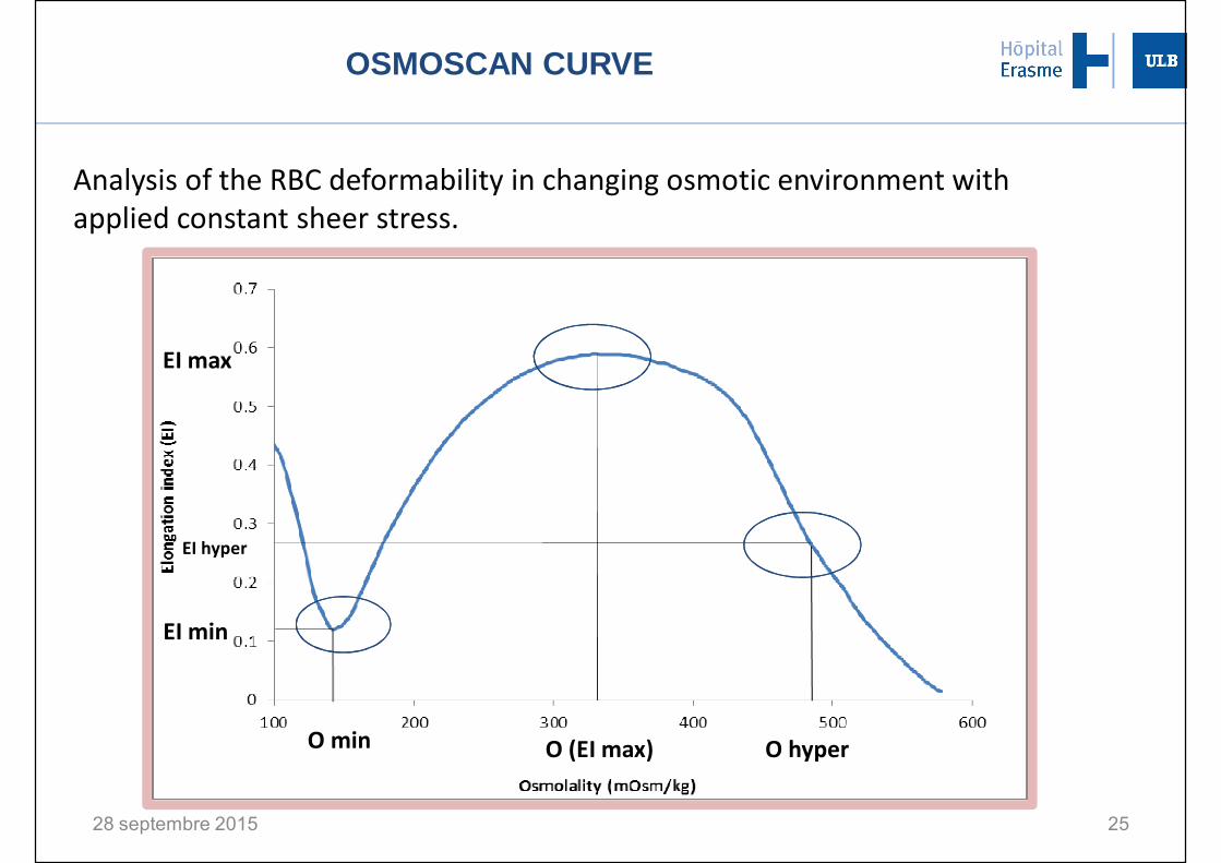
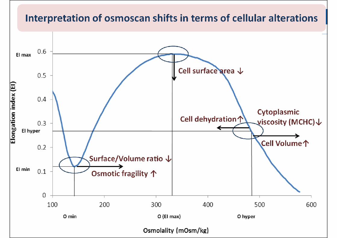
EI hyper
EI max
EI min
O (EI max)O min O hyper
OSMOSCAN CURVE
Analysis of the RBC deformability in changing osmotic environment with applied constant sheer stress.
28 septembre 2015 26
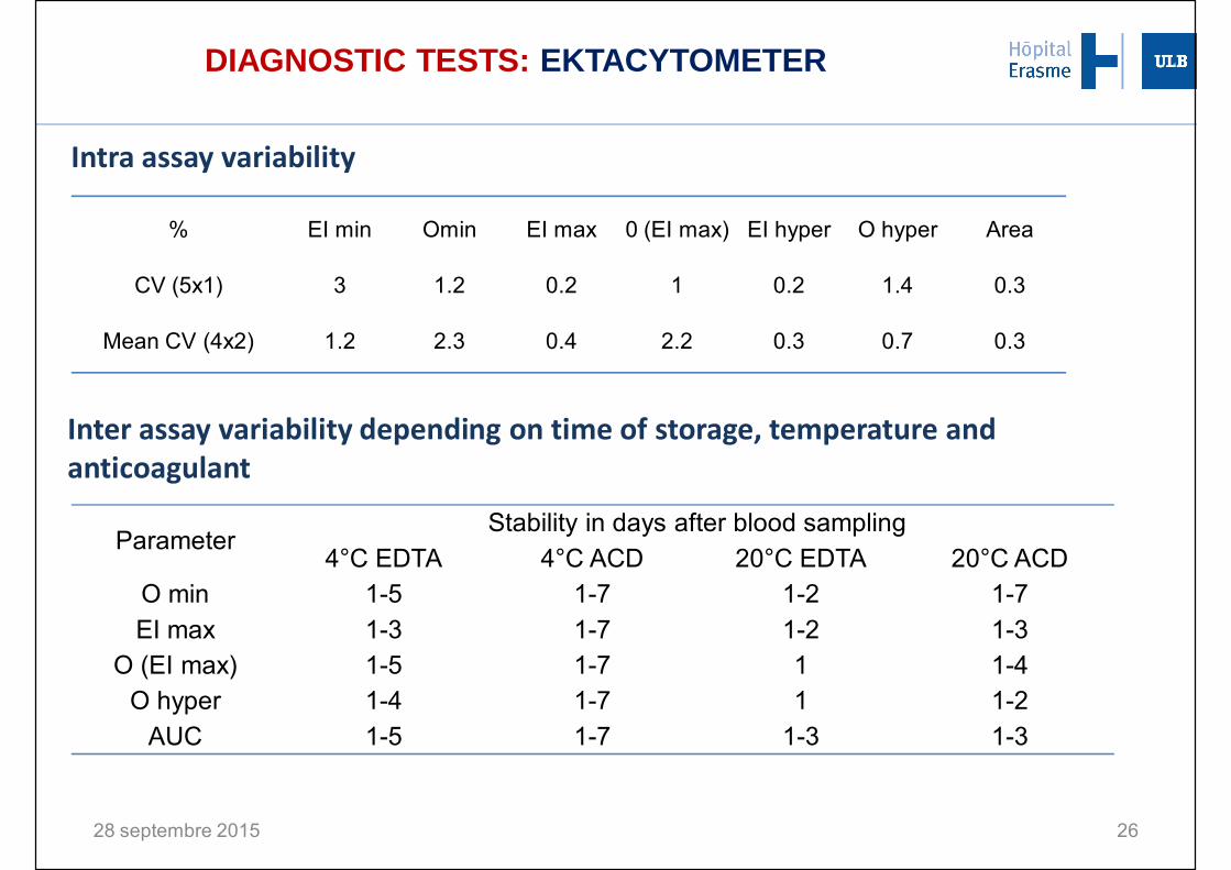
% EI min Omin EI max 0 (EI max) EI hyper O hyper Area
CV (5x1) 3 1.2 0.2 1 0.2 1.4 0.3
Mean CV (4x2) 1.2 2.3 0.4 2.2 0.3 0.7 0.3
Intra assay variability
Inter assay variability depending on time of storage, temperature and anticoagulant
ParameterStability in days after blood sampling
4°C EDTA 4°C ACD 20°C EDTA 20°C ACDO min 1-5 1-7 1-2 1-7EI max 1-3 1-7 1-2 1-3
O (EI max) 1-5 1-7 1 1-4O hyper 1-4 1-7 1 1-2
AUC 1-5 1-7 1-3 1-3
DIAGNOSTIC TESTS: EKTACYTOMETER
28 septembre 2015 27
28 septembre 2015 28
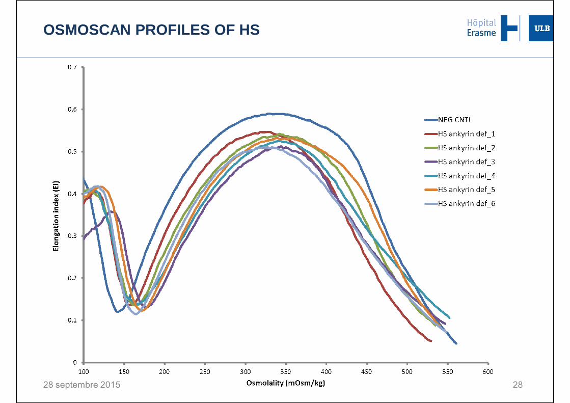
OSMOSCAN PROFILES OF HS

OSMOSCAN PROFILES OF OTHER RBC PATHOLOGIES
28 septembre 2015 29
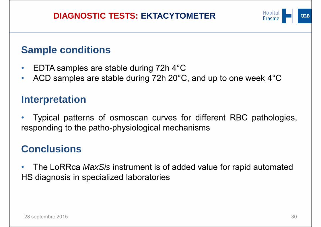
Sample conditions
• EDTA samples are stable during 72h 4°C• ACD samples are stable during 72h 20°C, and up to one week 4°C
Interpretation
• Typical patterns of osmoscan curves for different RBC pathologies,responding to the patho-physiological mechanisms
Conclusions• The LoRRca MaxSis instrument is of added value for rapid automated HS diagnosis in specialized laboratories
28 septembre 2015 30
DIAGNOSTIC TESTS: EKTACYTOMETER
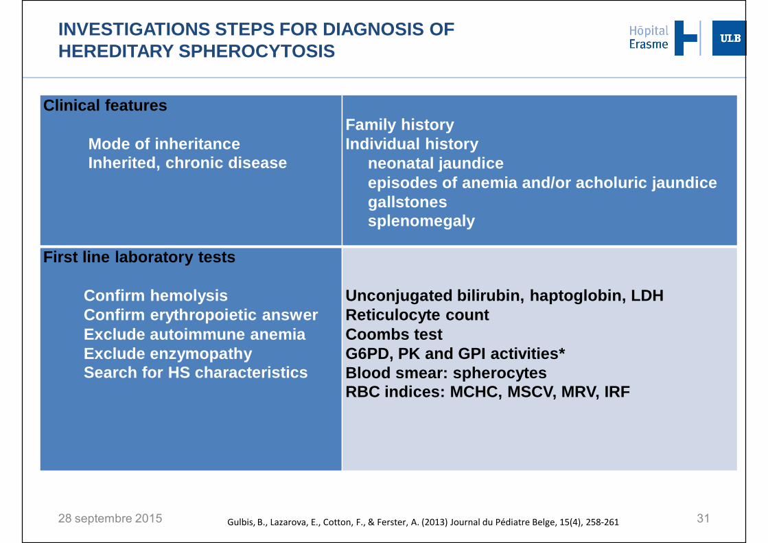
INVESTIGATIONS STEPS FOR DIAGNOSIS OF HEREDITARY SPHEROCYTOSIS
Clinical features
Mode of inheritanceInherited, chronic disease
Family historyIndividual history
neonatal jaundiceepisodes of anemia and/or acholuric jaundicegallstonessplenomegaly
First line laboratory tests
Confirm hemolysisConfirm erythropoietic answerExclude autoimmune anemiaExclude enzymopathySearch for HS characteristics
Unconjugated bilirubin, haptoglobin, LDHReticulocyte countCoombs testG6PD, PK and GPI activities*Blood smear: spherocytesRBC indices: MCHC, MSCV, MRV, IRF
28 septembre 2015 31Gulbis, B., Lazarova, E., Cotton, F., & Ferster, A. (2013) Journal du Pédiatre Belge, 15(4), 258-261
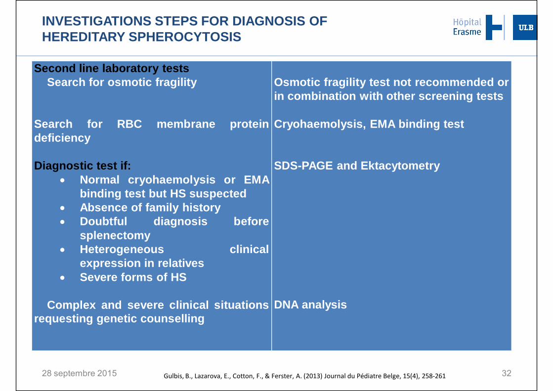
INVESTIGATIONS STEPS FOR DIAGNOSIS OF HEREDITARY SPHEROCYTOSIS
Second line laboratory testsSearch for osmotic fragility
Search for RBC membrane proteindeficiency
Diagnostic test if:• Normal cryohaemolysis or EMA
binding test but HS suspected• Absence of family history• Doubtful diagnosis before
splenectomy• Heterogeneous clinical
expression in relatives• Severe forms of HS
Complex and severe clinical situationsrequesting genetic counselling
Osmotic fragility test not recommended orin combination with other screening tests
Cryohaemolysis, EMA binding test
SDS-PAGE and Ektacytometry
DNA analysis
28 septembre 2015 32Gulbis, B., Lazarova, E., Cotton, F., & Ferster, A. (2013) Journal du Pédiatre Belge, 15(4), 258-261
CONCLUSIONS
§ Clinical features, family history
§ Laboratory investigation§ Screening tests (morphology, reticulocyte parameters; EMA, CH)§ Confirmatory tests (ektacytometry and/or SDS-PAGE)
§ Clinician-biologist communication
33