Development of giant bacteriophage KZ is independent of...
31
1 1 Development of giant bacteriophage фKZ is independent of the 2 host transcription apparatus 3 Running title: The transcriptional scheme of giant phage фKZ 4 5 Pieter-Jan Ceyssens 1,Ω,1 , Leonid Minakhin 2,Ω , An Van den Bossche 1 , Maria Yakunina 3 , Evgeny Klimuk 3,4,5 , 6 Bob Blasdel 1 , Jeroen De Smet 1 , Jean-Paul Noben 6 , Udo Bläsi 7 , Konstantin Severinov 2,3,4,5* , and Rob 7 Lavigne 1* 8 9 1 Division of Gene Technology, KULeuven, Kasteelpark Arenberg 21, 3001 Heverlee, Belgium 10 2 Waksman Institute of Microbiology, Rutgers University, NJ, USA 11 3 St. Petersburg State Polytechnical University, St. Petersburg, Russia 12 4 Institutes of Molecular Genetics and Gene Biology, Russian Academy of Sciences, Moscow, Russia 13 5 Skolkovo Institute of Science and Technology, Skolkovo, Russia 14 6 Biomedical Research Institute, Hasselt University, Agoralaan Building C, 3590 Diepenbeek, Belgium 15 7 Max F Perutz Laboratories, University of Vienna, Dr. Bohr-Gasse 9, 1030 Vienna, Austria 16 17 18 Ω Contributed equally 19 * Corresponding authors: 20 [email protected] 21 [email protected] 22 1 Current address: Scientific Institute of Public Health (WIV-ISP), J. Wytsmanstraat 14, 1050 Brussels, Belgium JVI Accepts, published online ahead of print on 25 June 2014 J. Virol. doi:10.1128/JVI.01347-14 Copyright © 2014, American Society for Microbiology. All Rights Reserved. on July 8, 2018 by guest http://jvi.asm.org/ Downloaded from
Transcript of Development of giant bacteriophage KZ is independent of...
1
1
Development of giant bacteriophage фKZ is independent of the 2
host transcription apparatus 3
Running title: The transcriptional scheme of giant phage фKZ 4
5
Pieter-Jan Ceyssens1,Ω,1, Leonid Minakhin2,Ω, An Van den Bossche1, Maria Yakunina3, Evgeny Klimuk3,4,5, 6
Bob Blasdel1, Jeroen De Smet1, Jean-Paul Noben6, Udo Bläsi7, Konstantin Severinov2,3,4,5*, and Rob 7
Lavigne1* 8
9
1 Division of Gene Technology, KULeuven, Kasteelpark Arenberg 21, 3001 Heverlee, Belgium 10
2 Waksman Institute of Microbiology, Rutgers University, NJ, USA 11
3 St. Petersburg State Polytechnical University, St. Petersburg, Russia 12
4 Institutes of Molecular Genetics and Gene Biology, Russian Academy of Sciences, Moscow, Russia 13
5 Skolkovo Institute of Science and Technology, Skolkovo, Russia 14
6 Biomedical Research Institute, Hasselt University, Agoralaan Building C, 3590 Diepenbeek, Belgium 15
7 Max F Perutz Laboratories, University of Vienna, Dr. Bohr-Gasse 9, 1030 Vienna, Austria 16
17
18
Ω Contributed equally 19
* Corresponding authors: 20
1 Current address: Scientific Institute of Public Health (WIV-ISP), J. Wytsmanstraat 14, 1050 Brussels, Belgium
JVI Accepts, published online ahead of print on 25 June 2014J. Virol. doi:10.1128/JVI.01347-14Copyright © 2014, American Society for Microbiology. All Rights Reserved.
on July 8, 2018 by guesthttp://jvi.asm
.org/D
ownloaded from
2
ABSTRACT 23
Pseudomonas aeruginosa bacteriophage фKZ is the type representative of the ‘giant’ phage genus 24
characterized by unusually large virions and genomes. By unraveling the transcriptional map of the ~280 25
kb фKZ genome to single-nucleotide resolution, we combine 369 фKZ genes into 134 operons. Early 26
transcription is initiated from highly conserved AT-rich promoters distributed across the фKZ genome 27
and located on the same strand of the genome. Early transcription does not require phage or host 28
protein synthesis. Transcription of middle and late genes is dependent on protein synthesis and 29
mediated by poorly conserved middle and late promoters. Unique to фKZ is its ability to complete its 30
infection in the absence of bacterial RNA polymerase (RNAP) enzyme activity. We propose that 31
transcription of the фKZ genome is performed by the consecutive action of two фKZ-encoded, non-32
canonical multisubunit RNAPs, one of which is packed within the virion, another being the product of 33
early genes. This unique, rifampicin-resistant transcriptional machinery is conserved within the diverse 34
giant phage genus. 35
IMPORTANCE 36
The data presented in this paper offer, for the first time, an insight into the complex transcriptional 37
scheme of giant bacteriophages. We show that Pseudomonas aeruginosa giant phage фKZ is able to 38
infect and lyse its host cell and produce phage progeny in the absence of functional bacterial 39
transcriptional machinery. This unique property can be attributed to two phage-encoded putative RNAP 40
enzymes, which contain very distant homologues of bacterial β and β’-like RNAP subunits. 41
42
43
44
on July 8, 2018 by guesthttp://jvi.asm
.org/D
ownloaded from
3
INTRODUCTION 45
Transcription is driven by DNA-dependent RNA polymerases (RNAPs), which synthesize RNA from DNA 46
templates [1]. RNAPs can be classified into two unrelated families: small single-subunit enzymes 47
(ssRNAPs) encoded by some bacteriophages and also found in mitochondria and chloroplasts, and large 48
multisubunit cellular enzymes (msRNAPs) transcribing genes in bacterial, archaeal, and eukaryal 49
genomes. The catalytic activity of enzymes from both families is accomplished through a common two-50
metal ion mechanism. The canonical bacterial msRNAP is a 400 kDa complex consisting of five core 51
subunits (α2ββ’ω) which are directed to specific promoter sequences by a variety of σ factors [2]. The 52
two largest RNAP subunits, β and β’, contain conserved double-psi beta-barrel (DPBB) domains that 53
together form the active center [3-5]. Members of the ssRNAP family rely on different catalytic domains 54
and amino acid motifs for catalysis of the RNA polymerization reaction and are related to DNA 55
polymerases and reverse transcriptases [6,7]. Bacterial RNAPs are inactivated by the antibiotic 56
Rifampicin (Rif), which acts by binding to the β-subunit pocket deep inside the active site channel, 57
preventing synthesis of RNA longer than 3-4 nt [8]. 58
Bacterial RNAPs play a key role during the infection of bacterial cell by bacteriophages. Most known 59
phages do not encode their own RNAP but redirect the host transcription machinery to viral promoters 60
by relying on very strong promoters recognized by host RNAP and/or modifying the host RNAP specificity 61
by phage-encode factors [9,10]. Some phages like T7, N4, Xp10, and phiEcoM-GJ1 depend on their own 62
single-subunit RNAPs to transcribe a subset of viral genes [11-15]. In these phages, two basic 63
transcriptional strategies have been described. Firstly, some phages, like T7, recruit the bacterial RNAP at 64
the onset of infection to transcribe the phage-encoded ssRNAP gene from an early promoter. Upon 65
completion of this task, the bacterial RNAP is inactivated using post-translational modifications or RNAP-66
binding proteins [17,18] and the viral RNAP proceeds to transcribe middle and late phage genes. The N4-67
on July 8, 2018 by guesthttp://jvi.asm
.org/D
ownloaded from
4
like viruses have evolved an inverse transcriptional strategy. A ssRNAP is carried within their capsids and 68
is injected with phage DNA upon infection to transcribe early phage promoters [18-20]. A second N4-69
encoded ssRNAP, a product of an early gene, transcribes middle phage genes. While early and middle N4 70
transcription is independent of bacterial transcription machinery, N4 recruits the host RNAP at the late 71
stage of infection through the action of N4 single-stranded DNA-binding protein [21]. A third strategy 72
may be used by the giant Bacillus subtilis phage PBS2 [22]. Forty years ago, the development of this 73
phage was reported to be independent of the addition of Rif, suggesting that host RNAP is not involved 74
in viral transcription throughout the infection. In follow-up studies, the purification of a highly unusual 75
multisubunit Rif-resistant RNAP complex from PBS2-infected cells was reported [23,24]. However, 76
further study of this interesting phage and its transcription strategy has been hampered by the absence 77
of its genome sequence. 78
Giant ‘фKZ-like’ bacteriophages form a very distant branch of myoviruses [25]. The type virus фKZ 79
possesses an unusually large 280 kb genome, which displays little evolutionary relatedness to other 80
genera. When the фKZ genome was first reported in 2002, a function could be assigned to only 19% of its 81
306 predicted genes [26]. The фKZ virion is composed of no less than 50 different proteins [27,28]. 82
Intriguingly, three of virion proteins, gp178, gp80, and gp180, were found to resemble bacterial RNAP β 83
and β’ subunits. It was hypothesized that these proteins form a highly unusual virion RNAP and are co-84
injected with the phage DNA and participate in early transcription, possibly through recruitment of host-85
encoded factors [27-30]. Two other фKZ proteins, Gp73 and Gp74, also resemble β/β-like subunits but 86
are not associated with virions, suggesting that the phage may encode an additional, non-virion RNAP 87
[26]. 88
In this paper, we uncover the unique transcriptional scheme of giant phage фKZ. We demonstrate that 89
phage development and transcription of its genes is not sensitive to Rifampicin, proving that viral-90
on July 8, 2018 by guesthttp://jvi.asm
.org/D
ownloaded from
5
encoded transcription enzymes are involved in phage gene expression. We also map conserved motifs 91
located upstream of фKZ genes belonging to different temporal expression classes. These motifs are 92
distinct from promoters recognized by host RNAP. We propose that they are recognized by phage-93
encoded RNAPs which, based on our analysis, contain additional subunits that were not revealed by 94
earlier work. The фKZ transcription scheme is likely conserved and characteristic for other phages of the 95
giant phages genus. 96
97
98
MATERIALS and METHODS 99
Bacteriophages, Bacterial strains and Growth Conditions 100
In all experiments, the P. aeruginosa strain PAO1 strain was grown in standard LB medium, in some cases 101
supplemented with Rifampicin (400µg/ml) or Chloramphenicol (100 µg/ml). Bacteriophage фKZ was 102
amplified from confluent lysed agar plates, purified by 0.45µm filtration and concentrated after 103
incubation in 10% PEG8000/1M NaCl according to standard procedures [31]. P. aeruginosa and its 104
derivative strains were grown overnight in 5 ml LB medium to reach saturation. Cells were diluted 1:100 105
in 2-500 mL fresh medium, grown at 37°C and, in infection experiments, infected with MOI 5 at OD600 nm 106
of 0.3. Cell growth (or phage infection) was halted at indicated time points by rapid cooling in ice water 107
or by methods specific for RNA-seq sampling (see below). Cells were harvested by centrifugation (6,000 x 108
g), flash-frozen and stored at -80°C until use. The efficiency of infection was always checked by cell 109
counts of the infected culture five minutes post infection, which should contain <5% surviving cells 110
compared to the non-infected culture. 111
112
on July 8, 2018 by guesthttp://jvi.asm
.org/D
ownloaded from
6
Pull-down experiments 113
For the analysis of the bacterial RNAP complex upon phage infection, we used an available P. aeruginosa 114
PAO1 rpoA::strep strain [32]. For the identification of protein interaction partners of the structural β-like 115
subunit gp178, its gene was cloned with a 6xhis-tag coding sequence into the expression vector 116
pHERD20T [33]. This plasmid was electroporated to freshly prepared electro-competent P. aeruginosa 117
PAO1 cells. The obtained cells were tested for growth and sensitivity to φKZ infection upon 118
overexpression of the protein by 1% arabinose. Infected P. aeruginosa cells were harvested by 119
centrifugation and resuspended in 8 ml resuspension buffer (10 mM Tris pH8,0, 150 mM NaCl, 0,1% (v/v) 120
NP-40, 2,5 mM Pefabloc® SC (Merck)) containing 1 mg/ml lysozyme (Sigma). After one freeze-thaw cycle, 121
the mixture was supplemented with 800 µl 10x BugBuster® Protein extraction reagent (Novagen) and 10 122
µl Benzonase nuclease (Novagen) and incubated at room temperature for 15 min with gently agitation. 123
For His6-mediated purification, the soluble fraction was loaded on a Bio-Rad Poly-Prep® Chromatography 124
column containing 1 ml pre-washed Ni-NTA agarose beads (Qiagen). The beads were washed with two 125
successive wash steps of 10 ml and 5 ml resuspension buffer containing 25 mM imidazole and 50 mM 126
imidazole respectively and eluted with 5 ml resuspension buffer containing 250 mM imidazole. The 127
eluted fractions were pooled and concentrated by ultrafiltration (Amicon Ultra 3K, Millipore), heat 128
denaturated for 5 min at 95°C and loaded on a 10% SDS-PAGE gel. The Coomassie-stained gel (Simply 129
Blue Safestain; Invitrogen) was cut into slices, which were subjected to trypsin digestion previous to 130
mass spectrometry analysis. 131
LC-MS/MS analysis and data analysis 132
An Easy-nLC 1000 liquid chromatograph (Thermo Scientific) was on-line coupled to a mass calibrated 133
LTQ-Orbitrap Velos Pro (Thermo Scientific) via a Nanospray Flex ion source (Thermo Scientific) using 134
sleeved 30 µm ID stainless steel emitters. The SpeedVac dried tryptic peptide mixture was dissolved in 20 135
on July 8, 2018 by guesthttp://jvi.asm
.org/D
ownloaded from
7
µl buffer A (0.1% v/v formic acid in Milli-Q water) and then loaded, concentrated and desalted on a 136
trapping pre-column (Acclaim PepMap 100 C18, Thermo Scientific) at a buffer A flow rate of 5 µl/min for 137
5 minutes. The peptide mixture was separated on an Acclaim PepMap RSLC C18 column (Thermo 138
Scientific) at a flow rate of 250 nL/min of buffer A with a linear gradient in 40 minutes of 0 to 70 % buffer 139
B (0.1% v/v formic acid in acetonitrile(Biosolve)). MS data were acquired in a data-dependent mode 140
under direct control of the Xcalibur software (version 2.2.SP1.48), selecting the fragmentation events 141
based on the top six precursor abundances in the survey scan (350–2000 Th). The isolation window for 142
MS/MS fragmentation was set to 2 Th and the normalized collision energy, Q-value and activation time 143
were 30%, 0.25 and 10 ms, respectively. 144
The analysis of the mass spectrometric RAW data was carried out using Proteome Discoverer software 145
v.1.3 (Thermo Scientific) with build-in Sequest v.1.3.0339 and interfaced with an in-house Mascot v.2.4 146
server (Matrix Science). MS/MS spectra were searched against a database containing all P. aeruginosa 147
PAO1 proteins and all ‘stop-to-stop’ protein sequences in all six frames of phage phiKZ. Peptide scoring 148
for identification was based on following search criteria: enzyme trypsin, maximum missed cleavages 2, 149
precursor mass tolerance 10 ppm and fragment mass tolerance 0.5 Da. 150
RNA-seq analysis 151
Cells intended for RNA sequencing were rapidly suspended in 1/10th volumes of ice cold stop solution 152
(10% phenol in ethanol) and chilled to inhibit RNA transcription and degradation at indicated time 153
points. Cells were then collected by centrifugation (6,000 x g) and Total RNA was extracted from the 154
resulting cell pellet by resuspending it in TRIzol (Ambion) and performing a classical phenol chloroform 155
extraction followed by ethanol precipitation. Remaining DNA was removed using TURBO DNAse 156
(Ambion), which efficiency was checked by the absence of reporter PCR products of both bacterial and 157
phage genes. Subsequently, the samples were processed into directional cDNA libraries with the TruSeq 158
on July 8, 2018 by guesthttp://jvi.asm
.org/D
ownloaded from
8
Stranded mRNA Sample Prep Kit and run on an Illumina HiSeq sequencer. For each of our comparative 159
analyses we used the total gene reads mapped to the host (P. aeruginosa PAO1) and viral (Pseudomonas 160
phage phiKZ) annotated gene features using the CLC Genomics Workbench 6.0.2. software. The 161
comparisons of relative levels of expression for gene features between different time points after 162
infection within both the phage and host genomes were made with the negative binomial distribution 163
test using the DESeq Bioconductor package in R. All raw RNA-seq data was submitted at the GEO 164
repository, with series record GSE58494. 165
Primer extension 166
In vivo primer extension reactions were done essentially as described elsewhere [34]. P. aeruginosa 167
PAO1 cells were infected with фKZ (MOI 5) at OD600 of 0.3 and harvested at different time points 168
specified in the experiments performed. When necessary, rifampicin and chloramphenicol were added 169
to the infected culture at designated time points at final concentrations 150 μg/mL and 100 μg/mL, 170
respectively. Total RNA was extracted using Trizol reagent (Life Technologies) according to the 171
manufacturer’s procedure. For each primer extension reaction, 10 μg of total RNA was reverse-172
transcribed with 100 units of M-MLV reverse transcriptase (Life Technologies) in the presence of 10 pmol 173
of γ-32P end-labeled primer. The reactions were treated with RNase H, precipitated with ethanol and 174
dissolved in formamide loading buffer. To precisely identify the 5′ ends of the primer extension products, 175
DNA sequencing reactions containing both the corresponding PCR-amplified фKZ genome fragments and 176
end-labeled primers used for the primer extension reaction were performed. The reaction products were 177
resolved on 6-8 % (w/v) polyacrylamide sequencing gels and visualized using a PhosphorImager 178
(Molecular Dynamics). 179
Time-lapse microscopy 180
on July 8, 2018 by guesthttp://jvi.asm
.org/D
ownloaded from
9
For microscopic recordings, the infected culture was diluted a thousand times, spotted on an LB agar pad 181
and recorded in real time for 5 h. For this, we used a temperature controlled (Okolab Ottaviano) Ti-182
Eclipse inverted microscope (Nikon) equipped with a TI-CT-E motorised condensor and a CoolSnap HQ2 183
FireWire CCD-camera. Images were acquired using the NIS-Elements AR 3.2 software (Nikon) as 184
described previously [35]. 185
186
RESULTS 187
The transcriptional map of фKZ 188
To investigate transcription regulation during фKZ infection, we first acquired a blueprint of the 189
transcriptional scheme of this giant phage by performing RNA-seq of total RNA prepared from cells 190
collected at different times post-infection. This approach allowed us to globally detect phage 191
transcriptional units belonging to different temporal classes at single-nucleotide resolution and to study 192
the impact of phage infection on the P. aeruginosa transcriptome in a single experiment. 193
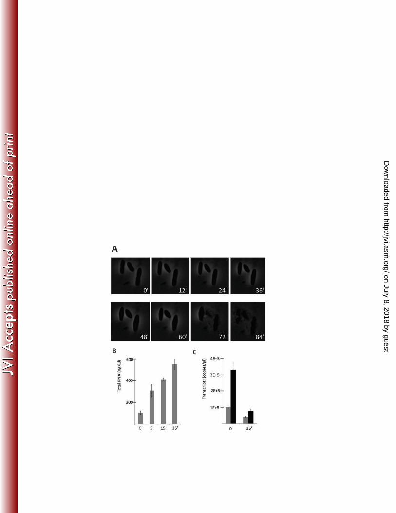
DNA-free total RNA was extracted from P. aeruginosa PAO1 cells immediately before (0 minute) and 5, 194
15, and 35 minutes after infection. In the course of infection, the host cells marginally increased in size 195
but acquired a rugby-ball shape (Figure 1a). The amount of total intracellular RNA extracted from cells in 196
the later stages of infection increased over fivefold compared to non-infected cells (Figure 1b). This 197
increase in total RNA was not due to host transcription, since an RT-PCR control of two household genes, 198
oprL and 5S rRNA, showed a significant decrease in transcript abundance 35 minutes post infection 199
(Figure 1c). RNA-seq analysis was performed on total RNA samples (in duplicates, 5x10⁶ reads/sample). 200
In addition, early (10 min) and late (35 min) transcription was analyzed by deeper (non-directional) RNA 201
sequencing (5x10⁷ reads/sample) (Table S1). Reads originating from the phage and the host were next 202
on July 8, 2018 by guesthttp://jvi.asm
.org/D
ownloaded from
10
mapped to respective genome sequences. A drastic accumulation of phage mRNA as the infection 203
proceeded was observed. Late in infection, 36.9% of all reads mapped to the phage genome, which 204
represented up to 98.5% of all non-rRNA and non-tRNA host RNA reads. 205
The results allowed us to identify 134 фKZ operons, spanning an average of 2,075 bp and containing an 206
average of 2.7 genes per operon (Figure 2, Table S2). When comparing these transcriptional units to the 207
original genome annotation [26], many events of intergenic transcription initiation were observed. We 208
used this information to expand the predicted gene feature content of the genome from the original 306 209
to 369 coding sequences (+20.5%), removing the annotation of ORF24 and ORF117 due to a lack of sense 210
transcripts, and updating start codon positions of 27 ORFs (Table S3). The resulting updated фKZ 211
annotation file (GenBank AF399011.1) corresponds only by 74% to the initial version submitted in 2002. 212
Most of newly identified ORFs display clear Shine-Dalgarno sequences. The majority of the newly 213
annotated genes (72%) also have a counterpart in the closely related фKZ-like viruses PA7 and фPA3 [36], 214
although we propose an alternative start codon for 48% of these genes. Many of the newly predicted 215
proteins contain structural features such as hydrophobic stretches (e.g., ORF 117.1), signal peptides (e.g., 216
ORF 166.1) or conserved domains (e.g., PAAR domain in ORF 163.1, Pfam 05488, E=1.96e-04), hinting at 217
functional importance of these proteins in the infection cycle (Table S3). We also observed cases of 218
antisense transcription, for example involving genes encoding structural proteins 29 and 30 (Figure S1). 219
The function of these antisense RNA (asRNA) might be to regulate expression levels by interfering with 220
sense RNA transcription [37], protect the primary transcript by masking single-stranded binding sites of 221
endoribonuclease E [38], or by blocking translation by occluding ribosome binding sites [39]. 222
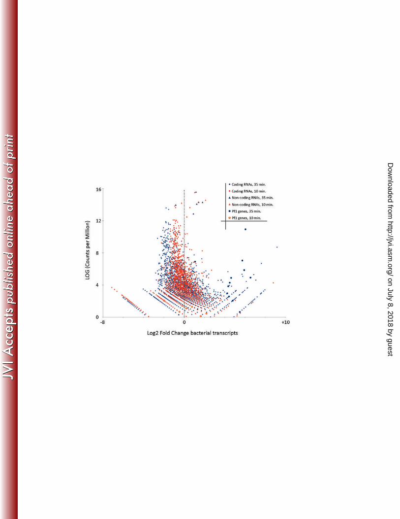
Differential gene expression during phage infection 223
When comparing transcripts 10 and 35 minutes of post-infection, we observed that, as expected, the 224
majority of virion-associated structural gene transcripts were significantly upregulated at later time 225
on July 8, 2018 by guesthttp://jvi.asm
.org/D
ownloaded from
11
point. The accumulation of phage transcripts coincides with a clear pattern of general decrease in 226
abundance of bacterial transcripts (Figure 3). Remarkably, halfway through infection only 1.5% of mRNAs 227
in the infected cells is from bacterial origin. To find out whether the phage employs any host 228
transcription shut-off mechanism associated with factor-dependent inhibition of host RNAP activity, we 229
affinity purified the bacterial RNAP from cells collected at various times post-infection [32]. Although the 230
composition of the bacterial RNAP changed subtly as the infection proceeded, no фKZ proteins were co-231
purified alongside the complex at any time point. Moreover, enzyme purified from infected cells was as 232
active as control enzyme purified from control uninfected cells (data not shown) suggesting the absence 233
of phage-encoded host RNAP inhibitors and/or modifications. 234
Remarkably, only a single bacterial operon, including genes PA0718 to PA0728, shows significant and 235
easily discernible upregulation during the infection (Figure 3). These genes encode proteins of the 236
filamentous prophage Pf1, raising the intriguing possibility that Pf1 tries to escape the infected cell 237
before lysis occurs. To test this idea, we performed quantitative PCR on DNA extracted from фKZ-238
infected cells to determine whether new copies of the Pf1 genome are produced during infection. 239
However, we did not see a significant rise in Pf1 genome copies, compared to the housekeeping gene 240
oprL and contrasting an average 57-fold increase in фKZ genome copies (data not shown). 241
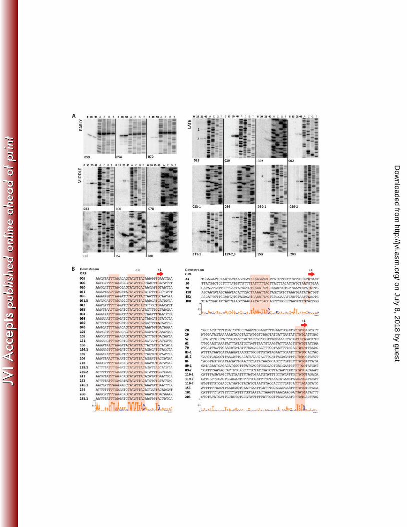
Mapping of early, middle, and late фKZ promoters 242
All фKZ-related viruses are characterized by the presence of similar AT-rich intergenic motifs (5’-243
TATATTAC-3’ in фKZ) that are unidirectionally distributed throughout their genomes. Inspection of the 244
фKZ genome sequence revealed 28 such highly conserved motifs. These motifs have been assumed to 245
act as phage-specific promoters [e.g., 26, 30, 40-42]. Indeed, our RNA-seq data clearly indicates that 246
after 5 minutes of фKZ infection, all transcription is performed on the leading strand and is exclusively 247
associated with these motifs (Figure 2, red arrows, and Figure 4 with alignment of the corresponding 248
on July 8, 2018 by guesthttp://jvi.asm
.org/D
ownloaded from
12
sequences), supporting their likely function as early promoters. Comparison of sequences surrounding 249
this “core” motif revealed an additional upstream motif of lower conservation (5’-TTTaA-3’) (Figure 4). 250
The early фKZ promoters mostly drive transcription of blocks of short genes with unknown function. 251
Primer extension analysis of total RNA purified at 0, 10 and 30 minutes of фKZ infection using primers 252
annealing downstream of three randomly chosen early promoters confirmed RNA-seq data. Each primer 253
extension product appeared after 10 minutes of infection and did not decline afterwards, suggesting that 254
the early transcripts are either stable over the course of infection or that early promoters are not fully 255
turned off at middle and late stages of infection and reach a steady state because the level of early 256
transcription is balanced by degradation. (Figure 4). The 5’ ends of primer extension products, which 257
should correspond to the transcription start sites, are located ~10 bp downstream from the center of the 258
core TATATTAC motif and are associated with an additional conserved 5’-TG-3’ motif. 259
In contrast to early transcription starts, no common sequence motifs were found in front of 5’ ends of 260
middle or late фKZ transcripts 5’ ends. For several non-early transcripts, primer extension analysis using 261
total RNA purified 10, 25, and 40 minutes post-infection was performed. The results are shown in Figure 262
2, blue and green arrows, and Figure 4. The middle promoters are distributed throughout the genome 263
but are located on both strands. They are united by only a weak AT-rich motif (5’-AAanntTAC-3’) 264
centered at position -24 with respect to the transcription start site (Figure 4). Primer extension products 265
corresponding to middle фKZ transcripts appear 10 minute post-infection and peak at around 25 266
minutes. These transcripts decrease in abundance late in infection suggesting differences in half-lives 267
compared to early phage mRNAs or stronger downregulation of middle transcription at late stage of 268
infection. 269
Late transcription is predominantly associated with structural and lysis-associated genes, which are 270
distributed throughout the genome. Late transcripts are most abundant 40 minutes post-infection 271
on July 8, 2018 by guesthttp://jvi.asm
.org/D
ownloaded from
13
(Figures 2 and 4). No sequence conservation upstream of 5’ ends of late transcripts could be detected 272
apart from a 5’-TATG-3’ motif overlapping with the transcription start site (Figure 4). Transcription of 273
several operons is initiated from multiple late promoters. In particular, the operon encoding the major 274
capsid protein (gp120, operon #52 in Table S2) is initiated from no less than three promoters. 275
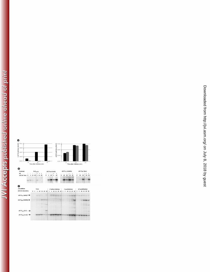
фKZ infection is resistant to rifampicin 276
With the exception of giant Bacillus phage PBS2 [22-24], all currently known phages require the bacterial 277
host transcriptional machinery at least during part of the infection. However, none of the фKZ transcripts 278
have sequences resembling bacterial promoters upstream of their 5’ ends. To test if host RNAP is 279
involved in фKZ infection, we analyzed the sensitivity of infection to Rif. Infection with P. aeruginosa 280
phage LUZ19 was used as a control. LUZ19 encodes a T7-like ssRNAP but requires host RNAP for early 281
transcription [43]. The addition of 400 µg/ml Rif to bacterial cultures prior to LUZ19 infection completely 282
abolished progeny phage production. In contrast, фKZ infected cultures produced similar amounts of 283
progeny both in the presence and in the absence of Rif (Figure 5a). 284
To confirm that transcription from all фKZ promoters is indeed Rif-resistant, primer extension reactions 285
described above were repeated on total RNA extracted from P. aeruginosa cells infected with the phage 286
in the presence of Rif. As can be seen (Figure 5b), the presence of Rif from the onset of infection did not 287
change the levels of early (P54), middle (P152) or late (P153) фKZ transcripts but completely abolished a 288
transcript from host promoter PrpoB2. These observations show that фKZ has the capacity to initiate and 289
complete the infection cycle in the absence of transcription by host RNAP, strongly suggesting the 290
reliance on Rif-independent phage-encoded RNAP(s). However, it cannot be totally excluded that the 291
phage virion contains factor(s) eliminating the Rif-sensitivity of the host RNAP and somehow direct it to 292
phage DNA only. 293
294
on July 8, 2018 by guesthttp://jvi.asm
.org/D
ownloaded from
14
Early transcription does not require protein synthesis 295
To investigate which temporal classes of phage transcription require newly synthesized phage proteins, 296
we purified total RNA from фKZ-infected P. aeruginosa cells supplemented with chloramphenicol (Cm) 297
before or during фKZ infection. Primer extension analysis showed that the overall kinetics of early phage 298
transcription does not depend on newly synthesized phage proteins (Figure 5c). With Cm being added at 299
different time points, the profiles of early transcript abundances changed slightly, likely showing that 300
new phage proteins may somehow attenuate transcription from early promoters and/or participate in 301
their processing or decay (Figure 5c). In contrast, transcription from middle (P50) and late (P29) phage 302
transcripts is clearly translation-dependent. Indeed, both P50- and P29-initiated transcripts were 303
completely absent if Cm was added prior to phage infection (Figure 5c). The partial transcription activity 304
from P50 observed with Cm added shortly after infection may be explained by incomplete blockage of 305
protein translation. When protein synthesis was ceased 10 minutes post-infection, transcription from 306
middle and late promoters was not affected. Taken together, the data clearly demonstrate that фKZ 307
transcription is performed by Rif-resistant phage-encoded RNAP. Early transcription must be performed 308
by an enzyme originating from the virion, while middle and late transcription must be performed by an 309
enzyme that is fully or partially synthesized de novo after infection. 310
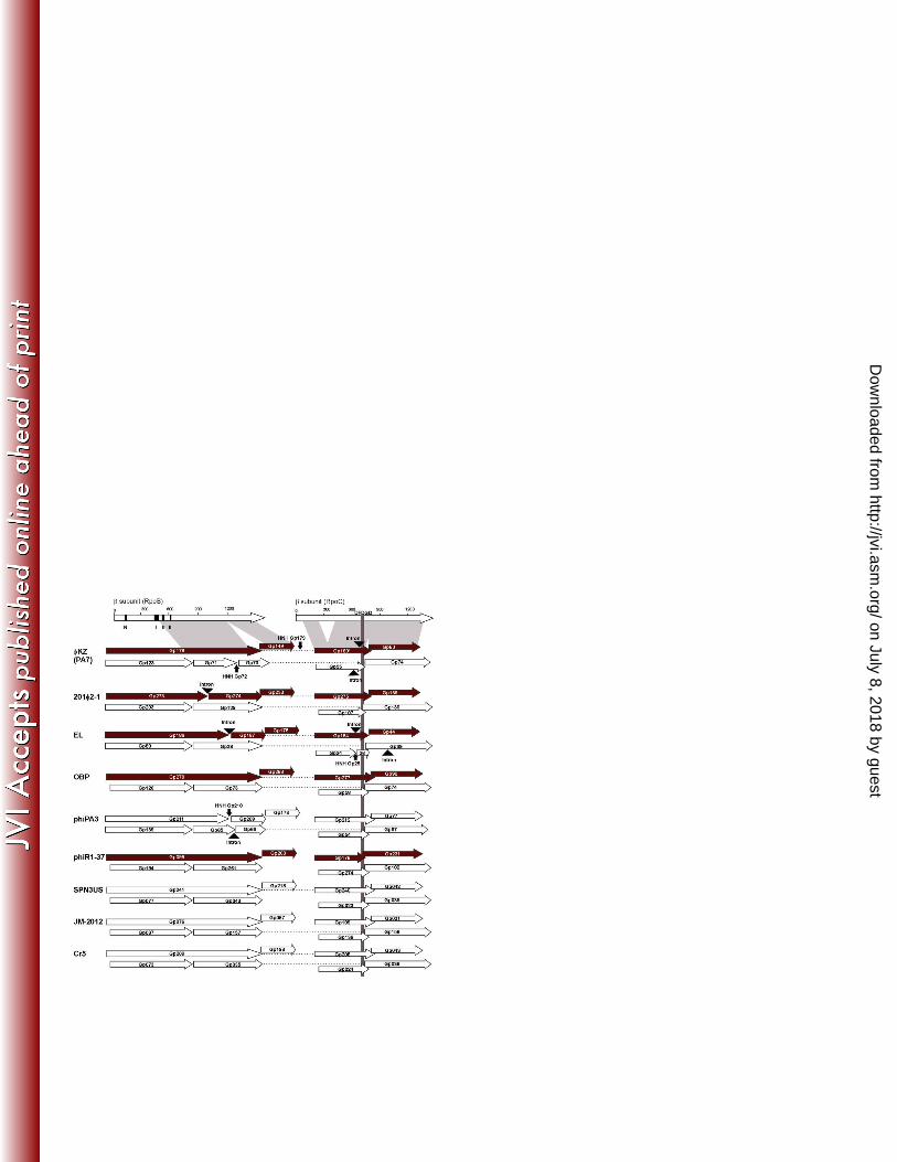
Two sets of split RNAP β and β’-like proteins are encoded by all known giant phage genomes 311
Careful in silico reanalysis of the фKZ genome merged with previously published mass spectrometric data 312
[27,28] indicated that фKZ encodes four β/β’-like virion-associated proteins (gp178, gp149, gp180, and 313
gp80) that may constitute vRNAP. Some of these proteins (e.g., gp149) show barely detectable 314
resemblance to known bacterial β or β’ RNAP subunits, and identification of similarities is further 315
hindered by interspersions by mobile genetic elements (Figure 6, Table S4). All putative vRNAP subunits 316
are products of middle and/or late genes. In contrast, four additional proteins with similarities to 317
on July 8, 2018 by guesthttp://jvi.asm
.org/D
ownloaded from
15
fragments of bacterial β/β’ subunits -- gp123, gp71/73, gp74, and gp55 -- are the products of early genes 318
and should therefore be part of the nvRNAP complex (Figure 6, Table S4). These eight RNAP subunits are 319
conserved in all known фKZ-related phages infecting a wide range of Gram-negative species belonging to 320
Pseudomonas, Salmonella, Erwinia, Vibrio, Cronobacter and Yersinia genera (Figure 6, Table S4). 321
Remarkably, no recognizable homologous to bacterial RNAP assembly subunit α or promoter specificity 322
σ subunit are predicted in any of giant phage genomes. One would assume that these phage-encoded 323
β/β’ subunits recruit the bacterial RNAP α subunit as an assembly platform [2,3]. However, the co-324
immunopurification experiment described above demonstrated that purification of bacterial RNAP 325
complex from infected cells through a tag positioned on the α subunit failed to identify phage RNAP 326
subunits. This suggests that the bacterial α subunit does not form hybrid(s) with any of the phage-327
encoded RNAPs subunits. 328
In a reciprocal experiment, we expressed 6xHis-tagged gp178 in P. aeruginosa cells and infected the 329
resulting strain with фKZ. After 10 and 25 minutes of infection, we co-purified the tagged gp178-His6. As 330
a negative control, non-infected cells were used. Because of the low stringency wash conditions, high 331
amounts of background proteins were identified in both non-infected and infected samples (Table S5). 332
However, while in the presence of gp178-His6 spectral counts for three other predicted vRNAP subunits 333
(gp80/gp149/gp180) were markedly increased, host bacterial RNAP and nvRNAP subunits were present 334
in negligible amounts (Table S5). The result indicates that the phage-encoded RNAP subunits from virion 335
and non-virion sets likely do not form hybrid(s) with host RNAP subunits as well as with each other. 336
337
DISCUSSION 338
on July 8, 2018 by guesthttp://jvi.asm
.org/D
ownloaded from
16
Pseudomonas phage фKZ is the type virus of a genus of remarkably large Myoviridae that are only 339
marginally related to other known phages. Although they were originally uniquely associated with 340
Pseudomonas, фKZ-related phages have been discovered that target a variety of Gram-negative bacteria 341
[27-30, 40-42]. A remarkable feature of these phages is that they all contain two sets of proteins 342
distantly related to fragments of bacterial multisubunit RNAP β and β’ subunits. Proteins from one set 343
were found in the virion for several giant phages [27-30]. Here, we show that the entire фKZ genome, 344
comprising ~280 kb and encoding 369 genes grouped into 134 operons, can be transcribed entirely by 345
viral-encoded RNAP. Host RNAP does not seem to contribute significantly to фKZ gene expression, since 346
phage infection proceeds normally in the presence of high concentrations of Rifampicin. 347
Global RNA profiling of фKZ allows us to come up with a general view of the of фKZ transcription. The 348
vRNAP is injected in the bacterial cell and initiates transcription from 28 early promoters characterized 349
by a highly conserved AT-rich consensus element and a less conserved upstream motif. All early 350
promoters are located on the same strand and are distributed throughout the circular permuted genome 351
of фKZ (Figure 2). Phage protein synthesis is not needed for early transcription to occur, indicating that 352
all determinants of early transcription are provided from the virion. Transcription of middle and late 353
promoters requires phage protein synthesis. At least one of these promoter classes must be recognized 354
by nvRNAP, whose subunits are encoded by early genes. Consensus elements for middle and late 355
promoters are distinct but not very well conserved. Since middle and late transcripts have clearly 356
different temporal kinetics and different promoter consensus elements, a question arises how is the 357
differential expression achieved. In principle, two scenarios are possible. First, middle transcription can 358
be catalyzed by vRNAP modified by a product(s) of an early gene, with late transcription performed by 359
nvRNAP. A second possibility is that both middle and late transcription is catalyzed by nvRNAP, whose 360
promoter specificity is modified by a middle gene product at the onset of late stage of infection. 361
on July 8, 2018 by guesthttp://jvi.asm
.org/D
ownloaded from
17
Purification and characterization of фKZ RNAPs from infected cultures at different time-points of 362
infection will be needed to determine which scenario actually takes place. 363
In both 4-polypeptide sets of phage β /β’-like subunits, one pair of proteins jointly forms a counterpart of 364
a full-sized cellular RNAP subunit homolog (Figure 6). Remarkably, the split of the β-like phage proteins 365
roughly coincides with the natural split in the β counterpart from archaeal RNAP, while the split of the 366
β’-like phage subunits is analogous to the subdivision of the β’ in cyanobacterial RNAPs [44, 45]. Such 367
splits are also tolerated by E. coli RNAP and occur at surface exposed loops of RNAP. The two largest 368
subunits of bacterial RNAPs jointly form the catalytic center of the enzyme, and are held together by a 369
dimer of α subunits, which are strictly required for RNAP assembly but not for promoter melting [46]. 370
Remarkably, фKZ does not encode a recognizable α homolog. Moreover, the affinity purification data as 371
well as the analysis of virion polypeptide content suggest that the phage RNAP subunit fragments do not 372
associate with the host α subunit. These observations suggest that фKZ and, by extension, other giant 373
phage RNAPs may either use a novel protein for assembly of large RNAP subunit fragments or assembly 374
occurs without an α homolog or analog. To experimentally investigate this interesting question, as well 375
as to determine which factors, if any, determine promoter specificity of viral RNAPs, purification of these 376
enzymes from infected cells and фKZ virions will be required. 377
To date, the giant phages PBS2 (infecting Gram-positive Bacilli) and фKZ (infecting Gram-negative 378
pseudomonads) are the only phages known that are independent of the host transcription apparatus. 379
Both giant phages encode Rif-resistant, multisubunit RNAPs [This work; 23]. Comparison of the numbers 380
of subunits (five in PBS2 and four predicted in фKZ) as well as their approximate sizes suggests that both 381
RNAP complexes of these giant phages might be related. If this conjecture is true, then giant phages will 382
be unique in their ability to infect both Gram-negative and Gram-positive hosts. Independence from host 383
transcription apparatus could be an important trait that contributes to such versatility. Determination of 384
on July 8, 2018 by guesthttp://jvi.asm
.org/D
ownloaded from
18
the PBS2 genomic sequence ongoing in one of our laboratories should help answer this question and 385
may shed more light on the highly unusual multisubunit bacteriophage RNAPs. 386
387
388
389
ACKNOWLEDGEMENTS 390
J. De Smet holds a predoctoral fellowships of the ‘Agentschap voor Innovatie door Wetenschap en 391
Technologie in Vlaanderen’ (IWT, Belgium). A. Van den Bossche and P.J. Ceyssens are pre- and 392
postdoctoral fellows supported by the ‘Fonds voor Wetenschappelijk Onderzoek’ (FWO, Belgium). This 393
research was supported by Grant G.0323.09 from the FWO and SBO-project 100042 of the IWT. The 394
work in U.B´s laboratory was supported by the Austrian Science Fund through project RNA-REG 43, 395
subproject AF4311. The work was also supported by the National Institutes of Health (grant GM59259 to 396
K.S.); the Russian Academy of Sciences Presidium Program in Molecular and Cellular Biology and the 397
Ministry of Education and Science of the Russian Federation project 14.B25.31.0004 grant to K.S. and 398
Federal targeted program “Scientific and scientific-pedagogical personnel of innovative Russia 2009-399
2013” to L.M. 400
401
402
403
404
on July 8, 2018 by guesthttp://jvi.asm
.org/D
ownloaded from
19
REFERENCES 405
1. Cramer P. 2002. Multisubunit RNA polymerases. Curr. Opin. Struct. Biol. 12: 89-97 406
2. Darst SA. 2001. Bacterial DNA polymerase. Curr. Opin. Struct. Biol. 11: 155-162 407
3. Lane WJ, Darst SA. 2010a. Molecular evolution of multisubunit RNA polymerases: structural analysis. J. Mol. 408
Biol. 395: 686-704 409
4. Lane WJ, Darst SA. 2010b. Molecular evolution of multisubunit RNA polymerases: sequence analysis. J. Mol. 410
Biol. 395: 671-85 411
5. Iyer LM, Koonin EV, Aravind L. 2003. Evolutionary connection between the catalytic subunits of DNA-412
dependent RNA polymerases and eukaryotic RNA-dependent RNA polymerases and the origin of RNA 413
polymerases. BMC Struct. Biol. 28: 1 414
6. Cermakian N, Ikeda TM, Miramontes P, Lang BF, Gray MW, Cedergren R. 1997. On the evolution of 415
the single-subunit RNA polymerases. J. Mol. Evol. 45:671-81 416
7. Cheetham GM, Steitz TA. 1999. Structure of a transcribing T7 RNA polymerase initiation complex. 417
Science 286:2305-9 418
8. Campbell EA, Korzheva N, Mustaev A, Murakami K, Nair S, Goldfarb A, Darst SA. 2001. Structural mechanism 419
for rifampicin inhibition of bacterial RNA polymerase. Cell 104: 901-12 420
9. Nechaev S, Severinov K. 2003. Bacteriophage-induced modifications of host RNA polymerase. Annu. Rev. 421
Microbiol. 57: 301-22. 422
10. Nechaev S, Severinov K. 2008. The elusive object of desire--interactions of bacteriophages and their hosts. Curr. 423
Opin. Microbiol. 11: 186-93. 424
11. Steitz TA. 2004. The structural basis of the transition from initiation to elongation phases of transcription, as 425
well as translocation and strand separation, by T7 RNA polymerase. Curr. Opin. Struct. Biol. 14: 4-9 426
12. Kazmierczak KM, Davydova EK, Mustaev AA, Rothman-Denes LB. 2002. The phage N4 virion RNA polymerase 427
catalytic domain is related to single-subunit RNA polymerases. EMBO J. 21: 5815-23 428
on July 8, 2018 by guesthttp://jvi.asm
.org/D
ownloaded from
20
13. Jamalludeen N, Kropinski AM, Johnson RP, Lingohr E, Harel J, Gyles CL. 2008. Complete genomic sequence of 429
bacteriophage phiEcoM-GJ1, a novel phage that has myovirus morphology and a podovirus-like RNA 430
polymerase. Appl. Environ. Microbiol. 74: 516-25 431
14. Semenova E, Djordjevic M, Shraiman B, Severinov K. 2005. The tale of two RNA polymerases: transcription 432
profiling and gene expression strategy of bacteriophage Xp10. Mol. Microbiol. 55: 764-77. 433
15. Sousa R, Mukherjee S. 2003. T7 RNA polymerase. Prog Nucleic Acid Res. Mol. Biol. 73: 1–41. 434
16. Severinova E, Severinov K. 2007. Localization of the Escherichia coli RNA polymerase beta' subunit residue 435
phosphorylated by bacteriophage T7 kinase Gp0.7. J Bacteriol. 188: 3470-6. 436
17. Savalia D, Robins W, Nechaev S, Molineux I, Severinov K. 2010. The role of the T7 Gp2 inhibitor of host RNA 437
polymerase in phage development. J Mol Biol. 402:118-26. 438
18. Falco SC, Laan KV, Rothman-Denes LB. 1977. Virion-associated RNA polymerase required for bacteriophage N4 439
development. Proc. Natl. Acad. Sci. USA 74: 520-3 440
19. Gleghorn ML, Davydova EK, Rothman-Denes LB, Murakami KS. 2008. Structural basis for DNA-hairpin 441
promoter recognition by the bacteriophage N4 virion RNA polymerase. Mol. Cell 32: 707-17 442
20. Ceyssens PJ, Brabban A, Rogge L, Lewis MS, Pickard D, Goulding D, Dougan G, Noben JP, Kropinski A, Kutter E, 443
Lavigne R. 2010. Molecular and physiological analysis of three Pseudomonas aeruginosa phages belonging to 444
the "N4-like viruses". Virology 405: 26-30 445
21. Cho NY, Choi M, Rothman-Denes LB. 1995. The bacteriophage N4-coded single-stranded DNA-binding protein. 446
N4SSB. is the transcriptional activator of Escherichia coli RNA polymerase at N4 late promoters. J. Mol. Biol. 447
246: 461-471 448
22. Price AR, Frabotta M. 1972. Resistance of bacteriophage PBS2 infection to rifampicin, an inhibitor of Bacillus 449
subtilis RNA synthesis. Biochem. Biophys. Res. Commun. 48: 1578-85 450
23. Clark S. 1978. Transcriptional specificity of a multisubunit RNA polymerase induced by Bacillus subtilis 451
bacteriophage PBS2. J. Virol. 25: 224-37 452
24. Clark S, Losick R, Pero J. 1974. New RNA polymerase from Bacillus subtilis infected with phage PBS2. Nature 453
252: 21-4 454
25. Hendrix RW. 2009. Jumbo bacteriophages. Curr. Top. Microbiol. Immunol. 328: 229-40. 455
on July 8, 2018 by guesthttp://jvi.asm
.org/D
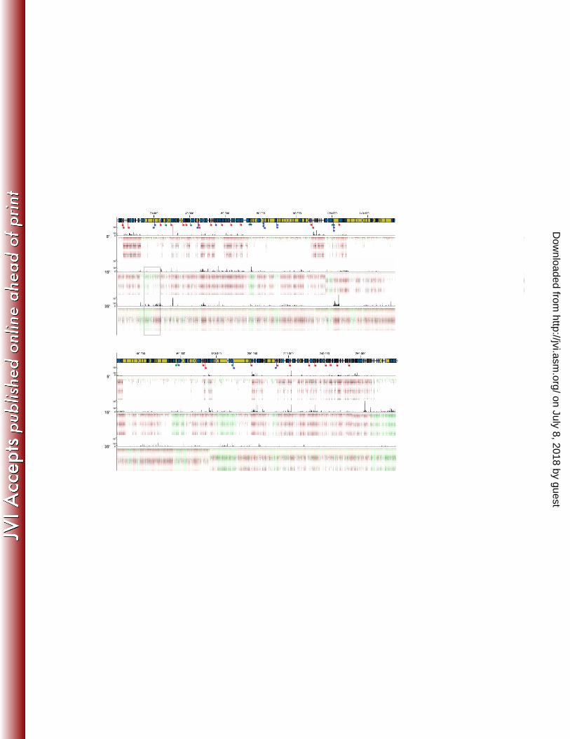
ownloaded from
21
26. Mesyanzhinov VV, Robben J, Grymonprez B, Kostyuchenko VA, Bourkaltseva MV, Sykilinda NN, Krylov VN, 456
Volckaert G. 2002. The genome of bacteriophage phiKZ of Pseudomonas aeruginosa. J. Mol. Biol. 317: 1-19 457
27. Thomas JA, Weintraub ST, Wu W, Winkler DC, Cheng N, Steven AC, Black LW. 2012. Extensive proteolysis of 458
head and inner body proteins by a morphogenetic protease in the giant Pseudomonas aeruginosa phage φKZ. 459
Mol. Microbiol. 84: 324-39 460
28. Lecoutere E, Ceyssens PJ, Miroshnikov KA, Mesyanzhinov VV, Krylov VN, Noben JP, Robben J, Hertveldt K, 461
Volckaert G, Lavigne R. 2009. Identification and comparative analysis of the structural proteomes of phiKZ and 462
EL, two giant Pseudomonas aeruginosa bacteriophages. Proteomics 9: 3215-9. 463
29. Thomas JA, Rolando MR, Carroll CA, Shen PS, Belnap DM, Weintraub ST, Serwer P, Hardies SC. 2008. 464
Characterization of Pseudomonas chlororaphis myovirus 201varphi2-1 via genomic sequencing, mass 465
spectrometry, and electron microscopy. Virology 376: 330-8 466
30. Skurnik M, Hyytiäinen HJ, Happonen LJ, Kiljunen S, Datta N, Mattinen L, Williamson K, Kristo P, Szeliga M, 467
Kalin-Mänttäri L, Ahola-Iivarinen E, Kalkkinen N, Butcher SJ. 2012. Characterization of the genome, proteome, 468
and structure of yersiniophage R1-37. J. Virol. 86: 12625-42. 469
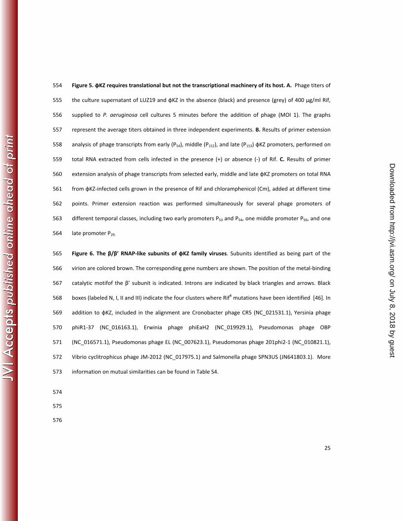
31. Ceyssens PJ, Lavigne R, Mattheus W, Chibeu A, Hertveldt K, Mast J, Robben J, Volckaert G. 2006. Genomic 470
analysis of Pseudomonas aeruginosa phages LKD16 and LKA1: establishment of the phiKMV subgroup within the 471
T7 supergroup. J. Bacteriol. 188: 6924-31 472
32. Klimuk E, Akulenko N, Makarova KS, Ceyssens PJ, Volchenkov I, Lavigne R, Severinov K. 2013. Host RNA 473
polymerase inhibitors encoded by KMV-like phages of Pseudomonas. Virology 436: 67-74. 474
33. Qiu D, Damron FH, Mima T, Schweizer HP, Yu HD. 2008. PBAD-based shuttle vectors for functional 475
analysis of toxic and highly regulated genes in Pseudomonas and Burkholderia spp. and other 476
bacteria. Appl. Environ. Microbiol. 74: 7422-6 477
34. Sevostyanova A, Djordjevic M, Kuznedelov K, Naryshkina T, Gelfand MS, Severinov K, Minakhin L. 478
2007. Temporal regulation of viral transcription during development of Thermus thermophilus 479
bacteriophage phiYS40. J. Mol. Biol. 366: 420-35. 480
on July 8, 2018 by guesthttp://jvi.asm
.org/D
ownloaded from

22
35. Cenens W, Mebrhatu MT, Makumi A, Ceyssens PJ, Lavigne R, Van Houdt R, Taddei F, Aertsen A. 2013. 481
Expression of a novel P22 ORFan gene reveals the phage carrier state in Salmonella Typhimurium. PLoS Genet. 482
9: e1003269. 483
36. Monson R, Foulds I, Foweraker J, Welch M, Salmond GP. 2011. The Pseudomonas aeruginosa generalized 484
transducing phage phiPA3 is a new member of the phiKZ-like group of 'jumbo' phages, and infects model 485
laboratory strains and clinical isolates from cystic fibrosis patients. Microbiology 157: 859-67. 486
37. Toledo-Arana A, Dussurget O, Nikitas G, Sesto N, Guet-Revillet H, Balestrino D, Loh E, Gripenland J, Tiensuu T, 487
Vaitkevicius K. 2009. The Listeria transcriptional landscape from saprophytism to virulence. Nature 459: 950-6. 488
38. Stazic D, Lindell D, Steglich C. 2011. Antisense RNA protects mRNA from RNase E degradation by RNA-RNA 489
duplex formation during phage infection. Nucleic Acids Res. 39: 4890-9 490
39. Kawano M, Aravind L, Storz G. 2007. An antisense RNA controls synthesis of an SOS-induced toxin evolved from 491
an antitoxin. Mol. Microbiol. 64: 738-54 492
40. Cornelissen A, Hardies SC, Shaburova OV, Krylov VN, Mattheus W, Kropinski AM, Lavigne R. 2012. Complete 493
genome sequence of the giant virus OBP and comparative genome analysis of the diverse ΦKZ-related phages. 494
J. Virol. 86: 1844-52 495
41. Hertveldt K, Lavigne R, Pleteneva E, Sernova N, Kurochkina L, Korchevskii R, Robben J, Mesyanzhinov V, 496
Krylov VN, Volckaert G. 2005. Genome comparison of Pseudomonas aeruginosa large phages. J. Mol. Biol. 354: 497
536-45 498
42. Dömötör D, Becságh P, Rákhely G, Schneider G, Kovács T. 2012. Complete genomic sequence of Erwinia 499
amylovora phage PhiEaH2. J. Virol. 86: 10899. 500
43. Ceyssens PJ, Glonti T, Kropinski NM, Lavigne R, Chanishvili N, Kulakov L, Lashkhi N, Tediashvili M, Merabishvili 501
M. 2011. Phenotypic and genotypic variations within a single bacteriophage species. Virol. J. 8:1 502
44. Severinov K. 2000. RNA polymerase structure-function: insights into points of transcriptional regulation. Curr. 503
Opin. Microbiol. 3: 118-25 504
45. Xie WQ, Jäger K, Potts M. 1989. Cyanobacterial RNA polymerase genes rpoC1 and rpoC2 correspond to rpoC of 505
Escherichia coli. J. Bacteriol. 171: 1967-73 506
on July 8, 2018 by guesthttp://jvi.asm
.org/D
ownloaded from
23
46. Young BA, Gruber TM, Gross CA. 2004. Minimal machinery of RNA polymerase holoenzyme sufficient for 507
promoter melting. Science 303:1382-4. 508
47. Jatsenko T, Tover A, Tegova R, Kivisaar M. 2010. Molecular characterization of Rifr. mutations in Pseudomonas 509
aeruginosa and Pseudomonas putida. Mutat. Res. 683:106-14. 510
511
512
513
514
515
516
517
518
519
520
521
522
523
524
525
526
527
528
529
530
531
on July 8, 2018 by guesthttp://jvi.asm
.org/D
ownloaded from
24
FIGURE LEGENDS 532
Figure 1. Total RNA extraction from фKZ-infected cells. A. Time-lapse recording of three P. aeruginosa 533
cells infected with фKZ at time 0. B. Total RNA was extracted in triplicate from 200 μl infected cells at the 534
indicated times post-infection and quantified spectrophotometrically. C. Transcript abundance of 5S 535
rRNA (black) and oprL (grey) per μl extracted RNA, as determined by RT-qPCR with three biological and 536
technical replicates. 537
Figure 2. RNA-seq analysis of the фKZ transcriptome. Genome-wide overview of reads mapped to the 538
sense (red) or antisense (green) strand of фKZ genome at samples taken 5, 15, and 35 minutes after 539
infection. Phage genes encoding structural proteins are indicated in yellow. Early, middle, and late 540
promoters examined in more detail are indicated as red, green and blue arrows, respectively. An 541
example of the antisense transcription in more detail can be found in Figure S1. 542
Figure 3. Impact of phage infection on bacterial transcriptome. Volcano plot of the P. aeruginosa 543
transcriptome as compared to the uninfected sample, with each dot representing an ORF, 10 (blue) and 544
35 (red) minutes post-infection. Triangles indicate structural RNAs, large circles indicate genes which are 545
located in the Pf1 operon. 546
Figure 4. Mapping the фKZ promoters. A. Kinetics of accumulation of selected early, middle, and late 547
фKZ transcripts revealed by primer extension assay is shown. Numbers of genes located downstream of 548
primer extension end points and time points when infected cells were collected and processed for RNA 549
purification are indicated. B. Alignments of фKZ promoters sequences. Consensus sequences of early, 550
middle, and late promoters derived from 28, 6, and 16 sequences, respectively. Experimentally defined 551
transcription start sites are in bold. The corresponding sequence logos are depicted below the 552
alignments. Red bars delineate conserved promoter elements. 553
on July 8, 2018 by guesthttp://jvi.asm
.org/D
ownloaded from
25
Figure 5. фKZ requires translational but not the transcriptional machinery of its host. A. Phage titers of 554
the culture supernatant of LUZ19 and фKZ in the absence (black) and presence (grey) of 400 µg/ml Rif, 555
supplied to P. aeruginosa cell cultures 5 minutes before the addition of phage (MOI 1). The graphs 556
represent the average titers obtained in three independent experiments. B. Results of primer extension 557
analysis of phage transcripts from early (P54), middle (P152), and late (P153) фKZ promoters, performed on 558
total RNA extracted from cells infected in the presence (+) or absence (-) of Rif. C. Results of primer 559
extension analysis of phage transcripts from selected early, middle and late фKZ promoters on total RNA 560
from фKZ-infected cells grown in the presence of Rif and chloramphenicol (Cm), added at different time 561
points. Primer extension reaction was performed simultaneously for several phage promoters of 562
different temporal classes, including two early promoters P53 and P54, one middle promoter P50, and one 563
late promoter P29. 564
Figure 6. The β/β’ RNAP-like subunits of фKZ family viruses. Subunits identified as being part of the 565
virion are colored brown. The corresponding gene numbers are shown. The position of the metal-binding 566
catalytic motifof the β’ subunit is indicated. Introns are indicated by black triangles and arrows. Black 567
boxes (labeled N, I, II and III) indicate the four clusters where RifR mutations have been identified [46]. In 568
addition to φKZ, included in the alignment are Cronobacter phage CR5 (NC_021531.1), Yersinia phage 569
phiR1-37 (NC_016163.1), Erwinia phage phiEaH2 (NC_019929.1), Pseudomonas phage OBP 570
(NC_016571.1), Pseudomonas phage EL (NC_007623.1), Pseudomonas phage 201phi2-1 (NC_010821.1), 571
Vibrio cyclitrophicus phage JM-2012 (NC_017975.1) and Salmonella phage SPN3US (JN641803.1). More 572
information on mutual similarities can be found in Table S4. 573
574
575
576
on July 8, 2018 by guesthttp://jvi.asm
.org/D
ownloaded from
5'
10³
0
15'
35'
10²
10
10³
4
0
10
10³
4
0
5'
10³
0
15'
35'
10²
10
10³
4
0
10
10³
4
0
on July 8, 2018 by guesthttp://jvi.asm
.org/D
ownloaded from
![Problem Solutions for Chapter 2 - SubodhTripathi · Problem Solutions for Chapter 2 ... 3 sin 2 (ωt - kz) = [1 ... fiber = )) = [] ...](https://static.fdocument.org/doc/165x107/5b91934109d3f2f8508bd726/problem-solutions-for-chapter-2-subodhtripathi-problem-solutions-for-chapter.jpg)