Development of a Monoclonal Antibody to Detect αs1-casein ...
4
Development of a Monoclonal Antibody to Detect αs1-casein in the Milk of Healthy and Mastitis-Affected Goats Ming PANG 1 Xiong GUAN 1 Chen-Xiang ZUO 1 Ming-Jie LIU 1 Saad REHMAN 1 Qin-Lei FAN 2 Ping LU 2 De-Kun CHEN 1 Wen-Tao MA 1,a 1 Veterinary Immunology Laboratory, College of Veterinary Medicine, Northwest Agriculture and Forestry University, Yangling 712100, Shaanxi Province, CHINA 2 China Animal Health and Epidemiology Center, Qingdao 266032, Shandong Province, CHINA a ORCID: 0000-0002-4747-1489 Article ID: KVFD-2019-22287 Received: 17.03.2018 Accepted: 26.08.2019 Published Online: 29.08.2019 How to Cite This Article Pang M, Guan X, Zuo CX, Liu MJ, Rehman S, Fan QL, Lu P, Chen DK, Ma WT: Development of a monoclonal antibody to detect αs1-casein in the milk of healthy and mastitis-affected goats. Kafkas Univ Vet Fak Derg, 26 (1): 71-74, 2020. DOI: 10.9775/kvfd.2019.22287 Abstract This study aimed to evaluate the expression level of caseins during mastitis of goats. Whole goat caseins were used as primary antigens for mouse immunization. A monoclonal antibody (mAb) named 5B with high specificity to goat αs1-casein was developed. Further results showed that mAb 5B can successfully be applied to western blot and ELISA. In addition, immunofluorescence analysis using this mAb showed increased milk αs1-casein level in mastitis-affected goats. In conclusion, this study has established an effective tool to evaluate the expression level of αs1-casein during mastitis development. Keywords: αs1-casein, Monoclonal antibody, ELISA, Mastitis, Immunofluorescence, Goat Sağlıklı ve Mastitisli Keçi Sütünde αs1-kazeini Belirleyen Monoklonal Antikor Geliştirilmesi Öz Bu çalışma keçilerde mastitis sırasında kazein ekspresyon seviyesini değerlendirmeyi amaçlamıştır. Farelerde immünizasyon için birincil antijen olarak keçi kazeinleri kullanıldı. Keçi αs1-kazeine yüksek spesifite gösteren ve 5B olarak adlandırılan bir monoklonal antikor (mAb) geliştirildi. Sonuçlar mAb 5B’nin western blot ve ELISA’ya başarıyla uygulanabileceğini gösterdi. Ek olarak, geliştirilen mAb kullanılarak yapılan immünofloresan analizi, mastitisten etkilenen keçilerde süt αs1-kazein seviyesinin arttığını gösterdi. Sonuç olarak, bu çalışma ile mastitis gelişimi sırasında αs1-kazeinin ekspresyon seviyesini değerlendirmek için etkili bir faktör belirlenmiştir. Anahtar sözcükler: αs1-kazein, Monoklonal antikor, ELISA, Mastitis, İmmünofloresans, Keçi INTRODUCTION Goat milk and its productions begin gaining attention because of their easy digestibility and lower allergenic properties compared to the cow milk [1] . An important role for goat milk is to provide the necessary nutrients for infants affected by cow milk allergy [2] . In addition, the nutritional value of goat milk has been widely accepted considering the total protein, fat, vitamin and mineral contents [3,4] . Caseins in milk account for more than 80% of total proteins and consist of αs1-, αs2-, β-, and κ-caseins and αs1-casein is one of the most important highly- phosphorylated proteins among caseins [3] . Goat milk contains less αs1-casein (5%-20%) compared to (30%- 35%) cow milk, making goat milk more similar to human milk and less likely to cause allergy [3] . In addition, αs1- casein influences the coagulation properties of milk [3] . With the development of hybridoma technology, monoclonal antibodies (mAbs) against caseins were used to determine the content of β-casein in bovine milk [5] . However, up to the present, mAbs against goat caseins have not yet been developed. In this paper, we used purified goat caseins for mouse immunization. Two strains of hybridoma cells targeting goat caseins (named 5B and 7H) were screened by indirect İleşim (Correspondence) +86-29-87092134 [email protected] Kafkas Universitesi Veteriner Fakultesi Dergisi ISSN: 1300-6045 e-ISSN: 1309-2251 Journal Home-Page: http://vetdergikafkas.org Online Submission: http://submit.vetdergikafkas.org Research Article Kafkas Univ Vet Fak Derg 26 (1): 71-74, 2020 DOI: 10.9775/kvfd.2019.22287
Transcript of Development of a Monoclonal Antibody to Detect αs1-casein ...
Development of a Monoclonal Antibody to Detect αs1-casein in the Milk of Healthy and Mastitis-Affected Goats
Ming PANG 1 Xiong GUAN 1 Chen-Xiang ZUO 1 Ming-Jie LIU 1 Saad REHMAN 1 Qin-Lei FAN 2 Ping LU 2 De-Kun CHEN 1 Wen-Tao MA 1,a
1 Veterinary Immunology Laboratory, College of Veterinary Medicine, Northwest Agriculture and Forestry University, Yangling 712100, Shaanxi Province, CHINA2 China Animal Health and Epidemiology Center, Qingdao 266032, Shandong Province, CHINAa ORCID: 0000-0002-4747-1489
Article ID: KVFD-2019-22287 Received: 17.03.2018 Accepted: 26.08.2019 Published Online: 29.08.2019
How to Cite This Article
Pang M, Guan X, Zuo CX, Liu MJ, Rehman S, Fan QL, Lu P, Chen DK, Ma WT: Development of a monoclonal antibody to detect αs1-casein in the milk of healthy and mastitis-affected goats. Kafkas Univ Vet Fak Derg, 26 (1): 71-74, 2020. DOI: 10.9775/kvfd.2019.22287
AbstractThis study aimed to evaluate the expression level of caseins during mastitis of goats. Whole goat caseins were used as primary antigens for mouse immunization. A monoclonal antibody (mAb) named 5B with high specificity to goat αs1-casein was developed. Further results showed that mAb 5B can successfully be applied to western blot and ELISA. In addition, immunofluorescence analysis using this mAb showed increased milk αs1-casein level in mastitis-affected goats. In conclusion, this study has established an effective tool to evaluate the expression level of αs1-casein during mastitis development.
Keywords: αs1-casein, Monoclonal antibody, ELISA, Mastitis, Immunofluorescence, Goat
Sağlıklı ve Mastitisli Keçi Sütünde αs1-kazeini Belirleyen Monoklonal Antikor Geliştirilmesi
ÖzBu çalışma keçilerde mastitis sırasında kazein ekspresyon seviyesini değerlendirmeyi amaçlamıştır. Farelerde immünizasyon için birincil antijen olarak keçi kazeinleri kullanıldı. Keçi αs1-kazeine yüksek spesifite gösteren ve 5B olarak adlandırılan bir monoklonal antikor (mAb) geliştirildi. Sonuçlar mAb 5B’nin western blot ve ELISA’ya başarıyla uygulanabileceğini gösterdi. Ek olarak, geliştirilen mAb kullanılarak yapılan immünofloresan analizi, mastitisten etkilenen keçilerde süt αs1-kazein seviyesinin arttığını gösterdi. Sonuç olarak, bu çalışma ile mastitis gelişimi sırasında αs1-kazeinin ekspresyon seviyesini değerlendirmek için etkili bir faktör belirlenmiştir.
Anahtar sözcükler: αs1-kazein, Monoklonal antikor, ELISA, Mastitis, İmmünofloresans, Keçi
INTRODUCTIONGoat milk and its productions begin gaining attention because of their easy digestibility and lower allergenic properties compared to the cow milk [1]. An important role for goat milk is to provide the necessary nutrients for infants affected by cow milk allergy [2]. In addition, the nutritional value of goat milk has been widely accepted considering the total protein, fat, vitamin and mineral contents [3,4]. Caseins in milk account for more than 80% of total proteins and consist of αs1-, αs2-, β-, and κ-caseins and αs1-casein is one of the most important highly-phosphorylated proteins among caseins [3]. Goat milk
contains less αs1-casein (5%-20%) compared to (30%-35%) cow milk, making goat milk more similar to human milk and less likely to cause allergy [3]. In addition, αs1-casein influences the coagulation properties of milk [3]. With the development of hybridoma technology, monoclonal antibodies (mAbs) against caseins were used to determine the content of β-casein in bovine milk [5]. However, up to the present, mAbs against goat caseins have not yet been developed.
In this paper, we used purified goat caseins for mouse immunization. Two strains of hybridoma cells targeting goat caseins (named 5B and 7H) were screened by indirect
İletişim (Correspondence) +86-29-87092134 [email protected]
Kafkas Universitesi Veteriner Fakultesi DergisiISSN: 1300-6045 e-ISSN: 1309-2251
Journal Home-Page: http://vetdergikafkas.orgOnline Submission: http://submit.vetdergikafkas.org
Research ArticleKafkas Univ Vet Fak Derg26 (1): 71-74, 2020DOI: 10.9775/kvfd.2019.22287
72αs1-casein Expression Under Mastitis
ELISA. To explore the eff ect of mastitis on αs1-casein, we analyzed the level of αs1-casein of goat milk samples collected from healthy goats, goats with sub-clinical mastitis and clinical mastitis by indirect competitive ELISA. Immunofl uorescence of goat mammary epithelial cells was carried out to further examine αs1-casein expression profile in lipopolysaccharide (LPS)-induced mastitis in vitro.
MATERIAL and METHODS
Ethical Approval
Animal-related experiments in the present study were approved by the Research Ethics Committee of Northwest A&F University according to the guidelines of the Ministry of Health in China for the care and use of laboratory animals.
Antigen (Whole Goat Caseins) Preparation
Goat caseins were extracted using isoelectric precipitation. Briefly, 10 g of whole goat milk powder (purchased from Kabtrita, Holland) was dissolved in 10 mL of 2M Na2HPO4•12H2O buff er. The pH was adjusted to 4.2 using 1M CH3COOH solution (preheated to 40ºC). After complete precipitation at 37ºC, the solution was centrifuged at 4000g for 10 min, and the precipitate was washed with 20 mL of 95% (v/v) ethanol 3 times to remove lipid. Next, 1:1 (v/v) ethanol-ether mixture was added to wash the precipitate 3 times. Final precipitation was washed twice with ether and was placed in a fume hood until all ether volatilized. Casein powder was preserved at -80ºC and its purity and molecular weight were analyzed by SDS-PAGE [6].
Mouse Immunization, Cell Fusion, Hybridoma Cell Screening and Ascites Prepareation
Mouse immunization, cell fusion, hybridoma cell screening and ascites preparation were conducted as previously reported by our group [6].
Competitive Indirect ELISA
Competitive indirect ELISA was conducted as previously reported [6]. Specifically, the dilution of goat milk was 15 foldwith PBST and the dilution of mAb 5B was 7 fold with PBST.
Milk Sample Collection and Processing
For clinical mastitis samples, milk with the detection of fl akes and clots with gland swelling or systemic illness such as fever, depression, weakness and dehydration were selected. For sub-clinical mastitis diagnosis, the California Mastitis Test was used as previously reported [7]. All the milk was stored at 4ºC after collection and was used for further assays within 1 h.
Isolation and Culture of Goat Mammary Epithelial Cells (MECs)
Goat MEC isolation was performed as previously reported [6].
After isolation, the cells were cultured in DMEM/F12 medium (Thermo Fisher Scientific, New York, USA) supplemented with 10% fetal bovine serum (Thermo Fisher Scientific), 1% GlutaMAX Supplement (Thermo Fisher Scientific) and 100 units mL-1 of penicillin-streptomycin (Sigma Aldrich, Missouri, USA). The culture medium was refreshed every 24 h. The cells were cultured at 37ºC in 5% CO2 and were passaged when they were 80% confl uent.
LPS Stimulation and Immuno� uorescence Staining
The in vitro mastitis model was established by adding LPS (Sigma Aldrich) to the culture medium at a final concentration of 10 μg mL-1. Meanwhile, the control group was treated with culture medium of the same volume. All cells were treated for 12 h before immunofluorescence analysis. Procedures of immunofl uorescence were the same as previously reported [8].
Statistical Analysis
Statistical analyses were performed using GraphPad Prism version 5.0 (GraphPad Software Inc., San Diego, CA, USA). Diff erences were assessed using t-tests or one-way ANOVA with Dunnett test. P<0.05 was considered significant.
RESULTS
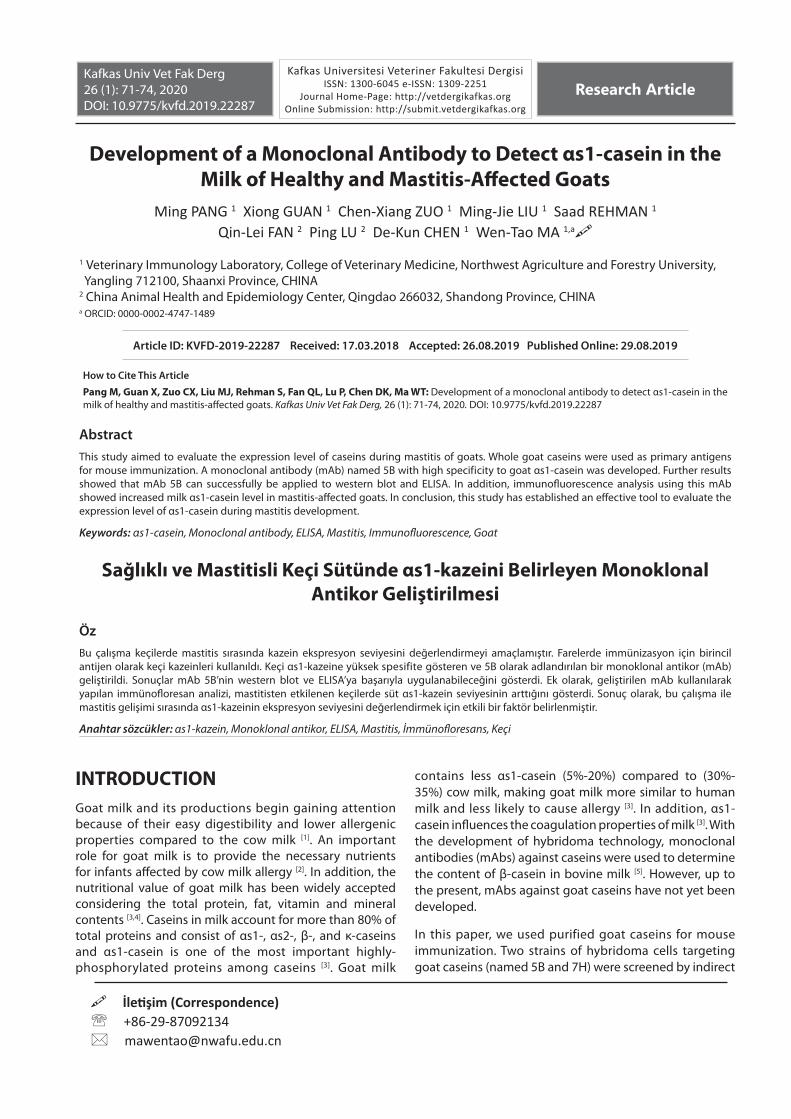
Isoelectric point precipitation method was used for thepurification of the goat casein and was identified by SDS-PAGE. After precipitation, caseins were purified andcontained αs1-casein because lane 3 and lane 4 has anidentical band at 23.9 kDa (Fig. 1). Consistent with a previous study, purified αs1-casein showed a higher molecularweight when analyzed by SDS-PAGE than expected [9].
The titers of sera collected from the immunized mice 7-10days after the forth immunization were determined byindirect ELISA. All immunized mice produced anti-goat
Fig 1. SDS-PAGE analysis of caseins. Lane 1, bovine milk powder; lane 2, goat milk powder; lane 3, total goat caseins; lane 4, goat αs1-casein; lane 5, bovine α-casein
73
PANG, GUAN, ZUO, LIUREHMAN, FAN, LU, CHEN, MA
casein antibodies. Several mice were selected for further experiment and were injected with 100 μg of immunogen intravenously 3 days before cell fusion. Then, hybridoma secreting antibodies specific to goat caseins but un-recognizable to bovine milk powder were selected by
indirect ELISA, and were subcloned by limiting dilution method. Two stable hybridoma cell lines were established and named as 5B and 7H. These two hybridoma cell lines showed strong proliferation ability and high specificity to goat caseins and was chosen for further studies.
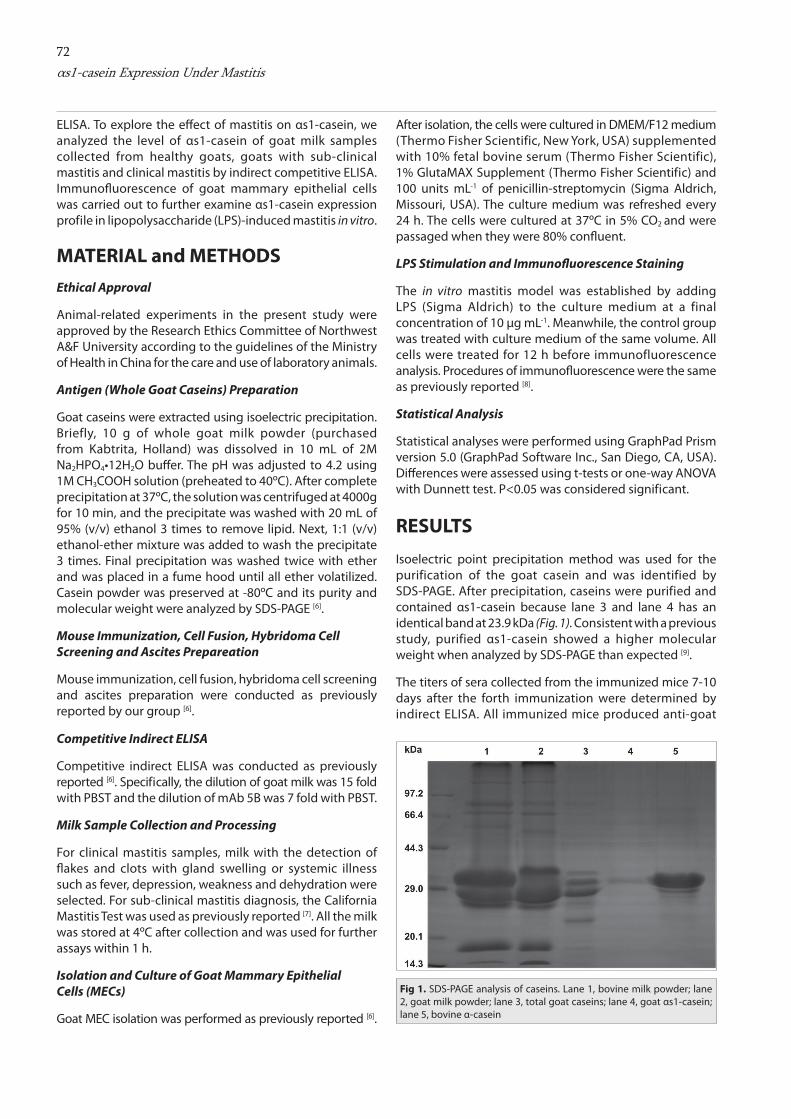
Cross-reactivity of mAbs 5B and 7H was analyzed by indirect ELISA using goat casein, bovine milk, goat milk, goat milk powder and bovine milk powder as antigens. The results showed that these two mAbs both specifically recognized goat caseins and had no cross-reactivity with bovine caseins (Table 1), which was supported by western blot analysis using mAb 5B (Fig. 2A). In addition, the specificity of mAb 5B was further evaluated using diff erent subtypes of goat caseins. The result showed that this mAb only reacted with goat αs1-casein, while no other reactions existed between mAb 5B and purified β- and κ-caseins (Fig. 2B).
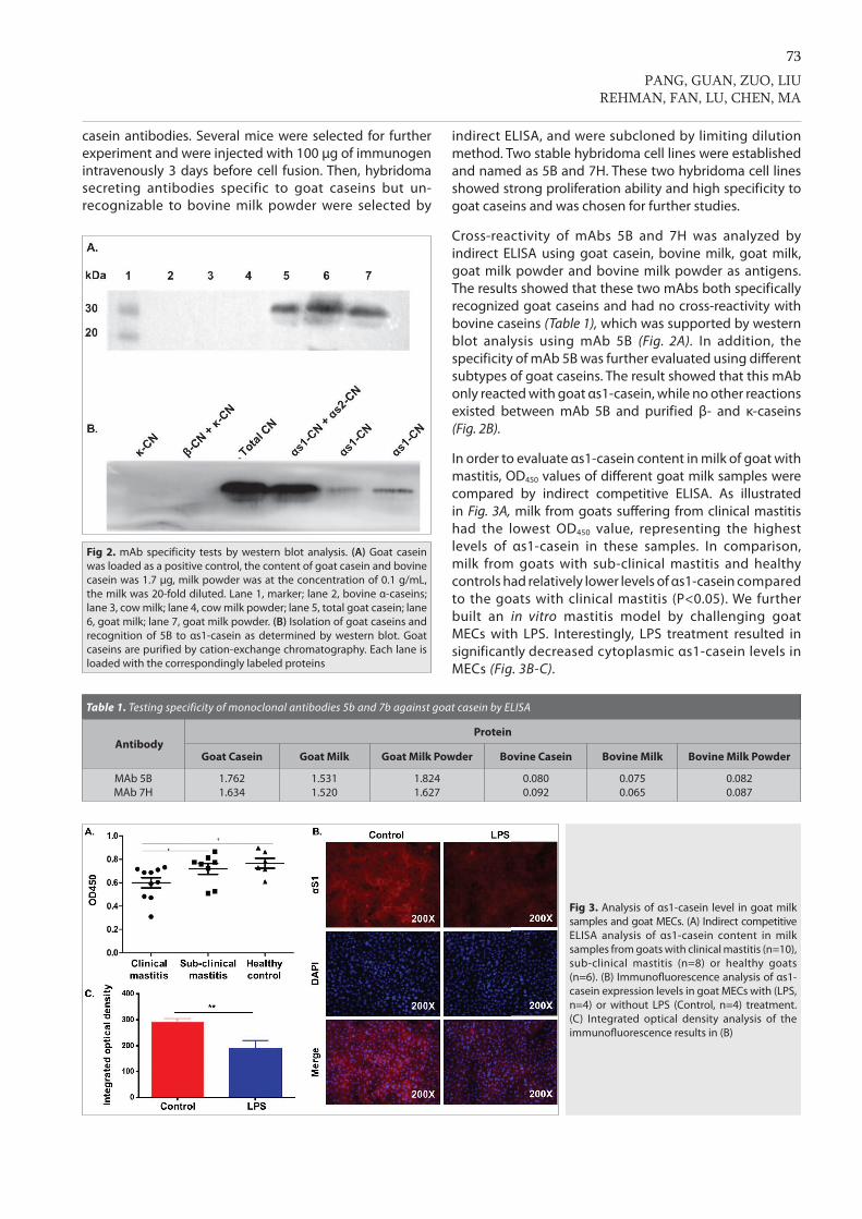
In order to evaluate αs1-casein content in milk of goat with mastitis, OD450 values of diff erent goat milk samples were compared by indirect competitive ELISA. As illustrated in Fig. 3A, milk from goats suff ering from clinical mastitis had the lowest OD450 value , representing the highest levels of αs1-casein in these samples. In comparison, milk from goats with sub-clinical mastitis and healthy controls had relatively lower levels of αs1-casein comparedto the goats with clinical mastitis (P<0.05). We further built an in vitro mastitis model by challenging goat MECs with LPS. Interestingly, LPS treatment resulted in significantly decreased cytoplasmic αs1-casein levels in MECs (Fig. 3B-C).
Fig 2. mAb specificity tests by western blot analysis. (A) Goat casein was loaded as a positive control, the content of goat casein and bovine casein was 1.7 μg, milk powder was at the concentration of 0.1 g/mL, the milk was 20-fold diluted. Lane 1, marker; lane 2, bovine α-caseins; lane 3, cow milk; lane 4, cow milk powder; lane 5, total goat casein; lane 6, goat milk; lane 7, goat milk powder. (B) Isolation of goat caseins and recognition of 5B to αs1-casein as determined by western blot. Goat caseins are purified by cation-exchange chromatography. Each lane is loaded with the correspondingly labeled proteins
Fig 3. Analysis of αs1-casein level in goat milksamples and goat MECs. (A) Indirect competitive ELISA analysis of αs1-casein content in milk samples from goats with clinical mastitis (n=10), sub-clinical mastitis (n=8) or healthy goats (n=6). (B) Immunofl uorescence analysis of αs1-casein expression levels in goat MECs with (LPS, n=4) or without LPS (Control, n=4) treatment. (C) Integrated optical density analysis of the immunofl uorescence results in (B)
Table 1. Testing specificity of monoclonal antibodies 5b and 7b against goat casein by ELISA
AntibodyProtein
Goat Casein Goat Milk Goat Milk Powder Bovine Casein Bovine Milk Bovine Milk Powder
MAb 5BMAb 7H
1.7621.634
1.5311.520
1.8241.627
0.0800.092
0.0750.065
0.0820.087

74αs1-casein Expression Under Mastitis
DISCUSSIONCasein expression level is an important index when evaluating the lactation function of the mammary gland. While mAbs against β-casein [10] and κ-casein [11] have been developed, no studies have reported the successful development of mAbs against αs1-casein, the major part (38%) of total caseins in milk [12]. In addition, considering that αs1-casein is the most important protein causing milk allergy [13], mAbs with high specificity to goat αs1-casein are therefore of critical importance for people to avoid milk allergy.
Mammary epithelial cells constitute an important part of the mammary gland and are the structural basis for the lactation of the mammary gland. The secretion of caseins is affected by the condition of MECs. By using mAbs against αs1-casein, we can measure its expression level in MECs under different conditions. Intriguingly, our data suggest that the concentration of αs1-casein was significantly increased in milk samples of clinical mastitis-suffered goats, indicating that the milk of these goats may be problematic and chances of allergy are higher. However, we found the expression of αs1-casein in the cytoplasm of MECs was decreased after stimulation of LPS in vitro, consistent with a previous study [14]. The severely milk yield loss in goats with clinical mastitis may be the main reason for the higher concentration of αs1-casein in milk of clinical mastitis-affected goats. However, we could not rule out the possibility that whether an enhanced αs1-casein secretion ability accounted for less αs1-caseins retention. Thus, future studies are needed to further explore the underlying mechanisms.
In conclusion, a mAb named 5B that specifically reacted with goat αs1-casein was developed in the present study. This mAb can successfully be applied to western blot, ELISA and immunofluorescence and served as a convenient tool to assess the dynamics of αs1-casein during mastitis development. More future studies investigating the biological function of αs1-casein and its relationship with certain diseases are encouraged. Specially, studies concerning the involvement of αs1-casein in milk allergy will lay important foundation for elucidating the underlying mechanisms and for effective therapy development.
ConfliCt of interest
The authors declare that no conflict of interest exists.
funding
This work was supported by National Natural Science Foundation of China (31902282), Qinghai province Major
R&D and Transformation Project (2018-NK-125) and Key industrial innovation chains of Shaanxi province (2018ZDCXL- NY-01-06).
REFERENCES
1. Clark S, Mora Garcia MB: A 100-year review: Advances in goat milk research. J Dairy Sci, 100 (12): 10026-10044, 2017. DOI: 10.3168/jds.2017-13287
2. Turck D: Cow’s milk and goat’s milk. World Rev Nutr Diet, 108, 56-62, 2013. DOI: 10.1159/000351485
3. Haenlein GFW: Goat milk in human nutrition. Small Ruminant Res, 51 (2): 155-163, 2004. DOI: 10.1016/j.smallrumres.2003.08.010
4. Moreno-Montoro M, Olalla-Herrera M, Rufian-Henares JA, Martinez RG, Miralles B, Bergillos T, Navarro-Alarcon M, Jauregi P: Antioxidant, ACE-inhibitory and antimicrobial activity of fermented goat milk: Activity and physicochemical property relationship of the peptide components. Food Funct, 8, 2783-2791, 2017. DOI: 10.1039/c7fo00666g
5. Liao Y, Weber D, Xu W, Durbin-Johnson BP, Phinney BS, Lonnerdal B: Absolute quantification of human milk caseins and the whey/casein ratio during the first year of lactation. J Proteome Res, 16 (11): 4113-4121, 2017. DOI: 10.1021/acs.jproteome.7b00486
6. Ma W, Wang Y, Gao F, Ning M, Liu A, Li Y, Gao Y, Lu P, Chen D: Development of a monoclonal antibody against bovine α-casein to evaluate functional status of mammary epithelial cells during mastitis. Kafkas Univ Vet Fak Derg, 25 (4): 445-450, 2019. DOI: 10.9775/kvfd.2018.20897
7. Mahlangu P, Maina N, Kagira J: Prevalence, risk factors, and anti-biogram of bacteria isolated from milk of goats with subclinical mastitis in Thika East Subcounty, Kenya. J Vet Med, 2018: 3801479, 2018. DOI: 10.1155/2018/3801479
8. Li N, Ma W, Shen Q, Zhang M, Du Z, Wu C, Niu B, Liu W, Hua J: Reconstitution of male germline cell specification from mouse embryonic stem cells using defined factors in vitro. Cell Death Differ, 2019. DOI: 10.1038/s41418-019-0280-2
9. Schulmeister U, Hochwallner H, Swoboda I, Focke-Tejkl M, Geller B, Nystrand M, Harlin A, Thalhamer J, Scheiblhofer S, Keller W, Niggemann B, Quirce S, Ebner C, Mari A, Pauli G, Herz U, Valenta R, Spitzauer S: Cloning, expression, and mapping of allergenic determinants of alphaS1-casein, a major cow’s milk allergen. J Immunol, 182 (11): 7019-7029, 2009. DOI: 10.4049/jimmunol.0712366
10. Castillo DS, Cassola A: Novel sensitive monoclonal antibody based competitive enzyme-linked immunosorbent assay for the detection of raw and processed bovine beta-casein. PLoS One, 12 (7): e0182447, 2017. DOI: 10.1371/journal.pone.0182447
11. Summer A, Santus E, Casanova L, Joerg H, Rossoni A, Nicoletti C, Donofrio G, Mariani P, Malacarne M: Characterization of a monoclonal antibody for kappa-casein B of cow’s milk. J Dairy Sci, 93 (2): 796-800, 2010. DOI: 10.3168/jds.2009-2636
12. Cakebread J, Hodgkinson A, Wallace O, Callaghan M, Hurford D, Wieliczko R, Harris P, Haigh B: Bovine milk derived skimmed milk powder and whey protein concentrate modulates Citrobacter rodentium shedding in the mouse intestinal tract. PeerJ, 6: e5359, 2018. DOI: 10.7717/peerj.5359
13. Sicherer SH: Clinical implications of cross-reactive food allergens. J Allergy Clin Immunol, 108 (6): 881-890, 2001. DOI: 10.1067/mai.2001.118515
14. Kobayashi K, Oyama S, Uejyo T, Kuki C, Rahman MM, Kumura H: Underlying mechanisms involved in the decrease of milk secretion during Escherichia coli endotoxin induced mastitis in lactating mice. Vet Res, 44:119, 2013. DOI: 10.1186/1297-9716-44-119

![Review Article Bioactive Peptides: A Review - BASclbme.bas.bg/bioautomation/2011/vol_15.4/files/15.4_02.pdf · Review Article Bioactive Peptides: A Review ... casein [145]. Other](https://static.fdocument.org/doc/165x107/5acd360f7f8b9a93268d5e73/review-article-bioactive-peptides-a-review-article-bioactive-peptides-a-review.jpg)