Department of Pediatric Surgery CM UMK Bydgoszcz, Poland · PDF fileIn special cases ... Make...
78
Department of Pediatric Surgery CM UMK Bydgoszcz, Poland
Transcript of Department of Pediatric Surgery CM UMK Bydgoszcz, Poland · PDF fileIn special cases ... Make...
Department of Pediatric Surgery
CM UMK Bydgoszcz, Poland
Preferred term is congenital hyperinsulinism
Incidence 1:50,000 live births
Unregulated secretion of insulin in pancreatic
β-cell hyperplasia due to alterations of the
K+-ATP channel (mutations of four genes, for
the following: Kir6.2 and sulfonylurea,
glucokinase, glutamate dehydrogenase
receptor)
Persistent hypoglycemia is often resistant to
therapy
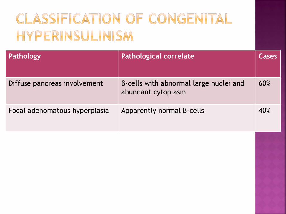
Pathology Pathological correlate Cases
Diffuse pancreas involvement β-cells with abnormal large nuclei and
abundant cytoplasm
60%
Focal adenomatous hyperplasia Apparently normal β-cells 40%
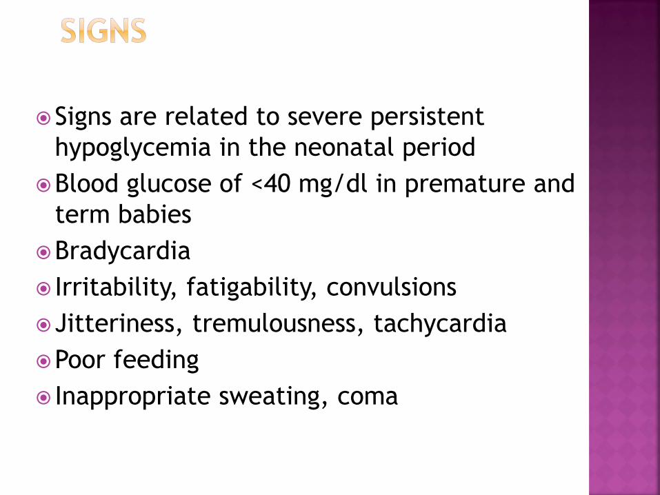
Signs are related to severe persistent
hypoglycemia in the neonatal period
Blood glucose of <40 mg/dl in premature and
term babies
Bradycardia
Irritability, fatigability, convulsions
Jitteriness, tremulousness, tachycardia
Poor feeding
Inappropriate sweating, coma
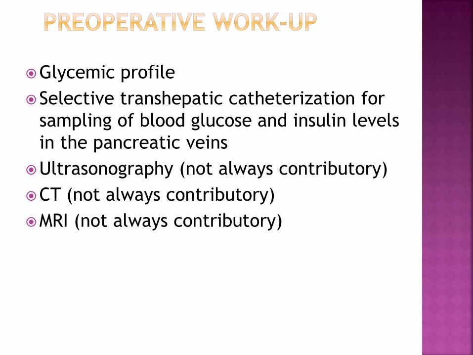
Glycemic profile
Selective transhepatic catheterization for
sampling of blood glucose and insulin levels
in the pancreatic veins
Ultrasonography (not always contributory)
CT (not always contributory)
MRI (not always contributory)
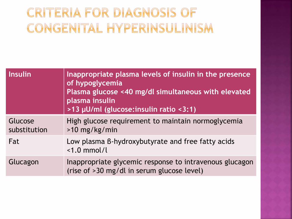
Insulin Inappropriate plasma levels of insulin in the presence
of hypoglycemia
Plasma glucose <40 mg/dl simultaneous with elevated
plasma insulin
>13 μU/ml (glucose:insulin ratio <3:1)
Glucose
substitution
High glucose requirement to maintain normoglycemia
>10 mg/kg/min
Fat Low plasma β-hydroxybutyrate and free fatty acids
<1.0 mmol/l
Glucagon Inappropriate glycemic response to intravenous glucagon
(rise of >30 mg/dl in serum glucose level)
Maintenance of normal glycemia by glucose
infusion (up to 15–20 mg/kg/min) and/or
high-calorie enteral feeding
Diazoxide, insulin antagonist (up to 20
mg/kg/day) (however, many side-effects)
Followed by octreotide or glucagon for
diazoxide-resistant cases
Hydrochlorothiazide (synergistic action with
diazoxide)
Purpose is to reduce the mass of insulin-producing β-
cells
Control of hyperinsulinism is achieved through the
extent of pancreas resection
For diffuse form, usually 95%
For focal form, less radical excision (after pancreatic
venous sampling and with the help of frozen-section
biopsies during surgery)
Resection of more than 98% may result in endocrine
and exocrine insufficiency
Resection of less than 95% may result in failure to
cure or recurrence
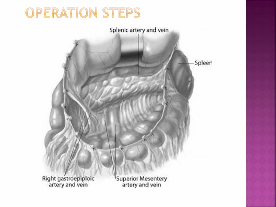
Make an upper transverse abdominal incision
Open the omentum major
Expose the pancreas after mobilization of the
duodenum (Kocher maneuver)
Perform the resection: options include the
tail, body, uncinate process and the majority
of the head, sparing the spleen
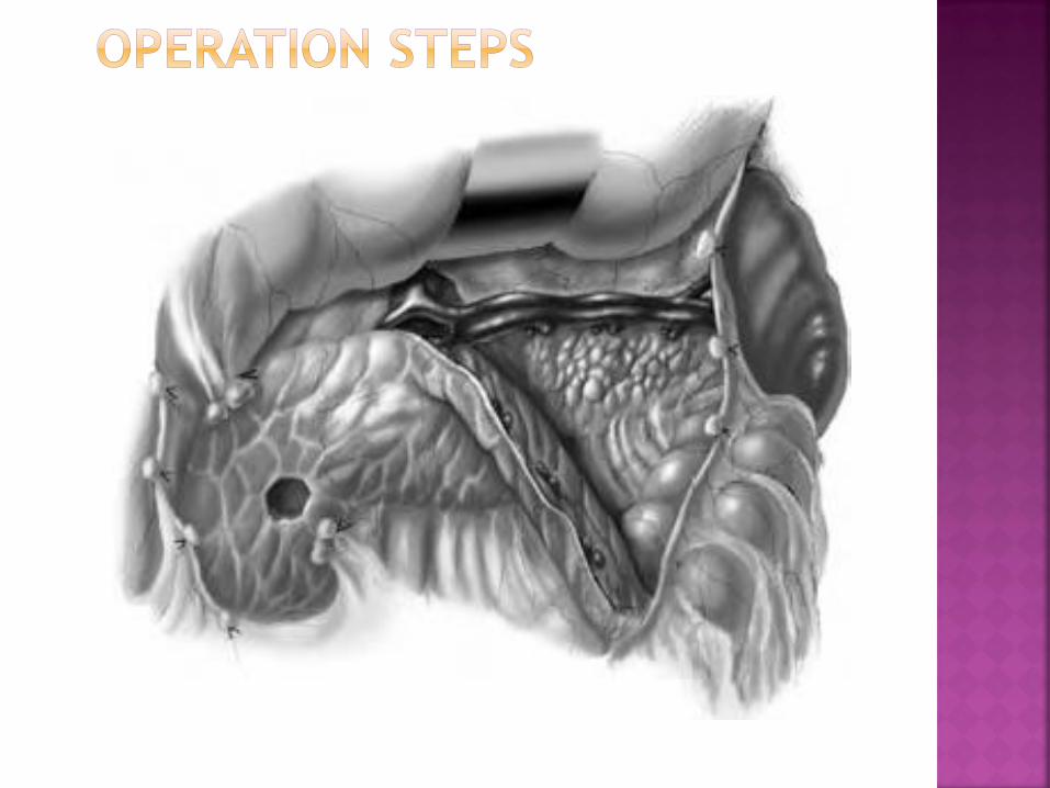
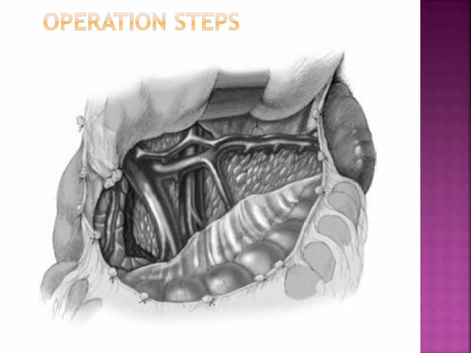
Dissect the pancreas and ligate all small
pancreatic arterial and venous branches
Leave a sliver of pancreatic tissue to the left
of the common bile duct and on the surface
of the duodenum
Ligate the pancreatic duct with a
nonabsorbable stitch
Seal the pancreatic parenchyma with
collagen glue or equivalent
Ensure peritoneal drainage
Gastric tube on suction
Intravenous nutrition for 5–7 days
Frequent serum glucose tests
Administration of insulin as required
Antibiotics for 5 days
Peritoneal drain to be removed 3–4 days
postoperatively
Follow adaptation for 4–6 months
50% cure after <95% resection, 19% cure after
>95% resection
Better results in focal disease assuming
complete excision and normal remaining
pancreas
Diabetes mellitus in 15%
Possible exocrine insufficiency
General considerations
Incidence: 1:10,000 live births
End result of a destructive inflammatory
process, unknown etiology (viral, toxic)
Intrahepatic hypoplasia (Alagille’s syndrome)
Alpha-1-antitrypsin deficiency
Thick bile syndrome (inspissated bile
syndrome) after hemolysis
Neonatal hepatitis (intrauterine viral
infection), giant cell hepatitis
Sepsis with jaundice
Cystic fibrosis
PFIC (progressive familial intrahepatic
cholestasis or Byler disease)
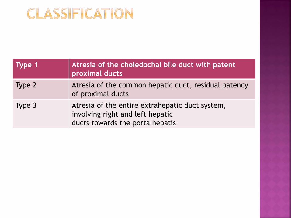
Type 1 Atresia of the choledochal bile duct with patent
proximal ducts
Type 2 Atresia of the common hepatic duct, residual patency
of proximal ducts
Type 3 Atresia of the entire extrahepatic duct system,
involving right and left hepatic
ducts towards the porta hepatis
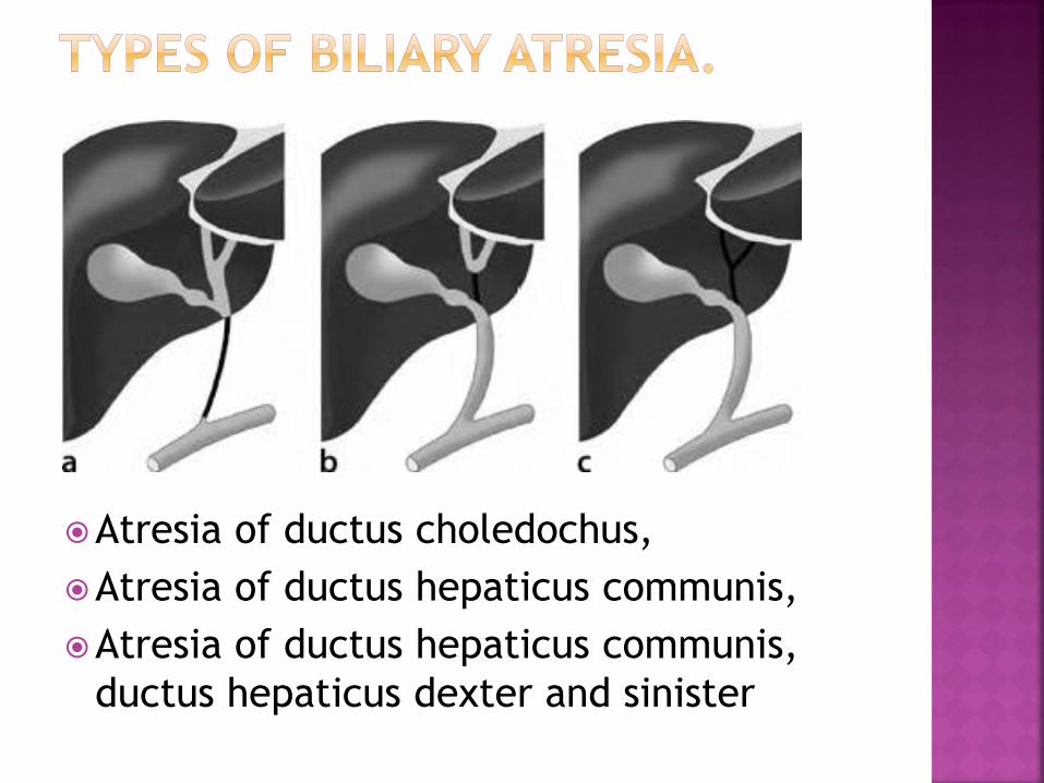
Atresia of ductus choledochus,
Atresia of ductus hepaticus communis,
Atresia of ductus hepaticus communis,
ductus hepaticus dexter and sinister
Early jaundice (first 36 h after birth)
Jaundice may appear at 3 weeks
Total bilirubin >12 μmol/l in term infants, >15 μmol/l in
premature infants
Prolonged jaundice >8 days in term infants and >14 days in
prematures
Conjugated bilirubin >15% of total bilirubin
Nonpigmented stools
Dark urine
Increased size of liver and spleen
Later on ascites
Associated malformations (malrotation, situs inversus,
polysplenia, preduodenal portal vein, absent inferior vena
cava, cardiac defects)
Exclude infections, metabolic, endocrine
disease or genetic disorder
Blood sample: bilirubin, transaminases,
alkaline phosphatase,
γ-glutamyl transferase (γ-GT), coagulation
factors
Cytomegalovirus (CMV) and hepatitis virus
Urine: bilirubin, urobilinogen (lacking)
Ultrasound (not very specific), exclude
choledochal cyst
Liver biopsy (in 10% there is difficulty of interpretation)
Transcutaneously as a needle biopsy (beware of
Bleeding!; small amount of material obtained)
Laparoscopically (bleeding control, adequate amount of
material obtained). Can also be combined with a
cholangiography
In special cases (not mandatory)
Hepatobiliary excretion scans (technetium-labeled
agents)
Percutaneous cholangiography (easier laparoscopically)
Endoscopic retrograde cholangio-pancreatography (ERCP)
Vitamin K, i.v. (1 mg・day–1) for 4 days preoperatively
Blood typing, cross-match
Perioperative antibiotics
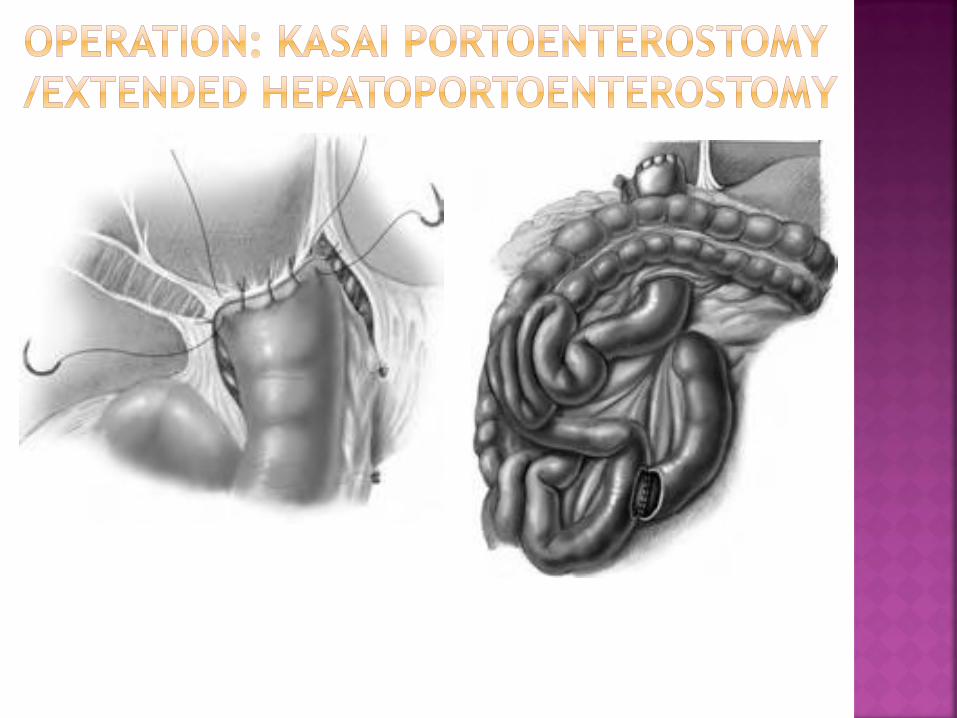
Perform the operation early, before liver
fibrosis or cirrhosis
Place the patient in a prone position on the
operating table to permit cholangiography
Make an incision usable for possible future
liver transplantation (transverse upper
abdominal subcostal incision)
Be prepared for possible Roux-en-Y
anastomosis
Inspect the abdominal cavity (search for
malrotation, situs inversus, preduodenal portal
vein). Confirm diagnosis of biliary atresia
(gallbladder may be hidden between segments 5 and
4)
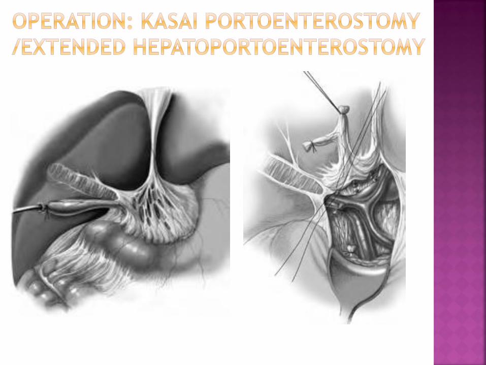
Dissect the hepatoduodenal ligament
Mobilize the liver
Mobilize the gallbladder from its liver bed; used as a
guide to the fibrous remnant of the hepatic duct.
This will lead to the porta hepatis
Prepare the hepatic artery, right and left branches
Ligate all lymphatic vessels
Prepare the portal vein; follow the right and left
branches as far as possible and expose the porta
hepatis behind the bifurcation of the portal vein (for
extended hepatoportoenterostomy; not performed in
the original Kasai procedure)
When necessary, exteriorize the liver
Ligate all small portal branches to the caudate lobe
Section Arantius’ ligament (or ligamentum venosum),
which helps mobilization of the left portal branch
Widely expose the porta hepatis
Cholecystectomy: excise remaining tract and tissue
of porta hepatis flush with liver capsule
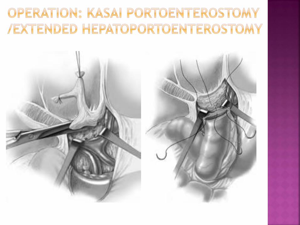
Extensive numbers of bile ducts present posteriorly and
laterally
Ensure hemostasis; replace liver; create Roux-en-Y loop
Level of jejunal section approximately 10 cm from the
Treitz ligament
Length of jejunal loop at least 50 cm, placed in retrocolic
position
Make a 3-cm-long incision on the antimesenteric border
Make an anastomosis of jejunal loop to tissue at the porta
hepatis, going far on both lateral sides, posteriorly on the
caudate lobe and anteriorly on the quadrate lobe
Any cystic dilatation not containing bile is considered not
to be communicating and should be excised
Occasionally, gallbladder and distal bile duct
are not affected by the atretic process and
the gallbladder can be used for the
anastomosis at the porta hepatis
If the residual segment of the proximal bile
duct is long enough, a hepaticojejunostomy
is feasible (rare type I cystic lesion)
The main problem is ascending cholangitis (best
prevention: long loop of jejunum)
Gastric tube on suction
Initially total parenteral nutrition (TPN), but enteral
nutrition as soon as possible
Antibiotics for 5–7 days, discuss continuous (for 2–3
months) prophylactic oral antibiotics (cephalosporin)
Prednisolone, although there is no general agreement on
steroids, we recommend
• days 1,2: 5 mg/kg
• day 3: 2 mg/kg
• days 4–14: 1 mg/kg
■ Cholestyramine (if bile flow)
■ Choleretics (ursodeoxycholine)
■ Vitamins A, D, E, K
Depends on grade of liver fibrosis or cirrhosis
(late operation, more liver destruction)
When operation is performed in first 6–8
weeks of life, chances of obtaining bile flow
is approximately 70%–90%; beyond 12 weeks
of age chances decrease to 35%
5-year survival rate with native liver is
approximately 60%
Portoenterostomy and transplantation are
now complementary procedures and give
good quality of life and a 80%–90% survival
rate
Incidence: 1:100,000 live births
Female:male ratio 3:1 to 4:1
60% of cases are diagnosed before 10 years of age
Most frequent etiology
Common pancreaticobiliary channel
Pressure in pancreatic duct higher than in the bile
duct
Reflux of pancreatic juice in the common bile duct
damages endothelium, causing cystic dilatation
Other etiologies are also possible, such as
obstruction of the distal common bile duct and
genetic reasons
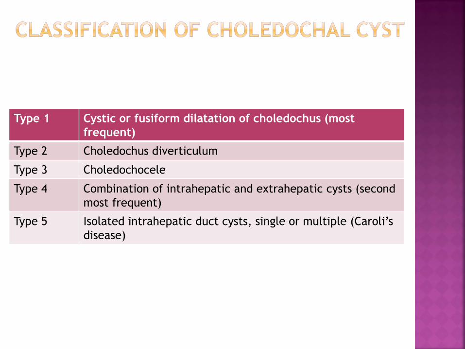
Type 1 Cystic or fusiform dilatation of choledochus (most
frequent)
Type 2 Choledochus diverticulum
Type 3 Choledochocele
Type 4 Combination of intrahepatic and extrahepatic cysts (second
most frequent)
Type 5 Isolated intrahepatic duct cysts, single or multiple (Caroli’s
disease)
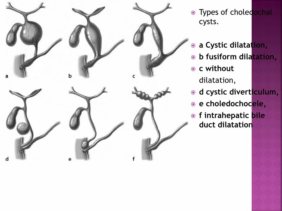
Types of choledochal
cysts.
a Cystic dilatation,
b fusiform dilatation,
c without
dilatation,
d cystic diverticulum,
e choledochocele,
f intrahepatic bile
duct dilatation
Usually during the first decade of life
Some are asymptomatic (prenatal diagnosis)
This classic triad only present in 6% of cases
Abdominal mass
Intermittent episodes of jaundice
Intermittent episodes of abdominal pain
Recurrent cholangitis
Pancreatitis
Biliary calculi
Pancreatic duct calculi
Pancreatitis
Cyst rupture, biliary peritonitis
Portal hypertension
Liver fibrosis or cirrhosis
Cholangiocarcinoma
Occasionally diagnosed antenatally
Ultrasonography
CT or magnetic resonance cholangiopancreatography
(MRCP)
ERCP
Percutaneous transhepatic cholangiography
Hepatobiliary scintigraphy
Not all investigations are necessary, decision
according to infrastructure
Early operation prevents complications
Occasional acute pancreatitis that is resistant to
conservative treatment with a gallbladder under
tension requires percutaneous drainage of the
gallbladder, in order to achieve resolution of
pancreatitis before surgery
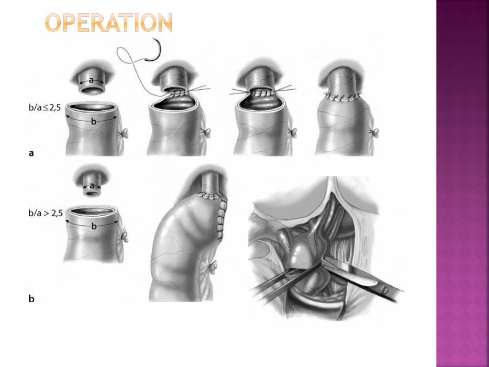
Aim of surgery is complete removal of the cyst
Cystenterostomy should not be done, because of
potential complications (cholangitis, cholelithiasis,
pancreatolithiasis, biliary cirrhosis,
cholangiocarcinoma)
Perioperative antibiotics

Make a high transverse incision
Check appearance of liver and spleen
Perform liver biopsy
Sample bile aspirated from the cyst for culture and
pancreatic enzyme concentration
Cholangiography to delineate precise anatomy,
intrahepatic ducts and pancreaticobiliary junction
Mobilize the gallbladder and cystic duct
Care must be taken to avoid damage to an aberrant
right hepatic artery, usually very adherent to the
cystic wall
Lift the cyst and gallbladder with a tape placed
under the bile duct (pay attention to the portal vein,
often adherent to the posterior wall of the cyst)
Dissect the common hepatic duct at its bifurcation
Further dissect the bile duct to within the head of
the pancreas
Remove the entire cyst; oversew the distal duct end
In difficult cases, open the cyst and remove mucosa
from the bottom of the cyst
Possible calculi and debris of intrahepatic and
pancreatic ducts should be cleared (intraoperative
endoscopy)
Make a roux-en-Y loop anastomosis to the hepatic
duct bifurcation (wide hilar anastomosis)
Occasionally, a transduodenal sphincteroplasty is
necessary (difficulty in removal calculi from long
common channel)
Postoperative care
As in biliary atresia, without prednisone and
choleretics
Prognosis
Low mortality
10% complications: cholangitis, pancreatitis,
anastomotic stricture (even late), calculi,
cholangiocarcinoma
Operation before 5 years of age limits
complications
Not as frequent as in adults. More frequently
discovered within the last three decades, probably
because of improvements in diagnostic techniques
(ultrasonography)
Two peaks of appearance: first in infancy and second
in early adolescence with a steady increase
thereafter
Female:male ratio 1:1 in infancy, 2–4:1 in prepuberty
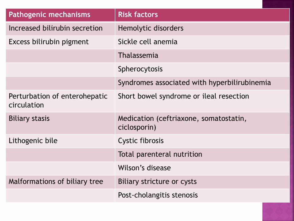
Pathogenic mechanisms Risk factors
Increased bilirubin secretion Hemolytic disorders
Excess bilirubin pigment Sickle cell anemia
Thalassemia
Spherocytosis
Syndromes associated with hyperbilirubinemia
Perturbation of enterohepatic
circulation
Short bowel syndrome or ileal resection
Biliary stasis Medication (ceftriaxone, somatostatin,
ciclosporin)
Lithogenic bile Cystic fibrosis
Total parenteral nutrition
Wilson’s disease
Malformations of biliary tree Biliary stricture or cysts
Post-cholangitis stenosis
May be asymptomatic
In infancy nonspecific signs (poor feeding, vomiting,
irritability)
Abdominal pain (right upper quadrant or
epigastrium, nausea, vomiting)
Ileus
Biliary colic, acute cholecystitis, choledocholithiasis
with obstructive jaundice, pancreatitis
Detailed history, search for any general
condition
Ultrasonography
X-ray of abdomen without preparation
Magnetic resonance cholangiography
ERCP, with possible papillotomy

Make a right subcostal incision
Dissect the hepatoduodenal ligament and gallbladder
Ligate and section the cystic artery
Dissect the cystic duct and perform a
cholangiography in order to visualize concrements in
the duct
Ligate and section the cystic duct
Visualize the choledochus and excise the gallbladder
in an anterograde fashion
Ensure hemostasis of the gallbladder bed
Drainage not mandatory
T-tube drainage in cases of choledochus exploration
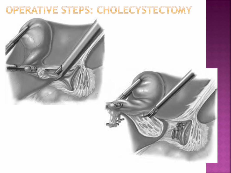
Has become the standard technique
If gallbladder is under tension, it may be punctured and
its extremity grasped with instruments
Gallbladder dissection begins close to Hartmann’s pouch;
window created above and behind the cystic duct and
artery
Cholangiogram
Section the cystic artery and cystic duct between clips
Dissect the gallbladder in a retrograde fashion
Remove the gallbladder through the umbilical port
(incision can be widened)
Remove the entire pneumoperitoneum
Choledocholithiasis must be treated (through laparoscopy,
conversion to open exploration or postoperative ERCP and
extraction)
Rapid recovery after laparoscopy
Standard postoperative care according to any intra-
abdominal procedure
Prognosis is good
The portal venous system drains blood from the
stomach, pancreas, gallbladder, spleen, and
intestines into the liver.
Portosystemic anastomoses exist in four main areas,
1. the gastro-esophageal veins via the cardiac vein
and perforating esophageal veins,
2. the retroperitoneum via the pancreaticoduodenal
veins and the retroperitoneal-paravertebral veins,
3. gastrorenal-splenorenal vein, and
4. the hemorrhoidal plexus.
Portal hypertension is defined as elevation of the
portal venous-IVC pressure gradient above 10-12 mm
Hg.
Portal hypertension in children can be divided into
two major categories based upon the anatomic
location of the increased portal resistance.
Extrahepatic portal hypertension (EHPH) is most
commonly the result of portal vein obstruction due
to thrombosis.
Intrahepatic portal hypertension (IHPH) is typically
associated with congenital liver or biliary diseases in
children. Biliary atresia is by far the most common
cause of IHPH in children.
Children with IHPH usually present between several
months to one year of life with severe hepatic
dysfunction, manifested by jaundice, hepatic
encephalopathy, and malnutrition complicated by
poor growth and increased susceptibility to
infections.
Extrahepatic portal hypertension most commonly
presents in the first decade of life with
gastrointestinal hemorrhage from esophageal
varices.
In a child presenting with an initial episode of
gastrointestinal bleeding, abdominal ultrasonography
is used to confirm the etiology. The presence of
portal vein thrombosis, the extent of collateral
formation, and the direction of portal vein flow is
established by this noninvasive and relatively
inexpensive diagnostic exam.
Upper endoscopy is used to identify and quantitate
esophageal varices.
This procedure is possible therapeutic intervention
(i.e., sclerotherapy, banding).
Angiography is a much more rarely used diagnostic
and potentially therapeutic modality used in certain
cases of portal hypertension.
In the acute setting, massive hemorrhage is managed
with intensive care monitoring, transfusion of red
blood cells and fresh frozen plasma, and potentially
intubation and sedation to minimize agitation that
increases variceal pressure. Octreotide infusions
effectively control acute hemorrhage in the vast
majority of children with portal hypertension and
variceal bleeding. Endoscopic variceal banding or
sclerotherapy is occasionally used in cases where
hemorrhage does not resolve with supportive care.
Once the patient has stabilized, endoscopy with
sclerotherapy or banding is employed to prevent
repeat episodes of hemorrhage.
Surgical treatment of portal hypertension can be
either direct, which involves ligation of the varices
themselves, or indirect, in which the portal venous
system is decompressed with a surgical shunt.
Examples include portocaval, mesocaval and central
splenorenal shunts.
Selective portosystemic shunts shunt a portion of
portal blood into the systemic circulation, with
distal splenorenal shunt being the most common.
Recently, selected children with EHPH due to portal
vein thrombosis have been successfully treated by
surgical creation of a mesenterico-portal venous
bypass (Rex shunt).
Treatment of intrahepatic portal hypertension
focuses on the primary liver disease.
For advanced cirrhosis and other intrahepatic
sources of portal hypertension, liver transplantation
is the definitive treatment.
For patients with EHPH, sclerotherapy is effective in
the treatment of acute variceal bleeding in up to
75% of patients. However, several follow-up sessions
are necessary to obliterate the varices, and a
rebleeding rate of 5-25% is expected.
Persistent variceal bleeding as well as hypersplenism
may require a surgical shunt.
Selective shunts have proven successful for the
control of bleeding, thrombocytopenia, and
leukopenia, without creating great risk of
encephalopathy.
The pancreas develops in the 4th week of gestation
and begins as two buds, dorsal and ventral, from the
endoderm of the duodenum.
The growing dorsal portion of the developing
pancreas spans across the hepatic diverticulum while
the ventral portion lies below moving more distal.
The dorsal and ventral portions fuse in week ten.
The distal portion will create the duct of Wirsung
while the proximal portion may obliterate or form
the duct of Santorini. 10% of the population will
have a double collecting system in the pancreas.
Fetal insulin production begins in the fifth
gestational month and the exocrine function is
present at birth.
The pancreas is a retroperitoneal organ located at
the vertebral L1-L2 level.
The head of the pancreas lies to the right of the
vertebral column and, along with the uncinate
process, is intimately adherent to the duodenum.
The body of the pancreas lies anterior to the
superior mesenteric artery and vein, and the portal
vein.
The arterial supply of the pancreas is derived from
the gastroduodenal artery, superior mesenteric
artery, and the splenic artery. The head of the
pancreas receives arterial blood via the four
pancreaticoduodenal arteries (i.e., anterior superior,
anterior inferior, posterior superior, and posterior
inferior). Venous drainage is via the splenic and
portal vein.
The vast majority of cases of pancreatitis in children
are from blunt abdominal injury. In the pediatric
population, nearly 40% of cases of traumatic
pancreatitis are attributable to bicycle-related
injury.
After trauma, the most common causes of
pancreatitis in children are drug therapy
(corticosteroids, azathioprine, thiazides,
furosemide, tetracyclines, and valproic acid), viral
infection (Epstein-Barr, Coxsackie, enterovirus, and
mumps), and bacterial infection. Cystic fibrosis,
biliary disease, vasculitic diseases (systemic lupus,
Henoch-Schonlein purpura), and type I and V
hyperlipidemias are also associated with acute
pancreatitis in the pediatric population.
Serum amylase, trypsinogen, and lipase levels are
useful to establish the diagnosis of acute
pancreatitis. An elevated serum amylase is the usual
biochemical abnormality associated with acute
pancreatitis. Because amylase production occurs
from other nonpancreatic sources (i.e., salivary
gland), elevated serum amylase is relatively
nonspecific. Calculation of the amylase clearance
may be helpful and is normally less than 5%.
Trypsinogen and lipase are produced almost
exclusively by the pancreas; elevated serum levels
are more specific for pancreatitis.
Computed tomography (CT) is the best radiographic
study to image the pancreas in cases of severe or
complicated pancreatitis. Abdominal CT is often
obtained as part of the trauma evaluation.
Ultrasound is sometimes useful, but often only
provides limited visualization of the pancreas due to
its retroperitoneal location and interposed bowel gas
which further limits the study.
Endoscopic retrograde cholangiopancreatography
(ERCP) is an invasive test that can accurately
delineate pancreatic ductal anatomy. ERCP causes
pancreatitis in 5-10% of cases and is generally
avoided during the early phases of acute
pancreatitis.
Medical management is the mainstay of treatment for
pancreatitis. Volume resuscitation is essential to counter
retroperitoneal third space fluid losses.
Nasogastric decompression is recommended to avoid
gastric distention and patients are initially maintained
NPO with nasogastric decompression.
Pain management is essential. Meperidine is preferred
because it does not cause sphincter of Oddi contraction
like morphine does.
Hyper-alimentation may be necessary if the course of
pancreatitis is prolonged. Enteral feeding distal to the
ligament of Treitz via duodenal feeding tube is the
preferred method of providing nutrition in refractory
cases. The majority of cases of pancreatitis are self-
limited and resolve spontaneously with supportive
therapy.
In severe cases (i.e., necrotizing pancreatitis,
infected pancreatic necrosis), surgical intervention
may be necessary for irrigation and/or debridement
of the pancreas.
The morality rate in this scenario approaches 15%.
Pancreatic cysts are broadly classified based on
etiology. These are simply categorized as:
1. congenital,
2. retention,
3. pseudocysts,
4. neoplastic,
5. parasitic.
Congenital cysts of the pancreas are a rare finding in
children. The cysts may be unilocular or multilocular
and are most commonly found in the body or tail of
the pancreas. These cysts are lined with true
epithelium and most commonly contain
nonenzymatic fluid. The majority of these lesions
are asymptomatic unless they are large.
Symptomatic cysts are excised.
Pancreatic retention cysts occasionally occur in
children and are associated with chronic obstruction
of the pancreatic ductal system.
These cysts are filled with enzyme containing fluid.
Surgical treatment is by excision or internal
drainage.
Approximately 90% of pseudocysts occur secondary to
trauma. This condition is more common in males (nearly
3:1). Patients present with symptoms (most common to
least) of vomiting, abdominal pain, abdominal mass,
fever, and anorexia.
Pseudocysts are usually located in the lesser sac and the
cyst wall consists of granulation tissue. If the cyst
communicates with the pancreatic ductal system, high
amylase levels are measurable with the cyst fluid. Useful
diagnostic tests include serum amylase, ultrasound, and
CT.
Surgical treatment is indicated for large, persistent, or
infected/symptomatic cysts. The surgical options include
external drainage, cystgastrostomy, cystjejunostomy, or
excision. Surgical therapy is associated with low
mortality, minimal morbidity, and low recurrence.
Annular pancreas is a rare congenital anomaly that occurs
due to abnormal rotation of the pancreatic ventral bud. It
is the most common of the congenital pancreatic
abnormalities. The annular pancreas usually completely
encircles the second portion of the duodenum. This
anomaly is associated with Down’s syndrome,
abnormalities of rotation, duodenal atresia, and biliary
atresia. Seventy percent of children with this lesion are
symptomatic and will present with high intestinal
obstruction. The emesis is most often bilious but can be
nonbilious as well. Surgical therapy consists of bypass
anastomosis: duodenoduodenostomy or
duodenojejunostomy. Division of the pancreatic tissue is
not recommended due to the high association of fistula
formation. Gastrojejunostomy is not recommended due to
associated growth problems and the risk of marginal
ulcers.
Pancreas divisum is a congenital anomaly in which
the dorsal and ventral pancreatic tissue fail to fuse
in utero. Pancreas divisum is identifiable in 10-15%
of the population. This condition is usually
asymptomatic, however there may be an increased
incidence of pancreatitis due to the inability of the
accessory duct to adequately drain the pancreatic
tissue. Diagnosis is made exclusively with ERCP.
Treatment, if necessary, is by endoscopic or surgical
sphincterotomy of the accessory ampulla.
Ectopic pancreatic tissue is frequently identified in
the duodenum, colon, pylorus, appendix, or a
Meckel’s diverticulum.
Ectopic pancreas can cause local inflammation and
bleeding and is noted in approximately 3% of
postmortem examinations.


![Ivyspring International Publisher TheraannoossttiiccssTheranostics 2012, 2(6) 578 from small animal models [1]. The lung metastasis model is often established by the tail vein injection](https://static.fdocument.org/doc/165x107/60015c82bddbc723df459880/ivyspring-international-publisher-theraannoossttiiccss-theranostics-2012-26-578.jpg)