`Degraded' RNA profiles in Arthropoda and beyond a single band, explained by a splitting of the 28S...
14
Submitted 4 July 2015 Accepted 4 November 2015 Published 1 December 2015 Corresponding author Anne Marie Power, [email protected] Academic editor Mar´ ıa ´ Angeles Esteban Additional Information and Declarations can be found on page 11 DOI 10.7717/peerj.1436 Copyright 2015 McCarthy et al. Distributed under Creative Commons CC-BY 4.0 OPEN ACCESS ‘Degraded’ RNA profiles in Arthropoda and beyond Sean D. McCarthy, Michel M. Dugon and Anne Marie Power School of Natural Sciences, Ryan Institute for Environmental, Marine and Energy Research, National University of Ireland Galway, Ireland ABSTRACT The requirement for high quality/non-degraded RNA is essential for an array of molecular biology analyses. When analysing the integrity of rRNA from the barnacle Lepas anatifera (Phylum Arthropoda, Subphylum Crustacea), atypical or sub- optimal rRNA profiles that were apparently degraded were observed on a bioanalyser electropherogram. It was subsequently discovered that the rRNA was not degraded, but arose due to a ‘gap deletion’ (also referred to as ‘hidden break’) in the 28S rRNA. An apparent excision at this site caused the 28S rRNA to fragment under heat- denaturing conditions and migrate along with the 18S rRNA, superficially presenting a ‘degraded’ appearance. Examination of the literature showed similar observations in a small number of older studies in insects; however, reading across multiple disciplines suggests that this is a wider issue that occurs across the Animalia and beyond. The current study shows that the 28S rRNA anomaly goes far beyond insects within the Arthropoda and is widespread within this phylum. We confirm that the anomaly is associated with thermal conversion because gap-deletion patterns were observed in heat-denatured samples but not in gels with formaldehyde-denaturing. Subjects Aquaculture, Fisheries and Fish Science, Genomics, Marine Biology, Taxonomy, Zoology Keywords Hidden break, Denaturing, Taxonomy, Gap deletion, Degraded rNA, Bioanalyser INTRODUCTION Anomalies in the gel migration of the 28S subunit rRNA in denatured samples have mostly appeared in the older literature (Applebaum, Ebstein & Wyatt, 1966; Ishikawa & Newburgh, 1972; Fujiwara & Ishikawa, 1986). More recent studies have also mentioned anomalies observed during routine RNA extraction and quality control steps using both automated (Winnebeck, Millar & Warman, 2010; Asai et al., 2014) and traditional (Macharia, Ombura & Aroko, 2015) electrophoresis methods. The Agilent bioanalyser is the gold standard for RNA quality analysis prior to deep sequencing and gene expression experiments. This method generates an electropherogram showing an RNA profile with peaks representing the various subunit components, as well as an RNA integrity number (RIN). This analysis is routinely carried out following RNA denaturation using a heating step (Krupp, 2005). An alternative to the bioanalyser is using an electrophoresis denaturing gel to confirm the integrity of the RNA sample. This also requires a heating step to prevent secondary structure formation. Several denaturing reagents are commonly used for this including methyl mercury hydroxide, urea, guanidine thiocyanate, formamide, DMSO and How to cite this article McCarthy et al. (2015), ‘Degraded’ RNA profiles in Arthropoda and beyond. PeerJ 3:e1436; DOI 10.7717/peerj.1436
Transcript of `Degraded' RNA profiles in Arthropoda and beyond a single band, explained by a splitting of the 28S...
Submitted 4 July 2015Accepted 4 November 2015Published 1 December 2015
Corresponding authorAnne Marie Power,[email protected]
Academic editorMarıa Angeles Esteban
Additional Information andDeclarations can be found onpage 11
DOI 10.7717/peerj.1436
Copyright2015 McCarthy et al.
Distributed underCreative Commons CC-BY 4.0
OPEN ACCESS
‘Degraded’ RNA profiles in Arthropodaand beyondSean D. McCarthy, Michel M. Dugon and Anne Marie Power
School of Natural Sciences, Ryan Institute for Environmental, Marine and Energy Research,National University of Ireland Galway, Ireland
ABSTRACTThe requirement for high quality/non-degraded RNA is essential for an array ofmolecular biology analyses. When analysing the integrity of rRNA from the barnacleLepas anatifera (Phylum Arthropoda, Subphylum Crustacea), atypical or sub-optimal rRNA profiles that were apparently degraded were observed on a bioanalyserelectropherogram. It was subsequently discovered that the rRNA was not degraded,but arose due to a ‘gap deletion’ (also referred to as ‘hidden break’) in the 28S rRNA.An apparent excision at this site caused the 28S rRNA to fragment under heat-denaturing conditions and migrate along with the 18S rRNA, superficially presentinga ‘degraded’ appearance. Examination of the literature showed similar observationsin a small number of older studies in insects; however, reading across multipledisciplines suggests that this is a wider issue that occurs across the Animalia andbeyond. The current study shows that the 28S rRNA anomaly goes far beyond insectswithin the Arthropoda and is widespread within this phylum. We confirm that theanomaly is associated with thermal conversion because gap-deletion patterns wereobserved in heat-denatured samples but not in gels with formaldehyde-denaturing.
Subjects Aquaculture, Fisheries and Fish Science, Genomics, Marine Biology, Taxonomy,ZoologyKeywords Hidden break, Denaturing, Taxonomy, Gap deletion, Degraded rNA, Bioanalyser
INTRODUCTIONAnomalies in the gel migration of the 28S subunit rRNA in denatured samples have
mostly appeared in the older literature (Applebaum, Ebstein & Wyatt, 1966; Ishikawa &
Newburgh, 1972; Fujiwara & Ishikawa, 1986). More recent studies have also mentioned
anomalies observed during routine RNA extraction and quality control steps using
both automated (Winnebeck, Millar & Warman, 2010; Asai et al., 2014) and traditional
(Macharia, Ombura & Aroko, 2015) electrophoresis methods. The Agilent bioanalyser is
the gold standard for RNA quality analysis prior to deep sequencing and gene expression
experiments. This method generates an electropherogram showing an RNA profile with
peaks representing the various subunit components, as well as an RNA integrity number
(RIN). This analysis is routinely carried out following RNA denaturation using a heating
step (Krupp, 2005). An alternative to the bioanalyser is using an electrophoresis denaturing
gel to confirm the integrity of the RNA sample. This also requires a heating step to prevent
secondary structure formation. Several denaturing reagents are commonly used for this
including methyl mercury hydroxide, urea, guanidine thiocyanate, formamide, DMSO and
How to cite this article McCarthy et al. (2015), ‘Degraded’ RNA profiles in Arthropoda and beyond. PeerJ 3:e1436;DOI 10.7717/peerj.1436
formaldehyde, with the latter being used most frequently (Masek et al., 2005). The problem
that arises is that, for some groups of organisms, accurate analysis of the RNA quality is
impossible unless non-heat-denaturing steps are followed.
In most animals, RNA denaturation generates 28S and 18S rRNA fragments (∼4,000
and ∼2,000 nucleotides (nt) respectively), which migrate separately on the denaturing
gel. It is widely accepted that, in intact rRNA, the 28S and 18S peak ratios must be close to
2:1 (Skrypina et al., 2003). But this is not the case for all groups: in the 1970s, studies on
the 28S rRNA in insects found that heat-denaturing RNA for several minutes resulted in
just a single band, explained by a splitting of the 28S rRNA into two fragments ‘α’ and ‘β’
(Ishikawa & Newburgh, 1972; Ishikawa, 1976) with this split likely occurring in the variable
D7a region cleavage site (Gillespie et al., 2006). At the cleavage site, heat-denaturing the
hydrogen bonds holding α and β fragments together, causes these to migrate together with
the 18S fragment because all three fragments (18S, α, and β) are similar in size and form a
single band on RNA gels, approximately at the 18S position. State-of-the-art RNA profile
analysis using bioanalyser electropherograms can also display a ‘degraded’ appearance in
RNA due to the presence of a single peak ∼ at the 18S position, as previously described by
Winnebeck, Millar & Warman (2010).
Very often, the anomalous RNA profile described above has been discovered by accident
during quality control steps and this phenomenon has not been subjected to detailed
study, with some notable exceptions (Fujiwara & Ishikawa, 1986; Ogino et al., 1990; Sun et
al., 2012). This phenomenon has mostly been described as a ‘hidden break’ (Ishikawa &
Newburgh, 1972; Ishikawa, 1977; Fujiwara & Ishikawa, 1986; Ogino et al., 1990; Winnebeck,
Millar & Warman, 2010; Macharia, Ombura & Aroko, 2015), but may more correctly
be termed a ‘gap deletion’ because the cleavage is actually due to the excision of a short
sequence from the rRNA precursors (Ware et al., 1983; Sun et al., 2012). Several authors
have wondered why this observation is limited to insects (Ogino et al., 1990; Winnebeck,
Millar & Warman, 2010) and have recommended further study (Sun et al., 2012). Others
have suggested that the gap deletion occurs more widely—e.g., in most protostomes
(Fujiwara & Ishikawa, 1986); however, there are exceptions of individual species within
a phylum lacking the deletion: e.g., Caenorhabditis elegans in Phylum Nematoda (Zarlenga
& Dame, 1992) and aphids in class Insecta (Ogino et al., 1990).
Clearly, this question has both practical and theoretical relevance since more knowledge
will save researchers a lot of time during optimisation and quality control of RNA
extractions in a variety of animal groups. It could also provide important information
about the maturation of rRNAs, molecules that are fundamentally important in the field of
evolution and systematics, as well as being associated with certain genetic diseases (Henras
et al., 2015). The present study came about during routine RNA extractions steps in stalked
barnacles whilst investigating bioadhesive genes (Jonker et al., 2012; Jonker et al., 2014).
This study is the first documentation of a gap deletion in non-insect arthropods including
stalked barnacles (Crustacea), spiders (Chelicerata) and centipedes (Myriapoda). In
addition, we review the prevalence of this gap deletion in groups of animals beyond the
arthropods, as well as in non-animal organisms.
McCarthy et al. (2015), PeerJ, DOI 10.7717/peerj.1436 2/14
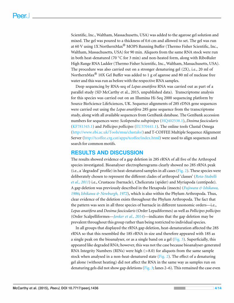
Figure 1 Species analysed for the gap deletion. (A) Spider Grammostola porteri; (B) Centipede Scolopen-dra subspinipes; and C) Barnacle Lepas anatifera.
MATERIALS AND METHODSTissue samples were obtained from Chilean rose tarantula Grammostola porteri (Che-
licerata) and giant Asian centipede Scolopendra subspinipes (Myriapoda). Both of these
organisms are not easily obtained and so we were limited to one individual specimen for
each (n = 1); however, RNA was extracted from three replicate tissue samples from each
individual. For barnacles (Crustacea), three species in different taxonomic orders were
sampled, this time with tissues from three individuals of each species (n = 3). Specimens
were instantly killed and frozen by immersion in liquid nitrogen. Immediately afterwards,
specimens were left to thaw for a few minutes before being dissected. Approximately
2 cm3 of tissue was excised from the muscle mass surrounding the fovea on the lateral
parts of the cephalothorax of Grammostola porteri and divided into three samples for
analysis. Approximately 1 cm3 of tissue was recovered from the deep oblique and lateral
longitudinal muscles of Scolopendra subspinipes and then divided into three samples.
Approximately 1 cm3 of prosoma and peduncle tissues were dissected from each of
three individuals of Lepas anatifera (Fig. 1), Dosima fascicularis and Pollicipes pollicipes
barnacles. Tissues from all specimens were snap-frozen in liquid nitrogen, and stored
at −80 ◦C.
Tissues were homogenized using the IKA® ULTRA-TURRAX T18 homogeniser in
1 ml RLT lysis buffer with 10 ul beta-mercaptoethanol (QIAGEN RNeasy Mini Kit;
Qiagen, Hilden Germany). Next, total RNA was extracted with QIAGEN RNeasy Mini
Kit including DNase I treatment (QIAGEN DNase I, RNase-free), according to the
manufacturer’s instructions. For the analyses of quality and integrity, RNA (100–200 ng)
was electrophoretically separated with an Agilent 2100 Bioanalyser using an RNA 6000
Nano Chip Kit according to manufacturer’s instructions. Each sample was heat-denatured
at 70 ◦C for 3 min prior to separation. Aliquots from the same RNA samples without
heat-denaturation were also run together with heat-denatured samples on the same
Bioanalyser chip.
Samples were also run on a formaldehyde-denaturing RNA gel. One gram of agarose
was added to 90 ml of nuclease free water and transferred to a 50 ◦C water bath until
equilibrated. In a fume hood, 10 ml of NorthernMax® 10X Gel Buffer (Thermo Fisher
McCarthy et al. (2015), PeerJ, DOI 10.7717/peerj.1436 3/14
Scientific, Inc., Waltham, Massachusetts, USA) was added to the agarose gel solution and
mixed. The gel was poured to a thickness of 0.6 cm and allowed to set. The gel was run
at 60 V using 1X NorthernMax® MOPS Running Buffer (Thermo Fisher Scientific, Inc.,
Waltham, Massachusetts, USA) for 90 min. Aliquots from the same RNA stock were run
in both heat-denatured (70 ◦C for 3 min) and non-heated form, along with RiboRuler
High Range RNA Ladder (Thermo Fisher Scientific, Inc., Waltham, Massachusetts, USA).
The procedure was also carried out on a stronger denaturing gel (2X), i.e., 20 ml of
NorthernMax® 10X Gel Buffer was added to 1 g of agarose and 80 ml of nuclease free
water and this was run as before with the respective RNA samples.
Deep sequencing by RNA-seq of Lepas anatifera RNA was carried out as part of a
parallel study (SD McCarthy et al., 2015, unpublished data). Transcriptome analysis
for this species was carried out on an Illumina Hi-Seq 2000 sequencing platform by
Source BioScience LifeSciences, UK. Sequence alignments of 28S rDNA gene sequences
were carried out using the Lepas anatifera 28S gene sequence from the transcriptome
study, along with all available sequences from GenBank database. The GenBank accession
numbers for sequences were: Scolopendra subspinipes (HQ402538.1), Dosima fascicularis
(KF781345.1) and Pollicipes pollicipes (EU370441.1). The online tools Clustal Omega
(http://www.ebi.ac.uk/Tools/msa/clustalo/) and T-COFFEE Multiple Sequence Alignment
Server (http://tcoffee.crg.cat/apps/tcoffee/index.html) were used to align sequences and
search for common motifs.
RESULTS AND DISCUSSIONThe results showed evidence of a gap deletion in 28S rRNA of all five of the Arthropod
species investigated. Bioanalyser electropherograms clearly showed no 28S rRNA peak
(i.e., a ‘degraded’ profile) in heat-denatured samples in all cases (Fig. 2). These species were
deliberately chosen to represent the different clades of arthropod ‘classes’ (Rota-Stabelli
et al., 2011) i.e., Crustacea (barnacle), Chelicerata (spider) and Myriapoda (centipede).
A gap deletion was previously described in the Hexapoda (insects) (Fujiwara & Ishikawa,
1986; Ishikawa & Newburgh, 1972), which is also within the Phylum Arthropoda. Thus,
clear evidence of the deletion exists throughout the Phylum Arthropoda. The fact that
the pattern was seen in all three species of barnacle in different taxonomic orders—i.e.,
Lepas anatifera and Dosima fascicularis (Order Lepadiformes) as well as Pollicipes pollicipes
(Order Scalpelliformes—Jonker et al., 2014)—indicates that the gap deletion may be
prevalent throughout this group rather than being restricted to individual species.
In all groups that displayed the rRNA gap deletion, heat-denaturation affected the 28S
rRNA so that this resembled the 18S rRNA in size and therefore appeared with 18S as
a single peak on the bioanalyser, or as a single band on a gel (Fig. 3). Superficially, this
appeared like degraded RNA; however, this was not the case because bioanalyser-generated
RNA Integrity Numbers (RINs) were high (>8.0) for aliquots from the same sample
stock when analysed in a non-heat-denatured state (Fig. 2). The effect of a denaturing
gel alone (without heating) did not affect the RNA in the same way as samples run on
denaturing gels did not show gap deletions (Fig. 3; lanes 2–6). This remained the case even
McCarthy et al. (2015), PeerJ, DOI 10.7717/peerj.1436 4/14
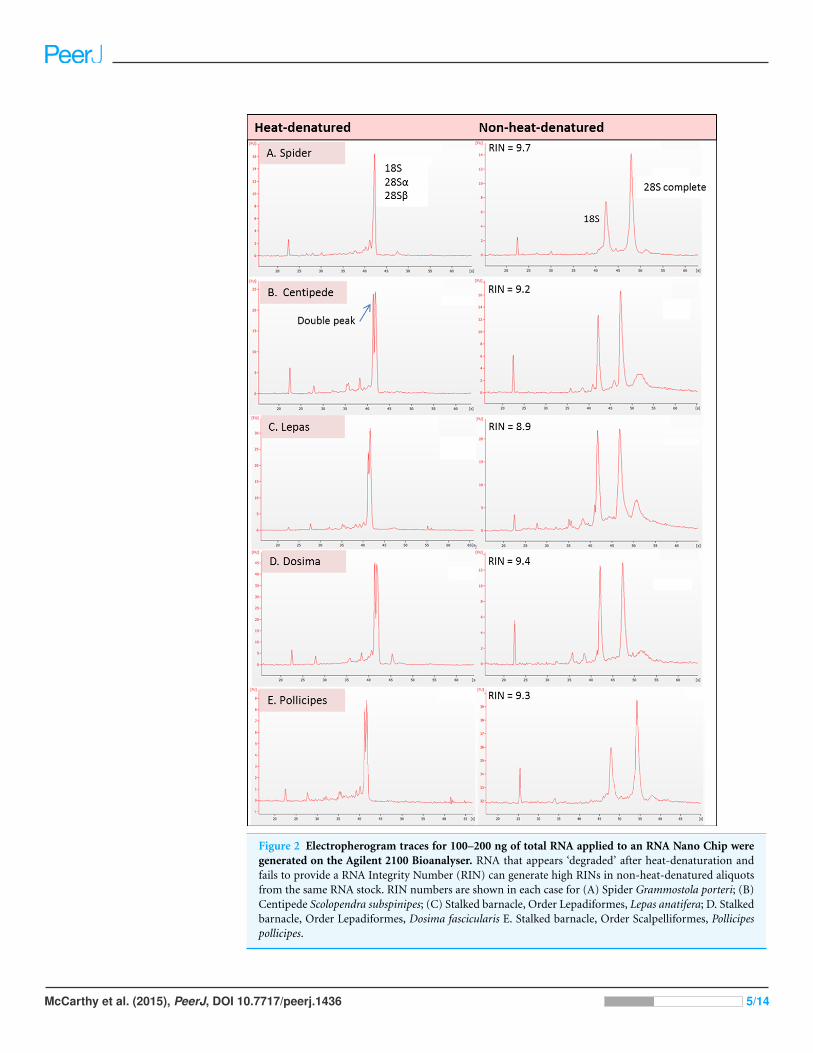
Figure 2 Electropherogram traces for 100–200 ng of total RNA applied to an RNA Nano Chip weregenerated on the Agilent 2100 Bioanalyser. RNA that appears ‘degraded’ after heat-denaturation andfails to provide a RNA Integrity Number (RIN) can generate high RINs in non-heat-denatured aliquotsfrom the same RNA stock. RIN numbers are shown in each case for (A) Spider Grammostola porteri; (B)Centipede Scolopendra subspinipes; (C) Stalked barnacle, Order Lepadiformes, Lepas anatifera; D. Stalkedbarnacle, Order Lepadiformes, Dosima fascicularis E. Stalked barnacle, Order Scalpelliformes, Pollicipespollicipes.
McCarthy et al. (2015), PeerJ, DOI 10.7717/peerj.1436 5/14
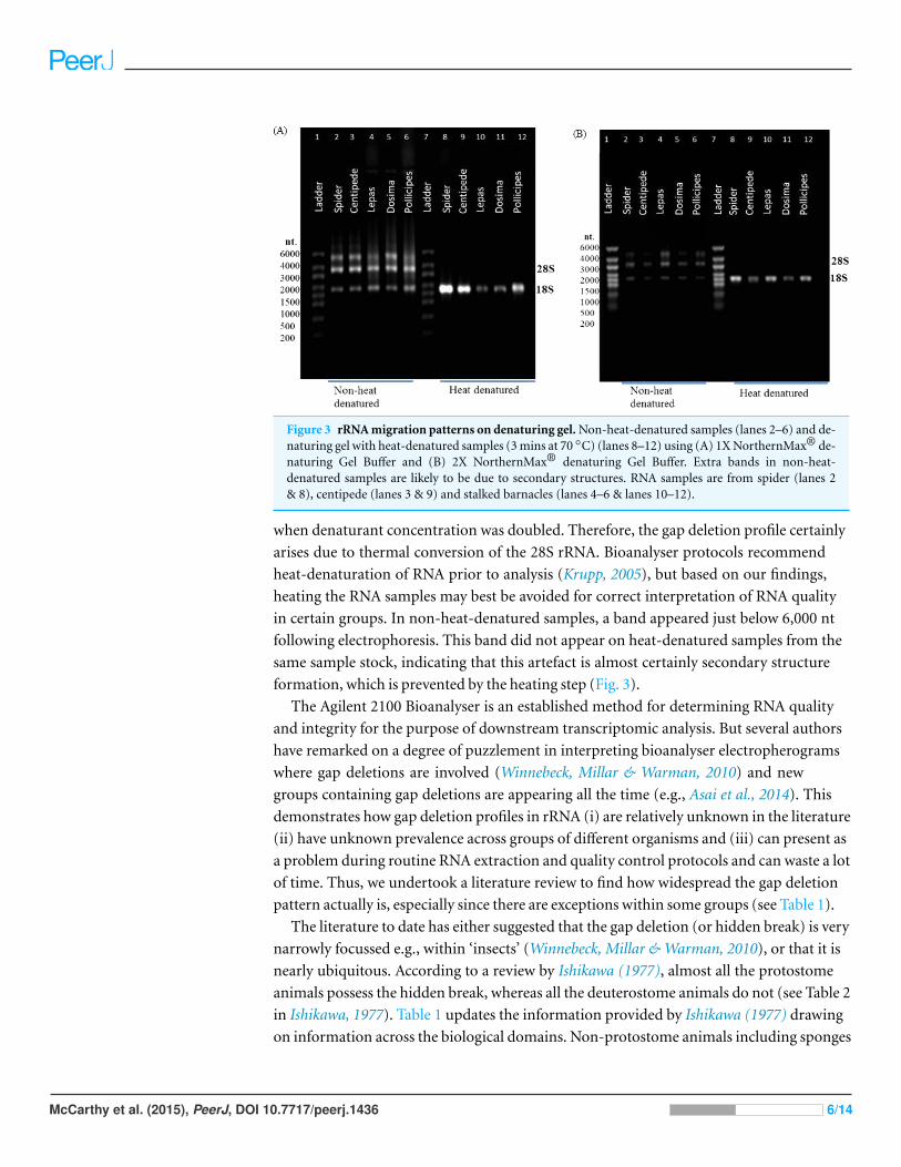
Figure 3 rRNA migration patterns on denaturing gel. Non-heat-denatured samples (lanes 2–6) and de-naturing gel with heat-denatured samples (3 mins at 70 ◦C) (lanes 8–12) using (A) 1X NorthernMax® de-naturing Gel Buffer and (B) 2X NorthernMax® denaturing Gel Buffer. Extra bands in non-heat-denatured samples are likely to be due to secondary structures. RNA samples are from spider (lanes 2& 8), centipede (lanes 3 & 9) and stalked barnacles (lanes 4–6 & lanes 10–12).
when denaturant concentration was doubled. Therefore, the gap deletion profile certainly
arises due to thermal conversion of the 28S rRNA. Bioanalyser protocols recommend
heat-denaturation of RNA prior to analysis (Krupp, 2005), but based on our findings,
heating the RNA samples may best be avoided for correct interpretation of RNA quality
in certain groups. In non-heat-denatured samples, a band appeared just below 6,000 nt
following electrophoresis. This band did not appear on heat-denatured samples from the
same sample stock, indicating that this artefact is almost certainly secondary structure
formation, which is prevented by the heating step (Fig. 3).
The Agilent 2100 Bioanalyser is an established method for determining RNA quality
and integrity for the purpose of downstream transcriptomic analysis. But several authors
have remarked on a degree of puzzlement in interpreting bioanalyser electropherograms
where gap deletions are involved (Winnebeck, Millar & Warman, 2010) and new
groups containing gap deletions are appearing all the time (e.g., Asai et al., 2014). This
demonstrates how gap deletion profiles in rRNA (i) are relatively unknown in the literature
(ii) have unknown prevalence across groups of different organisms and (iii) can present as
a problem during routine RNA extraction and quality control protocols and can waste a lot
of time. Thus, we undertook a literature review to find how widespread the gap deletion
pattern actually is, especially since there are exceptions within some groups (see Table 1).
The literature to date has either suggested that the gap deletion (or hidden break) is very
narrowly focussed e.g., within ‘insects’ (Winnebeck, Millar & Warman, 2010), or that it is
nearly ubiquitous. According to a review by Ishikawa (1977), almost all the protostome
animals possess the hidden break, whereas all the deuterostome animals do not (see Table 2
in Ishikawa, 1977). Table 1 updates the information provided by Ishikawa (1977) drawing
on information across the biological domains. Non-protostome animals including sponges
McCarthy et al. (2015), PeerJ, DOI 10.7717/peerj.1436 6/14
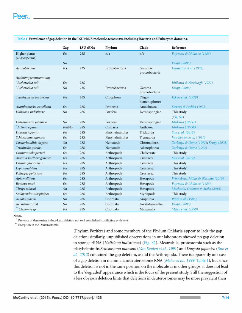
Table 1 Prevalence of gap deletion in the LSU rRNA molecule across taxa including Bacteria and Eukaryota domains.
Gap LSU rRNA Phylum Clade Reference
Higher plants(angiosperms)
Yes 25S n/a n/a Fujiwara & Ishikawa (1986)
No Krupp (2005)
Actinobacillus Yes 23S Proteobacteria Gamma-proteobacteria
Haraszthy et al. (1992)
Actimomycetemcomitans*Escherichia coli Yes 23S Ishikawa & Newburgh (1972)*Escherichia coli No 23S Proteobacteria Gamma-
proteobacteriaKrupp (2005)
Tetrahymena pyriformis Yes 26S Ciliophora Oligo-hymenophorea
Eckert et al. (1978)
Acanthamoeba castellanii Yes 26S Protozoa Amoebozoa Stevens & Pachler (1972)
Haliclona indistincta No 28S Porifera Demospongiae This study
(Fig. S2)
Halichondria japonica No 28S Porifera Demospongiae Ishikawa (1975a)*Actinia equina Yes/No 28S Cnidaria Anthozoa Ishikawa (1975b)
Dugesia japonica Yes 28S Platyhelminthes Tricladida Sun et al. (2012)
Schistosoma mansoni Yes 28S Platyhelminthes Trematoda Van Keulen et al. (1991)
Caenorhabditis elegans No 28S Nematoda Chromadorea Zarlenga & Dame (1992); Krupp (2005)
Trichinella spiralis Yes 28S Nematoda Adenophorea Zarlenga & Dame (1992)
Grammostola porteri Yes 28S Arthropoda Chelicerata This study
Artemia parthenogenetica Yes 28S Arthropoda Crustacea Sun et al. (2012)
Dosima fascicularis Yes 28S Arthropoda Crustacea This study
Lepas anatifera Yes 28S Arthropoda Crustacea This study
Pollicipes pollicipes Yes 28S Arthropoda Crustacea This study
Apis mellifera Yes 28S Arthropoda Hexapoda Winnebeck, Millar & Warman (2010)
Bombyx mori Yes 28S Arthropoda Hexapoda Fujiwara & Ishikawa (1986)
Thrips tabacai No 28S Arthropoda Hexapoda Macharia, Ombura & Aroko (2015)
Scolopendra subspinipes Yes 28S Arthropoda Myriapoda This study
Xenopus laevis No 28S Chordata Amphibia Ware et al. (1983)
Avian/mammal No 28S Chordata Aves/Mammalia Krupp (2005)**Ctenomys sp. Yes 28S Chordata Mammalia Melen et al. (1999)
Notes.* Presence of denaturing induced gap deletion not well established (conflicting evidence).
** Exception in the Deuterostomia.
(Phylum Porifera) and some members of the Phylum Cnidaria appear to lack the gap
deletion; similarly, unpublished observations in our laboratory showed no gap deletion
in sponge rRNA (Haliclona indistincta) (Fig. S2). Meanwhile, protostomia such as the
platyhelminths Schistosoma mansoni (Van Keulen et al., 1991) and Dugesia japonica (Sun et
al., 2012) contained the gap deletion, as did the Arthropoda. There is apparently one case
of a gap deletion in mammalian/deuterostome RNA (Melen et al., 1999; Table 1), but since
this deletion is not in the same position on the molecule as in other groups, it does not lead
to the ‘degraded’ appearance which is the focus of the present study. Still the suggestion of
a less obvious deletion hints that deletions in deuterostomes may be more prevalent than
McCarthy et al. (2015), PeerJ, DOI 10.7717/peerj.1436 7/14
current studies suggest. The gap deletion is therefore almost ubiquitous in protostome
groups and its prevalence is scattered across other animal clades. It should be noted that
there are also some exceptional groups of protostomes lacking the gap deletion; e.g., within
the insects, the aphids lack the deletion (Ishikawa, 1977) as do certain Nematoda (Table 1).
Further afield, within Eukaryota the gap appears to be found in some single celled or-
ganisms and even higher plants (Applebaum, Ebstein & Wyatt, 1966; Eckert et al., 1978; Fu-
jiwara & Ishikawa, 1986). For example, gap deletions may be found in ciliophore (Tetrahy-
mena spp.) and amoeboid LSU rRNA (Eckert et al., 1978; Ishikawa & Newburgh, 1972).
It may even occur in LSU rRNAs across several biological domains. Within the bacteria,
the proteobacterium Actinobacillus actinomycetemcomitans may also display a gap deletion
(Haraszthy et al., 1992), though evidence in Escherichia coli is more ambiguous (Table 1).
Gould (1967) and Ishikawa & Newburgh (1972) were the first to suggest thermo-
conversion of rRNA in insects and our results support this observation, as gap deletions
were seen in heat-denatured arthropod samples, whereas non-heat-denaturing methods
alone (denaturing gels) did not have this effect (Fig. 3). Experiments involving urea-
denaturation may have used heat simultaneously and these are difficult to interpret in
this context (Ishikawa & Newburgh, 1972; Summer, Gramer & Droge, 2009; Sun et al.,
2012). However, our results suggest that formaldehyde denaturation did not have a gap
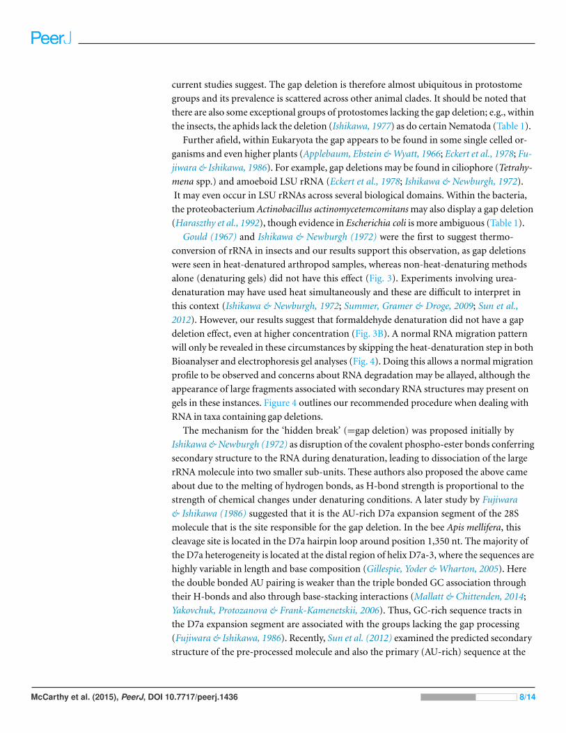
deletion effect, even at higher concentration (Fig. 3B). A normal RNA migration pattern
will only be revealed in these circumstances by skipping the heat-denaturation step in both
Bioanalyser and electrophoresis gel analyses (Fig. 4). Doing this allows a normal migration
profile to be observed and concerns about RNA degradation may be allayed, although the
appearance of large fragments associated with secondary RNA structures may present on
gels in these instances. Figure 4 outlines our recommended procedure when dealing with
RNA in taxa containing gap deletions.
The mechanism for the ‘hidden break’ (=gap deletion) was proposed initially by
Ishikawa & Newburgh (1972) as disruption of the covalent phospho-ester bonds conferring
secondary structure to the RNA during denaturation, leading to dissociation of the large
rRNA molecule into two smaller sub-units. These authors also proposed the above came
about due to the melting of hydrogen bonds, as H-bond strength is proportional to the
strength of chemical changes under denaturing conditions. A later study by Fujiwara
& Ishikawa (1986) suggested that it is the AU-rich D7a expansion segment of the 28S
molecule that is the site responsible for the gap deletion. In the bee Apis mellifera, this
cleavage site is located in the D7a hairpin loop around position 1,350 nt. The majority of
the D7a heterogeneity is located at the distal region of helix D7a-3, where the sequences are
highly variable in length and base composition (Gillespie, Yoder & Wharton, 2005). Here
the double bonded AU pairing is weaker than the triple bonded GC association through
their H-bonds and also through base-stacking interactions (Mallatt & Chittenden, 2014;
Yakovchuk, Protozanova & Frank-Kamenetskii, 2006). Thus, GC-rich sequence tracts in
the D7a expansion segment are associated with the groups lacking the gap processing
(Fujiwara & Ishikawa, 1986). Recently, Sun et al. (2012) examined the predicted secondary
structure of the pre-processed molecule and also the primary (AU-rich) sequence at the
McCarthy et al. (2015), PeerJ, DOI 10.7717/peerj.1436 8/14
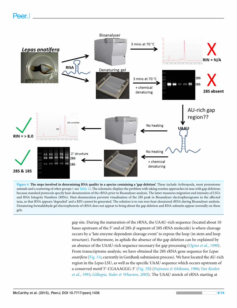
Figure 4 The steps involved in determining RNA quality in a species containing a ‘gap deletion’. These include Arthropoda, most protostomeanimals and a scattering of other groups (-see Table 1). The schematic displays the problem with taking routine approaches in taxa with gap deletionsbecause standard protocols specify heat-denaturation of the rRNA prior to Bioanalyser analysis. The latter measures migration and intensity of LSUsand RNA Integrity Numbers (RINs). Heat-denaturation prevents visualisation of the 28S peak in Bioanalyser electropherograms in the affectedtaxa, so that RNA appears ‘degraded’ and a RIN cannot be generated. The solution is to run non-heat-denatured rRNA during Bioanalyser analysis.Denaturing formaldehyde gel electrophoresis of rRNA does not appear to bring about the gap deletion and RNA subunits appear normally on thesegels.
gap site. During the maturation of the rRNA, the UAAU-rich sequence (located about 10
bases upstream of the 5′ end of 28S-β segment of 28S rRNA molecule) is where cleavage
occurs by a ‘late enzyme dependent cleavage event’ to expose the loop (in stem and loop
structure). Furthermore, in aphids the absence of the gap deletion can be explained by
an absence of the UAAU-rich sequence necessary for gap processing (Ogino et al., 1990).
From transcriptome analysis, we have obtained the 28S rRNA gene sequence for Lepas
anatifera (Fig. 5A; currently in GenBank submission process). We have located the AU-rich
region in the Lepas LSU, as well as the specific UAAU sequence which occurs upstream of
a conserved motif 5′-CGAAAGGG-3′ (Fig. 5B) (Fujiwara & Ishikawa, 1986; Van Keulen
et al., 1991; Gillespie, Yoder & Wharton, 2005). The UAAU stretch of rRNA starting at
McCarthy et al. (2015), PeerJ, DOI 10.7717/peerj.1436 9/14
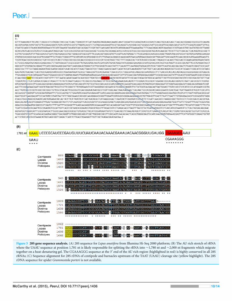
Figure 5 28S gene sequence analysis. (A) 28S sequence for Lepas anatifera from Illumina Hi-Seq 2000 platform; (B) The AU rich stretch of rRNAwhere the UAAU sequence at position 1,781 nt is likely responsible for splitting the rRNA into ∼1,780 nt and ∼2,000 nt fragments which migratetogether on a heat-denaturing gel. The CGAAAGGG sequence at the 3′ end of the AU rich region (highlighted in red) is highly conserved in all 28SrRNAs; (C) Sequence alignment for 28S rDNA of centipede and barnacles upstream of the TAAT (UAAU) cleavage site (yellow highlight). The 28SrDNA sequence for spider Grammostola porteri is not available.
McCarthy et al. (2015), PeerJ, DOI 10.7717/peerj.1436 10/14
position 1,781 nt in Lepas is likely responsible for splitting the rRNA into two fragments
of ∼1,780 nt and ∼2,000 nt, which migrate together on a heat-denaturing gel due to their
similar size. On the Agilent bioanalyser output (Fig. 2) we observed a double-headed peak
for heat-denatured samples of the centipede and barnacles, which is likely due to the small
difference in fragment size following cleavage. This UAAU sequence was present across the
three barnacle species tested (Fig. 5C). While complete 28S rDNA across all four species
with available sequence data was highly conserved (Fig. S1), the LSU sequence is not yet
available for the Grammostola spider species.
The gap deletion is not thought to cause any functional problem for mature rRNA
because unless denatured, the subunits remain bound together at the normal loop (Sun
et al., 2012). But many researchers extracting RNA may experience practical concerns
about apparent degradation: a Bioanalyser analysis of heat-denatured RNA is likely to
yield an output reading such as ‘RIN number not applicable (N/A)’ due to complete
absence of a detectable 28S fragment. In fact, these may be extractions of high quality
and the sample just requires analysis in its non-heated form to yield a RIN. The present
study adds to the knowledge about which organism groups display the ‘degraded’ RNA
appearance on analysis. Researcher awareness of the 28s rRNA cleavage activated during
the heat-denaturing process will eliminate unnecessary troubleshooting in the pursuit of
high quality RNA profiles and RINs.
ACKNOWLEDGEMENTSWe would like to thank our colleagues at School of Natural Sciences, NUIG and the
barnacle stranding sightings community on ‘theamazinggoosebarnacle’ facebook page.
Special thanks to the two reviewers whose comments were exceptionally helpful in
improving the manuscript.
ADDITIONAL INFORMATION AND DECLARATIONS
FundingThis research was funded by Science Foundation Ireland, Contract grant number:
09RFPMTR2311 awarded to AMP http://www.sfi.ie/ and a Beaufort Marine Research
Award grant-aided by the Marine Institute, Department of Communications, Marine and
Natural Resources, Government of Ireland. The funders had no role in study design, data
collection and analysis, decision to publish, or preparation of the manuscript.
Grant DisclosuresThe following grant information was disclosed by the authors:
Science Foundation Ireland: 09RFPMTR2311.
Marine Institute, Department of Communications, Marine and Natural Resources,
Government of Ireland.
Competing InterestsThe authors declare there are no competing interests.
McCarthy et al. (2015), PeerJ, DOI 10.7717/peerj.1436 11/14
Author Contributions• Sean D. McCarthy conceived and designed the experiments, performed the experiments,
analyzed the data, contributed reagents/materials/analysis tools, wrote the paper,
prepared figures and/or tables, reviewed drafts of the paper.
• Michel M. Dugon contributed reagents/materials/analysis tools, wrote the paper,
reviewed drafts of the paper, animal dissection and expertise.
• Anne Marie Power conceived and designed the experiments, analyzed the data, wrote
the paper, prepared figures and/or tables, reviewed drafts of the paper.
DNA DepositionThe following information was supplied regarding the deposition of DNA sequences:
GenBank, Lepas anatifera 28S sequence: KU052603.
Supplemental InformationSupplemental information for this article can be found online at http://dx.doi.org/
10.7717/peerj.1436#supplemental-information.
REFERENCESApplebaum SW, Ebstein RP, Wyatt GR. 1966. Dissociation of ribonucleic acid from silkmoth
pupae by heat and dimethylsulfoxide: evidence for specific cleavage points. Journal of MolecularBiology 21(1):29–41 DOI 10.1016/0022-2836(66)90077-5.
Asai S, Ianora A, Lauritano C, Lindeque PK, Carotenuto Y. 2014. High-quality RNA extractionfrom copepods for Next Generation Sequencing: a comparative study. Marine Genomics Epubahead of print Dec 26 2014 DOI 10.1016/j.margen.2014.12.004.
Eckert WA, Kaffenberger W, Krohne G, Franke WW. 1978. Introduction of hidden breaks duringrRNA maturation and ageing in Tetrahymena pyriformis. European Journal of Biochemistry87(3):607–616 DOI 10.1111/j.1432-1033.1978.tb12413.x.
Fujiwara H, Ishikawa H. 1986. Molecular mechanism of introduction of the hidden break intothe 28S rRNA of insects: implication based on structural studies. Nucleic Acids Research14(16):6393–6401 DOI 10.1093/nar/14.16.6393.
Gillespie JJ, Johnston JS, Cannone JJ, Gutell RR. 2006. Characteristics of the nuclear (18S,5.8S, 28S and 5S) and mitochondrial (12S and 16S) rRNA genes of Apis mellifera (Insecta:Hymenoptera): structure, organization, and retrotransposable elements. Insect MolecularBiology 15(5):657–686 DOI 10.1111/j.1365-2583.2006.00689.x.
Gillespie JJ, Yoder MJ, Wharton RA. 2005. Predicted secondary structure for 28S and 18SrRNA from Ichneumonoidea (Insecta: Hymenoptera: Apocrita): impact on sequencealignment and phylogeny estimation. Journal of Molecular Evolution 61(1):114–137DOI 10.1007/s00239-004-0246-x.
Gould HJ. 1967. The nature of high molecular weight fragments of ribosomal RNA. Journal ofMolecular Biology 29(2):307–312 DOI 10.1016/0022-2836(67)90218-5.
Haraszthy VI, Sunday GJ, Bobek LA, Motley TS, Preus H, Zambon JJ. 1992. Identification andanalysis of the gap region in the 23S ribosomal RNA from Actinobacillus actinomycetemcomitans.Journal of Dental Research 71(9):1561–1568 DOI 10.1177/00220345920710090401.
McCarthy et al. (2015), PeerJ, DOI 10.7717/peerj.1436 12/14
Henras AK, Plisson-Chastang C, O’Donohue MF, Chakraborty A, Gleizes PE. 2015. Anoverview of pre-ribosomal RNA processing in eukaryotes. WIREs RNA 6:225–242DOI 10.1002/wrna.1269.
Ishikawa H. 1975a. Comparative studies on the thermal stability of animal ribosomal RNA’s–III.Sponge (Porifera) and the other species (Tetrahymena and Lachnus). Comparative Biochemistryand Physiology 51(1):81–85.
Ishikawa H. 1975b. Comparative studies on the thermal stability of animal ribosomal RNA’s.II. Sea-anemones (Coelenterata). Comparative Biochemistry and Physiology 50(1):1–4DOI 10.1016/S0010-406X(75)80194-0.
Ishikawa H. 1976. The fragments from the 28 S ribosomal RNA of Galleria mellonella withunesterified uridine at the 3′-termini. Biochimica et Biophysica ACTA 425(2):185–195DOI 10.1016/0005-2787(76)90024-1.
Ishikawa H. 1977. Evolution of ribosomal RNA. Comparative Biochemistry and Physiology58(1):1–7 DOI 10.1016/0300-9629(77)90002-0.
Ishikawa H, Newburgh RW. 1972. Studies of the thermal conversion of 28 S RNA ofGalleria mellonella (L.) to an 18 S product. Journal of Molecular Biology 64(1):135–144DOI 10.1016/0022-2836(72)90325-7.
Jonker JL, Abram F, Pires E, Varela Coelho A, Grunwald I, Power AM. 2014. Adhesive proteinsof stalked and acorn barnacles display homology with low sequence similarities. PLoS ONE9(10):e108902 DOI 10.1371/journal.pone.0108902.
Jonker JL, Von Byern J, Flammang P, Klepal W, Power AM. 2012. Unusual adhesive productionsystem in the barnacle Lepas anatifera: an ultrastructural and histochemical investigation.Journal of Morphology 273(12):1377–1391 DOI 10.1002/jmor.20067.
Krupp G. 2005. Stringent RNA quality control using the Agilent 2100 bioanalyzer. Available athttp://www.chem.agilent.com/Library/applications/5989-1086EN.pdf.
Macharia RW, Ombura FL, Aroko EO. 2015. Insects’ RNA profiling reveals absence of “hiddenbreak” in 28S ribosomal RNA molecule of onion thrips, Thrips tabaci. Journal of Nucleic Acids2015:965294 DOI 10.1155/2015/965294.
Mallatt J, Chittenden KD. 2014. The GC content of LSU rRNA evolves across topological andfunctional regions of the ribosome in all three domains of life. Molecular Phylogenetics andEvolution 72:17–30 DOI 10.1016/j.ympev.2013.12.007.
Masek T, Vopalensky V, Suchomelova P, Pospisek M. 2005. Denaturing RNA electrophoresis inTAE agarose gels. Analytical Biochemistry 336(1):46–50 DOI 10.1016/j.ab.2004.09.010.
Melen GJ, Pesce CG, Rossi MS, Kornblihtt AR. 1999. Novel processing in a mammalian nuclear28S pre-rRNA: tissue-specific elimination of an ‘intron’ bearing a hidden break site. EMBOJournal 18(11):3107–3118 DOI 10.1093/emboj/18.11.3107.
Ogino K, Eda-Fujiwara H, Fujiwara H, Ishikawa H. 1990. What causes the aphid 28S rRNA tolack the hidden break? Journal of Molecular Evolution 30(6):509–513 DOI 10.1007/BF02101106.
Rota-Stabelli O, Campbell L, Brinkmann H, Edgecombe GD, Longhorn SJ, Peterson KJ,Pisani D, Philippe H, Telford MJ. 2011. A congruent solution to arthropod phylogeny:phylogenomics, microRNAs and morphology support monophyletic Mandibulata. Proceedingsof the Royal Society B: Biological Science 278(1703):298–306 DOI 10.1098/rspb.2010.0590.
Skrypina NA, Timofeeva AV, Khaspekov GL, Savochkina LP, Beabealashvilli RSh. 2003.Total RNA suitable for molecular biology analysis. Journal of Biotechnology 105(1–2):1–9DOI 10.1016/S0168-1656(03)00140-8.
McCarthy et al. (2015), PeerJ, DOI 10.7717/peerj.1436 13/14
Stevens AR, Pachler PF. 1972. Discontinuity of 26 s rRNA in Acanthamoeba castellani. Journal ofMolecular Biology 66(2):225–237 DOI 10.1016/0022-2836(72)90475-5.
Summer HR, Gramer R, Droge P. 2009. Denaturing urea polyacrylamide gel electrophoresis (UreaPAGE). Journal of Visualized Experiments 32:e1485 DOI 10.3791/1485.
Sun S, Xie H, Sun Y, Song J, Li Z. 2012. Molecular characterization of gap region in 28SrRNA molecules in brine shrimp Artemia parthenogenetica and planarian Dugesia japonica.Biochemistry 77(4):411–417.
Van Keulen H, Mertz PM, LoVerde PT, Shi H, Rekosh DM. 1991. Characterization of a54-nucleotide gap region in the 28S rRNA gene of Schistosoma mansoni. Molecular andBiochemical Parasitology 45(2):205–214 DOI 10.1016/0166-6851(91)90087-M.
Ware VC, Tague BW, Clark CG, Gourse RL, Brand RC, Gerbi SA. 1983. Sequence analysis of 28Sribosomal DNA from the amphibian Xenopus laevis. Nucleic Acids Research 11(22):7795–7817DOI 10.1093/nar/11.22.7795.
Winnebeck EC, Millar CD, Warman GR. 2010. Why does insect RNA look degraded? Journal ofInsect Science 10(1) DOI 10.1673/031.010.14119.
Yakovchuk P, Protozanova E, Frank-Kamenetskii MD. 2006. Base-stacking and base-pairingcontributions into thermal stability of the DNA double helix. Nucleic Acids Research34(2):564–574 DOI 10.1093/nar/gkj454.
Zarlenga DS, Dame JB. 1992. The identification and characterization of a break within the largesubunit ribosomal RNA of Trichinella spiralis: comparison of gap sequences within the genus.Molecular and Biochemical Parasitology 51(2):281–289 DOI 10.1016/0166-6851(92)90078-X.
McCarthy et al. (2015), PeerJ, DOI 10.7717/peerj.1436 14/14