Corrections - PNAS · Vladimir L. Buchman, which appeared in issue 51, December 18, 2012, of Proc...
7
Increased lipolysis and altered lipid homeostasis protect γ-synuclein–null mutant mice from diet-induced obesity Steven Millership a,1 , Natalia Ninkina a,1 , Irina A. Guschina a , Jessica Norton a , Riccardo Brambilla a,b , Pieter J. Oort c , Sean H. Adams c , Rowena J. Dennis d , Peter J. Voshol d , Justin J. Rochford d,2 , and Vladimir L. Buchman a,2 a School of Biosciences, Cardiff University, Cardiff CF10 3AX, United Kingdom; b Division of Neuroscience, Institute of Experimental Neurology, San Raffaele Foundation and University, 20132 Milan, Italy; c Obesity and Metabolism Research Unit, US Department of Agriculture/Agricultural Research Service Western Human Nutrition Research Center, Davis, CA 95616; and d Institute of Metabolic Science, University of Cambridge Metabolic Research Laboratories and National Institute for Health Research Cambridge Biomedical Research Centre, Addenbrooke’s Hospital, Cambridge CB2 0QQ, United Kingdom Edited* by Stephen O’Rahilly, University of Cambridge, Cambridge, United Kingdom, and approved November 12, 2012 (received for review June 13, 2012) Synucleins are a family of homologous proteins principally known for their involvement in neurodegeneration. γ-Synuclein is highly expressed in human white adipose tissue and increased in obesity. Here we show that γ-synuclein is nutritionally regulated in white adipose tissue whereas its loss partially protects mice from high-fat diet (HFD)–induced obesity and ameliorates some of the associated metabolic complications. Compared with HFD-fed WT mice, HFD-fed γ-synuclein–null mutant mice display increased lipolysis, lipid oxida- tion, and energy expenditure, and reduced adipocyte hypertrophy. Knockdown of γ-synuclein in adipocytes causes redistribution of the key lipolytic enzyme ATGL to lipid droplets and increases lipolysis. γ-Synuclein–deficient adipocytes also contain fewer SNARE com- plexes of a type involved in lipid droplet fusion. We hypothesize that γ-synuclein may deliver SNAP-23 to the SNARE complexes un- der lipogenic conditions. Via these independent but complementary roles, γ-synuclein may coordinately modulate lipid storage by influ- encing lipolysis and lipid droplet formation. Our data reveal γ-syn- uclein as a regulator of lipid handling in adipocytes, the function of which is particularly important in conditions of nutrient excess. U nderstanding the link between increased adiposity and the development of metabolic disease may reveal novel thera- peutic targets to counter the rising pandemic of obesity. Inhib- iting adipose tissue expansion alone is likely to worsen metabolic outcome, as evidenced by human syndromes of lipodystrophy, whereby inappropriately decreased adipose mass causes severe metabolic disorders (1). Indeed, adipose tissue dysfunction and/ or exceeded adipose storage capacity may underlie ectopic lipid accumulation and lipotoxicity in obesity (2). Therefore, a major challenge is to identify pathways via which adiposity can be re- duced without concomitant increases in circulating lipids and attendant metabolic disease. Achieving this goal requires a better understanding of the molecular mechanisms that regulate lipid metabolism and storage in adipocytes, particularly in times of energy surplus. γ-Synuclein belongs to the synuclein family of proteins, whose founder member α-synuclein is best known for its links with neurodegenerative diseases, most notably Parkinson disease (3). To date, no clear cellular role is attributed to γ-synuclein, and ablation of γ-synuclein causes only minor changes in the nervous system (4–7). Recently, we and others have reported high levels of γ-synuclein expression in adipose tissue of humans and other mammals (8, 9). Moreover, expression of γ-synuclein is increased in the adipose tissue of obese humans and decreased during caloric restriction (8). Here we demonstrate that γ-synuclein–null mice display sig- nificantly reduced adiposity and fewer metabolic derangements compared with WT mice following high-fat feeding. This appears to result from increased adipocyte lipolysis coupled to enhanced whole-body lipid oxidation and energy expenditure. At a molec- ular level, we identify dual roles for γ-synuclein independently regulating lipid droplet fusion and adipocyte lipolysis to co- ordinately regulate triglyceride (TG) storage in adipocytes. To- gether, our observations reveal that γ-synuclein is a regulator of lipid metabolism and, hence, a potential therapeutic target for treatment of obesity and associated metabolic diseases. Results γ-Synuclein Expression Is Nutritionally Regulated in Adipocytes. γ-Synuclein is abundantly expressed in adipose tissue (8, 9) (Fig. S1). Western blotting of fractionated white adipose tissue (WAT) demonstrated that γ-synuclein was expressed in the mature adi- pocyte but not the stromal cell fraction (Fig. 1A). When adipo- cytes were subjected to subcellular fractionation, γ-synuclein was recovered in the infranatant, containing cytosol and membranes. No γ-synuclein was detected in the fraction containing lipid droplet-associated proteins in the presence or absence of lipo- lytic stimulation, which was sufficient to induce the translocation of hormone-sensitive lipase (HSL; Fig. 1B). Immunostaining of adipocytes differentiated from MEFs in vitro also revealed dif- fuse, partly punctuate cytoplasmic staining, and did not suggest an association of γ-synuclein with lipid droplets (Fig. 1C). Consistent with previous observations in patients with obesity (8), γ-synuclein mRNA expression was dramatically increased in the epididymal and s.c. WAT of mice fed a high-fat diet (HFD) for 11 wk (Fig. 1D) and gradually decreased after mice fed an HFD for 10 d were switched to a low-fat diet (LFD) or fasted (Fig. 1E). Parallel changes in γ-synuclein protein levels were observed in epididymal WAT following dietary manipulation (Fig. 1F). γ-Synuclein–Null Mice Are Resistant to Diet-Induced Obesity. The nutritional regulation of γ-synuclein expression in adipose tissue led us to investigate whether it could play a role in the de- velopment of obesity. WT and γ-synuclein −/− mice (9 wk old) were fed an LFD or HFD for a period of 11 wk. LFD-fed WT and γ-synuclein −/− mice showed no differences in weight gain. In contrast, during HFD-feeding, γ-synuclein −/− mice gained sub- stantially less weight than their WT counterparts (Fig. 2A). Quantitative MRI analysis of body fat volume during the study Author contributions: J.J.R. and V.L.B. designed research; S.M., N.N., I.A.G., J.N., P.J.O., R.J.D., J.J.R., and V.L.B. performed research; R.B. contributed new reagents/analytic tools; S.M., N.N., S.H.A., P.J.V., J.J.R., and V.L.B. analyzed data; and S.M., N.N., J.J.R., and V.L.B. wrote the paper. The authors declare no conflict of interest. *This Direct Submission article had a prearranged editor. Freely available online through the PNAS open access option. 1 S.M. and N.N. contributed equally to this work. 2 To whom correspondence may be addressed. E-mail: [email protected] or buchmanvl@ cf.ac.uk. This article contains supporting information online at www.pnas.org/lookup/suppl/doi:10. 1073/pnas.1210022110/-/DCSupplemental. www.pnas.org/cgi/doi/10.1073/pnas.1210022110 PNAS | December 18, 2012 | vol. 109 | no. 51 | 20943–20948 CELL BIOLOGY Downloaded by guest on February 19, 2020 Downloaded by guest on February 19, 2020 Downloaded by guest on February 19, 2020
Transcript of Corrections - PNAS · Vladimir L. Buchman, which appeared in issue 51, December 18, 2012, of Proc...
Increased lipolysis and altered lipid homeostasisprotect γ-synuclein–null mutant mice fromdiet-induced obesitySteven Millershipa,1, Natalia Ninkinaa,1, Irina A. Guschinaa, Jessica Nortona, Riccardo Brambillaa,b, Pieter J. Oortc,Sean H. Adamsc, Rowena J. Dennisd, Peter J. Voshold, Justin J. Rochfordd,2, and Vladimir L. Buchmana,2
aSchool of Biosciences, Cardiff University, Cardiff CF10 3AX, United Kingdom; bDivision of Neuroscience, Institute of Experimental Neurology, San RaffaeleFoundation and University, 20132 Milan, Italy; cObesity and Metabolism Research Unit, US Department of Agriculture/Agricultural Research Service WesternHuman Nutrition Research Center, Davis, CA 95616; and dInstitute of Metabolic Science, University of Cambridge Metabolic Research Laboratories andNational Institute for Health Research Cambridge Biomedical Research Centre, Addenbrooke’s Hospital, Cambridge CB2 0QQ, United Kingdom
Edited* by Stephen O’Rahilly, University of Cambridge, Cambridge, United Kingdom, and approved November 12, 2012 (received for review June 13, 2012)
Synucleins are a family of homologous proteins principally knownfor their involvement in neurodegeneration. γ-Synuclein is highlyexpressed in human white adipose tissue and increased in obesity.Here we show that γ-synuclein is nutritionally regulated in whiteadipose tissue whereas its loss partially protects mice from high-fatdiet (HFD)–induced obesity and ameliorates some of the associatedmetabolic complications. Comparedwith HFD-fedWTmice, HFD-fedγ-synuclein–null mutant mice display increased lipolysis, lipid oxida-tion, and energy expenditure, and reduced adipocyte hypertrophy.Knockdown of γ-synuclein in adipocytes causes redistribution of thekey lipolytic enzyme ATGL to lipid droplets and increases lipolysis.γ-Synuclein–deficient adipocytes also contain fewer SNARE com-plexes of a type involved in lipid droplet fusion. We hypothesizethat γ-synuclein may deliver SNAP-23 to the SNARE complexes un-der lipogenic conditions. Via these independent but complementaryroles, γ-synuclein may coordinately modulate lipid storage by influ-encing lipolysis and lipid droplet formation. Our data reveal γ-syn-uclein as a regulator of lipid handling in adipocytes, the function ofwhich is particularly important in conditions of nutrient excess.
Understanding the link between increased adiposity and thedevelopment of metabolic disease may reveal novel thera-
peutic targets to counter the rising pandemic of obesity. Inhib-iting adipose tissue expansion alone is likely to worsen metabolicoutcome, as evidenced by human syndromes of lipodystrophy,whereby inappropriately decreased adipose mass causes severemetabolic disorders (1). Indeed, adipose tissue dysfunction and/or exceeded adipose storage capacity may underlie ectopic lipidaccumulation and lipotoxicity in obesity (2). Therefore, a majorchallenge is to identify pathways via which adiposity can be re-duced without concomitant increases in circulating lipids andattendant metabolic disease. Achieving this goal requires a betterunderstanding of the molecular mechanisms that regulate lipidmetabolism and storage in adipocytes, particularly in times ofenergy surplus.γ-Synuclein belongs to the synuclein family of proteins, whose
founder member α-synuclein is best known for its links withneurodegenerative diseases, most notably Parkinson disease (3).To date, no clear cellular role is attributed to γ-synuclein, andablation of γ-synuclein causes only minor changes in the nervoussystem (4–7). Recently, we and others have reported high levelsof γ-synuclein expression in adipose tissue of humans and othermammals (8, 9). Moreover, expression of γ-synuclein is increasedin the adipose tissue of obese humans and decreased duringcaloric restriction (8).Here we demonstrate that γ-synuclein–null mice display sig-
nificantly reduced adiposity and fewer metabolic derangementscompared with WT mice following high-fat feeding. This appearsto result from increased adipocyte lipolysis coupled to enhancedwhole-body lipid oxidation and energy expenditure. At a molec-ular level, we identify dual roles for γ-synuclein independently
regulating lipid droplet fusion and adipocyte lipolysis to co-ordinately regulate triglyceride (TG) storage in adipocytes. To-gether, our observations reveal that γ-synuclein is a regulator oflipid metabolism and, hence, a potential therapeutic target fortreatment of obesity and associated metabolic diseases.
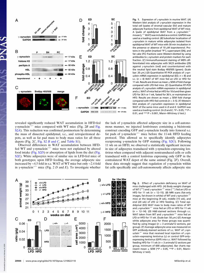
Resultsγ-Synuclein Expression Is Nutritionally Regulated in Adipocytes.γ-Synuclein is abundantly expressed in adipose tissue (8, 9) (Fig.S1). Western blotting of fractionated white adipose tissue (WAT)demonstrated that γ-synuclein was expressed in the mature adi-pocyte but not the stromal cell fraction (Fig. 1A). When adipo-cytes were subjected to subcellular fractionation, γ-synuclein wasrecovered in the infranatant, containing cytosol and membranes.No γ-synuclein was detected in the fraction containing lipiddroplet-associated proteins in the presence or absence of lipo-lytic stimulation, which was sufficient to induce the translocationof hormone-sensitive lipase (HSL; Fig. 1B). Immunostaining ofadipocytes differentiated from MEFs in vitro also revealed dif-fuse, partly punctuate cytoplasmic staining, and did not suggestan association of γ-synuclein with lipid droplets (Fig. 1C).Consistent with previous observations in patients with obesity
(8), γ-synuclein mRNA expression was dramatically increased inthe epididymal and s.c.WAT ofmice fed a high-fat diet (HFD) for11 wk (Fig. 1D) and gradually decreased after mice fed an HFDfor 10 d were switched to a low-fat diet (LFD) or fasted (Fig. 1E).Parallel changes in γ-synuclein protein levels were observed inepididymal WAT following dietary manipulation (Fig. 1F).
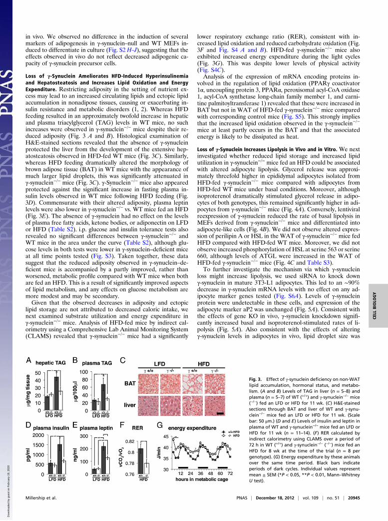
γ-Synuclein–Null Mice Are Resistant to Diet-Induced Obesity. Thenutritional regulation of γ-synuclein expression in adipose tissueled us to investigate whether it could play a role in the de-velopment of obesity. WT and γ-synuclein−/− mice (9 wk old)were fed an LFD or HFD for a period of 11 wk. LFD-fed WTand γ-synuclein−/− mice showed no differences in weight gain. Incontrast, during HFD-feeding, γ-synuclein−/− mice gained sub-stantially less weight than their WT counterparts (Fig. 2A).Quantitative MRI analysis of body fat volume during the study
Author contributions: J.J.R. and V.L.B. designed research; S.M., N.N., I.A.G., J.N., P.J.O., R.J.D.,J.J.R., and V.L.B. performed research; R.B. contributed new reagents/analytic tools; S.M.,N.N., S.H.A., P.J.V., J.J.R., and V.L.B. analyzed data; and S.M., N.N., J.J.R., and V.L.B. wrotethe paper.
The authors declare no conflict of interest.
*This Direct Submission article had a prearranged editor.
Freely available online through the PNAS open access option.1S.M. and N.N. contributed equally to this work.2To whom correspondence may be addressed. E-mail: [email protected] or [email protected].
This article contains supporting information online at www.pnas.org/lookup/suppl/doi:10.1073/pnas.1210022110/-/DCSupplemental.
www.pnas.org/cgi/doi/10.1073/pnas.1210022110 PNAS | December 18, 2012 | vol. 109 | no. 51 | 20943–20948
CELL
BIOLO
GY
Dow
nloa
ded
by g
uest
on
Feb
ruar
y 19
, 202
0 D
ownl
oade
d by
gue
st o
n F
ebru
ary
19, 2
020
Dow
nloa
ded
by g
uest
on
Feb
ruar
y 19
, 202
0
revealed significantly reduced WAT accumulation in HFD-fedγ-synuclein−/− mice compared with WT mice (Fig. 2B and Fig.S2A). This reduction was confirmed postmortem by determiningthe mass of dissected epididymal, s.c., and retroperitoneal de-pots, as well as fat pad mass to body mass ratios for all threedepots (Fig. 2C, Fig. S2 B and C, and Table S1).Observed differences in WAT accumulation between HFD-
fed WT and γ-synuclein−/− mice were not explained by alteredfood intake (Fig. S2D) or absorption of lipids from the diet (Fig.S2E). White adipocytes were of similar size in LFD-fed mice ofboth genotypes; upon HFD feeding, the average adipocyte sizeincreased by ∼4.5 fold in s.c. WAT of WT mice but only ∼2.4 foldin γ-synuclein−/− mice (Fig. 2 D and E). To investigate whether
the lack of γ-synuclein affected adipocyte size in a cell-autono-mous manner, we injected lentiviruses containing a bicistronicconstruct encoding GFP and γ-synuclein locally into femoral s.c.fat pads of γ-synuclein−/− mice before the 11-wk HFD feedingprotocol. This allowed us to specifically identify adipocytesreexpressing γ-synuclein by GFP staining (Fig. S2G). Following11 wk on an HFD, we observed a statistically significant increasein size of adipocytes transduced with γ-synuclein–expressing len-tivirus when compared with adjacent nontransduced cells or cellstransduced with a control lentivirus expressing only GFP in thecontralateral WAT depot of the same animal (Fig. 2F). Overall,these data strongly suggest that regulation of γ-synuclein withinfat cells specifically and cell-autonomously affects adipocyte size
Fig. 1. Expression of γ-synuclein in murine WAT. (A)Western blot analysis of γ-synuclein expression in thetotal cell lysates of stromal–vascular (SV) and matureadipocyte fractions from epididymal WAT of WTmice.A lysate of epididymal WAT from a γ-synuclein−/−
mouse(γ−/−WAT)was includedasacontrol.GAPDHwasused as a loading control. (B) Subcellular localization ofγ-synuclein in mature white adipocytes isolated fromepididymalWAT ofWTmice with 30min incubation inthe presence or absence of 10 μM isoproterenol. Pro-teins in the pellet (marked “P”), supernatant (SN), andfat cake (FC) fractions were Western-blotted by usingantibodies to γ-synuclein and protein markers for eachfraction. (C) Immunofluorescent staining of MEFs dif-ferentiated into adipocytes with SK23 antibodies (29)against γ-synuclein (red) and counterstained withthe neutral lipid stain Bodipy 493/505 (green). (Scalebar: 20 μm.) (D) Quantitative RT-PCR analysis of γ-syn-uclein mRNA expression in epididymal (ED; n = 8) ands.c. (n = 4) WAT of WT mice fed an LFD or HFD for11wk. Results are shownasmean±SEMof fold changecompared with LFD-fed mice. (E) Quantitative RT-PCRanalysis of γ-synuclein mRNA expression in epididymalands.c.WATofmice fedanHFDfor10dandthengivenLFD for 36 h or 1 wk, fasted for 36 h, or maintained onHFD. Results are shown as mean ± SEM fold changecompared with HFD-fed controls (n = 3–5). (F) Westernblot analysis of γ-synuclein expression in epididymalWAT of the same mice used in D and E. GAPDH wasused as a loading control. (InDandE, *P< 0.05, **P<0.01, and ***P < 0.001, Mann–Whitney U test.)
Fig. 2. Effect of γ-synuclein deficiency on WAT ofmice challenged with HFD. (A) Body weight changesof WT (+/+) and γ-synuclein−/− mice (−/−) fed an LFD orHFD for 11 wk (n = 12–15). (B) MRI scans (fat-onlyimages, fat shown in white) of WT and γ-synuclein−/−
mice at the beginning (9 wk), middle (15 wk), andend (20 wk) of LFD or HFD feeding. (C) Total epi-didymal (ED) WAT mass to body mass ratios of WTand γ-synuclein−/− mice fed an LFD or HFD for 11 wk(n = 12–14). (D) H&E-stained sections through s.c.WAT taken from WT and γ-synuclein−/− mice fed anLFD or HFD for 11 wk. (Scale bar: 50 μm.) (E) Averagewhite adipocyte area for these groups was quanti-fied by using ImageJ (n = 3 animals/12 sections, pergroup). (F) Average adipocyte area was measured onGFP antibody-stained sections of s.c. WAT of γ-syn-uclein−/− mice that received local injection of γ-syn-uclein–expressing lentivirus (γ) or control GFP-only–expressing lentivirus (GFP) into s.c. WAT followed byfeeding HFD for 11 wk (n = 3 animals/12 sections pergroup, minimum of 600 adipocytes). Bar charts rep-resent mean ± SEM (*P < 0.05, **P < 0.01, Mann–Whitney U test).
20944 | www.pnas.org/cgi/doi/10.1073/pnas.1210022110 Millership et al.
Dow
nloa
ded
by g
uest
on
Feb
ruar
y 19
, 202
0
in vivo. We observed no difference in the induction of severalmarkers of adipogenesis in γ-synuclein–null and WT MEFs in-duced to differentiate in culture (Fig. S2 H–J), suggesting that theeffects observed in vivo do not reflect decreased adipogenic ca-pacity of γ-synuclein precursor cells.
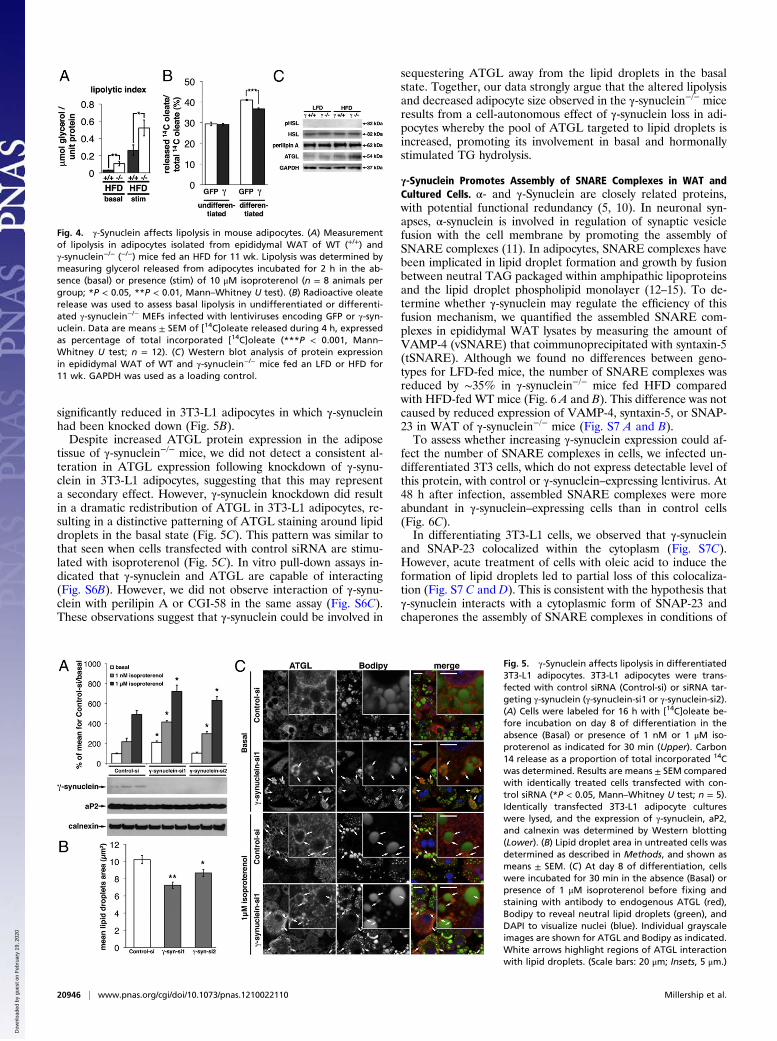
Loss of γ-Synuclein Ameliorates HFD-Induced Hyperinsulinemiaand Hepatosteatosis and Increases Lipid Oxidation and EnergyExpenditure. Restricting adiposity in the setting of nutrient ex-cess may lead to an increased circulating lipids and ectopic lipidaccumulation in nonadipose tissues, causing or exacerbating in-sulin resistance and metabolic disorders (1, 2). Whereas HFDfeeding resulted in an approximately twofold increase in hepaticand plasma triacylglycerol (TAG) levels in WT mice, no suchincreases were observed in γ-synuclein−/− mice despite their re-duced adiposity (Fig. 3 A and B). Histological examination ofH&E-stained sections revealed that the absence of γ-synucleinprotected the liver from the development of the extensive hep-atosteatosis observed in HFD-fed WT mice (Fig. 3C). Similarly,whereas HFD feeding dramatically altered the morphology ofbrown adipose tissue (BAT) in WT mice with the appearance ofmuch larger lipid droplets, this was significantly attenuated inγ-synuclein−/− mice (Fig. 3C). γ-Synuclein−/− mice also appearedprotected against the significant increase in fasting plasma in-sulin levels observed in WT mice following HFD feeding (Fig.3D). Commensurate with their altered adiposity, plasma leptinlevels were also lower in γ-synuclein−/− vs. WT mice fed an HFD(Fig. 3E). The absence of γ-synuclein had no effect on the levelsof plasma free fatty acids, ketone bodies, or adiponectin on LFDor HFD (Table S2). i.p. glucose and insulin tolerance tests alsorevealed no significant differences between γ-synuclein−/− andWT mice in the area under the curve (Table S2), although glu-cose levels in both tests were lower in γ-synuclein–deficient miceat all time points tested (Fig. S3). Taken together, these datasuggest that the reduced adiposity observed in γ-synuclein–de-ficient mice is accompanied by a partly improved, rather thanworsened, metabolic profile compared with WT mice when bothare fed an HFD. This is a result of significantly improved aspectsof lipid metabolism, and any effects on glucose metabolism aremore modest and may be secondary.Given that the observed decreases in adiposity and ectopic
lipid storage are not attributed to decreased caloric intake, wenext examined substrate utilization and energy expenditure inγ-synuclein−/− mice. Analysis of HFD-fed mice by indirect cal-orimetry using a Comprehensive Lab Animal Monitoring System(CLAMS) revealed that γ-synuclein−/− mice had a significantly
lower respiratory exchange ratio (RER), consistent with in-creased lipid oxidation and reduced carbohydrate oxidation (Fig.3F and Fig. S4 A and B). HFD-fed γ-synuclein−/− mice alsoexhibited increased energy expenditure during the light cycles(Fig. 3G). This was despite lower levels of physical activity(Fig. S4C).Analysis of the expression of mRNA encoding proteins in-
volved in the regulation of lipid oxidation (PPARγ coactivator1α, uncoupling protein 3, PPARα, peroxisomal acyl-CoA oxidase1, acyl-CoA synthetase long-chain family member 1, and carni-tine palmitoyltransferase 1) revealed that these were increased inBAT but not in WAT of HFD-fed γ-synuclein−/− mice comparedwith corresponding control mice (Fig. S5). This strongly impliesthat the increased lipid oxidation observed in the γ-synuclein−/−mice at least partly occurs in the BAT and that the associatedenergy is likely to be dissipated as heat.
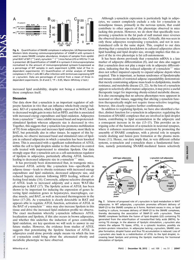
Loss of γ-Synuclein Increases Lipolysis in Vivo and in Vitro. We nextinvestigated whether reduced lipid storage and increased lipidutilization in γ-synuclein−/− mice fed an HFD could be associatedwith altered adipocyte lipolysis. Glycerol release was approxi-mately threefold higher in epididymal adipocytes isolated fromHFD-fed γ-synuclein−/− mice compared with adipocytes fromHFD-fed WT mice under basal conditions. Moreover, althoughisoproterenol dramatically stimulated glycerol release in adipo-cytes of both genotypes, this remained significantly higher in adi-pocytes from γ-synuclein−/− mice (Fig. 4A). Conversely, lentiviralreexpression of γ-synuclein reduced the rate of basal lipolysis inMEFs derived from γ-synuclein−/− mice and differentiated intoadipocyte-like cells (Fig. 4B). We did not observe altered expres-sion of perilipin A or HSL in the WAT of γ-synuclein−/− mice fedHFD compared with HFD-fed WT mice. Moreover, we did notobserve increased phosphorylation of HSL at serine 563 or serine660, although levels of ATGL were increased in the WAT ofHFD-fed γ-synuclein−/− mice (Fig. 4C and Table S3).To further investigate the mechanism via which γ-synuclein
loss might increase lipolysis, we used siRNA to knock downγ-synuclein in mature 3T3-L1 adipocytes. This led to an ∼90%decrease in γ-synuclein mRNA levels with no effect on any ad-ipocyte marker genes tested (Fig. S6A). Levels of γ-synucleinprotein were undetectable in these cells, and expression of theadipocyte marker aP2 was unchanged (Fig. 5A). Consistent withthe effects of gene KO in vivo, γ-synuclein knockdown signifi-cantly increased basal and isoproterenol-stimulated rates of li-polysis (Fig. 5A). Also consistent with the effects of alteringγ-synuclein levels in adipocytes in vivo, lipid droplet size was
Fig. 3. Effect of γ-synuclein deficiency on non-WATlipid accumulation, hormonal status, and metabo-lism. (A and B) Levels of TAG in liver (n = 5–8) andplasma (n = 5–7) of WT (+/+) and γ-synuclein−/− mice(−/−) fed an LFD or HFD for 11 wk. (C) H&E-stainedsections through BAT and liver of WT and γ-synu-clein−/− mice fed an LFD or HFD for 11 wk. (Scalebar: 50 μm.) (D and E) Levels of insulin and leptin inplasma of WT and γ-synuclein−/− mice fed an LFD orHFD for 11 wk (n = 11–14). (F) RER calculated byindirect calorimetry using CLAMS over a period of72 h in WT (+/+) and γ-synuclein−/− (−/−) mice fed anHFD for 8 wk at the time of the trial (n = 8 pergenotype). (G) Energy expenditure by these animalsover the same time period. Black bars indicateperiods of dark cycles. Individual values representmean ± SEM (*P < 0.05, **P < 0.01, Mann–WhitneyU test).
Millership et al. PNAS | December 18, 2012 | vol. 109 | no. 51 | 20945
CELL
BIOLO
GY
Dow
nloa
ded
by g
uest
on
Feb
ruar
y 19
, 202
0
significantly reduced in 3T3-L1 adipocytes in which γ-synucleinhad been knocked down (Fig. 5B).Despite increased ATGL protein expression in the adipose
tissue of γ-synuclein−/− mice, we did not detect a consistent al-teration in ATGL expression following knockdown of γ-synu-clein in 3T3-L1 adipocytes, suggesting that this may representa secondary effect. However, γ-synuclein knockdown did resultin a dramatic redistribution of ATGL in 3T3-L1 adipocytes, re-sulting in a distinctive patterning of ATGL staining around lipiddroplets in the basal state (Fig. 5C). This pattern was similar tothat seen when cells transfected with control siRNA are stimu-lated with isoproterenol (Fig. 5C). In vitro pull-down assays in-dicated that γ-synuclein and ATGL are capable of interacting(Fig. S6B). However, we did not observe interaction of γ-synu-clein with perilipin A or CGI-58 in the same assay (Fig. S6C).These observations suggest that γ-synuclein could be involved in
sequestering ATGL away from the lipid droplets in the basalstate. Together, our data strongly argue that the altered lipolysisand decreased adipocyte size observed in the γ-synuclein−/− miceresults from a cell-autonomous effect of γ-synuclein loss in adi-pocytes whereby the pool of ATGL targeted to lipid droplets isincreased, promoting its involvement in basal and hormonallystimulated TG hydrolysis.
γ-Synuclein Promotes Assembly of SNARE Complexes in WAT andCultured Cells. α- and γ-Synuclein are closely related proteins,with potential functional redundancy (5, 10). In neuronal syn-apses, α-synuclein is involved in regulation of synaptic vesiclefusion with the cell membrane by promoting the assembly ofSNARE complexes (11). In adipocytes, SNARE complexes havebeen implicated in lipid droplet formation and growth by fusionbetween neutral TAG packaged within amphipathic lipoproteinsand the lipid droplet phospholipid monolayer (12–15). To de-termine whether γ-synuclein may regulate the efficiency of thisfusion mechanism, we quantified the assembled SNARE com-plexes in epididymal WAT lysates by measuring the amount ofVAMP-4 (vSNARE) that coimmunoprecipitated with syntaxin-5(tSNARE). Although we found no differences between geno-types for LFD-fed mice, the number of SNARE complexes wasreduced by ∼35% in γ-synuclein−/− mice fed HFD comparedwith HFD-fed WT mice (Fig. 6 A and B). This difference was notcaused by reduced expression of VAMP-4, syntaxin-5, or SNAP-23 in WAT of γ-synuclein−/− mice (Fig. S7 A and B).To assess whether increasing γ-synuclein expression could af-
fect the number of SNARE complexes in cells, we infected un-differentiated 3T3 cells, which do not express detectable level ofthis protein, with control or γ-synuclein–expressing lentivirus. At48 h after infection, assembled SNARE complexes were moreabundant in γ-synuclein–expressing cells than in control cells(Fig. 6C).In differentiating 3T3-L1 cells, we observed that γ-synuclein
and SNAP-23 colocalized within the cytoplasm (Fig. S7C).However, acute treatment of cells with oleic acid to induce theformation of lipid droplets led to partial loss of this colocaliza-tion (Fig. S7 C and D). This is consistent with the hypothesis thatγ-synuclein interacts with a cytoplasmic form of SNAP-23 andchaperones the assembly of SNARE complexes in conditions of
Fig. 4. γ-Synuclein affects lipolysis in mouse adipocytes. (A) Measurementof lipolysis in adipocytes isolated from epididymal WAT of WT (+/+) andγ-synuclein−/− (−/−) mice fed an HFD for 11 wk. Lipolysis was determined bymeasuring glycerol released from adipocytes incubated for 2 h in the ab-sence (basal) or presence (stim) of 10 μM isoproterenol (n = 8 animals pergroup; *P < 0.05, **P < 0.01, Mann–Whitney U test). (B) Radioactive oleaterelease was used to assess basal lipolysis in undifferentiated or differenti-ated γ-synuclein−/− MEFs infected with lentiviruses encoding GFP or γ-syn-uclein. Data are means ± SEM of [14C]oleate released during 4 h, expressedas percentage of total incorporated [14C]oleate (***P < 0.001, Mann–Whitney U test; n = 12). (C) Western blot analysis of protein expressionin epididymal WAT of WT and γ-synuclein−/− mice fed an LFD or HFD for11 wk. GAPDH was used as a loading control.
Fig. 5. γ-Synuclein affects lipolysis in differentiated3T3-L1 adipocytes. 3T3-L1 adipocytes were trans-fected with control siRNA (Control-si) or siRNA tar-geting γ-synuclein (γ-synuclein-si1 or γ-synuclein-si2).(A) Cells were labeled for 16 h with [14C]oleate be-fore incubation on day 8 of differentiation in theabsence (Basal) or presence of 1 nM or 1 μM iso-proterenol as indicated for 30 min (Upper). Carbon14 release as a proportion of total incorporated 14Cwas determined. Results are means ± SEM comparedwith identically treated cells transfected with con-trol siRNA (*P < 0.05, Mann–Whitney U test; n = 5).Identically transfected 3T3-L1 adipocyte cultureswere lysed, and the expression of γ-synuclein, aP2,and calnexin was determined by Western blotting(Lower). (B) Lipid droplet area in untreated cells wasdetermined as described in Methods, and shown asmeans ± SEM. (C) At day 8 of differentiation, cellswere incubated for 30 min in the absence (Basal) orpresence of 1 μM isoproterenol before fixing andstaining with antibody to endogenous ATGL (red),Bodipy to reveal neutral lipid droplets (green), andDAPI to visualize nuclei (blue). Individual grayscaleimages are shown for ATGL and Bodipy as indicated.White arrows highlight regions of ATGL interactionwith lipid droplets. (Scale bars: 20 μm; Insets, 5 μm.)
20946 | www.pnas.org/cgi/doi/10.1073/pnas.1210022110 Millership et al.
Dow
nloa
ded
by g
uest
on
Feb
ruar
y 19
, 202
0
increased lipid availability, despite not being a constituent ofthese complexes itself.
DiscussionOur data show that γ-synuclein is an important regulator of adi-pocyte function in vivo that can influence whole-body energy bal-ance. KO of γ-synuclein, which is highly expressed in WAT, leadsto decreased weight gain inmice fed anHFD, and this is associatedwith increased energy expenditure and lipid oxidation. Adipocytesfrom γ-synuclein−/−mice exhibit increased basal and isoproterenol-stimulated lipolysis whereas adipocyte size is decreased in vivo.Together, this suggests that γ-synuclein loss promotes mobilizationof TG from adipocytes and increases lipid oxidation, most likely inBAT, but potentially also in other tissues. In support of this hy-pothesis, we observe increased basal and stimulated rates of lipol-ysis in cultured adipocytes in which γ-synuclein has been knockeddown. This is associated with a significant redistribution of ATGLwithin the cell to lipid droplets similar to that observed in controlcells treated with isoproterenol to stimulate lipolysis. Our datastrongly argue that γ-synuclein acts as a cell-autonomous regulatorof lipolysis in adipocytes, particularly affecting ATGL function,leading to decreased adipocyte size in γ-synuclein−/− mice.It has previously been demonstrated that, in transgenic mice,
increased ATGL activity like γ-synuclein loss—specifically inadipose tissue—leads to obesity resistance with increased energyexpenditure and lipid oxidation, decreased adipocyte size, andreduced hepatic steatosis following HFD feeding, without af-fecting food intake (16). Conversely, adipose-selective disruptionof ATGL leads to increased adiposity and a more WAT-likephenotype in BAT (17). The lipolytic action of ATGL has beenshown to be important for inducing the expression of genes fa-voring lipid oxidation genes in hepatocytes, macrophages, car-diac muscle, and BAT, as well as increased thermogenesis in thelatter (17–20). As γ-synuclein is clearly detectable in BAT andappears able to regulate ATGL function, activation of ATGL inthe BAT of γ-synuclein−/− mice may also increase lipolysis in thistissue and drive the increased lipid oxidation profile we observe.The exact mechanism whereby γ-synuclein influences ATGLlocalization and lipolysis, if this also occurs in brown adipocytes,and whether this underlies the induction of genes controllinglipid oxidation, are key questions we will seek to answer in ourfuture studies. However, the evidence from studies of ATGLsuggests that potentiating the lipolytic function of ATGL inadipocytes could alone provide ample means via which the lossof γ-synuclein could bring about the attenuated obesity andmetabolic phenotype we have observed.
Although γ-synuclein expression is particularly high in adipo-cytes, we cannot completely exclude a role for γ-synuclein innonadipose tissues, primarily in the nervous system, that couldcontribute to other aspects of the phenotype observed in micelacking this protein. However, we do show that specifically reex-pressing γ-synuclein in the fat pads of null mutant mice reversesthe observed decrease in adipocyte size. Critically, this was seen inonly those adipocytes reexpressing γ-synuclein, and not in non-transduced cells in the same depot. This, coupled to our datashowing that γ-synuclein knockdown in cultured adipocytes alterslipid handling and lipid droplet size, strongly implies that this as-pect of the in vivo phenotype is fat cell-autonomous.It has been shown previously that γ-synuclein mRNA is a late
marker of adipocyte differentiation (8), and our data also suggestthat γ-synuclein does not play a major role in adipocyte differenti-ation, indicating that the reduced adiposity of γ-synuclein−/− micedoes not reflect impaired capacity to expand adipose depots whenrequired. This is important, as human syndromes of lipodystrophyand mouse models of restricted adipose expandability demonstratethat merely constraining adipose mass leads to dyslipidemia, insulinresistance, and metabolic disease (21, 22). As the loss of γ-synucleinappears to selectively affectmature adipocytes, itmay prove a usefultherapeutic target for improving obesity-related metabolic disease.It is also encouraging that no adverse phenotypes were apparent inneuronal or other tissues, suggesting that altering γ-synuclein func-tion therapeutically might not require tissue-selective targeting;however, this clearly requires further confirmation.In addition to regulating lipolysis, we have also identified a fur-
ther, distinct function for γ-synuclein in adipocytes: regulating theformation of SNARE complexes that are involved in lipid dropletfusion, contributing to lipid accumulation in the adipocyte anda consequent increase in its size (13–15). This provides an in-triguing echo of the function of α-synuclein in neuronal synapses,where it enhances neurotransmitter exocytosis by promoting theassembly of SNARE complexes, with a pivotal role in synapticvesicle docking and fusion pore formation at the plasma mem-brane (11). Hence, our data imply that, in very different cellularsystems, α-synuclein and γ-synuclein share a fundamental func-tion, namely potentiating SNARE-mediated fusion selectively
Fig. 6. Quantification of SNARE complexes in adipocytes. (A) RepresentativeWestern blots showing coimmunoprecipitation of VAMP-4 with syntaxin-5used to assess SNARE complex abundance. Analysis of samples from epidid-ymalWAT ofWT (+/+) and γ-synuclein−/− (−/−) mice fed an LFD or HFD for 11wkis presented. (B) Quantification of VAMP-4 in syntaxin-5 immunoprecipitatesnormalized to the amount of immunoprecipitated syntaxin-5,expressed asa percentage of WT samples in each experiment (±SEM, total of five in-dependent experiments). (C) Quantification of similar analysis of SNAREcomplexes in 3T3-L1 cells 48 h after infection with lentiviruses expressing GFPor γ-synuclein. Data are percentages of control from a mean of three in-dependent experiments. (In B and C, *P < 0.05, Mann–Whitney U test.)
Fig. 7. Scheme of proposed role of γ-synuclein in lipid metabolism in WATadipocytes. In WT adipocytes, γ-synuclein promotes efficient delivery ofSNAP-23 to the SNARE complex as it forms. Nutrient excess in vivo, or lipidloading in culture, increases delivery of SNAP-23 to the SNARE complexes,thereby decreasing the association of SNAP-23 with γ-synuclein. TheseSNARE complexes facilitate the fusion of lipid droplets (LD) containing TGgenerated from the esterification of nonesterified fatty acids (NEFA), fa-voring TG storage. In the absence of lipolytic stimulation, γ-synuclein alsoretains the adipose TG lipase, ATGL, in a cytosolic location, possibly viaprotein–protein interaction. In adipocytes lacking γ-synuclein, SNARE com-plex formation, droplet fusion and thus TG accumulation is reduced. Loss ofγ-synuclein also leads to a redistribution of ATGL to the lipid droplet, in-creasing basal lipolysis and priming the lipolytic machinery for hormonalstimulation of lipolysis.
Millership et al. PNAS | December 18, 2012 | vol. 109 | no. 51 | 20947
CELL
BIOLO
GY
Dow
nloa
ded
by g
uest
on
Feb
ruar
y 19
, 202
0
under physiological and/or environmental conditions that requirethis process to occur with increased efficiency.We have demonstrated that, in conditions of increased lipid
supply, i.e., in HFD-fed mice, the lack of γ-synuclein substantiallyattenuates of SNARE complex assembly in adipocytes. We pro-pose that γ-synuclein delivers SNAP-23 to the forming SNAREcomplex, and that this delivery is increased at times of nutrientexcess and increased lipid droplet formation or enlargement.Conversely, overexpression of γ-synuclein in preadipocytes in-creases the abundance of SNARE complexes. It is thereforeplausible that the decreased SNARE complex formation seen inγ-synuclein–deficient adipocytes reduces TAG incorporation intothe lipid droplet. Ultimately, this involves γ-synuclein in two key,independent aspects of adipocyte biology regulating lipid dropletformation and TG lipolysis (Fig. 7). Hence, these twin roles forγ-synuclein may act in a concerted manner, converging to regulatethe size of lipid droplets and thereby adipocytes.In conclusion, our study reveals an important role of γ-synu-
clein in adipocyte lipid metabolism and suggests that increasedγ-synuclein expression could contribute to the development ofobesity-related metabolic disease. In addition, inhibiting γ-syn-uclein expression or function may represent a novel potentialtherapeutic avenue for the treatment of this prevalent condition.
MethodsAnimals. Production, genotyping, and initial characterization of γ-synuclein–null mutant mice on a C57Bl6J genetic background have been describedpreviously (4). The colony was maintained by backcross breeding, andintercrosses were used for production of experimental cohorts (details pro-vided in SI Methods). From the age of 9 wk, animals were fed a 10% energy-from-fat LFD or a 45% energy-from-fat HFD (23). Body mass changes andfood consumption were measured weekly. All animal work was carried outin accordance with the UK Animals (Scientific Procedures) Act (1986) andapproved by the Ethical Review Committee of Cardiff University.
Metabolic Analysis.Metabolic rate inWT and γ-synuclein−/−mice in the eighthweek of HFD feeding was measured by indirect calorimetry with the use ofa CLAMS (Columbus Instruments). RER (volume of CO2/volume of O2) andenergy expenditure [using constants according to a previous publication(24)] was determined over a 72-h period.
Isolation of Cells from WAT and Adipocyte Lipolysis. Mature adipocytes andthe preadipocyte-containing stromal cell fraction were isolated from epi-didymal fat depots as described previously (25). Mature WAT adipocyte li-polysis was quantified by glycerol release under basal [i.e., 200 nM (R)-(-)-N6-(2-phenylisopropyl)adenosine] and catecholamine-stimulated [i.e., 200 nM(R)-(-)-N6-(2-phenylisopropyl)adenosine plus 10 μM isoproterenol] conditions.In 3T3-L1 adipocytes and MEFs, lipolysis was determined by 14C release fromcells loaded overnight with [14C]oleate, serum-deprived for 2 h, then stimu-lated for 30 min with or without 1 nM or 1 μM isoproterenol as indicated,essentially as described previously (26).
Expression Analysis. Levels of RNA were measured by using quantitative RT-PCR and proteins by quantitative Western blotting as described previously(27). Oligonucleotide primers, primary antibodies and dilutions used areshown in SI Methods.
In Vitro Cell Culture Experiments. Primary cultures of MEFs from embryonicday 12.5 embryos weremaintained and differentiated essentially as describedpreviously (28). 3T3-L1 cells were cultured, transfected, and differentiated aspreviously described (29). Cells were transfected by siRNA at days 3 and 6 ofdifferentiation by using Lipofectamine RNA iMAX transfection reagent(Invitrogen). Lipid droplet areas were calculated by using Volocity 6.1 (Per-kin-Elmer). Analysis was performed on >2,400 droplets in randomly selectedfields. For reexpression studies, lentiviruses were added to cells for 48 h (106
virus transducing units per milliliter of medium). Additional methods aredescribed in SI Methods.
ACKNOWLEDGMENTS. We thank Derek Scarborough, Stephen Paisey, PavelTokarchuk, and Gurdeep Kooner for help with some experiments. This workwas supported by Wellcome Trust Grant 075615/Z/04/z (to V.L.B.), MedicalResearch Council (MRC) New Investigator Research Grant GO800203 (to J.J.R.),andMRC Centre for Obesity and RelatedMetabolic Diseases Grant GO600717.
1. Rochford JJ (2010) Molecular mechanisms controlling human adipose tissue de-velopment: Insights from monogenic lipodystrophies. Expert Rev Mol Med 12:e24.
2. Unger RH, Scherer PE (2010) Gluttony, sloth and the metabolic syndrome: A roadmapto lipotoxicity. Trends Endocrinol Metab 21(6):345–352.
3. Venda LL, Cragg SJ, Buchman VL, Wade-Martins R (2010) α-Synuclein and dopamine atthe crossroads of Parkinson’s disease. Trends Neurosci 33(12):559–568.
4. Ninkina N, et al. (2003) Neurons expressing the highest levels of gamma-synuclein areunaffected by targeted inactivation of the gene. Mol Cell Biol 23(22):8233–8245.
5. Robertson DC, et al. (2004) Developmental loss and resistance to MPTP toxicity ofdopaminergic neurones in substantia nigra pars compacta of gamma-synuclein,alpha-synuclein and double alpha/gamma-synuclein null mutant mice. J Neurochem89(5):1126–1136.
6. Al-Wandi A, et al. (2010) Absence of alpha-synuclein affects dopamine metabolismand synaptic markers in the striatum of aging mice. Neurobiol Aging 31(5):796–804.
7. Guschina I, et al. (2011) Lipid classes and fatty acid patterns are altered in the brain ofγ-synuclein null mutant mice. Lipids 46(2):121–130.
8. Oort PJ, et al. (2008) Gamma-synuclein is an adipocyte-neuron gene coordinatelyexpressed with leptin and increased in human obesity. J Nutr 138(5):841–848.
9. Frandsen PM, Madsen LB, Bendixen C, Larsen K (2009) Porcine gamma-synuclein:Molecular cloning, expression analysis, chromosomal localization and functional ex-pression. Mol Biol Rep 36(5):971–979.
10. Senior SL, et al. (2008) Increased striatal dopamine release and hyperdopaminergic-like behaviour in mice lacking both alpha-synuclein and gamma-synuclein. EurJ Neurosci 27(4):947–957.
11. Burré J, et al. (2010) Alpha-synuclein promotes SNARE-complex assembly in vivo andin vitro. Science 329(5999):1663–1667.
12. Andersson L, et al. (2006) PLD1 and ERK2 regulate cytosolic lipid droplet formation.J Cell Sci 119(Pt 11):2246–2257.
13. Boström P, et al. (2007) SNARE proteins mediate fusion between cytosolic lipiddroplets and are implicated in insulin sensitivity. Nat Cell Biol 9(11):1286–1293.
14. Boström P, et al. (2005) Cytosolic lipid droplets increase in size by microtubule-de-pendent complex formation. Arterioscler Thromb Vasc Biol 25(9):1945–1951.
15. Olofsson SO, et al. (2009) Lipid droplets as dynamic organelles connecting storage andefflux of lipids. Biochim Biophys Acta 1791(6):448–458.
16. Ahmadian M, et al. (2009) Adipose overexpression of desnutrin promotes fatty aciduse and attenuates diet-induced obesity. Diabetes 58(4):855–866.
17. Ahmadian M, et al. (2011) Desnutrin/ATGL is regulated by AMPK and is required fora brown adipose phenotype. Cell Metab 13(6):739–748.
18. Ong KT, Mashek MT, Bu SY, Greenberg AS, Mashek DG (2011) Adipose triglyceridelipase is a major hepatic lipase that regulates triacylglycerol turnover and fatty acidsignaling and partitioning. Hepatology 53(1):116–126.
19. Chandak PG, et al. (2010) Efficient phagocytosis requires triacylglycerol hydrolysis byadipose triglyceride lipase. J Biol Chem 285(26):20192–20201.
20. Haemmerle G, et al. (2011) ATGL-mediated fat catabolism regulates cardiac mito-chondrial function via PPAR-α and PGC-1. Nat Med 17(9):1076–1085.
21. Moitra J, et al. (1998) Life without white fat: A transgenic mouse. Genes Dev 12(20):3168–3181.
22. Schweiger M, Lass A, Zimmermann R, Eichmann TO, Zechner R (2009) Neutral lipidstorage disease: Genetic disorders caused by mutations in adipose triglyceride lipase/PNPLA2 or CGI-58/ABHD5. Am J Physiol Endocrinol Metab 297(2):E289–E296.
23. Van Heek M, et al. (1997) Diet-induced obese mice develop peripheral, but notcentral, resistance to leptin. J Clin Invest 99(3):385–390.
24. Elia M, Livesey G (1992) Energy expenditure and fuel selection in biological systems:The theory and practice of calculations based on indirect calorimetry and tracermethods. World Rev Nutr Diet 70:68–131.
25. Honnor RC, Dhillon GS, Londos C (1985) cAMP-dependent protein kinase and lipolysisin rat adipocytes. I. Cell preparation, manipulation, and predictability in behavior.J Biol Chem 260(28):15122–15129.
26. Gandotra S, et al. (2011) Human frame shift mutations affecting the carboxyl ter-minus of perilipin increase lipolysis by failing to sequester the adipose triglyceridelipase (ATGL) coactivator AB-hydrolase-containing 5 (ABHD5). J Biol Chem 286(40):34998–35006.
27. Ninkina N, et al. (2009) Gamma-synucleinopathy: Neurodegeneration associated withoverexpression of the mouse protein. Hum Mol Genet 18(10):1779–1794.
28. Soukas A, Socci ND, Saatkamp BD, Novelli S, Friedman JM (2001) Distinct transcrip-tional profiles of adipogenesis in vivo and in vitro. J Biol Chem 276(36):34167–34174.
29. Payne VA, et al. (2008) The human lipodystrophy gene BSCL2/seipin may be essentialfor normal adipocyte differentiation. Diabetes 57(8):2055–2060.
20948 | www.pnas.org/cgi/doi/10.1073/pnas.1210022110 Millership et al.
Dow
nloa
ded
by g
uest
on
Feb
ruar
y 19
, 202
0
Corrections
BIOCHEMISTRY, CHEMISTRYCorrection for “Dicyanovinylnaphthalenes for neuroimaging ofamyloids and relationships of electronic structures and geome-tries to binding affinities,” by Andrej Petri�c, Scott A. Johnson,Hung V. Pham, Ying Li, Simon �Ceh, Amalija Golobi�c, Eric D.Agdeppa, Gerald Timbol, Jie Liu, Gyochang Keum, NagichettiarSatyamurthy, Vladimir Kepe, Kendall N. Houk, and Jorge R.Barrio, which appeared in issue 41, October 9, 2012, of Proc NatlAcad Sci USA (109:16492–16497; first published September 25,2012; 10.1073/pnas.1214134109).The authors note that an additional affiliation should be listed
for Gyochang Keum. The new affiliation should appear as Centerfor Neuro-Medicine, Brain Science Institute, Korea Institute ofScience and Technology, Hwarangro 14-gil 5 Seoungbuk-gu, Seoul136-791, Republic of Korea. The corrected author and affiliationlines appear below. The online version has been corrected.
Andrej Petri�ca,b, Scott A. Johnsonc, Hung V. Phamc,Ying Lic, Simon �Ceha, Amalija Golobi�ca, Eric D. Agdeppad,Gerald Timbold, Jie Liud, Gyochang Keumd,e,Nagichettiar Satyamurthyd, Vladimir Keped,Kendall N. Houkc, and Jorge R. Barriod
aDepartment of Chemistry and Chemical Technology, University of Ljubljana,1000 Ljubljana, Slovenia; bEN-FIST Centre of Excellence, Dunajska 156, SI-1000Ljubljana, Slovenia; cDepartment of Chemistry and Biochemistry, University ofCalifornia, Los Angeles, CA 90095; dDepartment of Molecular and MedicalPharmacology, The David Geffen School of Medicine, University of California,Los Angeles, CA 90095; and eCenter for Neuro-Medicine, Brain ScienceInstitute, Korea Institute of Science and Technology Hwarangro 14-gil 5Seoungbuk-gu, Seoul 136-791, Republic of Korea
www.pnas.org/cgi/doi/10.1073/pnas.1302644110
CELL BIOLOGYCorrection for “Increased lipolysis and altered lipid homeostasisprotect γ-synuclein–null mutant mice from diet-induced obesity,”by Steven Millership, Natalia Ninkina, Irina A. Guschina, JessicaNorton, Ricardo Brambilla, Pieter J. Oort, Sean H. Adams,Rowena J. Dennis, Peter J. Voshol, Justin J. Rochford, andVladimir L. Buchman, which appeared in issue 51, December 18,2012, of Proc Natl Acad Sci USA (109:20943–20948; first pub-lished December 3, 2012; 10.1073/pnas.1210022110).The authors note that the author name Ricardo Brambilla
should instead appear as Riccardo Brambilla. The corrected au-thor line appears below. The online version has been corrected.
Steven Millership, Natalia Ninkina, Irina A. Guschina,Jessica Norton, Riccardo Brambilla, Pieter J. Oort,Sean H. Adams, Rowena J. Dennis, Peter J. Voshol,Justin J. Rochford, and Vladimir L. Buchman
www.pnas.org/cgi/doi/10.1073/pnas.1302920110
IMMUNOLOGYCorrection for “Previously undescribed grass pollen antigens arethe major inducers of T helper 2 cytokine-producing T cells inallergic individuals,” by Véronique Schulten, Jason A. Greenbaum,Michael Hauser, Denise M. McKinney, John Sidney, Ravi Kolla,Cecilia S. Lindestam Arlehamn, Carla Oseroff, Rapheul Alam,David H. Broide, Fatima Ferreira-Briza, Howard M. Grey,Alessandro Sette, and Bjoern Peters, which appeared in issue 9,February 26, 2013, of Proc Natl Acad Sci USA (110:3459–3464;first published February 11, 2013; 10.1073/pnas.1300512110).The authors note that the author name Fatima Ferreira-Briza
should instead appear as Fatima Ferreira. The corrected authorline appears below. The online version has been corrected.
Véronique Schulten, Jason A. Greenbaum,Michael Hauser, Denise M. McKinney, John Sidney,Ravi Kolla, Cecilia S. Lindestam Arlehamn, Carla Oseroff,Rapheul Alam, David H. Broide, Fatima Ferreira,Howard M. Grey, Alessandro Sette, and Bjoern Peters
www.pnas.org/cgi/doi/10.1073/pnas.1303818110
IMMUNOLOGYCorrection for “Lymphocyte-derived ACh regulates local innatebut not adaptive immunity,” by Colin Reardon, Gordon S.Duncan, Anne Brüstle, Dirk Brenner, Michael W. Tusche, PederOlofsson, Mauricio Rosas-Ballina, Kevin J. Tracey, and Tak W.Mak, which appeared in issue 4, January 22, 2013, of Proc NatlAcad Sci USA (110:1410-1415; first published January 7, 2013;10.1073/pnas.1221655110).The authors note that the author name Peder Olofsson should
instead appear as Peder S. Olofsson. The corrected author lineappears below. The online version has been corrected.
Colin Reardon, Gordon S. Duncan, Anne Brüstle,Dirk Brenner, Michael W. Tusche, Peder S. Olofsson,Mauricio Rosas-Ballina, Kevin J. Tracey, and Tak W. Mak
www.pnas.org/cgi/doi/10.1073/pnas.1302921110
NEUROSCIENCECorrection for “Representational gain in cortical area underliesincrease of memory strength,” by Kasia M. Bieszczad andNorman M. Weinberger, which appeared in issue 8, February 23,2010, of Proc Natl Acad Sci USA (107:3793–3798; first publishedFebruary 4, 2010; 10.1073/pnas.1000159107).The authors note that the National Institutes of Health Grant
DC-02938 should instead appear as DC-010013.
www.pnas.org/cgi/doi/10.1073/pnas.1303439110
www.pnas.org PNAS | March 26, 2013 | vol. 110 | no. 13 | 5269
CORR
ECTIONS