Correction of ß-thalassemia mutant by base editor in human ... · S HORT ARTICLE Correction of...
12
SHORT ARTICLE Correction of β-thalassemia mutant by base editor in human embryos Puping Liang 1,2 , Chenhui Ding 2 , Hongwei Sun 1 , Xiaowei Xie 1 , Yanwen Xu 2 , Xiya Zhang 1 , Ying Sun 1 , Yuanyan Xiong 1 , Wenbin Ma 1 , Yongxiang Liu 2 , Yali Wang 2 , Jianpei Fang 3 , Dan Liu 4 , Zhou Songyang 1,2,4& , Canquan Zhou 2& , Junjiu Huang 1,2& 1 Key Laboratory of Gene Engineering of the Ministry of Education, Guangzhou Key Laboratory of Healthy Aging Research and State Key Laboratory of Biocontrol, SYSU-BCM Joint Research Center, School of Life Sciences, Sun Yat-sen University, Guangzhou 510275, China 2 Key Laboratory of Reproductive Medicine of Guangdong Province, School of Life Sciences and the First Affiliated Hospital, Sun Yat-sen University, Guangzhou 510275, China 3 Department of Pediatrics, Second Affiliated Hospital, Sun Yat-sen University, Guangzhou 510120, China 4 Verna and Marrs Mclean Department of Biochemistry and Molecular Biology, Baylor College of Medicine, One Baylor Plaza, Houston, TX 77030, USA & Correspondence: [email protected] (Z. Songyang), [email protected] (C. Zhou), [email protected] (J. Huang) Received September 7, 2017 Accepted September 15, 2017 ABSTRACT β-Thalassemia is a global health issue, caused by mutations in the HBB gene. Among these mutations, HBB -28 (A>G) mutations is one of the three most common mutations in China and Southeast Asia patients with β-thalassemia. Correcting this mutation in human embryos may prevent the disease being passed onto future generations and cure anemia. Here we report the first study using base editor (BE) system to correct disease mutant in human embryos. Firstly, we produced a 293T cell line with an exogenous HBB -28 (A>G) mutant fragment for gRNAs and targeting efficiency evaluation. Then we collected primary skin fibroblast cells from a β-thalassemia patient with HBB -28 (A>G) homozygous mutation. Data showed that base editor could precisely correct HBB -28 (A>G) mutation in the patient’ s primary cells. To model homozygous mutation disease embryos, we constructed nuclear transfer embryos by fusing the lymphocyte or skin fibroblast cells with enucleated in vitro matured (IVM) oocytes. Notably, the gene correction efficiency was over 23.0% in these embryos by base editor. Although these embryos were still mosaic, the percentage of repaired blastomeres was over 20.0%. In addition, we found that base editor variants, with narrowed deamination win- dow, could promote G-to-A conversion at HBB -28 site precisely in human embryos. Collectively, this study demonstrated the feasibility of curing genetic disease in human somatic cells and embryos by base editor system. KEYWORDS β-thalassemia, HBB -28 (A>G), base editor, human embryo INTRODUCTION The explosive growth of human genomic data has revealed unprecedented numbers of disease-causing point mutations. Repairing such mutations may offer the best, and in some cases, only cure for genetic diseases. We and other groups have sought to correct disease mutant by combining CRISPR/Cas9 and homology directed repair (HDR) in human tripronulcear zygotes and diploid zygotes. However, low efficiency, mosaicism, off-target cleavage, and unin- tended homologous recombination (between target site and endogenous homologous genomic DNA sequence) remain obstacles that hamper the clinical applications of such Electronic supplementary material The online version of this article (doi:10.1007/s13238-017-0475-6) contains supplementary material, which is available to authorized users. Puping Liang, Chenhui Ding, Hongwei Sun, and Xiaowei Xie have contributed equally to this work. © The Author(s) 2017. This article is an open access publication Protein Cell 2017, 8(11):811–822 DOI 10.1007/s13238-017-0475-6 Protein & Cell Protein & Cell
Transcript of Correction of ß-thalassemia mutant by base editor in human ... · S HORT ARTICLE Correction of...
SHORT ARTICLE
Correction of β-thalassemia mutant by baseeditor in human embryos
Puping Liang1,2, Chenhui Ding2, Hongwei Sun1, Xiaowei Xie1, Yanwen Xu2, Xiya Zhang1, Ying Sun1,Yuanyan Xiong1, Wenbin Ma1, Yongxiang Liu2, Yali Wang2, Jianpei Fang3, Dan Liu4, Zhou Songyang1,2,4&,Canquan Zhou2&, Junjiu Huang1,2&
1 Key Laboratory of Gene Engineering of the Ministry of Education, Guangzhou Key Laboratory of Healthy Aging Researchand State Key Laboratory of Biocontrol, SYSU-BCM Joint Research Center, School of Life Sciences, Sun Yat-sen University,Guangzhou 510275, China
2 Key Laboratory of Reproductive Medicine of Guangdong Province, School of Life Sciences and the First Affiliated Hospital,Sun Yat-sen University, Guangzhou 510275, China
3 Department of Pediatrics, Second Affiliated Hospital, Sun Yat-sen University, Guangzhou 510120, China4 Verna and Marrs Mclean Department of Biochemistry and Molecular Biology, Baylor College of Medicine, One Baylor Plaza,Houston, TX 77030, USA
& Correspondence: [email protected] (Z. Songyang), [email protected] (C. Zhou),[email protected] (J. Huang)
Received September 7, 2017 Accepted September 15, 2017
ABSTRACT
β-Thalassemia is a global health issue, caused bymutations in the HBB gene. Among these mutations,HBB −28 (A>G) mutations is one of the three mostcommon mutations in China and Southeast Asiapatients with β-thalassemia. Correcting this mutation inhuman embryos may prevent the disease being passedonto future generations and cure anemia. Here we reportthe first study using base editor (BE) system to correctdisease mutant in human embryos. Firstly, we produceda 293T cell line with an exogenous HBB −28 (A>G)mutant fragment for gRNAs and targeting efficiencyevaluation. Then we collected primary skin fibroblastcells from a β-thalassemia patient with HBB −28 (A>G)homozygous mutation. Data showed that base editorcould precisely correct HBB −28 (A>G) mutation in thepatient’s primary cells. To model homozygous mutationdisease embryos, we constructed nuclear transferembryos by fusing the lymphocyte or skin fibroblastcells with enucleated in vitro matured (IVM) oocytes.
Notably, the gene correction efficiency was over 23.0%in these embryos by base editor. Although theseembryos were still mosaic, the percentage of repairedblastomeres was over 20.0%. In addition, we found thatbase editor variants, with narrowed deamination win-dow, could promote G-to-A conversion at HBB −28 siteprecisely in human embryos. Collectively, this studydemonstrated the feasibility of curing genetic disease inhuman somatic cells and embryos by base editorsystem.
KEYWORDS β-thalassemia, HBB −28 (A>G), baseeditor, human embryo
INTRODUCTION
The explosive growth of human genomic data has revealedunprecedented numbers of disease-causing point mutations.Repairing such mutations may offer the best, and in somecases, only cure for genetic diseases. We and other groupshave sought to correct disease mutant by combiningCRISPR/Cas9 and homology directed repair (HDR) inhuman tripronulcear zygotes and diploid zygotes. However,low efficiency, mosaicism, off-target cleavage, and unin-tended homologous recombination (between target site andendogenous homologous genomic DNA sequence) remainobstacles that hamper the clinical applications of such
Electronic supplementary material The online version of thisarticle (doi:10.1007/s13238-017-0475-6) contains supplementarymaterial, which is available to authorized users.
Puping Liang, Chenhui Ding, Hongwei Sun, and Xiaowei Xie have
contributed equally to this work.
© The Author(s) 2017. This article is an open access publication
Protein Cell 2017, 8(11):811–822DOI 10.1007/s13238-017-0475-6 Protein&Cell
Protein
&Cell
approaches (Kang et al., 2016; Liang et al., 2015; Tang et al.,2017). In a recent report, it was found that diploid humanzygotes, distinct from pluripotent cells, tends to repair DNAdouble strand break (DSB) using endogenous homologoussequence (Ma et al., 2017), consistent with what we havefound in human tripronuclear zygotes (Liang et al., 2015). Inthe study, highly efficient repair of the mutant allele wasachieved using the wild-type (WT) allele in heterozygoushuman zygotes through CRISPR/Cas9 (Ma et al., 2017).However, homozygous mutant embryos could not berepaired in way because of the lack of WT alleles. Addi-tionally, recombination may occur with similar but not iden-tical endogenous sequences, leading to unexpectedmutations, as we found in human tripronuclear zygotes inwhich HBB recombined with HBD (Liang et al., 2015). Usingbase editors to directly repair point mutations may representan efficient and highly specific alternative.
The base editor is a RNA-protein complex, adapted fromthe CRISPR/Cas9 system and cytidine deaminase (Komoret al., 2016). The effector protein is composed of cytidinedeaminase (rAPOBEC1), Cas9, and uracil DNA glycosylaseinhibitor (UGI). It can deaminate cytidine (C) to uridine(U) without inducing DNA DSB, and finally result in C-to-T (orG-to-A) conversion in the target DNA sequence (Hohmann,2017; Komor et al., 2016; Liang et al., 2015). Efficient baseediting at single-base resolution has been reported in plant,yeast, human cells, mouse zygotes, and human tripronu-clear zygotes (Chen et al., 2017; Kim et al., 2017b, c; Komoret al., 2016; Li et al., 2016, 2017a, b; Liang et al., 2017; Luand Zhu, 2016; Ren et al., 2017; Zhou et al., 2017); Zonget al., 2017). Intriguingly, mouse embryos and pups with100% point mutation efficiency (free of mosacism), as wellas human tripronuclear zygotes has been generated (Kimet al., 2017c; Li et al., 2017a; Liang et al., 2017). However,whether base editors can repair homozygous T>C (or A>G)disease mutant in human embryos remains to be tested.
β-Thalassemia, a common genetic disease in Mediter-ranean countries, North Africa, the Middle East, India, Cen-tral Asia, and Southeast Asia, is a major problem of globalhealth (Cao and Galanello, 2010; Galanello and Origa, 2010;Weatherall, 2010). Genetic mutations, which will lead to thereduction of hemoglobin β chain (β-globin) and erythrocytes,finally cause oxygen shortage, bone deformity, organ dys-function and even organ failure in many parts of the humanbody (Cao and Galanello, 2010). Based on the severity ofthe disease, β-thalassemia can be classified into β-tha-lassemia minor (also called β-thalassemia carrier), β-tha-lassemia intermedia, and β-thalassemia major (Cooley’sanemia) (Cao and Galanello, 2010). Without treatment,patients with β-thalassemia major usually die before age 5.Thalassemia major patients require lifelong blood transfu-sion and iron chelation treatment to survive, often accom-panied by numerous complications, including arrhythmia,congestive heart failure, hypothyroidism, hypoparathy-roidism, hypogonadism, diabetes, osteoporosis, liver cirrho-sis, and infection (Chern et al., 2007; Wu et al., 2017). To
date, allogeneic bone marrow transplantation (BMT) is theonly curative therapy, but BMT is limited by human leukocyteantigen (HLA) compatibility. β-Thalassemia is mainly causedby mutations in the HBB gene, of which −28 (A>G) mutationis a common defect reducing the transcription of HBB (Orkinet al., 1983). Patients with homozygous or compoundheterozygous −28 (A>G) mutation may develop severeanemia or intermedia anemia (Cao and Galanello, 2010;Orkin et al., 1983). Correcting the −28 (A>G) mutation bybase editing should help to ameliorate anemia. Here, wereport the efficient correction of −28 (A>G) mutation inhuman primary cells and human embryos by base editors.
RESULTS
Correcting HBB −28 (A>G) mutation in human cell lineby base editor
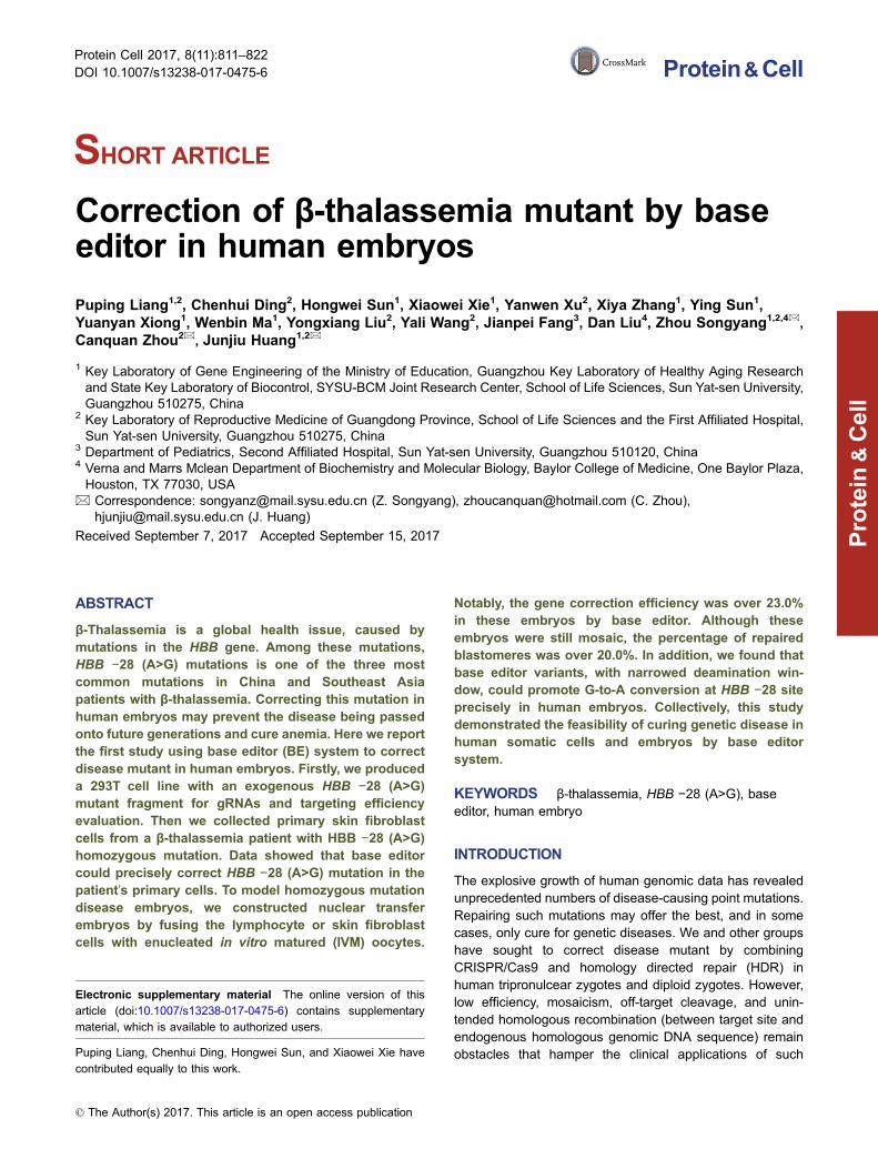
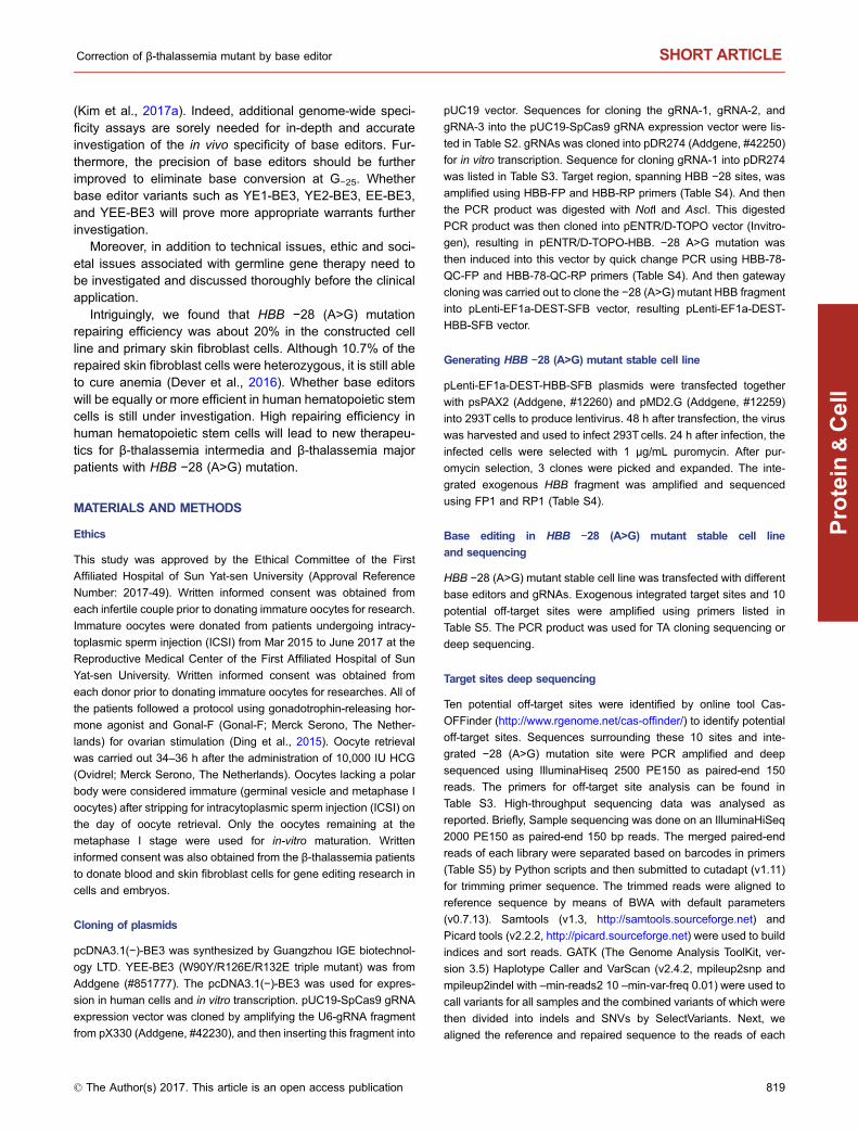
Of the two base editors (BE), BE2 (rAPOBEC1-dCas9-UGI)and BE3 (rAPOBEC1-nCas9-UGI), BE3 showed higherediting efficiency (Kim et al., 2017a). We therefore decidedto repair HBB −28 (A>G) mutation using BE3. HBB −28(A>G) mutation, in which the wild-type A at position −28(A−28) is replaced with G in patients (G−28), locates in theATA box upstream of the first exon of HBB (Fig. 1A) (Orkinet al., 1983). Three gRNAs targeting this mutant HBB allelewere designed to convert C (on the complementary strand)to T (Figs. 1A and S1). We found that G at position −25(G−25) might also be converted to A by these gRNAs(Fig. 1A). To test the deamination activity of these threegRNAs, we cloned the DNA fragment surrounding the HBB−28 (A>G) mutation into a lentiviral vector for stable inte-gration in 293T cells. After selection with puromycin, threedifferent cell clones were picked and verified by PCR(Fig. 1B). PCR primers (FP1 & RP1), that could specificallyamplify this exogenous HBB −28 (A>G) mutant fragment,were designed (Fig. 1B). Sanger sequencing of this PCRamplicons indicated a clear G at HBB position −28 in thesecell clones (Fig. 1B).
Next, we co-transfected the gRNA and the BE3 expres-sion vectors into clone #3. Cells transfected with GFP wereincluded as a control. After 48 h, the cells were harvested.Target sites were amplified with FP1 and RP1 primers.Sanger sequencing of the PCR amplicons revealed obviousG>A conversion using the three gRNAs (Fig. S2). TA cloningand sequencing further confirmed active conversion in thesecells (Fig. 1C). The conversion efficiency was 46.7% (14/30)for gRNA-1 (Fig. 1C). And consistent with previous findingsin human cells and mouse embryos, we found proximal-sitedeamination using gRNA-2 (Fig. 1C) (Liang et al., 2017). Off-target deamination could be a concern in base editing, so wefurther investigated off-target deamination in this HBB −28(A>G) mutant cell line. We again co-transfected BE3 toge-ther with either gRNA-1 or gRNA-2 into clone #3. GFPtransfected cells were used as a control. The cells wereharvested for genomic DNA extraction 48 h after
SHORT ARTICLE Puping Liang et al.
812 © The Author(s) 2017. This article is an open access publication
Protein
&Cell
A
B
C D
Exon
1 2 3
5′ 3′
3′ 5′
G-28 G-25
5′ LTR 3′ LTRHBB
FP1 RP1
HBBG-28
# 3
WT
#1 #2 #3
GFP control
gRNA-1
gRNA-2
gRNA-3
G-28 G-25
30/30
16/3014/30
20/295/291/292/291/29
27/281/28
gRNA-1gRNA-2GFP
Tota
l seq
uenc
ing
read
sw
ith ta
rget
G-A
con
vers
ion
(%)
G -28G -2
5OT1OT2
OT3OT4OT5
OT6OT7
OT8OT9
OT10
40
30
20
10
0
Figure 1. Correcting HBB −28 (A>G) mutation in human cell line. (A) Schematic of HBB −28 (A>G) mutation. The exons are
labeled with blue boxes. −28 (A>G) mutation was in red and indicated with red line (G−28). The −25 (G), next to G−28, was in blue and
indicated with blue line (G−25). And gRNAs were labeled with black arrow. (B) Generation of HBB −28 (A>G) mutant stable cell lines.
A fragment of HBB gene, containing the −28 (A>G) mutation, was cloned into a lentiviral vector. Packaged lentivirus was used to
infect 293T. Virus-infected cells were selected by puromycin. 7 days after selection, single clones of cells were picked. The up panel
showed the design of the recombined lentivirus vector. HBB gene fragment containing −28 (A>G) mutation was labeled with green
box. LTR (long terminal repeat) region of lentiviral vector was labeled with blue arrowhead. PCR primer used to specifically amplify
HBB fragment from integrated provirus were showed. The down panel showed the results of one wild-type 293T cells and three
clones, amplified using FP1 and RP1. Representative sequencing chromatographs of the PCR amplicons of #3 clone were shown.
The mutant base (G−28) was indicated by red arrowheads. (C) Precise repairing of HBB −28 (A>G) mutation by base editor 3 in the
HBB −28 (A>G) mutant stable cell line. TA cloning sequencing showed clear G>A conversion at the target site. The frequency of each
allele is shown. (D) Deep sequencing to detect on-target and off-target deamination at 10 potential off-target sites in HBB −28 (A>G)
mutant stable cell line. Bars represent mean ± SEM (n = 3). Significance was calculated using a two-tailed unpaired t test (*P < 0.05,
**P < 0.01).
Correction of β-thalassemia mutant by base editor SHORT ARTICLE
© The Author(s) 2017. This article is an open access publication 813
Protein
&Cell
transfection. The exogenously integrated HBB DNA frag-ment and 10 potential off-target sites were PCR amplified fordeep sequencing. We found 16.3% and 26.0% G>A con-versions at the target sites for gRNA-1 and gRNA-2respectively, significantly higher than the rate of 1.2% in GFPcontrol cells (Fig. 1D). And in line with data in Fig. 1C, wefound that both the G−28 and G−25 at the target region couldbe deaminated by BE3 (Fig. 1D). We found higher G>Aconversion efficiency at G−28 and G−25 using gRNA-2(Fig. 1D). Moreover, we did not found any off-target deami-nation at the 10 potential off-target sites examined for bothgRNAs, indicating high specificity (Fig. 1D). Taken together,these results clearly indicate the feasibility of repairing HBB−28 (A>G) in human cells in situ by base editing.
Correcting HBB −28 (A>G) mutation in primary skinfibroblast cells of a β-thalassemia patient by baseediting
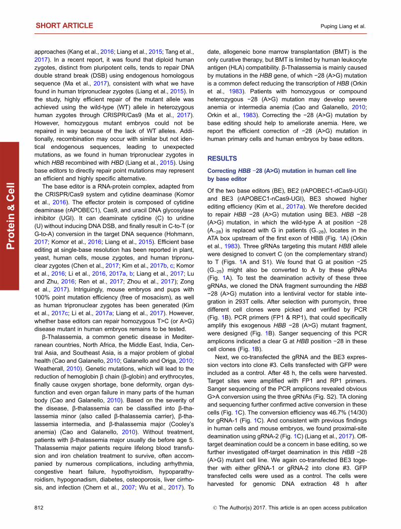
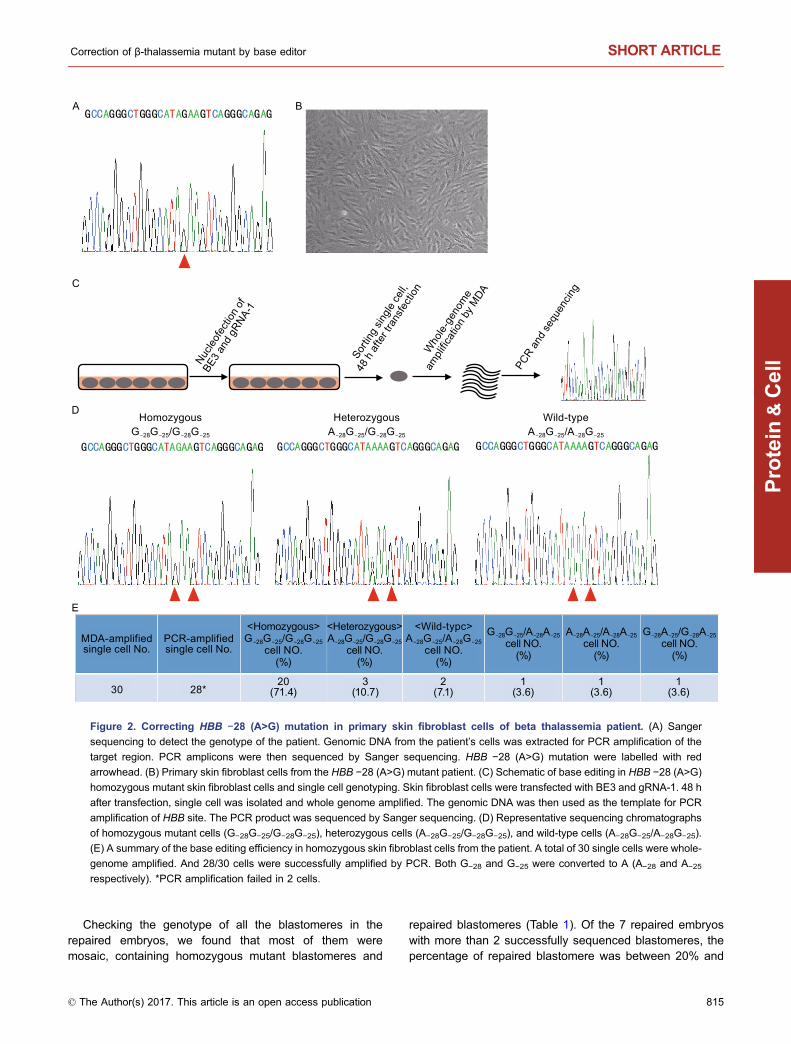
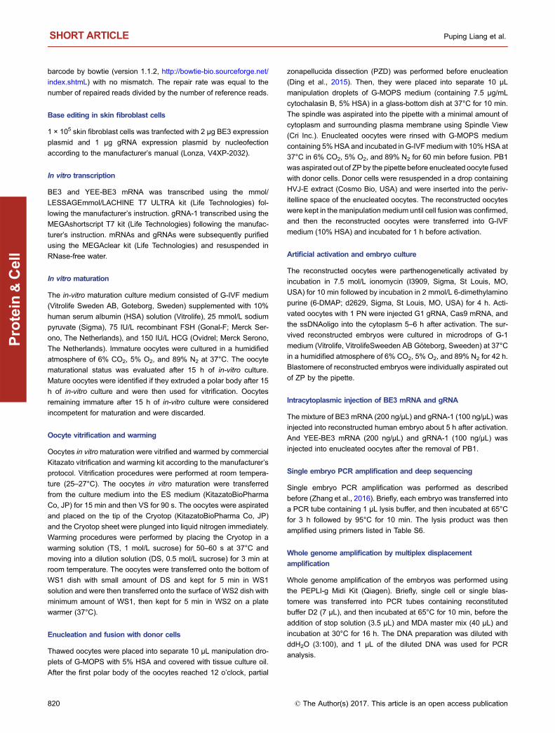
Inspired by the high efficiency and specificity of repairingHBB −28 (A>G) mutation by base editing, we sought tocorrect HBB −28 (A>G) mutation in patient’s cells. We iso-lated and cultured the skin fibroblast cells from a homozy-gous −28 (A>G) mutant patient (Fig. 2A and 2B). Aftertransfection of BE3 and gRNA-1 into these cells by nucleo-fection, we achieved 80%–90% transfection efficiency(Fig. S3). At 48 h after transfection, the cells were used forsingle cell sorting (Fig. 2C). The sorted cells were wholegenome amplified by multiplex displacement amplification(MDA), and then the HBB locus was PCR amplified(Fig. 2C). Here, we also observed efficient repairing of thehomozygous mutation to heterozygotes or WT bases asshown by Sanger sequencing.
We found 2 wild-type cells (2/28, 7.1%) with the genotypeof A−28G−25/A−28G−25, proving precise repair of both mutantalleles (Fig. 2D and 2E). Additionally, only one mutant allele(A−28G−25/G−28G−25) was repaired in 3/28 (10.7%) cells,resulting in heterozygosity (Fig. 2E). Consistent with ourprevious data using human cell lines (Fig. 1D), we also foundG>A conversion at G−25 of the target site in 3/28 (10.7%)cells, highlighting the need for developing base editor vari-ants with a narrower deamination window to improve theprecision of base editing (Figs. 1D and 2E). Here, these datashowed that 5/28 (17.8%) cells was repaired precisely,demonstrating the feasibility of repairing HBB −28 (A>G)mutation in situ.
Correcting HBB −28 (A>G) mutation in cloned humanembryos by BE3
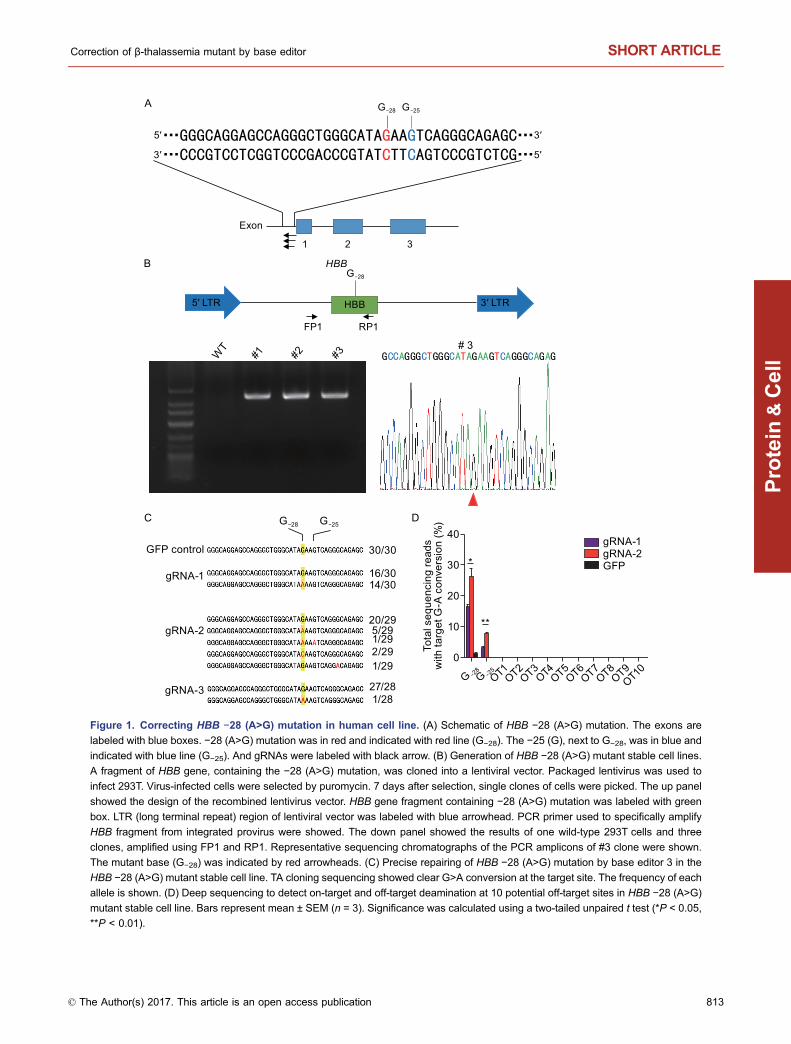
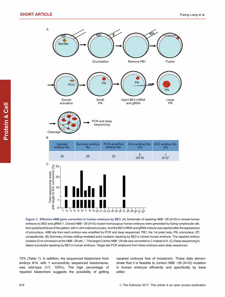
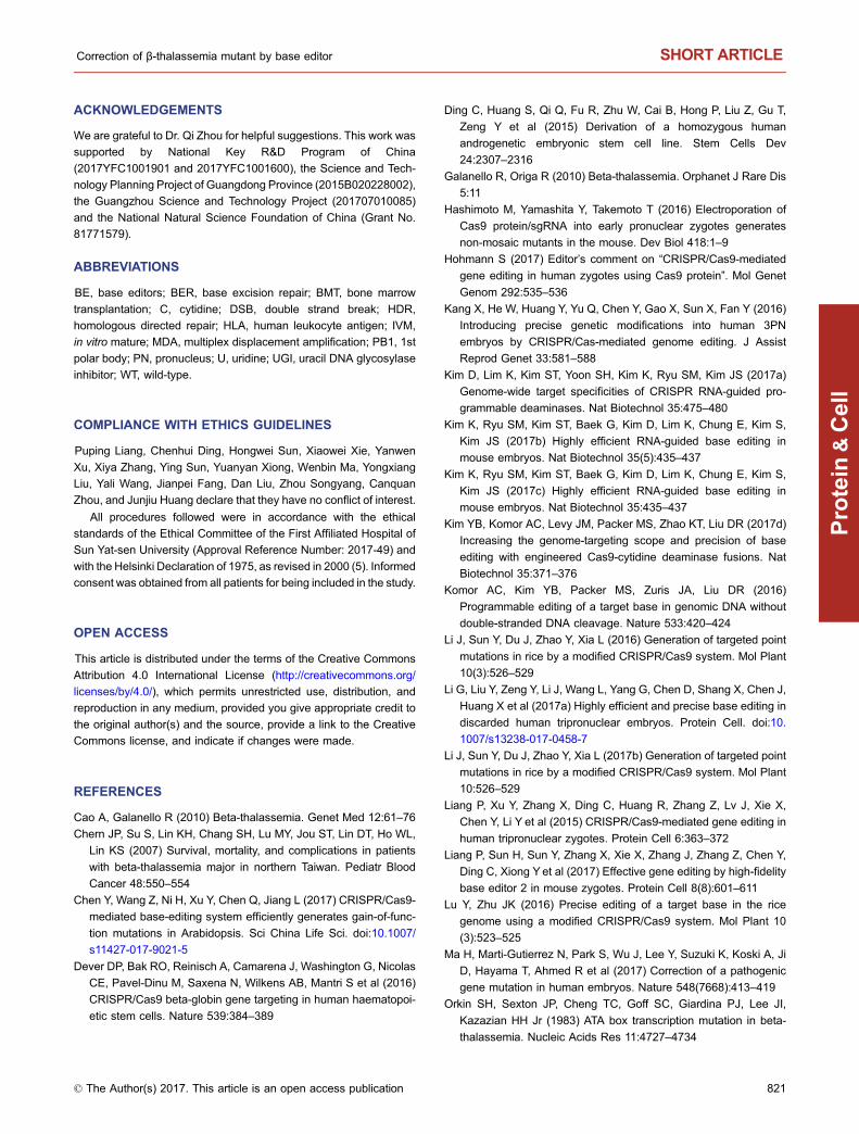
Next, we tested the feasibility of repairing HBB −28 (A>G)mutation in human embryos. To model disease embryos, wegenerated cloned human embryos by nuclear transfer
(Fig. 3A). The 1st polar body (PB1) and spindle of the in vitromatured oocytes were removed, and then the oocytes werefused with lymphocyte cells from peripheral blood of thepatient. The reconstructed oocytes were activated and cul-tured until the appearance of pronucleus (PN). Approxi-mately 5–6 h later, BE3 mRNA (200 ng/μL) and gRNA-1(100 ng/μL) were injected into the cytoplasm after theappearance of pronucleus (Fig. S4). Of the 30 embryosinjected, 26 survived (Fig. 3B). 48 h later, the HBB site ofeach embryo was PCR amplified individually. And then thePCR products were detected by Sanger sequencing anddeep sequencing. HBB site was successfully amplified in22/26 embryos (Fig. 3B). Interestingly, in these clonedembryos, we found high point mutation repairing efficiency,which was between 7.0% and 25.9% among the repairedembryos (Figs. 3C, S5 and Table S1). Analysis of the datashowed that G−28 was converted to either A or C in 45.4%(10/22) of the injected embryos (Fig. 4B). In embryo #17,G−28 was converted to C. In the other 9 embryos, G−28 wasconverted to A, representing precise mutation repairing(Fig. 3B and 3C). Furthermore, we did not find deaminationat G−25, indicating highly efficient and specific point mutationrepairing in these embryos (Fig. 3C).
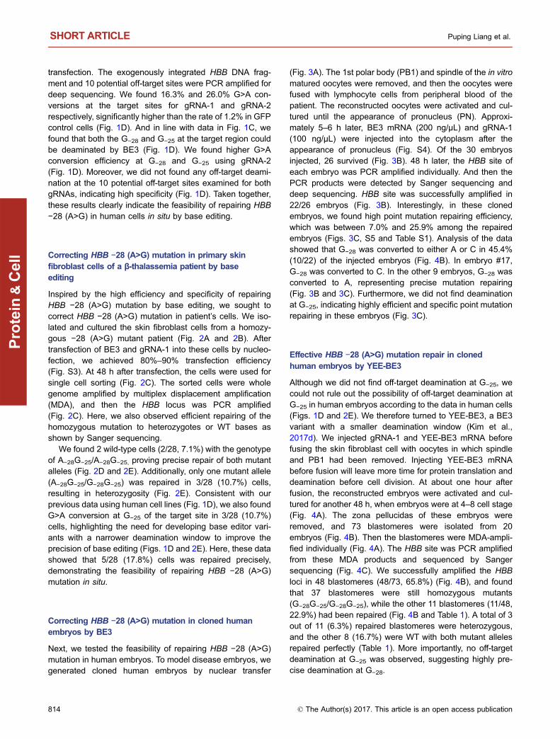
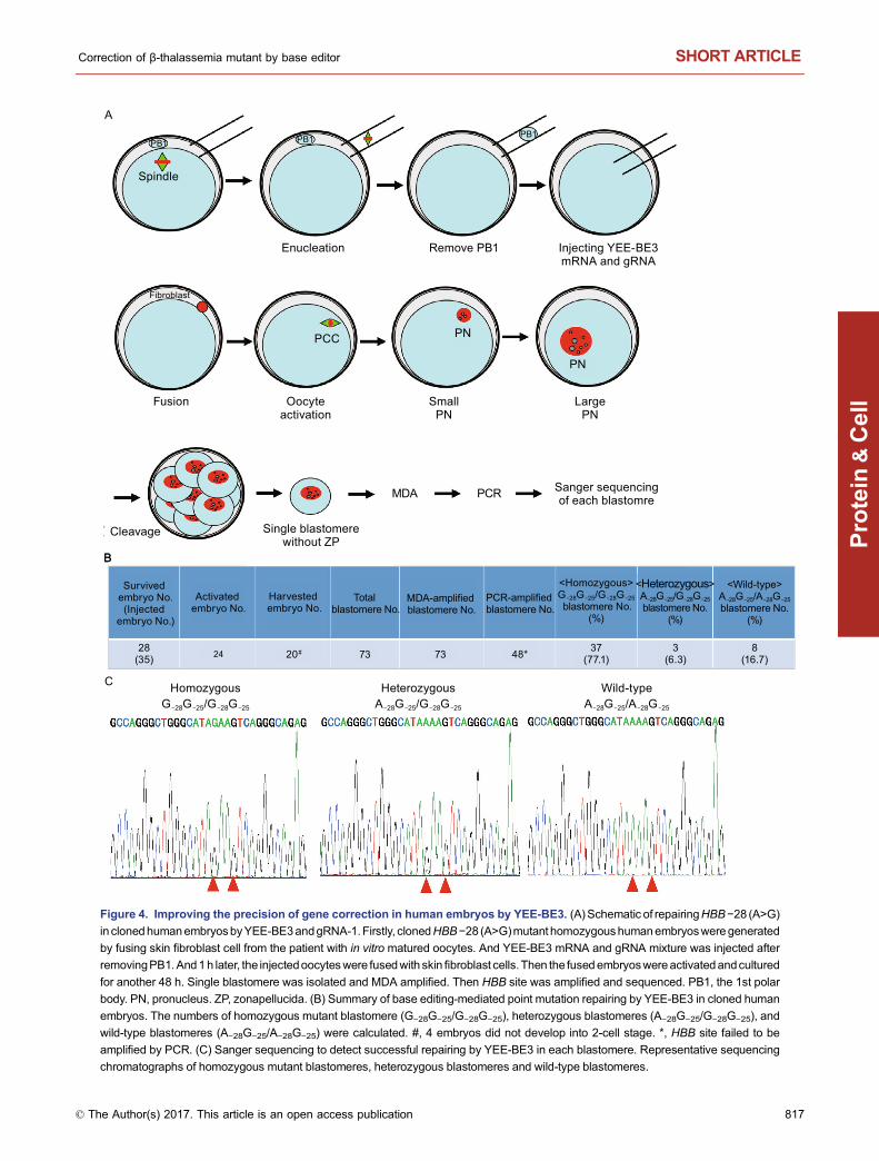
Effective HBB −28 (A>G) mutation repair in clonedhuman embryos by YEE-BE3
Although we did not find off-target deamination at G−25, wecould not rule out the possibility of off-target deamination atG−25 in human embryos according to the data in human cells(Figs. 1D and 2E). We therefore turned to YEE-BE3, a BE3variant with a smaller deamination window (Kim et al.,2017d). We injected gRNA-1 and YEE-BE3 mRNA beforefusing the skin fibroblast cell with oocytes in which spindleand PB1 had been removed. Injecting YEE-BE3 mRNAbefore fusion will leave more time for protein translation anddeamination before cell division. At about one hour afterfusion, the reconstructed embryos were activated and cul-tured for another 48 h, when embryos were at 4–8 cell stage(Fig. 4A). The zona pellucidas of these embryos wereremoved, and 73 blastomeres were isolated from 20embryos (Fig. 4B). Then the blastomeres were MDA-ampli-fied individually (Fig. 4A). The HBB site was PCR amplifiedfrom these MDA products and sequenced by Sangersequencing (Fig. 4C). We successfully amplified the HBBloci in 48 blastomeres (48/73, 65.8%) (Fig. 4B), and foundthat 37 blastomeres were still homozygous mutants(G−28G−25/G−28G−25), while the other 11 blastomeres (11/48,22.9%) had been repaired (Fig. 4B and Table 1). A total of 3out of 11 (6.3%) repaired blastomeres were heterozygous,and the other 8 (16.7%) were WT with both mutant allelesrepaired perfectly (Table 1). More importantly, no off-targetdeamination at G−25 was observed, suggesting highly pre-cise deamination at G−28.
SHORT ARTICLE Puping Liang et al.
814 © The Author(s) 2017. This article is an open access publication
Protein
&Cell
Checking the genotype of all the blastomeres in therepaired embryos, we found that most of them weremosaic, containing homozygous mutant blastomeres and
repaired blastomeres (Table 1). Of the 7 repaired embryoswith more than 2 successfully sequenced blastomeres, thepercentage of repaired blastomere was between 20% and
MDA-amplifiedsingle cell No.
PCR-amplifiedsingle cell No.
<Homozygous>G-28G-25/G-28G-25
cell NO.(%)
<Heterozygous>A-28G-25/G-28G-25
cell NO.(%)
G-28G-25/A-28A-25cell NO.
(%)
A-28A-25/A-28A-25cell NO.
(%)
G-28A-25/G-28A-25cell NO.
(%)
<Wild-typc>A-28G-25/A-28G-25
cell NO.(%)
3020
(71.4)3
(10.7)2
(7.1)1
(3.6)1
(3.6)1
(3.6)28*
A
C
B
D
Nucleo
fecti
on o
f
BE3
and
gRNA-
1
Sorti
ng si
ngle
cell,
48 h
afte
r tra
nsfe
ction
Who
le-g
enom
e
ampli
ficat
ion b
y MDA
PCR
and
sequ
encin
g
E
Wild-typeHeterozygousHomozygousG-28G-25/G-28G-25 A-28G-25/G-28G-25 A-28G-25/A-28G-25
Figure 2. Correcting HBB −28 (A>G) mutation in primary skin fibroblast cells of beta thalassemia patient. (A) Sanger
sequencing to detect the genotype of the patient. Genomic DNA from the patient’s cells was extracted for PCR amplification of the
target region. PCR amplicons were then sequenced by Sanger sequencing. HBB −28 (A>G) mutation were labelled with red
arrowhead. (B) Primary skin fibroblast cells from the HBB −28 (A>G) mutant patient. (C) Schematic of base editing in HBB −28 (A>G)
homozygous mutant skin fibroblast cells and single cell genotyping. Skin fibroblast cells were transfected with BE3 and gRNA-1. 48 h
after transfection, single cell was isolated and whole genome amplified. The genomic DNA was then used as the template for PCR
amplification of HBB site. The PCR product was sequenced by Sanger sequencing. (D) Representative sequencing chromatographs
of homozygous mutant cells (G−28G−25/G−28G−25), heterozygous cells (A−28G−25/G−28G−25), and wild-type cells (A−28G−25/A−28G−25).
(E) A summary of the base editing efficiency in homozygous skin fibroblast cells from the patient. A total of 30 single cells were whole-
genome amplified. And 28/30 cells were successfully amplified by PCR. Both G−28 and G−25 were converted to A (A−28 and A−25
respectively). *PCR amplification failed in 2 cells.
Correction of β-thalassemia mutant by base editor SHORT ARTICLE
© The Author(s) 2017. This article is an open access publication 815
Protein
&Cell
75% (Table 1). In addition, the sequenced blastomere fromembryo #14, with 1 successfully sequenced blastomeres,was wild-type (1/1, 100%). The high percentage ofrepaired blastomere suggests the possibility of getting
repaired embryos free of mosaicism. These data demon-strate that it is feasible to correct HBB −28 (A>G) mutationin human embryos efficiently and specifically by baseeditor.
Spindle
Enucleation
Oocyteactivation
SmallPN
Inject BE3 mRNAand gRNA
PCR and deepsequencing
Cleavage
LargePN
FusionRemove PB1
LymphocytePB1PB1PB1
PCC PN PN
PN
A
B
Injectedembryo No.
PCR-amplifiedembryo No.
G>A embryo No.(%)
G>C embryo No.(%)
Survived embryo No.
30 26 22 9(40.9)
1(4.5)*
C
Tota
l seq
uenc
ing
read
sw
ith ta
rget
G-A
/C c
onve
rsio
n (%
)
1 2 3 4 5 6 7 8 9 10 11 12 13 14 15 16 17 18 19 20 21 22
30
20
10
0
1
Figure 3. Effective HBB gene correction in human embryos by BE3. (A) Schematic of repairing HBB −28 (A>G) in cloned human
embryos by BE3 and gRNA-1. Cloned HBB −28 (A>G) mutant homozygous human embryos were generated by fusing lymphocyte cell,
fromperipheral bloodof the patient, with in vitromaturedoocytes. And theBE3mRNAandgRNAmixturewas injected after theappearance
of pronucleus. HBB site from each embryo was amplified by PCR and deep sequenced. PB1, the 1st polar body. PN, pronucleus. ZP,
zonapellucida. (B) Summary of base editing-mediated point mutation repairing by BE3 in cloned human embryos. The repaired embryo
containsG>A conversion at theHBB −28 site. *, The target G at theHBB−28 site was converted toC instead of A. (C) Deep sequencing to
detect successful repairing by BE3 in human embryos. Target site PCR amplicons from these embryos were deep sequenced.
SHORT ARTICLE Puping Liang et al.
816 © The Author(s) 2017. This article is an open access publication
Protein
&Cell
Spindle
Enucleation
Oocyteactivation
SmallPN
Injecting YEE-BE3 mRNA and gRNA
Single blastomerewithout ZP
Sanger sequencingof each blastomre
Cleavage
LargePN
Fusion
Remove PB1
Fibroblast
PB1PB1PB1
PCC PN
PN
MDA PCR
A
B
C
B
Survived embryo No.
(Injected embryo No.)
Activated embryo No.
Harvested embryo No.
Total blastomere No.
MDA-amplified blastomere No.
PCR-amplified blastomere No.
24 20# 48*28(35)
37(77.1)
3(6.3)
8(16.7)
<Homozygous>G-28G-25/G-28G-25 blastomere No.
(%)
<Heterozygous>A-28G-25/G-28G-25blastomere No.
(%)
<Wild-type>A-28G-25/A-28G-25blastomere No.
(%)
73 73
Wild-typeHeterozygousHomozygousG-28G-25/G-28G-25 A-28G-25/G-28G-25 A-28G-25/A-28G-25
Figure 4. Improving the precision of gene correction in human embryos by YEE-BE3. (A) Schematic of repairingHBB−28 (A>G)
in clonedhumanembryosbyYEE-BE3andgRNA-1.Firstly, clonedHBB−28 (A>G)mutant homozygoushumanembryosweregenerated
by fusing skin fibroblast cell from the patient with in vitro matured oocytes. And YEE-BE3 mRNA and gRNA mixture was injected after
removingPB1.And1h later, the injectedoocyteswere fusedwith skinfibroblast cells. Then the fusedembryoswereactivatedandcultured
for another 48 h. Single blastomere was isolated and MDA amplified. Then HBB site was amplified and sequenced. PB1, the 1st polar
body. PN, pronucleus. ZP, zonapellucida. (B) Summary of base editing-mediated point mutation repairing by YEE-BE3 in cloned human
embryos. The numbers of homozygous mutant blastomere (G−28G−25/G−28G−25), heterozygous blastomeres (A−28G−25/G−28G−25), and
wild-type blastomeres (A−28G−25/A−28G−25) were calculated. #, 4 embryos did not develop into 2-cell stage. *, HBB site failed to be
amplified by PCR. (C) Sanger sequencing to detect successful repairing by YEE-BE3 in each blastomere. Representative sequencing
chromatographs of homozygous mutant blastomeres, heterozygous blastomeres and wild-type blastomeres.
Correction of β-thalassemia mutant by base editor SHORT ARTICLE
© The Author(s) 2017. This article is an open access publication 817
Protein
&Cell
DISCUSSION
Taken together, our data highlight the tremendous potentialof correcting homozygous disease and compoundheterozygous mutations by base editing in human somaticcells and embryos. Although we did not achieve 100% repairin human embryos, we and other groups have reported100% base editing in mouse embryos (Kim et al., 2017c;Liang et al., 2017). By injecting BE3 protein and optimizingthe injection time of the base editors, 100% repair of diseasemutations may be achieved, as reported in CRISPR/Cas9system (Hashimoto et al., 2016). Injecting BE3 protein mayalso help to improve the specificity of base editing mediatedgene correction in human embryos. Moreover, we observedG>C mutation, caused by base excision repair (BER), in
human cells and embryos (Komor et al., 2016). Therefore,developing new methods to inhibit base excision repair isneeded, such as adding chemical inhibitors and overex-pressing UGI (Wang et al., 2017). Additionally, while wefound no indel formation in the cloned human embryos,indels have been observed in base editing in human cellsand mouse embryos (Kim et al., 2017c; Komor et al., 2016;Liang et al., 2017). Further investigation is needed to blockindel formation to improve the safety of base editing. Whe-ther BE2 will lead to fewer indel at the HBB −28 sites willneed further investigation.
Although we did not find off-target effects at the top 10potential off-target sites examined, the specificity of baseeditors needs more comprehensive investigation throughgenome-wide specificity assays, such as Digenome-seq
Table 1. Summary of base editing-mediated point mutation repairing by YEE-BE3 in human embryos
Embryo ID Blastomere No. PCR-amplifiedblastomere No.*
<Homozygous>G−28G−25/G−28G−25
blastomere No. (%)
<Heterozygous>A−28G−25/G−28G−25
blastomere No. (%)
<Wild-type>A−28G−25/A−28G−25
balstomere No. (%)
#1 2 1 1 0 0
#2 5 2 2 0 0
#3 7 4 3 1(25)
0
#4 3 1 1 0 0
#5 6 3 3 0 0
#6 6 3 2 0 1(33.3)
#7 2 2 2 0 0
#8 1 1 1 0 0
#9 4 4 4 0 0
#10 6 3 3 0 0
#11 1 1 1 0 0
#12 4 4 3 1(25)
0
#13 3 3 2 0 1(33.3)
#14 2 1 0 0 1(100)
#15 6 5 4 1(20)
0
#16 2 1 1 0 0
#17 2 1 1 0 0
#18 5 4 1 0 3(75)
#19 5 3 1 0 2(66.7)
#20 1 1 1 0 0
A total of 20 embryos were harvested for single blastomere genotyping. In some blastomeres, both alleles were repaired. In one blastomere,
only one mutant allele was repaired.
* Some blastomeres failed to be amplified by PCR.
SHORT ARTICLE Puping Liang et al.
818 © The Author(s) 2017. This article is an open access publication
Protein
&Cell
(Kim et al., 2017a). Indeed, additional genome-wide speci-ficity assays are sorely needed for in-depth and accurateinvestigation of the in vivo specificity of base editors. Fur-thermore, the precision of base editors should be furtherimproved to eliminate base conversion at G−25. Whetherbase editor variants such as YE1-BE3, YE2-BE3, EE-BE3,and YEE-BE3 will prove more appropriate warrants furtherinvestigation.
Moreover, in addition to technical issues, ethic and soci-etal issues associated with germline gene therapy need tobe investigated and discussed thoroughly before the clinicalapplication.
Intriguingly, we found that HBB −28 (A>G) mutationrepairing efficiency was about 20% in the constructed cellline and primary skin fibroblast cells. Although 10.7% of therepaired skin fibroblast cells were heterozygous, it is still ableto cure anemia (Dever et al., 2016). Whether base editorswill be equally or more efficient in human hematopoietic stemcells is still under investigation. High repairing efficiency inhuman hematopoietic stem cells will lead to new therapeu-tics for β-thalassemia intermedia and β-thalassemia majorpatients with HBB −28 (A>G) mutation.
MATERIALS AND METHODS
Ethics
This study was approved by the Ethical Committee of the First
Affiliated Hospital of Sun Yat-sen University (Approval Reference
Number: 2017-49). Written informed consent was obtained from
each infertile couple prior to donating immature oocytes for research.
Immature oocytes were donated from patients undergoing intracy-
toplasmic sperm injection (ICSI) from Mar 2015 to June 2017 at the
Reproductive Medical Center of the First Affiliated Hospital of Sun
Yat-sen University. Written informed consent was obtained from
each donor prior to donating immature oocytes for researches. All of
the patients followed a protocol using gonadotrophin-releasing hor-
mone agonist and Gonal-F (Gonal-F; Merck Serono, The Nether-
lands) for ovarian stimulation (Ding et al., 2015). Oocyte retrieval
was carried out 34–36 h after the administration of 10,000 IU HCG
(Ovidrel; Merck Serono, The Netherlands). Oocytes lacking a polar
body were considered immature (germinal vesicle and metaphase I
oocytes) after stripping for intracytoplasmic sperm injection (ICSI) on
the day of oocyte retrieval. Only the oocytes remaining at the
metaphase I stage were used for in-vitro maturation. Written
informed consent was also obtained from the β-thalassemia patients
to donate blood and skin fibroblast cells for gene editing research in
cells and embryos.
Cloning of plasmids
pcDNA3.1(−)-BE3 was synthesized by Guangzhou IGE biotechnol-
ogy LTD. YEE-BE3 (W90Y/R126E/R132E triple mutant) was from
Addgene (#851777). The pcDNA3.1(−)-BE3 was used for expres-
sion in human cells and in vitro transcription. pUC19-SpCas9 gRNA
expression vector was cloned by amplifying the U6-gRNA fragment
from pX330 (Addgene, #42230), and then inserting this fragment into
pUC19 vector. Sequences for cloning the gRNA-1, gRNA-2, and
gRNA-3 into the pUC19-SpCas9 gRNA expression vector were lis-
ted in Table S2. gRNAs was cloned into pDR274 (Addgene, #42250)
for in vitro transcription. Sequence for cloning gRNA-1 into pDR274
was listed in Table S3. Target region, spanning HBB −28 sites, was
amplified using HBB-FP and HBB-RP primers (Table S4). And then
the PCR product was digested with NotI and AscI. This digested
PCR product was then cloned into pENTR/D-TOPO vector (Invitro-
gen), resulting in pENTR/D-TOPO-HBB. −28 A>G mutation was
then induced into this vector by quick change PCR using HBB-78-
QC-FP and HBB-78-QC-RP primers (Table S4). And then gateway
cloning was carried out to clone the −28 (A>G) mutant HBB fragment
into pLenti-EF1a-DEST-SFB vector, resulting pLenti-EF1a-DEST-
HBB-SFB vector.
Generating HBB −28 (A>G) mutant stable cell line
pLenti-EF1a-DEST-HBB-SFB plasmids were transfected together
with psPAX2 (Addgene, #12260) and pMD2.G (Addgene, #12259)
into 293T cells to produce lentivirus. 48 h after transfection, the virus
was harvested and used to infect 293T cells. 24 h after infection, the
infected cells were selected with 1 μg/mL puromycin. After pur-
omycin selection, 3 clones were picked and expanded. The inte-
grated exogenous HBB fragment was amplified and sequenced
using FP1 and RP1 (Table S4).
Base editing in HBB −28 (A>G) mutant stable cell line
and sequencing
HBB −28 (A>G) mutant stable cell line was transfected with different
base editors and gRNAs. Exogenous integrated target sites and 10
potential off-target sites were amplified using primers listed in
Table S5. The PCR product was used for TA cloning sequencing or
deep sequencing.
Target sites deep sequencing
Ten potential off-target sites were identified by online tool Cas-
OFFinder (http://www.rgenome.net/cas-offinder/) to identify potential
off-target sites. Sequences surrounding these 10 sites and inte-
grated −28 (A>G) mutation site were PCR amplified and deep
sequenced using IlluminaHiseq 2500 PE150 as paired-end 150
reads. The primers for off-target site analysis can be found in
Table S3. High-throughput sequencing data was analysed as
reported. Briefly, Sample sequencing was done on an IlluminaHiSeq
2000 PE150 as paired-end 150 bp reads. The merged paired-end
reads of each library were separated based on barcodes in primers
(Table S5) by Python scripts and then submitted to cutadapt (v1.11)
for trimming primer sequence. The trimmed reads were aligned to
reference sequence by means of BWA with default parameters
(v0.7.13). Samtools (v1.3, http://samtools.sourceforge.net) and
Picard tools (v2.2.2, http://picard.sourceforge.net) were used to build
indices and sort reads. GATK (The Genome Analysis ToolKit, ver-
sion 3.5) Haplotype Caller and VarScan (v2.4.2, mpileup2snp and
mpileup2indel with –min-reads2 10 –min-var-freq 0.01) were used to
call variants for all samples and the combined variants of which were
then divided into indels and SNVs by SelectVariants. Next, we
aligned the reference and repaired sequence to the reads of each
Correction of β-thalassemia mutant by base editor SHORT ARTICLE
© The Author(s) 2017. This article is an open access publication 819
Protein
&Cell
barcode by bowtie (version 1.1.2, http://bowtie-bio.sourceforge.net/
index.shtmL) with no mismatch. The repair rate was equal to the
number of repaired reads divided by the number of reference reads.
Base editing in skin fibroblast cells
1 × 10⁵ skin fibroblast cells was tranfected with 2 μg BE3 expression
plasmid and 1 μg gRNA expression plasmid by nucleofection
according to the manufacturer’s manual (Lonza, V4XP-2032).
In vitro transcription
BE3 and YEE-BE3 mRNA was transcribed using the mmol/
LESSAGEmmol/LACHINE T7 ULTRA kit (Life Technologies) fol-
lowing the manufacturer’s instruction. gRNA-1 transcribed using the
MEGAshortscript T7 kit (Life Technologies) following the manufac-
turer’s instruction. mRNAs and gRNAs were subsequently purified
using the MEGAclear kit (Life Technologies) and resuspended in
RNase-free water.
In vitro maturation
The in-vitro maturation culture medium consisted of G-IVF medium
(Vitrolife Sweden AB, Goteborg, Sweden) supplemented with 10%
human serum albumin (HSA) solution (Vitrolife), 25 mmol/L sodium
pyruvate (Sigma), 75 IU/L recombinant FSH (Gonal-F; Merck Ser-
ono, The Netherlands), and 150 IU/L HCG (Ovidrel; Merck Serono,
The Netherlands). Immature oocytes were cultured in a humidified
atmosphere of 6% CO2, 5% O2, and 89% N2 at 37°C. The oocyte
maturational status was evaluated after 15 h of in-vitro culture.
Mature oocytes were identified if they extruded a polar body after 15
h of in-vitro culture and were then used for vitrification. Oocytes
remaining immature after 15 h of in-vitro culture were considered
incompetent for maturation and were discarded.
Oocyte vitrification and warming
Oocytes in vitromaturation were vitrified and warmed by commercial
Kitazato vitrification and warming kit according to the manufacturer’s
protocol. Vitrification procedures were performed at room tempera-
ture (25–27°C). The oocytes in vitro maturation were transferred
from the culture medium into the ES medium (KitazatoBioPharma
Co, JP) for 15 min and then VS for 90 s. The oocytes were aspirated
and placed on the tip of the Cryotop (KitazatoBioPharma Co, JP)
and the Cryotop sheet were plunged into liquid nitrogen immediately.
Warming procedures were performed by placing the Cryotop in a
warming solution (TS, 1 mol/L sucrose) for 50–60 s at 37°C and
moving into a dilution solution (DS, 0.5 mol/L sucrose) for 3 min at
room temperature. The oocytes were transferred onto the bottom of
WS1 dish with small amount of DS and kept for 5 min in WS1
solution and were then transferred onto the surface of WS2 dish with
minimum amount of WS1, then kept for 5 min in WS2 on a plate
warmer (37°C).
Enucleation and fusion with donor cells
Thawed oocytes were placed into separate 10 µL manipulation dro-
plets of G-MOPS with 5% HSA and covered with tissue culture oil.
After the first polar body of the oocytes reached 12 o’clock, partial
zonapellucida dissection (PZD) was performed before enucleation
(Ding et al., 2015). Then, they were placed into separate 10 µL
manipulation droplets of G-MOPS medium (containing 7.5 µg/mL
cytochalasin B, 5% HSA) in a glass-bottom dish at 37°C for 10 min.
The spindle was aspirated into the pipette with a minimal amount of
cytoplasm and surrounding plasma membrane using Spindle View
(Cri Inc.). Enucleated oocytes were rinsed with G-MOPS medium
containing 5%HSA and incubated in G-IVFmediumwith 10%HSA at
37°C in 6% CO2, 5% O2, and 89% N2 for 60 min before fusion. PB1
was aspirated out of ZP by the pipette before enucleated oocyte fused
with donor cells. Donor cells were resuspended in a drop containing
HVJ-E extract (Cosmo Bio, USA) and were inserted into the periv-
itelline space of the enucleated oocytes. The reconstructed oocytes
were kept in the manipulation medium until cell fusion was confirmed,
and then the reconstructed oocytes were transferred into G-IVF
medium (10% HSA) and incubated for 1 h before activation.
Artificial activation and embryo culture
The reconstructed oocytes were parthenogenetically activated by
incubation in 7.5 mol/L ionomycin (I3909, Sigma, St Louis, MO,
USA) for 10 min followed by incubation in 2 mmol/L 6-dimethylamino
purine (6-DMAP; d2629, Sigma, St Louis, MO, USA) for 4 h. Acti-
vated oocytes with 1 PN were injected G1 gRNA, Cas9 mRNA, and
the ssDNAoligo into the cytoplasm 5–6 h after activation. The sur-
vived reconstructed embryos were cultured in microdrops of G-1
medium (Vitrolife, VitrolifeSweeden AB Göteborg, Sweeden) at 37°C
in a humidified atmosphere of 6% CO2, 5% O2, and 89% N2 for 42 h.
Blastomere of reconstructed embryos were individually aspirated out
of ZP by the pipette.
Intracytoplasmic injection of BE3 mRNA and gRNA
The mixture of BE3 mRNA (200 ng/μL) and gRNA-1 (100 ng/μL) was
injected into reconstructed human embryo about 5 h after activation.
And YEE-BE3 mRNA (200 ng/μL) and gRNA-1 (100 ng/μL) was
injected into enucleated oocytes after the removal of PB1.
Single embryo PCR amplification and deep sequencing
Single embryo PCR amplification was performed as described
before (Zhang et al., 2016). Briefly, each embryo was transferred into
a PCR tube containing 1 μL lysis buffer, and then incubated at 65°C
for 3 h followed by 95°C for 10 min. The lysis product was then
amplified using primers listed in Table S6.
Whole genome amplification by multiplex displacement
amplification
Whole genome amplification of the embryos was performed using
the PEPLI-g Midi Kit (Qiagen). Briefly, single cell or single blas-
tomere was transferred into PCR tubes containing reconstituted
buffer D2 (7 μL), and then incubated at 65°C for 10 min, before the
addition of stop solution (3.5 μL) and MDA master mix (40 μL) and
incubation at 30°C for 16 h. The DNA preparation was diluted with
ddH2O (3:100), and 1 μL of the diluted DNA was used for PCR
analysis.
SHORT ARTICLE Puping Liang et al.
820 © The Author(s) 2017. This article is an open access publication
Protein
&Cell
ACKNOWLEDGEMENTS
We are grateful to Dr. Qi Zhou for helpful suggestions. This work was
supported by National Key R&D Program of China
(2017YFC1001901 and 2017YFC1001600), the Science and Tech-
nology Planning Project of Guangdong Province (2015B020228002),
the Guangzhou Science and Technology Project (201707010085)
and the National Natural Science Foundation of China (Grant No.
81771579).
ABBREVIATIONS
BE, base editors; BER, base excision repair; BMT, bone marrow
transplantation; C, cytidine; DSB, double strand break; HDR,
homologous directed repair; HLA, human leukocyte antigen; IVM,
in vitro mature; MDA, multiplex displacement amplification; PB1, 1st
polar body; PN, pronucleus; U, uridine; UGI, uracil DNA glycosylase
inhibitor; WT, wild-type.
COMPLIANCE WITH ETHICS GUIDELINES
Puping Liang, Chenhui Ding, Hongwei Sun, Xiaowei Xie, Yanwen
Xu, Xiya Zhang, Ying Sun, Yuanyan Xiong, Wenbin Ma, Yongxiang
Liu, Yali Wang, Jianpei Fang, Dan Liu, Zhou Songyang, Canquan
Zhou, and Junjiu Huang declare that they have no conflict of interest.
All procedures followed were in accordance with the ethical
standards of the Ethical Committee of the First Affiliated Hospital of
Sun Yat-sen University (Approval Reference Number: 2017-49) and
with the Helsinki Declaration of 1975, as revised in 2000 (5). Informed
consent was obtained from all patients for being included in the study.
OPEN ACCESS
This article is distributed under the terms of the Creative Commons
Attribution 4.0 International License (http://creativecommons.org/
licenses/by/4.0/), which permits unrestricted use, distribution, and
reproduction in any medium, provided you give appropriate credit to
the original author(s) and the source, provide a link to the Creative
Commons license, and indicate if changes were made.
REFERENCES
Cao A, Galanello R (2010) Beta-thalassemia. Genet Med 12:61–76Chern JP, Su S, Lin KH, Chang SH, Lu MY, Jou ST, Lin DT, Ho WL,
Lin KS (2007) Survival, mortality, and complications in patients
with beta-thalassemia major in northern Taiwan. Pediatr Blood
Cancer 48:550–554Chen Y, Wang Z, Ni H, Xu Y, Chen Q, Jiang L (2017) CRISPR/Cas9-
mediated base-editing system efficiently generates gain-of-func-
tion mutations in Arabidopsis. Sci China Life Sci. doi:10.1007/
s11427-017-9021-5
Dever DP, Bak RO, Reinisch A, Camarena J, Washington G, Nicolas
CE, Pavel-Dinu M, Saxena N, Wilkens AB, Mantri S et al (2016)
CRISPR/Cas9 beta-globin gene targeting in human haematopoi-
etic stem cells. Nature 539:384–389
Ding C, Huang S, Qi Q, Fu R, Zhu W, Cai B, Hong P, Liu Z, Gu T,
Zeng Y et al (2015) Derivation of a homozygous human
androgenetic embryonic stem cell line. Stem Cells Dev
24:2307–2316Galanello R, Origa R (2010) Beta-thalassemia. Orphanet J Rare Dis
5:11
Hashimoto M, Yamashita Y, Takemoto T (2016) Electroporation of
Cas9 protein/sgRNA into early pronuclear zygotes generates
non-mosaic mutants in the mouse. Dev Biol 418:1–9Hohmann S (2017) Editor’s comment on “CRISPR/Cas9-mediated
gene editing in human zygotes using Cas9 protein”. Mol Genet
Genom 292:535–536Kang X, He W, Huang Y, Yu Q, Chen Y, Gao X, Sun X, Fan Y (2016)
Introducing precise genetic modifications into human 3PN
embryos by CRISPR/Cas-mediated genome editing. J Assist
Reprod Genet 33:581–588Kim D, Lim K, Kim ST, Yoon SH, Kim K, Ryu SM, Kim JS (2017a)
Genome-wide target specificities of CRISPR RNA-guided pro-
grammable deaminases. Nat Biotechnol 35:475–480Kim K, Ryu SM, Kim ST, Baek G, Kim D, Lim K, Chung E, Kim S,
Kim JS (2017b) Highly efficient RNA-guided base editing in
mouse embryos. Nat Biotechnol 35(5):435–437Kim K, Ryu SM, Kim ST, Baek G, Kim D, Lim K, Chung E, Kim S,
Kim JS (2017c) Highly efficient RNA-guided base editing in
mouse embryos. Nat Biotechnol 35:435–437Kim YB, Komor AC, Levy JM, Packer MS, Zhao KT, Liu DR (2017d)
Increasing the genome-targeting scope and precision of base
editing with engineered Cas9-cytidine deaminase fusions. Nat
Biotechnol 35:371–376Komor AC, Kim YB, Packer MS, Zuris JA, Liu DR (2016)
Programmable editing of a target base in genomic DNA without
double-stranded DNA cleavage. Nature 533:420–424Li J, Sun Y, Du J, Zhao Y, Xia L (2016) Generation of targeted point
mutations in rice by a modified CRISPR/Cas9 system. Mol Plant
10(3):526–529Li G, Liu Y, Zeng Y, Li J, Wang L, Yang G, Chen D, Shang X, Chen J,
Huang X et al (2017a) Highly efficient and precise base editing in
discarded human tripronuclear embryos. Protein Cell. doi:10.
1007/s13238-017-0458-7
Li J, Sun Y, Du J, Zhao Y, Xia L (2017b) Generation of targeted point
mutations in rice by a modified CRISPR/Cas9 system. Mol Plant
10:526–529Liang P, Xu Y, Zhang X, Ding C, Huang R, Zhang Z, Lv J, Xie X,
Chen Y, Li Y et al (2015) CRISPR/Cas9-mediated gene editing in
human tripronuclear zygotes. Protein Cell 6:363–372Liang P, Sun H, Sun Y, Zhang X, Xie X, Zhang J, Zhang Z, Chen Y,
Ding C, Xiong Y et al (2017) Effective gene editing by high-fidelity
base editor 2 in mouse zygotes. Protein Cell 8(8):601–611Lu Y, Zhu JK (2016) Precise editing of a target base in the rice
genome using a modified CRISPR/Cas9 system. Mol Plant 10
(3):523–525Ma H, Marti-Gutierrez N, Park S, Wu J, Lee Y, Suzuki K, Koski A, Ji
D, Hayama T, Ahmed R et al (2017) Correction of a pathogenic
gene mutation in human embryos. Nature 548(7668):413–419Orkin SH, Sexton JP, Cheng TC, Goff SC, Giardina PJ, Lee JI,
Kazazian HH Jr (1983) ATA box transcription mutation in beta-
thalassemia. Nucleic Acids Res 11:4727–4734
Correction of β-thalassemia mutant by base editor SHORT ARTICLE
© The Author(s) 2017. This article is an open access publication 821
Protein
&Cell
Ren B, Yan F, Kuang Y, Li N, Zhang D, Lin H, Zhou H (2017) A
CRISPR/Cas9 toolkit for efficient targeted base editing to induce
genetic variations in rice. Sci China Life Sci 60(5):516–519Tang L, Zeng Y, Du H, Gong M, Peng J, Zhang B, Lei M, Zhao F,
Wang W, Li X et al (2017) CRISPR/Cas9-mediated gene editing
in human zygotes using Cas9 protein. Mol Genet Genom
292:525–533Wang L, Xue W, Yan L, Li X, Wei J, Chen M, Wu J, Yang B, Yang L,
Chen J (2017) Enhanced base editing by co-expression of free
uracil DNA glycosylase inhibitor. Cell Res. doi:10.1038/cr.2017.
111
Weatherall DJ (2010) The inherited diseases of hemoglobin are an
emerging global health burden. Blood 115:4331–4336Wu HP, Lin CL, Chang YC, Wu KH, Lei RL, Peng CT, Weng T, Tai
YM, Chao YH (2017) Survival and complication rates in patients
with thalassemia major in Taiwan. Pediatr Blood Cancer 64:135–138
Zhang X, Liang P, Ding C, Zhang Z, Zhou J, Xie X, Huang R, Sun Y,
Sun H, Zhang J et al (2016) Efficient Production of Gene-
Modified Mice using Staphylococcus aureus Cas9. Sci Rep
6:32565
Zong Y, Wang Y, Li C, Zhang R, Chen K, Ran Y, Qiu JL, Wang D,
Gao C (2017) Precise base editing in rice, wheat and maize with
a Cas9- cytidine deaminase fusion. Nat Biotechnol 35(5):438–440
Zhou C, Zhang M, Wei Y, Sun Y, Sun Y, Pan H, Yao N, Zhong W, Li
Y, Li W, Yang H (2017) Highly efficient base editing in human
tripronuclear zygotes. Protein Cell. doi:10.1007/s13238-017-
0459-6
SHORT ARTICLE Puping Liang et al.
822 © The Author(s) 2017. This article is an open access publication
Protein
&Cell