Correction of immunosuppression in aged septic rats by ...
11
RESEARCH ARTICLE Open Access Correction of immunosuppression in aged septic rats by human ghrelin and growth hormone through the vagus nerve- dependent inhibition of TGF-β production Mian Zhou 1 , Monowar Aziz 1 , Mahendar Ochani 1 and Ping Wang 1,2* Abstract Background: Co-administration of human ghrelin and growth hormone (GH) reverse immunosuppression in septic aged animals, but the mechanism remains elusive. Here, we hypothesize that ghrelin and GH co-treatment restores the immune response in aged septic rats by inhibiting the production of transforming growth factor-β (TGF-β), an immunoregulatory cytokine, through the vagus nerve. Methods: Male aged Fischer rats (22–23-month-old) were made septic by cecal ligation and puncture (CLP) with or without dissecting the vagus nerve (vagotomy). Human ghrelin and GH or vehicle (PBS) were administrated subcutaneously at 5 h post CLP. After 20 h of CLP, serum and spleens were harvested. Results: Serum TGF-β levels were increased in septic aged rats, while ghrelin and GH treatment significantly reduced its levels. Expression of TGF-β in the spleen was upregulated after sepsis, while ghrelin and GH treatment significantly inhibited its expression. TNF-α and IL-6 levels were significantly reduced after ex vivo LPS stimulation of splenocytes from rats that underwent CLP compared to sham rats; while these levels were significantly higher in splenocytes from ghrelin and GH-treated CLP rats compared to vehicle-treated CLP rats. Ghrelin and GH treatment reduced program death receptor-1 (PD-1) expression, increased human leukocyte antigen-DR (HLA-DR) expression, attenuated lymphopenia, and cleaved caspase-3 levels in the spleen of septic aged rats. Vagotomy diminished the beneficial effects of ghrelin and GH treatment in septic rats. In vitro, the addition of ghrelin, GH, or ghrelin and GH together had no effect on restoring immune response in splenocytes from CLP rats following LPS stimulation, indicating the requirement of the vagus nerve for ghrelin and GH’s effect. Conclusions: Ghrelin and GH attenuate immunosuppression in aged septic rats through the vagus nerve- dependent inhibition of TGF-β production. Keywords: Ghrelin, Aging, Sepsis, Immunosuppression, Vagus nerve © The Author(s). 2020 Open Access This article is licensed under a Creative Commons Attribution 4.0 International License, which permits use, sharing, adaptation, distribution and reproduction in any medium or format, as long as you give appropriate credit to the original author(s) and the source, provide a link to the Creative Commons licence, and indicate if changes were made. The images or other third party material in this article are included in the article's Creative Commons licence, unless indicated otherwise in a credit line to the material. If material is not included in the article's Creative Commons licence and your intended use is not permitted by statutory regulation or exceeds the permitted use, you will need to obtain permission directly from the copyright holder. To view a copy of this licence, visit http://creativecommons.org/licenses/by/4.0/. * Correspondence: [email protected] 1 Center for Immunology and Inflammation, The Feinstein Institutes for Medical Research, Manhasset, New York, USA 2 Departments of Surgery and Molecular Medicine, Donald and Barbara Zucker School of Medicine at Hofstra/Northwell, Manhasset, New York, USA Molecular Medicine Zhou et al. Molecular Medicine (2020) 26:71 https://doi.org/10.1186/s10020-020-00195-x
Transcript of Correction of immunosuppression in aged septic rats by ...
RESEARCH ARTICLE Open Access
Correction of immunosuppression in agedseptic rats by human ghrelin and growthhormone through the vagus nerve-dependent inhibition of TGF-β productionMian Zhou1, Monowar Aziz1, Mahendar Ochani1 and Ping Wang1,2*
Abstract
Background: Co-administration of human ghrelin and growth hormone (GH) reverse immunosuppression in septicaged animals, but the mechanism remains elusive. Here, we hypothesize that ghrelin and GH co-treatment restoresthe immune response in aged septic rats by inhibiting the production of transforming growth factor-β (TGF-β), animmunoregulatory cytokine, through the vagus nerve.
Methods: Male aged Fischer rats (22–23-month-old) were made septic by cecal ligation and puncture (CLP) with orwithout dissecting the vagus nerve (vagotomy). Human ghrelin and GH or vehicle (PBS) were administratedsubcutaneously at 5 h post CLP. After 20 h of CLP, serum and spleens were harvested.
Results: Serum TGF-β levels were increased in septic aged rats, while ghrelin and GH treatment significantlyreduced its levels. Expression of TGF-β in the spleen was upregulated after sepsis, while ghrelin and GHtreatment significantly inhibited its expression. TNF-α and IL-6 levels were significantly reduced after ex vivoLPS stimulation of splenocytes from rats that underwent CLP compared to sham rats; while these levels weresignificantly higher in splenocytes from ghrelin and GH-treated CLP rats compared to vehicle-treated CLP rats.Ghrelin and GH treatment reduced program death receptor-1 (PD-1) expression, increased human leukocyteantigen-DR (HLA-DR) expression, attenuated lymphopenia, and cleaved caspase-3 levels in the spleen of septicaged rats. Vagotomy diminished the beneficial effects of ghrelin and GH treatment in septic rats. In vitro, theaddition of ghrelin, GH, or ghrelin and GH together had no effect on restoring immune response insplenocytes from CLP rats following LPS stimulation, indicating the requirement of the vagus nerve for ghrelinand GH’s effect.
Conclusions: Ghrelin and GH attenuate immunosuppression in aged septic rats through the vagus nerve-dependent inhibition of TGF-β production.
Keywords: Ghrelin, Aging, Sepsis, Immunosuppression, Vagus nerve
© The Author(s). 2020 Open Access This article is licensed under a Creative Commons Attribution 4.0 International License,which permits use, sharing, adaptation, distribution and reproduction in any medium or format, as long as you giveappropriate credit to the original author(s) and the source, provide a link to the Creative Commons licence, and indicate ifchanges were made. The images or other third party material in this article are included in the article's Creative Commonslicence, unless indicated otherwise in a credit line to the material. If material is not included in the article's Creative Commonslicence and your intended use is not permitted by statutory regulation or exceeds the permitted use, you will need to obtainpermission directly from the copyright holder. To view a copy of this licence, visit http://creativecommons.org/licenses/by/4.0/.
* Correspondence: [email protected] for Immunology and Inflammation, The Feinstein Institutes forMedical Research, Manhasset, New York, USA2Departments of Surgery and Molecular Medicine, Donald and BarbaraZucker School of Medicine at Hofstra/Northwell, Manhasset, New York, USA
Molecular MedicineZhou et al. Molecular Medicine (2020) 26:71 https://doi.org/10.1186/s10020-020-00195-x
IntroductionSepsis is a life-threatening condition that arises due to adysregulated immune response to infection leading toexcessive inflammation and organ injury (Singer et al.2016). Although an uncontrolled inflammatory syn-drome is the prevailing theory for sepsis, subsequent de-velopment of an immunosuppressive phenomenon isincreasingly being recognized (Delano and Ward 2016;Hotchkiss et al. 2013). Studies with human septic pa-tients demonstrate that immunosuppression is the pre-dominant cause of morbidity and mortality in sepsis(Hotchkiss et al. 2013; Boomer et al. 2011). Immunosup-pression is commonly encountered in elderly patientsthat develop sepsis, resulting in impaired efficacy infighting against invading pathogens (Hotchkiss et al.2013; Boomer et al. 2011). Persistent inflammation, Tcell exhaustion, and lymphocytopenia are observed inelderly patients after sepsis (Inoue et al. 2014). Those pa-tients are susceptible to secondary infection with lowersurvival rates compared to young septic patients (Inoueet al. 2014). Studies have shown that severe sepsis andseptic shock are mainly observed in elderly patients(> 65 years of age), giving rise to significantly highermortality rate of about 80% (Boomer et al. 2011;Martin et al. 2006; Opal et al. 2005; Hepper et al.2013). In-depth understanding of the pathophysiologyand the development of novel therapeutics are ur-gently needed to protect the elderly from sepsis.The pleiotropic cytokine transforming growth factor-β
(TGF-β) is a key regulator of the immune response toinfection (Yoshimura et al. 2010; Santarpia et al. 2015).TGF-β is produced by a wide number of cells includingleukocytes as well as epithelial cells (Taylor 2009). Itcontrols the differentiation, proliferation and activationof immune cells (Taylor 2009; Weehuizen et al. 2012).There are 3 isoforms of TGF-β that have been identifiedin mammals, namely TGF-β1, -β2 and -β3. These iso-forms of TGF-β have similar biological function but areexpressed in different tissues (Govinden and Bhoola2003). Among these three isoforms, TGF-β1 is predom-inantly expressed by the cells of the immune system(Govinden and Bhoola 2003). TGF-β has multiple im-munosuppressive properties as evidenced by the fact thatTGF-β knockout mice develop multi-organ autoimmuneinflammatory disease and die shortly after birth (Govindenand Bhoola 2003). Although the levels of TGF-β are ele-vated in patients with sepsis (Marie et al. 1996), its role ininducing immunosuppression is not fully understood.Ghrelin is a small peptide predominantly produced by
the gastrointestinal tract (Collden et al. 2017). Ghrelinbinds to the growth hormone secretagogue receptor(GHSR)-1a to promote the release of growth hormone(GH) (Arvat et al. 2000). Beyond its metabolic function,ghrelin has anti-inflammatory and anti-apoptotic functions
as well (Wu and Kral 2004; Wu et al. 2007a; Gonzalez-Reyet al. 2006; Zhou et al. 2017). Our previous studies showedthat co-treatment of ghrelin and GH mitigated organ injuryand improved survival in aged rats after sepsis (Wu et al.2009a; Yang et al. 2016). Even though we recently demon-strated that the combined treatment with ghrelin and GH(GG) were able to reverse immunosuppression in aged sep-tic rats, the underlying mechanism involving the potentialrole of TGF-β in GG-mediated restoration of immune re-sponse had not been studied (Zhou et al. 2017). We there-fore investigated whether the effect of GG on restoringimmune response in aged animals with sepsis was mediatedthrough the modulation of TGF-β production. We furtheraimed to determine the contribution of the vagus nerve forGG-mediated restoration of immune function in aged sep-tic rats.
Material and methodsAnimal model of sepsis and vagotomyAged male Fisher rats (22–23month-old) were obtainedfrom Charles River Laboratories via the National Institutefor Aging, National Institutes of Health (NIH). Animalswere housed in a temperature-controlled room with a 12h light-dark cycle and fed a standard Purina rat chow diet.Rats were allowed to acclimate to the environment for atleast 5 days before being used for experiments.Sepsis was induced in aged rats by cecal ligation and
puncture (CLP) (Yang et al. 2016). Briefly, rats were anes-thetized by 2% isoflurane inhalation. The abdomen wasshaved and cleaned with iodine and alcohol solution, anda 2-cm midline incision was made. The cecum was thenexposed and 70% of its length was ligated using 4–0 silksuture distal to the ileocecal valve. The cecum was punc-tured twice with an 18-G needle and a small amount offeces was extruded. The cecum was then returned to theabdominal cavity, the abdominal incision was closed inlayers, and the animals were resuscitated with 30ml/kgbody weight normal saline subcutaneously. In sham rats,the same surgical procedure was performed with the ex-ception that their cecum was not ligated or punctured.Vagotomy procedure was performed on a group of rats
at the time of CLP as reported previously (Wu et al.2007b; Williams et al. 2003). The trunks of the subdiaph-ragmatic vagus nerve were transected. Briefly, the dorsaland ventral branches of the vagus nerve were dissectedand each branch of the nerve was tied with surgical su-tures at 2 points separated by about 1 cm, and then sev-ered between the sutures. After surgery, animals wereallowed to eat and drink food and water, respectively.
Administration of human ghrelin and human growthhormone into aged septic ratsHuman ghrelin (Phoenix Pharmaceuticals, Belmont, CA)and human GH (ProSpec, Ness Ziona, Israel) were
Zhou et al. Molecular Medicine (2020) 26:71 Page 2 of 11
dissolved in normal saline. A 500 μl mixture of humanghrelin and GH (GG) was prepared and injected sub-cutaneously to CLP animals at 5 h after CLP. Each GGtreated animal received 80 nmol/kg human ghrelin and50 μg/kg human GH in a single bolus dose.
Splenocyte isolation and stimulationSpleens were harvested at 20 h after CLP or sham-operation and homogenized by gentle grinding betweenfrosted glass slides, followed by passage through a 70-μm cell strainer to obtain single cell suspensions (BDBiosciences, San Jose, CA). The suspension was centri-fuged at 450×g for 5 min and the pellet was suspendedinto 44% percoll solution (Sigma, St. Louis, MO), thencareflly overlaid on top of 66% percoll solution, centri-fuged at 800×g for 30 min at room temperature. Afterdensity gradient centrifuge, leukocytes formed a whitefluffy ring at the interface between 44% percoll and 66%percoll and were collected into RPMI-1640 medium(Life Technologies, Grand Island, NY) containing 10%heat-inactivated fetal bovine serum, 2mM L-glutamine,100 U/ml penicillin, 100 μg/ml streptomycin,10 mMHEPES and 0.5 μM 2-mercaptaethanol. Cell viability wasgreater than 90%. To evaluate the immune responses ofthese cells, 2 × 10⁶ cells were plated into a 24-well plateand stimulated with LPS (100 ng/ml, Sigma, St. Louis,MO) for 5 h. The released proinflammatory cytokines inthe culture medium were measured by enzyme-linkedimmunosorbent assay (ELISA).
Measurement of cytokines and analysis of lymphocyte,monocyte, and basophil numbersCytokine levels were quantified using ELISA kits specificfor rat TNF-α, IL-6 (BD Biosciences) and TGF-β1(eBioscience, San Diego, CA). All measurements wereperformed according to manufacturer’s instructions.Lymphocyte, monocyte, and basophil numbers in theblood were analyzed by using a Cell-DYN 3700 analyzer(Abbott, Abbott Park, IL).
Western blot analysisSpleen tissues were homogenized and lysed in RIPA buf-fer, 10 mM Tris buffer, pH 7.5 containing 0.1% Triton-X100, 1 mM EDTA, 1 mM EGTA, protease inhibitor tab-let (Thermo Fisher, Waltham, MA), phosphatase inhibi-tor tablet (Thermo Fisher). Protein concentration wasdetermined by Bio-Rad DC protein assay kit (Bio-Rad,Hercules, CA). Tissue lysates were electrophoresed on4–12% NuPAGE Bis-Tris Gel (Life Technologies) andtransferred to nitrocellulose membranes. The mem-branes were then blocked with 0.1% casein in Tris buffersaline and incubated with anti-TGF-β (Proteintech, Chi-cago, IL), -cleaved caspase-3 (Cell Signaling Technolo-gies, Danvers, MA), and -β-actin (Sigma) Abs overnight
at 4 °C. After washing, membranes were incubated withinfrared dye-labeled respective secondary Abs (LI-COR,Lincoln, NE). The Odyssey infrared image system (LI-COR) was used to analyze the target bands and inten-sities of bands were qualified using Image Studio Litesoftware (LI-COR).
Immunohistochemical analysisSpleen tissues were fixed in 10% buffered formalin solu-tion for 2 days and processed for paraffin sections usingstandard histology procedures. Paraffin sections weredewaxed in 100% xylenes and rehydrated in series con-centrations of alcohols of 100, 95, 70% to water. Then,antigen retrieval procedure was performed using antigenunmasking solution (Vector Labs, Burlingame, CA).Slides were then incubated with rabbit anti-rat PD-1 (1:50 dilution, Abcam, Cambridge, MA) and rabbit anti-ratHLA-DR (1:50 dilution, Proteintech, Chicago, IL) anti-bodies overnight at 4 °C. Slides were washed with Tris-buffered saline containing 0.02% Triton x-100 and fur-ther reacted with biotinylated anti-rabbit IgG (VectorLabs). Then, the slides were incubated with peroxidaseconjugated avidin followed by reaction with substrate,DAB (Vector Labs). Slides were counterstained withhematoxylin and evaluated using a Nikon microscope.
Statistical analysisAll data are expressed as mean ± SEM and compared byone-way ANOVA and Student-Newman-Keuls (SNK)test and Student’s t-test. Differences in values were con-sidered significant when P < 0.05.
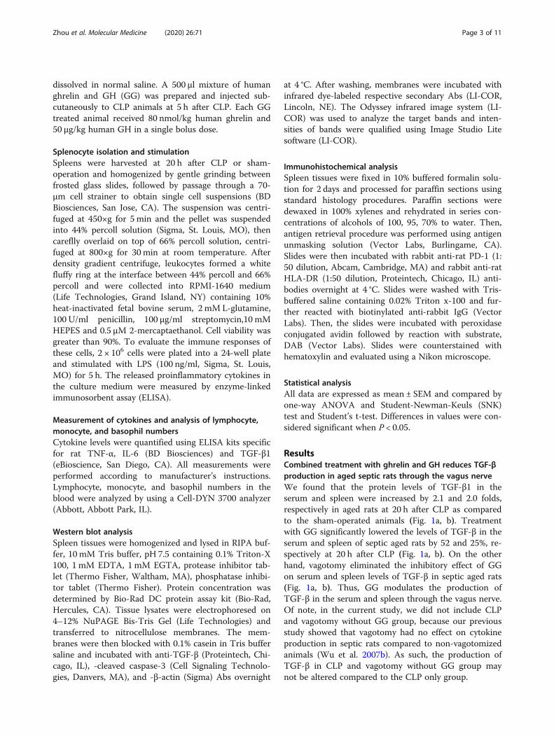
ResultsCombined treatment with ghrelin and GH reduces TGF-βproduction in aged septic rats through the vagus nerveWe found that the protein levels of TGF-β1 in theserum and spleen were increased by 2.1 and 2.0 folds,respectively in aged rats at 20 h after CLP as comparedto the sham-operated animals (Fig. 1a, b). Treatmentwith GG significantly lowered the levels of TGF-β in theserum and spleen of septic aged rats by 52 and 25%, re-spectively at 20 h after CLP (Fig. 1a, b). On the otherhand, vagotomy eliminated the inhibitory effect of GGon serum and spleen levels of TGF-β in septic aged rats(Fig. 1a, b). Thus, GG modulates the production ofTGF-β in the serum and spleen through the vagus nerve.Of note, in the current study, we did not include CLPand vagotomy without GG group, because our previousstudy showed that vagotomy had no effect on cytokineproduction in septic rats compared to non-vagotomizedanimals (Wu et al. 2007b). As such, the production ofTGF-β in CLP and vagotomy without GG group maynot be altered compared to the CLP only group.
Zhou et al. Molecular Medicine (2020) 26:71 Page 3 of 11
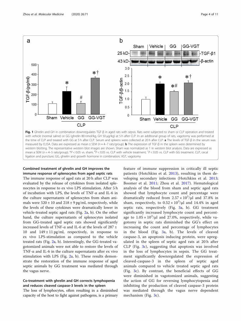
Combined treatment of ghrelin and GH improves theimmune response of splenocytes from aged septic ratsThe immune response of aged rats at 20 h after CLP wasevaluated by the release of cytokines from isolated sple-nocytes in response to ex vivo LPS stimulation. After 5 hof incubation with LPS, the levels of TNF-α and IL-6 inthe culture supernatants of splenocytes from sham ani-mals were 520 ± 10 and 218 ± 9 pg/ml, respectively, whilethe levels of these cytokines were dramatically lower invehicle-treated septic aged rats (Fig. 2a, b). On the otherhand, the culture supernatants of splenocytes isolatedfrom GG-treated aged septic rats showed significantlyincreased levels of TNF-α and IL-6 at the levels of 287 ±10 and 149 ± 11 pg/ml, respectively, in response toex vivo LPS-stimulation as compared to the vehicletreated rats (Fig. 2a, b). Interestingly, the GG-treated va-gotomized animals were not able to restore the levels ofTNF-α and IL-6 in the culture supernatants after ex vivostimulation with LPS (Fig. 2a, b). These results demon-strate the restoration of the immune response of agedseptic animals by GG treatment was mediated throughthe vagus nerve.
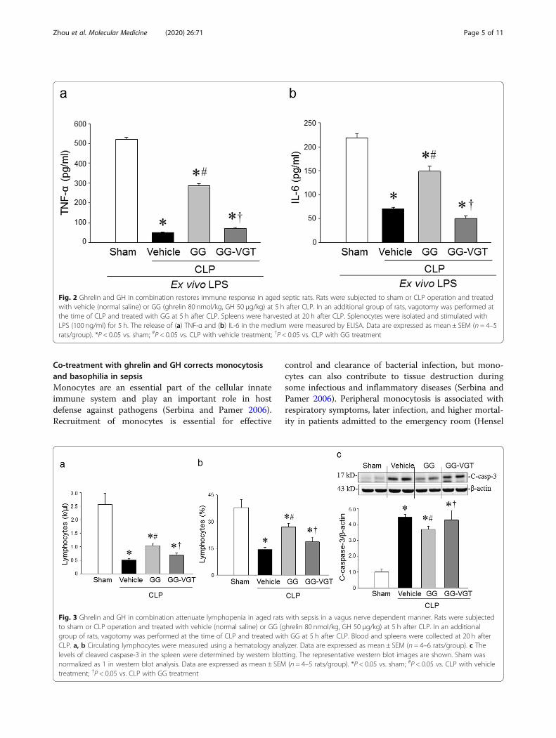
Co-treatment with ghrelin and GH corrects lymphopeniaand reduces cleaved caspase-3 levels in the spleenThe loss of lymphocytes, often resulting in a diminishedcapacity of the host to fight against pathogens, is a primary
feature of immune suppression in critically ill septicpatients (Hotchkiss et al. 2013), resulting in them de-veloping secondary infections (Hotchkiss et al. 2013;Boomer et al. 2011; Zhou et al. 2017). Hematologicalanalysis of the blood from sham and septic aged ratsshowed that lymphocyte count and percentage weredramatically reduced from 2.57 × 10³/μl and 37.8% insham, respectively, to 0.52 × 10³/μl and 14.4% in agedseptic rats, respectively (Fig. 3a, b). GG treatmentsignificantly increased lymphocyte count and percent-age to 1.05 × 10³/μl and 27.0%, respectively, while va-gotomy in septic rats diminished the GG’s effect onincreasing the count and percentage of lymphocytesin the blood (Fig. 3a, b). The levels of cleavedcaspase-3, an apoptosis inducing protein, were upreg-ulated in the spleen of septic aged rats at 20 h afterCLP (Fig. 3c), suggesting that apoptosis was involvedin the loss of lymphocytes in sepsis. The GG treat-ment significantly downregulated the expression ofcleaved-caspase-3 in the spleen of septic agedanimals compared to vehicle treated septic aged rats(Fig. 3c). By contrast, the beneficial effects of GGwere diminished in vagotomized animals, suggestingthe action of GG for reversing lymphocytopenia andinhibiting the production of cleaved caspase-3 proteinwas mediated through the vagus nerve dependentmechanism (Fig. 3c).
Fig. 1 Ghrelin and GH in combination downregulates TGF-β in aged rats with sepsis. Rats were subjected to sham or CLP operation and treatedwith vehicle (normal saline) or GG (ghrelin 80 nmol/kg, GH 50 μg/kg) at 5 h after CLP. In an additional group of rats, vagotomy was performed atthe time of CLP and treated with GG at 5 h after CLP. Serum and spleens were collected at 20 h after CLP. a The levels of TGF-β in the serum wasmeasured by ELISA. Data are expressed as mean ± SEM (n = 4–7 rats/group). b The expression of TGF-β in the spleen were determined bywestern blotting. The representative western blot images are shown. Sham was normalized as 1 in western blot analysis. Data are expressed asmean ± SEM (n = 4–5 rats/group). *P < 0.05 vs. sham; #P < 0.05 vs. CLP with vehicle treatment; †P < 0.05 vs. CLP with GG treatment. CLP, cecalligation and puncture; GG, ghrelin and growth hormone in combination; VGT, vagotomy
Zhou et al. Molecular Medicine (2020) 26:71 Page 4 of 11
Co-treatment with ghrelin and GH corrects monocytosisand basophilia in sepsisMonocytes are an essential part of the cellular innateimmune system and play an important role in hostdefense against pathogens (Serbina and Pamer 2006).Recruitment of monocytes is essential for effective
control and clearance of bacterial infection, but mono-cytes can also contribute to tissue destruction duringsome infectious and inflammatory diseases (Serbina andPamer 2006). Peripheral monocytosis is associated withrespiratory symptoms, later infection, and higher mortal-ity in patients admitted to the emergency room (Hensel
Fig. 3 Ghrelin and GH in combination attenuate lymphopenia in aged rats with sepsis in a vagus nerve dependent manner. Rats were subjectedto sham or CLP operation and treated with vehicle (normal saline) or GG (ghrelin 80 nmol/kg, GH 50 μg/kg) at 5 h after CLP. In an additionalgroup of rats, vagotomy was performed at the time of CLP and treated with GG at 5 h after CLP. Blood and spleens were collected at 20 h afterCLP. a, b Circulating lymphocytes were measured using a hematology analyzer. Data are expressed as mean ± SEM (n = 4–6 rats/group). c Thelevels of cleaved caspase-3 in the spleen were determined by western blotting. The representative western blot images are shown. Sham wasnormalized as 1 in western blot analysis. Data are expressed as mean ± SEM (n = 4–5 rats/group). *P < 0.05 vs. sham; #P < 0.05 vs. CLP with vehicletreatment; †P < 0.05 vs. CLP with GG treatment
Fig. 2 Ghrelin and GH in combination restores immune response in aged septic rats. Rats were subjected to sham or CLP operation and treatedwith vehicle (normal saline) or GG (ghrelin 80 nmol/kg, GH 50 μg/kg) at 5 h after CLP. In an additional group of rats, vagotomy was performed atthe time of CLP and treated with GG at 5 h after CLP. Spleens were harvested at 20 h after CLP. Splenocytes were isolated and stimulated withLPS (100 ng/ml) for 5 h. The release of (a) TNF-α and (b) IL-6 in the medium were measured by ELISA. Data are expressed as mean ± SEM (n = 4–5rats/group). *P < 0.05 vs. sham; #P < 0.05 vs. CLP with vehicle treatment; †P < 0.05 vs. CLP with GG treatment
Zhou et al. Molecular Medicine (2020) 26:71 Page 5 of 11
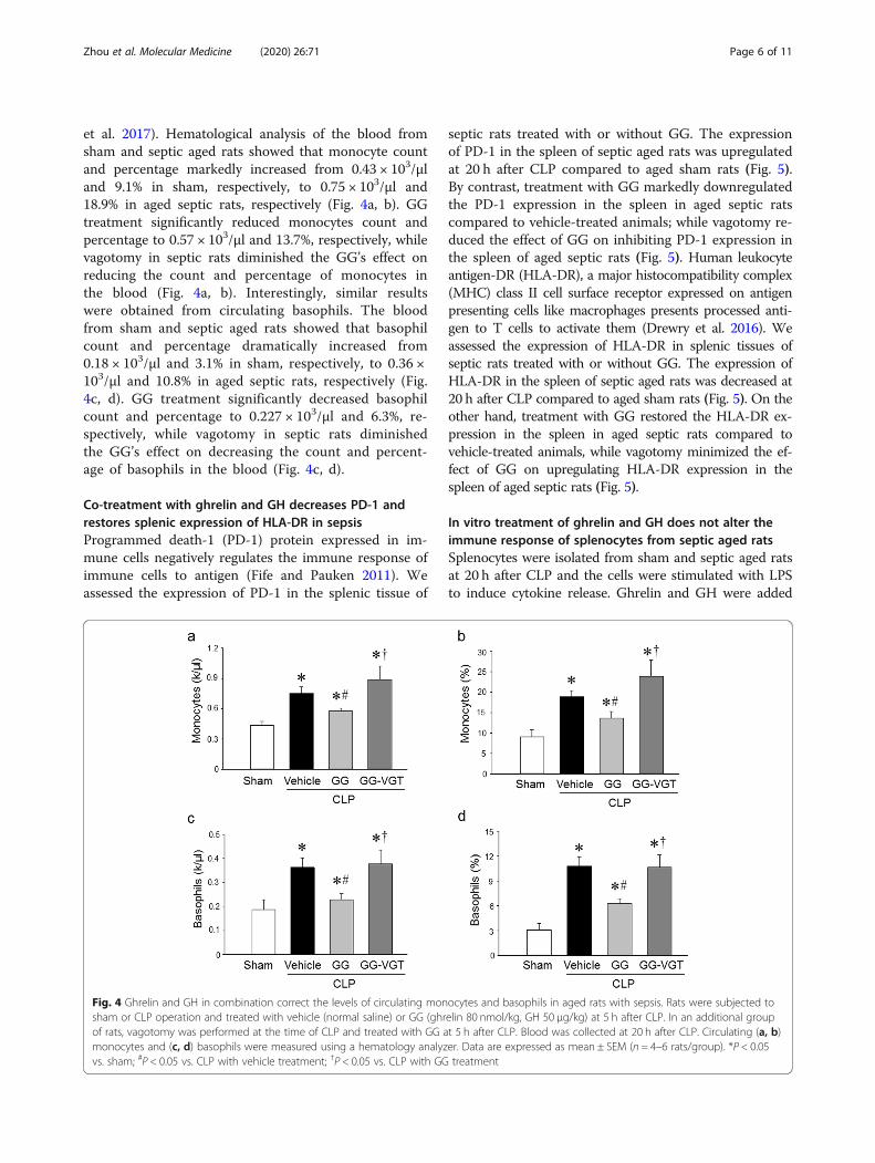
et al. 2017). Hematological analysis of the blood fromsham and septic aged rats showed that monocyte countand percentage markedly increased from 0.43 × 10³/μland 9.1% in sham, respectively, to 0.75 × 10³/μl and18.9% in aged septic rats, respectively (Fig. 4a, b). GGtreatment significantly reduced monocytes count andpercentage to 0.57 × 10³/μl and 13.7%, respectively, whilevagotomy in septic rats diminished the GG’s effect onreducing the count and percentage of monocytes inthe blood (Fig. 4a, b). Interestingly, similar resultswere obtained from circulating basophils. The bloodfrom sham and septic aged rats showed that basophilcount and percentage dramatically increased from0.18 × 103/μl and 3.1% in sham, respectively, to 0.36 ×10³/μl and 10.8% in aged septic rats, respectively (Fig.4c, d). GG treatment significantly decreased basophilcount and percentage to 0.227 × 10³/μl and 6.3%, re-spectively, while vagotomy in septic rats diminishedthe GG’s effect on decreasing the count and percent-age of basophils in the blood (Fig. 4c, d).
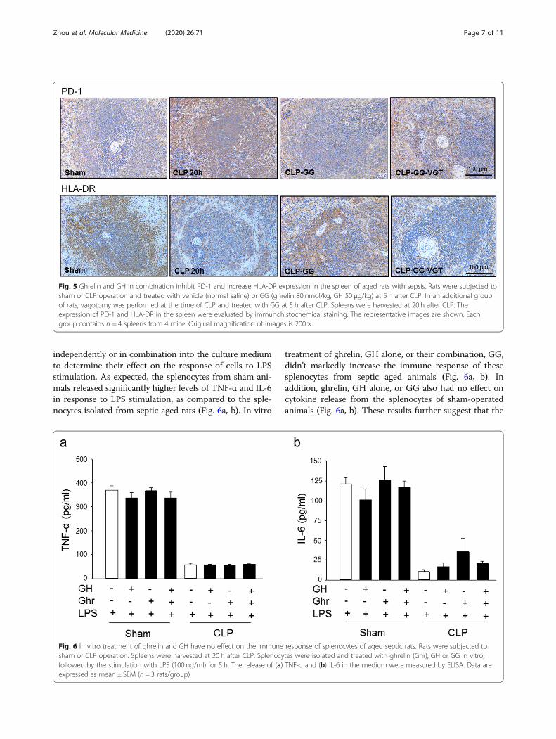
Co-treatment with ghrelin and GH decreases PD-1 andrestores splenic expression of HLA-DR in sepsisProgrammed death-1 (PD-1) protein expressed in im-mune cells negatively regulates the immune response ofimmune cells to antigen (Fife and Pauken 2011). Weassessed the expression of PD-1 in the splenic tissue of
septic rats treated with or without GG. The expressionof PD-1 in the spleen of septic aged rats was upregulatedat 20 h after CLP compared to aged sham rats (Fig. 5).By contrast, treatment with GG markedly downregulatedthe PD-1 expression in the spleen in aged septic ratscompared to vehicle-treated animals; while vagotomy re-duced the effect of GG on inhibiting PD-1 expression inthe spleen of aged septic rats (Fig. 5). Human leukocyteantigen-DR (HLA-DR), a major histocompatibility complex(MHC) class II cell surface receptor expressed on antigenpresenting cells like macrophages presents processed anti-gen to T cells to activate them (Drewry et al. 2016). Weassessed the expression of HLA-DR in splenic tissues ofseptic rats treated with or without GG. The expression ofHLA-DR in the spleen of septic aged rats was decreased at20 h after CLP compared to aged sham rats (Fig. 5). On theother hand, treatment with GG restored the HLA-DR ex-pression in the spleen in aged septic rats compared tovehicle-treated animals, while vagotomy minimized the ef-fect of GG on upregulating HLA-DR expression in thespleen of aged septic rats (Fig. 5).
In vitro treatment of ghrelin and GH does not alter theimmune response of splenocytes from septic aged ratsSplenocytes were isolated from sham and septic aged ratsat 20 h after CLP and the cells were stimulated with LPSto induce cytokine release. Ghrelin and GH were added
Fig. 4 Ghrelin and GH in combination correct the levels of circulating monocytes and basophils in aged rats with sepsis. Rats were subjected tosham or CLP operation and treated with vehicle (normal saline) or GG (ghrelin 80 nmol/kg, GH 50 μg/kg) at 5 h after CLP. In an additional groupof rats, vagotomy was performed at the time of CLP and treated with GG at 5 h after CLP. Blood was collected at 20 h after CLP. Circulating (a, b)monocytes and (c, d) basophils were measured using a hematology analyzer. Data are expressed as mean ± SEM (n = 4–6 rats/group). *P < 0.05vs. sham; #P < 0.05 vs. CLP with vehicle treatment; †P < 0.05 vs. CLP with GG treatment
Zhou et al. Molecular Medicine (2020) 26:71 Page 6 of 11
independently or in combination into the culture mediumto determine their effect on the response of cells to LPSstimulation. As expected, the splenocytes from sham ani-mals released significantly higher levels of TNF-α and IL-6in response to LPS stimulation, as compared to the sple-nocytes isolated from septic aged rats (Fig. 6a, b). In vitro
treatment of ghrelin, GH alone, or their combination, GG,didn’t markedly increase the immune response of thesesplenocytes from septic aged animals (Fig. 6a, b). Inaddition, ghrelin, GH alone, or GG also had no effect oncytokine release from the splenocytes of sham-operatedanimals (Fig. 6a, b). These results further suggest that the
Fig. 6 In vitro treatment of ghrelin and GH have no effect on the immune response of splenocytes of aged septic rats. Rats were subjected tosham or CLP operation. Spleens were harvested at 20 h after CLP. Splenocytes were isolated and treated with ghrelin (Ghr), GH or GG in vitro,followed by the stimulation with LPS (100 ng/ml) for 5 h. The release of (a) TNF-α and (b) IL-6 in the medium were measured by ELISA. Data areexpressed as mean ± SEM (n = 3 rats/group)
Fig. 5 Ghrelin and GH in combination inhibit PD-1 and increase HLA-DR expression in the spleen of aged rats with sepsis. Rats were subjected tosham or CLP operation and treated with vehicle (normal saline) or GG (ghrelin 80 nmol/kg, GH 50 μg/kg) at 5 h after CLP. In an additional groupof rats, vagotomy was performed at the time of CLP and treated with GG at 5 h after CLP. Spleens were harvested at 20 h after CLP. Theexpression of PD-1 and HLA-DR in the spleen were evaluated by immunohistochemical staining. The representative images are shown. Eachgroup contains n = 4 spleens from 4 mice. Original magnification of images is 200 ×
Zhou et al. Molecular Medicine (2020) 26:71 Page 7 of 11
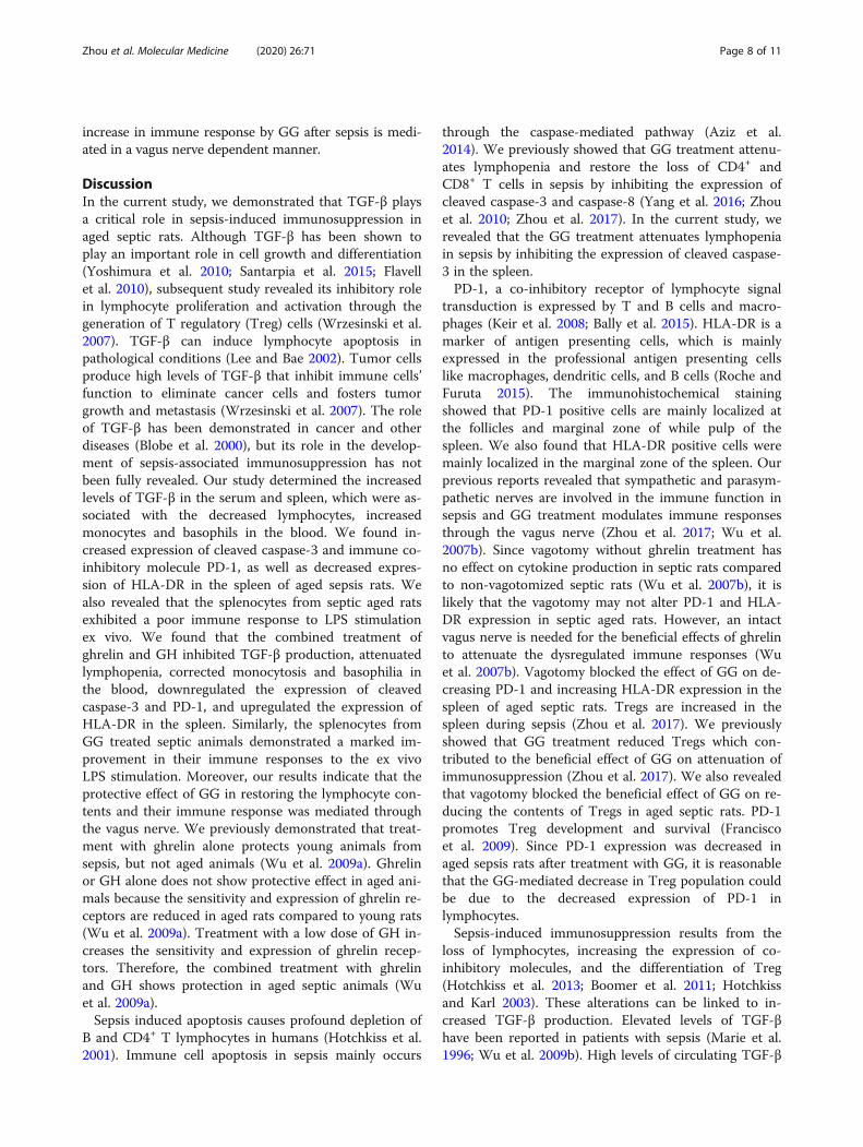
increase in immune response by GG after sepsis is medi-ated in a vagus nerve dependent manner.
DiscussionIn the current study, we demonstrated that TGF-β playsa critical role in sepsis-induced immunosuppression inaged septic rats. Although TGF-β has been shown toplay an important role in cell growth and differentiation(Yoshimura et al. 2010; Santarpia et al. 2015; Flavellet al. 2010), subsequent study revealed its inhibitory rolein lymphocyte proliferation and activation through thegeneration of T regulatory (Treg) cells (Wrzesinski et al.2007). TGF-β can induce lymphocyte apoptosis inpathological conditions (Lee and Bae 2002). Tumor cellsproduce high levels of TGF-β that inhibit immune cells’function to eliminate cancer cells and fosters tumorgrowth and metastasis (Wrzesinski et al. 2007). The roleof TGF-β has been demonstrated in cancer and otherdiseases (Blobe et al. 2000), but its role in the develop-ment of sepsis-associated immunosuppression has notbeen fully revealed. Our study determined the increasedlevels of TGF-β in the serum and spleen, which were as-sociated with the decreased lymphocytes, increasedmonocytes and basophils in the blood. We found in-creased expression of cleaved caspase-3 and immune co-inhibitory molecule PD-1, as well as decreased expres-sion of HLA-DR in the spleen of aged sepsis rats. Wealso revealed that the splenocytes from septic aged ratsexhibited a poor immune response to LPS stimulationex vivo. We found that the combined treatment ofghrelin and GH inhibited TGF-β production, attenuatedlymphopenia, corrected monocytosis and basophilia inthe blood, downregulated the expression of cleavedcaspase-3 and PD-1, and upregulated the expression ofHLA-DR in the spleen. Similarly, the splenocytes fromGG treated septic animals demonstrated a marked im-provement in their immune responses to the ex vivoLPS stimulation. Moreover, our results indicate that theprotective effect of GG in restoring the lymphocyte con-tents and their immune response was mediated throughthe vagus nerve. We previously demonstrated that treat-ment with ghrelin alone protects young animals fromsepsis, but not aged animals (Wu et al. 2009a). Ghrelinor GH alone does not show protective effect in aged ani-mals because the sensitivity and expression of ghrelin re-ceptors are reduced in aged rats compared to young rats(Wu et al. 2009a). Treatment with a low dose of GH in-creases the sensitivity and expression of ghrelin recep-tors. Therefore, the combined treatment with ghrelinand GH shows protection in aged septic animals (Wuet al. 2009a).Sepsis induced apoptosis causes profound depletion of
B and CD4+ T lymphocytes in humans (Hotchkiss et al.2001). Immune cell apoptosis in sepsis mainly occurs
through the caspase-mediated pathway (Aziz et al.2014). We previously showed that GG treatment attenu-ates lymphopenia and restore the loss of CD4+ andCD8+ T cells in sepsis by inhibiting the expression ofcleaved caspase-3 and caspase-8 (Yang et al. 2016; Zhouet al. 2010; Zhou et al. 2017). In the current study, werevealed that the GG treatment attenuates lymphopeniain sepsis by inhibiting the expression of cleaved caspase-3 in the spleen.PD-1, a co-inhibitory receptor of lymphocyte signal
transduction is expressed by T and B cells and macro-phages (Keir et al. 2008; Bally et al. 2015). HLA-DR is amarker of antigen presenting cells, which is mainlyexpressed in the professional antigen presenting cellslike macrophages, dendritic cells, and B cells (Roche andFuruta 2015). The immunohistochemical stainingshowed that PD-1 positive cells are mainly localized atthe follicles and marginal zone of while pulp of thespleen. We also found that HLA-DR positive cells weremainly localized in the marginal zone of the spleen. Ourprevious reports revealed that sympathetic and parasym-pathetic nerves are involved in the immune function insepsis and GG treatment modulates immune responsesthrough the vagus nerve (Zhou et al. 2017; Wu et al.2007b). Since vagotomy without ghrelin treatment hasno effect on cytokine production in septic rats comparedto non-vagotomized septic rats (Wu et al. 2007b), it islikely that the vagotomy may not alter PD-1 and HLA-DR expression in septic aged rats. However, an intactvagus nerve is needed for the beneficial effects of ghrelinto attenuate the dysregulated immune responses (Wuet al. 2007b). Vagotomy blocked the effect of GG on de-creasing PD-1 and increasing HLA-DR expression in thespleen of aged septic rats. Tregs are increased in thespleen during sepsis (Zhou et al. 2017). We previouslyshowed that GG treatment reduced Tregs which con-tributed to the beneficial effect of GG on attenuation ofimmunosuppression (Zhou et al. 2017). We also revealedthat vagotomy blocked the beneficial effect of GG on re-ducing the contents of Tregs in aged septic rats. PD-1promotes Treg development and survival (Franciscoet al. 2009). Since PD-1 expression was decreased inaged sepsis rats after treatment with GG, it is reasonablethat the GG-mediated decrease in Treg population couldbe due to the decreased expression of PD-1 inlymphocytes.Sepsis-induced immunosuppression results from the
loss of lymphocytes, increasing the expression of co-inhibitory molecules, and the differentiation of Treg(Hotchkiss et al. 2013; Boomer et al. 2011; Hotchkissand Karl 2003). These alterations can be linked to in-creased TGF-β production. Elevated levels of TGF-βhave been reported in patients with sepsis (Marie et al.1996; Wu et al. 2009b). High levels of circulating TGF-β
Zhou et al. Molecular Medicine (2020) 26:71 Page 8 of 11
during sepsis caused by pneumonia have been shown tocorrelate with higher tissue injury scores and mortality(Wu et al. 2009b). Increased levels of TGF-β are associ-ated with the reduced bacterial clearance and increasedorgan injury (Weehuizen et al. 2012). It has been shownthat bacteria-induced immunosuppression is mediatedthrough the induction of TGF-β, followed by the de-crease in the expression of surface MHCII on the den-dritic cells (DC), thereby inhibiting DC activation (Bosioet al. 2007). TGF-β plays a prominent role in the differ-entiation of immunoparalyzed DC, leading to immuno-suppression after lung infection by E. coli (Roquilly et al.2017). TGF-β can suppress the release of proinflamma-tory mediators such as TNF-α and IL-1β from mono-cytes and macrophage (Blobe et al. 2000; Pellacani et al.2001). Patients with sepsis can have both increased ordecreased levels of TGF-β (Marie et al. 1996; Pellacaniet al. 2001; White et al. 2010), which could be due to thevariant stages of sepsis in these patients.In sepsis, the host response to infection is associated
with sustained inflammation to eliminate pathogens inthe early phase and immune suppression in the laterphase (van der Poll et al. 2017). Patients with sepsisshow signs of both excessive inflammation and immunesuppression, although the extent of which may vary be-tween individuals (van der Poll et al. 2017). TGF-β sig-naling is important to maintain immune homeostasis insepsis and other infectious conditions. The dysregulationof TGF-β has been associated with tissue injury andhigher mortality in sepsis (Wu et al. 2009b; Lekkou et al.2014). Sepsis survivors have been found to have higherlevels of TGF-β in early time points and lower levels ofTGF-β at 10 days after hospitalization (Lekkou et al.2014). In contrast, the non-survivors have lower levels ofTGF-β in early stages and higher levels of TGF-β at 10days after their admission in the hospital (Lekkou et al.2014). Elderly are particularly susceptible to infectionsdue to insufficient immunity. Aged septic patients havefeatures consistent with immunosuppression that resultin an inability to clear infections and provide a predis-position to developing nosocomial infections (Hotchkisset al. 2013; Boomer et al. 2011; Hotchkiss and Karl2003). The gene expression profile showed that TGF-βexpression in elderly septic patients is higher as com-pared to young septic patients (Vieira da Silva PellegrinaD, et al. 2015). The higher TGF-β in elderly septic pa-tients correlates with their suppressed immune response,higher morbidity, and mortality (Hotchkiss et al. 2013;Boomer et al. 2011). Immunosuppression was observedin aged rats at 20 h after CLP. These animals demon-strated apoptosis in T cells, an increase of co-inhibitorymolecules, and Treg differentiation (Zhou et al. 2017).Our current study demonstrates the upregulation ofTGF-β is associated with the immunosuppression in
aged animal with sepsis. It has been shown that im-munosuppression occurs at a much later time point aftersepsis onset in young animals (Unsinger et al. 2012). Al-though the circulating TGF-β was increased in aged sep-tic rats at 20 h after CLP, this increase was not observedin young rats at 3 months of age 20 h after CLP (datanot shown). Thus, the levels of TGF-β between youngand aged animals reflect the differentiated immune re-sponse between different age groups during sepsis.TGF-β can be used as a prognostic marker of sepsis-
induced immunosuppression in the elderly population.Inhibition of TGF-β has been shown to ameliorate organinjury and improve survival in sepsis (Weehuizen et al.2012; Bae et al. 2014). Blockade of TGF-β attenuated im-munosuppression and reduced the susceptibility to sec-ondary infection following sepsis (Roquilly et al. 2017).Thus, modulating TGF-β could be a potential thera-peutic target for elderly patients with sepsis. Administra-tion of ghrelin was found to reduce TGF-β expressionand exert an anti-fibrotic effect in patients with systemicsclerosis (Ota et al. 2013). Here, we demonstrated thatghrelin reduced TGF-β levels in the serum in septic agedanimals and ameliorated sepsis-induced immune sup-pression. Human ghrelin has been administered into hu-man subjects in clinical studies, showing excellentefficacy and safety profile (Narula and deBoisblanc2015). Ghrelin has 93% homology in its amino acid se-quence between human and rat or human and mouse(Kojima and Kangawa 2005). In the current study, weused human ghrelin as a clinically relevant approach forimplementing ghrelin as a novel therapeutic for elderlyseptic patients.Communication between the nervous and immune
systems is important for the regulation of immune func-tion and inflammation (Tracey 2007; Abe and Inoue2018; Inoue et al. 2016). The vagus nerve has beenshown to play a critical role in the inflammatory reflexand mediates a rapid homeostatic response to protectagainst organ injury (Tracey 2007; Abe and Inoue 2018;Inoue et al. 2016). The anti-inflammatory effect ofghrelin is mainly mediated through the vagus nerve asvagotomy diminishes the protective effect of ghrelin onseptic animals (Wu et al. 2007b). The afferent vagusnerve can be activated by peripheral inflammatorystimulation and the signal is transmitted to the efferentvagus nerve to suppress the inflammation (Abe andInoue 2018; Borsody and Weiss 2005). The activation ofthe vagus nerve during infection results in a protectiveadvantage to the host, while a defect with the anti-inflammatory pathway of the vagus nerve contributes todisease pathology (Tracey 2007; Tracey 2005). Inaddition, the immunomodulatory effect of the efferentvagal nerve on macrophages is mediated through thestimulation of cholinergic activity of the parasympathetic
Zhou et al. Molecular Medicine (2020) 26:71 Page 9 of 11
nervous system (Tracey 2007). The spleen is a primaryorgan of the cholinergic anti-inflammatory pathway(Tracey 2007; Inoue et al. 2016). The impaired choliner-gic anti-inflammatory pathway in aged rats is responsiblefor the robust inflammatory response after the onset ofsepsis resulting in early morbidity and mortality in agedseptic animals (Wu et al. 2009a). On the other hand,aged animals that survived sepsis showed an impairedimmune response to LPS stimulation (Zhou et al. 2017).This indicates that these aged animals cannot removebacteria efficiently during secondary infection, whichmay lead to higher mortality rates after sepsis (Wu et al.2009a; Yang et al. 2016). Despite the anti-inflammatoryactivity of the vagus nerve, activation of the immune re-sponse has been shown to occur in immune dysregu-lated conditions (Corcoran et al. 2005). Vagus nervestimulation has been shown to increases immune activityby elevating plasma levels of inflammatory cytokines,such as TNF-α and IL-6 in humans with depression(Corcoran et al. 2005). Depression has been character-ized as a dysregulation of immune function and has beenassociated with immunosuppression (Blume et al. 2011;Zhang et al. 2018).
ConclusionsIn conclusion, we identified that GG maintained the im-mune response in aged septic rats through the inhibitionof TGF-β via vagal stimulation. The decreased expres-sion of TGF-β improved the immune response to infec-tion in aged septic rats by maintaining lymphocytecounts, correcting monocytosis and basophilia thatcorrelated with the decreased expression of cleavedcaspase-3, reducing lymphocyte inhibitory receptor PD-1, and increasing HLA-DR expression.
AbbreviationsCLP: Cecal ligation and puncture; GG: Ghrelin and growth hormone incombination; GH: Growth hormone; GHSR: Growth hormone secretagoguereceptor; PD-1: Programmed death-1; TGF-β: Transforming growth factor-β;Treg: T regulatory cells
AcknowledgementsWe thank Dr. William Royster for critically reviewing the manuscript.
Authors’ contributionsData curation, Mian Zhou and Mahendar Ochani; Formal analysis, Mian Zhouand Monowar Aziz; Funding acquisition, Ping Wang, Monowar Aziz;Investigation, Mian Zhou and Mahendar Ochani; Methodology, Mian Zhouand Mahendar Ochani; Project administration, Ping Wang; Resources, PingWang; Supervision, Monowar Aziz and Ping Wang; Writing – original draft,Mian Zhou and Monowar Aziz; Writing – review & editing, Mian Zhou,Monowar Aziz and Ping Wang. The author(s) read and approved the finalmanuscript.
FundingThis work was supported by the National Institutes of Health grants:R35GM118337 (PW) and R01GM129633 (MA).
Availability of data and materialsThe manuscript does not contain any supplemental or supportive data file.
Ethics approval and consent to participateAll experiments were performed in accordance with the NIH guidelines forthe use of experimental animals, and the study was approved by theInstitutional Animal Care and Use Committee (IACUC) of the FeinsteinInstitutes for Medical Research.
Consent for publicationAuthors agreed to this publication.
Competing interestsThe authors declare no conflicts of interest.
Received: 29 April 2020 Accepted: 3 July 2020
ReferencesAbe C, Inoue T. Role of C1 neurons in anti-inflammatory reflex: mediation
between afferents and efferents. Neurosci Res. 2018;136:6-12.Arvat E, et al. Preliminary evidence that ghrelin, the natural GH secretagogue
(GHS)-receptor ligand, strongly stimulates GH secretion in humans. JEndocrinol Investig. 2000;23:493–5.
Aziz M, Jacob A, Wang P. Revisiting caspases in sepsis. Cell Death Dis. 2014;5:e1526.
Bae JS, Lee W, Son HN, Lee YM, Kim IS. Anti-transforming growth factor beta-induced protein antibody ameliorates vascular barrier dysfunction andimproves survival in sepsis. Acta Physiol. 2014;212:306–15.
Bally AP, et al. NF-κB regulates PD-1 expression in macrophages. J Immunol.2015;194:4545–54.
Blobe GC, Schiemann WP, Lodish HF. Role of transforming growth factor beta inhuman disease. N Engl J Med. 2000;342:1350–8.
Blume J, Douglas SD, Evans DL. Immune suppression and immune activation indepression. Brain Behav Immun. 2011;25:221–9.
Boomer JS, et al. Immunosuppression in patients who die of sepsis and multipleorgan failure. Jama. 2011;306:2594–605.
Borsody MK, Weiss JM. The subdiaphragmatic vagus nerves mediate activation oflocus coeruleus neurons by peripherally administered microbial substances.Neuroscience. 2005;131:235–45.
Bosio CM, Bielefeldt-Ohmann H, Belisle JT. Active suppression of thepulmonary immune response by Francisella tularensis Schu4. J Immunol.2007;178:4538–47.
Collden G, Tschop MH, Muller TD. Therapeutic potential of targeting the ghrelinpathway. Int J Mol Sci. 2017;18(4):798.
Corcoran C, Connor TJ, O'Keane V, Garland MR. The effects of vagus nervestimulation on pro- and anti-inflammatory cytokines in humans: apreliminary report. Neuroimmunomodulation. 2005;12:307–9.
Delano MJ, Ward PA. Sepsis-induced immune dysfunction: can immune therapiesreduce mortality? J Clin Invest. 2016;126:23–31.
Drewry AM, et al. Comparison of monocyte human leukocyte antigen-DRexpression and stimulated tumor necrosis factor alpha production asoutcome predictors in severe sepsis: a prospective observational study. CritCare. 2016;20:334.
Fife BT, Pauken KE. The role of the PD-1 pathway in autoimmunity and peripheraltolerance. Ann N Y Acad Sci. 2011;1217:45–59.
Flavell RA, Sanjabi S, Wrzesinski SH, Licona-Limon P. The polarization of immunecells in the tumour environment by TGFbeta. Nat Rev Immunol. 2010;10:554–67.
Francisco LM, et al. PD-L1 regulates the development, maintenance, and functionof induced regulatory T cells. J Exp Med. 2009;206:3015–29.
Gonzalez-Rey E, Chorny A, Delgado M. Therapeutic action of ghrelin in a mousemodel of colitis. Gastroenterology. 2006;130:1707–20.
Govinden R, Bhoola KD. Genealogy, expression, and cellular function oftransforming growth factor-beta. Pharmacol Ther. 2003;98:257–65.
Hensel M, et al. Peripheral monocytosis as a predictive factor for adverseoutcome in the emergency department: survey based on a register study.Medicine (Baltimore). 2017;96:e7404.
Hepper HJ, Sieber C, Walger P, Bahrmann P, Singler K. Infections in the elderly.Crit Care Clin. 2013;29:757–74.
Hotchkiss RS, Karl IE. The pathophysiology and treatment of sepsis. N Engl J Med.2003;348:138–50.
Hotchkiss RS, Monneret G, Payen D. Sepsis-induced immunosuppression: fromcellular dysfunctions to immunotherapy. Nat Rev Immunol. 2013;13:862–74.
Zhou et al. Molecular Medicine (2020) 26:71 Page 10 of 11
Hotchkiss RS, et al. Sepsis-induced apoptosis causes progressive profounddepletion of B and CD4+ T lymphocytes in humans. J Immunol. 2001;166:6952–63.
Inoue S, et al. Persistent inflammation and T cell exhaustion in severe sepsis inthe elderly. Crit Care. 2014;18:R130.
Inoue T, et al. Vagus nerve stimulation mediates protection from kidneyischemia-reperfusion injury through alpha7nAChR+ splenocytes. J Clin Invest.2016;126:1939–52.
Keir ME, Butte MJ, Freeman GJ, Sharpe AH. PD-1 and its ligands in tolerance andimmunity. Annu Rev Immunol. 2008;26:677–704.
Kojima M, Kangawa K. Ghrelin: structure and function. Physiol Rev. 2005;85:495–522.Lee KY, Bae SC. TGF-beta-dependent cell growth arrest and apoptosis. J Biochem
Mol Biol. 2002;35:47–53.Lekkou A, Mouzaki A, Siagris D, Ravani I, Gogos CA. Serum lipid profile, cytokine
production, and clinical outcome in patients with severe sepsis. J Crit Care.2014;29:723–7.
Marie C, Cavaillon JM, Losser MR. Elevated levels of circulating transforminggrowth factor-beta 1 in patients with the sepsis syndrome. Ann Intern Med.1996;125:520–1.
Martin GS, Mannino DM, Moss M. The effect of age on the development andoutcome of adult sepsis. Crit Care Med. 2006;34:15–21.
Narula T, deBoisblanc BP. Ghrelin in Critical Illness. Am J Respir Cell Mol Biol.2015;53:437–42.
Opal SM, Girard TD, Ely EW. The immunopathogenesis of sepsis in elderlypatients. Clin Infect Dis. 2005;41(Suppl 7):S504–12.
Ota Y, et al. Ghrelin attenuates collagen production in lesional fibroblasts frompatients with systemic sclerosis. Clin Immunol. 2013;147:71–8.
Pellacani A, et al. Down-regulation of high mobility group-I(Y) protein contributesto the inhibition of nitric-oxide synthase 2 by transforming growth factor-beta1. J Biol Chem. 2001;276:1653–9.
Roche PA, Furuta K. The ins and outs of MHC class II-mediated antigenprocessing and presentation. Nat Rev Immunol. 2015;15:203–16.
Roquilly A, et al. Local modulation of antigen-presenting cell development afterresolution of pneumonia induces long-term susceptibility to secondaryinfections. Immunity. 2017;47:135–147.e5.
Santarpia M, et al. Programmed cell death protein-1/programmed cell deathligand-1 pathway inhibition and predictive biomarkers: understandingtransforming growth factor-beta role. Transl Lung Cancer Res. 2015;4:728–42.
Serbina NV, Pamer EG. Monocyte emigration from bone marrow during bacterialinfection requires signals mediated by chemokine receptor CCR2. NatImmunol. 2006;7:311–7.
Singer M, et al. The third international consensus definitions for sepsis and septicshock (Sepsis-3). Jama. 2016;315:801–10.
Taylor AW. Review of the activation of TGF-beta in immunity. J Leukoc Biol. 2009;85:29–33.
Tracey KJ. Fat meets the cholinergic antiinflammatory pathway. J Exp Med. 2005;202:1017–21.
Tracey KJ. Physiology and immunology of the cholinergic antiinflammatorypathway. J Clin Invest. 2007;117:289–96.
Unsinger J, et al. Interleukin-7 ameliorates immune dysfunction and improvessurvival in a 2-hit model of fungal sepsis. J Infect Dis. 2012;206:606–16.
van der Poll T, van de Veerdonk FL, Scicluna BP, Netea MG. Theimmunopathology of sepsis and potential therapeutic targets. Nat RevImmunol. 2017;17:407–20.
Vieira da Silva Pellegrina D, et al. Septic shock in advanced age: Transcriptomeanalysis reveals altered molecular signatures in neutrophil granulocytes. PLoSOne. 2015;10:e0128341.
Weehuizen TA, et al. Expression and function of transforming growth factor betain melioidosis. Infect Immun. 2012;80:1853–7.
White M, et al. Transforming growth factor beta-1 and interleukin-17 genetranscription in peripheral blood mononuclear cells and the human responseto infection. Cytokine. 2010;50:322–7.
Williams DL, Grill HJ, Cummings DE, Kaplan JM. Vagotomy dissociates short- andlong-term controls of circulating ghrelin. Endocrinology. 2003;144:5184–7.
Wrzesinski SH, Wan YY, Flavell RA. Transforming growth factor-beta and theimmune response: implications for anticancer therapy. Clin Cancer Res. 2007;13:5262–70.
Wu HP, et al. Plasma transforming growth factor-beta1 level in patients withsevere community-acquired pneumonia and association with diseaseseverity. J Formos Med Assoc. 2009b;108:20–7.
Wu JT, Kral JG. Ghrelin: integrative neuroendocrine peptide in health and disease.Ann Surg. 2004;239:464–74.
Wu R, et al. Ghrelin attenuates sepsis-induced acute lung injury and mortality inrats. Am J Respir Crit Care Med. 2007a;176:805–13.
Wu R, et al. Ghrelin down-regulates proinflammatory cytokines in sepsis throughactivation of the vagus nerve. Ann Surg. 2007b;245:480–6.
Wu R, et al. Ghrelin hyporesponsiveness contributes to age-relatedhyperinflammation in septic shock. Ann Surg. 2009a;250:126–33.
Yang WL, et al. Combined administration of human ghrelin and human growthhormone attenuates organ injury and improves survival in aged septic rats.Mol Med. 2016;22:124–35.
Yoshimura A, Wakabayashi Y, Mori T. Cellular and molecular basis for theregulation of inflammation by TGF-beta. J Biochem. 2010;147:781–92.
Zhang HX, et al. Difference in proinflammatory cytokines produced bymonocytes between patients with major depressive disorder and healthycontrols. J Affect Disord. 2018;234:305–10.
Zhou M, Wu R, Dong W, Leong J, Wang P. Accelerated apoptosis contributes toaging-related hyperinflammation in endotoxemia. Int J Mol Med. 2010;25:929–35.
Zhou M, Yang WL, Aziz M, Ma G, Wang P. Therapeutic effect of human ghrelinand growth hormone: attenuation of immunosuppression in septic agedrats. Biochim Biophys Acta. 2017;1863:2584–93.
Publisher’s NoteSpringer Nature remains neutral with regard to jurisdictional claims inpublished maps and institutional affiliations.
Zhou et al. Molecular Medicine (2020) 26:71 Page 11 of 11


![Current progress in stem cell therapy for type 1 diabetes mellitus · 2020. 7. 8. · protocol [4]. Over the past two decades, continuous im-provements in islet isolation and immunosuppression](https://static.fdocument.org/doc/165x107/614a44b112c9616cbc694e61/current-progress-in-stem-cell-therapy-for-type-1-diabetes-mellitus-2020-7-8.jpg)


![Ca2+ Entry (SOCE) Contributes to Muscle Contractility in ... · physiological role in young and aged skeletal muscle. We found that reagents that prevent [Ca2+] o entry reduce contractile](https://static.fdocument.org/doc/165x107/5fbbf98d4e86af3f2a7e3a76/ca2-entry-soce-contributes-to-muscle-contractility-in-physiological-role.jpg)