Comparison of rhGDF-5/β-TCP and rhPDGF/ β-TCP effects … · approval for use in orofacial...
49
- 1 - Comparison of rhGDF-5/β-TCP and rhPDGF/β-TCP effects on Periodontal Wound Healing/Regeneration in One-Wall Intrabony Defects Hyuk-Rak Kwon The Graduate School Yonsei University Department of Dental Science
Transcript of Comparison of rhGDF-5/β-TCP and rhPDGF/ β-TCP effects … · approval for use in orofacial...
- 1 -
Comparison of rhGDF-5/β-TCP and
rhPDGF/β-TCP effects on Periodontal
Wound Healing/Regeneration
in One-Wall Intrabony Defects
Hyuk-Rak Kwon
The Graduate School
Yonsei University
Department of Dental Science
- 2 -
Comparison of rhGDF-5/β-TCP and
rhPDGF/β-TCP effects on Periodontal
Wound Healing/Regeneration
in One-Wall Intrabony Defects
비교
연구
A Dissertation Thesis
Submitted to the Department of Dental Science,
the Graduate School of Yonsei University
in partial fulfillment of the
requirements for the degree of
Doctor of Philosophy of Dental Science
Hyuk-Rak Kwon
June 2008
- 3 -
This certifies that the dissertation thesis
of Hyuk-Rak Kwon is approved.
──────────────────────────────
Thesis Supervisor: Chong-Kwan Kim
──────────────────────────────
Kyoo-Sung Cho ──────────────────────────────
Seong-Ho Choi
──────────────────────────────
Kyoung-Nam Kim ──────────────────────────────
Yun-Jung Yoo
The Graduate School
Yonsei University
June 2008
- 4 -
감사의감사의감사의감사의 글글글글
본 논문이 완성되기까지 졸업 후부터 늘 끊임없는 지도와 관심을 보내주신
김종관 교수님께 진심으로 감사 드립니다. 그리고 박사 과정 동안 많은
관심으로 이끌어 주신 채중규 교수님, 조규성 교수님, 최성호 교수님께도
감사의 말씀 올립니다. 또한 논문 완성 과정에서 많은 조언을 해 주신 김경남
교수님, 유윤정 교수님께도 감사 드립니다.
연구 내내 많은 도움을 주신 치주과학 교실 여러분, 특히 김영택 선생님,
박정철 선생님께 고마움을 전합니다.
그리고, 언제나 아낌없는 사랑과 헌신적인 도움으로 나의 버팀목이 되어준
사랑하는 아내와 항상 끊임없이 뛰어 노는 예쁜 딸 지인이와 개구쟁이 아들
순헌이에게도 사랑하는 마음을 전합니다.
마지막으로, 항상 곁에서 든든하게 기도해주시는 양가 부모님께 감사의
마음을 담아 이 논문을 드립니다.
2008년 6월

- 5 -
저자 씀
i
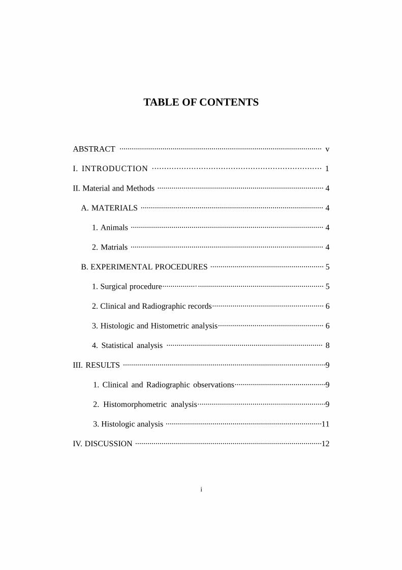
TABLE OF CONTENTS
ABSTRACT ··································································································· v
I. INTRODUCTION ····································································· 1
II. Material and Methods ·················································································· 4
A. MATERIALS ·························································································· 4
1. Animals ······························································································· 4
2. Matrials ······························································································· 4
B. EXPERIMENTAL PROCEDURES ························································ 5
1. Surgical procedure··············································································· 5
2. Clinical and Radiographic records······················································· 6
3. Histologic and Histometric analysis···················································· 6
4. Statistical analysis ············································································· 8
III. RESULTS ····································································································9
1. Clinical and Radiographic observations·············································9
2. Histomorphometric analysis·······························································9
3. Histologic analysis ·············································································11
IV. DISCUSSION ····························································································12
ii
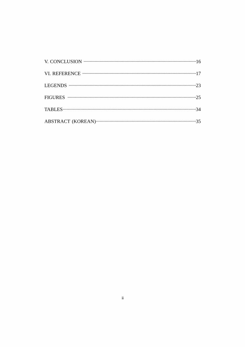
V. CONCLUSION ···························································································16
VI. REFERENCE ····························································································17
LEGENDS ·······································································································23
FIGURES ········································································································25
TABLES···········································································································34
ABSTRACT (KOREAN)·················································································35
iii
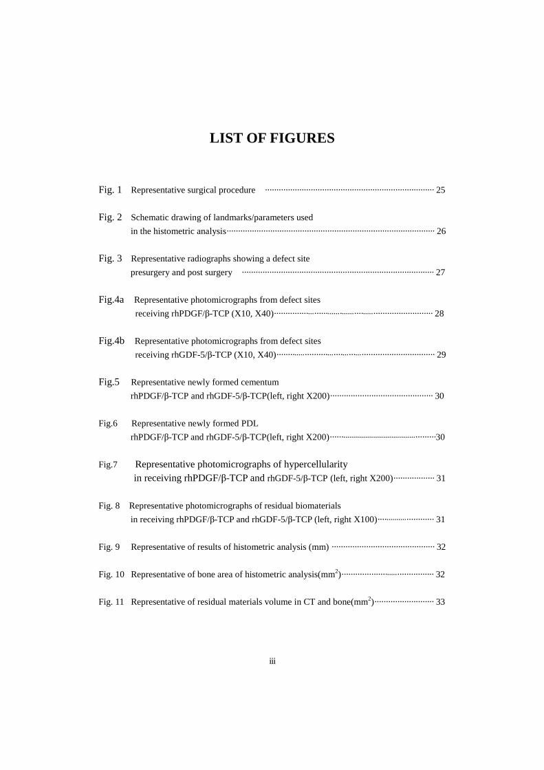
LIST OF FIGURES
Fig. 1 Representative surgical procedure ·········································································· 25 Fig. 2 Schematic drawing of landmarks/parameters used
in the histometric analysis··························································································· 26
Fig. 3 Representative radiographs showing a defect site
presurgery and post surgery ···················································································· 27
Fig.4a Representative photomicrographs from defect sites
receiving rhPDGF/β-TCP (X10, X40)········································································ 28
Fig.4b Representative photomicrographs from defect sites
receiving rhGDF-5/β-TCP (X10, X40)······································································· 29
Fig.5 Representative newly formed cementum
rhPDGF/β-TCP and rhGDF-5/β-TCP(left, right X200)············································· 30
Fig.6 Representative newly formed PDL
rhPDGF/β-TCP and rhGDF-5/β-TCP(left, right X200)···················································30
Fig.7 Representative photomicrographs of hypercellularity in receiving rhPDGF/β-TCP and rhGDF-5/β-TCP (left, right X200)·················· 31
Fig. 8 Representative photomicrographs of residual biomaterials
in receiving rhPDGF/β-TCP and rhGDF-5/β-TCP (left, right X100)·························· 31
Fig. 9 Representative of results of histometric analysis (mm) ············································· 32
Fig. 10 Representative of bone area of histometric analysis(mm2)········································· 32
Fig. 11 Representative of residual materials volume in CT and bone(mm2)·························· 33
iv
LIST OF TABLE
Table 1. Histomorphometric analysis (group means ± SD in mm/mm2)········ 34
Table 2. Residual materials (mean ± SD in mm2)··········································· 34
v
Abstract
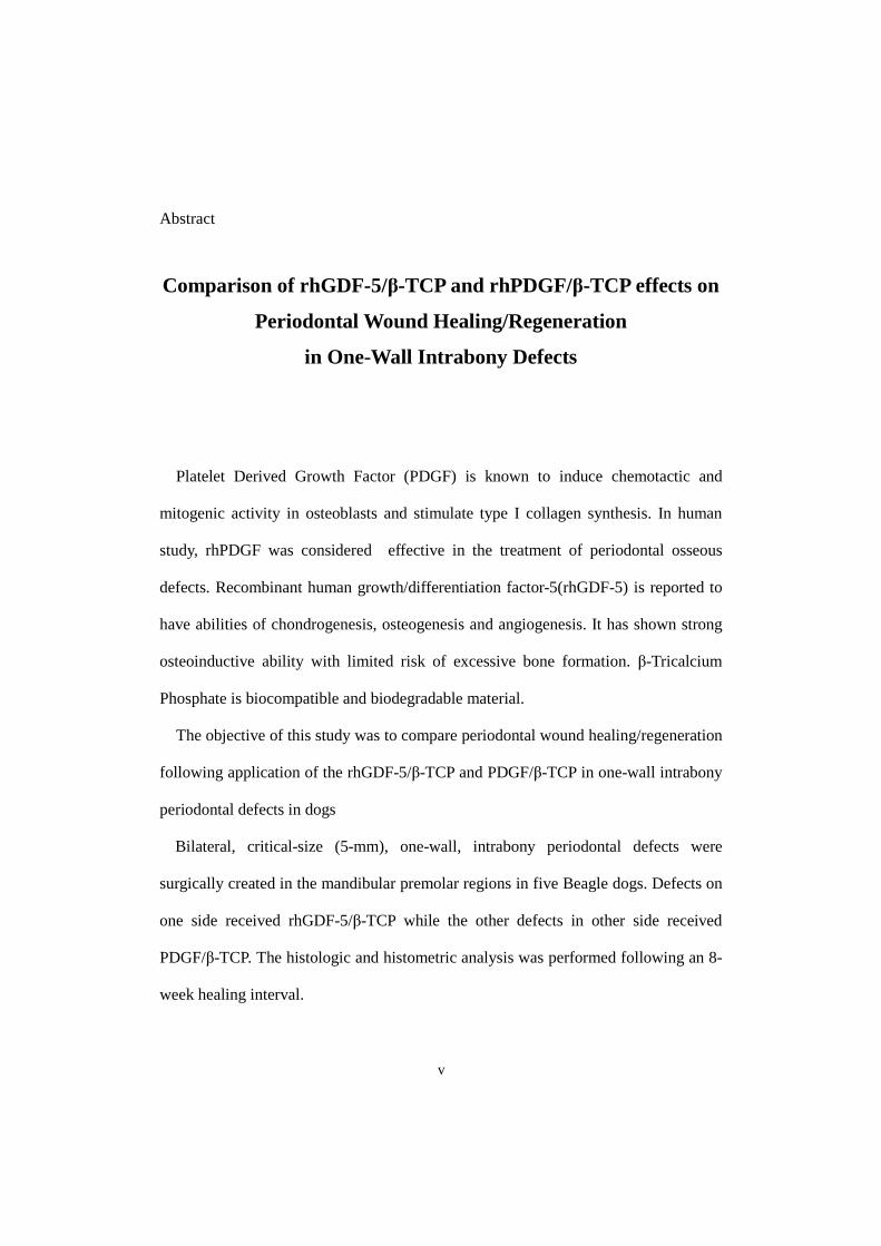
Comparison of rhGDF-5/β-TCP and rhPDGF/β-TCP effects on
Periodontal Wound Healing/Regeneration
in One-Wall Intrabony Defects
Platelet Derived Growth Factor (PDGF) is known to induce chemotactic and
mitogenic activity in osteoblasts and stimulate type I collagen synthesis. In human
study, rhPDGF was considered effective in the treatment of periodontal osseous
defects. Recombinant human growth/differentiation factor-5(rhGDF-5) is reported to
have abilities of chondrogenesis, osteogenesis and angiogenesis. It has shown strong
osteoinductive ability with limited risk of excessive bone formation. β-Tricalcium
Phosphate is biocompatible and biodegradable material.
The objective of this study was to compare periodontal wound healing/regeneration
following application of the rhGDF-5/β-TCP and PDGF/β-TCP in one-wall intrabony
periodontal defects in dogs
Bilateral, critical-size (5-mm), one-wall, intrabony periodontal defects were
surgically created in the mandibular premolar regions in five Beagle dogs. Defects on
one side received rhGDF-5/β-TCP while the other defects in other side received
PDGF/β-TCP. The histologic and histometric analysis was performed following an 8-
week healing interval.

vi
Clinical healing was generally uneventful. Sites implanted with rhGDF-5/β-TCP
exhibited significantly enhanced cementum and bone formation compared to
rhPDGF/β-TCP. Cementum regeneration averaged 4.49±0.48mm (Mean±SD) vs
2.72±0.91mm(p<0.05) for the rhGDF-5/β-TCP, rhPDGF/β-TCP respectively.
Corresponding values for bone regeneration height averaged 3.08±0.74mm vs
1.29±0.78mm (p<0.05). Cementum regeneration included cellular/acellular
cementum with or without a functionally oriented periodontal ligament in rhGDF-5/β-
TCP sites, whereas rhPDGF/β-TCP sites showed acellular cementum predominantly
Also in PDL fiber, mixed (extrinsic, intrinsic) fibers was observed in both groups.
Regenerated cementum generally extended above the alveolar crest in both treatment
groups. In newly formed bone, lamellar bone with primary osteon was seen in
rhGDF-5/β-TCP, whereas PDGF/ β-TCP showed newly formed bone including
woven and lamellar bone. Both sites receiving rhGDF-5/β-TCP or rhPDGF/β-TCP
showed some residual biomaterial apparently undergoing resorption. Sites receiving
rhPDGF/β-TCP commonly showed more residual biomaterial sequestered in
connective tissue than rhGDF-5/β-TCP.
In conclusion, rhGDF-5/β-TCP shows significance in supporting regeneration of the
periodontal healing/regeneration compared to rhPDGF/β-TCP.
Key words: Recombinant human growth/differentiation factor-5, Platelet Derived
Growth Factor, β-Tricalcium Phosphate
1
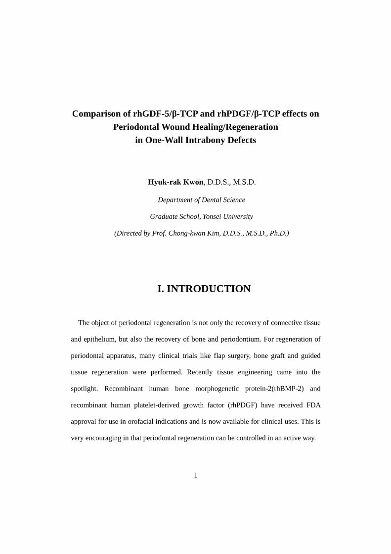
Comparison of rhGDF-5/β-TCP and rhPDGF/β-TCP effects on Periodontal Wound Healing/Regeneration
in One-Wall Intrabony Defects
Hyuk-rak Kwon , D.D.S., M.S.D.
Department of Dental Science
Graduate School, Yonsei University
(Directed by Prof. Chong-kwan Kim, D.D.S., M.S.D., Ph.D.)
I. INTRODUCTION
The object of periodontal regeneration is not only the recovery of connective tissue
and epithelium, but also the recovery of bone and periodontium. For regeneration of
periodontal apparatus, many clinical trials like flap surgery, bone graft and guided
tissue regeneration were performed. Recently tissue engineering came into the
spotlight. Recombinant human bone morphogenetic protein-2(rhBMP-2) and
recombinant human platelet-derived growth factor (rhPDGF) have received FDA
approval for use in orofacial indications and is now available for clinical uses. This is
very encouraging in that periodontal regeneration can be controlled in an active way.

2
For tissue engineering, three components: cells, scaffolds, and signaling
molecules (Babensee et al., 2000; Bartold et al., 2006) are needed. Blood supply
could be the fourth factor. Many scaffolds like collagen sponge which is concerned
as the ‘gold standard’ for BMP carriers have been studied. (McPherson, 1992;
Wikesjo et al., 1999; Uludag et al., 2001) β-Tircalcium Phosphate, which is regarded
as the best carrier for signaling molecules, and poly(lactic-''co''-glycolic acid)(PLGA).
However, there is weak point with this carrier such as collapse of the provided
space. For these reasons, other carriers were developed for the new delivery systems.
β-TCP can be a good substitute for collagen (Urist et al. 1984; Jung et al. 2006). β-
TCP can support the frame and release the signaling molecules slowly.
Recombinant human platelet-derived growth factor (rhPDGF) is known to
induce chemotactic and mitogenic activity in osteoblasts and stimulate type I collagen
synthesis (Rutherford et al., 1992; Park et al., 1995; Giannobile et al., 1996). In
human study, rhPDGF was considered safe and effective in the treatment of
periodontal osseous defects (Oates et al., 1993; Camelo et al., 2003; Nevins et al.,
2003; Nevins et al., 2005).
Nevins et al.(2005) reported that treatment with rhPDGF stimulate a significant
increase in the rate of CAL gain, reduce gingival recession at 3 months post-surgery,
and improve bone fill when compared to β-TCP bone substitute at 6 months(Nevins et
al., 2005).
Recently, FDA (Food and Drug Administration, USA) approved the clinical use

3
of rhPDGF for chronic skin wounds in diabetic patients (Regranex, Ethicon, USA)
and for periodontally related osseous defects (GEM21s, BioMimetic Therapeutics,
USA).
Recombinant human growth/differentiation factor-5(GDF-5) is also called
Cartilage-derived morphogenetic protein-1(CDMP-1) or bone morphogenetic protein-
14(BMP-14). This growth factor is known to have a chondrogenic effect. It is
reported that rhGDF-5 is a suitable factor for enhancing healing in bone defects and
ectopic bone formation (Spiro et al., 2000; Poehling et al., 2006; Yoshimoto et al.,
2006).
The objective of this study was to compare periodontal wound healing/
regeneration following rhGDF-5/β-TCP and PDGF/β-TCP application in one-wall
intrabony periodontal defects in beagle dogs.
4
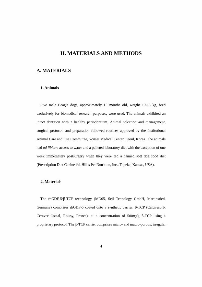
II. MATERIALS AND METHODS
A. MATERIALS
1. Animals
Five male Beagle dogs, approximately 15 months old, weight 10-15 kg, bred
exclusively for biomedical research purposes, were used. The animals exhibited an
intact dentition with a healthy periodontium. Animal selection and management,
surgical protocol, and preparation followed routines approved by the Institutional
Animal Care and Use Committee, Yonsei Medical Center, Seoul, Korea. The animals
had ad libitum access to water and a pelleted laboratory diet with the exception of one
week immediately postsurgery when they were fed a canned soft dog food diet
(Prescription Diet Canine i/d, Hill’s Pet Nutrition, Inc., Topeka, Kansas, USA).
2. Materials
The rhGDF-5/β-TCP technology (MD05, Scil Tchnology GmbH, Martinsried,
Germany) comprises rhGDF-5 coated onto a synthetic carrier, β-TCP (Calciresorb,
Ceraver Osteal, Roissy, France), at a concentration of 500㎍/g β-TCP using a
proprietary protocol. The β-TCP carrier comprises micro- and macro-porous, irregular
5
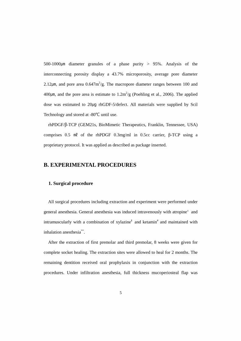
500-1000㎛ diameter granules of a phase purity > 95%. Analysis of the
interconnecting porosity display a 43.7% microporosity, average pore diameter
2.12㎛, and pore area 0.647m2/g. The macropore diameter ranges between 100 and
400㎛, and the pore area is estimate to 1.2m2/g (Poehling et al., 2006). The applied
dose was estimated to 20㎍ rhGDF-5/defect. All materials were supplied by Scil
Technology and stored at -80℃ until use.
rhPDGF/β-TCP (GEM21s, BioMimetic Therapeutics, Franklin, Tennessee, USA)
comprises 0.5㎖ of the rhPDGF 0.3mg/ml in 0.5cc carrier, β-TCP using a
proprietary protocol. It was applied as described as package inserted.
B. EXPERIMENTAL PROCEDURES
1. Surgical procedure
All surgical procedures including extraction and experiment were performed under
general anesthesia. General anesthesia was induced intravenously with atropine∥ and
intramuscularly with a combination of xylazine¶ and ketamin# and maintained with
inhalation anesthesia** .
After the extraction of first premolar and third premolar, 8 weeks were given for
complete socket healing. The extraction sites were allowed to heal for 2 months. The
remaining dentition received oral prophylaxis in conjunction with the extraction
procedures. Under infiltration anesthesia, full thickness mucoperiosteal flap was
6
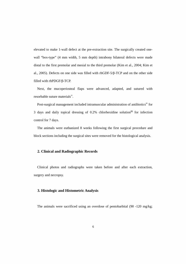
elevated to make 1-wall defect at the pre-extraction site. The surgically created one-
wall “box-type” (4 mm width, 5 mm depth) intrabony bilateral defects were made
distal to the first premolar and mesial to the third premolar (Kim et al., 2004; Kim et
al., 2005). Defects on one side was filled with rhGDF-5/β-TCP and on the other side
filled with rhPDGF/β-TCP.
Next, the mucoperiosteal flaps were advanced, adapted, and sutured with
resorbable suture materials††.
Post-surgical management included intramuscular administration of antibiotics‡‡ for
3 days and daily topical dressing of 0.2% chlorhexidine solution§§ for infection
control for 7 days.
The animals were euthanized 8 weeks following the first surgical procedure and
block sections including the surgical sites were removed for the histological analysis.
2. Clinical and Radiographic Records
Clinical photos and radiographs were taken before and after each extraction,
surgery and necropsy.
3. Histologic and Histometric Analysis
The animals were sacrificed using an overdose of pentobarbital (90 -120 mg/kg;
7
IV). Block sections including defect sites and tooth, surrounding alveolar bone and
soft tissues were collected. The block specimens were fixed in 10% buffered formalin
for 10 days, decalcified in 5% nitric acid for 7 days, trimmed, dehydrated and
embedded in paraffin. Serial sections, 4µm thick, were cut in a mesial-distal direction
at 80µm intervals. The sections were stained using hematoxylin and eosin.
The four most central sections of each defect site were observed using light
microscopy∥∥. Histometric analysis was performed using image analysis software¶¶.
The following parameters were recorded.
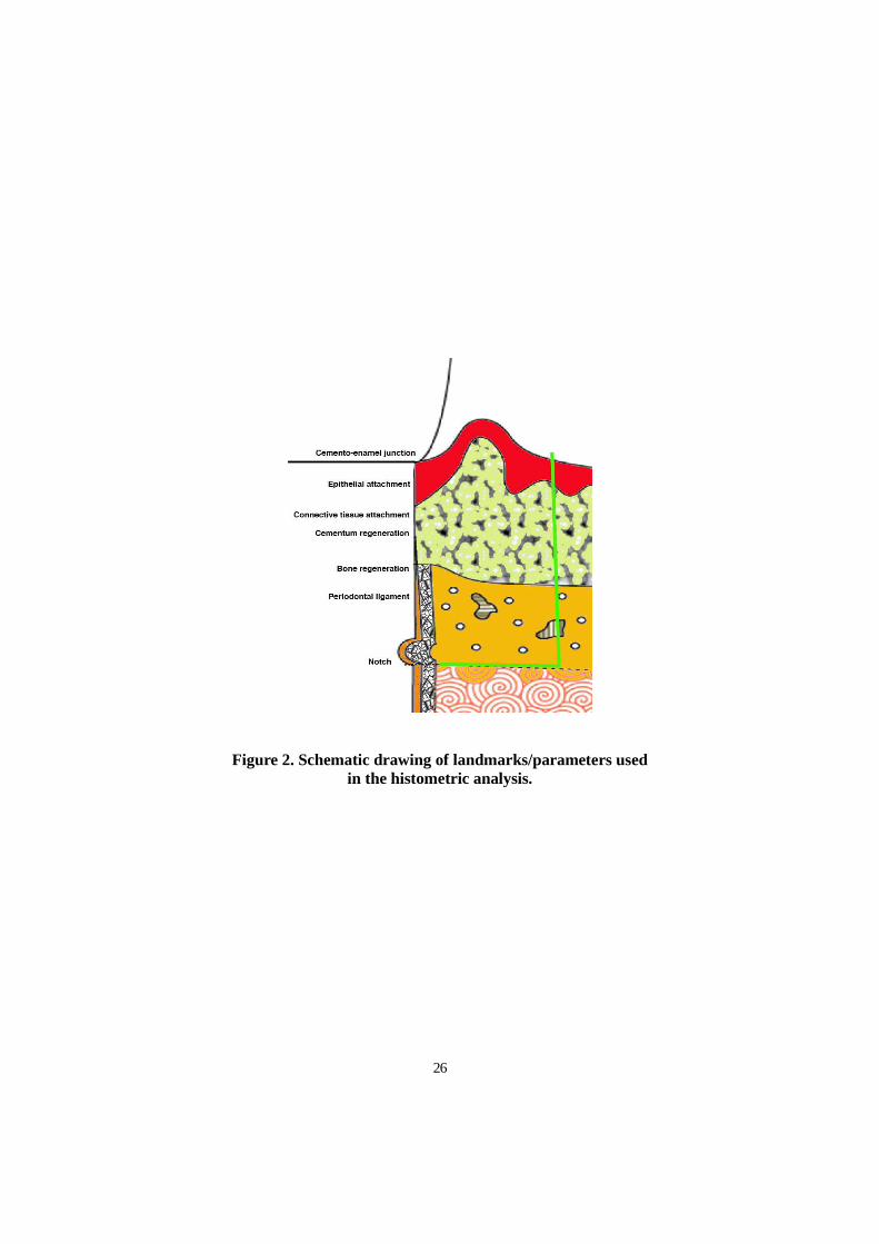
◦ Defect height: distance from the apical extension of the root surface notch to the
cemento-enamel junction (CEJ)
◦ Epithelial attachment: distance from the CEJ to the apical extension of an
epithelial attachment on the root surface. This parameter included any gingival
recession.
◦ Cementum regeneration: distance from the apical extension of the root surface
notch to the coronal extension of newly formed cementum or a cementum-like
substance on the root surface.
◦ Bone regeneration (height): distance from the apical extension of the root surface
notch to the coronal extension of newly formed bone along the root surface;
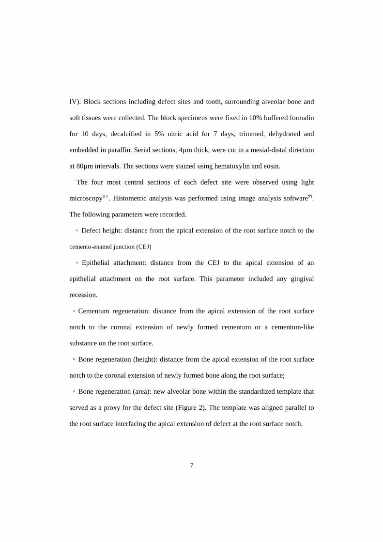
◦ Bone regeneration (area): new alveolar bone within the standardized template that
served as a proxy for the defect site (Figure 2). The template was aligned parallel to
the root surface interfacing the apical extension of defect at the root surface notch.
8
◦ Unresorbed biomaterial: volume of the remained biomaterial in newly formed
bone and connective tissue.
◦ Root resorption
◦ Ankylosis
4. Statistical Analysis
The experimental groups of rhGDF-5/β-TCP and rhPDGF/β-TCP were compared
using student t-test (p < 0.05) with Statistics software##.
0.04 mg/kg; Kwangmyung Pharmaceutical Ind. Co. Ltd., Seoul, Korea∥
¶ Rompun, Bayer Korea Co., Seoul, Korea
# Ketara, Yuhan Co., Seoul, Korea
** Gerolan, Choongwae Pharmaceutical Co., Seoul, Korea
†† Vicryl 5.0 Polyglactin 910, Ethicon, Johnson & Johnson, New Jersey, USA
‡‡ Cefazoline Sodium 20mg/kg; Yuhan Corporation, Seoul, Korea
§§ Hexamedin®, Bukwang Pharmaceutical Co., Seoul, Korea
Olympus Multi∥∥ -view microscope BH2, Tokyo, Japan
¶¶ Image-Pro Plus, Media Cybernetic, Silver Springs, MD, USA
## Microsoft Office Excel 2007, Microsoft Co., Redmond, Washington, USA
9
III. RESULTS
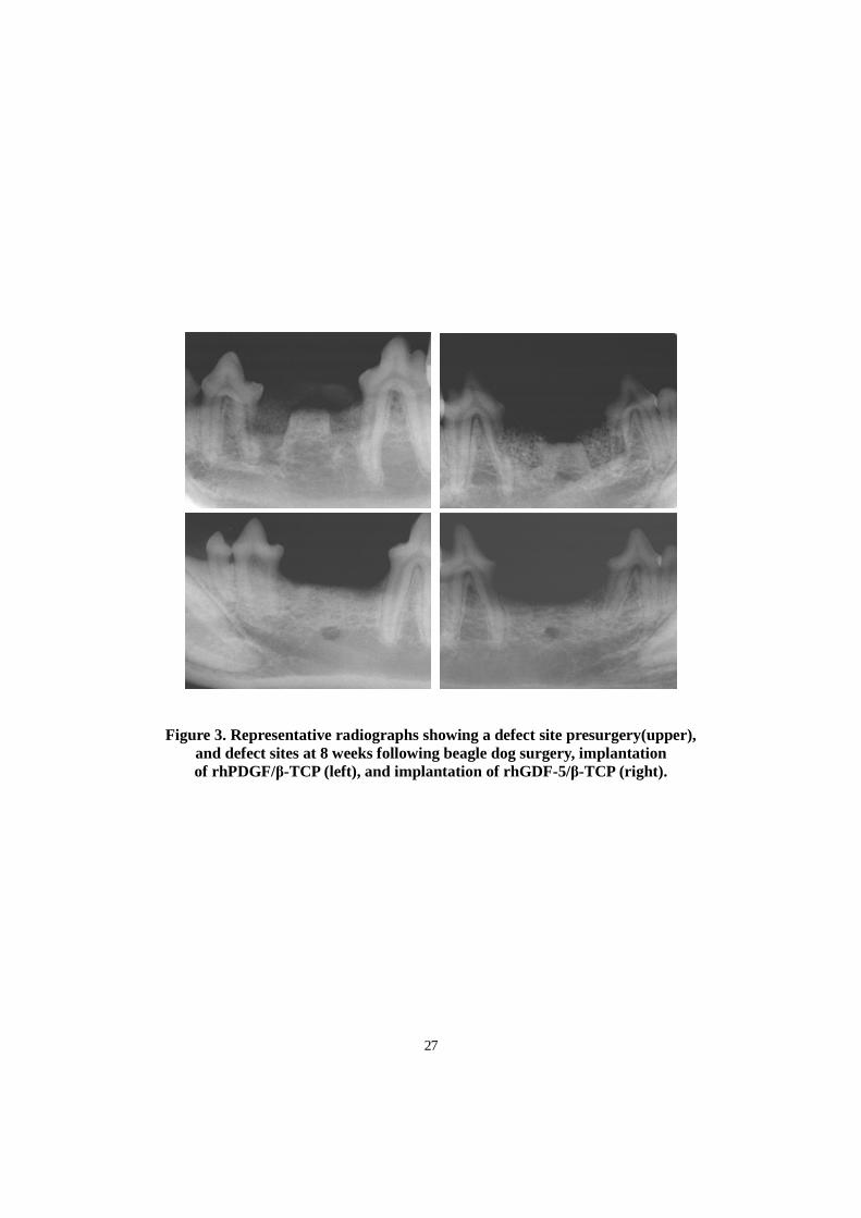
1. Clinical and Radiographic Observations
All sites including rhGDF-5/β-TCP and rhPDGF/β-TCP groups healed
uneventfully. Figure 1 and Figure 3 showed the clinical and radiographic observation
at surgery and after 8 weeks healing. Both groups showed bone formation at defect
sites.
2. Histomorphometric Analysis
Histomorphometric analysis is shown on Table 1.
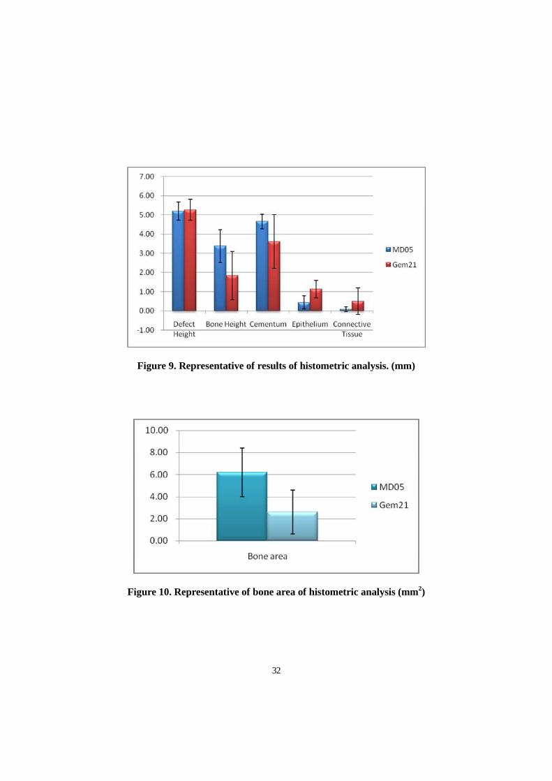
Defect height of rhGDF-5/β-TCP group was 5.16±0.43 mm and the defect height
of rhPDGF/β-TCP group was 5.15±0.17 mm. These values had no significant
difference (p>0.05). But, bone regeneration height and area showed significant
differences between the 2 groups. Bone regeneration height was 3.08±0.74 mm and
1.29±0.78 mm in rhGDF-5/β-TCP and rhPDGF/β-TCP group (p<0.05). Cementum
regeneration height was 4.49±0.48 mm and 2.72±0.91 mm in rhGDF-5/β-TCP and
rhPDGF/β-TCP group. Values of epithelial attachments averaged 0.52±0.40 mm and
1.17±0.52 mm in rhGDF-5/β-TCP and rhPDGF/β-TCP group Values of connective
tissue attachments averaged 0.14±0.25 mm and 1.25±0.84 mm in rhGDF-5/β-TCP
10
and rhPDGF/β-TCP group. rhPDGF/β-TCP group showed significantly higher
epithelial attachment (p<0.05)(Figure 9).
Mean values of bone regeneration area were 6.51±0.93 mm2 and 1.44±1.04 mm2
for rhGDF-5/β-TCP and rhPDGF/β-TCP respectively with significant differences
between the 2 groups (p<0.05)(Figure 10).
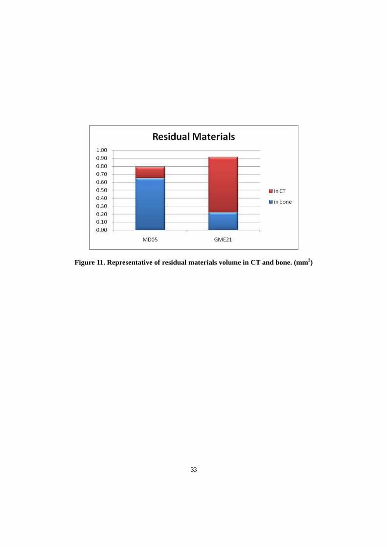
The total volume of residual materials was 0.79±0.58 mm2 , 0.91± 0.42 mm2 for
rhGDF-5/β-TCP and rhPDGF/β-TCP respectively. The volume of residual materials
were 0.65±0.55 mm2 and 0.22±0.17 mm2 for rhGDF-5/β-TCP and rhPDGF/β-TCP
respectively in the newly formed bone. On the other hand, in the connective tissue.
the values were 0.14±0.03 mm2 and 0.69±0.42 mm2 rhGDF-5/β-TCP and rhPDGF/β-
TCP respectively. There was a significant difference in the volume of the residual
material inside the connective tissue. But, there was no significant difference between
2 groups in total residual material volumes (Table 2, Figure 11).
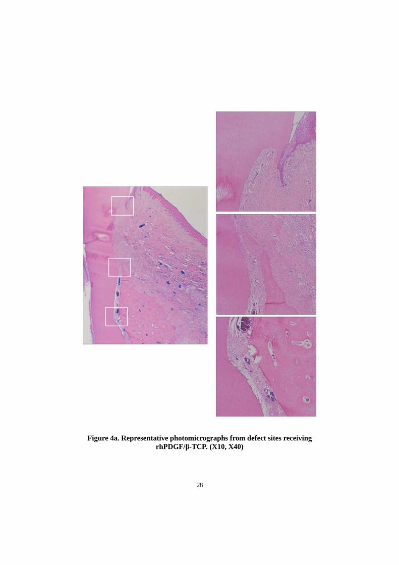
3. Histologic Analysis
Histologic observations are shown on Figure 4, 5, 6, 7, 8.
Both groups showed significant bone and cementum formation. In newly formed
bone, predominant lamellar bone with primary osteon was observed in rhGDF-5/β-
TCP. In contrast, lamellar and woven bone was observed in rhPDGF/β-TCP. Both
groups showed enhanced cementum regeneration. However, not only the length of
11
cementum, but also the density and thickness were enhanced in rhGDF-5/β-TCP
group(Figure 5). The rhPDGF/β-TCP groups were mostly acellular, but the rhGDF-
5/β-TCP groups were mixed (cellular/acellular,) cementum. Periodontal ligament
fibers were seen perpendicularly and parallelly in both groups (Figure 5, 6).
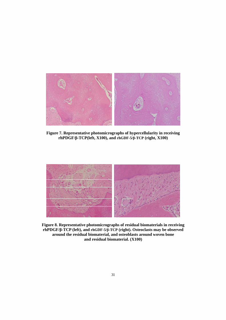
Most of the β-TCP particles in rhGDF-5/β-TCP were seen in the bone area, which
was lined with osteoid representing biodegradation. Besides, Most of β-TCP particles
in rhPDGF/β-TCP were sequestered in the connective tissue area (Figure 8).
12
IV. DISCUSSION
Dennison et al(1994). reported that PDGF might be valuable in promoting new
connective tissue attachment in the periodontal wound. Mumford et al(2001).
reported the effect of PDGF-BB on the proliferation of PDL and concluded that there
may be cell-specific differences critical to periodontal wound healing. These studies
supported that PDGF-BB promoted periodontal wound healing. And when PDGF-BB
is delivered to promote periodontal tissue engineering of tooth-supporting osseous
defects, there is a direct effect on bone turn-over (Sarment et al. 2006).
Recombinant human platelet-derived growth factor-BB (rhPDGF-BB) is potent
mitogenic and chemotactic protein for PDL fibroblasts and alveolar bone cells and
improves angiogenesis, while bone allograft offers a biological matrix conductive to
cell growth and may contribute osteoinductive bone matrix proteins (Nevins et al.
2003). And Nevins et al. demonstrated that the use of rhPDGF-BB was safe and
effective in the treatment of periodontal osseous defects in human clinical study
(Nevins et al. 2005).
Recently, rhPDGF-BB (GEM21s, Biomimetic Therapeutics) and rhBMP-2 (Infuse,
Medtronic sofamor Danek) received FDA approval for a clinical use. These are the
first proven safe materials for clinical use in tissue engineering.
Recombinant human growth/differentiation factor-5 (rhGDF-5) is a member of the
transforming growth factor-β superfamily and plays a pivotal role in the process of
13
joint formation (Chang et al. 1994; Storm et al. 1994). In addition to joint
development, rhGDF-5 has shown activity in promoting angiogenesis and
tendon/ligament morphogenesis (Wolfman et al. 1997; Yamashita et al. 1997). As well
as these basic properties, rhGDF-5 was reported suitable factor for enhancing healing
in bone defect and ectopic bone formation with chondrogenesis, osteogenesis
properties (Francis-West et al. 1999; Spiro et al. 2000; Yoshimoto et al. 2006).
rhPDGF-BB, which was first reported as having wound healing properties, and
rhGDF-5, which was first reported as having chondrogenesis properties, were both
applied to periodontal defects and studied for periodontal regeneration. In this study,
the 1-wall intrabony defects may provide the proper preclinical model for the
evaluation of the periodontal regeneration(Kim et al. 2004).
For evaluation of periodontal regeneration, cementum and bone regeneration height,
and bone regeneration area were measured. In both groups, bone was enhanced,
although bone regeneration area and height were significantly higher in the rhGDF-
5/β-TCP group than in rhPDGF/β-TCP group (Table 1, Figure5, 6). rhPDGF/β-TCP
group has bone regeneration in 1-wall intrabony defects approximately 30 percent of
the defect height, rhGDF-5/β-TCP group(3.08±0.74 mm) has better effect on bone
regeneration in 1-wall intrabony defects compared to rhPDGF/β-TCP group.(1.29±
0.78mm)(Table 1)
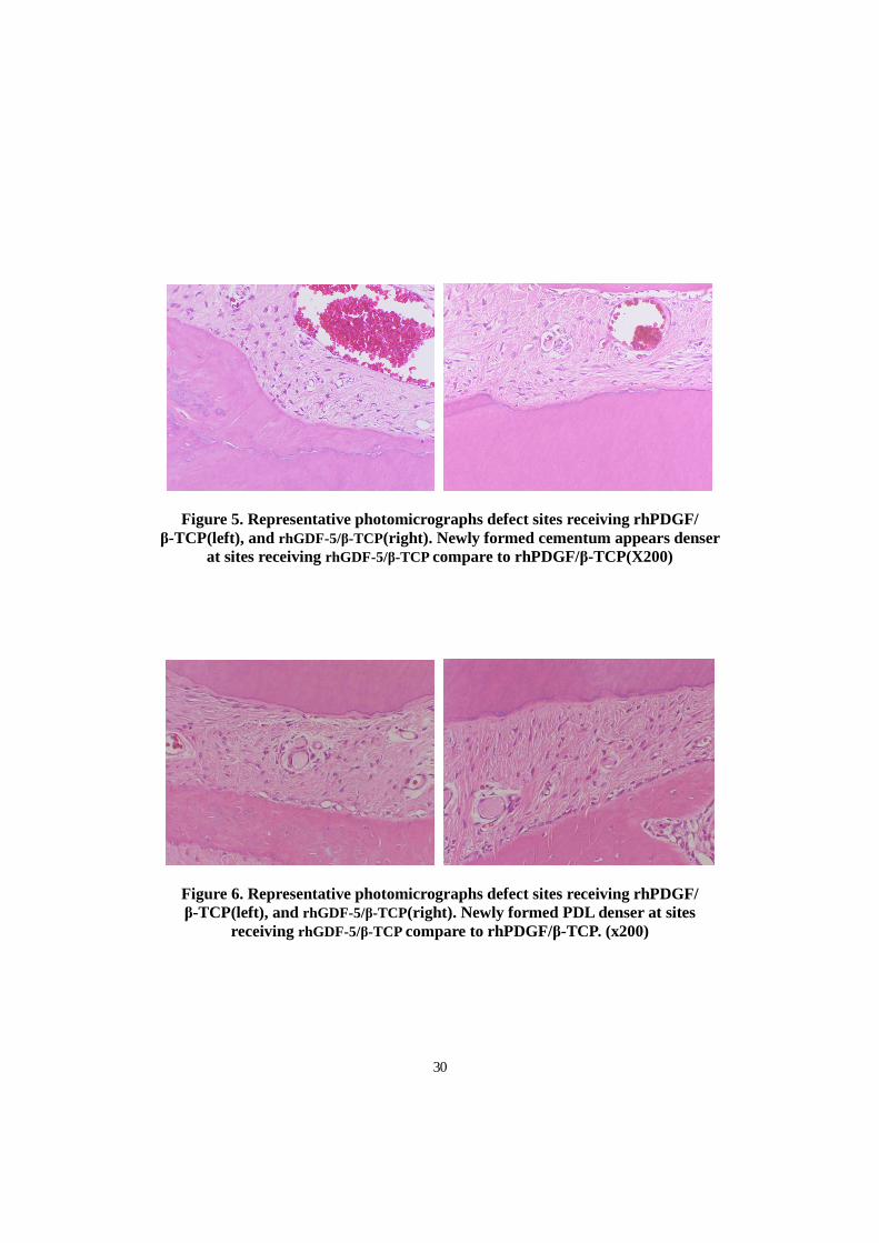
Both groups showed enhanced cementum regeneration, 4.49±0.48mm and
2.72±0.91mm, rhGDF-5/β-TCP and rhPDGF/β-TCP respectively. However, there
14
was stastically significant enhancement in rhGDF-5/β-TCP than rhPDGF/β-TCP
(Table 1, Figure 5). Moreover, not only the length of cementum, but also the density
and thickness were more enhanced in rhGDF-5/β-TCP group in histologic analysis
(Figure 5). rhPDGF/β-TCP groups were mostly acellular but the rhGDF-5/β-TCP
groups were mixed (cellular/acellular,) cementum. Periodontal regeneration includes
de novo cementogenesis, osteogenesis and the formation of periodontal ligament
fibers. Cementum seems to have the potential for the regeneration of periodontal
apparatus.
The properties of cementogenesis should be regarded as an important factor for the
periodontal regeneration. In this aspect, rhGDF-5 seems to be optimal for periodontal
regeneration.
Both groups showed little resorption of tooth. Unlike BMPs, both growth factors
could be applied to periodontal defects without ankylosis or resorption of tooth
(Wikesjo et al. 1999; Wikesjo et al. 1999).
Periodontal ligament, which is the major reference point of periodontal
regeneration, were found regulary arranged in both groups. Periodontal ligament
fibers were seen perpendicularly and parallelly in both groups (Figure 5, 6). Most of
the β-TCP particles in rhGDF-5/β-TCP were seen in the bone area, which was lined
with osteoid representing biodegradation. Besides, Most of β-TCP particles in
rhPDGF/β-TCP were sequestered in the connective tissue area (Figure 8). That means
that rhGDF-5/β-TCP has more potential for the new bone formations.
15
A remarkable finding about these growth factors is that both groups showed
hypercellularity in the newly formed bone (Figure 7). Regardless of the quantity of
bone formation, both groups showed active cellularity. It could be concluded that,
both growth factors accelerate biodegradation of a biomaterial used as a carrier in the
periodontal defects, being displaced with bone in an active way (Koo et al. 2007).
Biodegradation and formation of bone are the results of activities of growth factors.
The rhGDF-5 seems to have more potent capacity of osteoconduction and
osteoinduction than rhPDGF.
Though rhGDF-5/β-TCP showed enhanced effect on bone and cementum
formation compared to rhPDGF/β-TCP group, both groups showed characteristics of
hypercellularity and cementum formation. In the point of cementogenesis, long-term
studies about these biomaterials are needed.
16
V. CONCLUSION
The results show that rhGDF-5/β-TCP has a higher efficacy in bone and cementum
regeneration compared to rhPDGF/β-TCP in one-wall intrabony defects of dogs. The
β-TCP carrier exhibited timely resorption and no appreciable adverse reactions
affecting bone formation making it a candidate matrix for rhGDF-5 and rhPDGF.
In conclusion, rhGDF-5/β-TCP supports bone and cementum formation in
advanced periodontal defects. Application of rhGDF-5/β-TCP appears safe as it is
associated with limited adverse effects, if any.
17
IV. REFERENCES
Babensee, J. E., McIntire, L. V. and Mikos, A. G. Growth factor delivery for tissue
engineering. Pharm Res 17(5): 497-504, 2000.
Bartold, P. M., Xiao, Y., Lyngstaadas, S. P., Paine, M. L. and Snead, M. L.
Principles and applications of cell delivery systems for periodontal regeneration.
Periodontol 2000 41: 123-135, 2006.
Camelo, M., Nevins, M. L., Schenk, R. K., Lynch, S. E. and Nevins, M. Periodontal
regeneration in human Class II furcations using purified recombinant human platelet-
derived growth factor-BB (rhPDGF-BB) with bone allograft. Int J Periodontics
Restorative Dent 23(3): 213-225, 2003.
Chang, S. C., Hoang, B., Thomas, J. T., Vukicevic, S., Luyten, F. P., Ryba, N. J.,
Kozak, C. A., Reddi, A. H. and Moos, M., Jr. Cartilage-derived morphogenetic
proteins. New members of the transforming growth factor-beta superfamily
predominantly expressed in long bones during human embryonic development. J Biol
Chem 269(45): 28227-28234, 1994.
Dennison, D. K., Vallone, D. R., Pinero, G. J., Rittman, B. and Caffesse, R. G.
Differential effect of TGF-beta 1 and PDGF on proliferation of periodontal ligament
cells and gingival fibroblasts. J Periodontol 65(7): 641-648, 1994.
18
Francis-West, P. H., Abdelfattah, A., Chen, P., Allen, C., Parish, J., Ladher, R., Allen,
S., MacPherson, S., Luyten, F. P. and Archer, C. W. Mechanisms of GDF-5 action
during skeletal development. Development 126(6): 1305-1315, 1999.
Giannobile, W. V., Hernandez, R. A., Finkelman, R. D., Ryan, S., Kiritsy, C. P.,
D'Andrea, M. and Lynch, S. E. Comparative effects of platelet-derived growth
factor-BB and insulin-like growth factor-I, individually and in combination, on
periodontal regeneration in Macaca fascicularis. J Periodontal Res 31(5): 301-312,
1996.
Jung, U. W., Choi, S. Y., Pang, E. K., Kim, C. S., Choi, S. H. and Cho, K. S. The
effect of varying the particle size of beta tricalcium phosphate carrier of recombinant
human bone morphogenetic protein-4 on bone formation in rat calvarial defects. J
Periodontol 77(5): 765-772, 2006.
Kim, C. S., Choi, S. H., Chai, J. K., Cho, K. S., Moon, I. S., Wikesjo, U. M. and Kim,
C. K. Periodontal repair in surgically created intrabony defects in dogs: influence of
the number of bone walls on healing response. J Periodontol 75(2): 229-235, 2004.
Kim, C. S., Choi, S. H., Cho, K. S., Chai, J. K., Wikesjo, U. M. and Kim, C. K.
Periodontal healing in one-wall intra-bony defects in dogs following implantation of
autogenous bone or a coral-derived biomaterial. J Clin Periodontol 32(6): 583-589,
2005.
19
Koo, K. T., Susin, C., Wikesjo, U. M., Choi, S. H. and Kim, C. K. Transforming
growth factor-beta1 accelerates resorption of a calcium carbonate biomaterial in
periodontal defects. J Periodontol 78(4): 723-729, 2007.
McPherson, J. M. The utility of collagen-based vehicles in delivery of growth
factors for hard and soft tissue wound repair. Clin Mater 9(3-4): 225-234, 1992.
Mumford, J. H., Carnes, D. L., Cochran, D. L. and Oates, T. W. The effects of
platelet-derived growth factor-BB on periodontal cells in an in vitro wound model. J
Periodontol 72(3): 331-340, 2001.
Nevins, M., Camelo, M., Nevins, M. L., Schenk, R. K. and Lynch, S. E. Periodontal
regeneration in humans using recombinant human platelet-derived growth factor-BB
(rhPDGF-BB) and allogenic bone. J Periodontol 74(9): 1282-1292, 2003.
Nevins, M., Giannobile, W. V., McGuire, M. K., Kao, R. T., Mellonig, J. T., Hinrichs,
J. E., McAllister, B. S., Murphy, K. S., McClain, P. K., Nevins, M. L., Paquette, D. W.,
Han, T. J., Reddy, M. S., Lavin, P. T., Genco, R. J. and Lynch, S. E. Platelet-derived
growth factor stimulates bone fill and rate of attachment level gain: results of a large
multicenter randomized controlled trial. J Periodontol 76(12): 2205-2215, 2005.
Oates, T. W., Rouse, C. A. and Cochran, D. L. Mitogenic effects of growth factors
on human periodontal ligament cells in vitro. J Periodontol 64(2): 142-148, 1993.
20
Park, J. B., Matsuura, M., Han, K. Y., Norderyd, O., Lin, W. L., Genco, R. J. and Cho,
M. I. Periodontal regeneration in class III furcation defects of beagle dogs using
guided tissue regenerative therapy with platelet-derived growth factor. J Periodontol
66(6): 462-477, 1995.
Poehling, S., Pippig, S. D., Hellerbrand, K., Siedler, M., Schutz, A. and Dony, C.
Superior effect of MD05, beta-tricalcium phosphate coated with recombinant human
growth/differentiation factor-5, compared to conventional bone substitutes in the rat
calvarial defect model. J Periodontol 77(9): 1582-1590, 2006.
Rutherford, R. B., Niekrash, C. E., Kennedy, J. E. and Charette, M. F. Platelet-
derived and insulin-like growth factors stimulate regeneration of periodontal
attachment in monkeys. J Periodontal Res 27(4 Pt 1): 285-290, 1992.
Sarment, D. P., Cooke, J. W., Miller, S. E., Jin, Q., McGuire, M. K., Kao, R. T.,
McClain, P. K., McAllister, B. S., Lynch, S. E. and Giannobile, W. V. Effect of
rhPDGF-BB on bone turnover during periodontal repair. J Clin Periodontol 33(2):
135-140, 2006.
Spiro, R. C., Liu, L., Heidaran, M. A., Thompson, A. Y., Ng, C. K., Pohl, J. and Poser,
J. W. Inductive activity of recombinant human growth and differentiation factor-5.
Biochem Soc Trans 28(4): 362-368, 2000.
21
Storm, E. E., Huynh, T. V., Copeland, N. G., Jenkins, N. A., Kingsley, D. M. and Lee,
S. J. Limb alterations in brachypodism mice due to mutations in a new member of
the TGF beta-superfamily. Nature 368(6472): 639-643, 1994.
Uludag, H., Gao, T., Porter, T. J., Friess, W. and Wozney, J. M. Delivery systems for
BMPs: factors contributing to protein retention at an application site. J Bone Joint
Surg Am 83-A Suppl 1(Pt 2): S128-135, 2001.
Urist, M. R., Lietze, A. and Dawson, E. Beta-tricalcium phosphate delivery system
for bone morphogenetic protein. Clin Orthop Relat Res (187): 277-280, 1984.
Wikesjo, U. M., Guglielmoni, P., Promsudthi, A., Cho, K. S., Trombelli, L., Selvig, K.
A., Jin, L. and Wozney, J. M. Periodontal repair in dogs: effect of rhBMP-2
concentration on regeneration of alveolar bone and periodontal attachment. J Clin
Periodontol 26(6): 392-400, 1999.
Wikesjo, U. M. and Selvig, K. A. Periodontal wound healing and regeneration.
Periodontol 2000 19: 21-39, 1999.
Wolfman, N. M., Hattersley, G., Cox, K., Celeste, A. J., Nelson, R., Yamaji, N., Dube,
J. L., DiBlasio-Smith, E., Nove, J., Song, J. J., Wozney, J. M. and Rosen, V. Ectopic
induction of tendon and ligament in rats by growth and differentiation factors 5, 6,
and 7, members of the TGF-beta gene family. J Clin Invest 100(2): 321-330, 1997.
22
Yamashita, H., Shimizu, A., Kato, M., Nishitoh, H., Ichijo, H., Hanyu, A., Morita, I.,
Kimura, M., Makishima, F. and Miyazono, K. Growth/differentiation factor-5 induces
angiogenesis in vivo. Exp Cell Res 235(1): 218-226, 1997.
Yoshimoto, T., Yamamoto, M., Kadomatsu, H., Sakoda, K., Yonamine, Y. and Izumi,
Y. Recombinant human growth/differentiation factor-5 (rhGDF-5) induced bone
formation in murine calvariae. J Periodontal Res 41(2): 140-147, 2006.
Zeichner-David, M. Regeneration of periodontal tissues: cementogenesis revisited.
Periodontol 2000 41: 196-217, 2006.
23
LEGENDS
Figure 1. Representative surgical procedure. Surgically created, critical-size, one-
wall, intrabony periodontal defect at the distal aspect of the mandibular 2nd and mesial
aspect of the mandibular 4th premolar teeth (left). Application of rhPDGF/β-TCP and
rhGDF-5/ß-TCP (left center). Mucoperiosteal flaps adapted and sutured for primary
intention healing (right center). Healing at week 8 (right). upper: rhPDGF/β-TCP,
lower: rhGDF-5/ß-TCP
Figure 2. Schematic drawing of landmarks/parameters used in the histometric
analysis.
Figure 3. Representative radiographs showing a defect site presurgery(upper), and
defect sites at 8 weeks following beagle dog surgery, implantation of rhPDGF/β-TCP
(left), and implantation of rhGDF-5/β-TCP (right).
Figure 4a. Representative photomicrographs from defect sites receiving rhPDGF/β-
TCP. (X10, X40)
Figure 4b. Representative photomicrographs from defect sites receiving rhGDF-5/β-
TCP. (X10, X40)
24
Figure 5. Representative photomicrographs defect sites receiving rhPDGF/β-
TCP(left), and rhGDF-5/β-TCP(right). Newly formed cementum appears denser at sites
receiving rhGDF-5/β-TCP compare to rhPDGF/β-TCP(X200)
Figure 6. Representative photomicrographs defect sites receiving rhPDGF/β-
TCP(left), and rhGDF-5/β-TCP(right). Newly formed PDL denser at sites receiving
rhGDF-5/β-TCP compare to rhPDGF/β-TCP. (x200)
Figure 7. Representative photomicrographs of hypercellularity in receiving
rhPDGF/β-TCP(left, X100), and rhGDF-5/β-TCP (right, X100)
Figure 8. Representative photomicrographs of residual biomaterials in receiving
rhPDGF/β-TCP (left), and rhGDF-5/β-TCP (right). Osteoclasts may be observed
around the residual biomaterial, and osteoblasts around woven bone and residual
biomaterial. (X100)
Figure 9. Representative of results of histometric analysis. (mm)
Figure 10. Representative of bone area of histometric analysis (mm2)
Figure 11. Representative of residual materials volume in CT and bone. (mm2)

25
FIGURES
Figure 1. Surgically created, critical-size, one-wall, intrabony periodontal defect at the distal aspect of the mandibular 2nd and mesial aspect of the mandibular
4th premolar teeth (left). Application of rhPDGF/β-TCP and rhGDF-5/ß-TCP (left center). Mucoperiosteal flaps adapted
and sutured for primary intention healing (right center). Healing at week 8 (right). upper: rhPDGF/β-TCP, lower: rhGDF-5/ß-TCP
26
Figure 2. Schematic drawing of landmarks/parameters used in the histometric analysis.
27
Figure 3. Representative radiographs showing a defect site presurgery(upper), and defect sites at 8 weeks following beagle dog surgery, implantation of rhPDGF/β-TCP (left), and implantation of rhGDF-5/β-TCP (right).
28
Figure 4a. Representative photomicrographs from defect sites receiving rhPDGF/β-TCP. (X10, X40)

29
Figure 4b. Representative photomicrographs from defect sites receiving rhGDF-5/β-TCP. (X10, X40)
30
Figure 5. Representative photomicrographs defect sites receiving rhPDGF/ β-TCP(left), and rhGDF-5/β-TCP(right). Newly formed cementum appears denser
at sites receiving rhGDF-5/β-TCP compare to rhPDGF/β-TCP(X200)
Figure 6. Representative photomicrographs defect sites receiving rhPDGF/ β-TCP(left), and rhGDF-5/β-TCP(right). Newly formed PDL denser at sites
receiving rhGDF-5/β-TCP compare to rhPDGF/β-TCP. (x200)
31
Figure 7. Representative photomicrographs of hypercellularity in receiving rhPDGF/β-TCP(left, X100), and rhGDF-5/β-TCP (right, X100)
Figure 8. Representative photomicrographs of residual biomaterials in receiving rhPDGF/β-TCP (left), and rhGDF-5/β-TCP (right). Osteoclasts may be observed
around the residual biomaterial, and osteoblasts around woven bone and residual biomaterial. (X100)
32
Figure 9. Representative of results of histometric analysis. (mm)
Figure 10. Representative of bone area of histometric analysis (mm2)
33
Figure 11. Representative of residual materials volume in CT and bone. (mm2)
34
TABLES
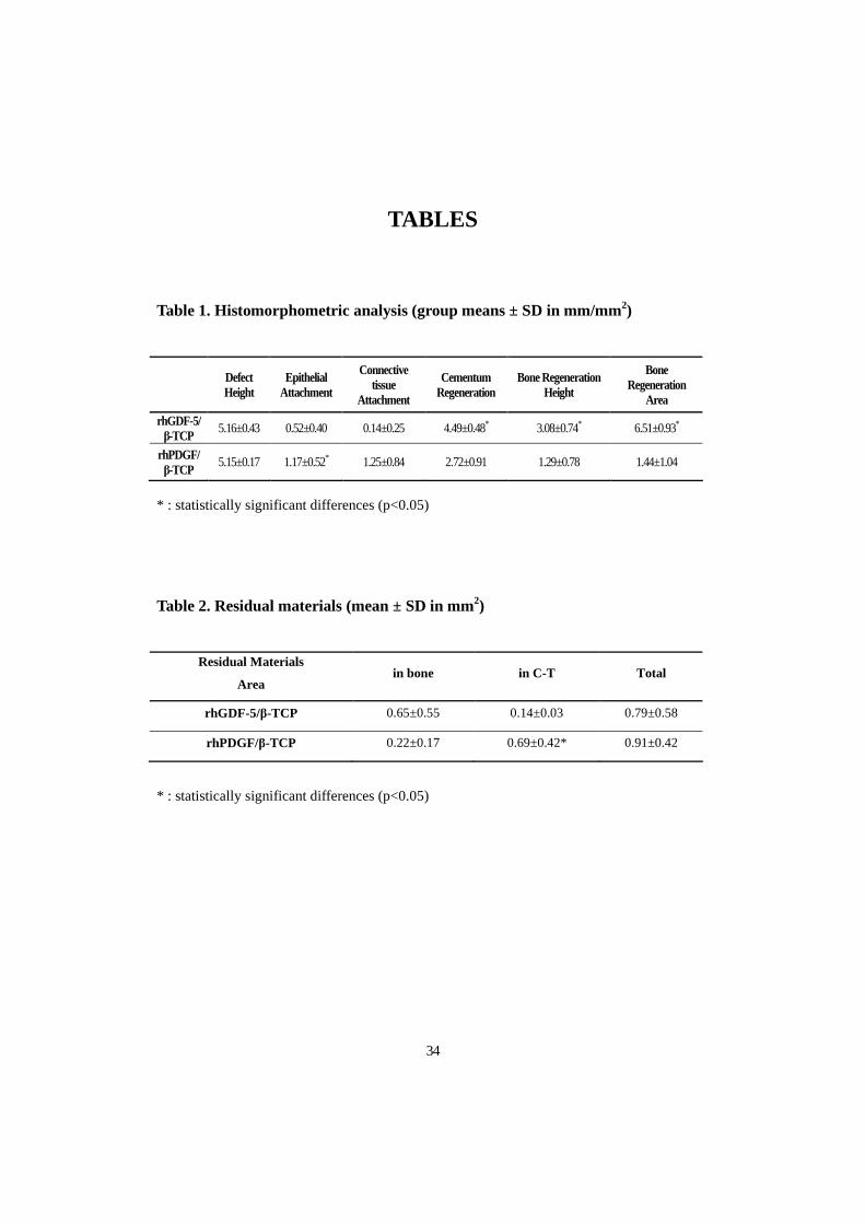
Table 1. Histomorphometric analysis (group means ± SD in mm/mm2)
Defect Height
Epithelial Attachment
Connective tissue
Attachment
Cementum Regeneration
Bone Regeneration Height
Bone Regeneration
Area
rhGDF-5/ β-TCP
5.16±0.43 0.52±0.40 0.14±0.25 4.49±0.48* 3.08±0.74* 6.51±0.93*
rhPDGF/ β-TCP
5.15±0.17 1.17±0.52* 1.25±0.84 2.72±0.91 1.29±0.78 1.44±1.04
* : statistically significant differences (p<0.05)
Table 2. Residual materials (mean ± SD in mm2)
Residual Materials
Area in bone in C-T Total
rhGDF-5/β-TCP 0.65±0.55 0.14±0.03 0.79±0.58
rhPDGF/β-TCP 0.22±0.17 0.69±0.42* 0.91±0.42
* : statistically significant differences (p<0.05)
35
국문요약국문요약국문요약국문요약
일벽성일벽성일벽성일벽성 골내결손에서골내결손에서골내결손에서골내결손에서
rhGDFrhGDFrhGDFrhGDF----5/ 5/ 5/ 5/ ββββ----TCPTCPTCPTCP와와와와 rhPDGF/ rhPDGF/ rhPDGF/ rhPDGF/ ββββ----TCPTCPTCPTCP의의의의
치주조직치주조직치주조직치주조직 치유치유치유치유 및및및및 재생에재생에재생에재생에 대한대한대한대한 비교비교비교비교
< 지도교수 김김김김 종종종종 관관관관 > 연세대학교 대학원 치의학과
권권권권 혁혁혁혁 락락락락
치주조직 재생의 목표는 결합조직, 상피뿐만 아니라 골과 인대 백악질의
재생에 있다. 치주조직의 재생에 많은 노력과 시도가 있어 왔으며 최근에는
조직공학이 주목을 받고 있다. Platelet Derived Growth Factor (PDGF)는 콜라겐
1형의 합성을 촉진하며 골 합성세포의 화학주성 및 유사분열의 활동성을
촉진한다고 알려졌으며 Recombinant human growth/differentiation factor-
5(rhGDF-5)는 과잉의 골을 형성할 정도의 제한된 위험성을 갖고 연골형성,
36
골형성, 혈관형성 능력을 보인다고 보고되어졌다. 하지만 PDGF와 rhGDF-5는
이식되어진 부위에서 일정기간 역할을 할 수 있도록 도와 주는 매개체가
필요한데 현재 β-Tricalcium Phosphate가 적합하다고 보고되어지고 있다. 이
연구의 목적은 성견의 치주결손 부위에 새롭게 개발되어진 rhGDF-5/β-
TCP를 적용시켜 이미 많은 논문에서 치주조직 재생에 유용하다고 보고되어진
PDGF/β-TCP와 비교하여 rhGDF-5/β-TCP의 가능성을 평가하는데 있다.
5마리의 성견 하악 소구치 부위에 수술적으로 양측에, 5mm 간격의
일벽성의 골내 결손을 양측성으로 형성하였다. 결손 부위의 한 편에는
rhGDF-5/β-TCP을 수여하였고 다른 부위에는 PDGF/β-TCP를 이식하여
비교부위를 형성하였다. 이후 8주의 치유기간을 가지고 희생 되어졌으며
조직학적분석과 조직계측학적 분석을 시행하였다.
임상적으로 염증소견 등 부작용이 양쪽 모두에서 발견되어지지 않았다.
조직학적 관찰결과 세포성/무세포성을 포함한 백악질의 양쪽 모두에서
재생되었으며 재생 되어진 백악질은 양쪽 실험 그룹 모두에서 치조골 위
37
부위까지 재형성이 이루어진 것이 관찰된다.
rhGDF-5/β-TCP와 rhPDGF/β-TCP이 이식 되어진 양쪽 부위에서 흡수가
진행되어지고 있는 잔존 이식재가 관찰되어졌다. 그러나 rhPDGF/β-TCP가 이식
되어진 부위에서 공통적으로 보다 많은 잔존 이식재 들이 존재 하였다.
rhGDF-5/β-TCP가 이식되어진 부위에서 rhPDGF/β-TCP가 이식되어진
부위보다 백악질과 골의 형성에서 통계학적으로 유의하게 우수한 능력을
나타냈다.
결론적으로 rhGDF-5/β-TCP 가 rhPDGF/β-TCP 보다 백악질, 골 형성
등에서 우수한 치주조직의 재생능력을 보여주고 있다. 이는 rhGDF-5/β-
TCP 의 임상적인 가능성을 나타낸다고 할 수 있을 것이다.
38
핵심되는말핵심되는말핵심되는말핵심되는말: : : : 조직공학, 이식재, 재생된 치주조직