CLONING, EXPRESSION AND PURIFICATION OF …Thyrotropin or thyroid stimulating hormone (TSH), a 28...
141
CLONING, EXPRESSION AND PURIFICATION OF RECOMBINANT FELINE THYROTROPIN (fTSH): EFFECT OF GLYCOSYLATION ON IMMUNOREACTIVITY AND BIOACTIVITY by SRUJANA RAYALAM (Under the Direction of Duncan C. Ferguson) ABSTRACT The genes encoding the feline common glycoprotein α (CGA) and feline thyrotropin (fTSH) β subunits were cloned and sequenced. The feline CGA gene encodes a 96 amino acid peptide and fTSHβ sequence encodes a 138 amino acid peptide. A FLAG tag was added to the 3’ end of the CGA gene to facilitate detection and purification. A single chain analogue of fTSH termed yoked fTSH (yfTSH) was developed by fusing C-terminus of the β subunit using a yoking peptide, CTP to the N-terminus of the α-subunit. Expression levels of 1 μg/ml were achieved for both heterodimeric and yoked fTSH forms in modified HEK293 (PEAK TM ) cells. The glycoproteins were purified in one step using anti-FLAG immunoaffinity column chromatography to high purity. Both heterodimeric and yoked glycoproteins were recognized with 40% detection by both commercial canine TSH immunoassay and an in-house canine TSH ELISA. The heterodimeric and yoked fTSH behaved immunologically parallel with the pituitary - source canine TSH in the in-house ELISA. The heterodimeric and yoked forms of fTSH were 12.5 and 3.4 % as potent as
Transcript of CLONING, EXPRESSION AND PURIFICATION OF …Thyrotropin or thyroid stimulating hormone (TSH), a 28...
CLONING, EXPRESSION AND PURIFICATION OF RECOMBINANT FELINE
THYROTROPIN (fTSH): EFFECT OF GLYCOSYLATION ON IMMUNOREACTIVITY
AND BIOACTIVITY
by
SRUJANA RAYALAM
(Under the Direction of Duncan C. Ferguson)
ABSTRACT
The genes encoding the feline common glycoprotein α (CGA) and feline thyrotropin (fTSH) β
subunits were cloned and sequenced. The feline CGA gene encodes a 96 amino acid peptide and
fTSHβ sequence encodes a 138 amino acid peptide. A FLAG tag was added to the 3’ end of the
CGA gene to facilitate detection and purification. A single chain analogue of fTSH termed yoked
fTSH (yfTSH) was developed by fusing C-terminus of the β subunit using a yoking peptide, CTP
to the N-terminus of the α-subunit. Expression levels of 1 µg/ml were achieved for both
heterodimeric and yoked fTSH forms in modified HEK293 (PEAKTM) cells. The glycoproteins
were purified in one step using anti-FLAG immunoaffinity column chromatography to high
purity. Both heterodimeric and yoked glycoproteins were recognized with 40% detection by both
commercial canine TSH immunoassay and an in-house canine TSH ELISA. The heterodimeric
and yoked fTSH behaved immunologically parallel with the pituitary - source canine TSH in the
in-house ELISA. The heterodimeric and yoked forms of fTSH were 12.5 and 3.4 % as potent as
pituitary source bovine TSH at displacing 125I-bTSH and 45 and 24 % as potent in stimulating
adenylate cyclase activity in human TSH receptor-expressing JP09 cells. Also, a reduced
maximal effect at maximal concentration (Emax) suggests the possibility of the recombinant
peptides acting as partial agonists of the human TSH receptor. For expression of fTSH in
baculovirus expression system, the signal peptide in both the subunits was replaced with the
honey bee mellitin signal sequence and the intron in the fTSH beta “mini gene” construct was
removed with an over-lap PCR. The expression levels as determined by immunoreactivity were
35 ± 15 ng/ml. Since purification of large quantities of recombinant fTSH for standardization by
protein assay was not successful, as an alternative, enzymatically desialylated fTSH expressed in
PEAKTM cells was prepared and used to characterize the bioactivity. Both the insect cell-
expressed and desialylated fTSH behaved immunologically parallel to pituitary-source canine
TSH. No significant change in the cAMP production and binding affinity were observed with
desialylation. However, the biological to immunological ratio for cAMP production increased
significantly with desialylation.
INDEX WORDS: thyrotropin; TSH; feline; pituitary; sequence; expression; glycosylation; bioactivity
CLONING, EXPRESSION AND PURIFICATION OF RECOMBINANT FELINE
THYROTROPIN (fTSH): EFFECT OF GLYCOSYLATION ON IMMUNOREACTIVITY
AND BIOACTIVITY
by
SRUJANA RAYALAM
B.V.Sc., Acharya N.G. Ranga Agricultural University, Tirupati, A.P, India, 1999
M.V.Sc., Acharya N.G. Ranga Agricultural University, Hyderabad, A.P, India, 2001
A Dissertation Submitted to the Graduate Faculty of The University of Georgia in Partial Fulfillment of the Requirements for the Degree
DOCTOR OF PHILOSOPHY
ATHENS, GEORGIA
2005

@ 2005 Srujana Rayalam
All rights reserved
CLONING, EXPRESSION AND PURIFICATION OF RECOMBINANT FELINE
THYROTROPIN (fTSH): EFFECT OF GLYCOSYLATION ON IMMUNOREACTIVITY
AND BIOACTIVITY
by
SRUJANA RAYALAM
Major Professor: Duncan C. Ferguson
Committee: Margarethe E. Hoenig Thomas F. Murray
Donald L. Evans James Michael Pierce
Electronic Version Approved: Maureen Grasso Dean of the Graduate School The University of Georgia December 2005

DEDICATION
This thesis does not exist without the love and support of my beloved husband Dhanunjay.
iv
ACKNOWLEDGEMENTS
I earnestly revere the lord Venkateswara for his boundless blessings which accompanied
me in all endeavors.
I place my profound etiquette, deep sense of gratitude and heartfelt thanks to my major
professor Duncan C. Ferguson for giving me the opportunity to study in this wonderful country
by accepting me as his graduate student, for his meticulous guidance, constant encouragement to
develop independent thinking and transcendent suggestions during the course of investigation.
I wish to express my sincere thanks to my graduate committee members, Dr. Margarethe
E. Hoenig, Dr. Thomas F. Murray, Dr. Donald L. Evans, and Dr. James M. Pierce for their
encouragement, guidance and support.
I am deeply indebted to Linda Eizenstat for her help and guidance in my first steps of
research. I extend many thanks to my colleagues Ryan Davis, Zachary Caffall, and Mathew
Taylor for their valuable help with my experiments.
I am grateful to Morris Animal Foundation for providing funding to this project.
It is time to surface my genuflect love and affectionate gratitude to my beloved parents
Sri Srinivasulu Reddy Rayalam and Smt. Lalitha who molded me into the present position and
whose boundless love and moral support is a constant source of motivation for me in shaping my
career. I would also like to express my deep sense of adorance to my dear brother Kiran for his
support through out my career.
Finally I owe my warmest thanks to my husband Dhanunjay whose presence allowed me
to keep my foot always firmly on the ground. His love and care were an essential input for this
study. I would also like to remember our bundle of joy Nitya Suhas whose arrival made these
moments more memorable.
v
TABLE OF CONTENTS
ACKNOWLEDGEMENTS……………………………………………………………...………..v
INTRODUCTION..……... ……………………………………………………………….1
REVIEW OF LITERATURE……………………………………………………………..4
CLONING AND SEQUENCING OF FELINE THYROTROPIN (fTSH): HETERODIMERIC AND YOKED CONSTRUCTS ………………………….……….45
EXPRESSION AND PURIFICATION OF FELINE THYROTROPIN (fTSH): IMMUNOLOGICAL DETECTION AND BIOACTIVITY OF HETERODIMERIC AND YOKED GLYCOPROTEINS……………………….……………..…………………….69
EXPRESSION OF RECOMBINANT FELINE THYROTROPIN IN BACULOVIRUS EXPRESSION SYSTEM: EFFECT OF SIALYLATION ON IMMUNOREACTIVITY AND BIOACTIVITY……………………………………………………………………98
SUMMARY AND DISCUSSION…...…………………………………………………126
INTRODUCTION
Thyrotropin or thyroid stimulating hormone (TSH), a 28 kilodaltons (kD) glycoprotein
synthesized and secreted from anterior pituitary gland, is the primary regulator of the function
and growth of thyroid gland. It is chemically and structurally similar to the gonadotropic
hormones, luteinizing hormone (LH), follicle stimulating hormone (FSH), and chorionic
gonadotropin (CG). Each of these hormones contain two noncovalently linked dissimilar
polypeptide chains, α and β. Within the same species, the α-subunit is identical and commonly
shared among all the four glycoprotein hormones, and it is the β-subunit that confers the
immunological and biological specificity (Pierce and Parsons, 1981).
A number of studies have been reported on the molecular biology, biosynthesis,
structure-function relationships, mechanism of action and bioactivity of thyrotropin that have
helped to understand regulation of the hypothalamic-pituitary-thyroid axis. During the past-
decade, molecular biology has been a powerful tool for the study of various aspects of the
pathogenesis of thyroid disease. The genes encoding the α and β subunits of thyrotropin from
various species as well as thyrotropin receptor from several species have been cloned and
expressed (Erwin et al, 1983; Godine et al., 1982; Chin et al., 1981; O’Brien and Headon, 1995;
Yang et al., 2000a; Hayashizaki et al., 1985; Maurer et al., 1984; Wolf et al., 1987; Croyle et al.,
1986; Yang et al., 2000b; Parmentier et al., 1987; Nguyen et al., 2002).
Hyperthyroidism is one of the most commonly diagnosed thyroid disorders in middle to
old aged cats. Over the past 20 years the prevalence of feline hyperthyroidism per veterinary
clinic visit according to a recent study was 2.1 % (Edinboro et al., 2004). Furthermore,
2
epizootiological studies have suggested links to dietary and/or environmental associations with
this disease not recognized prior to 1979. Canned cat food, cat litter, and pesticides have been
identified as possible risk factors for the disease (Martin, 2000). A diagnostic tool that can
sensitively detect changes in thyroid status is essential to understand the underlying thyroid
pathology and also to diagnose hyperthyroidism at an early stage. Currently available ultra-
sensitive human TSH assays eliminate the expensive and time consuming thyrotropin releasing
hormone (TRH) stimulation tests by distinguishing euthyroid patients from hyperthyroid patients
(Klee, 1987; Ehrmann, 1989). There is no commercially available source for fTSH and
biochemical purification of pituitary-source TSH is complicated by the presence of much higher
concentrations of LH and FSH, and only about 100 µg of TSH even in a human pituitary.
Therefore, we reasoned that recombinant fTSH engineered with an immunoaffinity tag would
advance the development of a recombinant peptide-based immunoassay by providing a pure
standard for the glycohormones.
A commercially available canine TSH immunoassay (Williams et al., 1996)) has been
evaluated for detection of feline TSH (fTSH). Although 68% of hyperthyroid cats had TSH
concentrations below the detection limit, the assay was not sensitive enough to distinguish
normal from low values (Graham et al., 2000). Therefore, feline-specific peptide reagents and
antibodies are necessary for a clinically useful immunoassay. Measurement of endogenous fTSH
would allow diagnosis of early hyperthyroidism where TSH levels are suppressed by a
hyperfunctioning thyroid gland. Also, a valid feline TSH assay would help characterize
chemicals which might directly or indirectly influence feline thyroid physiology, potentially
leading to hyperthyroidism. Since a commercially available pituitary source of fTSH does not
3
exist, the approach was taken to clone sequence and express recombinant fTSH which would
then allow development of a feline specific TSH immunoassay.
Ultrasensitive immunoassays, which generally utilize monoclonal antibodies to capture
the glycopeptide, reveal that glycosylation of plasma hormones is immunologically distinct from
pituitary stock because the ratio of circulating glycoforms appears to vary according to the
pathophysiology of the pituitary axis (Zerfaoui et al., 1996). Posttranslational glycosylation
produces extensive microheterogeneity of the glycopeptide with each glycovariant having a
different circulatory halflife and therefore bioactivity. The oligosaccharides have also been
shown to play a role in the proper folding, assembly, secretion, metabolic clearance, and the
bioactivity of TSH (Magner, 1990). The degree of sialylation varies between pituitary and
circulating isoforms and has been shown to influence the recognition by anti-human TSHβ
antibodies (Schaaf 1997). Therefore, the nature of glycosylation patterns of TSH plays an
important role in developing ultra sensitive immunoassays. Recombinant hTSH has been
approved by the FDA for diagnostic use in patients to increase thyroidal isotope uptake in
patients with thyroid cancer. Likewise, recombinant fTSH either in heterodimeric form or in
yoked form, may have potential as a pharmaceutical designed to increase the efficiency of uptake
of radioiodide by thyroid tissue in cats suffering from multinodular goiter or thyroid carcinoma.
HYPOTHESIS: The glycosylation pattern influences not only immunological detection but also biological
binding and signal transduction of recombinant feline thyrotropin.
REVIEW OF LITERATURE
1. Structure and biochemistry of TSH:
Thyrotropin (thyroid-stimulating hormone; TSH), a glycoprotein hormone is produced in
the anterior lobe of the pituitary upon stimulation by hypothalamic peptide, thyrotropin releasing
hormone (TRH) and is inhibited by thyroid hormones, somatostatin and dopamine (Chin et al.,
1993). TSH directs the production of thyroid hormones (T3 and T4) in the thyroid gland and in
turn is negatively regulated by T3 at the transcriptional level (Chin et al., 1993). Thyroid
hormones are necessary for maintaining body metabolism.
TSH belongs to glycoprotein hormone family along with luteinizing hormone (LH),
follicle stimulating hormone (FSH) and placental chorionic gonadotropin (CG). These
glycoproteins are heterodimers with noncovalently linked α and β subunits. The α-subunit is
commonly shared among all the pituitary glycoprotein hormones; however the carbohydrate
structure may vary (Nilson et al., 1986). The α-subunit is produced in excess of the β-subunit in
the anterior pituitary thyrotrophs (Magner, 1990). The β-subunit is different and confers the
immunological and biological specificity to each hormone. The synthesis of β-subunit appears to
be the rate-limiting step for TSH assembly (Taylor et al., 1995) and the alpha and beta subunit
assembly is necessary for full biological activity of the hormone (Taylor et al., 1995). Regions of
β-subunits of glycoprotein hormones which interact with the common α-subunit are homologous.
The alpha and beta subunits are synthesized from separate mRNAs coded by DNA from genes
on separate chromosomes that may differ among species (Pierce and Parsons, 1981). Thyrotropin
is highly glycosylated with N-linked complex carbohydrates, which account for approximately
21% and 12% of the total weight in the α and β chains, respectively, and are very important in
maintaining proper folding and bioactivity (Magner, 1990; Gesundheit et al., 1986).
The molecular weight of mammalian TSH can range from 28 to 30 kD, with the variation
being associated with heterogeneity of the oligosaccharide chains. The α subunit has a molecular
weight ranging from 20-22 kD, again due to differences in glycosylation, and consists of a
polypeptide chain of 92 amino acids in human and 96 residues in other mammalian species
(Gharib, 1990). The beta subunit has a molecular weight ranging from 15-18 kD and has one
asparagine-linked oligosaccharide unit.
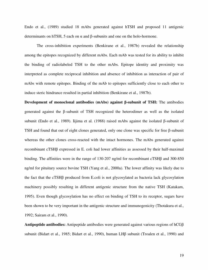
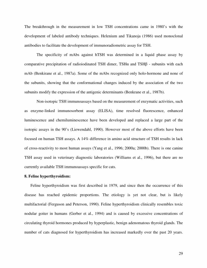
Crystal structure: The crystal structure of TSH has not yet been elucidated but the crystal
structure of human CG (hCG) and other members of glycoprotein hormone family have been
reported (Lapthorn et al, 1994; Wu et al., 1994). The crystal structure of hCG has revealed that
each subunit contains a central cysteine knot and three loops, two β-hairpin loops (L1 and L3) on
one side of cysteine knot, and a long loop (L2) on the other side (Fig. 1). The long loop in the α-
subunit (L2) has a two turn α-helix. Because of the presence of the cysteine knot, TSH is also
classified as a member of cysteine knot growth factor (CKGF) superfamily. The similarity of the
folds of the α and β subunits is best visualized by superpositioning the subunits. The α and β
subunits are aligned reciprocally at the interface and are associated with each other with a
minimal hydrophobic core. Unlike other CKGFs that exist as homo- or heterodimers with
interchain disulfide bridges, the glycoprotein hormones have noncovalently linked α and β
subunits stabilized by a segment of the β-subunit termed “seat-belt,” as it wraps around both the
subunits maintaining the tertiary structure. This seat belt arrangement has implications for
folding pathways during heterodimer formation as this process is governed by the precise order
of disulfide formation (Lapthorn et al., 1994).
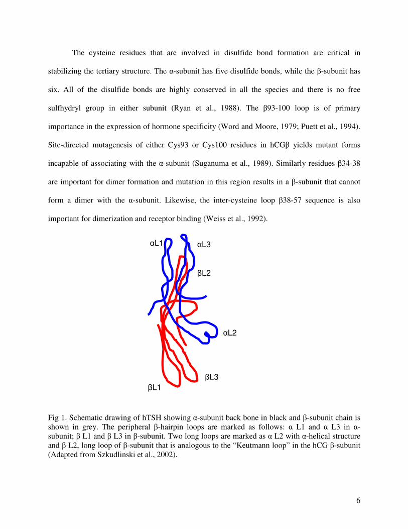
6
The cysteine residues that are involved in disulfide bond formation are critical in
stabilizing the tertiary structure. The α-subunit has five disulfide bonds, while the β-subunit has
six. All of the disulfide bonds are highly conserved in all the species and there is no free
sulfhydryl group in either subunit (Ryan et al., 1988). The β93-100 loop is of primary
importance in the expression of hormone specificity (Word and Moore, 1979; Puett et al., 1994).
Site-directed mutagenesis of either Cys93 or Cys100 residues in hCGβ yields mutant forms
incapable of associating with the α-subunit (Suganuma et al., 1989). Similarly residues β34-38
are important for dimer formation and mutation in this region results in a β-subunit that cannot
form a dimer with the α-subunit. Likewise, the inter-cysteine loop β38-57 sequence is also
important for dimerization and receptor binding (Weiss et al., 1992).
Fig 1. Schematic drawing of hTSH showing α-subunit back bone in black and β-subunit chain is shown in grey. The peripheral β-hairpin loops are marked as follows: α L1 and α L3 in α-subunit; β L1 and β L3 in β-subunit. Two long loops are marked as α L2 with α-helical structure and β L2, long loop of β-subunit that is analogous to the “Keutmann loop” in the hCG β-subunit (Adapted from Szkudlinski et al., 2002).
αL1 αL3
βL1
αL2
βL2
βL3
7
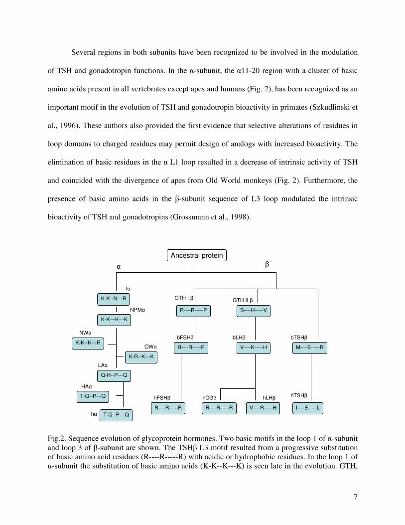
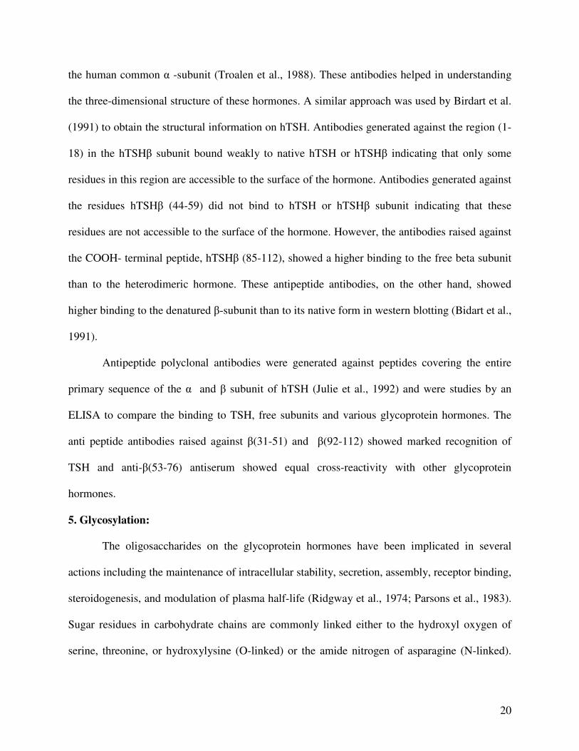
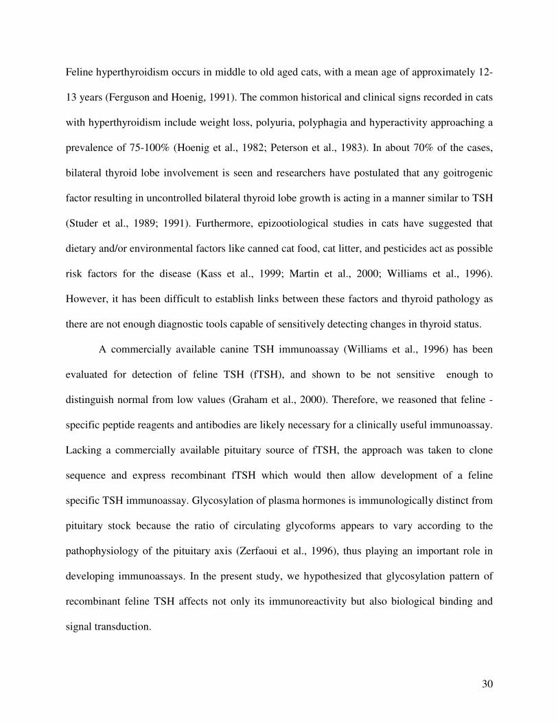
Several regions in both subunits have been recognized to be involved in the modulation
of TSH and gonadotropin functions. In the α-subunit, the α11-20 region with a cluster of basic
amino acids present in all vertebrates except apes and humans (Fig. 2), has been recognized as an
important motif in the evolution of TSH and gonadotropin bioactivity in primates (Szkudlinski et
al., 1996). These authors also provided the first evidence that selective alterations of residues in
loop domains to charged residues may permit design of analogs with increased bioactivity. The
elimination of basic residues in the α L1 loop resulted in a decrease of intrinsic activity of TSH
and coincided with the divergence of apes from Old World monkeys (Fig. 2). Furthermore, the
presence of basic amino acids in the β-subunit sequence of L3 loop modulated the intrinsic
bioactivity of TSH and gonadotropins (Grossmann et al., 1998).
Fig.2. Sequence evolution of glycoprotein hormones. Two basic motifs in the loop 1 of α-subunit and loop 3 of β-subunit are shown. The TSHβ L3 motif resulted from a progressive substitution of basic amino acid residues (R----R-----R) with acidic or hydrophobic residues. In the loop 1 of α-subunit the substitution of basic amino acids (K-K--K---K) is seen late in the evolution. GTH,
Ancestral protein
K-K--N---R
K-K—K---K
Q-H--P---Q
K-R--K---K
K-K--K---R
T-Q--P---Q
R----R-----P S----H-----V
M----E-----R R----R-----P V----K-----H
R----R-----R R----R-----R V----R-----H I----E-----L
T-Q--P---Q
α β
fα
NPMα
NWα
OWα
LAα
HAα
hα
GTH I β GTH II β
bTSHβ
hTSHβ
bLHβ bFSHβ
hFSHβ hLHβ hCGβ

8
gonadotropin; NPM, non primate mammals; NW, New World monkeys; OW, Old World monkeys; LA, Lower Apes; HA, Higher Apes; h, human; f, fish (Adapted from Grossmann et al., 1998).
Modeling of hTSH: Computer-aided modeling was attempted initially to predict a model for
three-dimensional structure for hTSH and, subsequently, secondary structure prediction was
made by computing the primary sequence in terms of flexibility, hydrophobicity and solvent
exposure (Ronin et al., 1990; Delege et al., 1988). According to this model, the surface of
interaction is made of two anti-parallel strands of β - pleated sheet running parallel to the α-helix
in the β-subunit and the N- and C- termini of the α-subunit (Ronin et al., 1990). Recently,
comparative modeling based on the crystal structures of hCG and hFSH has allowed construction
of more complete and accurate models for human, porcine and bovine TSH (Miguel et al., 2004).
These authors also reported that hTSHα chains interact with many amino acids on the Leucine
Rich Domains (LRD) surface and Cleavage Domain (CD) surface of human thyrotropin receptor
and the β chains do not interact with CD. Moreover, the observed higher affinity of bovine
thyrotropin and porcine thyrotropin relative to hTSH for the hTSH receptor has been explained
in terms of charge-charge interactions between the α chains and the receptor.
Common α subunit gene: The cDNA of the human α subunit was first cloned and reported in
1979 (Fiddes and Goodman., 1979). The common α subunit gene has subsequently been cloned
from a variety of species including cow (Erwin et al, 1983), rat (Godine et al., 1982), mouse
(Chin et al., 1981), horse (O’Brien M and Headon, 1995), sheep (Bello et al., 1989), pig
(Rettenberger et al., 1995) and dog (Yang et al., 2000b). The gene for the α subunit has been
localized to the long arm of chromosome 6 in man and chromosome 4 in the mouse and these
genes are 13.7 kb (cow), 9.4 kb (man) and 7.7 kb (rat) in length. Comparison of the cDNA
sequences of various species shows significant homology varying form 76 % between man and
9
rat to 96 % between rat and mouse (Burnside et al., 1988). The common α subunit gene in all
species contains four exons and three introns, and the coding sequences span from the second
exon to part of exon four. Gene mapping studies of pituitary RNA revealed a single transcription
start site downstream of the TATA boxes (Wondisford et al., 1991). However, multiple
regulatory elements for various hormonal and physiologic effectors can be anticipated as the α
subunit is expressed in both gonadotrophs and thyrotrophs.
A cyclic AMP-response element, containing the core 8-base motif T(G or T)ACGTCA
which is required for the induction of cAMP gene transcription, is present between residues 146
and 128 in the α subunit gene. The mRNAs for the common α subunits are about 800 bp in size
and encode a translational product of 14 kD in molecular weight containing 116-120 residues. A
24 amino acid signal peptide precedes a 92-96 amino acid apoprotein (Fiddes and Goodman,
1979; 1981).
TSHβ subunit gene: The gene encoding TSHβ subunit has been cloned and sequenced in man
(Hayashizaki et al., 1985), cattle (Maurer et al., 1984), mouse (Wolf et al., 1987), rat (Croyle et
al., 1986), chicken (Gregory and Porter, 1997), sheep (Bockmann et al, 1997), rabbit (Mathee et
al., 2004) and dog (Yang et al., 2000a). There are three exons and two introns in the beta subunit
gene. The first is only 37 bp and untranslated followed by a 3.9 kilo base intron. The function of
the first exon is unclear; however it has been speculated that exon I may interact directly with
thyroid hormone and its receptor and down regulate the TSHβ gene (Wondisford et al., 1988).
The second exon encodes the signal peptide and first 34 amino acids of the mature TSHβ
peptide; the third exon contains the remaining coding region and 3’ untranslated sequences.
Unlike rat and mouse β genes that contain two transcription sites due to alteration in of the distal
5’ TATA box, human TSHβ contains only one (Samuels et al., 1989).
10
The cyclic AMP response elements are present at the 5’- flanking region of human, rat
and mouse genes and contain more than 30 nucleotides with a highly conserved core sequence,
ATGACGTCAG. These regions are likely to be used for cAMP regulation (Tatsumi et al., 1988).
The mRNA for TSHβ is approximately 700 bases in size and encodes a protein of 138 amino
acids with 20 residues constituting signal peptide and 118 residues forming the β-subunit
apoprotein (Hayashizaki et al., 1985; Whitfield et al., 1986; Wondisford et al., 1988).
Single chain or yoked analogs of glycoprotein hormones: The functional activity of TSH
depends on the correct assembly of the subunits into heterodimers. The noncovalent association
of the subunits is an obligatory step for the formation of biologically active hormone (3). A novel
approach, namely, the construction of single chain analogs has been proven to be a promising
strategy for structure-function studies and in generation of hormones with increased stability and
activity (Narayan et al., 1995; Sugahara et al., 1996; Grossmann et al., 1997). Grossmann, et al,
showed that the genetic fusion of hTSH α- and β-subunits using the carboxy-terminal peptide of
the hCG β-subunit as a linker created a yoked form of hTSH whose receptor binding and
bioactivity were comparable to native hTSH, but had higher thermostability and a longer plasma
half-life (Grossmann et al., 1997).
Several features of CTP make it an ideal linker. It is rich in serine/proline content and
thus lacks any secondary structure (Puett et al., 1998). At the same time CTP also provides
sufficient distance and flexibility to facilitate the proper folding and interaction between the α-
and β-subunits. The crystal structure of hCG suggested the flexible nature of CTP wherein the
aminoacid residues 112-145, comprising entire length of CTP could not be traced (Lapthorn et
al., 1994). Further, site directed mutagenesis studies have shown that the CTP is not required for
subunit association or receptor binding, whereas the N-terminus of the β-subunit and C-terminus
11
of α-subunit appear to be important in receptor binding (Keutmann, 1992; Chen et al., 1992).
The CTP contains four O-linked glycosylation sites which play and important in increasing the
circulatory half life of the hCG (Matzuk et al., 1990).
Functional yoked hCG has been successfully expressed in both insect cells and Chinese
Hamster Ovary (CHO) cells (Narayan et al., 1995; Sugahara et al., 1995). It was reported that
yoked hormones are as effective as the heterodimer in receptor binding and activation (Narayan
et al., 1995). Fundamentally for the purpose of the present study, this approach also ensures
equimolar expression of the subunits and simultaneous affinity labeling of a single recombinant
peptide rather than a heterodimer.
2. TSH Receptor (TSHR):
The function of TSH is mediated by binding of TSH to its specific membrane receptors
and activation of signal transduction pathways. Activation of second messenger systems results
in a large variety of metabolic consequences like iodide uptake and release, thyroid peroxidase
generation, thyroid cell growth, thyroid hormone synthesis and release (Dumont et al., 1992).
TSH receptors are located on the basal membrane of thyroid follicle cells. Due to the clinical
significance of TSHR autoantibodies involved in the etiology of Graves’ disease and its
associated hyperthyroidism, the TSHR has been the focus of interest for past 20 years.
The human TSHR was first cloned by Parmentier et al. (1987). The feline TSHR
(fTSHR) was cloned in 2002 (Nguyen et al., 2002). Species comparison revealed that the feline
TSHR is closely related to the canine TSHR with 96% identity and 97% similarity in amino acid
sequence. The TSHR is a single chain glycoprotein containing 744 amino acids with a molecular
weight of about 100 kD. It consists of a 398-residue NH2-terminal large putative extracellular
domain, which contains six glycosylation sites and three disulfide binds followed by 346 amino
12
acids, comprising seven putative transmembrane segments (Libert et al., 1989; Misrahi et al.,
1990). The TSHR has been studied in a variety of in vitro and in vivo systems and has been
partially purified and characterized biochemically (Rees Smith et al., 1987).
The TSHR belongs to the G- protein coupled receptor (GPCR) super family having seven
transmembrane domains, with similarities to receptors for LH/CG and FSH. The extracellular
domain of the TSHR and the LH/CG receptors are much larger and more complex than the short
extracellular domains recognized by the smaller ligands such as adrenergic and cholinergic
agents. The TSHR has an additional 64 residues which are involved in autoantibody generation,
in the N- and C- terminus of extracellular domain that are homologous with LH/CG receptors
(Hidaka et al., 1993). However, the TSHR and gonadotropin receptor are both examples of single
receptors which can couple to both the cAMP and phosphoinositide signaling cascades (Vassart
and Dumont, 1992).
Together with newly discovered receptors (Nishi et al., 2000), TSHR, LHR and FSHR
are now classified as leucine-rich repeats (LRR)-containing G-protein coupled receptors.
Different modeling studies suggested 6-9 LRRs, each repeat containing a β - pleated sheet
oriented towards the interior circumference of the horseshoe-like tertiary structure, which binds
to the hormone molecule (Szkudlinski et al., 2002). TSHR is unique among glycoprotein
hormone receptors in that some mature receptors on the cell surface are cleaved into two
subunits (Rapoport et al., 1998; Rees Smith et at., 1988). Posttranslational modifications of
TSHR include glycosylation of the six asparagine residues.
The extracellular domain of the TSHR is involved in TSH binding and signal
transduction (Nagayama et al., 1992), and has been the target of a variety of autoantibodies. The
binding of autoantibodies to the receptor either stimulates or inhibits thyroid cell function and/or
13
growth, and may have serious physiological consequences. In Graves’ disease, the autoantibody
is stimulatory, and in Hashimoto’s disease, the autoantibody inhibits the binding of TSH to its
receptor thus resulting in idiopathic atrophy (Zakarija and McKenzie, 1991).
Ligand-receptor cross-reactivity has been investigated for a long time and it was reported
that TSH binds to LH/CG receptor and increases cAMP and Inositol phosphate levels (Hidaka et
al., 1993). Similarly, hCG binds to thyroid plasma membrane and displaces 125I-TSH from its
receptor (Azukizawa et al., 1979). Furthermore hCG can activate adenylate cyclase in Chinese
Hamster Ovary (CHO) cells stably transfected with the human TSH receptor (Tomer et al., 1992)
and increases iodide uptake and 3H-thymidine incorporation in FRTL-5 cells (Hershman et al.,
1988).
Second messenger systems:
TSH activates both the adenylate cyclase and the phospholipase C signaling pathways
(Field, 1975). Activation of phospholipase C is slower and requires larger quantities of TSH than
the activation of adenylate cyclase. Adenylate cyclase mediates most of the TSH’s cellular
effects. Studies are necessary to evaluate the physiological significance of dual signaling system
for the thyrotropin (Cho, 2002).
Adenylate Cyclase-cAMP System: Activation of adenylate cyclase leads to the production of
cAMP and cAMP in turn stimulates growth as well as function and differentiation of cultured
thyrocytes. It has been shown that constitutive activation of adenylate cyclase is sufficient to
promote hyperplasia of thyroid gland and hyperthyroidism (Ledent et al., 1992). Accumulation
of cAMP in the cytoplasm stimulates dissociation of regulatory and catalytic subunits of cAMP-
dependent protein kinase and subsequent phosphorylation of various cellular proteins. Growth
factors like IGF-I increase the sensitivity and maximum response of cyclic AMP to TSH
14
(Brenner et al., 1989). Catecholamines, which increase cAMP phosphodiesterase activity, are
other factors which can modulate the cAMP response (Berman et al., 1987).
The TSHR is associated with an inhibitory guanine nucleotide (Gi) binding protein that
exerts inhibitory control over adenylate cyclase activation. Adenosine diphosphate (ADP)
ribosylation of Gi binding protein by pertussis toxin abolishes the inhibitory effect of Gi and may
be involved in TSH stimulation of adenylate cyclase activity (Corda et al., 1987). Recently, it
was reported that TSH receptor signaling via cyclic AMP stabilizes the assembly and retention of
E-cadherin at the cell surface suggesting a new mechanism by which TSH supports maintenance
of thyroid follicular integrity (Larsson et al., 2004).
Phospholipase C system: Bone et al., (1986) reported that TSH also activates the inositol-lipid
signaling pathway. TSH stimulates hydrolysis of phosphotidylinositol-4, 5-biphosphate (PIP2) by
phospholipase C and results in the formation of diacylglycerol (DAG) and inositol-1, 4, 5-
triphosphate (IP3). IP3 mobilizes intracellular Ca2+, which activates Ca2+- and calmodulin -
dependent protein kinase (Ca-CaM). DAG activates protein kinase C (PKC) and both Ca-CaM
kinase and PKC phosphorylate numerous proteins in nucleus, plasma membranes and cytosol
resulting in cell responses including hormone secretion and gene expression. Interestingly, TSH-
induced production of hydrogen peroxide, which is an essential process for iodide organification,
a highly differentiated function of thyroid cells, is mediated not by cAMP, but by Ca2+ signaling
and phospholipase-A2 activation in FRTL-5 cells (Kimura et al., 1995).
3. Regulation of TSH
TSH subunits are synthesized separately as pre-α and pre-β-subunit precursors. These
precursor subunits contain signal peptide needed for intracellular passage. These precursors then
undergo post-translational modifications like glycosylation, truncation, sulfation, folding and
15
then the two subunits associate to form a dimer. The secretion of TSH is controlled by two major
factors: feedback effects of the unbound form of thyroid hormones and stimuli emanating from
the central nervous system like, thyrotropin releasing hormone (TRH), somatostatin, and
dopamine.
Thyroid hormones: An inverse relationship between the circulating thyroid hormone
concentrations and secretion of TSH has been well established (Tong, 1974). Thyroid hormones
T3 and T4 were recognized as the most important inhibitory factors in the control of TSH
secretion. In the serum of most domestic species, all but about 0.1 % of T4 is bound to plasma
proteins. Although it is T3 binding to nuclear receptors in the pituitary that governs the response,
the circulating concentrations of both T3 and T4 play an important role in the feedback process
(Larsen et al., 1981). The main action of thyroid hormones in the control of TSH secretion is to
regulate its gene expression. The nuclear receptors are transcriptional regulatory proteins and
binding of thyroid hormones activates the receptors. The hormone-receptor complex recognizes
and binds to specific sequences of DNA. The DNA-bound receptors influence gene transcription
by excluding polymerase and other DNA-binding regulatory proteins by competition. The
activated T3 receptor inhibits transcription of both β and α subunit gene expression even in the
absence of new protein synthesis (Shupnik et al., 1986). The nuclear receptor-T3 complex binds
to the cis-acting negative thyroid hormone responsive elements in the 5’-flank region of the
TSHβ gene and to a lesser extent the common α gene (Gurr and Kourides, 1985). This higher
affinity binding of T3 to TSHβ gene than TSHα gene may account for the observation that
thyroid hormones at different doses decreases TSH β mRNA levels more rapidly than TSH α
mRNA levels in the pituitaries of hypothyroid mice (Gurr et al., 1986).
16
Thyroid hormones are also able to modulate the TRH effects on TSH release by
decreasing the number of TRH receptors on the thyrotrophs and stimulating TRH degradation by
pituitary enzymes (De Lean et al., 1977; Bauer, 1987). In addition, thyroid hormones also reduce
TRH transcription in the paraventricular nuclei also contributing to the regulation of TSH
secretion (Koller et al., 1987).
Thyrotropin releasing hormone (TRH): TRH is synthesized in the hypothalamic
paraventricular nucleus. It is a tripeptide (pyroglutamyl-histidyl-proline-amide) and is
transported to median eminence and released into hypophysial portal blood (Jackson et al.,
1985). TRH is a major positive regulator of TSH secretion acting over a range of 10-11 to 10-7 M
(Vale, et al., 1972). In man, the serum TSH levels in response to intravenous administration of
TRH are observed within 2 to 5 minutes and peaks at 20 to 30 minutes and returns to basal levels
by 2 to 3 hrs (Jackson, 1982).
TRH significantly stimulated TSH glycosylation and subunit assembly (Taylor and
Weintraub, 1985). The effects of TRH are mostly post - translational, including alteration of
TSH bioactivity and metabolic clearance by influencing the carbohydrate components of TSH.
TSH secreted in response to TRH stimulation has a lower sulfate to mannose ratio, and a lower
sialic acid to mannose ratio, than does spontaneously secreted TSH (Gesundheit et al., 1986).
Presumably, TRH affects the final carbohydrate structure of secreted TSH by activating or
inhibiting glycosyltransferases or by altering the intracellular secretory pathways. TRH also
enhances the intrinsic bioactivity of TSH (Magner, 1990). In addition, TRH also stimulates the
transcriptional activity of the TSH subunit gene three-fold to five-fold in animal models
(Shupnik et al., 1990).
17
TRH binds to specific receptors on the plasma membrane of the pituitary cells (Hinkle
and Goh, 1982) which belong to large family of G-protein coupled receptors. TRH action is
mediated mainly through activation of phospholipase C (Drummond, 1986) rather than activation
of adenylate cyclase (Iriuchijima and Mor, 1986). TRH also induces an immediate and rapid
increase in intracellular free calcium which decays rapidly followed by an extended plateau of
elevated calcium. This biphasic action correlates with induction of calcium fluxes and secretary
activity in pituitary cells (Geras and Gershengorn, 1981).
Somatostatin: Somatostatin, a cyclic neuropeptide with two naturally occurring forms of 14 and
28 amino acids, is secreted from hypothalamic neurons located in the preoptic and
suprachiasmatic regions and released in the median eminence. Anti-somatostatin serum
administered to rats enhances the release of TSH due to cold or exogenous TRH (Vitger, 1987)
supporting the inhibitory role of somatostatin in TSH regulation. Similarly in human,
somatostatin reduces the elevated serum TSH levels in primary hypothyroidism, suppresses the
serum TSH response to TRH and prevents TSH release after administration of dopamine
antagonist (Scanlon, 1991). Somatostatin also decreases intracellular cAMP levels through
binding to its specific receptors coupled to an inhibitory guanyl nucleotide-binding protein (Gi),
thus reducing TSH gene expression (Ahlquist et al., 1987). Berelowitz et al (1980) have shown
that T3, in addition to its direct pituitary effect, also exerts a negative feedback effect on pituitary
TSH release through stimulation of somatostatin release from hypothalamus.
Dopamine: Dopamine inhibits the secretion of TSH by acting on its receptors on the
thyrotrophs. Shupnik et al., (1986b) showed that dopamine treatment of cultured pituitary cells
from hypothyroid rats results in decreased TSH mRNAs and suppressed transcription of both
TSH subunit genes and addition of cAMP partially overcomes dopamine suppression of
18
transcription suggesting that dopamine may decrease the circulating TSH level by decreasing
intracellular cAMP levels and thus interfering with cAMP-mediated stimulation of TSH-subunit
gene expression.
4. Immunogenicity of TSH:
Within a species, the α-subunit carries similar antigenic determinants which are species
specific for all the four glycoprotein hormones, TSH, LH, FSH and CG (Vaitukaitis et al., 1972).
The β - subunit possess hormone specific epitopes and gives functional specificity to these
hormones (Reichert et al., 1970).
Development of monoclonal antibodies (mAbs) against TSH dimer: A number of studies
have been reported on production and characterization of monoclonal antibodies against TSH
(Soos and Siddle 182; Soos et al., 1984; Benkirane et al., 1987a; Endo et al., 1989). The number
and spatial distribution of antigenic determinants on the surface of TSH molecule was studied in
detail using the monoclonal antibodies generated against intact human TSH. Using a similar
approach, two- or three- dimensional mappings of epitopes were proposed for human LH (Soos
and Siddle, 1983) and human CG (Schwarz et al., 1986). Also, a study of the functional role of
epitope site may give important information to elucidate the mechanism of hormone action on
the receptor (Hill et al., 1987).
Soos et al., (1987) characterized 23 mAbs against human TSH and postulated four
antigenic determinants on TSH. Only one of these epitopes is expressed in the free TSHβ-
subunit. Benkirane et al., (1987b) characterized 28 mAbs and postulated that human TSH has at
least 12 different antigenic regions, 2 of them on the α -subunit, 6 on the β-subunit either in free
form or in heterodimeric conformation and 4 antigenic regions expressed only on holo-hormone.
19
Endo et al., (1989) studied 18 mAbs generated against hTSH and proposed 11 antigenic
determinants on hTSH, 5 each on α and β-subunits and one on the holo-hormone.
The cross-inhibition experiments (Benkirane et al., 1987b) revealed the relationship
among the epitopes recognized by different mAbs. Each mAb was tested for its ability to inhibit
the binding of radiolabeled TSH to the other mAbs. Epitope identity and proximity was
interpreted as complete reciprocal inhibition and absence of inhibition as interaction of pair of
mAbs with remote epitopes. Binding of the mAb to epitopes sufficiently close to each other to
induce steric hindrance resulted in partial inhibition (Benkirane et al., 1987b).
Development of monoclonal antibodies (mAbs) against β-subunit of TSH: The antibodies
generated against the β-subunit of TSH recognized the heterodimer as well as the isolated
subunit (Endo et al., 1989). Iijima et al. (1988) raised mAbs against the isolated β-subunit of
TSH and found that out of eight clones generated, only one clone was specific for free β-subunit
whereas the other clones cross-reacted with the intact hormones. The mAbs generated against
recombinant cTSHβ expressed in E. coli had lower affinities as assessed by their half-maximal
binding. The affinities were in the range of 130-207 ng/ml for recombinant cTSHβ and 300-850
ng/ml for pituitary source bovine TSH (Yang et al., 2000a). The lower affinity was likely due to
the fact that the cTSHβ produced from E.coli is not glycosylated as bacteria lack glycosylation
machinery possibly resulting in different antigenic structure from the native TSH (Katakam,
1995). Even though glycosylation has no effect on binding of TSH to its receptor, sugars have
been shown to be very important in the antigenic structure and immunogenicity (Thotakura et al.,
1992; Sairam et al., 1990).
Antipeptide antibodies: Antipeptide antibodies were generated against various regions of hCGβ
subunit (Bidart et al., 1985; Bidart et al., 1990), human LHβ subunit (Troalen et al., 1990) and
20
the human common α -subunit (Troalen et al., 1988). These antibodies helped in understanding
the three-dimensional structure of these hormones. A similar approach was used by Birdart et al.
(1991) to obtain the structural information on hTSH. Antibodies generated against the region (1-
18) in the hTSHβ subunit bound weakly to native hTSH or hTSHβ indicating that only some
residues in this region are accessible to the surface of the hormone. Antibodies generated against
the residues hTSHβ (44-59) did not bind to hTSH or hTSHβ subunit indicating that these
residues are not accessible to the surface of the hormone. However, the antibodies raised against
the COOH- terminal peptide, hTSHβ (85-112), showed a higher binding to the free beta subunit
than to the heterodimeric hormone. These antipeptide antibodies, on the other hand, showed
higher binding to the denatured β-subunit than to its native form in western blotting (Bidart et al.,
1991).
Antipeptide polyclonal antibodies were generated against peptides covering the entire
primary sequence of the α and β subunit of hTSH (Julie et al., 1992) and were studies by an
ELISA to compare the binding to TSH, free subunits and various glycoprotein hormones. The
anti peptide antibodies raised against β(31-51) and β(92-112) showed marked recognition of
TSH and anti-β(53-76) antiserum showed equal cross-reactivity with other glycoprotein
hormones.
5. Glycosylation:
The oligosaccharides on the glycoprotein hormones have been implicated in several
actions including the maintenance of intracellular stability, secretion, assembly, receptor binding,
steroidogenesis, and modulation of plasma half-life (Ridgway et al., 1974; Parsons et al., 1983).
Sugar residues in carbohydrate chains are commonly linked either to the hydroxyl oxygen of
serine, threonine, or hydroxylysine (O-linked) or the amide nitrogen of asparagine (N-linked).
21
There are two Asn-linked sugar chains in the α-subunit, and the β-subunit has one or two chains
depending on the hormone. In human TSH, the two N-linked glycosylation sites are present at
asparagine residues 56 and 82 in the α-subunit, while only one glycosylation site is present at
asparagine residue 23 in the β-subunit (Pierce and Parsons, 1981). There is an additional O-
linked glycosylation site at threonine 43 in the free α-subunits secreted by bovine pituitaries
(Parsons, et al., 1983). This additional glycosylation may prevent dimerization with the β-subunit
(Pierce, 1986); however, the physiological role of the third oligosaccharide chain is not fully
understood.
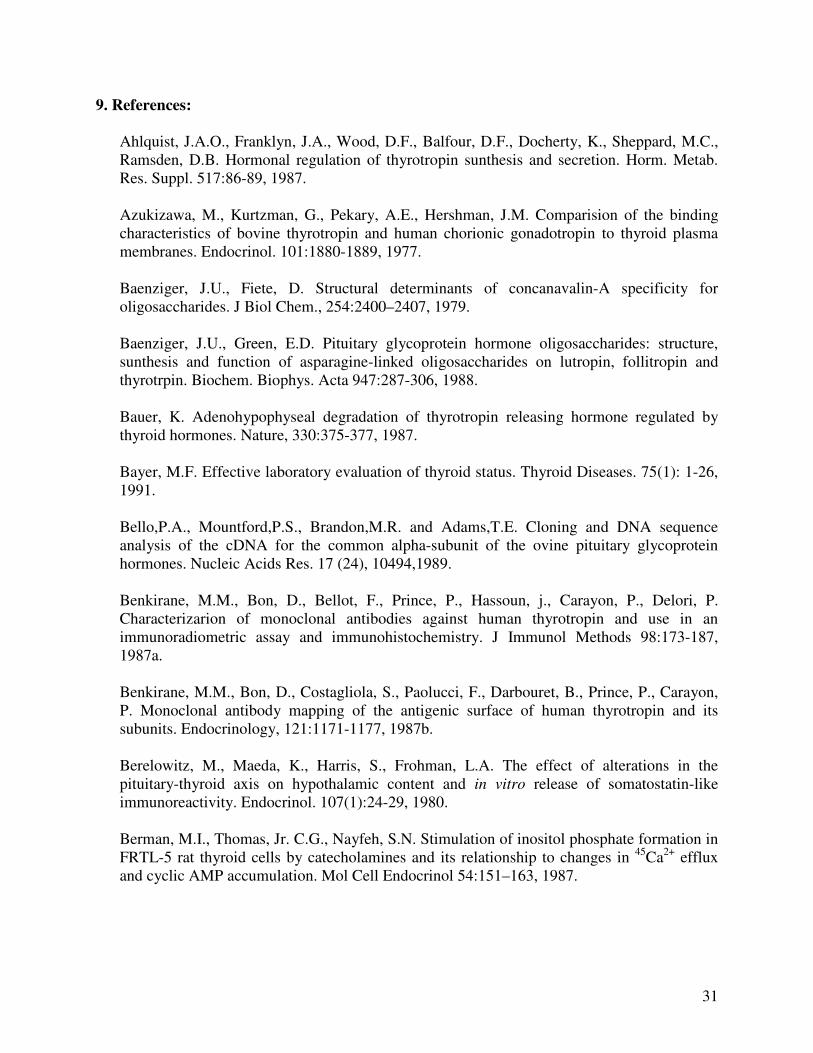
Glycosylation of TSH: The glycosylation of TSH subunits starts with transfer of precursor
oligosaccharides rich in mannose to the nascent peptide. The high mannose precursor
oligosaccharides protect nascent TSH subunits from intracellular proteolysis, aggregation and
also allow proper chain folding to occur so that the correct subunit tertiary confirmation is
attained (Weintraub et al., 1983). Tunicamycin, an antibiotic that blocks the reaction of UDP-
GlcNAc and dolichol phosphate in the first step of glycoprotein synthesis, inhibits the formation
of all dolichol carrier precursors, resulting in synthesis of nonglycosylated TSH subunits which
fail to fold and assemble properly, resulting in intracellular aggregation and proteolytic
degradation (Stricklant and Pierce, 1983). These high mannose precursors are later trimmed and
other sugar residues were added to make complex-type N-linked oligosaccharides. The transfer
of other sugar residues like fucose, β-acetylglucosamine, galactose, N-acetylgalactosamine,
sulfate and sialic acid residues is believed to occur principally in the Golgi apparatus (Kornfeld
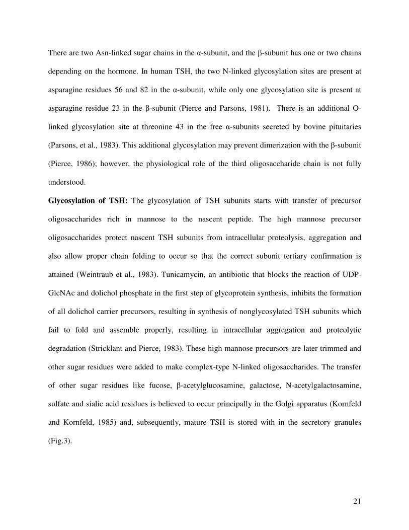
and Kornfeld, 1985) and, subsequently, mature TSH is stored with in the secretory granules
(Fig.3).
22
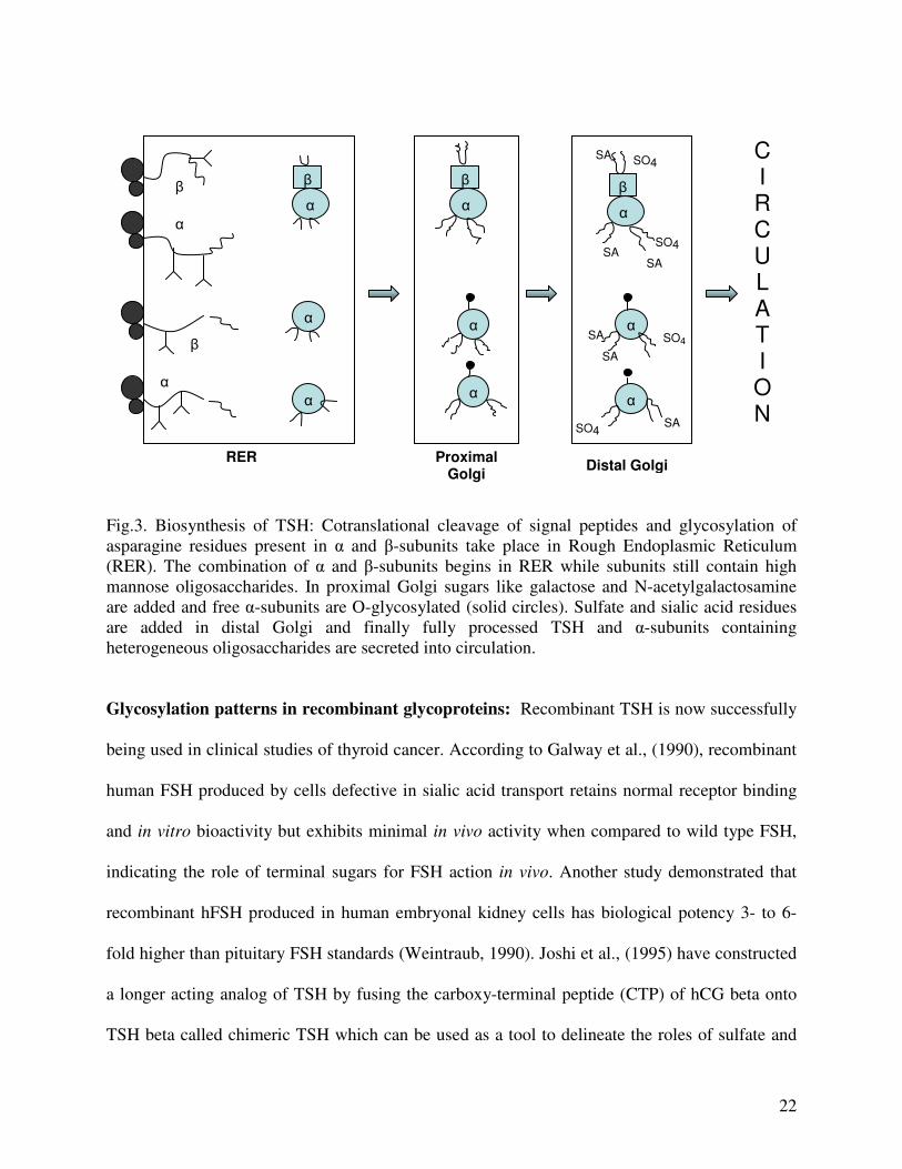
Fig.3. Biosynthesis of TSH: Cotranslational cleavage of signal peptides and glycosylation of asparagine residues present in α and β-subunits take place in Rough Endoplasmic Reticulum (RER). The combination of α and β-subunits begins in RER while subunits still contain high mannose oligosaccharides. In proximal Golgi sugars like galactose and N-acetylgalactosamine are added and free α-subunits are O-glycosylated (solid circles). Sulfate and sialic acid residues are added in distal Golgi and finally fully processed TSH and α-subunits containing heterogeneous oligosaccharides are secreted into circulation.
Glycosylation patterns in recombinant glycoproteins: Recombinant TSH is now successfully
being used in clinical studies of thyroid cancer. According to Galway et al., (1990), recombinant
human FSH produced by cells defective in sialic acid transport retains normal receptor binding
and in vitro bioactivity but exhibits minimal in vivo activity when compared to wild type FSH,
indicating the role of terminal sugars for FSH action in vivo. Another study demonstrated that
recombinant hFSH produced in human embryonal kidney cells has biological potency 3- to 6-
fold higher than pituitary FSH standards (Weintraub, 1990). Joshi et al., (1995) have constructed
a longer acting analog of TSH by fusing the carboxy-terminal peptide (CTP) of hCG beta onto
TSH beta called chimeric TSH which can be used as a tool to delineate the roles of sulfate and
β
α
β
α
α
α
β
α
α
α
β
α
β
α
SA
SA SA
SA
SO4
SO4
α
SO4
SO4
α
SA
SA
RER Proximal Golgi
CIRCULATION
Distal Golgi
23
sialic acid in the in vivo clearance and, thereby, the in vivo bioactivity of TSH. In recombination
studies performed by Sairam et al., (1990), LH formed by glycosylated native alpha- and beta-
subunits of the hormone was fully active but when one of the subunits was in the deglycosylated
form, the receptor binding activity was greatly reduced. These results demonstrate that LH
hormone glycosylation is essential for optimum receptor recognition in the sheep testis, further
emphasizing the importance of correct glycosylation for ovine LH alpha subunit function. Schaaf
et al (1997) showed for the first time for TSH that the two dominant intracellular signal
transduction systems (cAMP formation and IP release) are activated to different degrees by
hTSH glycosylation variants. A common polymorphism was recently discovered in the gene of
the LH beta-subunit containing two point mutations, which introduced to LH two amino acid
changes and an extra glycosylation site. The overall bioactivity of this variant is LH variant is
lower than that of the wild-type LH. The pathophysiological significance of this LH variant
remains open however, it is clear that clinicians monitoring LH levels in their patients have to be
aware of this common polymorphic form, which behaves aberrantly in several widely applied
immunoassay systems and may give rise to misleading LH levels (Huhtaniemi, 2000).
Glycosylation of glycoproteins in altered health conditions: A TSH bioassay system has been
developed using the CHO cell line transfected with the recombinant human TSH receptor. Using
this bioassay system, it has been shown that the secretion of TSH molecules with reduced
bioactivity is a common alteration in healthy patients. In a study comparing 25 patients with
central hypothyroidism, the ratio between biological(B) and immunological(I) activities of
circulating TSH was shown to be only 25 % that of normal subjects (Persani et al., 2000). Cyclic
AMP generation measured by radioimmunoassay assay is used to quantify the biological
response to the glycoproteins in this assay. Perez and Apfelbaum (1997) suggested that

24
glycosylation is not an essential step in the LH secretory process since the hormone, which is
normally secreted in a glycosylated form, was synthesized, transported, and released without the
carbohydrate side chains. Hashim et al (1990) reported that, under basal conditions, the majority
of prolactin secreted from the pituitary is glycosylated, but with hyperprolactinemia the capacity
for glycosylation is exceeded.
Terminal sulfation and sialylation of complex oligosaccharides: The complex
oligosaccharides of TSH and LH may terminate in either sulfate or sialic acid residues.
Variations in sialic acid content as well as differences in the complexity of the glycans determine
the full biological activity of FSH (Creus et al., 2001). Altered physiologic states alter the
oligosaccharide structure, which influences the clearance rate and therefore the serum
concentrations of TSH. For example, reduced intrinsic TSH bioactivity in pituitary
hypothyroidism results from increased sialylation of TSH (Oliveira et al., 2001). Thyroid
hormone and developmental factors also regulate the branching pattern and relative sialylation of
TSH carbohydrate chains, which may affect TSH action in vivo, by modifying hormonal
secretion, bioactivity and metabolic clearance (Weintraub et al., 1989).
The structure and distribution of sulfated and sialylated oligosaccharides on human TSH
have indicated that 25% of the oligosaccharides contain one sulfate, 21% have one sufate and
one sialic acid, 18% are neutral, 12% have two sialic acid residues, and 5 % have one sialic acid
residue (Baenziger and Green, 1988). Terminal sialic acid and sulfates are of special importance
because both the residues provide a net negative charge to the molecule (Drickamer, 1991).
Under in vitro conditions, sialic acid appears to the major factor affecting the charge
heterogeneity, metabolic clearance rate, and bioactivity of recombinant TSH (Szkundlinski et al.,
1993). Removal of the terminal sialic acid residues markedly reduces the half-life of TSH in the

25
circulation by enhancing its binding to asialohepatic receptors and thus being cleared from the
bloodstream (Sairam, 1983).
Extending N-glycosylation pathway: In the process of TSH biosynthesis, after the common
intermediate, Man8GlcNAc2-N-Asn is produced, exoglycosidases and a glycosyltransferase
catalyze trimming and elongation reactions, which yield, GlcNAcMan3GlcNAc2-N-Asn. In
mammalian cells, terminal glycosyltransferases can elongate this intermediate to produce hybrid
and complex N-glycans with terminal sialic acids. In contrast, insect cells have only extremely
low levels of these terminal glycosyltransferase activities, and in some cases, have a competing
exoglycosidase that can remove the terminal N-acetylglucosamine residue from
GlcNAcMan3GlcNAc2-N-Asn. Hence, the major processed N-glycan produced by insect cells is
usually the paucimannosidic structure, Man3GlcNAc2-N-Asn. Recently, genetically transformed
insect cells with mammalian β1,4-galactosylatransferase and α2,6-sialyltransferase genes have
been described (Hollister et al., 2001). Stably transformed insect cells with β1,4-
galactosyltransferase can be used as modified hosts for conventional baculovirus expression
vectors to produce mammalianized glycoprotein glycans which more closely resemble those
produced by higher eukaryotes (Hollister et al., 1998). The commercial Mimic™ Sf9 Insect Cell
line (Invitrogen, Carlsbad, CA), a derivative of the Sf9 insect cell line, was modified to stably
express a variety of mammalian glycosyltransferases. Typically, insect cells are unable to
process N-glycans to the extent that mammalian cells do. The addition of mammalian
glycosyltransferases like α 2,6-sialyltransferase, β4-galactosyltransferase, N-
acetylglucosaminyltransferase I and N-acetylglucosaminyltransferase II to the Mimic™ Sf9
Insect cells allows for production of biantennary, terminally sialyated N-glycans from insect
cells (Hollister et al., 2003).
26
Lectins: Lectins are carbohydrate-binding proteins. Although they were first discovered more
than 100 years ago in plants, they are now known to be present throughout nature (including the
microbial world, wherein they tend to be called by other names, such as hemagglutinins,
adhesins, and toxins). The interaction of lectin with glycans is as specific as enzyme-substrate or
antigen-antibody interactions. Lectins may bind with free sugar or with sugar residues of
polysaccharides, glycoproteins, or glycolipids which can be free or bound (as in cell
membranes). Lectin affinity chromatography with lectins, ricin and Concovanalin A (Con A) has
been used to analyze glycoprotein hormones in patients suffering with congenital disorders of
glycosylation (Ferrari et al., 2001). Con A permits the separation of molecules differing in the
extent of their carbohydrate branching, whereas ricin gives an estimation of the degree of their
sialylation. Pituitary TSH was more retained on Con A and less sialylated than circulating
hormone, suggesting that the carbohydrate chains of the pituitary form of the hormone are less
mature than those present in the circulating TSH form (Papandreou et al., 1993). Glycoproteins
applied to the lectin Con A are eluted in three general classes: 1) unbound glycopeptides that
have bisecting, triantennary and multiantennary complex structures, with low mannose content,
2) weakly bound glycoproteins that elute with 10 mmol/L α-methylglucopyranoside and have
biantennary complex or truncated hybrid oligosaccharides, and 3) firmly bound glycopeptides
that elute with 300 mmol/L -methylmannopyranoside and have high mannose or hybrid
oligosaccharides, corresponding to less mature TSH molecules ((Papandreou et al 1993;
Baenziger et al., 1979). A Lectin ELISA that uses biotinylated AAL (Aleuria aurantia lectin) and
a capture antibody specific for AGP was used to study the fucosylation on AGP (α1 Acid
Glycoprotein) (Ryden et al., 1999).Thus, lectin ELISA and lectin blotting will provide a
qualitative estimate of the sugars present in the glycoproteins.
27
6. Glycosylation and antigenicity:
The effect of glycosylation on the antigenic structure and biological activity of hTSH was
studied by Papandreou et al., in 1990. In a different study it was reported that equine pituitary
glycoprotein hormones and bovine TSH are resistant to endoglycosidase action in their native
forms (Swedlow et al., 1986; Lee et al., 1987). However, the oligosaccharides on β-subunit in
both heterodimeric conformation and free form are easily accessible whereas, the sugars on α-
subunit are hidden by subunit interaction as evident from the partial deglycosylation of native
hTSH on β-subunit and complete deglycosylation of free α and β-subunits (Ronin et al., 1990).
The capacity of mAbs to bind intact and deglycosylated forms of hTSH was tested by
using a panel of 14 mAbs generated against eight out of twelve known epitopes present in human
TSH. All the mAbs tested recognized both the native and deglycosylated hTSH identically. In
contrast, binding of fully deglycosylated hTSH to anti-β mAbs was lost while the binding to anti-
α mAbs was retained. This observation suggests that the epitopes specific for subunit association
as well as those present on the β-subunit are glycosylation - dependent. Deglycosylation of free
subunits did not alter the recognition indicating that the effect of glycosylation is seen only in
dimeric conformation. Also, mAbs raised against recombinant canine TSHβ expressed in E.coli
showed lower binding affinity towards pituitary - source bovine TSH likely due to the fact that
nonglycosylated recombinant canine TSHβ subunit has a different antigenic structure than the
native glycosylated peptide and the antibodies raised against it might be weakly reactive towards
the native heterodimeric hormone (Katakam, 1995).
To identify different epitopes contributing to the recognition of deglycosylated hTSH,
competitive binding assays with radiolabeled hTSH and free subunits either in their native form
or deglycosylated form were done. These experiments revealed that deglycosylated hTSH is five
28
times less immunoreactive towards anti-β as compared to anti- α antiserum. This finding
demonstrates that all the glycosylation-independent epitopes are localized in the α-subunit of
hTSH and glycosylation - dependent domains in the hTSHβ subunit (Papandreou et al., 1990).
7. Developments in TSH immunoassays:
TSH synthesis in the anterior pituitary and its secretion into the peripheral circulation are
under the positive control by TRH and negative feedback by thyroid hormones. Therefore, when
the hypothalamic-pituitary axis is intact, serum TSH levels reflects a direct thyroid hormone
action on the pituitary thyrotrophs. On the other hand, the negative feedback of thyroid hormones
on the pituitary, designed to counteract thyroid hormone fluctuations and to maintain
euthyroidism, amplifies the TSH response. A minor rise or fall of thyroid hormones, particularly
of free thyroxine (FT4), elicits about 10 times larger inverse change in pituitary TSH release in
man. This makes TSH an extremely sensitive indicator of thyroid status (Bayer, 1991).
Measurement of TSH not only replaces time consuming TSH stimulation tests, but a sensitive
TSH assay also makes it possible to distinguish euthyroid from hyperthyroid patients eliminating
the need for the TRH stimulation test. High sensitivity TSH tests have become the primary test
for thyroid function diagnosis (Klee and Hay, 1987).
Strategies to develop TSH immunoassay: The early TSH immunoassay was too insensitive for
clinical purposes (Werner et al., 1961) as it was based on cross-reaction of human and bovine
TSH on a hemagglutination inhibition technique. Utiger, (1965) reported the first practical
radioimmunoassay (RIA) sensitive enough for determining not only the raised but also the
normal concentrations of TSH. Even though the TSH RIA was continuously improved by using
more delicate methods for purification and iodination of the antigen and various solid phase
separation techniques, measurement of subnormal TSH concentrations was still not achieved.
29
The breakthrough in the measurement in low TSH concentrations came in 1980’s with the
development of labeled antibody techniques. Helenium and Tikanoja (1986) used monoclonal
antibodies to facilitate the development of immunoradiometric assay for TSH.
The specificity of mAbs against hTSH was determined in a liquid phase assay by
comparative precipitation of radioiodinated TSH dimer, TSHα and TSHβ - subunits with each
mAb (Benkirane et al., 1987a). Some of the mAbs recognized only holo-hormone and none of
the subunits, showing that the conformational changes induced by the association of the two
subunits modify the expression of the antigenic determinants (Benkrane et al., 1987b).
Non-isotopic TSH immunoassays based on the measurement of enzymatic activities, such
as enzyme-linked immunosorbent assay (ELISA), time resolved fluorescence, enhanced
luminescence and chemiluminescence have been developed and replaced a large part of the
isotopic assays in the 90’s (Liewendahl, 1990). However most of the above efforts have been
focused on human TSH assays. A 14% difference in amino acid structure of TSH results in lack
of cross-reactivity to most human assays (Yang et al., 1996; 2000a; 2000b). There is one canine
TSH assay used in veterinary diagnostic laboratories (Williams et al., 1996), but there are no
currently available TSH immunoassays specific for cats.
8. Feline hyperthyroidism:
Feline hyperthyroidism was first described in 1979, and since then the occurrence of this
disease has reached epidemic proportions. The etiology is yet not clear, but is likely
multifactorial (Ferguson and Peterson, 1990). Feline hyperthyroidism clinically resembles toxic
nodular goiter in humans (Gerber et al., 1994) and is caused by excessive concentrations of
circulating thyroid hormones produced by hyperplastic, benign adenomatous thyroid glands. The
number of cats diagnosed for hyperthyroidism has increased markedly over the past 20 years.
30
Feline hyperthyroidism occurs in middle to old aged cats, with a mean age of approximately 12-
13 years (Ferguson and Hoenig, 1991). The common historical and clinical signs recorded in cats
with hyperthyroidism include weight loss, polyuria, polyphagia and hyperactivity approaching a
prevalence of 75-100% (Hoenig et al., 1982; Peterson et al., 1983). In about 70% of the cases,
bilateral thyroid lobe involvement is seen and researchers have postulated that any goitrogenic
factor resulting in uncontrolled bilateral thyroid lobe growth is acting in a manner similar to TSH
(Studer et al., 1989; 1991). Furthermore, epizootiological studies in cats have suggested that
dietary and/or environmental factors like canned cat food, cat litter, and pesticides act as possible
risk factors for the disease (Kass et al., 1999; Martin et al., 2000; Williams et al., 1996).
However, it has been difficult to establish links between these factors and thyroid pathology as
there are not enough diagnostic tools capable of sensitively detecting changes in thyroid status.
A commercially available canine TSH immunoassay (Williams et al., 1996) has been
evaluated for detection of feline TSH (fTSH), and shown to be not sensitive enough to
distinguish normal from low values (Graham et al., 2000). Therefore, we reasoned that feline -
specific peptide reagents and antibodies are likely necessary for a clinically useful immunoassay.
Lacking a commercially available pituitary source of fTSH, the approach was taken to clone
sequence and express recombinant fTSH which would then allow development of a feline
specific TSH immunoassay. Glycosylation of plasma hormones is immunologically distinct from
pituitary stock because the ratio of circulating glycoforms appears to vary according to the
pathophysiology of the pituitary axis (Zerfaoui et al., 1996), thus playing an important role in
developing immunoassays. In the present study, we hypothesized that glycosylation pattern of
recombinant feline TSH affects not only its immunoreactivity but also biological binding and
signal transduction.
31
9. References:
Ahlquist, J.A.O., Franklyn, J.A., Wood, D.F., Balfour, D.F., Docherty, K., Sheppard, M.C., Ramsden, D.B. Hormonal regulation of thyrotropin sunthesis and secretion. Horm. Metab. Res. Suppl. 517:86-89, 1987. Azukizawa, M., Kurtzman, G., Pekary, A.E., Hershman, J.M. Comparision of the binding characteristics of bovine thyrotropin and human chorionic gonadotropin to thyroid plasma membranes. Endocrinol. 101:1880-1889, 1977. Baenziger, J.U., Fiete, D. Structural determinants of concanavalin-A specificity for oligosaccharides. J Biol Chem., 254:2400–2407, 1979. Baenziger, J.U., Green, E.D. Pituitary glycoprotein hormone oligosaccharides: structure, sunthesis and function of asparagine-linked oligosaccharides on lutropin, follitropin and thyrotrpin. Biochem. Biophys. Acta 947:287-306, 1988. Bauer, K. Adenohypophyseal degradation of thyrotropin releasing hormone regulated by thyroid hormones. Nature, 330:375-377, 1987. Bayer, M.F. Effective laboratory evaluation of thyroid status. Thyroid Diseases. 75(1): 1-26, 1991. Bello,P.A., Mountford,P.S., Brandon,M.R. and Adams,T.E. Cloning and DNA sequence analysis of the cDNA for the common alpha-subunit of the ovine pituitary glycoprotein hormones. Nucleic Acids Res. 17 (24), 10494,1989. Benkirane, M.M., Bon, D., Bellot, F., Prince, P., Hassoun, j., Carayon, P., Delori, P. Characterizarion of monoclonal antibodies against human thyrotropin and use in an immunoradiometric assay and immunohistochemistry. J Immunol Methods 98:173-187, 1987a. Benkirane, M.M., Bon, D., Costagliola, S., Paolucci, F., Darbouret, B., Prince, P., Carayon, P. Monoclonal antibody mapping of the antigenic surface of human thyrotropin and its subunits. Endocrinology, 121:1171-1177, 1987b. Berelowitz, M., Maeda, K., Harris, S., Frohman, L.A. The effect of alterations in the pituitary-thyroid axis on hypothalamic content and in vitro release of somatostatin-like immunoreactivity. Endocrinol. 107(1):24-29, 1980. Berman, M.I., Thomas, Jr. C.G., Nayfeh, S.N. Stimulation of inositol phosphate formation in FRTL-5 rat thyroid cells by catecholamines and its relationship to changes in 45Ca2+ efflux and cyclic AMP accumulation. Mol Cell Endocrinol 54:151–163, 1987.
32
Bidart, J.M., Ozturk, M., Bellet, D.H., Jolivet, M., Gras-Masse, H., Troalen, F., Bohuon, C.J., and Wands, J.R. Identification of epitopes associated with hCG and the beta hCG carboxyl terminus by monoclonal antibodies produced against a synthetic peptide. J Immunol Vol 134:457-464, 1985. Bidart, J.M., Troalen, F., Ghillani, P., Rouas, N., Razafindratsita, A., Bohuon, C., Bellet, D. Peptide immunogen mimicry of a protein-specific structural epitope on human choriogonadotropin. (Erratum in: Science 1991 Feb 22;251(4996):856) Science. 11;248(4956):736-9,1990. Bidart, J.M., Troalen, F., Ghillani, P., Puisieux, A., Bohuon, C., Bellet, D. Immunochemical mapping of antigenic regions on the human thyrotropin beta-subunit by antipeptide antibodies. J Biol Chem. 15;266(29):19238-44, 1991. Bockmann,J., Bockers,T.M., Winter,C., Wittkowski,W., Winterhoff,H., Deufel,T., Kreutz,M.R. Thyrotropin expression in hypophyseal pars tuberalis-specific cells is 3,5,3'-triiodothyronine, thyrotropin-releasing hormone, and pit-1 independent. Endocrinology 138 (3), 1019-1028, 1997.
Bone, E.A., Alling, O.W., Grollman, E.F. Norepinephrine and thyroid stimulating hormone induce Inositol phosphate accumulation in FRTL-5 cells. Endocrinol. 119:2193-2200, 1986. Brenner-Gati, L., Berg, K.A., Gershengorn, M.C. Insulin-like growth factor-I potentiates thyrotropin stimulation of adenylyl cyclase in FRTL-5 cells. Endocrinology, 125:1315–1320, 1989. Burnside, J., Buckland, P.R., Chin, W.W. Isolation and characterization of the gene encoding the α-subunit of the rat pituitary glycoprotein hormones. Gene, 70:67-64, 1988.
Chastain, C.B. Human thyroid stimulating hormone radioimmunoassay in the dog. J Amer Anima Hosp Ass. 14:368-369, 1978. Chen, F., Wang, Y., Puett, D. The carboxy-terminal region of the glycoprotein hormone alpha-subunit: contributions to receptor binding and signaling in human chorionic gonadotropin. Mol Endocrinol.6(6):914-9, 1992. Chin, W.W., Carr, F.E., Burnside, J., Darling, D.S.: Thyroid hormone regulation of thyrotropin gene expression. Recent Prog. Horm. Res. 48:393-414, 1993. Chin, W.W., Kronenberg, H.M., Dee, P.C., Maloof, F., Haben, J.F. Nucleotide sequence of the mRNA encoding the pre-α-subunit of mouse thyrotropin. Proc Natl Acad Sci USA. 78:5329-33, 1981. Cho B Y. Clinical applications of TSH receptor antibodies in thyroid diseases J Korean Med Sci. 17:293-301, 2002.
33
Corda, D., Sekara, R.D., Kohn, L.D. Thyrotropin effect on the availability of Ni regulatory protein in FRTL-5 rat thyroid cells to ADP-ribosylation by pertusis toxin, Eur J Biochem 166:475-481, 1987. Creus, S., Chaia, Z., Pellizzari, E.H., Cigorraga, S.B., Ulloa Aguirre A., Campo, S. Human FSH isoforms: Carbohydrate complexity as determinant of in vitro bioactivity Molecular and Cellular Endocrinology, 174(1-2): 41-9, 2001. Croyle, M.L., Bhattacharya, A., Gordon, D.F., Maurer, R.A. Analysis of the organization and nucleotide sequence of the chromosomal gene for the β-subunit of rat thyrotropin. DNA, 5:299-304, 1986. Delege, G., Clerc, F.F., Roux B., Gautheron, D.C. ANTHEPROT: a package for protein sequence analysis using microcomputer. CABIOS 4:351-156, 1988.
De Lean, A., Ferland, L., Drouin, J. Modulation of pituitary thyrotropin releasing hormone receptor levels by estrogens and thyroid hormones. Endocrinology, 100:1496-1504, 1977.
Drickamer, K. Clearing up glycoprotein hormones. Cell 67:1029-1032 (Minireview), 1991. Drummond, A.H. Inositol lipid metabolism and signal transduction in clonal pituitary cells. J Exp Biol 124:337-358, 1986.
Dumont, J.E., Lamy, F., Roger, P., Maenhaut, C. Physiological and pathological regulation of thyroid cell proliferation ands differentiation by thyrotropin and other factors. Physiol. Rev. 72:667-697, 1992. Edinboro, C.H., Scott-Moncrieff, C., Janovitz, E., Thacker, H.L., Glickman, L.T. Epidemiologic study of relationships between consumption of commercial canned food and risk of hyperthyroidism in cats. J Am Vet Med Assoc. 224(6):879-886,2004. Ehrmann, D.A., Sarne, D.H. Serum thyrotropin and the assessment of thyroid status. Ann Intern Med. 24:515-39, 1989. Endo, Y., Miyai, K., Iijima, Y., Nakajima, T., Eda, Y., Fujita, H., Unoki, M. Epitope mapping of human thyrotropin. Acta endocrinilogica (copenh) 120:201-209, 1989. Erwin, C.R., Croyle, M.L., Donelson, J.E., Maurer, R.A. Nucleotide sequence of cloned complementary deoxyribonucleic acid for the alpha subunit of bovine pituitary glycoprotein hormones. Biochemistry. 22:4856-60, 1983. Ferguson, D.C., Hoenig, M. Feline Hyperthyroidism In: Allen, D.G. ed. Small Animal Medicine. Philadelphia, J.B.Lippincott Co, 831-843, 1991. Ferguson, D.C., Peterson, M.E. In search of a cause for feline hyperthyroidism. In Proceedings. 8th Annu Meet Vet Med Forum, 765-768, 1990.
34
Ferrari, M.C., Parini, R., Di Rocco, M.D., Radetti, G., Beck-Peccoz, P., Persani, L. Lectin analyses of glycoprotein hormones in patients with congenital disorders of glycosylation. Eur J Endocrinol. 144(4):409-16, 2001. Fiddes, J.C., Goodman, H.M. Isolation, cloning and sequence analyses of the cDNA for the α-subunit of human chorionic gonagotropin. Nature. 281:351-6, 1979.
Fiddes, J.C., Goodman, H.M. The genes encoding the common α-subunit of the four human glycorpotien hormones. J Mol Appl Genet 1:3-18, 1981. Field, J.B. Thyroid stimulating hormone and cyclioc adenosime 3’ 5’ monophospahte in the regulation of thyroid gland function. Metabolism 24:381-393, 1975. Field, J.B., Ealey, P.A., Marshall, N.J., Cockcroft, S. Thyroid stimulating hormone stimulates increases in inositol phosphates as well as cyclic AMP in the FRTL-5 rat thyroid cell line. Biochem J 247:519-524, 1987. Galway, B., Hsueh, A.J.W., Keene, J.L., Yamoto, M., Fauser, C.J.M. and Boime, I. In Vitro and In Vivo Bioactivity of Recombinant Human Follicle Stimulating Hormone and Partially Deglycosylated Variants Selected by Transfected Eukariotic Cell Lines Endocrinology, 127(1): 93-100, 1990. Geras, E.J., Gershengorn, M.D. Evidence that TRH stimulates secretion of TSH by two calcium-mediated mechanisms. Am J Physiol 242:109-114, 1981. Gerber, H., Peter, H., Ferguson, D.C., Peterson, M.E. Etiopathology of feline toxic nodular goiter.Vet Clin North Am Small Anim Pract. 24(3):541-65, 1994.
Gesundheit, N., Weintraub BD. Mechanisms and regulation of TSH glycosylation Adv. Exp. Med Biol. 205:87-105, 1986. Gharib, S.D., Weirman, M.E., Shupnik, M.A., Chin, W.W. Molecular Biology of the Pituitary Gonadotropins. Endocrine Reviews, 11, 177-199, 1990.
Godine, J.E., Chin, W.W., Habener, J.F. Alpha subunit of rat pituitary glycoprotein hormones. Primary structure of the precursor determined from the nucleotide sequence of the cloned cDNAs. J Biol Chem. 257:8368-71, 1982. Graham, P.A., Refsal, K.R., Nachreiner, R.F., Provencher-Bollinger, A.L. The measurement of feline thyrotropin (TSH) using a commercial canine immunoradiometric assay. Proceedings. 18th ACVIM Forum, Seattle, WA, Abstract p.719, 2000. Gregory,C.C., Porter,T.E. Cloning and sequence analysis of a cDNA for the beta subunit of chicken thyroid-stimulating hormone. Gen. Comp. Endocrinol. 107 (2), 182-190,1997.
35
Grossmann, M., Leitolf, H., Weintraub, B.D., Szkulinski, M.W. A rational design strategy for protein hormone superagonists. Nature Biotechnol. 16:871-875,1998. Grossmann, M., Wong, R., Szkudlinski, M.W., Weintraub, B.D. Human thyroid-stimulating hormone (hTSH) subunit gene fusion produces hTSH with increased stability and serum half-life and compensates for mutagenesis-induced defects in subunit association.J Biol Chem. 22;272(34):21312-6, 1997.
Gurr, J.A., Kourides, I.A. Thyroid hormone regulation of thyrotropin alpha and beta gene transcription. DNA 4:301-307, 1985.
Gurr, J.A., Vrontakis, M.E., Athanasian, E.A., Wagner, C.R., Kourides, I.A. Hormanl regulation of thyrotropin alpha and beta subunit mRNAs. Horm. Metabol. Res. 18:382-385, 1986. Hashim, I.A., Aston, R., Butler, J., Mc.Gregor, A.N., Smith, C.R., Norman, M. The Proportion of Glycosylated Prolactin in Serum is Increased in Hyperprolactinemic States Journal of Clinical Endicrinology and Metabolism, 71(1): 111-115, 1990.
Hayashizaki, Y., Miyai, K., Kato, K., Matsubara, K. Molecular cloning of the human thyrotropin β-subunit gene. FEBS Lett, 188:394-400, 1985.
Helenium, T., Tikanoja, S. Asensitive and practical immunoradimetric assay of thyrotropin. Clin Chem. 32:514-518, 1986. Hershman, J.M., Lee, H.Y., Sugawara, M., Mirell, C.J., Pang, X.P., Yanagisawa, M. Human chorionic gonadotropin stimulates iodide uptake, adenylate cyclase, and deoxyribonucleic acid synthesis in cultured rat thyroid cells. J Clin Endocrinol Metab. 67:74-79, 1988. Hidaka, A., Ban, T., Panesar, N.S., Minegishi, T., Kohn, L.D., Tahara, K. Thyrotropin stimulation of the lutropin/chorionic gonadotropin receptor: different sites mediate agonist activity and high affinity binding. Thyroid, 4(4):447-457, 1994.
Hidaka, A., Minegishi, T., Kohn, L.D. Thyrotropin, like luteinizing hormone (LH) and chorionic gonadotropin (CG), increases cAMP and Inositol phosphate levels in cells with recombinant human LH/CG receptor. Biochem. Biophy. Res. Comm. 196(1):187-195, 1993. Hill, B.L., Lustbader, J.W., Erlanger, B.F. Characterization of a bioeffective monoclonal antibody against thyrotropin beta-subunit. Endocrinology 120:574-581, 1987. Hinkle, P.M., and Goh, K.B.C. Regulation of thyrotropin-releasing hormone receptors and responses by L-triiodothyronine in dispersed rat pituitary cell cultures. Endocrinology, 110:1725-1731, 1982. Hoenig, M., Goldschimdt, M.H., Ferguson, D.C. Toxic nodular goiter in cats. J Small Anima Pract 23:1-12, 1982.
36
Hollister, J., Conradt, H., Jarvis, D.L. Evidence for a sialic acid salvaging pathway in lepidopteran insect cells. Glycobiology, 13(6): 487-495, 2003. Hollister, J.R., Jarvis, D.L. Engineering lepidopteron insect cells for sialoglycoprotein production by genetic transformation with mammalian β1,4-galactosylatransferase and α2,6-sialyltransferase genes. Glycobiology, 11(1): 1-9, 2001. Hollister, J.R., Sharper, J.H., Jarvis, D.L. Stable expression of mammalian β1,4-galactosylatransferase extends the N-glycosylation pathway in insect cells. Glycobiology, 8(5): 473-480, 1998. Huhtaniemi, I.T. Polymorphism of gonadotropin action: clinical implications. Asian J Androl. 2 (4), pp. 241-6, 2000. Iijima, Y., Endo, Y., Hata, N., Fujita, H., Unoki, M., Miyai, K. Highly specific enzyme immunoassay for the β-subunit of human thyrotropin with the use of monoclonal antibodies. Clin Chem 34:98-102,1988. Iriuchijima, T and Mor, M. Regional dissociation of cyclic AMP and Inositol phosphate formation in response to thyrotropin-releasing hormone in the rat brain. J Neurochem 52:1944-1946, 1989. Jackson, I.M.D. Thyrotropin releasing hormone. New Engl. J. Med. 306:145-155, 1982.
Jackson, J.M.D., Wu, P., Lechan, R.M. Imuunohistochemical localization in the rat brain of the precursor for thyrotropin-releasing hormone, Science, 229:1097-1099, 1985. Joshi, L., Murata, Y., Wondisford, F.E., Szkudlinski, M.W., Desai, R., Weintraub, B.D. Recombinant thyrotropin containing a beta-subunit chimera with the human chorionic gonadotropin-beta carboxy-terminus is biologically active, with a prolonged plasma half-life: role of carbohydrate in bioactivity and metabolic clearance. Endocrinology, 136 (9): 3839-48, 1995. Julie C.L., Sergi, I., Caninne, C., Darbon., Ronin, C. Comparative mapping of human thyrotropin, gonadotropins, and free subunits with antipeptide antibodies. Endocrinology 131:1999-2009, 1992. Kass PH, Peterson ME, Levy J, James K, Becker DV, Cowgill LD. Evaluation of environmental, nutritional, and host factors in cats with hyperthyroidism. J Vet Intern Med 1999;13(4):323-329. Katakam, P.V.G. Purification and development of monoclonal antibodies against recombinant beta subunit of canine thyrotropin. Masters Thesis, University of Georgia, Athens, 1995.
37
Keutmann, H.T. Receptor-binding regions in human glycoprotein hormones. Mol Cell Endocrinol. 86(1-2):C1-6, 1992.
Kimura, T., Okajima, F., Sho, K., Kobayashi, I., Kondo, Y. Thyrotropin-induced hydrogen peroxide production in FRTL-5 thyroid cells is mediated not by adenosine 3',5'-monophosphate, but by Ca2+ signaling followed by phospholipase-A2 activation and potentiated by an adenosine derivative. Endocrinology, 136:116-123, 1995. Klee, G.G., Hay, I.D. Assessment of sensitive thyrotropin assays for an expanded role in thyroid function testing: proposed criteria for analytic performance and clinical utility. J. Clin. Endo. Metab. 64:461-471, 1987. Koller, K.J., Wolff, R.S., Warden, M.K., Zoeller, R.T. Thyroid hormones regulate levels of thyrotropin releasing hormone mRNA in the paraventricular nucleus. Proc Natl Acad Sci. USA, 84:7329-7333, 1987. Kornfeld, R., Kornfeld, S. Assembly of asparagine-linked oligosaccharides. Annu. Rev. Biochem. 54:631-664, 1985. Lapthorn, A.J., Harris, D.C., Littlejohn, A., Lustbader, J.W., Canfield, R.E., Machin, K.J., Morgan, F.J., Isaacs, N.W. Crystal structure of human chorionic gonadotropin. Nature, 369:455-461, 1994. Larsen, P.R., Silva, J.E., Kaplan, M.M. Relationship between circulating and intracellular thyroid hormones: Physiological and clinical implications. Endocrinol. Rev. 2:87-102, 1981. Larsson, F., Fagman, H., Nilsson, M. TSH receptor signaling via cyclic AMP inhibits cell surface degradation and internalization of E-cadherin in pig thyroid epithelium. Cell Mol Life Sci. 61(14):1834-42, 2004. Ledent, C., Dumont, J.E., Vassart, G., Parmentier, M. Thyroid expression of an A2 adenosine receptor transgene induces thyroid hyperplasia and hyperthyroidism. EMBO J. 11:537–542, 1992. Lee K.O., Gesundheit, N., Chen, H.C., Weintraub, B.D. Enzymatic deglycosylation of thyroid stimulating hormone with peptide N-glycosidase F and endo-β-acetly glucosaminidase F. Biochem Biophys Res Commun 138:230-237, 1987. Libert, F., Lefort, A., Gerard, C., Parmemtier, M., Perret, J., Ludgae, M., Dumont, J.E., Vassart, G. Cloning sequencing and expression of human thyrotropin receptor : Evidence for binding of auotantibodies. Biochem. Biophys. Res. Comm, 165(3): 1250-1255, 1989. Liewendahl, K. Assessment of thyoid status by laboratory methods: developments and perspectives. Scand. J. Clin. Lab. Invest. 50(201):83-92,1990.
38
Magner, J.A. Thyroid stimulating hormone: Biosynthesis, cell biology and bioactivity. Endocrinol. Rev. 11:345-385, 1990. Martin, K.M., Rossing, M.A., Ryland, L.M., DiGiacomo, R.F., Freitag, W.A. Evaluation of dietary and environmental risk factors for hyperthyroidisam in cats. J Am Vet Med Assoc. 217(6):853-856, 2000. Matthee,C.A., van Vuuren,B.J., Bell,D. and Robinson,T.J. A Molecular Supermatrix of the Rabbits and Hares (Leporidae) Allows for the Identification of Five Intercontinental Exchanges During the Miocene. Syst. Biol. 53 (3), 433-447,2004. Matzuk, M.M., Hsueh, A.J., Lapolt, P., Tsafriri, A., Keene, J.L., Boime, I. The biological role of the carboxyl-terminal extension of human chorionic gonadotropin [corrected] beta-subunit. Endocrinology. 126(1):376-83, 1990. Maurer, R.A., Croyle, M.L., Donelson, J.E. The sequence of a cloned cDNA for the beta subunit of bovine thyrotropin predicts a protein containing both NH2- and COOH- terminal extensions. J Biol Chem, 259:5024-27, 1984.
Miguel, R.N., Sanders, J., Jeffreys, J., Depraetere, H., Evane, M., Richards, T., Blundell, T.L., Smith, B,R., Furmaniak, J. Analysis of the thyrotropin receptor-thyrotropin interaction by comparative modeling, Thyroid, 14:991-1011, 2004. Misrahi, M., Loosefelt, H., Atger, M. Cloning, sequencing and expression of human TSH receptor. Biochem. Biophys. Res. Comm, 166(1): 194-401, 1990. Nagayama, Y., Rapoport, B. Role of carboxy-terminal half of the extracellular domain of the human thyrotropin receptor in signal transduction. Endocrinology, 131: 548-552, 1992.
Narayan, P., Wu, C., Puett, D. Functional expression of yoked human chorionic gonadotropin in baculovirus-infected insect cells. Mol Endocrinol, 9:1720-1726, 1995. Nguyen, L.Q., Arsevan. A.K., Gerber, H., Stein, B.S., Jameson, J.L., Kopp, P. Cloning of the cat TSH receptor and evidence against an autoimmune etiology of feline hyperthyroidism. Endocrinology, 143(2)395-402, 2002. Nilson, B., Rosen, S.W., Weintaub, B.D., Zopf, D.A. Differences in the carbohydrate moieties of the common α subunits of human chorionic gonadotropin, luteinizing hormone, follicle stilumating hormone and thyrotropin: preliminary structural inferences from direct methylation analysis. Endocrinology, 119:2737-2743, 1986.
Nishi, S., Hsu, S.Y., Zell, K., Hsueh, A.J. Characterization of two fly LGR (leucine-rich repeat-containing, G protein-coupled receptor) proteins homologous to vertebrate glycoprotein hormone receptors: constitutive activation of wild-type fly LGR1 but not LGR2 in transfected mammalian cells. Endocrinology 141: 4081-4090, 2000.
39
O’Brien M, Headon, D.R. The cloning and expression of the alpha subunit of equine glycoprotein hormones. Biochem Soc Trans. 3:347S, 1995. Oliveira, J.H., Persani, L., Beck Peccoz, P., Abucham, J. Investigating the paradox of hypothyroidism and increased serum thyrotropin levels Sheehan`s syndrome: Characterization of TSH Carbohydrate content and bioactivity Journal of Clinical Endocrinology and Metabolism, 86940 1694-9, 2001. Papandreou, M.J., Persani. L., Asteria, C., Ronin, C., Peccoz, P.B. Variable carbohydrate structures of circulating thyrotropin as studied by lectin affinity chromatography in different clinical conditions Journal of Clinical Endocrinology & Metabolism, 77:393-398, 1993. Papandreou MJ, Sergi I, Benkirane M, Ronin C. Carbohydrate-dependent epitope mapping of human thyrotropin. Mol Cell Endocrinol. 1;73(1):15-26, 1990. Parmentier, M., Libert, F., Maenhaut, C., Lefort, A., Gerard, C., Perret, J., Sande, J,V., Dumont, J.E., Vassart, G. Molecular cloning of the thyrotropin receptor. Science, 246:1620-1622, 1987. Parsons, T.F., Bloomfield, G.A., Pierce, J.G. Purification of an alternative form of the α-subunit of the glycoprotein hormones from bovine pituitaries and identification of its O-linked oligosaccharides. J. Biol. Chem. 258:240-244, 1983. Persani, L., Ferretti, E., Borgato, S., Faglia, G., Beck-Peccoz, P. Circulation Thyrotropin bioactivity in sporadic central hypothyroidism Journal of Clinical Endocrionology and Metabolism, 85(10) 3631-5, 2000. Perez, G.T ., Apfelbaum, M.E. Rates of proximal and distal glycosylation of luteinizing hormone by cultured rat pituitary cells Canadian Journal of Physiology and Parmacology 75: 825-831, 1997. Peterson, M.E., Knitzer, P.P., Cavanagh, P.G., Fox, P.R., Ferguson, D.C., Johnson, G.F., Becker, D.V. Feline hyperthyroidism: pretreatment clinical and laboratory evaluation of 131 cases. J Am Vet Med Assoc. 1;183(1):103-10, 1983. Pierce, J.G. Thyrotropin: chemistry. In: Ingbar, S., Braverman, L. (eds), Werner’s the thyroid. (5th ed.) Philadelphia, JB Lippincott, pp267, 1986. Pierce, J.G., Parsons, T.F. Glycoprotein hormones: structure and function. Annu Rev Biochem, 50:465-495, 1981. Puett, D., Huang, J., Xia, H. Delineation of subunit and receptor conmtact sites by site-directed mutagenesis of hCG β. In: Lustbader, J.W., Puett, D. and Ruddon, R.W. (eds), Glycoprotein Hormones: Structure, function and clinical implications. Pp122-134, New York, Springer, 1994.

40
Puett, D., Wu, C., Narayan, P. The tie that binds: design of biologically active single-chain human chorionic gonadotropins and a gonadotropin-receptor complex using protein engineering. Biol Reprod. 1998 Jun;58(6):1337-42, 1998. Rapoport, B., Chazenbalk, G,D., Jaume, J.C., McLachlan, S.M. The thyrotropin (TSH) receptor: interaction with TSH and autoantibodies. Endocr Rev 19: 673-716, 1998. Rees Smith, B., Furmaniak, J., Hashim, F.A., Jones, F.A., Davies, E., Howells, R.D., Nakajima, Y. The subunit structure of the thyrotropin receptor. Biochem. Soc. Trans. 15:51-55, 1987.
Rees Smith, B., McLachlan, S.M., Furmaniak, J. Autoantibodies to the thyrotropin receptor. Endocr Rev 9: 106-121, 1988. Reichert, Jr L.E., Midgley, Jr A.R., Niswender, G.D., Ward, D.N. Formation of a hybrid molecule from subunits of human and bovine luteinizing hormone. Endocrinology 125:1889-1897, 1970. Rettenberger,G., Bruch,J., Beattie,C.W., Moran,C., Fries,R., Hameister,H. Chromosomal assignment of seventeen porcine microsatellites and genes by use of a somatic cell hybrid mapping panel. Anim. Genet. 26 (4), 269-273, 1995.
Ridgway, E.C., Weintraub, B.D., Marloof, F. Metabolic clearance and production rates of human thyrotropin. J. Clin. Invest. 53:895-903, 1974. Ronin, C., Papandreou, M.J., Sergi, S., Labbe-Jullie, C., Medri, G., Hoffmann, T., Darbon, H. Glycosylation-dependent epitope mapping of human TSH (hTSH) isoforms. Int J Rad Appl Instrum B.17(7):651-6, 1990. Ryan, R.J., Charlesworth, M.C., McCormick, D.J., Milius, R.P., Keutmann, H.T. The glycoprotein hormones: recent studies of structure-funtion relationships. FASEB J. 2:2661-2669,1988. Ryden, I., Lundblad, A., Påhlsson, P. Lectin ELISA for Analysis of α1-Acid Glycoprotein Fucosylation in the Acute Phase Response. Clinical Chemistry, 45:2010-2012, 1999. Sairam, M.R. Gonadotropin hormones: relationship between structure and function with emphasis on antagonists. In: Li, C.H. (ed) Hormonal proteins and peptides. New York: Academic. 1-79, 1983. Sairam, M.R., Bhargavi, G.N., Yarney, T.A. Hormone glycosylation required for lutropin receptor recognition in sheep testis. FEBS Lett, 276 (1-2), pp. 143-6, 1990. Sairam, M.R., Linggen, J., Sairam. J., Bhargavi, G.N. Influence of carbohydrates on the antigenic structure of gonadotropins: distinction of agonist and antagonists. Biochem Cell Biol 68:889-93, 1990.
41
Samuels, M., Wood, W.M., Gordon, D.F., Demasters, B., Lillihei, K., Ridgway, E.C. Clinical and molecular studies of a thyrotropin secreting adenoma, J. Clin. Endocrinol. Metab. 68:1211-1215, 1989. Scanlon, M.F. Neuroendocrine control of thyrotropin secretion. In: Braverman, L.E, Utiger, R.D. (eds): The Thyrpoid- A Fundamental and Clinical Text (6th Ed). J.B. Lippincott, New York, pp230-256, 1991. Schaaf, L., Leiprecht, A., Saji, M., Hübner, U., Usadel, K.H., Kohn, L.D. Glycosylation variants of human TSH selectively activate signal transduction pathways. Mol Cell Endocrinol. 132 (1-2), pp. 185-94, 1997. Schwartz, S., Berger, P., Wick, G. The antigenic surface of human chorionic gonadotropin as mapped by murine monoclonal antibodies. Endocrinology 118:189-197, 1986. Shupnik, M.A., Ardisson, L.J., Meskell, M.J., Bornstein, J., Ridgway, E.C. Triiodithyronine (T3) regulation of thyrotropin subunit gene transcription is proportional to T3 nuclear receptor occupancy. Endocrinol. 118:367-371, 1986.
Shupnik, M.A., Greenspan, S.L., Ridgway, E.C. Transcriptional regulation of thyrotropin subunit genes by thyrotropin-releasing hormone and dopamine in pituitary cell culture. J. Bio. Chem., 261:12675-12679, 1986b. Shupnik, M.A., Rosenzweig, B.A., Showers, M.O. Interactions of thyrotropin-releasing hormone, phorbol ester, and forskolin-sensitive regions of the rat thyrotropin- β gene. Mol. Endocrinol. 4(6):829-836, 1990. Soos, M., Siddle K. Characterization of monoclonal antibodies directed against human thyroid stimulating hormone. J Immunol Methods 51:57-68, 1982. Soos, M., Siddle K. Characterization of monoclonal antibodies for human luteinizing hormone, and mapping of antigenic determinants on the hormone. Clin Chem Acta 133:263-274, 1983. Soos, M., Taylor, S.J., Gard, T., Siddle, K. A rapid sensitive two-site immunometric assay for TSH using monoclonal antibodies. J Immunol Methods 73:237-249, 1984. Stricklant, T.W., Pierce, J.G. The α-subunit of pituitary glycoprotein hormones: formation of three-dimensional structure during cell-free biosynthesis. J.Bio. Chem. 258:5927-5931, 1983. Studer H., Gerber, H. Pathogenesis of nontoxic diffuse and nodular goiter. In Braverman L.E., Utiger, R. eds. Werner’s and Ingbars’s The Thyroid. Philadelphia, JB Liipincott, 1107-1113, 1991.
42
Studer H, Peter HJ, Gerber H. Natural heterogeneity of thyroid cells: the basis for understanding thyroid function and nodular goiter growth. Endocr Rev. 10(2):125-35, 1989. Sugahara, T., Sato, A., Kudo, M., Ben-Menahem, D., Pixley, M.R., Hsueh, A.J., Boime, I. Expression of biologically active fusion genes encoding the common alpha subunit and the follicle-stimulating hormone beta subunit. Role of a linker sequence. J Biol Chem 271(18):10445-8, 1996. Sugahara T, Pixley MR, Minami S, Perlas E, Ben-Menahem D, Hsueh AJ, Boime I. Biosynthesis of a biologically active single peptide chain containing the human common alpha and chorionic gonadotropin beta subunits in tandem.Proc Natl Acad Sci U S A. 92(6):2041-5, 1995. Suganuma, N., Matzukm, M.M., Biome, I. Elimination of disulfide bonds affects assembly and secretion of the human chorionic gonadotropin beta subunit. J. Biol. Chem. 264:19302-29307, 1989. Swedlow, J.R., Matteri R.L., Papkoff, H. Deglycosylation of gonadotropins with an endoglycosidase. Proc Soc for Exp Biol Med 181: 432-437, 1986. Szkudlinski, M.W., Fremont, V., Ronin, C., Weintraub, B.D. Thyroid-stimulating hormone and thyroid-stimulating hormone receptor structure-function relationships. Physiol Rev. Apr;82(2):473-502, 2002. Szkundlinski, M.W., Thotakura, N.R., Bucci, I., Joshi, L.R., Tsai, A., East-Palmer,J., Shiloach, J., Weintraub, B.D. Purification and characterization of recombinant human thyrotropin (TSH) isoforms produced by Chinese hamster ovary cells :The role of sialylation and sulfation in TSH bioactivity. Endocrinol. 133:1460-1503, 1993. Szkudlinski, M.W., The, N., Grossmann, M., Tropea, J.E., Weintraub, B.D. Engineering human glycoprotein hormone superactive analogs. Nature Biotechnol 14: 1257-1263, 1996. Taylor, T., Weintaub, B.D. Thyrotropin (TSH)-releasing hormone regulation of TSH biosynthesis and glycosylation in normal and hypothyroid rat pituitaries. Endocrinol. 116:1969-76, 1985. Thotakura, N.R., Desai, R.K., Szkudlinski, M.W., Weintraub, B.D. The role of the oligosaccharide chains of thyrotropin α - β-subunits in hormone action. Endocrinology 131:82-8, 1992. Tomer, Y., Huber, G.K., Davies, T.F. Human chorionic gonadotropin (hCG) interacts directly with recombinant human TSH receptors. J Clin Endocrinol Metab 74,1477–1479, 1992.
43
Tong, W. The isolation and culture of thyroid cells.Methods Enzymol. 32(Part B):745-58, 1974. Troalen, F., Bellet, D.H., Ghillani, P., Puisieux, A., Bohuon, C.J., Bidart, J.M. Antigenic determinants on human choriogonadotropin alpha-subunit. II. Immunochemical mapping by a monoclonal antipeptide antibody. J Biol Chem. 25;263(21):10370-6, 1988. Troalen, F., Razafindratsita, A., Puisieux, A., Voeltzel, T., Bohuon, C., Bellet, D., Bidart, J.M. Structural probing of human lutropin using antibodies raised against synthetic peptides constructed by classical and multiple antigen peptide system approaches. Mol Immunol. 27(4):363-8, 1990. Utiger, R.D. Radioimmunoassay of human plasma thyrotropin. J.Clin. Invest. 44:1277-1286, 1965. Vaitukaitis, J.L., Ross, G.J., Reichert, Jr L.E., Ward, D.N. Immunological basis for within and between species cross-reactivity of luteinizing hormone. Endocrinology 91:1337-1342,1972. Vale, W., Grant, G., Amoss, M., Blackwell, R., Guillemin, R. Culture of enzymatically dispersed anterior pituitary cells: functional validation of a method. J Clin Endocrinol Metab 91:562, 1972.
Vassart, G., Dumont, J.E. The thyrotropin receptor and the regulation of thyrocyte function and growth. Endocr. Rev. 13:596-611, 1992. Vitger, R.T. Thyrotropin-releasing hormone and thyrotropin secretion. J. Lab. Clin. Med. 109:327-335, 1987. Weintraub, B.D. TSH Immunoassays: Relationship between glycosylation and bioactivity International Journal of Radiation Applications and Instrumentation, 17(7):657-669, 1990. Weintraub, B.D., Stannard, B.S., Meyers, L. Glycosylation of thyroid stimulating hormone in pituitary tumor cells. Influence of high-mannose oligosaccharide units on subunit aggregation, combination, and intracellular degradation. Endocrinol. 112:1331-1336, 1983. Weintraub, B.D., Wondisford, F.E., Farr, E.A., Steinfelder, H.J., Radovick, S., Gesundheit, N., Gyves, P.W., Taylor, T., DeCherney, G.S. Pre-translational and post-translational regulation of TSH synthesis in normal and neoplastic thyrotrophs. Horm Res. 32(1-3):22-4, 1989. Weiss, J., Axelrod, L., Whitcomb, R.W., Harris, P.E., Crowley, W.F., Jameson, J.L. Hypogonadism caused by a single amino acid substitution in the beta subunit of luteinizing hormone. New Engl. J. Med. 326:179-183, 1992.
44
Werner, S.C., Seegal, B.C., Osserman, E.F. Immunologcal and biological characterization of antibodies to beef thyrotropin preparations. J. Clin. Invest. 40:92-104, 1961. Whitfield, G.K., Powers, R.E., Gurr, J.A., Wolf, O., Kourides, I.A. Isolation of a gene encoding human thyrotropin β subunit. In: Mediros-Neto G, Gaitan, E. (eds) Frontiers in thyroidology. Plenium Medical Book Co., New York, pp 173-176, 1986. Williams, D.A., Scott-Moncrieff, C., Bruner, J., Sustarsic, D., Panosian-Sahakian, N., Unver, E.., el Shami, A.S. Validation of an immunoassay for canine thyroid stimulating hormone and changes in serum concentration following induction of hypothyroidism in dogs. J Am Vet Med Assoc, 209:1730-1732, 1996. Wolf, O., Kourides, I.A., Gurr, J.A. Expression of the gene for the beta sununit of mouse thyrotropin results in multiple mRNAs differing in their 5’-untranslated regions. J Biol Chem, 262:16596-603, 1987. Wondisford, F.E., Radovick, S., Moates, J.M., Usala, S.J., Weintraub, B.D. Isolation and characterization of the human thyrotropin β-subunit gene. Differences in gene structure and promoter function from murine specoes. J Biol Chem, 263:12538-42, 1988.
Wondisford, F.E., Magner, J.A., Weintraub, B.D. Chemistry and biosynthesis of thyrotropin. In : Braverman, L.E., Utiger, R.D. (eds), The Thyroid- A Fundamental and Clinical Text (6th Ed). J.B. Lippincott Co., New York, pp257-275, 1991.
Word, D.N., Moore, W.T., Jr. In: Animal models for Research on conception and fertility. N.J.Alexander (ed), pp. 151-163, Harper & Row, New York, 1979. Yang X., Ferguson, D.C., McGraw, R.A. Cloning, sequencing and characterization of equine and ovine thyrotropin beta genes. PhD thesis, The University of Georgia, 1996. Yang, X., McGraw, R.A., Su, X., Katakam. P. Grosse WM, Li OW, Ferguson DC. Canine thyrotropin β-subunit gene: cloning and expression in Escheria coli, generation of monoclonal antibodies and transient expression in the Chinese ovary cells. Domestic Animal Endocrinology 18, 363-378, 2000a. Yang, X., McGraw, R.A., Ferguson, D.C. cDNA cloning of canine common α gene and its co-expression with canine β gene in baculovirus expression system. Domestic Animal Endocrinology. 18:379-393, 2000b.
Zakarija, M., McKenzie, J.M. Autoantibodies to the TSH receptor. Exp. Clin. Endocrinol. 97(2/3):165-169, 1991. Zerfaoui, M., and Ronin, C. Glycosylation is the structural basis for the changes in polymorphism and immunoreactivity of pituitary glycoprotein hormones. European journal of Clinical Chemistry and Clinical Biochemistry, 34:749-753, 1996.
CLONING AND SEQUENCING OF FELINE THYROTROPIN (fTSH):
HETERODIMERIC AND YOKED CONSTRUCTS1
1S.Rayalam, L.D.Eizenstat, M.Hoenig, *D.C.Ferguson. Department of Physiology and Pharmacology, College of Veterinary Medicine, The University of Georgia, Athens, GA 30602, USA. Accepted by Domestic Animal Endocrinology.
Reprinted here with permission of publisher, 10/12/05.
46
Abstract
The genes encoding the mature common glycoprotein α (CGA) and hormone-specific β
subunits of feline thyroid stimulating hormone (fTSH) were cloned and sequenced. The feline
CGA gene was cloned from RNA extracted from the feline pituitary gland by the reverse
transcription polymerase chain reaction (RT-PCR).The gene fragment that encodes mature TSHβ
was cloned from feline genomic DNA after direct polymerase chain reaction (PCR). In both
cases, primers were based on the consensus sequences from TSH in other species. The resulting
510 bp PCR product for the CGA subunit included the full coding sequence for the 96 amino
acid mature subunit preceded by a 24 amino acid signal peptide. The 850 bp sequence of fTSHβ
genomic DNA consisted of two coding exons, an intron of 418 bp, and a 60 bp signal sequence.
The octapeptide immunoaffinity tag FLAG was added to 3’ end of the α gene to facilitate
detection and purification. Both genes were cloned independently downstream from the EF1α
promoter of the PEAK™ transfer vector to facilitate co-expression studies in PEAK™ cells
(modified human embryonic kidney (HEK) cells). A single chain analogue of fTSH termed
yoked fTSH (yfTSH) was developed by fusing the nucleotides encoding the C-terminus of the β-
subunit fused to the N-terminus of the α-subunit with DNA encoding the C-terminal peptide
(CTP) of human chorionic gonadotropin beta subunit as a linker peptide. The resulting single
chain analogue encoded from N-terminus to C-terminus: β-CTP-α-FLAG. The resulting DNA
sequence was cloned, sequenced, ligated and recloned into expression vector PEAK™. This
report constitutes the first cloning and sequencing of the genes encoding the subunits of feline
thyrotropin.

47
Running Title: Cloning and Sequencing of Feline TSH
*Corresponding author: Telephone: 706-542-5864 FAX: 706-542-3015 Email:
[email protected] (D.C. Ferguson)
This work was presented in part at the 23rd Annual Forum of the American College of Veterinary
Internal Medicine, Baltimore, MD, 2005, abstract 232.
Keywords: thyrotropin; TSH; feline; pituitary; sequence
48
1. Introduction
Thyrotropin (thyroid stimulating hormone, TSH), chorionic gonadotropin (CG), lutropin
(luteinizing hormone, LH) and follitropin (follicle stimulating hormone, FSH) are the members
of a glycoprotein hormone family. These hormones are structurally related heterodimers with a
common glycoprotein α (CGA) subunit noncovalently linked to a distinct β subunit which
confers immunological and biological specificity of each hormone (1). TSH is produced by the
anterior pituitary gland and, through its action on the thyroid gland, plays a major role in
thyroidal secretion and growth (2). The α and β subunits are each encoded by a single gene (3).
Hyperthyroidism is one of the most common endocrine disorders of cats, affecting
mainly middle to old aged cats and the lack of a feline-specific TSH assay has hindered early
diagnosis. A commercially available canine TSH immunoassay (4) has been evaluated for
detection of fTSH and a preliminary report showed that 68% of hyperthyroid cats had serum
immunoreactive TSH concentrations below the assay detection limit (5). However, regardless of
the species, the assay is not sensitive enough to distinguish normal from low values. As no
standard for pituitary feline TSH exists, feline-specific peptide reagents and antibodies are
necessary for development of a clinically useful immunoassay. Measurement of endogenous
fTSH would allow diagnosis of early hyperthyroidism where TSH levels are suppressed by a
hyperfunctioning thyroid gland. Also, a valid feline TSH assay would help characterize the
pathophysiological factors leading to hyperthyroidism. In 97 - 99% of the cases of feline
hyperthyroidism, thyroid adenomatous hyperplasia, involving one or both thyroid lobes is
noticed (6). As there is no physical connection between feline thyroid lobes, it was postulated
that either circulating factors, nutritional factors, or environmental factors such as goitrogens,
may interact to cause thyroid pathology in cats. A feline-specific TSH assay may also be helpful
49
in case of non-thyroidal illness or fluctuations of the circulating thyroid hormone levels towards
the values within the reference range.
The functional activity of TSH depends on the correct assembly of the subunits into
heterodimers. The noncovalent association of the subunits is an obligatory step for the formation
of biologically active hormone (1). Recently, single chain or yoked (also called tethered) analogs
of hTSH and hCG were constructed with the C-terminus of the β subunit fused using a yoking
peptide, CTP (carboxy terminal peptide) to the N-terminus of the α-subunit. The approach has
allowed more extensive structure-function studies and also has resulted in the generation of
hormones with increased stability and activity (7, 8). The tandem order of subunits, β-CTP-α,
was chosen based on studies suggesting the importance of the N-terminal region of hCGβ and C-
terminal region of the α-subunit in receptor binding and activation (9). From the standpoint of a
strategy for recombinant protein expression and purification, this approach also ensures
equimolar expression, detection and purification of the single chain glycoprotein.
The CGA gene of the glycoprotein hormones has been cloned from numerous species
including man (10), cattle (11), rat (12), mouse (13), horse (14), and dog (15). There are two N-
linked oligosaccharide chains attached to Asn56 and Asn82 and five intramolecular disulphide
bonds in the α subunit. A 24 amino acid leader sequence, which is cleaved prior to secretion, is
followed by a 96 amino acid mature protein for all species except man where TSHα is a 92
amino acid mature protein (16). The gene encoding the TSHβ subunit has been cloned and
sequenced in man (17), cattle (18), mouse (19), rat (20) dog (21) and equine (Genbank Accession
# U51789).
There are three exons and two introns in the beta subunit gene of most species. The
first exon is only 37 bp and is untranslated followed by a 3.9 kilobase intron. The function of the
50
first exon is unclear; however it has been speculated that exon 1 may interact directly with
thyroid hormone and its receptor and down regulate the TSHβ gene (22). Since a commercially
available pituitary source of fTSH does not exist, the approach was taken to clone, sequence and
express recombinant fTSH which would then allow development of a feline specific TSH
immunoassay. The fTSH α and β subunit sequences were independently cloned and have been
submitted to Genbank and are available with accession numbers AY972823 and AY972824
respectively.
2. Materials and methods
2.1. Materials
The cloning vectors TOPO TA™ and TOPO Blunt™ were from Invitrogen
(Carlsbad, CA). All the restriction enzymes were from New England Biolabs, Inc (Beverley,
MA). The RNA extraction kit, RNaqueous™ kit and Retroscript™ RT-PCR kit were from
Ambion Inc (Austin, TX). The DNA extraction kit, QIA Amp® DNA Blood Mini kit was
acquired from QIAGEN® (Valencia, CA). High Pure Plasmid Isolation® kit was from Roche
Diagnostics (Indianapolis, IN). The mammalian expression vector PEAK™ was obtained from
Edge Biosystems (Gaithersburg, MD). One Shot® cells were from Invitrogen (Carlsbad, CA).
The Rapid DNA ligation® kit was purchased from Roche Diagnostics Corporation (Indianapolis,
IN). The ABI PRISM® 3100 automated sequencer from Applied Biosystems (Foster City, CA)
was used by the Molecular Genetics Instrumentation Facility (MGIF) at the University of
Georgia. The programmable gradient thermal cycler (Model PTC-200) was the product of MJ
Research, Inc (Watertown, MA).
51
2.2. cDNA cloning and sequencing of feline CGA gene
The RNaqueous™ kit was used for RNA extraction according to the protocol and buffers
provided by supplier. Pituitary glands from two cats were obtained and processed as explained
below. In brief, frozen cat pituitary gland was ground under liquid nitrogen with 100 µl of lysis
buffer. Another 100 µl of lysis buffer was added to the tissue and an equal volume of 64%
ethanol was added, mixed gently, and transferred to a filter cartridge provided by the suppler and
centrifuged for 30-60 sec at 14000 rpm. After washing the filter cartridge twice, RNA was eluted
using 40 µl of preheated elution solution. DNase buffer and DNase were added to the eluted
RNA and incubated at 37°C for 30 min followed by DNase inactivation reagent and ethanol
precipitation of RNA. cDNA was generated using the Retrospect RT-PCR kit following
manufacturer’s instructions. In summary, 5 µl of newly prepared RNA was mixed with 4 pmol of
feline α signal sequence primer added up to 10 µl of nuclease free water incubated at 72oC for 3
minutes, then 2 µl of 10x RT buffer, 4 µl (200 µM) dNTPs, 1 µl RNase inhibitor and 1 µl reverse
transcriptase were added, incubated at 48oC for 1 hour followed by 92oC for 10 minutes. The two
primers flanking the coding sequence of the glycoprotein hormone α gene was based on the
consensus sequence from human, rat, mouse, bovine, equine and canine, were used to directly
amplify the α gene from the cDNA by PCR using the forward primer 5’- C AGT TAC TGA
GAA ATC ACA AGA CG - 3’ (feline-α-ss) and reverse primer 5’ - TTA AAT CTT GTG GTG
ATA GCA AGT GCT - 3’. The octapeptide immunoaffinity tag FLAG (DYKDDDDK) along
with Factor XA site (ATC GAA GGT CGT) was included subsequently using the reverse primer
5’ – GC TCT AGA TTA CTT ATC GTC ATC GTC CTT GTA GTC ACG ACC TTC GAT
AAT CTT GTG GTG ATA GC – 3’ and later a Not I cut site was incorporated to facilitate
cloning into the expression vector using another reverse primer 5’ – CAT AGC GGC CGC TTA
52
CTT ATC GTC ATC – 3’, and in both cases feline-α-ss was used as forward primer. The Eco RI
cut site in the vector was used for restriction digestion for later cloning into cloning and
sequencing vectors. The resulting PCR band on a 1.5% agarose gel was excised and then
purified. The purified PCR product was cloned into the TOPO TA™ cloning vector. The clonal
DNA was checked for insert by digesting the DNA with restriction enzymes Eco RI and Not I.
The positive clones were then sequenced. The CGA gene was then cloned into expression vector
PEAK™ digested with restriction enzymes Eco RI and Not I, using a Rapid DNA ligation kit
following the manufacturer’s instructions.
2.3. Cloning and sequencing of fTSHβ gene:
The QIA Amp® DNA Blood Mini kit was used for isolating feline genomic DNA
from white blood cells of two different cats separately, as per the manufacturer’s instructions. In
brief, the white blood cells were lysed with the lysis buffer at 56oC for 10 minutes and DNA
from lysed cells was purified using spin columns provided with the kit and purified DNA was
eluted with distilled water. Following the experience with the human (23) and equine TSH beta
sequences (unpublished work in our laboratory), the second intron was included and the
untranslated first exon and first intron were eliminated from the recombinant DNA construct.
This “mini-gene” construct gave higher expression levels of recombinant human and equine TSH
than constructs leaving out the second intron. Primers designed from consensus sequences of
human, bovine, equine and rat TSHβ were used to amplify fTSHβ gene along with the signal
peptide directly from genomic DNA as template. The forward degenerate primer 5’- GAA TTC
ATG ACT GCT A(CT)C T(AT)C CTG ATG TCC - 3’ was designed to recognize the DNA
sequence encoding for the signal peptide. The reverse primer 5’ - A TGC GGC CGC TTA GAT
AGA AAC TCC TAC CAC ATC GGA CTT CT - 3’ was designed to begin amplifying the
53
sequence upstream of TSHβ coding frame. Restriction sites for the construct were the same as
for the TSHα construct with Eco RI on the 5’ end and Not I on the 3’ end for subsequent cloning
into the PEAK™ expression vector. The subunit gene was amplified in a programmable thermal
cycler with a program of 4 min at 94oC for initial denaturation followed by 1 min cycling at
94oC, 1 min at an annealing temperature of 55oC and 1 minute of elongation at 72oC for 25
cycles. The PCR products were cloned into the TOPO TA™ cloning vector after extraction from
1.5% agarose gel. The positive clones were confirmed by sequencing. The expression vector
PEAK™ was linearized with restriction enzymes Eco RI and Not I and the β subunit gene was
cloned into it.
2.4. Amplification of fTSH α and β constructs for developing yoked fTSH:
The nucleotide sequence encoding the full length alpha subunit excluding the signal
sequence was fused in frame with the CTP to the 3’ terminus of the beta subunit containing the
coding sequence of the signal sequence and the secreted subunit. The second half of the CTP
with an Afl III cut site at the 5’ end was added on to the 5’ end of the α gene already cloned as
mentioned in 2.2. The forward primer 5’ - GAG ACG CGT CTC CCG GGG CCC TCG GAC
ACC CCG ATC CTC CCA CAA TTT CCT GAT GGA GAG - 3’ was used along with the
reverse primer 5’ – CAT AGC GGC CGC TTA CTT ATC GTC ATC – 3’ used in 2.2 to amplify
α needed for generating yoked construct. An Afl III restriction site was added to the first half of
the CTP sequence at the 3’ end and the resulting reverse primer 5’ - G ACG CGT TGG GCT
TGG AAG GCT GGC GGA AGG GGC CTT TGA GGA AGA GGA GTC CTG GAT AGA
AAC TCC TAC CAC ATC GGA C - 3’ was added on to the 3’ end of β gene and amplified in
PCR using the forward primer 5’- GAA TTC ATG ACT GCT ATC TAC CTG ATG TCC - 3’.
54
The two gene constructs, were cloned individually into the TOPO TA™ cloning vector and the
sequence was confirmed.
2.5. Construction of yoked fTSH:
The yfTSH gene was formed by three-way ligations between (i) feline CGA (isolated as
about 380 bp Afl III - Not I fragment from a 1.5% agarose gel) (ii) fTSHβ subunit (isolated as
about 880 bp Eco RI – Afl III fragment from a 1.5% agarose gel) and (iii) PEAK™ expression
vector linearized by double digestion with Eco RI and Not I and isolated from a 1.5% agarose
gel. The ligation reactions were carried out with T4 DNA ligase using a Rapid DNA ligation kit
following the manufacturer’s instructions. The ligation reaction was transformed into One
Shot™ cells and the plasmids with Eco RI - Not I inserts of the size wanted (1260 bp) were
identified by restriction enzyme digestion. The structure of the yfTSH gene was confirmed by
DNA sequencing as described in 2.2 and 2.3.
3. Results:
3.1. Nucleotide sequence encoding feline pituitary α gene
PCR amplification from the first strand cDNA with a 5’ primer located at the 5’ end of
signal peptide and a 3’ primer that adds Factor Xa site, FLAG tag and a Not I site yielded a
product about 0.5 kb when examined on a 1.5% agarose gel (Fig.1). The nucleotide sequence of
the 510 bp DNA fragment along with the deduced amino acid sequence is shown in Fig. 2. The
feline α gene encodes a 96 amino acid mature α subunit preceded by a 24 amino acid signal
peptide according to the consensus sequence from other species. There are two N-linked
glycosylation sites located at Asn56 and Asn82 (24), and ten cysteine residues that are involved
in forming five intramolecular disulphide bonds. The alignment of feline CGA gene with its
counterparts from dog, cattle, man and tiger shows that the sequence is highly conserved, but
55
with differences which are likely significant with regards to immunoassay detection. The
projected amino acid homology of the feline alpha subunit for the secreted 96 amino acid
glycoprotein was (amino acid identity, % homology): tiger (93, 96.8%), dog ( 92, 95.8%), cattle
(88, 91.6%), horse (77, 80%) and human (63, 68%) (Fig. 3).
3.2. Nucleotide sequence encoding feline TSHβ gene
PCR amplification of feline genomic DNA yielded a product of about 850 bp when
examined on a 1.5% agarose gel (Fig.1). The DNA fragment amplified represented the DNA
sequences of two exons and an intron of the fTSHβ gene (Fig.4). The nucleotide sequence of
fTSHβ is shown in Fig.5 along with deduced amino acid sequence. The amplified fTSHβ
sequence encodes for a 138 amino acid peptide with deduced 20 amino acid signal sequence. The
projected amino acid homology of the secreted feline beta subunit was (amino acids, %): dog
(111, 94%), cattle (107, 90.5%), horse (110, 93.2%) and human (104, 88%) (Fig 6). The 12
cysteine residues are important in maintaining the tertiary structure of TSH molecule by forming
six disulphide bridges (25, 26) and are conserved both in numbers and positions in feline, canine,
human, bovine and equine TSHβ. The unique N-glycosylation site at Asn23 of the mature
peptide is also conserved in all of the above species (1, 24).
3.3. Nucleotide sequence encoding yoked feline TSH construct
The PCR-amplified fTSHα and fTSHβ constructs with CTP were cloned into TOPO
TA™ separately and the sequence confirmed. The ligation of fTSHα and fTSHβ constructs with
CTP using T4 DNA ligase generated a 1260 bp single chain fTSH nucleotide when examined on
a 1.5% agarose gel (Fig 7). The DNA fragment after ligation represented the DNA sequences
encoding two exons and an intron of the mature fTSHβ, CTP and fTSHα, in the order (Fig.1).
The ligated DNA fragment was cloned into the expression vector PEAK™ and the sequence
56
confirmed. The nucleotide sequence of yfTSH is shown in Fig.8 along with its deduced amino
acid sequence.
4. Discussion:
This is the first report of the sequence of the genes encoding the α and β glycoprotein
subunits of TSH in the cat and the first time a single chain construct for a pituitary hormone of a
domestic animal has been prepared. It is interesting to note that sequence of the CGA gene is
highly conserved between cat and dog with each gene encoding for 96 amino acid residues and
only 4 residues being different in the secreted protein. Very high homology of 96.8% was
observed between the feline and tiger CGA gene and 91.6% homology between feline and
bovine CGA subunits. The human α subunit contains only 92 amino acids due to a four codon
deletion at or near the second intron in the human sequence relative to the others and may be
involved in the evolution of the α subunit gene in human (27). The sequence homology between
feline and human α subunits is less than 70%. However, the ten cysteine residues which form
five intramolecular disulphide bonds and play an important role in folding and stabilizing the
tertiary structure of the TSH, are completely conserved in both number and position among all
five species (26, 28, 29).
The fTSHβ sequence, when aligned with the TSHβ sequence of other species shows high
homology with homology of the human sequence being the lowest at 88%. Intron 2, which
interrupts the coding sequences of TSHβ peptide between amino acids 34 and 35, is not a highly
conserved sequence containing 412 bp in feline, 448 in human and 450 bp in canine. The peptide
sequence CAGYC (cysteine-alanine-glycine-tyrosine-cysteine) is highly conserved in TSHβ,
LHβ, hCGβ and FSHβ and is thought to be important in subunit combination (30). The 12
cysteine residues that are important for maintaining the tertiary structure of TSH molecule are
57
conserved in all five species compared. The presence of a tandem dinucleotide (GT)n repeat
sequence with ‘n’ equaling 10 was shown to be present in the canine TSHβ intron (21) whereas
for the repeat sequence in feline TSHβ, ‘n’ equals only 5. As for canine TSH, we propose that
this intron sequence may serve as an important genetic marker for mapping the canine TSH
genome.
The amino acid sequence comparisons of the β subunit genes from different species
indicate the presence of one potential N- glycosylation site. The oligosaccharide chains attached
to the protein moiety play an important role in TSH subunit folding and biologic activities (22).
The N-glycosylation site in fTSHβ located at Asn23 on the N-terminus of the mature peptide is
followed by two threonine residues and is well conserved (1, 24, 31). This glycosylation site is
also seen in TSHβ subunits from dog, man, cow, mouse, rat and horse (17, 18, 19, 20, 21
Genbank Accession # U51789).
Human FSH, hCG and hTSH were synthesized as biologically active single chains (32,
33, 34). Following this strategy the yoked fTSH construct developed in the present study will be
used for the expression of single chain fTSH where the linked α and β sequences fold properly,
form correct disulphide bonds and adopt a stable native conformation (expression, purification
and bioactivity reported in companion publication). The conversion of the fTSH heterodimer to a
yoked form represents an important model to further investigate structure-function relationships
of fTSH. This approach could also lead to the development of long acting fTSH agonists or
antagonists useful as therapeutic agents.
Thyrotropin is a heterodimer where subunits assemble early in the secretory phase and
only dimers are biologically active. Therefore, the subunit assembly is the rate limiting step in
production of functional heterodimers. Expression of heterodimer in single chain form might
58
avoid the problem of dominance of α subunit usually observed in the expression system. The
ability to overcome the limitations of subunit assembly and dissociation could lead to the
development of analogues that have therapeutic and diagnostic applications. Thus, the β and α
subunits have been genetically linked to form single chain hormones of the each member of
glycoprotein hormone family. The carboxy terminal peptide (CTP) of hCGβ which is used as a
linker contains several serine and proline residues. This region thus lacks significant secondary
structure, is flexible, and apparently permits the α subunit to assume proper orientation with
respect to the β subunit (35). Moreover, CTP has been shown to enhance the secretion rate of the
hTSH single chain form (31).
In summary, the successful cloning and sequencing of feline CGA and fTSHβ
genes will facilitate generation of recombinant TSH both in heterodimeric and yoked forms.
Recombinant feline TSH may provide a reproducible common standard for future feline-specific
immunoassays for TSH and its purity should allow development of anti-feline TSH antibodies
for sensitive fTSH immunoassays. Furthermore, recombinant feline TSH in heterodimeric and
yoked forms may be useful for the enhancement of radioiodide uptake during thyroid ablative
therapy, an approach shown particularly useful in imaging nonfunctional adenocarcinomas in
man (36), or in patients undergoing T4-suppressive therapy for thyroid tumors.
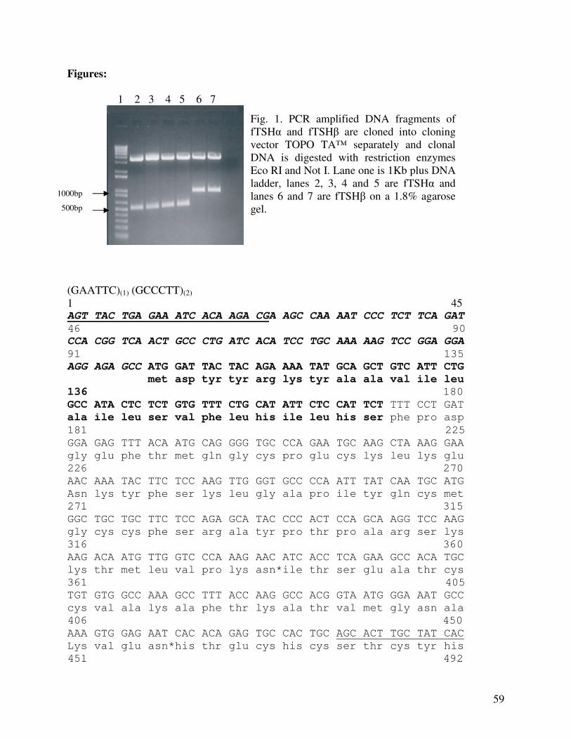
59
Figures:
1 2 3 4 5 6 7
(GAATTC)(1) (GCCCTT)(2) 1 45 AGT TAC TGA GAA ATC ACA AGA CGA AGC CAA AAT CCC TCT TCA GAT
46 90
CCA CGG TCA ACT GCC CTG ATC ACA TCC TGC AAA AAG TCC GGA GGA
91 135
AGG AGA GCC ATG GAT TAC TAC AGA AAA TAT GCA GCT GTC ATT CTG
met asp tyr tyr arg lys tyr ala ala val ile leu
136 180
GCC ATA CTC TCT GTG TTT CTG CAT ATT CTC CAT TCT TTT CCT GAT
ala ile leu ser val phe leu his ile leu his ser phe pro asp
181 225
GGA GAG TTT ACA ATG CAG GGG TGC CCA GAA TGC AAG CTA AAG GAA
gly glu phe thr met gln gly cys pro glu cys lys leu lys glu
226 270
AAC AAA TAC TTC TCC AAG TTG GGT GCC CCA ATT TAT CAA TGC ATG
Asn lys tyr phe ser lys leu gly ala pro ile tyr gln cys met
271 315
GGC TGC TGC TTC TCC AGA GCA TAC CCC ACT CCA GCA AGG TCC AAG
gly cys cys phe ser arg ala tyr pro thr pro ala arg ser lys
316 360
AAG ACA ATG TTG GTC CCA AAG AAC ATC ACC TCA GAA GCC ACA TGC
lys thr met leu val pro lys asn*ile thr ser glu ala thr cys
361 405
TGT GTG GCC AAA GCC TTT ACC AAG GCC ACG GTA ATG GGA AAT GCC
cys val ala lys ala phe thr lys ala thr val met gly asn ala
406 450
AAA GTG GAG AAT CAC ACA GAG TGC CAC TGC AGC ACT TGC TAT CAC
Lys val glu asn*his thr glu cys his cys ser thr cys tyr his
451 492
Fig. 1. PCR amplified DNA fragments of fTSHα and fTSHβ are cloned into cloning vector TOPO TA™ separately and clonal DNA is digested with restriction enzymes Eco RI and Not I. Lane one is 1Kb plus DNA ladder, lanes 2, 3, 4 and 5 are fTSHα and lanes 6 and 7 are fTSHβ on a 1.8% agarose gel.
1000bp
500bp
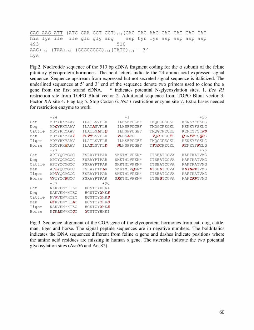
60
CAC AAG ATT (ATC GAA GGT CGT)(3)(GAC TAC AAG GAC GAT GAC GAT
his lys ile ile glu gly arg asp tyr lys asp asp asp asp
493 510
AAG)(4) (TAA)(5) (GCGGCCGC)(6)(TATG)(7) - 3’
Lys
Fig.2. Nucleotide sequence of the 510 bp cDNA fragment coding for the α subunit of the feline pituitary glycoprotein hormones. The bold letters indicate the 24 amino acid expressed signal sequence. Sequence upstream from expressed but not secreted signal sequence is italicized. The underlined sequences at 5’ and 3’ end of the sequence denote two primers used to clone the α
gene from the first strand cDNA. * indicates potential N-glycosylation sites. 1. Eco R1 restriction site from TOPO Blunt vector 2. Additional sequence from TOPO Blunt vector 3. Factor XA site 4. Flag tag 5. Stop Codon 6. Not 1 restriction enzyme site 7. Extra bases needed for restriction enzyme to work.
-24 +1 +26
Cat MDYYRKYAAV ILAILSVFLH ILHSFPDGEF TMQGCPECKL KENKYFSKLG
Dog MDCYRKYAAV ILAIASVFLH ILHSFPDGEF TMQGCPECKL KENKYFSKLG
Cattle MDYYRKYAAV ILAILSLFLQ ILHSFPDGEF TMQGCPECKL KENKYFSKPD
Man MDYYRKYAAI FLVTLSVFLH VLHSAPD--- -VQDCPECTL QENPFFSQPG
Tiger MDYYRKYAAV ILAILSVFLH ILHSFPDGEF TMQGCPECKL KENKYFSKLG
Horse MDYYRKHAAV ILATLSVFLD NLHSFPDGEF TTQDCPECKL RENKYFFKLG
+27 +76
Cat APIYQCMGCC FSRAYPTPAR SKKTMLVPKN* ITSEATCCVA KAFTKATVMG
Dog APIYQCMGCC FSRAYPTPAR SKKTMLVPKN* ITSEATCCVA KAFTKATVMG
Cattle APIYQCMGCC FSRAYPTPAR SKKTMLVPKN* ITSEATCCVA KAFTKATVMG
Man APLYQCMGCC FSRAYPTPLR SKKTMLVQKN* VTSESTCCVA KSYNRVTVMG
Tiger APVYQCMGCC FSRAYPTPAR SKKTMLVPKN* ITSEATCCVA KAFTKATVMG
Horse VPIYQCKGCC FSRAYPTPAR SRKTMLVPKN* ITSESTCCVA KAFIRVTVMG
+77 +96
Cat NAKVEN*HTEC HCSTCYHHKI
Dog NAKVEN*HTEC HCSTCYYHKS
Cattle NVRVEN*HTEC HCSTCYYHKS
Man GFKVEN*HTAC HCSTCYYHKS
Tiger NAKVEN*HTEC HCSTCYYHKS
Horse NIKLEN*HTQC YCSTCYHHKI
Fig.3. Sequence alignment of the CGA gene of the glycoprotein hormones from cat, dog, cattle, man, tiger and horse. The signal peptide sequences are in negative numbers. The bold/italics indicates the DNA sequences different from feline α gene and dashes indicate positions where the amino acid residues are missing in human α gene. The asterisks indicate the two potential glycosylation sites (Asn56 and Ans82).
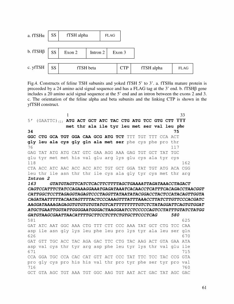
61
a. fTSHα b. fTSHβ c. yfTSH Fig.4. Constructs of feline TSH subunits and yoked fTSH 5’ to 3’. a. fTSHα mature protein is preceeded by a 24 amino acid signal sequence and has a FLAG tag at the 3’ end. b. fTSHβ gene includes a 20 amino acid signal sequence at the 5’ end and an intron between the exons 2 and 3. c. The orientation of the feline alpha and beta subunits and the linking CTP is shown in the yfTSH construct. 1 33
5’ (GAATTC)(1) ATG ACT GCT ATC TAC CTG ATG TCC GTG CTT TTT
met thr ala ile tyr leu met ser val leu phe
34 75
GGC CTG GCA TGT GGA CAA GCG ATG TCT TTT TGT TTT CCA ACT
gly leu ala cys gly gln ala met ser phe cys phe pro thr
76 117
GAG TAT ATG ATG CAT GTC GAA AGG AAA GAG TGT GCT TAT TGC
glu tyr met met his val glu arg lys glu cys ala tyr cys
118 162
CTA ACC ATC AAC ACC ACC ATC TGT GCT GGA TAT TGT ATG ACA CGG
leu thr ile asn thr thr ile cys ala gly tyr cys met thr arg
Intron 2
163 GTATGTAGTTCATCTCACTTCTTTTAGCTGAAAATTAGATAAACCTAGACT
CAGTCCATTTCTATCCAGAAAGGAAATGAGATAAATCACAACCTCATTTCACAGACCTAACGGT
CATTGGCTCCTTAGAGGTAGAGTCCCTAGGTTATAATATACGGACCTACTCCATACAGTTGGTA
CAGATAATTTTTACAATAGTTTTACTCCCAAAGTTTATTTAAACCTTATCTTGTTCCCACGATC
AAGGATAAAAGAGAGGTGTGTGTGTATGTCATTTTTTTTTGTCTCTATAGGATTCAGTGTGGAT
ATGCTGAATTGGTATTGGGGAATGGGACTAAGGAATCCTCCCCCAGTCCTATTTGTATCTATGG
GATGTAAGCGAATTAACATTTTGCTTCCTCTTCTGTGCTTCCCTCAG 580
581 625
GAT ATC AAT GGC AAA CTG TTT CTT CCC AAA TAT GCT CTG TCC CAA
asp ile asn gly lys leu phe leu pro lys tyr ala leu ser gln
626 670
GAT GTT TGC ACC TAC AGA GAC TTC CTG TAC AAG ACT GTA GAA ATA
asp val cys thr tyr arg asp phe leu tyr lys thr val glu ile
671 715
CCA GGA TGC CCA CAC CAT GTT ACT CCC TAT TTC TCC TAC CCG GTA
pro gly cys pro his his val thr pro tyr phe ser tyr pro val
716 760
GCT GTA AGC TGT AAA TGT GGC AAG TGT AAT ACT GAC TAT AGC GAC
SS fTSH beta CTP fTSH alpha FLAG
SS fTSH alpha FLAG
SS Exon 2 Intron 2 Exon 3
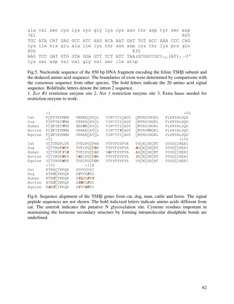
62
ala val ser cys lys cys gly lys cys asn thr asp tyr ser asp
761 805
TGC ATA CAT GAG GCC ATC AAG ACA AAT GAT TGT ACC AAA CCC CAG
cys ile his glu ala ile lys thr asn asp cys thr lys pro gln
806 835
AAG TCC GAT GTG GTA GGA GTT TCT ATC TAA(GCGGCCGC)(2)(AT)3 -3’
lys ser asp val val gly val ser ile stop
Fig.5. Nucleotide sequence of the 850 bp DNA fragment encoding the feline TSHβ subunit and the deduced amino acid sequence. The boundaries of exon were determined by comparisons with the consensus sequence from other species. The bold letters indicate the 20 amino acid signal sequence. Bold/italic letters denote the intron 2 sequence. 1. Eco R1 restriction enzyme site 2. Not 1 restriction enzyme site 3. Extra bases needed for restriction enzyme to work.
+1 +50
Cat FCFPTEYMMH VERKECAYCL TIN*TTICAGY CMTRDINGKL FLPKYALSQD
Dog FCFPTEYTMH VERKECAYCL TIN*TTICAGY CMTRDINGKL FLPKYALSQD
Human FCIPTEYTMH IERRECAYCL TIN*TTICAGY CMTRDINGKL FLPKYALSQD
Bovine FCIPTEYMMH VERKECAYCL TIN*TTVCAGY CMTRDVNGKL FLPKYALSQD
Equine FCIPTEYMMH VERKECAYCL TIN*TTICAGY CMTRDINGKL FLPKYALSQD
+51 +100
Cat VCTYRDFLYK TVEIPGCPHH VTPYFSYPVA VSCKCGKCNT DYSDCIHEAI
Dog VCTYRDFMYK TVEIPGCPRH VTPYFSYPVA ASCKCGKCNT DYSDCIHEAI
Human VCTYRDFIYR TVEIPGCPLH VAPYFSYPVA LSCKCGKCNT DYSDCIHEAI
Bovine VCTYRDFMYK TAEIPGCPRH VTPYFSYPVA ISCKCGKCNT DYSDCIHEAI
Equine VCTYRDFMYK TVEIPGCPDH VTPYFSYPVA VSCKCGKCNT DYSDCIHEAI
+101 +118
Cat KTNDCTKPQK SDVVGVSI
Dog KTNYCTKPQK SYVVGFSI
Human KTNYCTKPQK SYLVGFSV
Bovine KTNYCTKPQK SYMVGFSI
Equine KANYCTKPQK SYVVEFSI
Fig.6. Sequence alignment of the TSHβ genes from cat, dog, man, cattle and horse. The signal peptide sequences are not shown. The bold italicized letters indicate amino acids different from cat. The asterisk indicates the putative N glycosylation site. Cysteine residues important in maintaining the hormone secondary structure by forming intramolecular disulphide bonds are underlined.
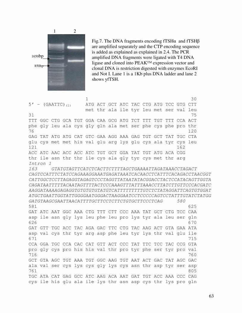
63
1 2
1 30
5’ - (GAATTC)(1) ATG ACT GCT ATC TAC CTG ATG TCC GTG CTT
met thr ala ile tyr leu met ser val leu
31 75
TTT GGC CTG GCA TGT GGA CAA GCG ATG TCT TTT TGT TTT CCA ACT
phe gly leu ala cys gly gln ala met ser phe cys phe pro thr
76 120
GAG TAT ATG ATG CAT GTC GAA AGG AAA GAG TGT GCT TAT TGC CTA
glu cys met met his val glu arg lys glu cys ala tyr cys leu
121 162
ACC ATC AAC ACC ACC ATC TGT GCT GGA TAT TGT ATG ACA CGG
thr ile asn thr thr ile cys ala gly tyr cys met thr arg
Intron 1
163 GTATGTAGTTCATCTCACTTCTTTTAGCTGAAAATTAGATAAACCTAGACT
CAGTCCATTTCTATCCAGAAAGGAAATGAGATAAATCACAACCTCATTTCACAGACCTAACGGT
CATTGGCTCCTTAGAGGTAGAGTCCCTAGGTTATAATATACGGACCTACTCCATACAGTTGGTA
CAGATAATTTTTACAATAGTTTTACTCCCAAAGTTTATTTAAACCTTATCTTGTTCCCACGATC
AAGGATAAAAGAGAGGTGTGTGTGTATGTCATTTTTTTTTGTCTCTATAGGATTCAGTGTGGAT
ATGCTGAATTGGTATTGGGGAATGGGACTAAGGAATCCTCCCCCAGTCCTATTTGTATCTATGG
GATGTAAGCGAATTAACATTTTGCTTCCTCTTCTGTGCTTCCCTCAG 580
581 625
GAT ATC AAT GGC AAA CTG TTT CTT CCC AAA TAT GCT CTG TCC CAA
asp ile asn gly lys leu phe leu pro lys tyr ala leu ser gln
626 670
GAT GTT TGC ACC TAC AGA GAC TTC CTG TAC AAG ACT GTA GAA ATA
asp val cys thr tyr arg asp phe leu tyr lys thr val glu ile
671 715
CCA GGA TGC CCA CAC CAT GTT ACT CCC TAT TTC TCC TAC CCG GTA
pro gly cys pro his his val thr pro tyr phe ser tyr pro val
716 760
GCT GTA AGC TGT AAA TGT GGC AAG TGT AAT ACT GAC TAT AGC GAC
ala val ser cys lys cys gly lys cys asn thr asp tyr ser asp
761 805
TGC ATA CAT GAG GCC ATC AAG ACA AAT GAT TGT ACC AAA CCC CAG
cys ile his glu ala ile lys thr asn asp cys thr lys pro gln
Fig.7. The DNA fragments encoding fTSHα and fTSHβ are amplified separately and the CTP encoding sequence is added as explained as explained in 2.4. The PCR amplified DNA fragments were ligated with T4 DNA ligase and cloned into PEAK™ expression vector and clonal DNA is restriction digested with enzymes EcoRI and Not I. Lane 1 is a 1Kb plus DNA ladder and lane 2 shows yfTSH.
1650bp
850bp
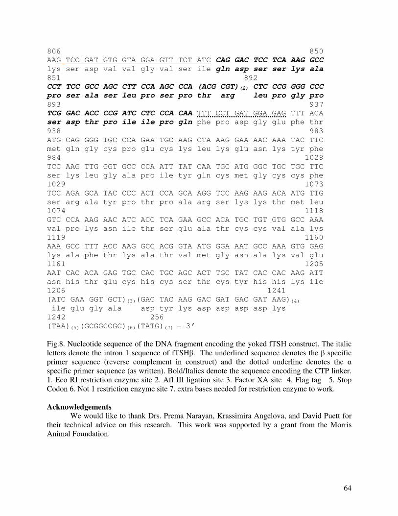
64
806 850
AAG TCC GAT GTG GTA GGA GTT TCT ATC CAG GAC TCC TCA AAG GCC
lys ser asp val val gly val ser ile gln asp ser ser lys ala
851 892
CCT TCC GCC AGC CTT CCA AGC CCA (ACG CGT)(2) CTC CCG GGG CCC
pro ser ala ser leu pro ser pro thr arg leu pro gly pro
893 937
TCG GAC ACC CCG ATC CTC CCA CAA TTT CCT GAT GGA GAG TTT ACA
ser asp thr pro ile ile pro gln phe pro asp gly glu phe thr
938 983
ATG CAG GGG TGC CCA GAA TGC AAG CTA AAG GAA AAC AAA TAC TTC
met gln gly cys pro glu cys lys leu lys glu asn lys tyr phe
984 1028
TCC AAG TTG GGT GCC CCA ATT TAT CAA TGC ATG GGC TGC TGC TTC
ser lys leu gly ala pro ile tyr gln cys met gly cys cys phe
1029 1073
TCC AGA GCA TAC CCC ACT CCA GCA AGG TCC AAG AAG ACA ATG TTG
ser arg ala tyr pro thr pro ala arg ser lys lys thr met leu
1074 1118
GTC CCA AAG AAC ATC ACC TCA GAA GCC ACA TGC TGT GTG GCC AAA
val pro lys asn ile thr ser glu ala thr cys cys val ala lys
1119 1160
AAA GCC TTT ACC AAG GCC ACG GTA ATG GGA AAT GCC AAA GTG GAG
lys ala phe thr lys ala thr val met gly asn ala lys val glu
1161 1205
AAT CAC ACA GAG TGC CAC TGC AGC ACT TGC TAT CAC CAC AAG ATT
asn his thr glu cys his cys ser thr cys tyr his his lys ile
1206 1241
(ATC GAA GGT GCT)(3)(GAC TAC AAG GAC GAT GAC GAT AAG)(4)
ile glu gly ala asp tyr lys asp asp asp asp lys
1242 256
(TAA)(5)(GCGGCCGC)(6)(TATG)(7) - 3’
Fig.8. Nucleotide sequence of the DNA fragment encoding the yoked fTSH construct. The italic letters denote the intron 1 sequence of fTSHβ. The underlined sequence denotes the β specific primer sequence (reverse complement in construct) and the dotted underline denotes the α specific primer sequence (as written). Bold/Italics denote the sequence encoding the CTP linker. 1. Eco RI restriction enzyme site 2. Afl III ligation site 3. Factor XA site 4. Flag tag 5. Stop Codon 6. Not 1 restriction enzyme site 7. extra bases needed for restriction enzyme to work. Acknowledgements
We would like to thank Drs. Prema Narayan, Krassimira Angelova, and David Puett for their technical advice on this research. This work was supported by a grant from the Morris Animal Foundation.
65
References
1. Pierce JG, Parsons TF. Glycoprotein hormones: structure and function. Annu Rev Biochem 1981;50:465-495.
2. Magner JA. Thyroid stimulating hormone: Biosynthesis, cell biology and bioactivity.
Endocrinol Rev 1990;11:345-385. 3. Vamvakopoulos NC, Kourides IA. Identification of separate mRNAs coding for the alpha
and beta subunits of thyrotropin. Proc Natl Acad Sci USA 1979;76:3809-13.
4. Williams DA, Scott-Moncrieff C, Bruner J, Sustarsic D, Panosian-Sahakian N, Unver E, el Shami AS. Validation of an immunoassay for canine thyroid stimulating hormone and changes in serum concentration following induction of hypothyroidism in dogs. J Am Vet Med Assoc. 1996;209:1730-1732.
5. Graham PA, Refal KR, Nachreiner RF and Provencher-Bollinger AL. The measurement
of feline thyrotropin using a commercial canine immunoradiometric assay: J Vet Intern Med 2000;14:342 (Abstract).
6. Turrel JM, Feldman EC, Nelson RW, Cain GR. Thyroid carcinoma causing
heperthyroidiam in cats: 14 cases (1981-1986). J Am Vet Med Assoc 193:359-364. 7. Grossmann M, Wong R, Mariusz W, Szkudlinski MW, Weintraub BD. Human Thyroid-
stimulating Hormone (hTSH) Subunit Gene Fusion Produces hTSH with Increased Stability and Serum Half-life and Compensates for Mutagenesis-induced Defects in Subunit Association. J Boil Chem. 1997;272:21312-21316.
8. Narayan P, Wu C, Puett D. Functional expression of yoked human chorionic
gonadotropin in baculovirus-infected insect cells. Mol Endocrinol. 1995;9:1720-1726. 9. Menahem DB, Shariff AJ, Hyde RK, Pixley MR, Srivasthava S, Berger P, Boime I. The
position of the α and β subunits in a single chain variant of human chorionic gonadotropin affects the heterodimeric interaction of the subunits and receptor binding epitopes. J Biol Chem. 2001;276:32,29871-29879.
10. Fiddes JC, Goodman HM. Isolation, cloning and sequence analyses of the cDNA for the α-subunit of human chorionic gonagotropin. Nature. 1979;281:351-6.
11. Erwin CR, Croyle ML, Donelson JE, Maurer RA. Nucleotide sequence of cloned
complementary deoxyribonucleic acid for the alpha subunit of bovine pituitary glycoprotein hormones. Biochemistry. 198322:4856-60.
12. Godine JE, Chin WW, Habener JF. Alpha subunit of rat pituitary glycoprotein hormones.
Primary structure of the precursor determined from the nucleotide sequence of the cloned cDNAs. J Biol Chem. 1982; 257:8368-71.
66
13. Chin WW, Kronenberg HM, Dee PC, Maloof F, Haben JF. Nucleotide sequence of the
mRNA encoding the pre-α-subunit of mouse thyrotropin. Proc Natl Acad Sci USA. 1981;78:5329-33.
14. O’Brien M, Headon DR. The cloning and expression of the alpha subunit of equine
glycoprotein hormones. Biochem Soc Trans. 1995;3:347S. 15. Yang X, McGraw RA, Ferguson DC. cDNA cloning of canine common α gene and its
co-expression with canine β gene in baculovirus expression system. Domest Anim Endocrinol 2000; 18, 379-393.
16. Gharib SD, Weirman ME, Shupnik MA, Chin WW. Molecular biology of the pituitary
gonadotropins. Endocrine Reviews. 1990;11, 177-199. 17. Hayashizaki Y, Miyai K, Kato K, Matsubara K. Molecular cloning of the human
thyrotropin β-subunit gene. FEBS Lett. 1985;188:394-400. 18. Maurer RA, Croyle ML, Donelson JE. The sequence of a cloned cDNA for the beta
subunit of bovine thyrotropin predicts a protein containing both NH2- and COOH- terminal extensions. J Biol Chem. 1984;259:5024-27.
19. Wolf O, Kourides IA, Gurr JA. Expression of the gene for the beta sununit of mouse
thyrotropin results in multiple mRNAs differing in their 5’-untranslated regions. J Biol Chem. 1987;262:16596-603.
20. Croyle ML, Bhattacharya A, Gordon DF, Maurer RA. Analysis of the organization and
nucleotide sequence of the chromosomal gene for the β-subunit of rat thyrotropin. DNA. 1986;5:299-304.
21. Yang X, McGraw RA, Su X, Katakam P, Grosse WM, Li OW, Ferguson DC.
Canine thyrotropin β-subunit gene: cloning and expression in Escheria coli, generation of monoclonal antibodies and transient expression in the Chinese ovary cells. Domest Anim Endocrinol 2000;18, 363-378.
22. Wondisford FE, Radovick S, Moates JM, Usala SJ, Weintraub BD. Isolation and
characterization of the human thyrotropin β-subunit gene. Differences in gene structure and promoter function from murine species. J Biol Chem. 1988;263:12538-42.
23. Joshi L, Murata Y, Wondisford FE, Szkudlinski MW, Desai R, Weintraub BD.
Recombinant thyrotropin containing a beta-subunit chimera with the human chorionic gonadotropin-beta carboxy-terminus is biologically active, with a prolonged plasma half-life: role of carbohydrate in bioactivity and metabolic clearance. Endocrinology 1995;136: 3839-3848.
67
24. Kornfeld R, Kornfeld S. Assembly of asparagine linked oligosaccharides. Annu Rev Biochem 1985;54:631-64.
25. Ryan RJ, Charlesworth MC, McCornick DJ, Milius RP, Keutmann HT. The glycoprotein
hormones: recent studies of structure-function relationships. FASEB J 1988;2:2661-9.
26. Lapthorn AJ, Harris DC, Littlejohn A, Lustbader JW, Canfield RE, Machin KJ, Morgan FJ, Isaacs NW. Crystal structure of human chorionic gonadotropin. Nature 1994;369:455-61.
27. Chin WW, Maizel JV, Jr, Habenner JF. Difference in sizes of human compared to murine α-subunits of the glycoprotein hormones arises by four-codon gene delation or insertion. Endocrinology 1983; 112:482-5.
28. Furuhashi M, Suzuki S, Sugunama N. Disulfide bonds 7-31 and 59-87 of the α subunit
play a different role in assembly of human chorionic gonadotropin and lutropin. Endocrinology 1996;137:4196-200.
29. Furuhashi M, Ando H, Bielinska M, Pixley MR, Shikone T, Hsueh AJ, Boime I.
Mutagenesis of cysteine residues in the human gonadotropin alpha subunit. Roles of individual disulphide bonds in secretion, assembly and biologic activity. J Biol Chem 1994;269:25543-48.
30. Hayashizaki Y, Hiraoka Y, Endo Y, Matsubara K. Thyroid-stimulating hormone (TSH) deficiency caused by a single base substitution in the CAGYC region of the β -subunit. EMBO J 1989;8:2291.
31. Bause E, Breuer W, Peters S. Investigation of the active site of oligosaccharyltransferase from pig liver using synthetic tripeptides as tools. Biochem J 1995;312:979-85.
32. Fares FA, Yamabe S, Menahem DB, Pixley M, Hsueh AJW, Boime I. Conversion of
thyrotropin heterodimer to a biologically active single-chain. Endocrinology 1998;139:2459-64.
33. Sugahara T, Sato A, Kudo M, Menahem DB, Pixley MR, Hsueh AJW, Boime I
Expression of biologically active fusion genes encoding the common subunit and the follicle-stimulating hormone ß subunit. J Biol Chem 1996;271:10445–10448.
34. Sugahara T, Pixley MR, Minami S, Perlas E, Menahem DB, Hsueh AJW, Boime I.
Biosynthesis of a biologically active single peptide chain containing the human common α and chorionic gonadotropin β subunits in tandem. Proc Natl Acad Sci USA 1995;92:2041–2045.
35. Menahem DB, Boime I. Converting heterodimeric gonadotropins to genetically linked
single chains : New approaches to structure activity relationships and analogue design. Trends in Endocrinology and Metabolism 1996;7(3):100-105.
68
36. Robbins RJ, Tuttle RM, Sonenberg M, Shaha A, Sharaf R, Robbins H, Fleisher M, Larson SM. Radioiodine ablation of thyroid remnants after preparation with recombinant human thyrotropin. Thyroid 2001;11:865-869.
EXPRESSION AND PURIFICATION OF FELINE THYROTROPIN
(fTSH): IMMUNOLOGICAL DETECTION AND BIOACTIVITY OF
HETERODIMERIC AND YOKED GLYCOPROTEINS2
2S. Rayalam, L.D. Eizenstat, R.R. Davis, M. Hoenig, *D.C. Ferguson. Department of Physiology and Pharmacology, College of Veterinary Medicine, The University of Georgia, Athens, GA 30602, USA. Accepted by Domestic Animal Endocrinology. Reprinted here with permission of publisher, 10/12/05.
70
Abstract
The goal of this study was to express and purify recombinant feline TSH as a possible
immunoassay standard or pharmaceutical agent. Previously cloned feline common glycoprotein
alpha (CGA) and beta subunits were ligated into the mammalian expression vector pEAK10. The
feline CGA-FLAG and beta subunits were cloned separately into the pEAK10 expression vector,
and transiently co-transfected into PEAK™ cells. Similarly, previously cloned and sequenced
yoked (single chain) fTSH (yfTSH) and the CGA-FLAG sequences were ligated into the same
vector, and stable cell lines selected by puromycin resistance. Expression levels of at least 1
µg/ml were achieved for both heterodimeric and yoked fTSH forms. The glycoproteins were
purified in one step using anti-FLAG immunoaffinity column chromatography to high purity.
The molecular weights of feline CGA-FLAG subunit, beta subunit and yfTSH were 20.4, 17, and
45 kilodaltons, respectively. Both heterodimeric and yoked glycoproteins were recognized with
approximately 40% detection by both a commercial canine TSH immunoassay and an in-house
canine TSH ELISA. The yoked glycoprotein exhibited parallelism with the heterodimeric form
in the in-house ELISA, supporting their possible use as immunoassay standards. In bioactivity
assays, the heterodimeric and yoked forms of fTSH were 12.5 and 3.4 % as potent as pituitary
source bovine TSH at displacing 125I-bTSH and 45 and 24 % as potent in stimulating adenylate
cyclase activity in human TSH receptor-expressing JP09 cells. However, in addition to reduced
receptor binding affinity, the recombinant glycohormones produced a reduced maximal effect at
maximal concentration (Emax) suggesting the possibility of the recombinant glycohormone
constructs acting as partial agonists of the human TSH receptor.
71
Running Title: Expression and Purification of Feline TSH
*Corresponding author: Telephone: 706-542-5864 FAX: 706-542-3015 Email:
[email protected] (D.C. Ferguson)
This work was presented in part at the 23rd Annual Forum of the American College of Veterinary
Internal Medicine, Baltimore, MD, 2005, abstract 233.
Keywords: thyrotropin; TSH; feline; pituitary; expression
72
1. Introduction
TSH is a glycoprotein hormone with an α subunit, common to Follicle stimulating
hormone (FSH), Luteinizing hormone (LH) and Chorionic gonadotropin (CG), and a hormone-
specific β subunit. The α and β subunits associate noncovalently and subunit assembly is
necessary for the biological activity of the hormone (1). The functional activity of these
hormones is dependent on the correct assembly of the subunits into the heterodimer.
The serum concentration of TSH, which is decreased in primary hyperthyroidism, and
increased in primary hypothyroidism, is a very sensitive indicator of thyroid function in man (2).
Currently available ultra-sensitive human TSH assays eliminate the expensive and time
consuming thyrotropin releasing hormone (TRH) stimulation tests by distinguishing euthyroid
patients from hyperthyroid patients (3, 4). Therefore TSH measurement has been widely used to
screen for thyroid dysfunction. Standards for human TSH (National Hormone and Pituitary
Program (NHPP) and World Health Organization (WHO)) continue to be cadaver-source
purified pituitary TSH. A commercial assay for canine TSH has been available since 1997 (7);
however, no widely available independent standard for this or other domestic animal TSH assays
have been developed.
Hyperthyroidism is the most commonly diagnosed thyroid disorders in middle to old
aged cats. The hospital prevalence of feline hyperthyroidism increased from 0.06 % for the
period of 1978 to 1982 to 0.3 % for the period of 1993 to 1997 and over 20 years the prevalence
of feline hyperthyroidism per visit was 2.1 % (8). Furthermore, epizootiological studies have
suggested links to dietary and/or environmental associations with this disease not recognized
prior to 1979. Canned cat food, cat litter, and pesticides have been identified as possible risk
factors for the disease (5, 6, 9). Canine TSH assays have been tried to measure TSH in feline
73
serum samples. In one study, 68% of hyperthyroid cats had TSH concentrations below the
detection limit, as might be expected; however the commercial canine TSH assay used is
generally not sensitive enough to distinguish normal from low values, particularly in the cat (10).
The development of a sensitive feline TSH (fTSH) assay has been hampered by the absence of
an available standard for fTSH. Biochemical purification of pituitary source TSH is complicated
by the presence of much higher concentrations of LH and FSH, and only about 100 µg of TSH
even in a human pituitary. Therefore, we reasoned that recombinant fTSH engineered with an
immunoaffinity tag would advance the development of a recombinant peptide-based
immunoassay to detect fTSH. The feline CGA subunit previously cloned and sequenced in our
lab encodes for a 96 amino acid protein and contains five intramolecular disulfide bonds. The
fTSHβ subunit encodes for a 138 amino acid protein and six disulfide bonds (see companion
paper). The α and β subunit structure predicted two and one N-linked oligosaccharides,
respectively. The oligosaccharides have been shown to play an important role in bioactivity of
human TSH (11).
Recently, single chain or yoked (also called tethered) analogs of hTSH and hCG have
been constructed with the C-terminus of the β subunit fused using a yoking peptide, the carboxy
terminal peptide (CTP) of hCGβ to the N-terminus of the α-subunit. Expression of these
recombinant single chain hormones has allowed extensive structure-function studies and also has
resulted in generation of hormones with increased stability and, in some cases, activity (12, 13).
Grossmann et al., showed that the genetic fusion of hTSH α- and β-subunits using the carboxy-
terminal peptide of the hCGβ subunit as a linker created a yoked form of hTSH whose receptor
binding and bioactivity were comparable to native hTSH, but had higher thermostability and a
longer plasma half-life (12). The linker CTP is not required for the assembly of the heterodimer
74
nor is it required for receptor activation (14, 15). In contrast, the presence of CTP linker appears
mainly important for the maximal expression of a functional single chain (17). The yfTSH-
FLAG previously cloned and constructed in our lab encodes a 1260 bp glycoprotein with the
sequence from N-terminus to C-terminus of β-CTP-α-FLAG.
The synthesis, purification, characterization, pharmacokinetic and toxicological
properties of recombinant hTSH for human use were described for the first time in 1993 (16). In
2003, our laboratory reported the expression of recombinant canine TSH in a baculovirus
expression system (18). The objective of the present study was to express higher quantities and
to purify biologically and immunologically active recombinant feline thyrotropin. In vitro
expression in mammalian cells allows the mammalian pattern of post-translational modifications
of the recombinant hormone and facilitates the investigation of structure-function relationships.
The ideal expression system for a mammalian glycoprotein is a mammalian cell line capable of
post-translational glycosylation with a mammalian pattern of sugars. The Chinese Hamster
Ovary (CHO) cells lack sulfotransferase and N-acetyl galactosamine transferase (19). Therefore
the recombinant glycoproteins produced from CHO cells are not sulfated to the same degree as
pituitary-derived glycoproteins. However, Human Embryonic Kidney cells (HEK 293) have been
shown to sulfate pituitary glycoproteins to a greater extent (20, 21), because of the presence of
higher levels of N-acetyl galactosamine transferase and sulfotransferase activities to those
expressed in the pituitary gland (22). Moreover, the biological potency of recombinant FSH
produced in HEK 293 cells was shown to be greater than pituitary FSH, while the biological
potency of recombinant FSH produced from CHO cells was less than that of pituitary FSH (23).
This work constitutes the first report of in vitro expression and purification of recombinant feline
thyrotropin. The demonstration of immunological recognition by antibodies generated against
75
pituitary-source TSH, and of bioactivity confirms that recombinant glycoprotein may be used to
standardize and improve clinical assays for feline TSH and has potential as a pharmaceutical
agent as well.
2. Materials and methods
2.1. Materials:
All restriction enzymes necessary for cloning into pEAK10 expression vector were
purchased from New England Biolabs® Inc (Beverley, MA). Expression vector pEAK10 and the
PEAKTM Stable Expression Kit were obtained from Edge Biosystems (Gaithersburg, MD). The
High Pure Plasmid Isolation kitTM was from Roche Diagnostics (Indianapolis, IN). One ShotTM
cells, fetal bovine serum (FBS) and penicillin-streptomycin were from Invitrogen Corporation
(Carlsbad, CA). The Rapid DNATM ligation kit was purchased from Roche Diagnostics
Corporation (Indianapolis, IN). Dulbecco’s Modified Eagles Medium (DMEM), Waymouth`s
MB752/1 medium, Nutrient Mixture F-12 Ham, puromycin, ANTI-FLAG® M2-Agarose Affinity
Gel, FLAG® Peptide, ANTI-FLAG® M2 monoclonal antibody, cAMP salt, anti-cAMP
polyclonal antibody, 3-isobutyl-1-methylxanthine (IBMX), Mouse IgG, Goat anti-rabbit-
Peroxidase conjugate and Tetramethyl-benzidine were all from Sigma (St. Louis, MO). Micro
BCA™ Protein Assay Kit, AminoLink® kit and West Dura Super Signal™ substrate were from
Pierce (Rockford, IL). Immulon™ 4HBX strips were from ThermoLabsystems (Franklin, MA),
and Canine TSH was from Scripps Laboratories (San Diego, CA) and Dr. A.F. Parlow (National
Hormone and Peptide Program (NHPP)). Immobilon™ transfer membrane was purchased from
Millipore (Bedford, MA). Forskolin was from Fisher Scientific (Pittsburg, PA). The automated
microplate reader was from Bio-Tek Instrument®, Inc (Winooski, VT). All the chemicals for
making buffers and other reagents were from Sigma (St. Louis, MO). The ABI PRISM® 3100
76
automated sequencer from Applied Biosystems (Foster City, CA) was used by the Molecular
Genetics Instrumentation Facility (MGIF) at the University of Georgia.
2.2. Construction of expression vectors
The expression vector pEAK10 was linearized by digesting with restriction enzymes Eco
RI and Not I. Previously cloned and sequenced feline CGA was digested with the same enzymes
and ligated with linearized pEAK10 vector DNA downstream of EF1α using the Rapid DNA™
ligation kit following the manufacturer’s instructions. The ligated DNA was transformed into
One Shot™ cells and the positive clones were checked for correct insertion by digesting with
Eco RI and Not I restriction enzymes. Similarly, the previously cloned and sequenced fTSH beta
subunit was cloned downstream of EF1α into the pEAK10 expression vector separately and
insertion confirmed by restriction digests. The feline CGA construct for yoked fTSH previously
cloned and sequenced was linearized by digesting with restriction enzymes Afl III and Not I. The
fTSH beta construct for yoked fTSH was digested with Eco RI and Afl III and both the linearized
subunits were double ligated with linearized pEAK10 vector as explained above. The ligated
DNA was transformed into One Shot™ cells and positive clones were checked for inserts by
digesting with Eco RI and Not I restriction enzymes.
2.3. Transient expression of heterodimeric fTSH
The PEAKTM Stable Expression kit was used following the manufacturer’s instructions.
The PEAKTM cells were maintained in DMEM media with 10% FBS and 1% penicillin-
streptomycin. Briefly, 6 µg each of feline CGA and fTSH beta plasmid DNA were mixed with
125 µl of 1.0 M CaCl2, 500 µl of 2x Phosphate buffer and the total volume was brought up to 1
ml with water. The DNA/Calcium Phosphate suspension was added dropwise to 75 cm2 flask
with 1.5 x10⁶ PEAKTM cells in 9 ml of media, incubated at 37oC and 5% CO2 for 5 hours,
77
washed and 10 ml of fresh media added. Five days after transfection the media was harvested,
filtered and frozen at -20oC and the purification was generally performed within four weeks. A
mock transfection with empty pEAK10 vector was also performed as a control.
2.4. Stable expression of feline CGA and yfTSH
The expression vector pEAK10-w/o toxic gene has a cell type independent EF-1 α
promoter (24) and a puromycin resistance gene. The pEAK10 plasmid DNA checked for feline
CGA gene was transfected into PEAKTM cells as explained in 2.3. The transfected cells were
selected by adding 0.5 µg puromycin per ml of culture medium 24-48 hrs post transfection. The
cells were split at 80% confluency and 24 hrs later puromycin was added. Two weeks after
transfection, puromycin was added directly while splitting and concentration gradually increased
at 0.5 µg/ml weekly up to 2.0 µg/ml. Similarly a stable cell line expressing yfTSH was also
developed by transfecting PEAKTM cells with pEAK10 plasmid DNA containing the yfTSH
gene. Mock transfections were performed in both cases as controls. The conditioned medium
from stable cell lines was collected and frozen at -20oC until purified.
2.5. Purification of heterodimeric and yoked forms of fTSH and feline CGA
A column was made with 1 ml of ANTI-FLAG® M2-Agarose Affinity Gel in a pasteur
pipette and equilibrated with 3 ml of 0.1 M glycine pH 3.5 and washed with 5 ml of tris
buffered saline (TBS: 50mM Tris HCl, 150 mM NaCl, pH 7.4). The frozen expression media
was thawed, filtered and percolated by gravity through the column 3 times. Following binding,
the column was washed with 10 ml of TBS and eluted with five one-column volumes of solution
containing 100 µg/ml FLAG peptide in TBS. The elution fractions were collected into glass vials
and siliconized pipette tips used at every point to minimize protein losses. All the elution
fractions were dialyzed against 0.03 M phosphate buffer for buffer exchange and also to remove
78
FLAG peptide. A small fraction was saved for a silver stain and ELISA and the rest lyophilized
in aliquots and stored at -800C.
2.6. Purification of heterodimeric fTSH using mAb column
An affinity column was made by coupling AminoLink® coupling gel with anti-cTSH
mAb (14H9.E2) following manufacturer’s instructions. The development of this monoclonal
antibody is described below. Recombinant heterodimeric fTSH was allowed to enter the gel bed
and incubated for 60 minutes at room temperature. Bound peptide was eluted using 0.1 M
glycine pH 2.5. The eluted fractions were treated in the same way as explained in 2.5.
2.7. Characterization of purified proteins
ELISA
The purified heterodimeric and yoked forms of fTSH was detected by in-house canine
TSH ELISA employing a capture monoclonal antibody (14H9.E2) (mAb) previously selected for
ovine/canine TSH beta and a polyclonal antibody (pAb) generated previously against pituitary-
source canine TSH (25). Pituitary-source canine TSH (NHPP) was used as a standard. Immulon
4HBX strips were coated with 1.5 µg per well of 14H9.E2 monoclonal antibody in 200 µl PBS
and subsequently blocked with blocking buffer (5 g/dl of sucrose and 2g/dl of bovine serum
albumin (BSA) in PBS pH 7.0). A standard curve with 100 µl of solution contained cTSH
ranging from 0.31 ng/ml to 20 ng/ml in an immunoassay buffer consisting of 25 mM Tris base,
10 mM EGTA, 0.15 mM KCl, 2 mg/ml BSA and 0.5% Tween pH 7.5. Anti-cTSH polyclonal
antibody was diluted in the immunoassay buffer at 100 ng IgG protein per 100 µl and mouse IgG
was added to the diluted polyclonal antibody at the rate of 100 µg/ml of antibody solution to
block the epitopes for mouse IgG. The pAb - mouse IgG mixture was added to the wells with
standards and samples. The assay was incubated at 4oC overnight to equilibrium and washed
79
three times with 300 µl of immunoassay wash buffer (20 mM Tris base, 0.15 M NaCl, 5 mM
EDTA, 0.5% Tween, pH 7). Goat anti rabbit- Immunoglobulin-peroxidase conjugate was then
added as a tertiary antibody (1:10,000 in immunoassay buffer). The assay was incubated for 60
minutes at room temperature, washed and TMB substrate added. The reaction was allowed to
proceed for 30 minutes or until a blue color develops and then stopped with 1 M HCl or 1M
H2SO4. The absorbance was read at 450nm in an automated microplate reader.
Protein assay
Apart from measurement of immunoassayable recombinant fTSH levels with ELISA, the
gravimetric measurement of total protein was performed by Bicinchoninic acid (BCA) protein
assay (26). Along with bovine serum albumin standards in BCA assay, bovine TSH standards
were also used for accurate measurement of recombinant TSH levels. The total protein and
percentage purity of recombinant peptide as determined by silver stain were used to arrive at the
gravimetric concentrations of purified recombinant fTSH.
Gel electrophoresis and immunoblotting
Purified fTSH in both heterodimeric and yoked forms (100-150 ng/lane) were heat-
denatured in reducing buffer (β-mercaptoethanol) and run on a 16.5 % SDS-PAGE as previously
described (27). The protein bands were subsequently transferred to an ImmobilonTM transfer
membrane with blotting buffer containing 25 mM Tris pH 8.3, 192 mM glycine and 10%
methanol. The blotted membrane was blocked with PBS pH 8.2 containing 5% nonfat dry milk
and 0.05% Tween 20 for 30 minutes at room temperature. Anti-FLAG® M2 monoclonal
antibody IgG protein was diluted in PBS containing 0.1% BSA and 0.05% Tween 20 (antibody
incubation buffer) to 5 µg/ml and allowed to bind overnight at 4oC. The membrane was then
washed three times with PBS and incubated with goat anti-mouse peroxidase conjugate in
80
antibody incubation buffer for 2 hours at room temperature. The protein band was visualized
using West Dura Super SignalTM substrate for 2 minutes and the lanes imaged and quantified
with a Fluor-S Max MultiImagerTM (Bio-Rad Laboratories, Inc. Hercules, CA). Protein bands in
the gel were also detected by silver staining and purity estimated by densitometry.
TSH Receptor Binding Assay
Competitive binding experiments were performed with a stable cell line of CHO cells
expressing the recombinant human TSH receptor (rhTSHR), clone JP09 (28). Pituitary source
bTSH was iodinated as described previously (36). The cells were maintained in Ham’s F12 (HF-
12) media supplemented with 10% fetal bovine serum and 1% penicillin-streptomycin and were
maintained with geneticin selection at 400 µg per ml of media. Cells were plated in 12-well
plates, grown until confluency and were incubated with 1 ng (105 cpm) 125I bTSH and various
dilutions of unlabelled TSH in binding buffer (278 mM sucrose, 5 mM Hepes, 1 mM CaCl2, 5
mM KCl, 1.23 mM KH2PO4, 1.2 mM MgSO4, 1 mM NaHCO3, 5.5 mM glucose, 0.1% BSA)
and incubated at room temperature for 6 hrs. The cells were washed and subsequently solubilized
with 1N NaOH and counted in an automated gamma counter (PerkinElmer, Inc., Shelton, CT).
TSH stimulation of JP09 cells
JP09 cells were grown in 12 well plates to confluence, the media removed and incubated
in Waymouth`s MB752/1 medium with 0.1% BSA and 0.8 mM isobutylmethylxanthine (IBMX)
for 15 min at 37oC. The cells were then incubated in various concentrations of peptide for 30 min
at 37oC in Waymouth`s medium containing 0.1% BSA and 0.8 mM IBMX immediately
following first incubation. The cells were also incubated with 0.1 mM forskolin to determine the
maximal cAMP response. The incubation medium was removed, and the cells were lysed in
81
100% ethanol at -20oC overnight. The extract was collected, dried under vacuum, and dissolved
in 1 ml of 50 mM sodium acetate pH 6.2.
Cyclic AMP Radioimmunoassay
Cyclic AMP was iodinated with 125I using a standard chloramine T method (29). Briefly,
1 mCi of 125I, 25 µl of deionized water, 5 µl of cAMP methyl ester (2.5 µg), 50 µl of 0.5 mM
sodium phosphate pH 7.4 and 10 µl chloramine T (165 mg/10 ml of 0.5 M sodium phosphate
buffer) were allowed to react for 60 seconds and the reaction terminated by adding 20 µl of
sodium metabisulfite (120 mg/10 ml of 0.5 M sodium phosphate buffer). Diluted reaction
mixture was added to a DEAE cellulose column. The bound labeled peptide was eluted with 5
mM, 10 mM and 50 mM concentrations of potassium phosphate pH 4.4. The first peak (in the
wash) constituted free iodide and the iodinated cAMP in the second peak (usually in 10 mM
potassium phosphate elution) was pooled. The radioimmunoassay standards were made using
cAMP salt ranging from 50 pmol/ml to 0.1 pmol/ml in 50 mM sodium acetate buffer pH 6.2. The
unknowns were diluted at 1:25 for TSH and forskolin - stimulated samples, and 1:10 for basal
samples in 50 mM sodium acetate buffer pH 6.2. For the RIA, 100 µl of standard or sample was
succinylated using acetylating reagent (2:1 of triethylamine and acetic anhydride) at 2.5 µl/tube
and 100 µl of anti cAMP pAb was added per tube. The tubes were incubated for 4 hrs at 4°C and
~ 4-5 nCi/tube of 125I – cAMP was added and again incubated 16-18 hours at 4°C. Then 100 µl of
carrier 10% BSA and 2 ml of ethanol was added to tubes, spun at 2000xg for 15 min, the
supernatant aspirated and the pellet counted in gamma counter.
2.7. Statistical analysis
The statistical analysis of binding assays and cAMP assays was carried out by using
GraphPad PRISM program. All graphical data were also prepared using GraphPad Prism.
82
Nonlinear regression was used to fit both binding and adenylate cyclase data to a sigmoidal
curve when plotted against the log of the TSH concentration. The goodness of fit is reported as R
and Emax and EC50 calculated. Significance of differences between Emax and EC50 for bTSH,
fTSH and yfTSH in adenylate cyclase and binding assays were calculated by performing
unpaired t tests. The slopes and intercepts for immunoreactivity data were also compared by
performing unpaired t tests. A p value of ≤ 0.05 was considered significant. Means and standard
error were reported as appropriate.
3. Results
3.1. Expression and purification of heterodimeric fTSH
The time course of expression of recombinant heterodimeric fTSH following transient
transfection showed an average plateau concentration of 1500 ± 400 ng/ml (mean ± SEM, n=6)
4 days after transfection (Fig. 1). The expression media was purified using ANTI-FLAG® M2-
Agarose Affinity Gel, and 47.6 ± 9.8 % (mean ± SEM, n=4 preparations) of immunoassayable
TSH protein was recovered. The recovery of the protein from the column was consistent with
each purification (Table 1). When the column eluate was analyzed on a 16.5 % SDS gel, protein
bands with molecular weights of 21 kDa and 17 kDa for fTSHα and fTSHβ, respectively, were
visualized with silver stain (Fig.2). Another band of about 23 kDa molecular weight (Fig. 2) for
free alpha was also observed with fTSH purified using ANTI-FLAG® M2-Agarose affinity gel,
indicating that the anti-FLAG column bound not only heterodimeric fTSH but also free alpha
subunit expressed in the media. The free alpha subunit was removed by an anti-cTSHβ
monoclonal antibody column as described in 2.6 (Fig. 2). This antibody has been shown in our
laboratory only to bind to canine TSH in a Western blot only when non-denaturing
electrophoretic conditions are used. (data not shown). A Western blot for purified protein using
83
anti-FLAG monoclonal antibody showed a broad band at about 21 kDa (Fig.3.) consistent with
the glycosylated α – FLAG subunit in heterodimeric form.
3.2. Expression and purification of yoked fTSH and feline CGA
The final structure of the expression vector cloned with yfTSH is shown in Fig.4. The
PEAKTM cells were stably transfected with feline CGA and yfTSH as explained in 2.4 to develop
a stable cell line expressing the recombinant protein. The peak media concentration of yfTSH
averaged 800 ± 350 ng/ml (mean ± SEM, n=6) and the expression media was purified using
ANTI-FLAG® M2-Agarose affinity gel with a recovery of 43 ± 3% (mean ± SEM, n=4
preparations). A western blot on the purified yfTSH using anti-FLAG monoclonal antibody
showed a band at about 45 kDa (Fig.3) and that for feline CGA showed at about 21 kDa.
3.3. Immunological characterization of recombinant fTSH
The average expression concentrations in medium as determined by the in-house ELISA
were 1500 ± 400 ng/ml and 800 ± 350 ng/ml (mean ± SEM, n=6 preparations) for heterodimeric
and yoked forms of fTSH respectively. Both the recombinant fTSH forms were also detected by
the commercially available canine TSH assay (Diagnostic Products Corp., Los Angeles, CA).
However, In-house assay detected about 38.5±2.8 % and DPC assay detected 35.7±4.8 %
(mean±SEM) of the recombinant fTSH in both forms as standardized by BCA protein assay.
Both the heterodimeric and yoked forms of fTSH showed immunological parallelism
with pituitary source cTSH (NHPP) in our in-house ELISA (Fig.5). The IC50 (ng/ml±SE, n=3)
concentrations for pituitary source cTSH (NHPP), heterodimeric and yoked forms of fTSH were
3.3±1.9, 4.3±1.2 and 3.8±1.2 and slopes were 1.36±0.2, 1.29±0.03 and 1.32±0.02 respectively.
Both the IC50 values and slopes of all the three peptides were not significantly different from
each other.
84
3.4. Biological activity of recombinant fTSH
To demonstrate the biological activity of recombinant forms of fTSH, hTSH receptor
binding inhibition and adenylate cyclase activity were measured. As shown in Fig.6, the
heterodimeric and yoked forms of fTSH were able to inhibit the binding of 125I-bTSH to the
hTSH receptor in JP09 cells but with different affinities (Table 2).
The bioactivities of recombinant fTSH constructs were also evaluated by potency to
stimulate adenylate cyclase in the human TSH receptor-expressing JP09 cells. Heterodimeric
fTSH was almost 2-fold more potent than yoked fTSH in stimulating cAMP production (Fig.7).
However, heterodimeric fTSH was 2-fold and yfTSH was about 4-fold less potent in signal
transduction than pituitary source bTSH (Table 2). The maximal cAMP concentrations upon
stimulation with 100 ng/ml of TSH peptides in comparison with forskolin stimulation are
significantly different from each other and also different from forskolin (Fig. 8).
The ratio between biological potency and immunoactivity has been used as an index of
the overall potency of circulating TSH in vitro (40). The Biological to Immunological ratio (B/I)
for yfTSH for binding and cAMP production was 27 % and 52 %, respectively, of that of
heterodimeric fTSH (Table. 3).
4. Discussion
Previously, we cloned and sequenced feline CGA and feline thyrotropin beta genes
separately (companion publication) and the present study constitutes the first report of the in
vitro expression and purification of recombinant feline thyrotropin in heterodimeric and yoked
forms. Lacking a commercially available source for pituitary fTSH, we reasoned that by
demonstrating immunological parallelism to other standards, recombinant fTSH can be
substituted for pituitary source fTSH as a standard. In fact, using similar purity criteria, we were
85
able to demonstrate that recombinant fTSH was more highly purified than available pituitary
source canine TSH standards (Fig. 9).
Immunoreactive and bioactive fTSH was efficiently expressed from PEAKTM cells, and
the expression media concentrations obtained (~1 µg/ml) were considerably higher than the
concentrations obtained with recombinant hTSH expression using CHO and other eukaryotic
cells (20, 30, 31). Furthermore, the synthesis of yoked fTSH indicates that the linked alpha and
beta subunits presumably folded with formation of disulfide bonds and a stable conformation.
The highly flexible connecting linker CTP apparently still allowed the interaction of alpha and
beta subunits, with recognition by both TSH receptor and anti-TSH antibodies. Surprisingly,
compared to available pituitary standards (canine for immunoassay and bovine for bioassay), the
recombinant glycoproteins were detected with equal affinity by the immunoassay systems, but
showed reduced receptor affinity and diminished maximal effect on the bioactivity assay,
consistent with a partial agonist.
The alpha and beta subunits were previously cloned into pEAK10 expression vector
separately and transiently transfected using calcium phosphate into PEAKTM cells. The
expression vector has a single EF-1α promoter which facilitates expression of only one gene at a
time. Interestingly, in a manner similar to pituitary thyrotrophs (37), equimolar expression of
feline CGA and fTSH beta subunits did not occur as shown by affinity chromatography
purification using anti-FLAG M2 resin (Fig.2). The biosynthesis of human recombinant
glycoprotein hormones in cell cultures have shown the presence of alpha subunit
immunoreactivity (32) and evidence that it is associated with ‘free alpha’ subunit which has a
slightly higher molecular weight due to an additional O-linked oligosaccharide (32, 33). This
sugar residue is located in a domain of the alpha subunit believed to normally interact with the
86
beta subunit during heterodimer formation (1). Therefore, the free alpha subunit expressed
independently from the beta subunit is subject to glycosylation with one additional sugar moiety
during post-translational processing. An affinity column made with agarose beads linked to a
beta-subunit specific monoclonal antibody (14H9.E4) captured heterodimeric fTSH alone
removing the free alpha subunit and increasing the purity of the recombinant fTSH preparation
(Fig.2).
Having demonstrated the expression and purification of recombinant fTSH in
heterodimeric and yoked forms, the next step was to assess the immunological activity of the
recombinant protein. Lacking pituitary source fTSH, the immunological activity of fTSH in both
forms was compared with pituitary source canine TSH (NHPP standard) and the results indicated
that both forms of fTSH showed immunological parallelism to pituitary-source TSH supporting
the possibility that recombinant fTSH could be substituted for pituitary source TSH as an
immunoassay standard. However, the in-house ELISA detected both heterodimeric and yoked
forms of the recombinant protein to an extent of about 40 % as standardized by BCA protein
assay and purity determined by silver stain. The reasons for this discrepancy in both assays is not
yet clear but could reflect differences in tertiary structures and/or glycosylation differences
between recombinant and pituitary source-TSH.
It had previously been shown that bioactive hCG, hTSH and hFSH could be produced as
single chain hormones (12, 13, 34). In this study, yoked fTSH exhibited 54% of the potency of
heterodimeric fTSH and 24% of the potency of pituitary source bTSH in adenylate cyclase
assays and 27% of the potency of heterodimeric fTSH and only 3.4 % of the potency of pituitary
source bTSH at inhibiting 125I-bTSH binding in JP09 cells. The heterodimeric fTSH showed 45
% and 12.5 % of the potency of pituitary source bTSH in adenylate cyclase and binding assays
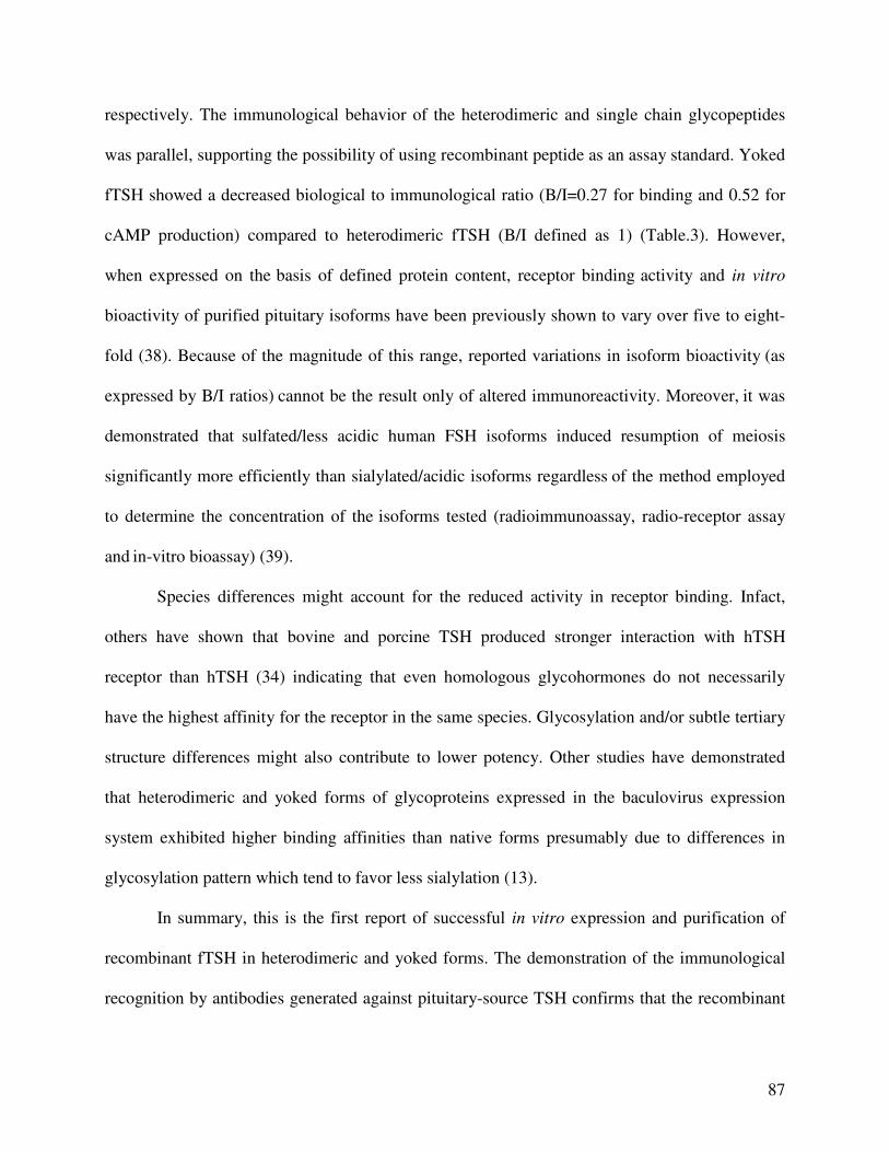
87
respectively. The immunological behavior of the heterodimeric and single chain glycopeptides
was parallel, supporting the possibility of using recombinant peptide as an assay standard. Yoked
fTSH showed a decreased biological to immunological ratio (B/I=0.27 for binding and 0.52 for
cAMP production) compared to heterodimeric fTSH (B/I defined as 1) (Table.3). However,
when expressed on the basis of defined protein content, receptor binding activity and in vitro
bioactivity of purified pituitary isoforms have been previously shown to vary over five to eight-
fold (38). Because of the magnitude of this range, reported variations in isoform bioactivity (as
expressed by B/I ratios) cannot be the result only of altered immunoreactivity. Moreover, it was
demonstrated that sulfated/less acidic human FSH isoforms induced resumption of meiosis
significantly more efficiently than sialylated/acidic isoforms regardless of the method employed
to determine the concentration of the isoforms tested (radioimmunoassay, radio-receptor assay
and in-vitro bioassay) (39).
Species differences might account for the reduced activity in receptor binding. Infact,
others have shown that bovine and porcine TSH produced stronger interaction with hTSH
receptor than hTSH (34) indicating that even homologous glycohormones do not necessarily
have the highest affinity for the receptor in the same species. Glycosylation and/or subtle tertiary
structure differences might also contribute to lower potency. Other studies have demonstrated
that heterodimeric and yoked forms of glycoproteins expressed in the baculovirus expression
system exhibited higher binding affinities than native forms presumably due to differences in
glycosylation pattern which tend to favor less sialylation (13).
In summary, this is the first report of successful in vitro expression and purification of
recombinant fTSH in heterodimeric and yoked forms. The demonstration of the immunological
recognition by antibodies generated against pituitary-source TSH confirms that the recombinant
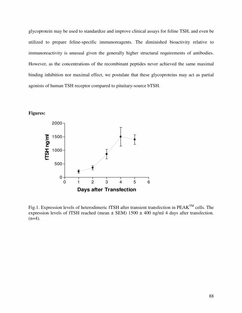
88
glycoprotein may be used to standardize and improve clinical assays for feline TSH, and even be
utilized to prepare feline-specific immunoreagents. The diminished bioactivity relative to
immunoreactivity is unusual given the generally higher structural requirements of antibodies.
However, as the concentrations of the recombinant peptides never achieved the same maximal
binding inhibition nor maximal effect, we postulate that these glycoproteins may act as partial
agonists of human TSH receptor compared to pituitary-source bTSH.
Figures:
0 1 2 3 4 5 60
500
1000
1500
2000
Days after Transfection
fTS
H n
g/m
l
Fig.1. Expression levels of heterodimeric fTSH after transient transfection in PEAKTM cells. The expression levels of fTSH reached (mean ± SEM) 1500 ± 400 ng/ml 4 days after transfection. (n=4).
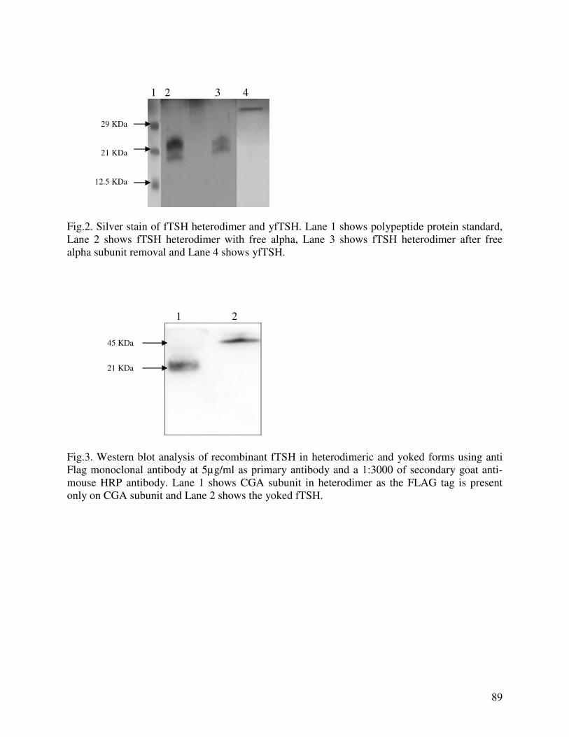
89
1 2 3 4
Fig.2. Silver stain of fTSH heterodimer and yfTSH. Lane 1 shows polypeptide protein standard, Lane 2 shows fTSH heterodimer with free alpha, Lane 3 shows fTSH heterodimer after free alpha subunit removal and Lane 4 shows yfTSH. 1 2
Fig.3. Western blot analysis of recombinant fTSH in heterodimeric and yoked forms using anti Flag monoclonal antibody at 5µg/ml as primary antibody and a 1:3000 of secondary goat anti-mouse HRP antibody. Lane 1 shows CGA subunit in heterodimer as the FLAG tag is present only on CGA subunit and Lane 2 shows the yoked fTSH.
45 KDa
21 KDa
29 KDa 21 KDa 12.5 KDa
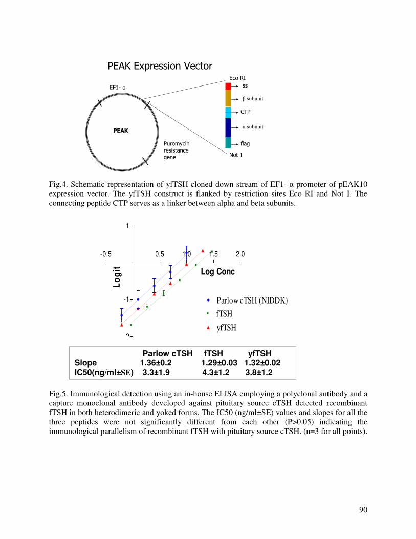
90
Not 1
Eco RI
ss
β subunit
α subunit
CTP
flag
EF1- α
PEAK
Puromycin resistancegene
PEAK Expression Vector
Fig.4. Schematic representation of yfTSH cloned down stream of EF1- α promoter of pEAK10 expression vector. The yfTSH construct is flanked by restriction sites Eco RI and Not I. The connecting peptide CTP serves as a linker between alpha and beta subunits.
-0.5 0.5 1.0 1.5 2.0
-2
-1
1
Parlow cTSH (NIDDK)
fTSH
yfTSH
Log Conc
Lo
git
Fig.5. Immunological detection using an in-house ELISA employing a polyclonal antibody and a capture monoclonal antibody developed against pituitary source cTSH detected recombinant fTSH in both heterodimeric and yoked forms. The IC50 (ng/ml±SE) values and slopes for all the three peptides were not significantly different from each other (P>0.05) indicating the immunological parallelism of recombinant fTSH with pituitary source cTSH. (n=3 for all points).
Parlow cTSH fTSH yfTSH Slope 1.36±0.2 1.29±0.03 1.32±0.02 IC50(ng/ml±SE) 3.3±1.9 4.3±1.2 3.8±1.2
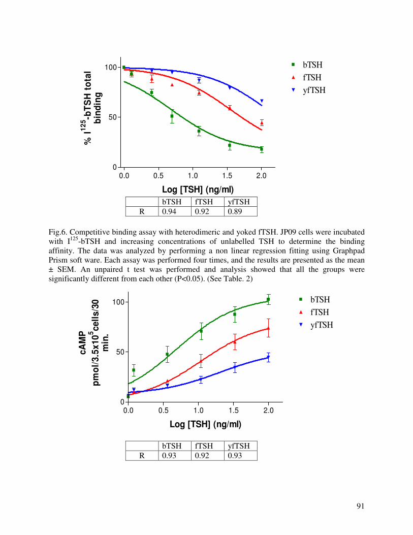
91
0.0 0.5 1.0 1.5 2.00
50
100 bTSH
fTSH
yfTSH
Log [TSH] (ng/ml)
% I
12
5-b
TS
H t
ota
lb
ind
ing
bTSH fTSH yfTSH
R 0.94 0.92 0.89
Fig.6. Competitive binding assay with heterodimeric and yoked fTSH. JP09 cells were incubated with I125-bTSH and increasing concentrations of unlabelled TSH to determine the binding affinity. The data was analyzed by performing a non linear regression fitting using Graphpad Prism soft ware. Each assay was performed four times, and the results are presented as the mean ± SEM. An unpaired t test was performed and analysis showed that all the groups were significantly different from each other (P<0.05). (See Table. 2)
0.0 0.5 1.0 1.5 2.00
50
100 bTSH
fTSH
yfTSH
Log [TSH] (ng/ml)
cA
MP
pm
ol/
3.5
x1
05c
ell
s/3
0m
in.
bTSH fTSH yfTSH
R 0.93 0.92 0.93

92
Fig.7. Stimulation of cAMP production by heterodimeric and yoked fTSH. JP09 cells were incubated with increasing concentrations of TSH, and intracellular cAMP levels were measured by RIA. The data was analyzed by performing nonlinear regression fitting using Graphpad Prism™ software to an equation describing a sigmoidal response. Results were presented as the mean ± SEM (n=4). An unpaired t test analysis of the data showed a significant difference between bovine and feline TSH, bovine and yoked feline TSH and feline and yoked feline TSH (P<0.05). (See Table.2)
Basal bTSH fTSH yfTSH Forskolin0
100
200
300
400
Basal
bTSH
fTSH
yfTSH
Forskolin
cA
MP
pm
ol/
3.5
x10
5cell
s/3
0 m
in.
Fig.8. Basal and maximal cAMP production in JP09 cells. JP09 cells were incubated in 1 ml of Waymouth`s medium with 0.1% BSA and 0.8 mM isobutylmethylxanthine for basal cAMP concentrations, 100 ng/ml of bTSH, fTSH and yfTSH as mentioned in 2.7 for TSH stimulated cAMP levels and 0.1 mM forskolin for maximal activation of adenylate cyclase and maximal cAMP production. Results were presented as the mean ± SEM (n=4). The maximal cAMP production upon stimulation with bTSH, recombinant fTSH and yfTSH were significantly different from each other (P < 0.05). 1 2 3
Fig.9. Silver stain of fTSH heterodimer and commercially available pituitary source cTSH standard. Lane 1 shows polypeptide protein standard, Lane 2 shows fTSH heterodimer and Lane 3 shows pituitary source cTSH.
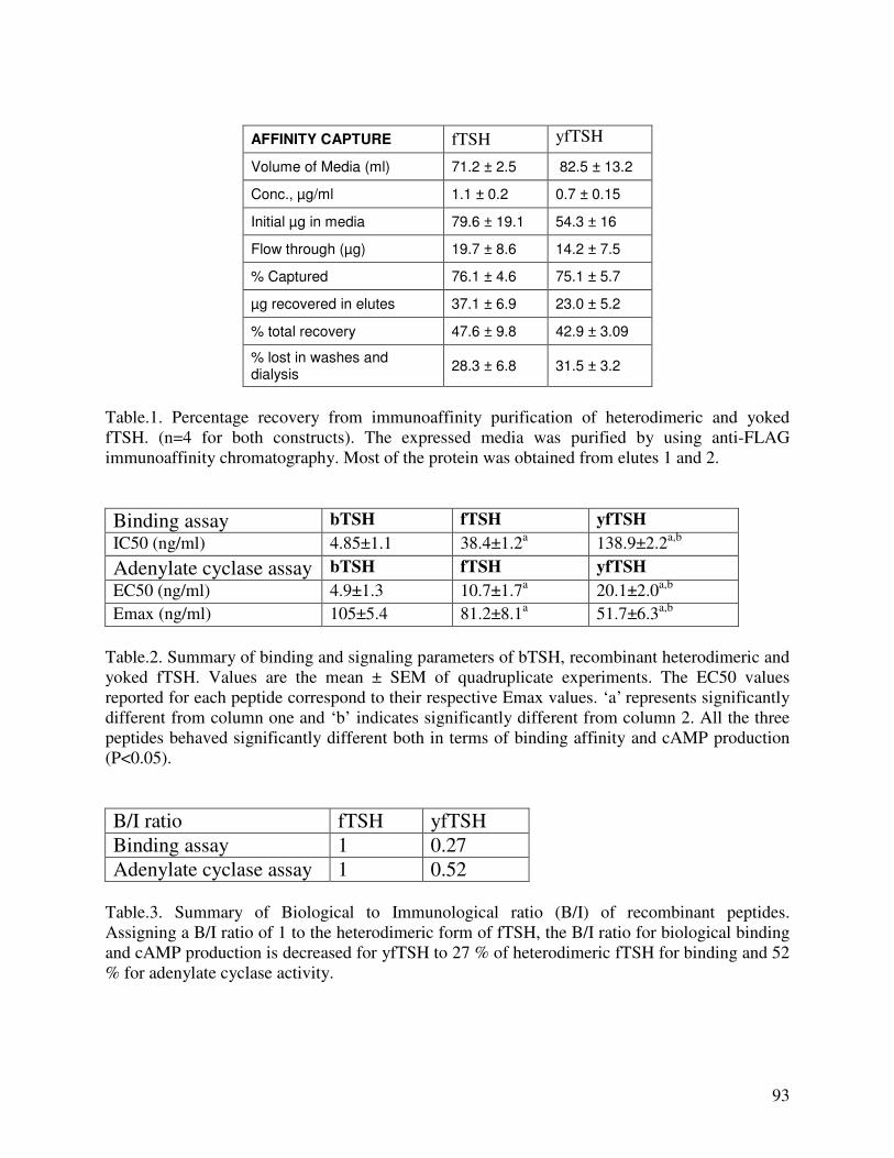
93
AFFINITY CAPTURE fTSH yfTSH
Volume of Media (ml) 71.2 ± 2.5 82.5 ± 13.2
Conc., µg/ml 1.1 ± 0.2 0.7 ± 0.15
Initial µg in media 79.6 ± 19.1 54.3 ± 16
Flow through (µg) 19.7 ± 8.6 14.2 ± 7.5
% Captured 76.1 ± 4.6 75.1 ± 5.7
µg recovered in elutes 37.1 ± 6.9 23.0 ± 5.2
% total recovery 47.6 ± 9.8 42.9 ± 3.09
% lost in washes and dialysis
28.3 ± 6.8 31.5 ± 3.2
Table.1. Percentage recovery from immunoaffinity purification of heterodimeric and yoked fTSH. (n=4 for both constructs). The expressed media was purified by using anti-FLAG immunoaffinity chromatography. Most of the protein was obtained from elutes 1 and 2.
Binding assay bTSH fTSH yfTSH
IC50 (ng/ml)
4.85±1.1 38.4±1.2a 138.9±2.2a,b
Adenylate cyclase assay bTSH fTSH yfTSH
EC50 (ng/ml)
4.9±1.3 10.7±1.7a 20.1±2.0a,b
Emax (ng/ml)
105±5.4 81.2±8.1a 51.7±6.3a,b
Table.2. Summary of binding and signaling parameters of bTSH, recombinant heterodimeric and yoked fTSH. Values are the mean ± SEM of quadruplicate experiments. The EC50 values reported for each peptide correspond to their respective Emax values. ‘a’ represents significantly different from column one and ‘b’ indicates significantly different from column 2. All the three peptides behaved significantly different both in terms of binding affinity and cAMP production (P<0.05).
B/I ratio fTSH yfTSH
Binding assay 1 0.27
Adenylate cyclase assay 1 0.52 Table.3. Summary of Biological to Immunological ratio (B/I) of recombinant peptides. Assigning a B/I ratio of 1 to the heterodimeric form of fTSH, the B/I ratio for biological binding and cAMP production is decreased for yfTSH to 27 % of heterodimeric fTSH for binding and 52 % for adenylate cyclase activity.
94
Acknowledgements
We would like to thank Drs. Krassimira Angelova and David Puett for providing the JP09 cells and for technical advice on this research. This work was supported by a grant from the Morris Animal Foundation. References
1. Pierce JG, Parsons TF. Glycoprotein hormones: structure and function. Annu Rev Biochem 1981;50:465-495. 2. Fiddes JC, Goodman HM. Isolation, cloning and sequence analyses of the cDNA for the α-subunit of human chorionic gonagotropin. Nature 1979;281:351-6. 3. Klee GG, Hay ID. Assessment of sensitive thyrotropin assays for an expanded role in thyroid function testing : proposed criteria for analytic performance and clinical utility. J Clin Endocrinol Metab 1987;64:461-71. 4. Ehrmann DA. Sarne DH. Serum thyrotropin and the assessment of thyroid status. Ann Intern Med 1989;24:515-39. 5. Kass PH, Peterson ME, Levy J, James K, Becker DV, Cowgill LD. Evaluation of environmental, nutritional, and host factors in cats with hyperthyroidism. J Vet Intern Med 1999;13(4):323-329. 6. Martin KM, Rossing MA, Ryland LM, DiGiacomo RF, Freitag WA. Evaluation of dietary and environmental risk factors for hyperthyroidisam in cats. J Am Vet Med Assoc 2000;217(6):853-856. 7. Williams DA, Scott-Moncrieff C, Bruner J, Sustarsic D, Panosian-Sahakian N, Unver E, el Shami AS. Validation of an immunoassay for canine thyroid stimulating hormone and changes in serum concentration following induction of hypothyroidism in dogs. J Am Vet Med Assoc 1996;209:1730-1732.
8. Edinboro CH, Scott-Moncrieff C, Janovitz E, Thacker HL, Glickman LT. Epidemiologic study of relationships between consumption of commercial canned food and risk of hyperthyroidism in cats. J Am Vet Med Assoc 2004;224(6):879-886.
9. Edinboro CH, Scott-Moncrieff C, Tetrick M and Glickman LT. Dietary and environmental risk factors for clinical hyperthyroidism in pet cats Ann Epidemiol 2002;12(7):509. 10. Graham PA, Refsal KR, Nachreiner RF, Provencher-Bollinger AL. The measurement of feline thyrotropin (TSH) using a commercial canine immunoradiometric assay: J. Vet. Intern. Med. 2000;14:342 (Abstract).
95
11. Thotakura NR, Szkudlinski MW, Weintraub BD. Structure-function studies of oligosaccharides of recombinant human thyrotrophin by sequential deglycosylation and resialylation. Glycobiology 1994;4:525-533.
12. Grossmann M, Wong R, Mariusz W, Szkudlinski MW, Weintraub BD. Human Thyroid-stimulating Hormone (hTSH) Subunit Gene Fusion Produces hTSH with Increased Stability and Serum Half-life and Compensates for Mutagenesis-induced Defects in Subunit Association. J Boil Chem 1997;272:21312-21316.
13. Narayan P, Wu C, Puett D. Functional expression of yoked human chorionic gonadotropin in baculovirus-infected insect cells. Mol Endocrinol 1995; 9:1720-1726.
14. El-Deiry S, Kaetzel D, Kennedy G, Nilson J, Puett D Site-directed mutagenesis of the human chorionic gonadotropin β-subunit: bioactivity of a heterologous hormone, bovine α-human des-(122-145)β. Mol Endocrinol 1989;3:1523-1528. 15. Matzuk MM, Hsueh AJW, Lapolt P, Tsafriri A, Keene JL, Biome I. The biological role of the carboxy terminal extension of human chorionic gonadotropin β subunit. Endocrinology 1990;126:376-383.
16. Cole ES, Lee K, Lauziere K, Kelton C, Chappel S, Weintraub B, Ferrara D, Peterson P, Bernasconi R., Edmunds T. et al. Recombinant human thyroid stimulating hormone: development of a biotechnology product for detection of metastatic lesions of thyroid carcinoma. Biotechnology 1993;11(9):1014–1024.
17. Fares FA, Yamabe S, Menahem DB, Pixley M, Hsueh JW Boime I. Conversion of Thyrotropin Heterodimer to a Biologically Active Single-Chain. Endocrinology 1998;139(5):2459-2464.
18. Yang X, McGraw RA, Ferguson DC. cDNA cloning of canine common α gene and its co-expression with canine β gene in baculovirus expression system. Domestic Animal Endocrinology 2000;18: 379-393.
19. Green ED, Baenziger JU. Asparagine-linked oligosaccharides on lutropin, follitropin, and thyrotropin: Structural elucidation of sulfated and sialylated oligosaccharides on bovine, ovine, and human pituitary glycoprotein hormones. J Biol Chem 1988;263(1):36-44.
20. Grossmann M, Szkudlinski MW, Tropea JE, Bishop LA, Thotakura NR, Schofield PR. and Weintraub BD. Expression of human thyrotropin in cell lines with different glycosylation patterns combined with mutagenesis of specific glycosylation sites: Characterization of a novel role for oligosaccharides in the in vitro and in vivo bioactivity. J. Biol. Chem 1995;270:29378–29385.
21. Joshi L, Murata Y, Wondisford FE, Szkudlinski MW, Desai R. and Weintraub BD. Recombinant thyrotropin containing a ß-subunit chimera with the human chorionic
96
gonadotropin-ß carboxy-terminus is biologically active, with a prolonged plasma half-life: role of carbohydrate in bioactivity and metabolic clearance. Endocrinology 1995; 136:3839–3848.
22. Flack MR, Bennet AP, Froehlich J, Anasti JN, Nisula BC. Increased biological activity due to basic isoforms in recombinant human follicle-stimulating hormone produced in a human cell line. J Clin Endocrinol Metab 1994;79(3):756-60.
23. Smith PL, Skelton TP, Fiete D, Dharmesh SM, Beranek MC, MacPhail L, Broze GJ Jr, Baenziger JU. The asparagine-linked oligosaccharides on tissue factor pathway inhibitor terminate with SO4-4GalNAc beta 1, 4GlcNAc beta 1,2 Mana alpha. J Biol Chem. 1992;267(27):19140-6.
24. Tokushige K, Moradpour D, Wakita T, Geissler M, Hayashi N and Wands JR. Comparison between cytomegalovirus promoter and elongation factor-1 α promoter-driven constructs in the establishment of cell lines expressing hepatitis C virus core protein. Journal of virological methods 1997;64:73-80.
25. Yang X, McGraw RA, Su X, Katakam P, Grosse WM, Li OW, Ferguson DC. Canine thyrotropin β-subunit gene: cloning and expression in Escheria coli, generation of monoclonal antibodies and transient expression in the Chinese ovary cells. Domestic Animal Endocrinology 2000;18:363-378.
26. Smith PK, Krohn RL, Hermanson GT, Mallia AK, Gartner FH, Provenzano MD, Fujimoto EK, Goeke NM, Olson BJ and Klenk DC Measurement of protein using bicinchonic acid. Anal. Biochem. 1985;150:76-85.
27. Laemmli UK. Cleavage of structural proteins during the assembly of the head of bacteriophage T4. Nature 1970;227:680-685.
28. Costagliola S, Swillens S, Niccoli P, Dumont JE, Vassart G, Ludgate M. Binding assay for thyrotropin receptor auto antibodies using the recombinant receptor protein. Endocrinology 1992;75:1540-1544.
29. Greenwood FC, Hunter WM, Glover JS. Preparation of 131I-labeled human growth hormone of high specific radioactivity, Biochemical Journal 1963;89:114-123.
30. Grossmann M, Szkudlinski MW, Zeng H, Kraiem Z, Ji I, Tropea JE, Ji HT, Weintraub BD. Role of carboxy-terminal residues of the α subunit in the expression and bioactivity of human thyroid-stimulating hormone, Mol Endocrinol 1995;9:948-958.
31. Grossmann M, Szkudlinski MW, Dias JA, Xia H, Wong R, Puett D, Weintraub BD. Site-directed mutagenesis of amino acids 33-44 of the common α subunit reveals different structural requirements for heterodimer expression among the glycoprotein hormones and suggests that cAMP production and growth promotion are potentially dissociable functions of hTSH, Mol Endocrinol 1996;10:769-779.
97
32. Dean DJ, Weintraub BD, and Rosen SW. De novo synthesis and secretion of heterogeneous forms of human chorionic gonadotropin and its free alpha-subunit in the human choriocarcinoma clonal cell line JEG-3, Endocrinology 1980;106: 849-858. 33. Parsons TF, Bloomfield GA, and Pierce JG. Purification of an alternate form of the alpha subunit of the glycoprotein hormones from bovine pituitaries and identification of its O-linked oligosaccharide, J. Biol. Chem 1983; 258: 240-244.
34. Sugahara T, Sato A, Kudo M, Ben-Menahem D, Pixley MR, Hsueh AJW and Boime I. Expression of biologically active fusion genes encoding the common α subunit and the follicle-stimulating hormone β subunit, J. Biol. Chem 1996;271:10445-10448.
35. Miguel RN, Sanders J, Jeffreys J, Depraetere H, Evane M, Richards T, Blundell TL, Smith BR, Furmaniak J. Analysis of the thyrotropin receptor-thyrotropin interaction by comparative modeling, Thyroid 2004;14:991-1011.
36. Chard T. An introduction to radioimmunoassay and related techniques: laboratory techniques in biochemistry and molecular biology 1995; Elsevier Biomedical press, Amsterdam, The Netherlands.
37. Kourides IA, Re RN, Weintraub BD, Ridgway EC, Maloof F. Metabolic clearance and secretion rates of subunits of human thyrotropin. J Clin Invest. 1977;59:508–516.
38. Stanton PG, Burgon PG, Hearn MTW et al. Structural and functional characterization of hFSH and hLH isoforms. Mol. Cell,. Endocrinology 1996; 125:133–141.
39. Yding Andersen C, Leonardsen L, Ulloa-Aguirre A. et al. FSH-induced resumption of meiosis in mouse oocytes: effects of different isoforms. Mol. Hum. Reprod 1999;5:726–731.
40. Chappel S. Editorial: biological to immunological ratios: reevaluation of a concept. J Clin Endocrinol Metab. 1990 ;70:1494–5.
98
EXPRESSION OF RECOMBINANT FELINE THYROTROPIN IN
BACULOVIRUS EXPRESSION SYSTEM: EFFECT OF SIALYLATION
ON IMMUNOREACTIVITY AND BIOACTIVITY3
3S. Rayalam, R.R.Davis, M.Hoenig, G. Alvarez-Manilla*, D.C.Ferguson. Department of Physiology and Pharmacology, College of Veterinary Medicine,*Complex Carbohydrate Research Center, The University of Georgia, Athens, GA 30602, USA. To be submitted to Domestic Animal Endocrinology.
99
Abstract
Previously cloned and sequenced feline CGA and fTSH beta subunits were expressed
in insect cells using a baculovirus expression system. The signal peptide in both the constructs
was replaced with honey bee mellitin signal sequence and the intron in the fTSH beta “mini
gene” construct was removed with an over-lap PCR. The constructs were ligated downstream of
polyhedrin promoter of baculovirus transfer vector pvl1393. The expression levels as determined
by immunoreactivity (35 ± 15 ng/ml) were considerably lower than the expression levels
obtained in mammalian expression system using PEAKTM cells. Since the relatively low
expression levels of fTSH in baculovirus expression system did not facilitate purification of large
quantities of recombinant fTSH for standardization by protein assay, as an alternative,
enzymatically desialylated fTSH expressed in PEAKTM cells was prepared and used to
characterize the bioactivity. The immunological activity of insect cell expressed fTSH and
desialylated fTSH was compared with pituitary source canine TSH and non desialylated fTSH.
The results from the in-house ELISA and the commercially available canine TSH assay indicated
that both insect cell expressed fTSH and desialylated fTSH showed immunological parallelism to
pituitary-source canine TSH and non desialylated fTSH. There was no significant change in the
bioactivity of desialylated fTSH in terms of cAMP production as compared to non
desialylatedfTSH. However, the biological to immunological ratio (B/I) for cAMP production
increased significantly upon desialylation. Sialic acid removal did not significantly change the
receptor binding affinity. Since an ideal immunoassay standard is glycosylation independent, this
study supports the possibility that recombinant fTSH could be substituted for pituitary source
TSH as an immunoassay standard.
1. Introduction:
TSH (Thyroid Stimulating Hormone), FSH (Follicle stimulating Hormone), CG
(Chorionic Gonadotropin), and LH (Luteinizing Hormone) belong to glycoprotein hormone
family. They are all structurally related heterodimers consisting of a common alpha subunit and a
hormone specific beta subunit (1, 2). The molecular weight of mammalian TSH can range from
28 to 30 kilo daltons (kDa), with variation being associated with heterogeneity of the
oligosaccharide chains. The alpha subunit has a molecular weight of approximately 14
kilodaltons and has two oligosaccharide units linked to asparagine residues whereas the beta
subunit has a molecular weight of 15 kilodaltons and has one asparagine linked oligosaccharide
unit. Like other glycoprotein hormones, TSH is highly glycosylated with N-linked complex
carbohydrates, which in the human molecule, account for approximately 21% and 12% of the
total weight in it’s α and β chains respectively (2, 3).
Several studies have been reported on the influence of carbohydrates on the in vivo
bioactivity of the hormone (4, 5, 6). Moreover, oligosaccharides are also required for
posttranslational subunit folding and assembly, protection from intracellular degradation, and
secretion of the heterodimer (4, 6, 7, 8, 9). Therefore, mammalian cells are required for
recombinant glycoprotein hormone production, and attempts to achieve high level expression of
glycoprotein hormones using prokaryotic systems have not been successful (7). In order to
elucidate the role of carbohydrate the hormone can either be deglycosylated or can be expressed
in different cell systems that have different glycosylation machinery and examine the effect of
different glycosylation patterns on function. A number of studies report deglycosylation of
glycoproteins chemically (10, 11), enzymatically (12), and also by using oligosaccharide
processing inhibitors (13), but these modifications are not only nonspecific but may also result in
101
partial degradation of the protein. Therefore, for better understanding of the effect of
glycosylation, recombinant feline thyrotropin (fTSH) was expressed in the baculovirus
expression system. For secreted and membrane associated proteins, a signal peptide at the
amino-terminus is responsible for targeting the polypeptide to the endoplasmic reticulum ER
(14). The efficiency of secretion of the baculovirus system can be increased by using signal
peptides of insect origin to direct the secretion of a foreign protein through the protein synthesis
and secretary pathways (15, 16).
The carbohydrates of the common alpha subunit are most important for biological
activity (17). As shown by deglycosylation studies of hTSH , the sialic acid residues at Asn 52
attenuate in vitro hormonal activity whereas they play a stimulatory role in hCG and FSH
(18,5,4). Interestingly, removal of terminal sialic acid residues of recombinant hTSH increased in
vitro bioactivy of hormone (5). The biological activity of resulting peptide showed a reduced
ability to stimulate adenylate cyclase, despite still binding to its receptor with high affinity (4).
There is an unusual sulfate group that terminates certain chains in TSH and LH which is not seen
in CG and found only to some extent in FSH. The sulfate moiety is covalently linked to N-
acetylgalactosamine in contrast to the usual terminal structure in which sialic acid is bound to
galactose. Pituitary TSH and LH contain predominantly sulfated oligosaccharides, due to the
presence of GalNAc-transferase and GalNAc-4-sulfotransferase in the pituitary thyrotrophs and
lutrophs, and terminate mostly in SO4-4GalNAcß1–4GlcNAcß1–2Manα (19). On the other hand
hTSH expressed in CHO cells terminates with complex sialylated oligosaccharides (20, 21).
However, Human Embryonic Kidney cells (HEK 293) sulfate pituitary glycoproteins to a greater
extent (18, 22), because of the presence of higher levels of N-acetyl galactosamine transferase
and sulfotransferase activities similar to those expressed in the pituitary gland (23).

102
In this study, we have tested the feasibility of using a baculovirus system to express
recombinant fTSH in quantities suitable for immunological studies. Enzymatic desialylation with
neuraminidase and lectin binding studies were also designed to investigate the role of sialylation
in the immunoreactivity and bioactivity of recombinant fTSH. The present study describes the
first attempt to express recombinant fTSH in insect cells using a baculovirus vector. Further, this
is the first report investigating the carbohydrate compositional analysis of recombinant fTSH and
elucidating the role of sialic acid on immunoreactivity and bioactivity. The demonstration of
immunological recognition by antibodies generated against pituitary-source TSH confirmed that
sialic acid does not influence immunoreactivity of recombinant fTSH. As previously reported for
hTSH (24), desialylation on the other hand, increased the biological to immunological (B/I) ratio
for cAMP production where as the B/I ratio for binding was unaltered. Recombinant fTSH
derived from insect cells will be useful in defining structure-function relations of hormone
analogs.
2. Materials and methods:
2.1. Materials:
All the restriction enzymes were from New England Biolabs, Inc (Beverley, MA). High
Pure Plasmid Isolation® kit and Rapid DNA ligation® kit were purchased from Roche
Diagnostics Corporation (Indianapolis, IN). One ShotTM cells, Sf 9 cells, Hi 5 cells, penicillin-
streptomycin, Express FiveTM media and Sf 900 media were from Invitrogen Corporation
(Carlsbad, CA). Dulbecco’s Modified Eagles Medium (DMEM), Waymouth`s MB752/1
medium, Nutrient Mixture F-12 Ham, puromycin, ANTI-FLAG® M2-Agarose Affinity Gel,
FLAG® Peptide, ANTI-FLAG® M2 monoclonal antibody, cAMP salt, anti-cAMP polyclonal
antibody, 3-isobutyl-1-methylxanthine (IBMX), Mouse IgG, Goat anti-rabbit-Peroxidase
103
conjugate, Tetramethyl-benzidine, neuraminidase, β-galactosidase and N-acetylhexosaminidase
were all from Sigma (St. Louis, MO). The BaculoGold Transfection Kit was purchased from
PharMingen (San Diego, CA). All the biotinylated lectins were obtained from Vector
Laboratories (Burlingame, CA). ConA SepharoseTM was obtained from Amersham Biosciences
(Piscataway, NJ). Micro BCA™ Protein Assay Kit, AminoLink® kit and West Dura Super
Signal™ substrate were from Pierce (Rockford, IL). Immulon™ 4HBX strips were from
ThermoLabsystems (Franklin, MA), and Canine TSH was from Scripps Laboratories (San
Diego, CA) and the National Hormone and Pituitary Program (NHPP, Torrance, CA).
Immobilon™ transfer membrane was purchased from Millipore (Bedford, MA). Forskolin was
from Fisher Scientific (Pittsburg, PA). The automated microplate reader was from Bio-Tek
Instrument®, Inc (Winooski, VT). All the chemicals for making buffers and other reagents were
from Sigma (St. Louis, MO).
2.2. PCR to add Honey Bee Mellitin (HBM) signal sequence:
The signal peptide in the previously cloned and sequenced feline CGA and fTSHβ DNA
constructs (25) were replaced with the honey bee mellitin (HBM) signal sequence (atgaaatt
cttagtcaac gttgcccttg tttttatggt cgtgtacatt tcttacatct atgcg, Genbank ACCESSION # NP
001011607) in two PCR reactions using two forward primers one at a time. The second part of
the HBM sequence was first added on to 5’ end of fTSHα with forward primer 5’ TTT ATG
GTC GTA TAC ATT TCT TAC ATC TAT GCG TTT TGT TTT CCA 3’ and subsequently the
first part of HBM signal sequence was added with forward primer 5’- GAA TTC ATG AAA
TTC TTA GTC AAC GTT GCC CTT GTT TTT ATG GTC GTA - 3’. In both cases, the reverse
primer 5’ – CAT AGC GGC CGC TTA CTT ATC GTC ATC – 3’ was used to amplify fTSHα-
FLAG. The same forward primers with the reverse primer 5’ - A TGC GGC CGC TTA GAT
104
AGA AAC TCC TAC CAC ATC GGA CTT CT - 3’ were used to replace the signal sequence in
the fTSHβ DNA construct. Appropriate restriction sites, Eco RI at the 5’ and Not I at the 3’ end
were included for the subsequent cloning into baculovirus transfer vector pvl1393.
2.3 Construction of transfer vector for fTSHα and fTSHβ:
Previously cloned and sequenced fTSHα was ligated into baculovirus transfer vector
pvl1393. The baculovirus transfer vector pvl1393 was linearized by digesting with restriction
enzymes Eco RI and Not I. Previously cloned and sequenced feline common alpha was digested
with the same enzymes and ligated with linearized pvl1393 vector DNA downstream of the
polyhedrin promoter using the Rapid DNA™ ligation kit following the manufacturer’s
instructions. The ligated DNA was transformed into One Shot™ cells and the positive clones
were checked for correct insertion by digesting with Eco RI and Not I restriction enzymes. For
development of the fTSH beta subunit construct, the intron was removed of the “mini-gene”
construct with an overlap PCR before ligating into the pvl1393 transfer vector. Internal primers
were designed on either side of the intron and two pieces of fTSHβ were amplified separately.
To amplify the fTSHβ gene towards the 5’ end of intron, the primers used were:
1. Forward primer 5’- GAA TTC ATG ACT GCT ATC TAC CTG ATG TCC - 3’
2. Reverse primer 5’- GCC ATT GAT ATC CCG TGT CAT ACA ATA - 3’
To amplify fTSHβ gene towards the 3’ end of intron the primers used were:
3. Forward primer 5’- A TGC GGC CGC TTA GAT AGA AAC TCC TAC CAC ATC GGA
CTT CT - 3’
4. Reverse primer 5’- TGT ATG ACA CGG GAT ATC AAT GGC AAA - 3’
The two pieces were then overlapped by using both the amplified pieces as templates and
primers 1 and 2 shown above (Fig.1). The fTSH beta subunit was then cloned downstream of the
105
polyhedrin promoter into the pvl1393 transfer vector separately and insertion confirmed by
restriction digests.
2.4. Generation of recombinant baculoviruses:
The recombinant baculoviruses were generated by using BaculoGoldTM Transfection Kit
following the manufacturer’s instructions. In brief, 2 µg of recombinant pvl1393 plasmid DNA
containing fTSHα and fTSHβ genes were mixed separately with 0.5 µg of linearized viral DNA
and incubated for 5 minutes. One milliliter of transfection buffer was then added and the mixture
overlaid on Sf9 cells cultured in a 60 mm plate in serum-free medium, and incubated at 270C for
4 hr. After incubation, the cotransfection plate was rinsed and 3 ml of fresh medium was
replaced. The cells were allowed to grow for 4-5 days at 27oC, and the medium was harvested.
The recombinant viruses in the culture medium were amplified 2 to 3 rounds and the viral titer
was determined by plaque assay. Single plaques were isolated from the plaque assay and
amplified four rounds before being used for expression.
2.5. Expression of recombinant heterodimeric fTSH:
For expression, Hi 5TM cells were used instead of Sf9 cells. The Hi 5 cells were seeded at
2x10⁷ cells/ 75 cm2 flask and the recombinant viruses with a titer of 107 plaque forming units/ml
were used for infection at a virus to cell ratio of 10. The recombinant viruses for fTSHα and
fTSHβ were added at 1:1 ratio for generation of recombinant heterodimeric fTSH. The cells were
allowed to grow at 270C for 4 days and the medium was harvested for testing the levels of
expression. The expression levels were determined in in-house canine TSH ELISA employing a
capture monoclonal antibody previously selected for ovine/canine TSH beta and a polyclonal
antibody generated previously against pituitary-source canine TSH as explained previously (26).
106
2.6. Purification of fTSH expressed in baculovirus expression system:
The expressed media was concentrated down 40 fold using a Vivacell 250 (Vivascience
AG, Hannover, Germany) gas pressurised ultrafiltration system. Anti-FLAGTM column
chromatography was not successful in purifying the insect cell expression medium. The FLAGTM
immunoaffinity tag cleavage was suspected and hence as affinity column was made by coupling
AminoLink® coupling gel with anti-cTSH mAb (14H9.E2) (26) following manufacturer’s
instructions. The concentrated media was allowed to enter the gel bed and incubated for 60
minutes at room temperature. Bound peptide was eluted using 0.1 M glycine pH 2.5. The eluted
fractions were dialyzed against 0.03 M phosphate buffer for buffer exchange.
2.7. Enzymatic deglycosylation of fTSH expressed in PEAKTM
cells:
Heterodimeric fTSH previously expressed in PEAKTM cells was enzymatically
deglycosylated by using exoglycosidase like neuraminidase. One µl of A. ureafaciens sialidase
(10 U/ml) was added to 20 µg of recombinant fTSH in 100 µl of 50 mM sodium phosphate (pH
5.0) and incubated overnight at 370C. The deglycosylated fTSH is then dialyzed against 0.03M
phosphate buffer (pH 7.2) for buffer exchange and ran over Con A column as described in 2.8.
2.8. Lectin binding studies:
One milliliter of Con A Sepharose was loaded into Pasteur pipettes and the column was
equilibrated with TBS (20 mM Tris HCL and 0.5 M NaCl, pH 7.4) and deglycosylated fraction
described in 2.7 was loaded onto the column. Con A - bound fTSH was eluted with 0.2 M
methyl- α-D mannopyranoside, pH 7.4 for first four fractions and pH 4.5 for last two fractions.
The elution fractions were collected into glass vials and siliconized pipette tips used at every
point to minimize protein losses. All the elution fractions were dialyzed against 0.03M phosphate
buffer for buffer exchange. The fractions were checked for the presence of sugars N-
107
acetylglucosamine, galactose and sialic acid by performing dot blots with biotinylated lectins
WGA (Wheat Germ agglutinin), RCA (Ricinus Communis agglutinin) and SNA (Sambucus
Nigra agglutinin) respectively. In brief, 2 µl of the elutes and non desialylated fTSH, a positive
control were blotted on a nitrocellulose membrane. and incubated with biotinylated lectins (10
µg/ml) for 2 hrs and after washing with TSS-T (20 mM Tris HCL, 0.5 M NaCl and Tween20
0.05%, pH 7.4) membrane was subsequently incubated with peroxidase conjugated Streptavidin
(1:1000) for one hour and the dot blot was visualized using West Dura Super SignalTM substrate
for 2 minutes and the dots imaged and quantified with a Fluor-S Max MultiImagerTM (Bio-Rad
Laboratories, Inc. Hercules, CA).
2.9. Immunological detection:
The enzymatically desialylated fTSH and fTSH expressed in the baculovirus expression
system were detected by in-house canine TSH ELISA employing a capture monoclonal antibody
(14H9.E2) (mAb) previously selected for ovine/canine TSH beta and a polyclonal antibody
(pAb) generated previously against pituitary-source canine TSH (30). Pituitary-source canine
TSH (NHPP) was used as the immunoassay standard. Immunological parallelism of desialylated
fTSH and fTSH expressed in the baculovirus expression system with pituitary-source canine
TSH and recombinant fTSH expressed in mammalian cells (PEAKTM cells) was evaluated as
explained previously (26).
2.10. Bioactivity studies:
TSH Receptor Binding Assay
Competitive binding experiments were performed with a stable cell line of CHO cells
expressing the recombinant human TSH receptor (rhTSHR), clone JP09 (28). Pituitary source
bTSH was iodinated as described previously (29). The cells were maintained in Ham’s F12 (HF-
108
12) media supplemented with 10% fetal bovine serum and 1% penicillin-streptomycin and were
maintained with geneticin selection at 400 µg per ml of media. The radioreceptor assay was
performed as described previously (26). The receptor-binding activity of the deglycosylated
fTSH in comparison to recombinant fTSH expressed in mammalian cells and pituitary source
bTSH was determined by their ability to displace 125I-bTSH from binding to hTSH receptors
expressed on the JP09 cells.
TSH stimulation of JP09 cells
JP09 cells were grown in 12 well plates to confluence, the media removed and incubated
in Waymouth`s MB752/1 medium with 0.1% BSA and 0.8 mM isobutylmethylxanthine (IBMX)
for 15 min at 37oC. The cells were then incubated in various concentrations of peptide for 30 min
at 37oC in Waymouth`s medium containing 0.1% BSA and 0.8 mM IBMX immediately
following first incubation. The cells were also incubated with 0.1 mM forskolin to determine the
TSH-independent maximal cAMP response. The incubation medium was removed, and the cells
were lysed in 100% ethanol at -20oC overnight. The extract was collected, dried under vacuum,
and dissolved in 1 ml of 50 mM sodium acetate pH 6.2. cAMP radioimmunoassay was
performed as described previously (26).
2.11. Statistical analysis
The statistical analysis of binding assays and cAMP assays was carried out by using
GraphPad PRISM program. All graphical data were also prepared using GraphPad Prism. Non
linear regression was used to fit both binding and adenylate cyclase data to a sigmoidal curve
when plotted against the log of the TSH concentration. The goodness of fit is reported as R and
Emax and EC50 calculated. Significance of differences between Emax and EC50 for bTSH,
fTSH and desialylated fTSH in adenylate cyclase and binding assays was calculated by

109
performing unpaired t tests. The slopes and intercepts for immunoreactivity data were also
compared by performing unpaired t tests. P value of ≤ 0.05 was considered significant. Means
and standard errors were reported as appropriate.
3. Results
3.1. Replacing signal sequence with HBM in both α and β-subunits and removal of intron
in fTSHβ:
PCR addition of HBM to previously cloned and sequenced (25) feline CGA DNA
constructs yielded a product of 420 bp when examined on a 1.5% agarose gel (Fig.1). The Intron
in fTSHβ was removed by an overlap PCR as explained in 2.3 (Fig.2) and the final PCR product
with HBM signal sequence of 432 bp was obtained as shown in Fig 3.
3.2. Expression of fTSH α and β-subunits and purification of heterodimeric fTSH:
Hi 5TM cells were infected, separately and concurrently, with fTSHα and fTSHβ
producing recombinant virus at an MOI of 10.0. Figure 4 shows a dot blot with anti-FLAGTM
monoclonal antibody on fTSHα expressed in Hi 5TM cells. The medium from mock-infected Hi
5TM cells was used as a negative control and recombinant fTSH expressed in PEAKTM cells (26)
was used as positive control. The expression levels of fTSHβ glycopeptide, as evaluated by the
in-house ELISA reached 35±10 ng/ml (n=4) four days after infection. Infection with fTSHα and
fTSHβ producing recombinant baculoviruses at a ratio of 1:1 resulted in expression of fTSH
(Fig.6) as evaluated by anti-FLAG western and ELISA (maximal expression: 35±15 ng/ml, n=4).
The expressed media was concentrated approximately 40 fold and purified as outlined in 2.6. A
silver stain showing purified heterodimeric fTSH expressed in Hi 5 cells along with fTSH
expressed in PEAKTM cells is shown in Figure 5. It is clear from the silver stain that the alpha
subunit is not present in equimolar amounts to the beta subunit in the purified sample. In fact, in
110
some preparations no alpha band was seen on silver stain and no-anti-FLAG immunoreactivity
seen on western blot.
3.3. Deglycosylation and lectin binding studies:
The desialylated fTSH was checked for the absence of sialic acid by doing dot blots using
different biotinylated lectins (Fig.7). The dot blots with lectins WGA, RCA and SNA showed
the presence of N-acetylglucosamine (GlcNAc), N-acetylgalactosamine (GalNAc) and absence
of sialic acid in neuraminidase treated recombinant fTSH expressed in PEAKTM cells.
3.4. Immunological parallelism of fTSH expressed in insect cells and desialylated fTSH:
The average expression concentrations of fTSHβ in medium as determined by the in-
house ELISA was 35 ± 10 ng/ml and that of heterodimeric fTSH was 35 ± 15 ng/ml (mean ±
SEM, n=4). Both the recombinant fTSH expressed in insect cells and desialylated fTSH were
also detected by the commercially available canine TSH assay (Diagnostic Products Corp., Los
Angeles, CA). However, In-house assay detected about 33±1.8 % (n=3) and DPC assay detected
32.7±2.8 % (mean±SEM) (n=3) of the desialylated fTSH in both forms as standardized by BCA
protein assay.
Both fTSH expressed in insect cells and desialylated fTSH showed immunological
parallelism with pituitary source cTSH (NHPP) and non desialylated fTSH in our in-house
ELISA (Fig.8). The IC50 (ng/ml±SE, n=3) concentrations for pituitary source cTSH (NHPP),
heterodimeric fTSH expressed in PEAKTM cells, fTSH expressed in insect cells and desialylated
fTSH were 5.3±1.9, 7.6±1.0, 6.96±1.5 and 8.8±2.2 and slopes were 1.40±0.07, 1.47±0.08,
1.39±0.09 and 1.42±0.05 respectively. Both the IC50 values and slopes of all three peptides were
not significantly different from each other.
111
3.5. Biological activity of recombinant fTSH:
The bioactivity of desialylated fTSH was compared with that of recombinant fTSH
expressed in PEAKTM cells and bovine TSH by comparison of hTSH receptor binding inhibition
and stimulation of adenylate cyclase activity. As shown in Fig. 9, desialylated fTSH was also
able to inhibit the binding of 125I-bTSH to the hTSH receptor in JP09 cells with an affinity not
significantly different from non desialylated fTSH (P>0.05) (Table 1).
The bioactivity of desialylated fTSH was evaluated by potency to stimulate adenylate
cyclase in the human TSH receptor-expressing JP09 cells. Desialylated fTSH was 2.3 -fold less
potent than bovine TSH in stimulating cAMP production (Fig.10). However, desialylation did
not alter the potency for stimulation of adenylate cyclase (Table 1). The EC50 (ng/ml) values for
desialylated and non - desialylated fTSH are not significantly different from each other (Fig. 8).
The Biological to Immunological ratio (B/I) for desialylated fTSH for binding was 96 % of that
of fTSH and is not significantly different from fTSH whereas, for cAMP production, the B/I
ratio was 16 % more for desialylated fTSH than non desialylated fTSH and is significantly
different (P<0.05) (Table. 2).
4. Discussion
The present study describes the first attempt to express previously cloned and
sequenced fTSHα and fTSHβ genes (25) in insect cells using a baculovirus expression vector.
Furthermore, this study reports on the effect of glycosylation on the immunoreactivity and also
bioactivity of the recombinant fTSH.
In the baculovirus system, fTSH was secreted but the expression levels determined by
immunoreactivity (35 ± 15 ng/ml) were considerably lower than the expression levels obtained
in mammalian expression system (~ 1 µg/ml) using PEAKTM cells (26). In contrast, others had
112
shown that the expression levels of recombinant human TSH were higher in the insect cells than
the levels obtained in eukaryotic cells (30). A study reported the substitution of natural signal
sequences with the signal sequences from honeybee mellitin promotes a high level of expression
of a glycosylated form of gp120 and efficient secretion (16). However, use of honeybee mellitin
signal peptide does not ensure proper processing of recombinant proteins (31).
Insect cells lack the capacity to process carbohydrate residues to complex-type,
terminally sialylated oligosaccharides, especially when a vector like pvl1393, containing the very
late viral polyhedrin promoter, was used for expression. As a result, glycoproteins produced
under such conditions usually contain high mannose-type precursor oligosaccharides (32, 33). In
contrast, hTSH expressed in CHO cells terminates with complex, sialylated oligosaccharides (20,
21). Also, other studies that analyzed insect cell-expressed glycoprotein carbohydrates by
compositional analysis suggested that the glycosylation machinery of insect cells does not
possess the same capacity to process N-linked oligosaccharides to complex-type carbohydrates as
mammalian cells (34).
The relatively low expression levels of fTSH in baculovirus expression system together
with the possible cleavage of the immunoaffinity tag did not facilitate purification of large
quantities of recombinant fTSH for standardization by protein assay. The observation that the
FLAGTM immunoaffinity tag was being cleaved also suggests problems with intracellular
processing or suggests the presence of protease cleaving the FLAG peptide. Parenthetically, even
with different baculovirus vectors and or yoking of the subunits, expression levels were no
higher for equine or canine TSH (unpublished data). Insect cells lack the capacity to process
sugar moieties to complex sialic acid terminated oligosaccharides especially when a very late
promoter like polyhedrin is used to express genes of interest, resulting in production of
113
glycoproteins with high mannose-type precursor oligosaccharides (34, 34, 38). Therefore, as an
alternative to insect cell expressed fTSH, we prepared enzymatically desialylated fTSH
expressed in mammalian expression system. Complete deglycosylation without denaturing the
protein was not possible because the oligosaccharides on the β-subunit in both heterodimeric
conformation and free form have been reported to be easily accessible whereas, the sugars on α-
subunit are hidden by subunit interaction (36). Moreover it is well-known that under in vitro
conditions, sialic acid appears to be the major factor affecting the bioactivity of recombinant
TSH (21). In vivo sialylation influences metabolic clearance rate as removal of the terminal sialic
acid residues markedly reduces the half-life of TSH in the circulation by enhancing its binding to
hepatocyte lectins responsible for clearance from the bloodstream (37).
Lacking pituitary source fTSH, the immunological activity of insect cells expressed
fTSH and desialylated fTSH was compared with pituitary source canine TSH (NHPP standard)
and non desialylated recombinant fTSH. The results from the in-house ELISA and the
commercially available canine TSH assay indicated that desialylated fTSH showed
immunological parallelism to pituitary-source canine TSH and recombinant fTSH supporting the
conclusion that the immunological reagents in both of these assays are not sialylation dependent.
These results do not rule out the immunological and biological impact of other sugars.
Glycosylation of plasma hormones has been shown to be immunologically distinct from pituitary
stock because the ratio of circulating glycoforms appears to vary according to the
pathophysiology of the pituitary axis (38), thus playing an important role in immunoreactivity
with many antibodies. For our recombinant fTSH expressed in PEAKTM cells to be used as a
standard in feline specific TSH assay, the immunoreactivity would ideally be independent of
glycosylation. Since recombinant fTSH, even after desialylation, showed immunological
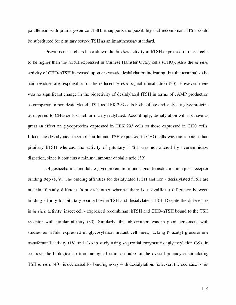
114
parallelism with pituitary-source cTSH, it supports the possibility that recombinant fTSH could
be substituted for pituitary source TSH as an immunoassay standard.
Previous researchers have shown the in vitro activity of hTSH expressed in insect cells
to be higher than the hTSH expressed in Chinese Hamster Ovary cells (CHO). Also the in vitro
activity of CHO-hTSH increased upon enzymatic desialylation indicating that the terminal sialic
acid residues are responsible for the reduced in vitro signal transduction (30). However, there
was no significant change in the bioactivity of desialylated fTSH in terms of cAMP production
as compared to non desialylated fTSH as HEK 293 cells both sulfate and sialylate glycoproteins
as opposed to CHO cells which primarily sialylated. Accordingly, desialylation will not have as
great an effect on glycoproteins expressed in HEK 293 cells as those expressed in CHO cells.
Infact, the desialylated recombinant human TSH expressed in CHO cells was more potent than
pituitary hTSH whereas, the activity of pituitary hTSH was not altered by neuraminidase
digestion, since it contains a minimal amount of sialic acid (39).
Oligosaccharides modulate glycoprotein hormone signal transduction at a post-receptor
binding step (8, 9). The binding affinities for desialylated fTSH and non - desialylated fTSH are
not significantly different from each other whereas there is a significant difference between
binding affinity for pituitary source bovine TSH and desialylated fTSH. Despite the differences
in in vitro activity, insect cell - expressed recombinant hTSH and CHO-hTSH bound to the TSH
receptor with similar affinity (30). Similarly, this observation was in good agreement with
studies on hTSH expressed in glycosylation mutant cell lines, lacking N-acetyl glucosamine
transferase I activity (18) and also in study using sequential enzymatic deglycosylation (39). In
contrast, the biological to immunological ratio, an index of the overall potency of circulating
TSH in vitro (40), is decreased for binding assay with desialylation, however; the decrease is not
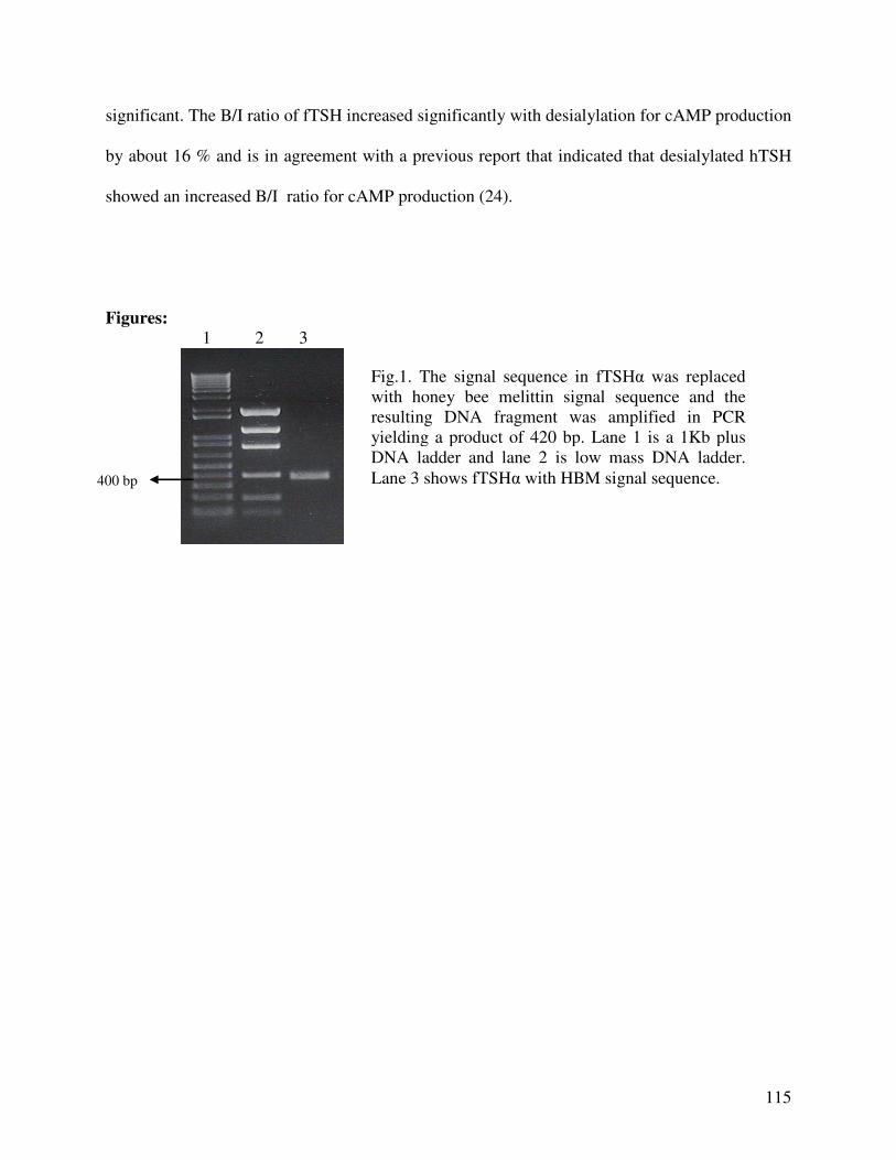
115
significant. The B/I ratio of fTSH increased significantly with desialylation for cAMP production
by about 16 % and is in agreement with a previous report that indicated that desialylated hTSH
showed an increased B/I ratio for cAMP production (24).
Figures:
1 2 3
Fig.1. The signal sequence in fTSHα was replaced with honey bee melittin signal sequence and the resulting DNA fragment was amplified in PCR yielding a product of 420 bp. Lane 1 is a 1Kb plus DNA ladder and lane 2 is low mass DNA ladder.
Lane 3 shows fTSHα with HBM signal sequence. 400 bp
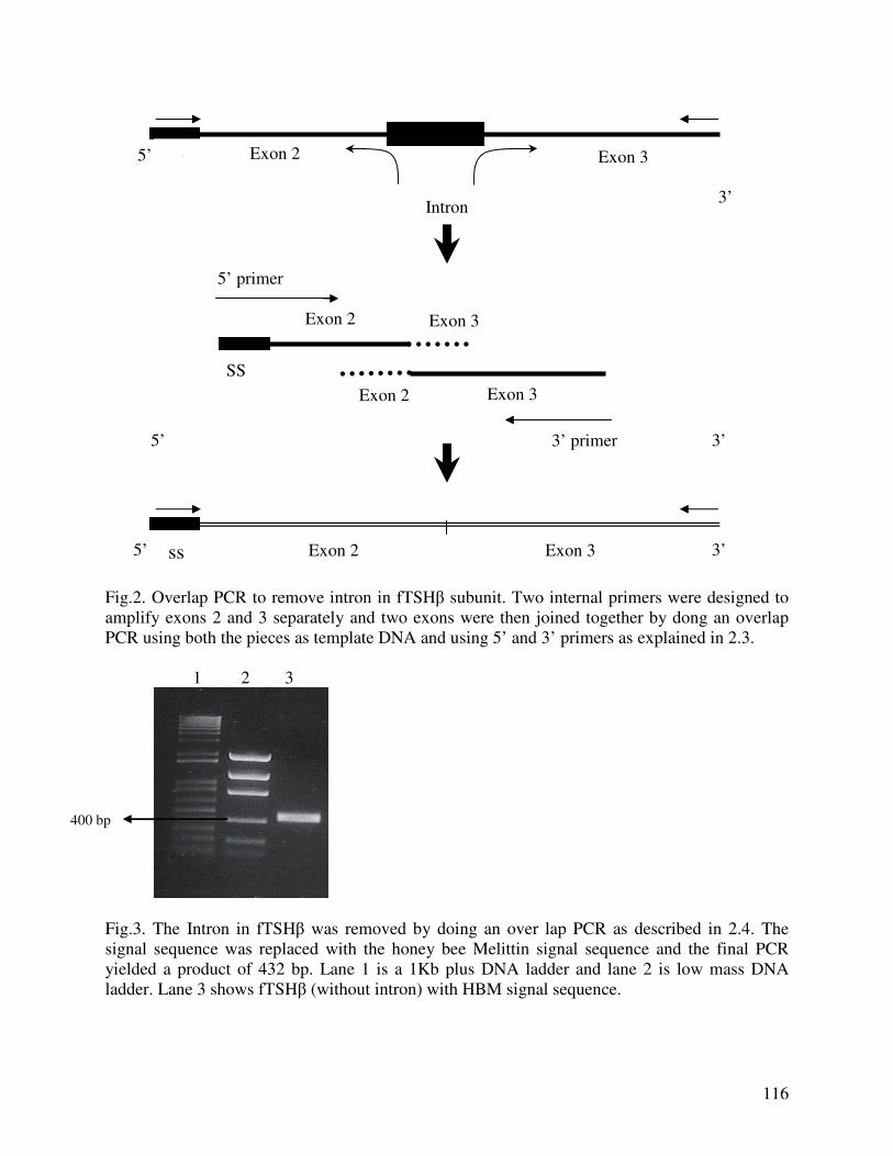
116
Intron
Fig.2. Overlap PCR to remove intron in fTSHβ subunit. Two internal primers were designed to amplify exons 2 and 3 separately and two exons were then joined together by dong an overlap PCR using both the pieces as template DNA and using 5’ and 3’ primers as explained in 2.3. 1 2 3
Fig.3. The Intron in fTSHβ was removed by doing an over lap PCR as described in 2.4. The signal sequence was replaced with the honey bee Melittin signal sequence and the final PCR yielded a product of 432 bp. Lane 1 is a 1Kb plus DNA ladder and lane 2 is low mass DNA ladder. Lane 3 shows fTSHβ (without intron) with HBM signal sequence.
Exon 3 Exon 2 SS
5’ 3’ ss
Exon 3 Exon 2 SS 5’
SS
Exon 2 Exon 3
Exon 2 Exon 3
3’ primer
5’ primer
3’
5’ 3’
400 bp
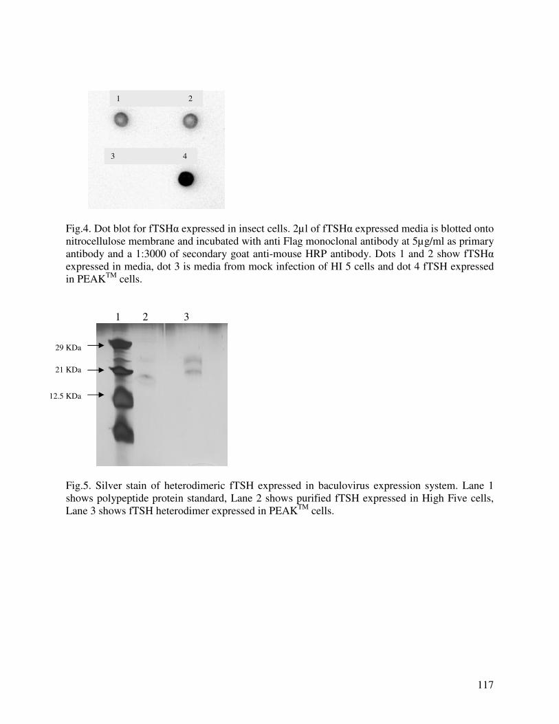
117
Fig.4. Dot blot for fTSHα expressed in insect cells. 2µl of fTSHα expressed media is blotted onto nitrocellulose membrane and incubated with anti Flag monoclonal antibody at 5µg/ml as primary antibody and a 1:3000 of secondary goat anti-mouse HRP antibody. Dots 1 and 2 show fTSHα expressed in media, dot 3 is media from mock infection of HI 5 cells and dot 4 fTSH expressed in PEAKTM cells. 1 2 3
Fig.5. Silver stain of heterodimeric fTSH expressed in baculovirus expression system. Lane 1 shows polypeptide protein standard, Lane 2 shows purified fTSH expressed in High Five cells, Lane 3 shows fTSH heterodimer expressed in PEAKTM cells.
1 2
3 4
29 KDa
21 KDa
12.5 KDa
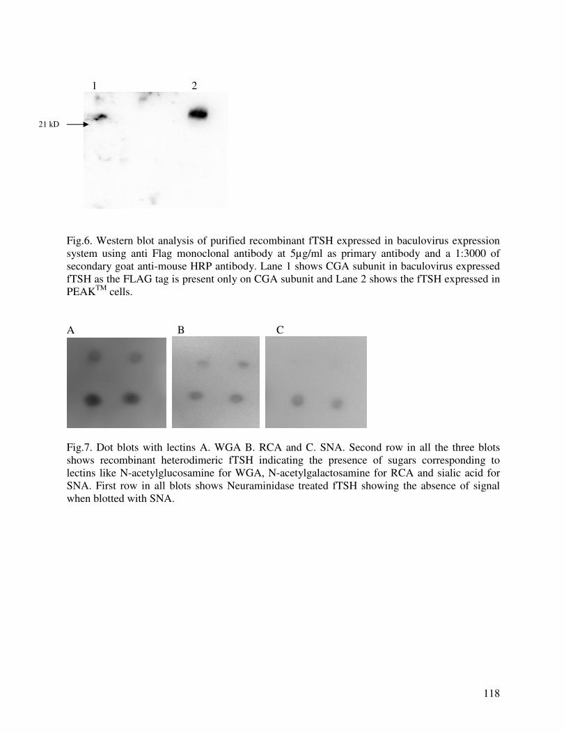
118
1 2
Fig.6. Western blot analysis of purified recombinant fTSH expressed in baculovirus expression system using anti Flag monoclonal antibody at 5µg/ml as primary antibody and a 1:3000 of secondary goat anti-mouse HRP antibody. Lane 1 shows CGA subunit in baculovirus expressed fTSH as the FLAG tag is present only on CGA subunit and Lane 2 shows the fTSH expressed in PEAKTM cells. A B C
Fig.7. Dot blots with lectins A. WGA B. RCA and C. SNA. Second row in all the three blots shows recombinant heterodimeric fTSH indicating the presence of sugars corresponding to lectins like N-acetylglucosamine for WGA, N-acetylgalactosamine for RCA and sialic acid for SNA. First row in all blots shows Neuraminidase treated fTSH showing the absence of signal when blotted with SNA.
21 kD
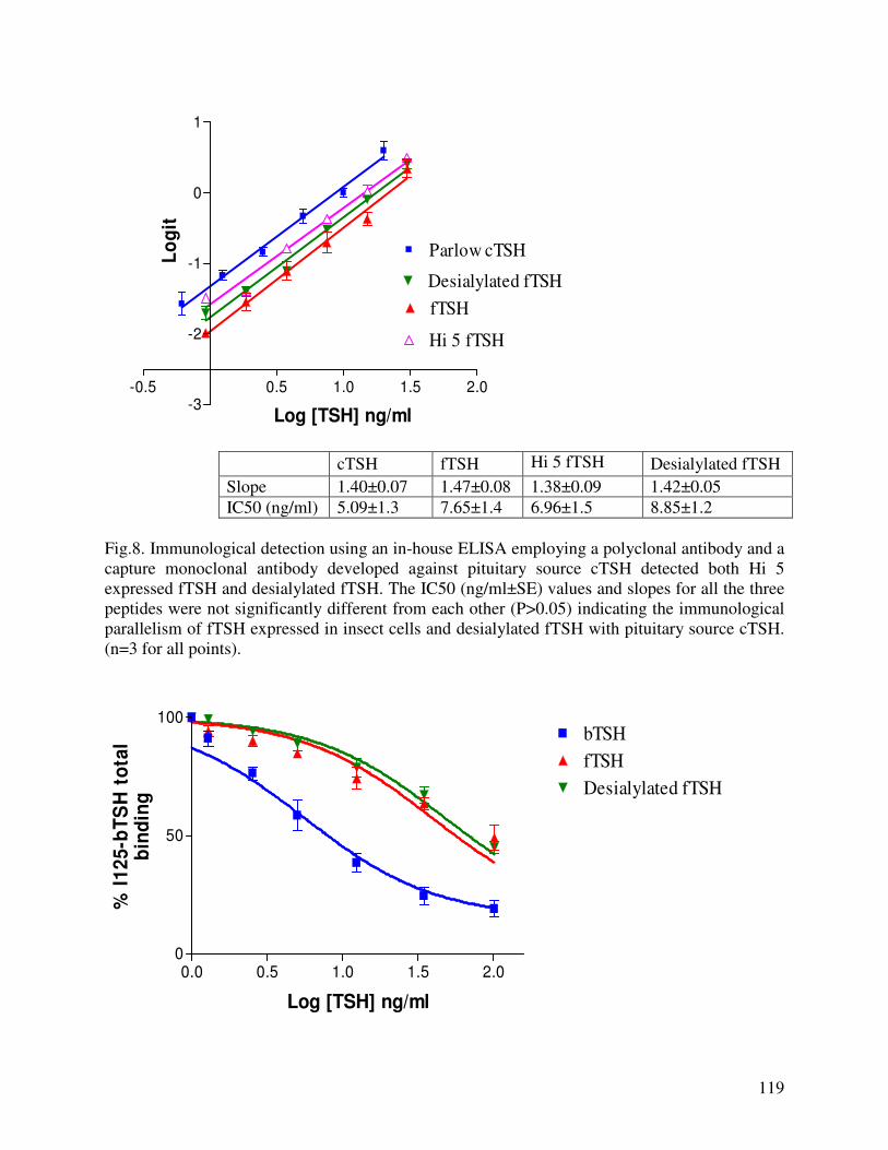
119
-0.5 0.5 1.0 1.5 2.0-3
-2
-1
0
1
Parlow cTSH
fTSH
Desialylated fTSH
Hi 5 fTSH
Log [TSH] ng/ml
Lo
git
cTSH fTSH Hi 5 fTSH Desialylated fTSH
Slope 1.40±0.07 1.47±0.08 1.38±0.09 1.42±0.05
IC50 (ng/ml) 5.09±1.3 7.65±1.4 6.96±1.5 8.85±1.2
Fig.8. Immunological detection using an in-house ELISA employing a polyclonal antibody and a capture monoclonal antibody developed against pituitary source cTSH detected both Hi 5 expressed fTSH and desialylated fTSH. The IC50 (ng/ml±SE) values and slopes for all the three peptides were not significantly different from each other (P>0.05) indicating the immunological parallelism of fTSH expressed in insect cells and desialylated fTSH with pituitary source cTSH. (n=3 for all points).
0.0 0.5 1.0 1.5 2.00
50
100bTSH
fTSH
Desialylated fTSH
Log [TSH] ng/ml
% I
12
5-b
TS
H t
ota
lb
ind
ing
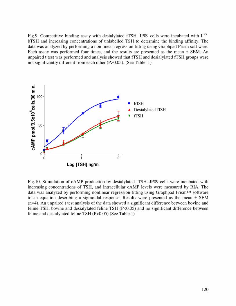
120
Fig.9. Competitive binding assay with desialylated fTSH. JP09 cells were incubated with I125-bTSH and increasing concentrations of unlabelled TSH to determine the binding affinity. The data was analyzed by performing a non linear regression fitting using Graphpad Prism soft ware. Each assay was performed four times, and the results are presented as the mean ± SEM. An unpaired t test was performed and analysis showed that fTSH and desialylated fTSH groups were not significantly different from each other (P>0.05). (See Table. 1)
0 1 20
50
100
bTSH
Desialylated fTSH
fTSH
Log [TSH] ng/ml
cA
MP
pm
ol/
3.5
x1
05ce
lls
/30 m
in.
Fig.10. Stimulation of cAMP production by desialylated fTSH. JP09 cells were incubated with increasing concentrations of TSH, and intracellular cAMP levels were measured by RIA. The data was analyzed by performing nonlinear regression fitting using Graphpad Prism™ software to an equation describing a sigmoidal response. Results were presented as the mean ± SEM (n=4). An unpaired t test analysis of the data showed a significant difference between bovine and feline TSH, bovine and desialylated feline TSH (P<0.05) and no significant difference between feline and desialylated feline TSH (P>0.05) (See Table.1)
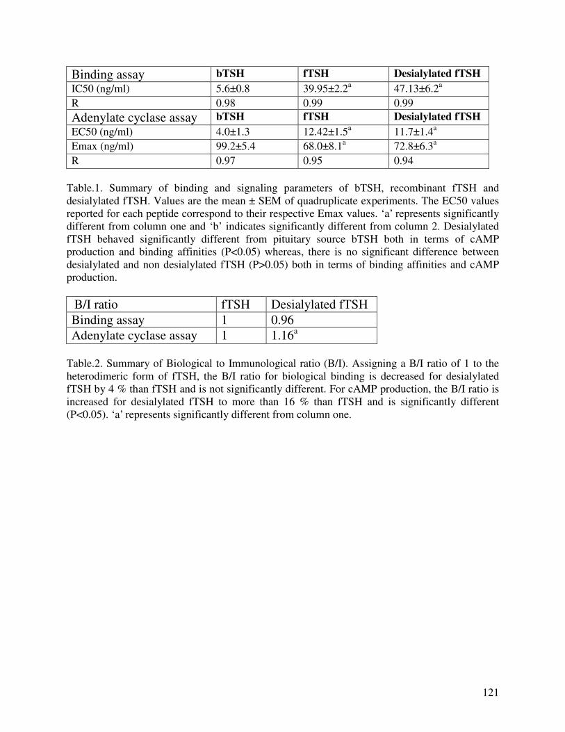
121
Binding assay bTSH fTSH Desialylated fTSH
IC50 (ng/ml)
5.6±0.8 39.95±2.2a 47.13±6.2a
R 0.98 0.99 0.99
Adenylate cyclase assay bTSH fTSH Desialylated fTSH
EC50 (ng/ml)
4.0±1.3 12.42±1.5a 11.7±1.4a
Emax (ng/ml)
99.2±5.4 68.0±8.1a 72.8±6.3a
R 0.97 0.95 0.94
Table.1. Summary of binding and signaling parameters of bTSH, recombinant fTSH and desialylated fTSH. Values are the mean ± SEM of quadruplicate experiments. The EC50 values reported for each peptide correspond to their respective Emax values. ‘a’ represents significantly different from column one and ‘b’ indicates significantly different from column 2. Desialylated fTSH behaved significantly different from pituitary source bTSH both in terms of cAMP production and binding affinities (P<0.05) whereas, there is no significant difference between desialylated and non desialylated fTSH (P>0.05) both in terms of binding affinities and cAMP production.
B/I ratio fTSH Desialylated fTSH
Binding assay 1 0.96
Adenylate cyclase assay 1 1.16a Table.2. Summary of Biological to Immunological ratio (B/I). Assigning a B/I ratio of 1 to the heterodimeric form of fTSH, the B/I ratio for biological binding is decreased for desialylated fTSH by 4 % than fTSH and is not significantly different. For cAMP production, the B/I ratio is increased for desialylated fTSH to more than 16 % than fTSH and is significantly different (P<0.05). ‘a’ represents significantly different from column one.
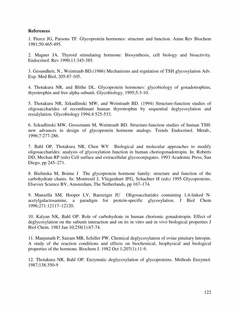
122
References
1. Pierce JG, Parsons TF. Glycoprotein hormones: structure and function. Annu Rev Biochem 1981;50:465-495. 2. Magner JA. Thyroid stimulating hormone: Biosynthesis, cell biology and bioactivity. Endocrinol. Rev 1990;11:345-385. 3. Gesundheit, N., Weintraub BD.(1986) Mechanisms and regulation of TSH glycosylation Adv. Exp. Med Biol, 205:87-105. 4. Thotakura NR, and Blithe DL. Glycoprotein hormones: glycobiology of gonadotrophins, thyrotrophin and free alpha subunit. Glycobiology, 1995;5:3-10. 5. Thotakura NR, Szkudlinski MW, and Weintraub BD. (1994) Structure-function studies of oligosaccharides of recombinant human thyrotrophin by sequential deglycosylation and resialylation. Glycobiology 1994;4:525-533. 6. Szkudlinski MW, Grossmann M, Weintraub BD. Structure-function studies of human TSH: new advances in design of glycoprotein hormone analogs. Trends Endocrinol. Metab., 1996;7:277-286. 7. Bahl OP, Thotakura NR, Chen WY Biological and molecular approaches to modify oligosaccharides: analysis of glycosylation function in human choriogonadotropin. In: Roberts DD, Mechan RP (eds) Cell surface and extracellular glycoconjugates. 1993 Academic Press, San Diego, pp 245–271. 8. Bielinska M, Boime I The glycoprotein hormone family: structure and function of the carbohydrate chains. In: Montreuil J, Vliegenhart JFG, Schachter H (eds) 1995 Glycoproteins. Elsevier Science BV, Amsterdam, The Netherlands, pp 167–174. 9. Manzella SM, Hooper LV, Baenziger JU Oligosaccharides containing 1,4-linked N- acetylgalactosamine, a paradigm for protein-specific glycosylation. J Biol Chem 1996;271:12117–12120. 10. Kalyan NK, Bahl OP. Role of carbohydrate in human chorionic gonadotropin. Effect of deglycosylation on the subunit interaction and on its in vitro and in vivo biological properties J Biol Chem. 1983 Jan 10;258(1):67-74. 11. Manjunath P, Sairam MR, Schiller PW. Chemical deglycosylation of ovine pituitary lutropin. A study of the reaction conditions and effects on biochemical, biophysical and biological properties of the hormone. Biochem J. 1982 Oct 1;207(1):11-9. 12. Thotakura NR, Bahl OP. Enzymatic deglycosylation of glycoproteins. Methods Enzymol. 1987;138:350-9
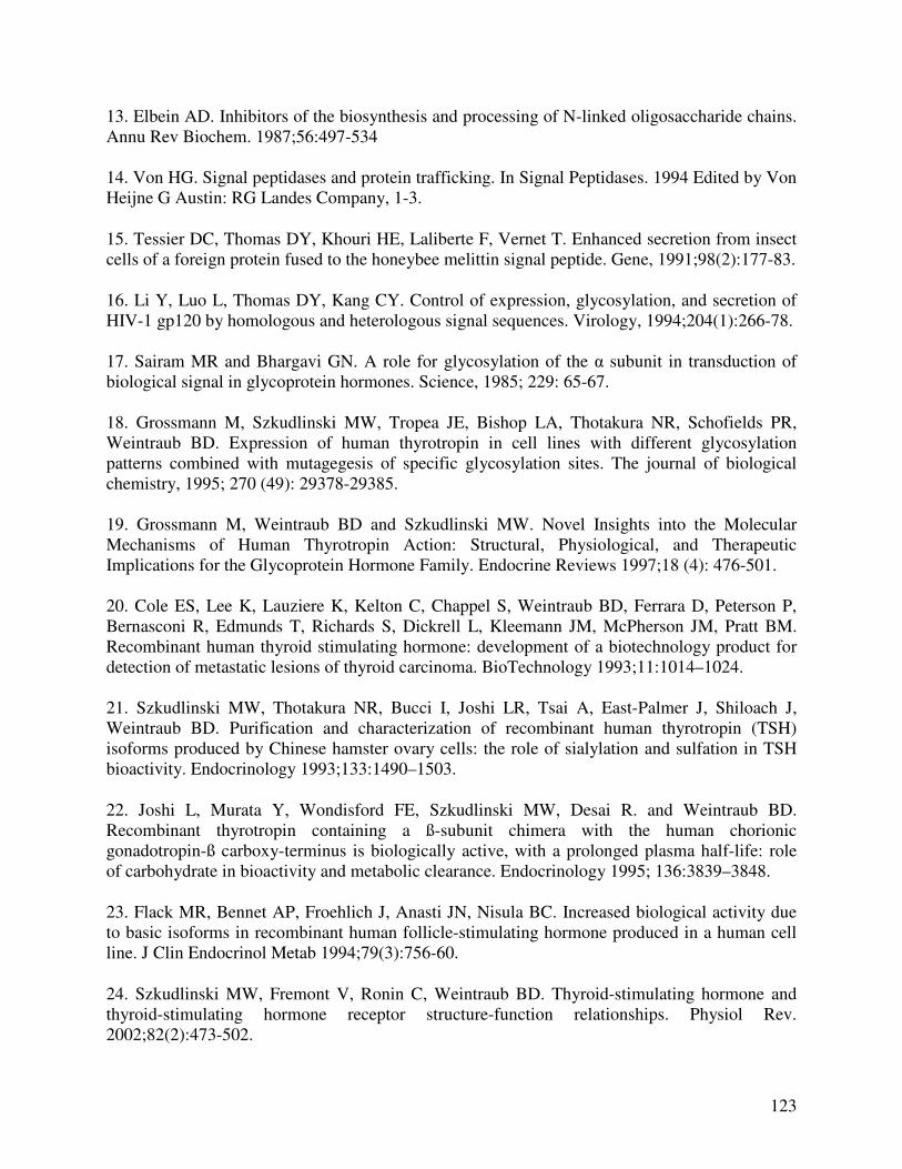
123
13. Elbein AD. Inhibitors of the biosynthesis and processing of N-linked oligosaccharide chains. Annu Rev Biochem. 1987;56:497-534
14. Von HG. Signal peptidases and protein trafficking. In Signal Peptidases. 1994 Edited by Von Heijne G Austin: RG Landes Company, 1-3.
15. Tessier DC, Thomas DY, Khouri HE, Laliberte F, Vernet T. Enhanced secretion from insect cells of a foreign protein fused to the honeybee melittin signal peptide. Gene, 1991;98(2):177-83.
16. Li Y, Luo L, Thomas DY, Kang CY. Control of expression, glycosylation, and secretion of HIV-1 gp120 by homologous and heterologous signal sequences. Virology, 1994;204(1):266-78. 17. Sairam MR and Bhargavi GN. A role for glycosylation of the α subunit in transduction of biological signal in glycoprotein hormones. Science, 1985; 229: 65-67. 18. Grossmann M, Szkudlinski MW, Tropea JE, Bishop LA, Thotakura NR, Schofields PR, Weintraub BD. Expression of human thyrotropin in cell lines with different glycosylation patterns combined with mutagegesis of specific glycosylation sites. The journal of biological chemistry, 1995; 270 (49): 29378-29385. 19. Grossmann M, Weintraub BD and Szkudlinski MW. Novel Insights into the Molecular Mechanisms of Human Thyrotropin Action: Structural, Physiological, and Therapeutic Implications for the Glycoprotein Hormone Family. Endocrine Reviews 1997;18 (4): 476-501. 20. Cole ES, Lee K, Lauziere K, Kelton C, Chappel S, Weintraub BD, Ferrara D, Peterson P, Bernasconi R, Edmunds T, Richards S, Dickrell L, Kleemann JM, McPherson JM, Pratt BM. Recombinant human thyroid stimulating hormone: development of a biotechnology product for detection of metastatic lesions of thyroid carcinoma. BioTechnology 1993;11:1014–1024. 21. Szkudlinski MW, Thotakura NR, Bucci I, Joshi LR, Tsai A, East-Palmer J, Shiloach J, Weintraub BD. Purification and characterization of recombinant human thyrotropin (TSH) isoforms produced by Chinese hamster ovary cells: the role of sialylation and sulfation in TSH bioactivity. Endocrinology 1993;133:1490–1503.
22. Joshi L, Murata Y, Wondisford FE, Szkudlinski MW, Desai R. and Weintraub BD. Recombinant thyrotropin containing a ß-subunit chimera with the human chorionic gonadotropin-ß carboxy-terminus is biologically active, with a prolonged plasma half-life: role of carbohydrate in bioactivity and metabolic clearance. Endocrinology 1995; 136:3839–3848.
23. Flack MR, Bennet AP, Froehlich J, Anasti JN, Nisula BC. Increased biological activity due to basic isoforms in recombinant human follicle-stimulating hormone produced in a human cell line. J Clin Endocrinol Metab 1994;79(3):756-60.
24. Szkudlinski MW, Fremont V, Ronin C, Weintraub BD. Thyroid-stimulating hormone and thyroid-stimulating hormone receptor structure-function relationships. Physiol Rev. 2002;82(2):473-502.
124
25. Rayalam S, Eizenstat LD, Hoenig M, Ferguson DC. Cloning and sequencing of feline thyrotropin (fTSH): heterodimeric and yoked constructs. Domest Anim Endocrinol 2000 (in press). 26. Rayalam S, Eizenstat LD, Davis RR, Hoenig M, Ferguson DC. Expression and purification of feline thyrotropin (fTSH): immunological detection and bioactivity of heterodimeric and yoked glycoproteins. Domest Anim Endocrinol 2000 (in press).
27. Yang X, McGraw RA, Su X, Katakam P, Grosse WM, Li OW, Ferguson DC. Canine thyrotropin β-subunit gene: cloning and expression in Escheria coli, generation of monoclonal antibodies and transient expression in the Chinese ovary cells. Domestic Animal Endocrinology 2000;18:363-378.
28. Libert F, Lefort A, Gerard C, Parmentier M, Perret J, Ludgate M, Dumont JE, Vassart G. Cloning, sequencing and expression of the human thyrotropin (TSH) receptor: evidence for binding of autoantibodies. Biochem Biophys Res Commun. 1989;165(3):1250-5. 29. Chard T. An introduction to radioimmunoassay and related techniques: laboratory techniques in biochemistry and molecular biology 1995; Elsevier Biomedical press, Amsterdam, The Netherlands.
30. Grossmann M, Wong R, Teh NG, Tropea JE, East-Palmer J, Weintraub BD, Szkudlinski MW. Expression of biologically active human thyrotropin (hTSH) in a baculovirus system: effect of insect cell glycosylation on hTSH activity in vitro and in vivo. Endocrinology 1997;138(1):92-100.
31. Jarvis, D.L., Summers, M.D., Garcia, A., Bohlmeyer, D.A. (1993) Influence of different peptides and prosequences on expression and secretion of human tissue plasminogenm activator in the baculovirus system. J Biol Chem, 268: 16754-16762. 32. Chen W, Shen QY, Bahl OP. Carbohydrate variant of the recombinant ß-subunit of human choriogonadotropin expressed in baculovirus expression system. J Biol Chem 1991;266:4081–4087. 33. Luckow VA. Baculovirus system for the expression of human gene products. Curr Opin Biotechnol 1993;4:564–572. 34. Maerz L, Altmann F, Staudacher E, Kubelka V. Protein glycosylation in insect cells. In: Montreuil J, Vliegenhart JFG Schachter H (eds) Glycoproteins. Elsevier Science BV, Amsterdam, The Netherlands. 1995;543–563. 35. Kidd M, Emery VC The use of baculoviruses as expression vectors. Appl Biochem Biotech 1993;42:137–155.
125
36. Ronin C, Papandreou MJ, Sergi S, Labbe-Jullie C, Medri G, Hoffmann T, Darbon H. Glycosylation-dependent epitope mapping of human TSH (hTSH) isoforms. Int J Rad Appl Instrum B. 1990;17(7):651-6. 37. Sairam M.. Gonadotropin hormones: relationship between structure and function with emphasis on antagonists. In: Li, C.H. (ed) Hormonal proteins and peptides. New York: Academic. 1-79, 1983. 38. Zerfaoui M, Ronin C. Glycosylation is the structural basis for the changes in polymorphism and immunoreactivity of pituitary glycoprotein hormones. European journal of Clinical Chemistry and Clinical Biochemistry, 1996;34:749-753.
39. Thotakura NR, Desai RK, Bates LG, Cole ES, Pratt BM, Weintraub BD. Biological activity and metabolic clearance of a recombinant human thyrotropin produced in Chinese hamster ovary cells. Endocrinology 1991;128:341–348.
40. Chappel S. Editorial: biological to immunological ratios: reevaluation of a concept. J Clin Endocrinol Metab. 1990 ;70:1494–5.
SUMMARY AND DISCUSSION
Thyrotropin (TSH) along with Follicle stimulating hormone (FSH), Luteinizing hormone
(LH) and Chorionic gonadotropin (CG) belong to the glycoprotein hormone family. All these
glycoprotein hormones share a common α subunit, and a hormone-specific β subunit. The α and
β subunits associate noncovalently and subunit assembly is necessary for the biological activity
of the hormone (Pierce and Parsons, Annu Rev Biochem, 50:465-495, 1981). It has been
reported that the α-subunit carries species-specific antigenic determinants, while the β-subunit
possess hormone-specific evolutionarily conserved determinants (Vaitukaitis et al.,
Endocrinology 91:1337-1342, 1972). TSH plays an important role in maintaining the normal
circulating levels of thyroid hormones through its action on the thyroid gland.
Hyperthyroidism is one of the most common endocrine disorders of cats, affecting
mainly middle to old aged cats and the lack of a feline-specific TSH assay has hindered early
diagnosis. Since a commercially available pituitary source of fTSH does not exist, we reasoned
that cloning, sequencing and expression of recombinant fTSH would allow development of a
feline specific TSH immunoassay. Feline pituitary RNA was extracted and an RT-PCR followed
by a PCR was done to amplify feline CGA gene along with signal peptide. The feline CGA gene
encoded a 96 amino acid protein and is highly conserved between cat and dog with only 4
residues being different in the secreted protein. Very high homology of 96.8% was observed
between the feline and tiger CGA gene and 91.6% homology between feline and bovine CGA
subunits. The sequence homology between feline and human α subunits is less than 70% because
of the four codon deletion in human CGA gene. The ten cysteine residues which are involved in

the formation of five intramolecular disulphide bonds are completely conserved in both number
and position among all species. The fTSH α subunit sequence has been submitted to Genbank
and is available with accession number AY972823.
For the fTSHβ gene feline genomic DNA was isolated and the “mini-gene” construct
along with signal peptide was amplified. The fTSHβ gene encoded for a protein of 118 amino
acids and showed high sequence homology of 94% with dog and low sequence homology of
88% with human. The 12 cysteine residues, involved in formation of six disulphide bridges are
conserved in feline, canine, human, bovine and equine TSHβ. Intron 2, is not a highly conserved
sequence containing 412 bp in feline, 448 in human and 450 bp in canine. The peptide sequence
CAGYC is highly conserved in TSHβ, LHβ, hCGβ and FSHβ and is thought to be important in
subunit combination (Hayashizaki et al., EMBO J 8:2291, 1989). The fTSHβ subunit sequence
has been submitted to Genbank and is available with accession number AY972824.
There are two N-linked glycosylation sites located at Asn56 and Asn82 in feline CGA
and are conserved in all other species. The unique N-glycosylation site at Asn23, followed by
two threonine residues in the mature TSH beta peptide, is also well conserved in all the species.
The oligosaccharide chains attached to the protein moiety have been shown to play an important
role in TSH subunit folding and biologic activities (Magner, J.A. Endocr. Rev. 11:345-385,
1990). This is the first report of the sequence of the genes encoding the α and β glycoprotein
subunits of TSH in the cat. It is clinically relevant that the predicted amino acid homologies of
feline CGA and fTSH beta subunits with dog TSH sequences were high 96 % and 94 %
respectively. Such homology likely accounts for the recognition of TSH in cat samples by
antibodies in canine TSH assays. However, it is interesting to note that even though the predicted
128
amino acid homology of fTSH beta subunit with human TSH beta was 88 % the human TSH
assay does not detect fTSH.
The correct assembly of the alpha and beta subunits noncovalently into heterodimers is
an obligatory step for the formation of biologically active hormone. Single chain or yoked
analogs of hTSH and hCG were constructed with the C-terminus of the β subunit fused using a
yoking peptide, CTP (carboxy terminal peptide) to the N-terminus of the α-subunit. CTP
contains several serine and proline residues and thus lacks significant secondary structure,
apparently, permitting the α subunit to assume proper orientation with respect to the β subunit.
The approach has resulted in the generation of hormones with increased in vivo stability and
activity (Grossmann et al., J Boil Chem. 272:21312-21316, 1997). The tandem order of
subunits, β-CTP-α, was chosen based on studies suggesting the importance of the N-terminal
region of hCGβ and C-terminal region of the α-subunit in receptor binding and activation. From
the standpoint of a strategy for recombinant protein expression and purification, this approach
also ensures equimolar expression, detection and purification of the single chain glycoprotein.
Therefore, the nucleotide sequence encoding the full length alpha subunit excluding the signal
sequence was fused in frame with the CTP to the 3’ terminus of the beta subunit containing the
coding sequence of the signal sequence and the secreted subunit. We demonstrated that the
yoked fTSH may act as a substitute for either pituitary source fTSH or recombinant
heterodimeric fTSH as a standard in immunoassay. The development of a permanent cell line
expressing yfTSH facilitates production of this recombinant peptide. Furthermore, recombinant
feline TSH in heterodimeric or yoked forms may be useful for the enhancement of radioiodide
uptake during thyroid ablative therapy.
129
The ideal expression system for a mammalian glycoprotein is a mammalian cell line
capable of post-translational glycosylation with a mammalian pattern of sugars. The most
commonly used Chinese Hamster Ovary (CHO) cells lack sulfotransferase and N-acetyl
galactosamine transferase resulting in the expression of glycoproteins that are not sulfated to the
same degree as pituitary-derived glycoproteins. However, in these studies we expressed the
recombinant hormones in human Embryonic Kidney cells (HEK 293) which have been shown to
sulfate pituitary glycoproteins to a greater extent (Grossmann et al., J. Biol. Chem. 270:29378–
29385, 1995), because of the presence of higher levels of N-acetyl galactosamine transferase and
sulfotransferase activities to those expressed in the pituitary gland. The feline CGA-FLAG, fTSH
beta and yoked fTSH constructs were ligated into pEAK10 expression vector separately and
expressed in PEAKTM cells. The molecular weights of feline CGA subunit, beta subunit and
yfTSH with glycosylation were 20.4, 17, and 45 kilodaltons, respectively. This is the first report
of the in vitro expression and purification of recombinant feline thyrotropin in heterodimeric and
yoked forms.
Expression levels of about 1 µg/ml were obtained for both heterodimeric and yoked
forms of fTSH and the levels obtained were considerably higher than the concentrations obtained
with recombinant hTSH expression using CHO and other eukaryotic cells (Grossmann et al.,
Mol Endocrinol 10:769-779, 1996). Furthermore, the synthesis of yoked fTSH indicates that the
linked alpha and beta subunits presumably folded with formation of disulfide bonds and a stable
conformation. However, in a manner similar to pituitary thyrotrophs, equimolar expression of
feline CGA and fTSH beta subunits did not occur owing to a single EF-1α promoter which
facilitates expression of only one gene at a time in the expression vector pEAK10. The ‘free
alpha’ subunit which has a slightly higher molecular weight due to an additional O-linked

130
oligosaccharide is removed by percolating the media though an affinity column made with
agarose beads linked to a beta-subunit specific monoclonal antibody (14H9.E4), which captured
heterodimeric fTSH alone removing the free alpha subunit and increasing the purity of the
recombinant fTSH preparation.
Lacking pituitary source fTSH, the immunological activity of fTSH in both forms was
compared with pituitary source canine TSH (NHPP standard) and the results indicated that both
forms of fTSH showed immunological parallelism to pituitary-source TSH supporting the
possibility that recombinant fTSH could be substituted for pituitary source TSH as an
immunoassay standard. However, the in-house ELISA detected both heterodimeric and yoked
forms of the recombinant protein to an extent of about 40 % as standardized by BCA protein
assay and purity determined by silver stain. Surprisingly, the recombinant glycoproteins showed
reduced receptor affinity and diminished maximal effect on the bioactivity assay.
The yoked fTSH exhibited 24% of the potency of pituitary source bTSH in adenylate
cyclase assays and only 3.4 % of the potency of pituitary source bTSH at inhibiting 125I-bTSH
binding in JP09 cells. The heterodimeric fTSH showed 45 % and 12.5 % of the potency of
pituitary source bTSH in adenylate cyclase and binding assays respectively. Yoked fTSH
showed a decreased biological to immunological ratio compared to heterodimeric fTSH.
However, receptor binding activity and in vitro bioactivity of purified pituitary isoforms have
been previously shown to vary over a five to eight-fold range when expressed on the basis of
defined protein content. Glycosylation and/or subtle tertiary structure differences might also
contribute to lower potency. Other studies have demonstrated that heterodimeric and yoked
forms of glycoproteins expressed in the baculovirus expression system exhibited higher binding
affinities than native forms presumably due to differences in glycosylation pattern which tend to
131
favor less sialylation. The demonstration of the immunological parallelism with pituitary source
canine TSH confirms that the recombinant glycoprotein may be used to standardize and improve
clinical assays for feline TSH, and even be utilized to prepare feline-specific immunoreagents.
Since the concentrations of the recombinant peptides never achieved the same maximal binding
inhibition nor maximal effect, we postulate that these glycoproteins may act as partial agonists of
human TSH receptor compared to pituitary-source bTSH. The reduced activity in receptor
binding might be accounted for species differences as well, since it was reported that
homologous glycohormones do not necessarily have the highest affinity for the receptor in the
same species.
Ultra-sensitive immunoassays will give misleading results when the glycosylation pattern
of the standard used in immunoassay is different from pituitary and circulating isoforms. For
recombinant fTSH expressed in PEAKTM cells to be used as a standard in feline specific TSH
assay, the immunoreactivity would ideally be independent of glycosylation. The glycosylation
machinery of insect cells does not possess the same capacity to process N-linked
oligosaccharides to complex-type carbohydrates as mammalian cells resulting in secretion of
high mannose-type precursor oligosaccharides (Luckow V.A, Curr Opin Biotechnol 4:564–572,
1993).
For expression of recombinant fTSH in baculovirus expression system, the intron in
fTSH beta subunit was removed with an over lap PCR and the signal peptide on both CGA and
beta subunits was substituted with insect cell specific honey bee melittin signal sequence. The
expression levels of only about 35 ng/ml were achieved as opposed to 1 µg/ml with PEAKTM
cells. This is in contrast to other studies which reported expression of recombinant hTSH at
1µg/ml in insect cells and 10-50 ng/ml of recombinant hTSH in mammalian cells. It is
132
interesting to note that the beta subunit expression seemed to dominate in insect cells, but it
appeared that the alpha subunit may have been partially degraded to release the C-terminal
FLAG tag. Since the relatively low expression levels of fTSH in the baculovirus expression
system did not facilitate purification of large quantities of recombinant fTSH for standardization
by protein assay, as an alternative, enzymatically desialylated fTSH expressed in PEAKTM cells
was prepared and used to characterize immunoreactivity and bioactivity. Complete
deglycosylation without denaturing the protein was likely not possible because the
oligosaccharides on β-subunit in both heterodimeric conformation and free forms have been
reported to be more easily accessible whereas, the sugars on α-subunit are hidden by subunit
interaction. Moreover it is well known that under in vitro conditions, sialic acid appears to be the
major factor affecting the bioactivity of recombinant TSH (Szkudlinski et al., Endocrinology
133:1490–1503, 1993).
The immunological activity of insect cell - expressed fTSH and desialylated fTSH was
compared with pituitary source canine TSH (NHPP standard) and non - desialylated fTSH and
the results indicated that both insect cell expressed fTSH and desialylated fTSH showed
immunological parallelism to pituitary-source TSH and non desialylated fTSH supporting the
possibility that the antibodies used in our in-house ELISA and the commercially available canine
TSH assay are not glycosylation dependent. There was no significant change in the bioactivity of
desialylated fTSH in terms of cAMP production as compared to non - desialylated fTSH.
However, the biological to immunological ratio (B/I) for cAMP production increased
significantly upon desialylation. The receptor binding affinity was also not significantly changed
by desialylation and in agreement with previous reports for human TSH.
133
The cloning of feline CGA and beta subunits enabled the use of the PEAKTM expression
system, which gives higher expression. The recombinant fTSH expressed in PEAK cells behaved
immunologically parallel to pituitary source canine TSH and changes in glycosylation pattern did
not affect the immunological recognition in both in-house ELISA and commercially available
canine TSH assay. However the biological to immunological ratio for cAMP production was
significantly increased upon desialylation of fTSH expressed in PEAKTM cells indicating that
sialic acid attenuates in vitro bioactivity in terms of cAMP production. The receptor binding
affinity was not affected upon neuraminidase treatment. Therefore, the recombinant glycopeptide
provides an ideal feline-specific TSH immunoassay standard which is not at least sialylation
dependent. However, more studies like determining the carbohydrate compositional analysis,
bioactivity studies using pituitary source fTSH and recombinant fTSH expressed in insect cells
and the generation of higher titer antibodies specific for fTSH will further advance the goal of
development of an ultrasensitive immunoassay for feline TSH.
![University of Western Australia · Web view, as it helps to completely degrade chitin degradation products, generated by secreted chitinases [43,44] and transported through outer](https://static.fdocument.org/doc/165x107/60d97f7be5724d3db967093f/university-of-western-australia-web-view-as-it-helps-to-completely-degrade-chitin.jpg)