Cloning and soluble expression of mature α-luffin from...
10
23 http://journals.tubitak.gov.tr/biology/ Turkish Journal of Biology Turk J Biol (2018) 42: 23-32 © TÜBİTAK doi:10.3906/biy-1708-12 Cloning and soluble expression of mature α-luffin from Luffa cylindrica in E. coli using SUMO fusion protein Shaghayegh NAMVAR 1,2, *, Farzaneh BARKHORDARI 2, *, Mozhgan RAIGANI 2 , Hoda JAHANDAR 1,3 , Leila NEMATOLLAHI 2 , Fatemeh DAVAMI 2, ** 1 Pharmaceutical Sciences Research Center, Pharmaceutical Sciences Branch, Islamic Azad University, Tehran, Iran 2 Biotechnology Research Center; Pasteur Institute of Iran, Tehran 3 Department of Biotechnology, Faculty of Advanced Sciences &Technology, Pharmaceutical Sciences Branch, Islamic Azad University, Tehran, Iran 1. Introduction α-Luffin is a single-chain type I ribosome-inactivating protein (RIP) found in Luffa cylindica seeds (Griffiths and Electricwala, 1987; Ng et al., 1992a; Ma et al., 2012). α-Luffin is one of three principle categories of RIPs identified from plant resources that have structural differences: type I RIPs consist of single chain polypeptides of approximately 30 kDa; type II category has an A chain catalytic domain common with type I and a B chain binding domain containing double chained RIPs of around 60 kDa (Lord et al., 1994); type III RIPs, synthesized as inactive precursors, undergo a proteolytic process before generating an active RIP (Peumans et al., 2001). No clinical indications have yet been reported for the third category, but type I RIPs have been broadly studied in biomedical fields for targeted drug delivery of immunotoxins in cancer immunotherapy. RIPs, which inhibit protein synthesis by their rRNA N glycosidases activity (Park et al., 2006), are widely distributed among plants, bacteria, and fungi. RIPs have been reported in approximately 17 different plant families, including Cucurbitaceae, Poaceae, and Euphorbiaceae (Sharma et al., 2004). RIPs show some properties including antibacterial (Pelegrini et al., 2008), antifungal (Parkash et al., 2002), and a multiplicity of antitumor (Cao et al., 2012; Shin et al., 2013), abortifacient (Yeung et al., 1988), and antiviral (for example, polio virus, cytomegalo virus, influenza virus, herpes simplex virus, and anti-human immunodeficiency virus (HIV)) activities (Barbieri et al., 1993; Park et al., 2006; Au et al., 2014). RIPs have attracted a lot of attention in biomedical research, especially immunotoxins in targeted therapy studies (Battelli et al., 1996). RIPs have been linked with carriers such as antibodies and hormones, in order to make them selectively toxic to a target cell. Numerous studies have been carried out to examine novel recombinant RIPs with desirable properties but, because of RIPs’ cellular toxicity, the expression of recombinant RIPs is a complicated process. Some results suggested that α-luffin inhibited the proliferation of cancer cell lines in a time- and dose- dependent manner and it was shown that the morphology of RIP-treated cells is similar to that of cells undergoing death by apoptosis (Griffiths et al., 1987). Abstract: α-Luffin, found in Luffa cylindrica seeds, is a type I ribosome inactivating proteins. Cytotoxic effects make it an appropriate candidate for the construction of immunotoxins and conjugates. Because of limited natural resources, recombinant technology is the best approach to achieve large-scale production of plant-based proteins. In the present study, α-luffin protein was expressed in E. coli and the effects of different temperature conditions, SUMO fusion tag, and cultivation strategies on total expression and solubility were investigated. Protein expression was evaluated at different intervals (0, 4, 6, 8, 24 h) postinduction. Our results showed that EnBase had higher efficiency than LB, and maximum solubility and total protein expression were achieved 24 h aſter induction at 30 °C and 25 °C, respectively. It was shown that SUMO tag is an effective strategy to improve protein solubility. Key words: E. coli expression system, fed-batch culture, ribosome inactivating proteins, soluble expression, α-luffin Received: 07.08.2017 Accepted/Published Online: 17.11.2017 Final Version: 15.02.2018 Research Article * ese authors contributed equally to this work. ** Correspondence: [email protected]
Transcript of Cloning and soluble expression of mature α-luffin from...
23
http://journals.tubitak.gov.tr/biology/
Turkish Journal of Biology Turk J Biol(2018) 42: 23-32© TÜBİTAKdoi:10.3906/biy-1708-12
Cloning and soluble expression of mature α-luffin from Luffa cylindrica inE. coli using SUMO fusion protein
Shaghayegh NAMVAR1,2,*, Farzaneh BARKHORDARI2,*, Mozhgan RAIGANI2,Hoda JAHANDAR1,3, Leila NEMATOLLAHI2, Fatemeh DAVAMI2,**
1Pharmaceutical Sciences Research Center, Pharmaceutical Sciences Branch, Islamic Azad University, Tehran, Iran2Biotechnology Research Center; Pasteur Institute of Iran, Tehran
3Department of Biotechnology, Faculty of Advanced Sciences &Technology,Pharmaceutical Sciences Branch, Islamic Azad University, Tehran, Iran
1. Introductionα-Luffin is a single-chain type I ribosome-inactivating protein (RIP) found in Luffa cylindica seeds (Griffiths and Electricwala, 1987; Ng et al., 1992a; Ma et al., 2012). α-Luffin is one of three principle categories of RIPs identified from plant resources that have structural differences: type I RIPs consist of single chain polypeptides of approximately 30 kDa; type II category has an A chain catalytic domain common with type I and a B chain binding domain containing double chained RIPs of around 60 kDa (Lord et al., 1994); type III RIPs, synthesized as inactive precursors, undergo a proteolytic process before generating an active RIP (Peumans et al., 2001). No clinical indications have yet been reported for the third category, but type I RIPs have been broadly studied in biomedical fields for targeted drug delivery of immunotoxins in cancer immunotherapy.
RIPs, which inhibit protein synthesis by their rRNA N glycosidases activity (Park et al., 2006), are widely distributed among plants, bacteria, and fungi. RIPs have been reported in approximately 17 different plant families, including Cucurbitaceae, Poaceae, and Euphorbiaceae
(Sharma et al., 2004). RIPs show some properties including antibacterial (Pelegrini et al., 2008), antifungal (Parkash et al., 2002), and a multiplicity of antitumor (Cao et al., 2012; Shin et al., 2013), abortifacient (Yeung et al., 1988), and antiviral (for example, polio virus, cytomegalo virus, influenza virus, herpes simplex virus, and anti-human immunodeficiency virus (HIV)) activities (Barbieri et al., 1993; Park et al., 2006; Au et al., 2014). RIPs have attracted a lot of attention in biomedical research, especially immunotoxins in targeted therapy studies (Battelli et al., 1996). RIPs have been linked with carriers such as antibodies and hormones, in order to make them selectively toxic to a target cell. Numerous studies have been carried out to examine novel recombinant RIPs with desirable properties but, because of RIPs’ cellular toxicity, the expression of recombinant RIPs is a complicated process.
Some results suggested that α-luffin inhibited the proliferation of cancer cell lines in a time- and dose-dependent manner and it was shown that the morphology of RIP-treated cells is similar to that of cells undergoing death by apoptosis (Griffiths et al., 1987).
Abstract: α-Luffin, found in Luffa cylindrica seeds, is a type I ribosome inactivating proteins. Cytotoxic effects make it an appropriate candidate for the construction of immunotoxins and conjugates. Because of limited natural resources, recombinant technology is the best approach to achieve large-scale production of plant-based proteins. In the present study, α-luffin protein was expressed in E. coli and the effects of different temperature conditions, SUMO fusion tag, and cultivation strategies on total expression and solubility were investigated. Protein expression was evaluated at different intervals (0, 4, 6, 8, 24 h) postinduction. Our results showed that EnBase had higher efficiency than LB, and maximum solubility and total protein expression were achieved 24 h after induction at 30 °C and 25 °C, respectively. It was shown that SUMO tag is an effective strategy to improve protein solubility.
Key words: E. coli expression system, fed-batch culture, ribosome inactivating proteins, soluble expression, α-luffin
Received: 07.08.2017 Accepted/Published Online: 17.11.2017 Final Version: 15.02.2018
Research Article
* These authors contributed equally to this work. ** Correspondence: [email protected]
NAMVAR et al. / Turk J Biol
24
Cancer is one of the therapeutic indications that have benefited more from recombinant drugs. In this field, antibodies designed for cancer cell eradication were used for targeted cancer therapy (Thomas et al., 2016). The monoclonal antibodies (mAb) linked to different cytotoxic agents have made a second generation of antibodies with considerable success in oncology. To date, only one immunotoxin, denileukin diftitox (DD; Ontak), has been approved by the US Food and Drug Administration (FDA) (Goldmacher and Kovtun, 2011; Flygare et al., 2013; Thomas et al., 2016).
The expression of a desired recombinant protein seemed to be more reasonable using a suitable expression system. The first FDA approved biopharmaceutical (Insulin) was produced in Escherichia coli, in 1982. Nowadays about 30% of biopharmaceuticals are produced in the bacterial cells. Moreover, 24% (94 products) of marketed biopharmaceuticals are used in anticancer therapies and 69% of these products are produced in E. coli (Sanchez-Garcia et al., 2016). According to the Southeast Collaboratory for Structural Genomics (SECSG) reports, 22.9% of proteins that have been expressed in E. coli are in soluble form. Coexpression with foldases and chaperones (Ikura et al., 2002), using weaker promoters (Studier and Moffatt, 1986), reducing the temperature, optimizing the cultivation strategies, and protein fusions technology are some of numerous strategies that may improve the solubility of expressed proteins.
Thioredoxin (TRX), NUS A, maltose binding protein (MBP), glutathione S-transferase (GST), ubiquitin (UB), and small ubiquitin-related modifier protein (SUMO) (Pryor and Leiting, 1997) are the most commonly used tags. These fusion motifs can improve correct protein folding and enhance the expression level and solubility rate of recombinant proteins. SUMO, around 11 kDa, belongs to a group of ubiquitin-like proteins that are mostly used by fusing to the N-terminal of the protein of interest (POI) to enhance its expression and solubility (Bird, 2011). SUMO is involved in cellular processes like apoptosis, nuclear–cytosolic transport, protein activation, and controlling the eukaryotic cell cycle. Fusion proteins need to be cleaved after expression, to promote native protein structure (Lee et al., 2008). SUMO protease can recognize the 3D structure of SUMO and cleave precisely within the amino acid sequence of target proteins. Reducing proteolytic degradation of the recombinant protein and simplifying the purification process are other advantages of SUMO fusion technology. Recombinant protein expression in E. coli, using SUMO, has increased the yield of difficult-to-express proteins (Shimokawa-Falcao et al., 2017).
In the present study, the recombinant α-luffin was expressed in E.coli. In addition, the effect of SUMO fusion tag, reduced temperature, and the cultivation strategy on
its total and soluble expression was studied. Furthermore, because of the toxic properties of α-luffin, we used rhamnose promoter for tightly controlling the expression and preventing overexpression of this protein.
2. Materials and methods2.1. Cloning of α-luffin geneAccording to sequence data in GenBank, the α-luffin gene with accession number X62371.1 was chemically synthesized, after codon optimization for protein expression in E. coli cells. Forward and reverse primers were designed based on the protocol of the Lucigen kit (Lucigen Corporation, USA). Amplification was achieved through polymerase chain reaction (PCR). The PCR products were analyzed by agarose gel electrophoresis. The expected size of PCR product was 750 bp.
After the amplification of the target gene, 1–3 μL of PCR product was mixed with 25 ng of pRham N-His SUMO Vector and transformed into E. coli 10G, chemically competent cells of the Lucigen kit. Transformation was performed according to the manufacturer’s instructions. Recombination between the vector and α-luffin gene occurred in vivo, within the bacterial host. 2.2. Screening for recombinantsTo confirm the recombination, the colonies were picked from a freshly prepared selective plate and plasmid extraction was performed using a Fermentas Gene JET plasmid miniprep kit according to the manufacturer’s instructions. The extracted plasmids were subjected to colony PCR and restriction enzyme digestion. PCR amplification was carried out using the specific primers and SUMO forward and pETite reverse primers included in the kit. PCR cycles were as follow: 25 cycles of 94 °C for 45 s, 55 °C for 45 s, and 72 °C for 45 s. The expected PCR product size was 750 bp. The PCR products were analyzed by agarose gel electrophoresis. Recombinant plasmid was also digested using EcoRV.2.3. Protein expressionA single colony of recombinant E. coli 10G was inoculated into LB growth medium containing 30 µg/mL kanamycin at 37 °C with 180 rpm shaking. After 3–4 h, when the OD600 of the culture reached 0.55–0.6, 1 mL of medium was centrifuged and kept at –20 °C as a negative control. Then 0.2% rhamnose was added to the remaining cells to induce expression. Bacterial pellets were collected 4, 6, 8, and 24 h after incubation.2.4. α-Luffin expression analysisFor detection of recombinant expression, SDS-PAGE and western blot analysis were performed. Induced cells were harvested and resuspended in 2X sample buffer (0.5 M Tris base pH 6.8, 25% glycerol, 4% SDS, 0.5% bromophenol blue). Samples were heated in a water bath
NAMVAR et al. / Turk J Biol
25
in 95 °C for 5–10 min and analyzed on 12% SDS-PAGE with Coomassie Brilliant Blue staining.
Western blotting was performed utilizing an anti-His antibody. The separated proteins were then transferred to a nitrocellulose membrane at 14 V for 40 min. The membrane was blocked with 3% nonfat dry milk in phosphate-buffered saline (PBS)-0.05% Tween 20 for 2 h. Then blots were incubated with the antibody at a dilution of 1:500 in PBS for 2 h at room temperature.
Visualization of the HRP-conjugated antibody was achieved using the DAB substrate. An 18 kDa His-conjugated protein and uninduced cells were used as the positive and negative controls, respectively.2.5. Effects of cultivation strategies and reduced temperatures on α-luffin expression/solubilityAccording to the Biosilta kit protocol for fed-batch Enbase cultivation, EnPresso two white tablets, containing culture media component, were solved in 50 mL of pure, distilled, and sterile water. For preparing the preculture, a single colony was inoculated in 5 mL of LB containing 30 µg/mL kanamycin and incubated for 6–8 h at 37 °C to obtain OD600 0.6 approximately, at which time 1:25 volume of the preculture medium was inoculated into the 5 mL of prepared EnBase culture medium supplemented with kanamycin (30 µg/mL). Then 12.5 µL of 600 IU Reagent A (glucose releasing enzyme) was added and the culture was incubated overnight with shaking at 200 rpm. After 15–18 h, 1 mL of grown medium was collected as before induction sample. According to the manufacturer’s instruction, in this step, a booster tablet (black tablet) was added. The booster tablet was composed of extra nutrients and polysaccharide substrate. Each tablet was solved in 5 mL of distilled sterile water and 0.5 mL of prepared medium was used in each study. Moreover, 12.5 µL of 600 IU Reagent A and rhamnose (at final concentration of 0.2%) were also added to the cultivation medium. Samples were collected 2, 4, 6, 8, 12, and 24 h after induction. The media were centrifuged and pellets were stored at –20 °C until further analysis.
The mentioned steps were repeated at two other induction temperatures, 25 °C and 30 °C, to investigate the influence of reduced temperature on solubility of recombinant α-luffin.2.6. Soluble expression analysisIn order to assess the solubility of recombinant α-luffin, samples were centrifuged at 9000 rpm for 3 min. Cell pellets were resuspended in TE buffer and sonicated for 8 cycles of 30 s pulses and 20 s intervals. The sonicated samples were centrifuged at 13,000 rpm for 20 min at 4 °C. The total protein sample was collected from the cell suspension after sonication. The clear supernatant was collected as a soluble fraction after the insoluble debris was pelleted. Protein concentration was measured using
absorbance at 280 nm by NanoDrop spectrophotometer. Total, pellet, and supernatant samples of each experiment were subjected to SDS-PAGE and protein bands were visualized by staining with Coomassie Brilliant Blue.
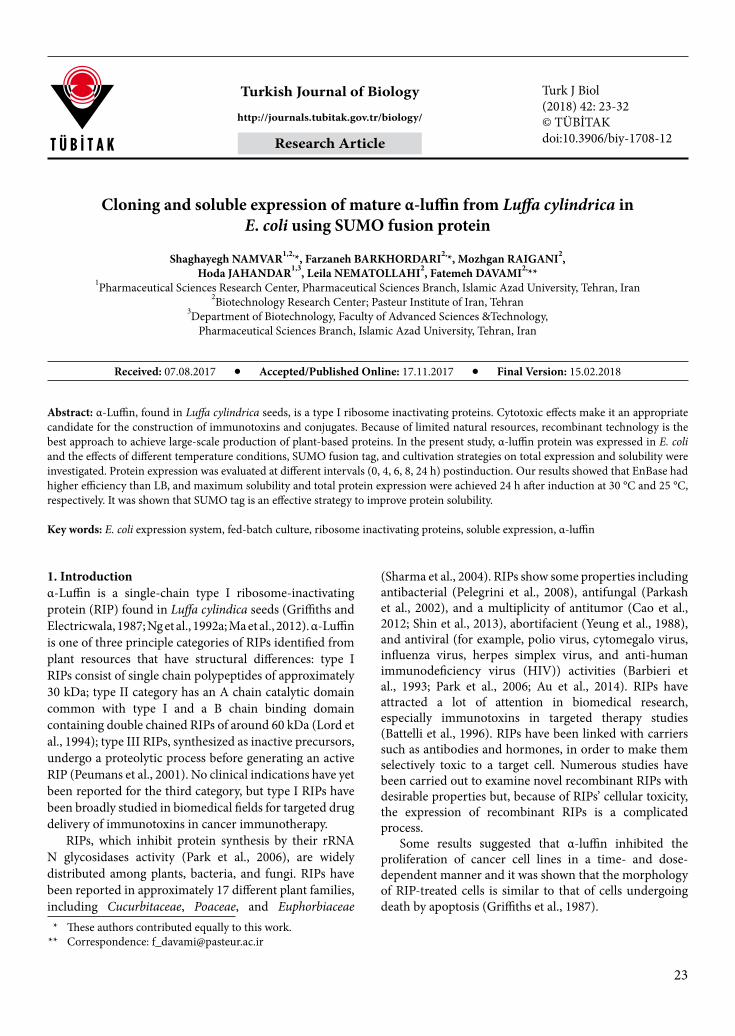
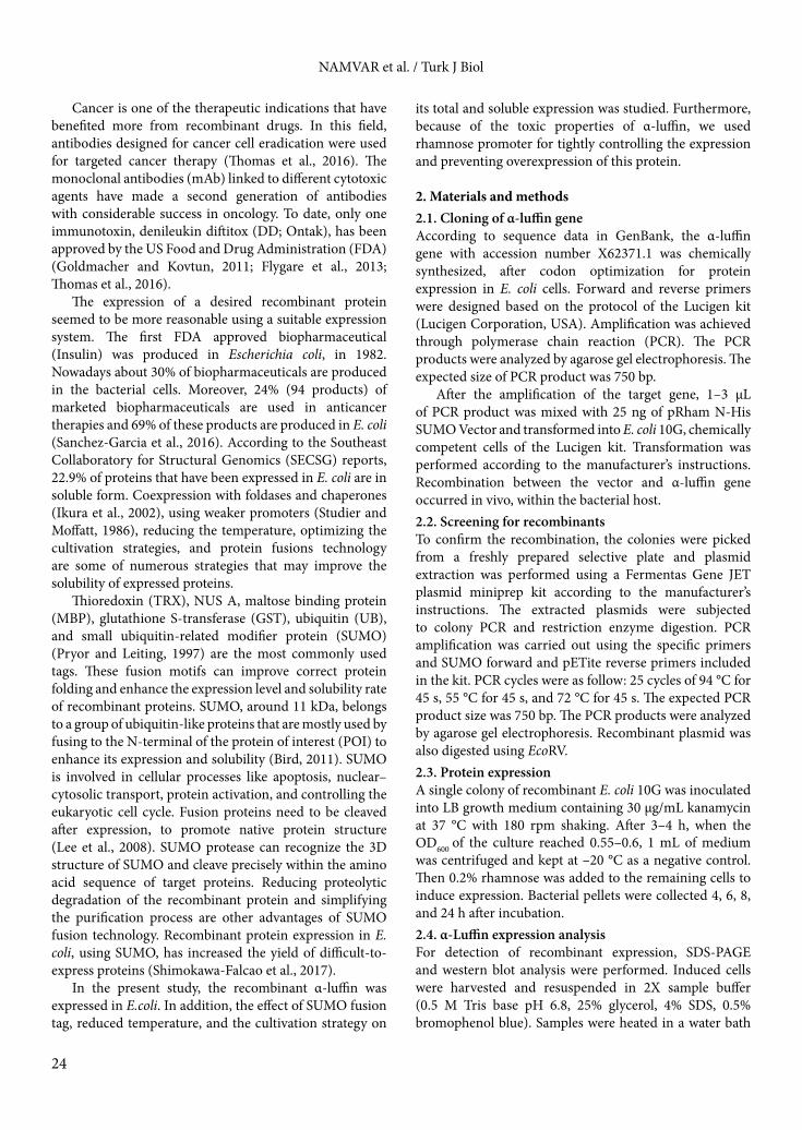
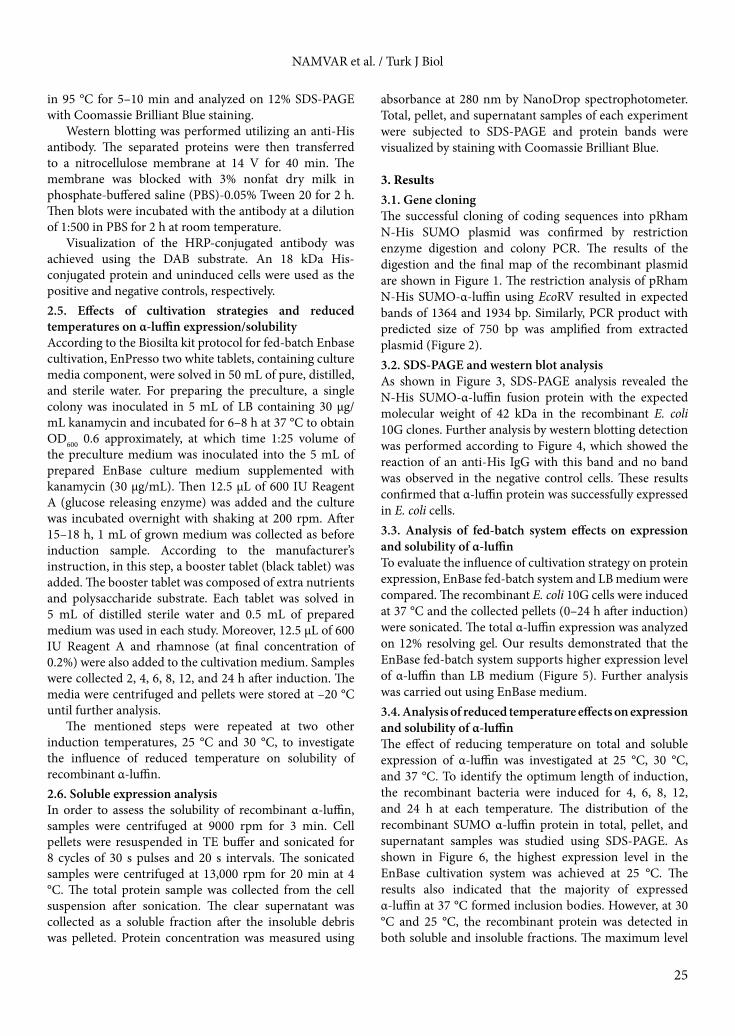
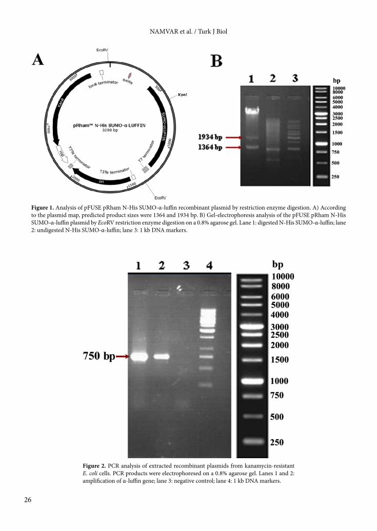
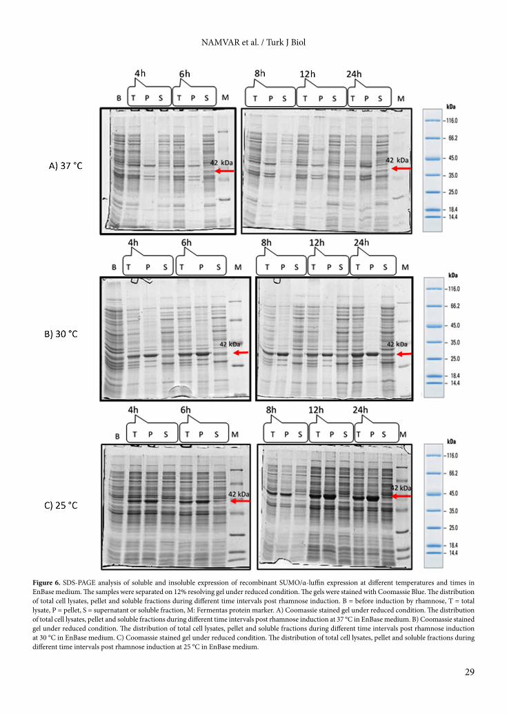
3. Results3.1. Gene cloningThe successful cloning of coding sequences into pRham N-His SUMO plasmid was confirmed by restriction enzyme digestion and colony PCR. The results of the digestion and the final map of the recombinant plasmid are shown in Figure 1. The restriction analysis of pRham N-His SUMO-α-luffin using EcoRV resulted in expected bands of 1364 and 1934 bp. Similarly, PCR product with predicted size of 750 bp was amplified from extracted plasmid (Figure 2).3.2. SDS-PAGE and western blot analysisAs shown in Figure 3, SDS-PAGE analysis revealed the N-His SUMO-α-luffin fusion protein with the expected molecular weight of 42 kDa in the recombinant E. coli 10G clones. Further analysis by western blotting detection was performed according to Figure 4, which showed the reaction of an anti-His IgG with this band and no band was observed in the negative control cells. These results confirmed that α-luffin protein was successfully expressed in E. coli cells.3.3. Analysis of fed-batch system effects on expression and solubility of α-luffinTo evaluate the influence of cultivation strategy on protein expression, EnBase fed-batch system and LB medium were compared. The recombinant E. coli 10G cells were induced at 37 °C and the collected pellets (0–24 h after induction) were sonicated. The total α-luffin expression was analyzed on 12% resolving gel. Our results demonstrated that the EnBase fed-batch system supports higher expression level of α-luffin than LB medium (Figure 5). Further analysis was carried out using EnBase medium.3.4. Analysis of reduced temperature effects on expression and solubility of α-luffinThe effect of reducing temperature on total and soluble expression of α-luffin was investigated at 25 °C, 30 °C, and 37 °C. To identify the optimum length of induction, the recombinant bacteria were induced for 4, 6, 8, 12, and 24 h at each temperature. The distribution of the recombinant SUMO α-luffin protein in total, pellet, and supernatant samples was studied using SDS-PAGE. As shown in Figure 6, the highest expression level in the EnBase cultivation system was achieved at 25 °C. The results also indicated that the majority of expressed α-luffin at 37 °C formed inclusion bodies. However, at 30 °C and 25 °C, the recombinant protein was detected in both soluble and insoluble fractions. The maximum level
NAMVAR et al. / Turk J Biol
26
Figure 1. Analysis of pFUSE pRham N-His SUMO-α-luffin recombinant plasmid by restriction enzyme digestion. A) According to the plasmid map, predicted product sizes were 1364 and 1934 bp. B) Gel-electrophoresis analysis of the pFUSE pRham N-His SUMO-α-luffin plasmid by EcoRV restriction enzyme digestion on a 0.8% agarose gel. Lane 1: digested N-His SUMO-α-luffin; lane 2: undigested N-His SUMO-α-luffin; lane 3: 1 kb DNA markers.
Figure 2. PCR analysis of extracted recombinant plasmids from kanamycin-resistant E. coli cells. PCR products were electrophoresed on a 0.8% agarose gel. Lanes 1 and 2: amplification of α-luffin gene; lane 3: negative control; lane 4: 1 kb DNA markers.

NAMVAR et al. / Turk J Biol
27
Figure 3. SDS-PAGE analysis of α-luffin expression in LB medium. Lane 1: Cell lysates before rhamnose induction; Lane 2: Cell lysates 4 h postinduction; Lane 3: Cell lysates 6 h postinduction; Lane 4: Cell lysates 8 h postinduction; Lane 5: Cell lysates 24 h postinduction; Lane 6: Fermentas Protein marker SM0671.
Figure 4. Western blot analysis of recombinant His tag-α-luffin. Lane 1: 42 kDa His tagged α-luffin; Lane 2: His tagged protein as positive control; Lane 3: Fermentas protein marker SM0671.

NAMVAR et al. / Turk J Biol
28
Figure 5. SDS-PAGE analysis of expressed recombinant SUMO/α-luffin at different temperatures and times in EnBase medium. The samples were separated on 12% resolving gel under reduced condition. The gels were stained with Coomassie Blue. A) Coomassie stained gel under reduced condition, EnBase medium at 37 °C. Lane 1: Cell lysates before rhamnose induction; Lane 2: Cell lysates 4 h postrhamnose induction; Lane 3: Cell lysates 6 h postrhamnose induction; Lane 4: Cell lysates 8 h postrhamnose induction; Lane 5: Cell lysates 12 h postrhamnose induction; Lane 6: Cell lysates 24 h postrhamnose induction; M: Fermentas protein marker SM0671. B) Coomassie stained gel under reduced condition, EnBase medium at 30 °C. Lane 1: Cell lysates before rhamnose induction; Lane 2: Cell lysates 4 h postrhamnose induction; Lane 3: Cell lysates 6 h postrhamnose induction; Lane 4: Cell lysates 8 h postrhamnose induction; Lane 5: Cell lysates 12 h postrhamnose induction; Lane 6: Cell lysates 24 h postrhamnose induction; M: Fermentas protein marker SM0671. C) Coomassie stained gel under reduced condition, EnBase medium at 25 °C. Lane 1: Cell lysates before rhamnose induction; Lane 2: Cell lysates 4 h postrhamnose induction; Lane 3: Cell lysates 6 h postrhamnose induction; Lane 4: Cell lysates 8 h postrhamnose induction; Lane 5: Cell lysates 12 h postrhamnose induction; Lane 6: Cell lysates 24 h postrhamnose induction; M: Fermentas protein marker SM0671.
NAMVAR et al. / Turk J Biol
29
Figure 6. SDS-PAGE analysis of soluble and insoluble expression of recombinant SUMO/α-luffin expression at different temperatures and times in EnBase medium. The samples were separated on 12% resolving gel under reduced condition. The gels were stained with Coomassie Blue. The distribution of total cell lysates, pellet and soluble fractions during different time intervals post rhamnose induction. B = before induction by rhamnose, T = total lysate, P = pellet, S = supernatant or soluble fraction, M: Fermentas protein marker. A) Coomassie stained gel under reduced condition. The distribution of total cell lysates, pellet and soluble fractions during different time intervals post rhamnose induction at 37 °C in EnBase medium. B) Coomassie stained gel under reduced condition. The distribution of total cell lysates, pellet and soluble fractions during different time intervals post rhamnose induction at 30 °C in EnBase medium. C) Coomassie stained gel under reduced condition. The distribution of total cell lysates, pellet and soluble fractions during different time intervals post rhamnose induction at 25 °C in EnBase medium.
NAMVAR et al. / Turk J Biol
30
of soluble expression for α-luffin was attained 12 and 24 h postinduction time at 30 °C.
As illustrated in Figures 5 and 6, the fed-batch system and reduced temperature can improve expression and solubility of α-luffin.
4. DiscussionNowadays targeted therapy is one of the best strategies in the treatment of cancer or other complicated diseases (Battelli et al., 1996). Antibody drug conjugates (ADCs) are a class of therapeutics consisting of the antigen-selectivity of MAbs in order to deliver highly potent toxins to tumor cell surface antigen (Trail, 2013). Many toxins have been considered as candidates for producing antibody drug conjugates (Flygare et al., 2013). For this approach, RIPs have been linked to carriers such as antibodies, hormones, and growth factors. RIPs containing immunotoxins can act on cells without inducing resistance and so they have more advantages in comparison to the conventional chemotherapeutic agents (Sobiya Raj and Jannet Vennila, 2013).
α-Luffin, a type I RIP, is one of the most toxic proteins from the luffin family. Several studies revealed its antiviral effects. On the other hand, antitumoral activities of this protein were confirmed in breast cancers, choriocarcinoma, and hepatoma in different cell lines (Sha et al., 2013).
Regarding their highly toxic nature, RIP family toxins have toxic effects on producing host cells. It especially occurs in luffin-a and -b from Luffa cylindrica (cytotoxic derivatives) (Ng et al., 1992a, 1992b; Ma et al., 2012).
Escherichia coli has been the workhorse of gene expression for many years, but unfortunately cytoplasmic expression of many recombinant toxins mostly has led to inclusion bodies formation, requiring comprehensive in vitro refolding steps to access their biological activities (Balduino et al., 2011; Lyukmanova et al., 2016; Shimokawa-Falcao et al., 2017). On the other hand, the refolding process is not cost effective in many cases; therefore enhancing protein solubility by other strategies is a better approach to obtain high production levels (Lee et al., 2008). In the present study, we aimed to enhance the total and soluble expression of α-luffin by reducing the induction temperature, fed-batch cultivation system, and SUMO fusion tag. Moreover, because of the toxic effect of recombinant α-luffin, the tunable rhamnose promoter was used.
First, the expression of recombinant α-luffin was confirmed at 37 °C in LB medium (Figures 3 and 4), and the total protein expression was not significantly altered at different times postinduction. However, in EnBase medium, the expression of recombinant protein increased with time postinduction, and maximum expression and solubility were obtained 12 and 24 h postinduction.
EnBase medium provides a controlled cultivation mode by continuous feeding with glucose, controlling pH changes, avoiding oxygen fluctuations, and other controlling processes on growth medium conditions (Panula-Perälä et al., 2008). Hence, the E. coli cells survived the harmful effect of metabolites and oxygen depletion and protein synthesis reduction (Krause et al., 2010). A similar outcome was reported by Krause et al., who developed a novel fed-batch based cultivation method. They demonstrated that the novel EnBase Flo cultivation system in shaken cultures can provide high cell densities without impairing the productivity per cell and improve yield of soluble recombinant proteins in shaken cultures in comparison to commonly used LB, Terrific broth, or mineral salt media (Krause et al., 2010). Due to the growth control in the EnBase cultivation system, accumulation of harmful metabolites can be diminished and development of anaerobic conditions can be avoided. Expression at three different temperatures, 25 °C, 30 °C, and 37 °C, was investigated. Samples were collected 0–24 h after induction by rhamnose.
Some previous studies have reported the toxic effect of RIPs on prokaryotic ribosomes (Girbes et al., 1993; Chaddock et al., 1994; Liu et al., 2010). Our observations did not show this phenomenon because of lower temperature cultivation (25 °C), which could reduce the toxic effect of α-luffin on E. coli cells only if it is performed in fed-batch mode. It may be explained by the fact that EnBase fed-batch mode supports high cell density by slow release of an essential nutrient (glucose). Furthermore, at 25 °C 24 h postinduction, OD600 is about twofold more than in a previous study at the same temperature and the same time in EnBase medium (unpublished data). We suggested that rhamnose promoter has an effective role on controlling the expression of recombinant α-luffin protein by its tunable mechanism of action. Although the rhamnose promoter is weaker than T7, lower expression of the α-luffin happens by this promoter and E. coli cells could survive the toxic effect of α-luffin and keep on the expression of recombinant protein up to 24 h postinduction. On the other hand, by using the rhamnose promoter system we prevent leaky expression in before induction samples that happened in the previous study with T7 promoter (unpublished data).
Furthermore, as illustrated in Figures 5 and 6, the highest level of total expression and soluble form was obtained at 25 °C and 30 °C, respectively. At lower temperatures, the number of functional ribosomes and level of protein synthesis in the cell are increased to compensate for the decrease in translational activity and α-luffin is a ribosome inactivating protein and so when more ribosomes are available the inhibiting effect is probably prevented (Yun et al., 1996). Therefore, the ribosome inhibitory toxic effect is less apparent at low
NAMVAR et al. / Turk J Biol
31
temperature cultivation. Furthermore, hydrophobic reactions that make inclusion bodies and aggregation of the recombinant proteins happen less (Krause et al., 2010) and for this reason a higher soluble form of α-luffin was achieved at 30 °C and 25 °C in comparison to 37 °C. On the other hand, the chaperone-like manner of SUMO tag and high activity of chaperones at 30 °C can improve the solubility rate of α-luffin (Rezaie et al., 2017).
In conclusion, the mature α-luffin gene was cloned and the recombinant α-luffin was successfully expressed in E. coli. Different induction parameters, such as growth temperature and incubation time, have been modified
to improve the expression levels of α-luffin especially in soluble form. The results revealed that a maximum yield of total protein expression was achieved by induction with 0.2% rhamnose and 24 h after induction at 25 °C. Furthermore, our data showed that the highest level of soluble protein expression was achieved at 30 °C in EnBase cultivation medium.
AcknowledgmentThis work was supported by a grant from Biotechnology Research Center, Pasteur Institute of Iran.
References
Au KY, Wang RR, Wong YT, Wong KB, Zheng YT, Shaw PC (2014). Engineering a switch-on peptide to ricin A chain for increasing its specificity towards HIV-infected cells. Biochim Biophys Acta 1840: 958-963.
Balduino KN, Spencer PJ, Malavasi NV, Chura-Chambi RM, Lemke LS, Morganti L (2011). Refolding by high pressure of a toxin containing seven disulfide bonds: Bothropstoxin-1 from Bothrops jararacussu. Molecular Biotechnology 48: 228-234.
Barbieri L, Battelli MG, Stirpe F (1993). Ribosome-inactivating proteins from plants. Biochim Biophys Acta 21: 3-4.
Battelli MG, Polito L, Bolognesi A, Lafleur L, Fradet Y, Stirpe F (1996). Toxicity of ribosome-inactivating proteins-containing immunotoxins to a human bladder carcinoma cell line. Int J Cancer 65: 485-490.
Bird LE (2011). High throughput construction and small scale expression screening of multi-tag vectors in Escherichia coli. Methods 55: 29-37.
Cao Y, Marks JD, Huang Q, Rudnick SI, Xiong C, Hittelman WN, Wen X, Marks JW, Cheung LH, Boland K, Li C et al (2012). Single-chain antibody-based immunotoxins targeting Her2/neu: design optimization and impact of affinity on antitumor efficacy and off-target toxicity. Mol Cancer Ther 11: 143-153.
Chaddock JA, Lord JM, Hartley MR, Roberts LM (1994). Pokeweed antiviral protein (PAP) mutations which permit E. coli growth do not eliminate catalytic activity towards prokaryotic ribosomes. Nucleic Acids Res 22: 1536-1540.
Flygare JA, Pillow TH, Aristoff P (2013). Antibody-drug conjugates for the treatment of cancer. Chem Biol Drug Des 81: 113-121.
Girbes T, Barbieri L, Ferreras M, Arias FJ, Rojo MA, Iglesias R, Alegre C, Escarmis C, Stirpe F (1993). Effects of ribosome-inactivating proteins on Escherichia coli and Agrobacterium tumefaciens translation systems. J Bacteriol 175: 6721-6724.
Goldmacher VS, Kovtun YV (2011). Antibody-drug conjugates: using monoclonal antibodies for delivery of cytotoxic payloads to cancer cells. Ther Deliv 2: 397-416.
Griffiths GD, Leek MD, Gee DJ (1987). The toxic plant proteins ricin and abrin induce apoptotic changes in mammalian lymphoid tissues and intestine. J Pathol 151: 221-229.
Griffiths JB, Electricwala A (1987). Production of tissue plasminogen activators from animal cells. Adv Biochem Eng Biotechnol 34: 147-166.
Ikura K, Kokubu T, Natsuka S, Ichikawa A, Adachi M, Nishihara K, Yanagi H, Utsumi S (2002). Co-overexpression of folding modulators improves the solubility of the recombinant guinea pig liver transglutaminase expressed in Escherichia coli. Prep Biochem Biotechnol 32: 189-205.
Krause M, Ukkonen K, Haataja T, Ruottinen M, Glumoff T, Neubauer A, Neubauer P, Vasala A (2010). A novel fed-batch based cultivation method provides high cell-density and improves yield of soluble recombinant proteins in shaken cultures. Microb Cell Fact 9: 11.
Lee CD, Sun HC, Hu SM, Chiu CF, Homhuan A, Liang SM, Leng CH, Wang TF (2008). An improved SUMO fusion protein system for effective production of native proteins. Protein Sci 17: 1241-1248.
Liu L, Wang R, He W, He F, Huang G (2010). Cloning and soluble expression of mature alpha-luffin from Luffa cylindrica and its antitumor activities in vitro. Acta Biochim Biophys Sin (Shanghai) 42: 585-592.
Lord JM, Roberts LM, Robertus JD (1994). Ricin: structure, mode of action, and some current applications. FASEB J 8: 201-208.
Lyukmanova EN, Shulepko MA, Shenkarev ZO, Kasheverov IE, Chugunov AO, Kulbatskii DS, Myshkin MY, Utkin YN, Efremov RG, Tsetlin VI et al (2016). Central loop of non-conventional toxin WTX from Naja kaouthia is important for interaction with nicotinic acetylcholine receptors. Toxicon 119: 274-279.
Ma C, Li Y, Li Z, Huang H, Xu K, Xu H, Bai J, Li X, Zhao G (2012). Synthesis and purification of a toxin-linked conjugate targeting epidermal growth factor receptor in Escherichia coli. Protein Expr Purif 83: 1-7.
NAMVAR et al. / Turk J Biol
32
Ng TB, Chan WY, Yeung HW (1992a). Proteins with abortifacient, ribosome inactivating, immunomodulatory, antitumor and anti-AIDS activities from Cucurbitaceae plants. Gen Pharmacol 23: 579-590.
Ng TB, Wong RN, Yeung HW (1992b). Two proteins with ribosome-inactivating, cytotoxic and abortifacient activities from seeds of Luffa cylindrica Roem. (Cucurbitaceae). Biochem Int 27: 197-207.
Panula-Perälä J, Šiurkus J, Vasala A, Wilmanowski R, Casteleijn MG, Neubauer P (2008). Enzyme controlled glucose auto-delivery for high cell density cultivations in microplates and shake flasks. Microbial Cell Factories 7: 31.
Park SW, Prithiviraj B, Vepachedu R, Vivanco JM (2006). Isolation and Purification of Ribosome-Inactivating Proteins. In: Loyola-Vargas VM, Vázquez-Flota F, editors. Plant Cell Culture Protocols. Totowa, NJ, USA: Humana Press, pp. 335-347.
Parkash A, Ng TB, Tso WW (2002). Isolation and characterization of luffacylin, a ribosome inactivating peptide with anti-fungal activity from sponge gourd (Luffa cylindrica) seeds. Peptides 23: 1019-1024.
Pelegrini PB, Murad AM, Silva LP, Dos Santos RC, Costa FT, Tagliari PD, Bloch C Jr, Noronha EF, Miller RN, Franco OL (2008). Identification of a novel storage glycine-rich peptide from guava (Psidium guajava) seeds with activity against Gram-negative bacteria. Peptides 29: 1271-1279.
Peumans WJ, Hao Q, Van Damme EJ (2001). Ribosome-inactivating proteins from plants: more than RNA N-glycosidases? FASEB J 15: 1493-506.
Pryor KD, Leiting B (1997). High-level expression of soluble protein in Escherichia coli using a His6-Tag and maltose-binding-protein double-affinity fusion system. Protein Expr Purif 10: 309-319.
Rezaie F, Davami F, Mansouri K, Amiri SA, Fazel R, Mahdian R, Davoudi N, Enayati S, Azizi M, Khalaj V (2017). Cytosolic expression of functional Fab fragments in Escherichia coli using a novel combination of dual SUMO expression cassette and EnBase® cultivation mode. J Appl Microbiol 123: 134-144.
Sanchez-Garcia L, Martín L, Mangues R, Ferrer-Miralles N, Vázquez E, Villaverde A (2016). Recombinant pharmaceuticals from microbial cells: a 2015 update. Microbial Cell Factories 15: 33.
Sha O, Niu J, Ng TB, Cho EYP, Fu X, Jiang W (2013). Anti-tumor action of trichosanthin, a type 1 ribosome-inactivating protein, employed in traditional Chinese medicine: a mini review. Cancer Chemother Pharmacol 71: 1387-1393.
Sharma N, Park SW, Vepachedu R, Barbieri L, Ciani M, Stirpe F, Savary BJ, Vivanco JM (2004). Isolation and characterization of an RIP (ribosome-inactivating protein)-like protein from tobacco with dual enzymatic activity. Plant Physio 134: 171-181.
Shimokawa-Falcao LH, Caporrino MC, Barbaro KC, Della-Casa MS, Magalhaes GS (2017). Toxin fused with SUMO tag: a new expression vector strategy to obtain recombinant venom toxins with easy tag removal inside the bacteria. Toxins (Basel) 9.
Shin MC, Zhang J, David AE, Trommer WE, Kwon YM, Min KA, Kim JH, Yang VC (2013). Chemically and biologically synthesized CPP-modified gelonin for enhanced anti-tumor activity. J Control Release 172: 169-178.
Sobiya Raj D, Jannet Vennila J (2013). Progress in ribosomal inactivating protein (RIP) studies: recent review of potential applications. International Journal of Pharmacy and Biological Sciences 3: 88-100.
Studier FW, Moffatt BA (1986). Use of bacteriophage T7 RNA polymerase to direct selective high-level expression of cloned genes. J Mol Biol 189: 113-130.
Thomas A, Teicher BA, Hassan R (2016). Antibody-drug conjugates for cancer therapy. Lancet Oncol 17: e254-262.
Trail P (2013). Antibody drug conjugates as cancer therapeutics. Antibodies 2: 113.
Yeung HW, Li WW, Feng Z, Barbieri L, Stirpe F (1988). Trichosanthin, alpha-momorcharin and beta-momorcharin: identity of abortifacient and ribosome-inactivating proteins. Int J Pept Protein Res 31: 265-268.
Yun HS, Hong J, Lim HC (1996). Regulation of ribosome synthesis in Escherichia coli: effects of temperature and dilution rate changes. Biotechnol Bioeng 52: 615-624.