Clinical Study - Hindawi Publishing Corporationdownloads.hindawi.com/journals/mi/2012/720976.pdf ·...
9
Hindawi Publishing Corporation Mediators of Inflammation Volume 2012, Article ID 720976, 8 pages doi:10.1155/2012/720976 Clinical Study IL-8, IL-10, TGF-β, and GCSF Levels Were Increased in Severe Persistent Allergic Asthma Patients with the Anti-IgE Treatment Arzu D. Yalcin, 1 Atil Bisgin, 2, 3 and Reginald M. Gorczynski 4 1 Allergy and Clinical Immunology Unit, Department of Internal Medicine, Antalya Training and Research Hospital, 07070 Antalya, Turkey 2 Department of Clinical and Experimental Medicine, Faculty of Health Sciences, Link¨ oping University, 58185 Link¨ oping, Sweden 3 Department of Medical Genetics, Faculty of Medicine, Cukurova University, 01330 Adana, Turkey 4 Division of Cellular and Molecular Biology, Toronto Hospital, University Health Network, Toronto, ON, Canada M5G 2C4 Correspondence should be addressed to Atil Bisgin, [email protected] Received 3 August 2012; Revised 18 October 2012; Accepted 22 November 2012 Academic Editor: Gustavo Duarte Pimentel Copyright © 2012 Arzu D. Yalcin et al. This is an open access article distributed under the Creative Commons Attribution License, which permits unrestricted use, distribution, and reproduction in any medium, provided the original work is properly cited. Background. Allergic asthma is showed an increase in Th2-cytokine and IgE levels and an accumulation activation of Th2 cells, eosinophils and mast cells. However, recent studies focused on cell-based mechanisms for the pathogenesis of allergic asthma. Objectives. In this study, we compare the anti-IgE treatment modality in the dynamics of immune system cytokine levels in severe persistent asthma (SPA) patients who had no other any allergic disease, newly diagnosed allergic asthma patients and healthy volunteers. Study Design. The study population consisted of 14 SPA patients, 14 newly diagnosed allergic asthma patients and 14 healthy volunteers included as controls. Cytokine levels were measured. Total and specific IgE levels of anti-IgE monoclonal antibody treated patients, serum high-sensitivity C-reactive protein (hsCRP) levels, FEV1/FVC rates and asthma control test (ACT) were measured for the clinical follow-up. Results. We observed that SPA patients presented increasing levels of IL-8, IL-10, TGF-β and GCSF during the anti-IgE treatment in period of sampling times at 4 months and 18 months. However this increase was not correlated neither with serum hsCRP levels nor FEV1/FVC rates. Conclusions. Our study gives a different perspective for the SPA and anti-IgE immunotherapy efficacy at the cell cytokine-linked step. 1. Introduction Asthma is the most common serious chronic lung disease that affects people of all ages with evidence for a growing prevalence in industrialized as well as in developing countries [1, 2]. With airway hyper-responsiveness being the physio- logical hallmark of asthma, it is also characterized by chronic inflammation of the respiratory tract, allergen-specific IgE production, infiltration of eosinophils, the recruitment of T cells into the airways, and alterations in the fine balance between type 1 helper T lymphocytes (Th1) and type 2 helper T lymphocytes (Th2) responses towards Th2 bias [3, 4]. Th2 cells secrete a panel of cytokines with several over- lapping functions including Interleukin-4 (IL-4), IL-5, IL- 13, and granulocyte-macrophage colony stimulating fac- tor (GM-CSF). By mediating differentiation of the Th2 subpopulation and eosinophils, as well as modulating B- cell proliferation and IgE switching, the Th2 cytokines are thought to play a prominent role in asthma [5, 6]. The sentinel Th1 cytokine, interferon gamma (IFNγ), and IL- 12 reciprocally stimulate their production and function during cell-mediated immunity and development of na¨ ıve T lymphocytes into Th1 cells. Evidence suggests a contrib- utory role of Th1 cells and their cytokines in asthmatic inflammation and airway hyper-responsiveness [7, 8]. The T cell subset of regulatory T cells (Treg) acts by expressing immunosuppressive cytokines, such as IL-10, of which impaired production has been reported in asthmatic patients [9]. Moreover, the lymphocyte lineage Th17 is increased in inflamed airways and characterized by the production of IL- 17 [10, 11]. This proinflammatory cytokine is capable of causing the release of other proinflammatory cytokines, such as IL-8, tumor necrosis factor alpha (TNFα), and GM-CSF,
-
Upload
hoangtuyen -
Category
Documents
-
view
223 -
download
1
Transcript of Clinical Study - Hindawi Publishing Corporationdownloads.hindawi.com/journals/mi/2012/720976.pdf ·...
Hindawi Publishing CorporationMediators of InflammationVolume 2012, Article ID 720976, 8 pagesdoi:10.1155/2012/720976
Clinical Study
IL-8, IL-10, TGF-β, and GCSF Levels Were Increased in SeverePersistent Allergic Asthma Patients with the Anti-IgE Treatment
Arzu D. Yalcin,1 Atil Bisgin,2, 3 and Reginald M. Gorczynski4
1 Allergy and Clinical Immunology Unit, Department of Internal Medicine, Antalya Training and Research Hospital,07070 Antalya, Turkey
2 Department of Clinical and Experimental Medicine, Faculty of Health Sciences, Linkoping University, 58185 Linkoping, Sweden3 Department of Medical Genetics, Faculty of Medicine, Cukurova University, 01330 Adana, Turkey4 Division of Cellular and Molecular Biology, Toronto Hospital, University Health Network, Toronto, ON, Canada M5G 2C4
Correspondence should be addressed to Atil Bisgin, [email protected]
Received 3 August 2012; Revised 18 October 2012; Accepted 22 November 2012
Academic Editor: Gustavo Duarte Pimentel
Copyright © 2012 Arzu D. Yalcin et al. This is an open access article distributed under the Creative Commons Attribution License,which permits unrestricted use, distribution, and reproduction in any medium, provided the original work is properly cited.
Background. Allergic asthma is showed an increase in Th2-cytokine and IgE levels and an accumulation activation of Th2 cells,eosinophils and mast cells. However, recent studies focused on cell-based mechanisms for the pathogenesis of allergic asthma.Objectives. In this study, we compare the anti-IgE treatment modality in the dynamics of immune system cytokine levels in severepersistent asthma (SPA) patients who had no other any allergic disease, newly diagnosed allergic asthma patients and healthyvolunteers. Study Design. The study population consisted of 14 SPA patients, 14 newly diagnosed allergic asthma patients and14 healthy volunteers included as controls. Cytokine levels were measured. Total and specific IgE levels of anti-IgE monoclonalantibody treated patients, serum high-sensitivity C-reactive protein (hsCRP) levels, FEV1/FVC rates and asthma control test (ACT)were measured for the clinical follow-up. Results. We observed that SPA patients presented increasing levels of IL-8, IL-10, TGF-βand GCSF during the anti-IgE treatment in period of sampling times at 4 months and 18 months. However this increase was notcorrelated neither with serum hsCRP levels nor FEV1/FVC rates. Conclusions. Our study gives a different perspective for the SPAand anti-IgE immunotherapy efficacy at the cell cytokine-linked step.
1. Introduction
Asthma is the most common serious chronic lung diseasethat affects people of all ages with evidence for a growingprevalence in industrialized as well as in developing countries[1, 2]. With airway hyper-responsiveness being the physio-logical hallmark of asthma, it is also characterized by chronicinflammation of the respiratory tract, allergen-specific IgEproduction, infiltration of eosinophils, the recruitment ofT cells into the airways, and alterations in the fine balancebetween type 1 helper T lymphocytes (Th1) and type 2helper T lymphocytes (Th2) responses towards Th2 bias[3, 4].
Th2 cells secrete a panel of cytokines with several over-lapping functions including Interleukin-4 (IL-4), IL-5, IL-13, and granulocyte-macrophage colony stimulating fac-tor (GM-CSF). By mediating differentiation of the Th2
subpopulation and eosinophils, as well as modulating B-cell proliferation and IgE switching, the Th2 cytokines arethought to play a prominent role in asthma [5, 6]. Thesentinel Th1 cytokine, interferon gamma (IFNγ), and IL-12 reciprocally stimulate their production and functionduring cell-mediated immunity and development of naıveT lymphocytes into Th1 cells. Evidence suggests a contrib-utory role of Th1 cells and their cytokines in asthmaticinflammation and airway hyper-responsiveness [7, 8]. TheT cell subset of regulatory T cells (Treg) acts by expressingimmunosuppressive cytokines, such as IL-10, of whichimpaired production has been reported in asthmatic patients[9]. Moreover, the lymphocyte lineage Th17 is increased ininflamed airways and characterized by the production of IL-17 [10, 11]. This proinflammatory cytokine is capable ofcausing the release of other proinflammatory cytokines, suchas IL-8, tumor necrosis factor alpha (TNFα), and GM-CSF,
2 Mediators of Inflammation
which have been associated with asthma in murine models,in humans or with disease severity [12–18].
Asthma—probably the most heterogeneous lung dis-ease—classification is based on severity and there is no uni-versally accepted utility in diagnosis of asthma and certainsubtypes. Current Global Initiative for Asthma (GINA)guidelines emphasize the need to evaluate asthma control toguide asthma management decisions. The Asthma ControlTest (ACT) questionnaire—a simple, self-administered, andrapidly completed assessment tool—is also appropriate toassess the patients and has an advantage of can be applied atall levels of healthcare. Symptoms in the most severe formof asthma, also called “severe persistent allergic asthma”,are thought to be precipitated by allergens. In additionto allergens, environmental factors or infectious pathogensoften trigger epithelial stress and altered innate immunitythat induce different types of inflammation, thereby resultingin the heterogeneous forms of asthma.
Most recent treatment modality—anti-IgE therapy—wasdeveloped for severe allergic asthma. Anti-IgE therapy affectsby lowering free IgE and leading to downregulation of high-affinity IgE receptors on circulating basophils and mast cells.So that the early and late phase responses to inhaled allergenswill be attenuated [19–22].
This study surveyed the levels of chosen serum IL-8,IL-17, TGF-β, and GCSF of the allergic asthma patientstreated with anti-IgE therapy to investigate their roles in thepathogenesis of disease perpetuation, and anti-IgE therapy’simpact on them.
2. Materials and Methods
2.1. Patients Samples. Twenty eight allergic asthma—allergicrhinitis patients were included in the study and dividedinto two groups according to the severity. In the first groupthere were 14 patients of 5 male and 9 female, whom weresuffering from severe persistent allergic asthma—allergicrhinitis and underwent anti-IgE therapy for 18 monthswithin the product label (omalizumab) every 2 weeks.Assessment of clinical changes and adverse effects were eval-uated at each bimonthly patient visit including vital signs,full physical examination, details of any allergy incidents,total and specific IgE levels, serum high-sensitivity C-reactiveprotein levels, pulmonary function test (FEV1/FVC rates),and asthma control test (ACT) (Quality Metric Incorp.). Aspirometry was performed at each visit (once or twice amonth depending on the patient’s visit schedule). Referencevalues for the Mediterranean population were used [23].Need for the steroid therapy and doses they were using weregiven in Tables 1 and 2. Blood samples were taken duringthese followups first in the time of diagnosis (Group IA), 4months after the anti-IgE therapy (Group IB), and at the 18thmonth of treatment during the remission (Group IC).
The other patients group included the newly diagnosedallergic asthma-allergic rhinitis patients (non-severe) (GroupII).
The healthy volunteers (group III, n = 14) had no historyof allergy/atopy, family atopy, cardiac and pulmonary dis-eases or smoking.
The study was approved by the local ethics committee,and written consent was obtained from all patients andhealthy volunteers.
2.2. Treatment Control. Patients were asked to describe theirasthma treatment at each outpatient visit, and the totalmonthly oral corticosteroid dose was recorded. In the caseof exacerbation, patients were asked to come to the hospital,if possible to the outpatient center at our pulmonary serviceduring business hours rather than the emergency room (ER)in order to facilitate treatment control. Nonetheless, datafor patients who came to the ER and discharge treatmentwere recovered, since the clinical histories at the hospital arecomputerized.
2.3. Skin Prick Test (SPT). Skin prick tests on the forearmwere performed in all patients using standardized latexextract containing high ammonia natural rubber latex, anda full set of 35 common and 35 food allergens. In addition,venom SPT was performed on one patient based on thesubject’s clinical history. SPTs were performed by skillednursing personnel. Positive tests were counted as wheals of3 mm in diameter after 20 minutes. Tests were comparedwith positive histamine controls and negative saline controls.Commercial extracts used were manufactured by Aller-gopharma (Germany). No intradermal tests were performed.
2.4. Treatment Protocol. Best Standard Care (BSC) follow-ing the recommendations of the GINA included inhaledcorticosteroids (fluticasone 500 mg bid), inhaled long-actingbeta-agonists (LABA) (salmeterol 50 mg bid), and oralmethyl-prednisolone. Prior to starting omalizumab treat-ment, patients underwent a run-in period of at least 18months. The protocol followed for decreasing oral steroidadministration was as follows; the daily dose was decreasedby 2 mg/day; if the patient remained stable, at the end of thetwo weeks the daily dose was decreased by a further 2 mg forthe following weeks. Steroid dose was then increased to theprevious level and the process was repeated.
2.5. Experimental Procedures. Concentrations of IL-8, IL-10, IL-17, TNF-α, TGF-β, and GCSF in the serum sampleswere quantified using ELISA kits. The assays were performedaccording to the recommendations of the manufacturerusing standard curve for every cytokine. The results werereported as means of duplicate measurements.
Total and specific IgE levels were enumerated by flu-oroenzyme immunoassay (ImmunoCAP—FEIA) using anImmunoCAP (Pharmacia, Uppsala, Sweden) kit. Valuesabove 100 kU/L and 0.35 kU/L for total and specific IgE levelswere considered abnormal.
Serum hs-CRP levels were measured using a hs-CRPassay (Behring Latex-Enhanced using the Behring Neph-elometer BN-100; Behring Diagnostics, Westwood, MA,USA). The sensitivity of the assay ranged 0.04–5.0 mg/L.
2.6. Statistical Analysis. All the data were analyzed by usingstudent t-test with the statistical package for the Social
Mediators of Inflammation 3
Table 1: Demographics of severe persistent asthma patients (Group I).
PatientAge (y)and Sex
Prick Test PositivityNumber ofInjection
Injection Dose ofOmalizumab
Inhalant SteroidDoses
Pre-omalizumab
Oral Steroid DosesPre-omalizumab
1 62 male Grass, tree, mold, mite 30 375 mg q. 2 weeks 600 μg 6 mg
2 39 male Grass, mite, cockroach 28 225 mg q. 2 weeks 500 μg 8 mg
3 50 male Wheat, Mite, tree 31 300 mg q. 2 weeks 400 μg 0
4 19 female Mold, mite, dog epithelia 27 225 mg q. 2 weeks 500 μg 0
5 18 femaleGrass, wheat, tree, mold, mite,cockroach, kiwi and orange, catepithelia
29 300 mg q. 2 weeks 500 μg 0
6 57 female Grass, tree, mite 33 300 mg q. 2 weeks 600 μg 6 mg
7 50 femaleGrass, wheat, tree, mold, mite,cockroach, tomato, eggplant,strawberry, dog and cat epithelia
34 300 mg q. 2 weeks 800 μg 8 mg
8 34 female Grass, tree, mite 29 300 mg q. 2 weeks 600 μg 6 mg
9 42 femaleGrass, wheat, tree, mold, mite,cockroach, honeybee, dog andcat epithelia
31 300 mg q. 2 weeks 1200 μg 12 mg
10 59 maleGrass, wheat, tree, mold, mite,cockroach, shrimp, perch, eggand latex
32 300 mg q. 2 weeks 1000 μg 10 mg
11 49 female Grass, tree, mite, dog epithelia 28 300 mg q. 2 weeks 800 μg 8 mg
12 37 female Mold, mite, cockroach 34 300 mg q. 2 weeks 400 μg 0
13 52 male Mold, mite, dog epithelia 21 300 mg q. 2 weeks 600 μg 6 mg
14 48 femaleGrass, wheat, tree, mite,cockroach
23 225 mg q. 2 weeks 400 μg 0
Table 2: Demographics of controlled allergic asthma patients (Group II) and control group (Group III).
PatientAge (y) and Gender
Group IIInhalant Steroid Doses
Group IIPrick Test Positivityof Group II
Age (y) and GenderGroup III
1 58 male 200 μg Grass, mite, cockroach 58 male
2 42 male 200 μg Grass, mite, cockroach 39 male
3 54 male 100 μg Wheat, mite, grass 55 male
4 22 female 100 μg Mold, mite 21 female
5 20 female 200 μg Grass, wheat, tree, mold, cat epithelia 19 female
6 62 female 100 μg Grass, tree, mite 58 female
7 49 female 100 μg Grass, wheat, mold, mite, dog and cat epithelia 50 female
8 40 female 200 μg Grass, cockroach, mite 38 female
9 43 female 100 μg Grass, tree, mold, mite, cat epithelia 43 female
10 59 male 100 μg Grass, tree, mold, mite 61 male
11 49 female 100 μg Mite, dog epithelia, tree 50 female
12 39 female 100 μg Mold, mite, cockroach 42 female
13 52 male 200 μg Mold, mite, cockroach, dog and cat epithelia 53 male
14 48 female 100 μg Grass, wheat, tree, mite, cockroach 50 female
Sciences 13.0 software for Windows (SPSS Inc., Chicago, III).A P value less than 0.05 was considered to be statisticallysignificant. GraphPad Prism version 5 (La Jolla, CA, USA)were used to plot the data and perform correlation analyses.All correlation analyses used Spearman’s Rho tests.
3. Results
Main demographic and clinical characteristics of study par-ticipants were summarized in Tables 1 and 2. Clinical datafrom the patients during the treatment with anti-IgE,
4 Mediators of Inflammation
indicated the beneficial effects on symptoms and perceivedquality of life without any exacerbation, as well as a reductionin unscheduled healthcare visits.
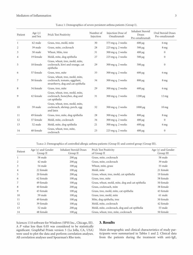
In Table 2, healthy and asthmatic patients are comparedas well as healthy and severe persistent asthma patients.Except of IL-8, IL-10, TGF-β, and GCSF in the anti-IgEtreated severe persistent asthma patients group, none ofthe cytokine concentrations in serum differ significantlybetween healthy and severe persistent asthma diseasedpatients (Figures 1, 2, 3, 4, and 5—only the significant datawith P values were given as in figures). These values werealso increased during the anti-IgE therapy and this differenceis significant between the fourth/eighteenth month of anti-IgE therapy and control group and newly diagnosed allergicasthma patients. In contrast, the levels of IL-8, TGF-β, andGCSF did not differ between newly diagnosed allergic asthmapatients, non-treated severe persistent asthma patients andcontrol. Moreover, the mean serum IL-10 levels were lowerthan the control group in newly diagnosed allergic asthmapatients and non-treated severe persistent asthma patients.Furthermore, IL-17 levels did not change between any groupsand are not associated with any clinical parameters ofasthma.
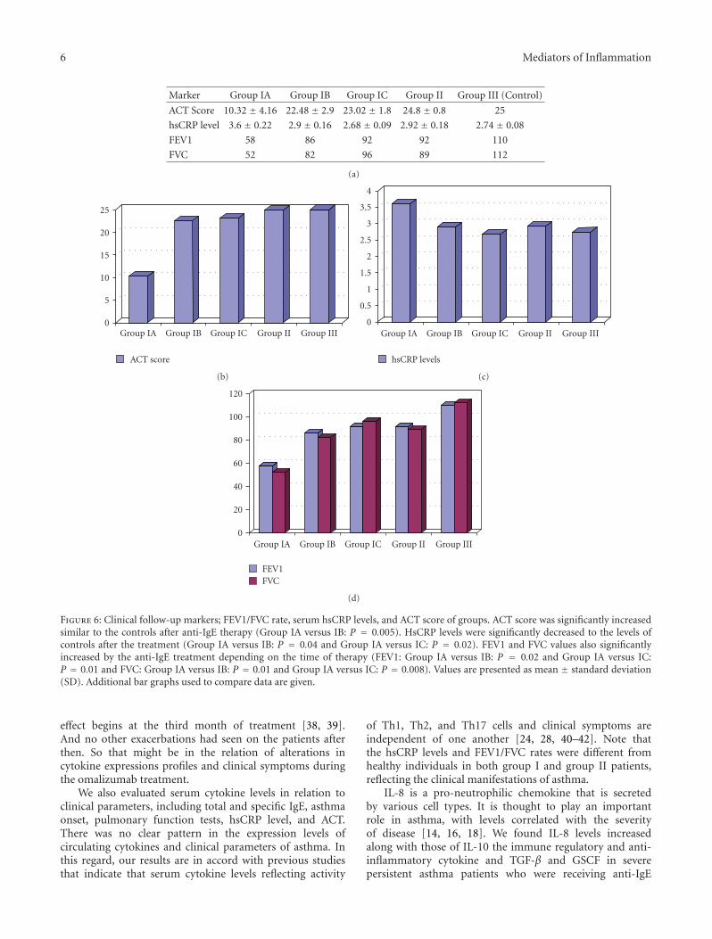
Results of serum cytokine measurements of the severepersistent asthma patients and distinct clinical parameterswere then further analyzed. However, this increase was notcorrelated neither with any of the clinical follow-up markersand serum hsCRP levels. Even though, ACT score of thepatients and serum hsCRP levels together with FEV1/FVCstatus were significantly different between two patient groups(see Figures 6(a), 6(b), 6(c), and 6(d)).
Prick tests were in all patients in Group I and GroupII were detected in mite and grass allergy. These resultscorrelated with specific IgE. The study subjects’ baselinecharacteristics are shown in Tables 1 and 2. The mean IgElevels were as follow: (Group IA: 551.99 IU/mL; Group II:144.25 IU/mL and Group III: 38.62 IU/mL).
4. Discussion
Multiple pathophysiological effects have been associated withimbalanced T cell activation and the presence or absenceof distinct immune mediators in asthma patients [18].Thus there has developed an increased interest in the roleof cytokines and chemokines for diagnosis and therapy.The present study aimed at understanding the patternof expression of several circulating cytokines in asthmaticindividuals. To assess the potential value of these cytokinesas biomarker for asthma, or certain asthma phenotypes, orprediction of anti-IgE therapy efficacy, we compared serumlevels of these asthma associated mediators in two groups:patients with severe persistent asthma treated with anti-IgEtherapy, and newly diagnosed allergic asthma patients. Anti-IgE therapy with omalizumab reduces serum levels of free IgEand downregulates expression of IgE receptors (Fc epsilonRI)on mast cells and basophils. In the airways of patients withmild allergic asthma, omalizumab reduces Fc epsilon RI+and IgE+ cells and causes a profound reduction in tissue
Group IA Group IB Group IC Group II Control0
10
20
30
40
IL-8
leve
ls
Figure 1: Plot graphic of serum IL-8 concentrations in all groups.The numbers of samples of the all groups are 14 for each. GroupIA: severe persistent asthma patients before the treatment. GroupIB: 4 months after the anti-IgE therapy, severe persistent asthmapatients. Group IC: 18 months after the anti-IgE therapy, severepersistent asthma patients. Group II: newly diagnosed controlledallergic asthma patients. Group III: healthy individuals as control.P values were as below: Group IA versus IB: P = 0.02, Group IAversus IC: P = 0.019, Group IA versus II: P = 0.42, Group IA versusControl: P = 0.46, Group IB versus IC: P = 0.27, Group IB versusControl: P = 0.25, Group IC versus Control: P = 0.16, and GroupII versus Control: P = 0.48.
eosinophilia, together with reductions in submucosal T-celland B-cell numbers. Omalizumab decreases Fc epsilonRIexpression on circulating dendritic cells, which might leadto a reduction in allergen presentation, T(h)2 cell activation,and proliferation. And our result of no difference betweenanti-IgE treated patients and the control group emphasizethe fundamental importance of anti-inflammatory effects ofomalizumab and IgE in allergic inflammation. A numberof authors have studied serum cytokines in asthmaticindividuals [24–28]. However, no study focused on therelation between these cytokine levels and anti-IgE therapy.
Distinct type of asthma is related to neutrophilic inflam-mation. However, there is still a multicellular process leadsto multicellular inflammation in the pathogenesis of asthma[29]. IL-8 takes role in the activation of neutrophils and isa potent chemoattractant of neutrophils during the airwayinflammation [30]. There is also growing evidence thatIL-17 is involved in the pathogenesis of asthma. IL-17orchestrates the neutrophilic influx into the airways and alsoenhances T-helper 2 (Th2) cell-mediated eosinophilic airwayinflammation in asthma [31, 32]. Moreover eosinophils arealso a central feature in asthma and are very prominent cells.And GM-CSF promotes eosinophil activation and survival[33]. Eosinophils are also thought to be an importantsource of the potent pro-fibrotic cytokine TGF-β, althoughnumerous other cells types including platelets, fibroblasts,smooth muscle and epithelial cells can also produce TGF-β. However, the precise role of eosinophil-derived TGF-βin airway remodeling is complicated and related to botheosinophils and mast cells [34–36].
Mediators of Inflammation 5
Group IA Group IB Group IC Group II Control0
2
4
6
8
IL-1
0 le
vels
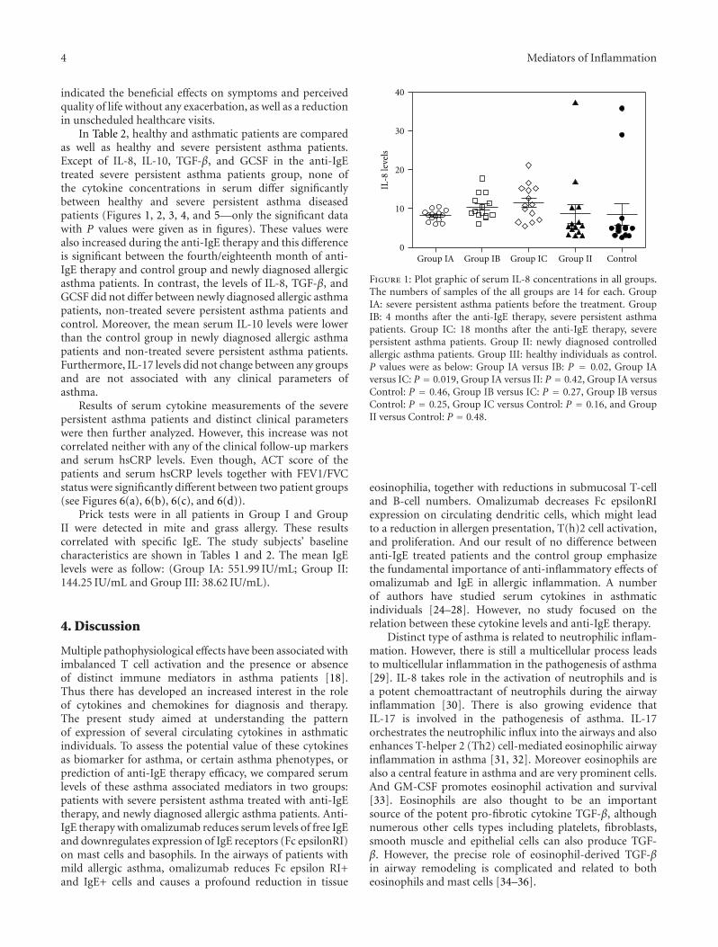
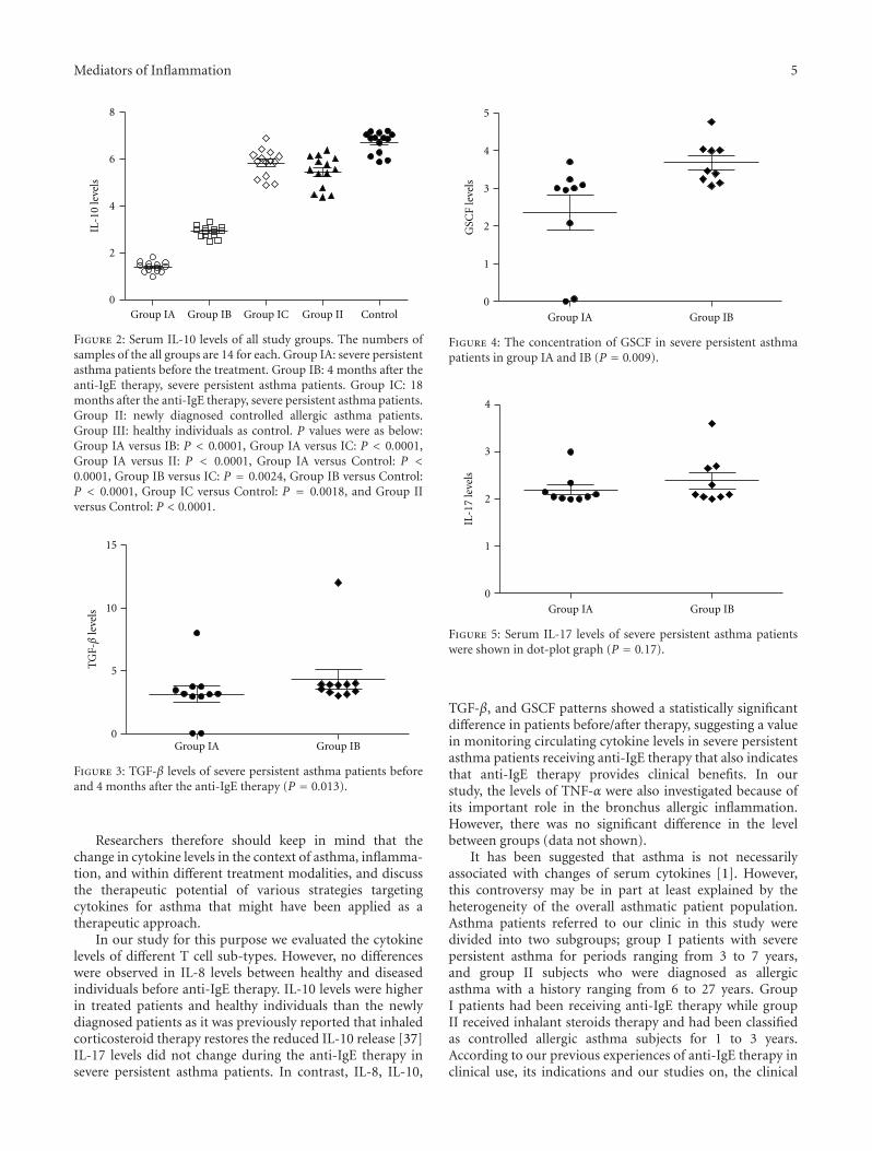
Figure 2: Serum IL-10 levels of all study groups. The numbers ofsamples of the all groups are 14 for each. Group IA: severe persistentasthma patients before the treatment. Group IB: 4 months after theanti-IgE therapy, severe persistent asthma patients. Group IC: 18months after the anti-IgE therapy, severe persistent asthma patients.Group II: newly diagnosed controlled allergic asthma patients.Group III: healthy individuals as control. P values were as below:Group IA versus IB: P < 0.0001, Group IA versus IC: P < 0.0001,Group IA versus II: P < 0.0001, Group IA versus Control: P <0.0001, Group IB versus IC: P = 0.0024, Group IB versus Control:P < 0.0001, Group IC versus Control: P = 0.0018, and Group IIversus Control: P < 0.0001.
Group IA Group IB0
5
10
15
TG
F-β
leve
ls
Figure 3: TGF-β levels of severe persistent asthma patients beforeand 4 months after the anti-IgE therapy (P = 0.013).
Researchers therefore should keep in mind that thechange in cytokine levels in the context of asthma, inflamma-tion, and within different treatment modalities, and discussthe therapeutic potential of various strategies targetingcytokines for asthma that might have been applied as atherapeutic approach.
In our study for this purpose we evaluated the cytokinelevels of different T cell sub-types. However, no differenceswere observed in IL-8 levels between healthy and diseasedindividuals before anti-IgE therapy. IL-10 levels were higherin treated patients and healthy individuals than the newlydiagnosed patients as it was previously reported that inhaledcorticosteroid therapy restores the reduced IL-10 release [37]IL-17 levels did not change during the anti-IgE therapy insevere persistent asthma patients. In contrast, IL-8, IL-10,
Group IA Group IB
0
1
2
3
4
5
GSC
F le
vels
Figure 4: The concentration of GSCF in severe persistent asthmapatients in group IA and IB (P = 0.009).
Group IA Group IB
0
1
2
3
4
IL-1
7 le
vels
Figure 5: Serum IL-17 levels of severe persistent asthma patientswere shown in dot-plot graph (P = 0.17).
TGF-β, and GSCF patterns showed a statistically significantdifference in patients before/after therapy, suggesting a valuein monitoring circulating cytokine levels in severe persistentasthma patients receiving anti-IgE therapy that also indicatesthat anti-IgE therapy provides clinical benefits. In ourstudy, the levels of TNF-α were also investigated because ofits important role in the bronchus allergic inflammation.However, there was no significant difference in the levelbetween groups (data not shown).
It has been suggested that asthma is not necessarilyassociated with changes of serum cytokines [1]. However,this controversy may be in part at least explained by theheterogeneity of the overall asthmatic patient population.Asthma patients referred to our clinic in this study weredivided into two subgroups; group I patients with severepersistent asthma for periods ranging from 3 to 7 years,and group II subjects who were diagnosed as allergicasthma with a history ranging from 6 to 27 years. GroupI patients had been receiving anti-IgE therapy while groupII received inhalant steroids therapy and had been classifiedas controlled allergic asthma subjects for 1 to 3 years.According to our previous experiences of anti-IgE therapy inclinical use, its indications and our studies on, the clinical
6 Mediators of Inflammation
Marker Group IA Group IB Group IC Group II Group III (Control)
ACT Score 10.32 ± 4.16 22.48 ± 2.9 23.02 ± 1.8 24.8 ± 0.8 25
hsCRP level 3.6 ± 0.22 2.9 ± 0.16 2.68 ± 0.09 2.92 ± 0.18 2.74 ± 0.08
FEV1 58 86 92 92 110
FVC 52 82 96 89 112
(a)
0
5
10
15
20
25
Group IA Group IB Group IC Group II Group III
ACT score
(b)
0
0.5
1
1.5
2
2.5
3
3.5
4
Group IA Group IB Group IC Group II Group III
hsCRP levels
(c)
0
20
40
60
80
100
120
Group IA Group IB Group IC Group II Group III
FEV1FVC
(d)
Figure 6: Clinical follow-up markers; FEV1/FVC rate, serum hsCRP levels, and ACT score of groups. ACT score was significantly increasedsimilar to the controls after anti-IgE therapy (Group IA versus IB: P = 0.005). HsCRP levels were significantly decreased to the levels ofcontrols after the treatment (Group IA versus IB: P = 0.04 and Group IA versus IC: P = 0.02). FEV1 and FVC values also significantlyincreased by the anti-IgE treatment depending on the time of therapy (FEV1: Group IA versus IB: P = 0.02 and Group IA versus IC:P = 0.01 and FVC: Group IA versus IB: P = 0.01 and Group IA versus IC: P = 0.008). Values are presented as mean ± standard deviation(SD). Additional bar graphs used to compare data are given.
effect begins at the third month of treatment [38, 39].And no other exacerbations had seen on the patients afterthen. So that might be in the relation of alterations incytokine expressions profiles and clinical symptoms duringthe omalizumab treatment.
We also evaluated serum cytokine levels in relation toclinical parameters, including total and specific IgE, asthmaonset, pulmonary function tests, hsCRP level, and ACT.There was no clear pattern in the expression levels ofcirculating cytokines and clinical parameters of asthma. Inthis regard, our results are in accord with previous studiesthat indicate that serum cytokine levels reflecting activity
of Th1, Th2, and Th17 cells and clinical symptoms areindependent of one another [24, 28, 40–42]. Note thatthe hsCRP levels and FEV1/FVC rates were different fromhealthy individuals in both group I and group II patients,reflecting the clinical manifestations of asthma.
IL-8 is a pro-neutrophilic chemokine that is secretedby various cell types. It is thought to play an importantrole in asthma, with levels correlated with the severityof disease [14, 16, 18]. We found IL-8 levels increasedalong with those of IL-10 the immune regulatory and anti-inflammatory cytokine and TGF-β and GSCF in severepersistent asthma patients who were receiving anti-IgE
Mediators of Inflammation 7
therapy. Anti-IgE (omalizumab) treatment attenuates boththe early- and late-phase responses to inhaled allergensin patients with asthma [19]. Further anti-inflammatoryeffects, including changes in interleukin levels, have beenobserved and postulated to contribute to the clinical efficacyof omalizumab treatment [20, 21]. Other studies in severepersistent allergic asthma patients receiving omalizumabtherapy have focused on modulation of serum soluble TNF-related apoptosis-inducing ligand, total antioxidant capacity,hydrogen peroxide, malondialdehyde and total nitric oxideconcentrations, and ceruloplasmin oxidase activity measure-ments, as markers of the efficacy of anti-IgE treatmentmodality [43–46]. Our data add IL-8, IL-10, TGF-β, andGCSF to this list.
Both local and systemic inflammation is associatedwith pathogenesis in asthma [47, 48]. To assess systemicinflammation, we monitored serum levels of CRP in patients.Because of possible confounding effects on CRP levels, sub-jects with kidney disease, heart disease, liver disease, diabetesmellitus, cancer, obesity, smoking history, and autoimmunedisease were excluded from our study. No correlation wasobserved between levels of any of the cytokines measured,clinical outcome, and serum hsCRP concentrations.
In conclusion, the present study documents evidencefor altered patterns in serum cytokines in severe persistentasthma patients following anti-IgE therapy. However, thebasal serum cytokine profiles excluding the IL-10, patternswere not different between healthy and asthmatic individ-uals, regardless of whether the latter were newly diagnosedallergic asthma or non-treated severe persistent asthmaticpatients. We believe this study provides a novel perspectiveon the mechanism of action of anti-IgE immunotherapyin severe persistent asthma patients and inflammatorymediators in defining clinical benefits.
Conflict of Interests
The authors declare no conflict of interests.
Authors’ Contributions
A. D. Yalcin and A. Bisgin contributed equally to this paper.A. D. Yalcin and A. Bisgin conceived and designed the study.Clinical followup: A. D. Yalcin. A. Bisgin analyzed the data.Contribution of reagents/materials: R. M. Gorczynski and A.Bisgin. Writing of the paper: A. Bisgin and A. D. Yalcin.
Acknowledgments
The authors thank all participating patients and volunteers.They would like to thank Dr. Nuray Erin for providinglaboratory assistance.
References
[1] G. P. Anderson, “Endotyping asthma: new insights into keypathogenic mechanisms in a complex, heterogeneous disease,”The Lancet, vol. 372, no. 9643, pp. 1107–1119, 2008.
[2] J. Lotvall, C. Akdis, L. B. Bacharier et al., “Asthma endotypes:a new approach to classification of disease entities within theasthma syndrome,” American Academy of Allergy, Asthma, andImmunology, vol. 127, no. 2, pp. 355–360, 2011.
[3] M. Larche, D. S. Robinson, and A. B. Kay, “The role of Tlymphocytes in the pathogenesis of asthma,” The Journal ofAllergy and Clinical Immunology, vol. 111, pp. 450–463, 2003.
[4] M. M. Epstein, “Targeting memory Th2 cells for the treatmentof allergic asthma,” Pharmacology & Therapeutics, vol. 109, pp.107–136, 2006.
[5] D. S. Robinson, Q. Hamid, S. Ying et al., “Predominant T(H2)-like bronchoalveolar T-lymphocyte population in atopicasthma,” The New England Journal of Medicine, vol. 326, no.5, pp. 298–304, 1992.
[6] M. Wills-Karp and F. D. Finkelman, “Untangling the complexweb of IL-4- and IL-13-mediated signaling pathways,” ScienceSignaling, vol. 1, no. 51, p. pe55, 2008.
[7] A. M. Cooper and S. A. Khader, “IL-12p40: an inherentlyagonistic cytokine,” Trends in Immunology, vol. 28, no. 1, pp.33–38, 2007.
[8] R. K. Kumar, D. C. Webb, C. Herbert, and P. S. Foster,“Interferon-γ as a possible target in chronic asthma,” Inflam-mation and Allergy, vol. 5, no. 4, pp. 253–256, 2006.
[9] M. John, S. Lim, J. Seybold et al., “Inhaled corticosteroidsincrease interleukin-10 but reduce macrophage inflammatoryprotein-1α, granulocyte-macrophage colony-stimulating fac-tor, and interferon-γ release from alveolar macrophages inasthma,” American Journal of Respiratory and Critical CareMedicine, vol. 157, no. 1, pp. 256–262, 1998.
[10] J. Pene, S. Chevalier, L. Preisser et al., “Chronically inflamedhuman tissues are infiltrated by highly differentiated Th17lymphocytes,” The Journal of Immunology, vol. 180, pp. 7423–7430, 2008.
[11] Y. H. Wang, K. S. Voo, B. Liu et al., “A novel subset of CD4+
TH2 memory/ effector cells that produce inflammatory IL-17 cytokine and promote the exacerbation of chronic allergicasthma,” Journal of Experimental Medicine, vol. 207, no. 11, pp.2479–2491, 2010.
[12] N. Yamashita, H. Tashimo, H. Ishida et al., “Attenuation ofairway hyperresponsiveness in a murine asthma model byneutralization of granulocyte-macrophage colony-stimulatingfactor (GM-CSF),” Cellular Immunology, vol. 219, no. 2, pp.92–97, 2002.
[13] F. L. Dente, S. Carnevali, M. L. Bartoli et al., “Profiles ofproinflammatory cytokines in sputum from different groupsof severe asthmatic patients,” Annals of Allergy, Asthma andImmunology, vol. 97, no. 3, pp. 312–320, 2006.
[14] J. K. Shute, B. Vrugt, I. J. D. Lindley et al., “Free and complexedinterleukin-8 in blood and bronchial mucosa in asthma,”American Journal of Respiratory and Critical Care Medicine,vol. 155, no. 6, pp. 1877–1883, 1997.
[15] M. A. Berry, B. Hargadon, M. Shelley et al., “Evidence of a roleof tumor necrosis factor alpha in refractory asthma,” The NewEngland Journal of Medicine, vol. 354, pp. 697–708, 2006.
[16] A. Jatakanon, C. Uasuf, W. Maziak, S. Lim, K. F. Chung, andP. J. Barnes, “Neutrophilic inflammation in severe persistentasthma,” American Journal of Respiratory and Critical CareMedicine, vol. 160, no. 5 I, pp. 1532–1539, 1999.
[17] S. K. Saha, C. Doe, V. Mistry et al., “Granulocyte-macrophagecolony-stimulating factor expression in induced sputum andbronchial mucosa in asthma and COPD,” Thorax, vol. 64, no.8, pp. 671–676, 2009.
[18] M. Silvestri, M. Bontempelli, M. Giacomelli et al., “Highserum levels of tumour necrosis factor-α and interleukin-8 in
8 Mediators of Inflammation
severe asthma: markers of systemic inflammation?” Clinicaland Experimental Allergy, vol. 36, no. 11, pp. 1373–1381, 2006.
[19] J. V. Fahy, H. E. Fleming, H. H. Wong et al., “The effectof an anti-IgE monoclonal antibody on the early- and late-phase responses to allergen inhalation in asthmatic subjects,”American Journal of Respiratory and Critical Care Medicine,vol. 155, no. 6, pp. 1828–1834, 1997.
[20] O. Noga, G. Hanf, I. Brachmann et al., “Effect of omalizumabtreatment on peripheral eosinophil and T-lymphocyte func-tion in patients with allergic asthma,” Journal of Allergy andClinical Immunology, vol. 117, no. 6, pp. 1493–1499, 2006.
[21] G. Hanf, I. Brachmann, J. Kleine-Tebbe et al., “Omalizumabdecreased IgE-release and induced changes in cellular immu-nity in patients with allergic asthma,” Allergy, vol. 61, no. 9,pp. 1141–1144, 2006.
[22] A. D. Yalcin, A. Bisgin, R. Cetinkaya, and S. Gumuslu, “Clinicalefficacy of omalizumab in severe persistent asthmaand co-morbid conditions,” EAACI. In press.
[23] J. Roca, J. Sanchis, and A. Agusti-Vidal, “Spirometric referencevalues from a Mediterranean population,” Clinical RespiratoryPhysiology, vol. 22, no. 3, pp. 217–224, 1986.
[24] C. Hollander, B. Sitkauskiene, R. Sakalauskas, U. Westin,and S. M. Janciauskiene, “Serum and bronchial lavage fluidconcentrations of IL-8, SLPI, sCD14 and sICAM-1 in patientswith COPD and asthma,” Respiratory Medicine, vol. 101, no. 9,pp. 1947–1953, 2007.
[25] A. Krogulska, K. Wasowska-Krolikowska, E. Polakowska, andS. Chrul, “Cytokine profile in children with asthma undergo-ing food challenges,” Journal of Investigational Allergology andClinical Immunology, vol. 19, no. 1, pp. 43–48, 2009.
[26] A. A. Litonjua, D. Sparrow, L. Guevarra, G. T. O’Connor, S.T. Weiss, and D. J. Tollerud, “Serum interferon-γ is associatedwith longitudinal decline in lung function among asthmaticpatients: the Normative Aging Study,” Annals of Allergy,Asthma and Immunology, vol. 90, no. 4, pp. 422–428, 2003.
[27] H. Nakamura, S. T. Weiss, E. Israel, A. D. Luster, J. M. Drazen,and C. M. Lilly, “Eotaxin and impaired lung function inasthma,” American Journal of Respiratory and Critical CareMedicine, vol. 160, no. 6, pp. 1952–1956, 1999.
[28] H. Tateno, H. Nakamura, N. Minematsu et al., “Plasmaeotaxin level and severity of asthma treated with corticos-teroid,” Respiratory Medicine, vol. 98, no. 8, pp. 782–790, 2004.
[29] S. T. Holgate, “Epithelium dysfunction in asthma,” Journal ofAllergy and Clinical Immunology, vol. 120, no. 6, pp. 1233–1244, 2007.
[30] R. E. T. Nocker, D. F. M. Schoonbrood, E. A. van de Graaf et al.,“Interleukin-8 in airway inflammation in patients with asthmaand chronic obstructive pulmonary disease,” InternationalArchives of Allergy and Immunology, vol. 109, no. 2, pp. 183–191, 1996.
[31] H. C. Hsu, P. A. Yang, J. Wang et al., “Interleukin 17-producingT helper cells and interleukin 17 orchestrate autoreactivegerminal center development in autoimmune BXD2 mice,”Nature Immunology, vol. 9, no. 2, pp. 166–175, 2008.
[32] Y. C. Sun, Q. T. Zhou, and W. Z. Yao, “Sputum interleukin-17 is increased and associated with airway neutrophilia inpatients with severe asthma,” Chinese Medical Journal, vol. 118,no. 11, pp. 953–956, 2005.
[33] S. G. Trivedi and C. M. Lloyd, “Eosinophils in the pathogenesisof allergic airways disease,” Cellular and Molecular Life Sci-ences, vol. 64, no. 10, pp. 1269–1289, 2007.
[34] E. M. Minshall, D. Y. M. Leung, R. J. Martin et al., “Eosinophil-associated TGF-β1 mRNA expression and airways fibrosis in
bronchial Asthma,” American Journal of Respiratory Cell andMolecular Biology, vol. 17, no. 3, pp. 326–333, 1997.
[35] J. Y. Cho, M. Miller, K. J. Baek et al., “Inhibition ofairway remodeling in IL-5-deficient mice,” Journal of ClinicalInvestigation, vol. 113, no. 4, pp. 551–560, 2004.
[36] C. E. Brightling, P. Bradding, F. A. Symon, S. T. Holgate,A. J. Wardlaw, and I. D. Pavord, “Mast-cell infiltration ofairway smooth muscle in asthma,” The New England Journalof Medicine, vol. 346, no. 22, pp. 1699–1705, 2002.
[37] F. Chung, “Anti-inflammatory cytokines in asthma andallergy: Interleukin-10, interleukin-12, interferon-γ,” Media-tors of Inflammation, vol. 10, no. 2, pp. 51–59, 2001.
[38] A. D. Yalcin, “Bisgin A The relation of sTRAIL levels andquality of life in severe persistent allergic asthma patients usingomalizumab,” Medical Science Monitor, vol. 18, no. 8, pp. LE9–LE10, 2012.
[39] A. D. Yalcin and A. Bisgin, “Omalizumab: anti-IgE therapy insevere allergic conditions,” Allergy & Therapy Journals, vol. 3,article 120.
[40] C. J. Corrigan and A. B. Kay, “CD4 T-lymphocyte activationin acute severe asthma. Relationship to disease severity andatopic status,” American Review of Respiratory Disease, vol. 141,no. 4, pp. 970–977, 1990.
[41] H. Saito, T. Hayakawa, H. Mita, Y. Yui, and T. Shida, “Augmen-tation of leukotriene C4 production by gamma interferon inleukocytes challenged with an allergen,” International Archivesof Allergy and Applied Immunology, vol. 87, no. 3, pp. 286–293,1988.
[42] A. Friebe and H. D. Volk, “Stability of tumor necrosis factor α,interleukin 6, and interleukin 8 in blood samples of patientswith systemic immune activation,” Archives of Pathology andLaboratory Medicine, vol. 132, no. 11, pp. 1802–1806, 2008.
[43] A. D. Yalcin, R. M. Gorczynski, G. E. Parlak et al., “Totalantioxidant capacity, hydrogen peroxide, malondialdehydeand total nitric oxide concentrations in patients with severepersistent allergic asthma: its relation to omalizumab treat-ment,” Clinical Laboratory, vol. 58, no. 1-2, pp. 89–96, 2012.
[44] A. D. Yalcin, A. Bisgin, A. Kargi, and R. M. Gorczynski, “Serumsoluble TRAIL levels in patients with severe persistent allergicasthma: its relation to Omalizumab treatment,” MedicalScience Monitor, vol. 18, no. 3, pp. 11–15, 2012.
[45] A. D. Yalcin, S. Gumuslu, G. E. Parlak et al., “Systemic levels ofceruloplasmin oxidase activity in allergic asthma and allergicrhinitis,” Immunopharmacol Immunotoxicol, vol. 34, no. 6, pp.1047–1053, 2012.
[46] C. A. Bates and P. E. Silkoff, “Exhaled nitric oxide inasthma: from bench to bedside,” Journal of Allergy and ClinicalImmunology, vol. 111, no. 2, pp. 256–262, 2003.
[47] M. B. Pepys and M. L. Baltz, “Acute phase proteins withspecial reference to C-reactive protein and related proteins(pentaxins) and serum amyloid A protein,” Advances inImmunology, vol. 34, pp. 141–212, 1983.
[48] P. Jousilahti, V. Salomaa, K. Hakala, V. Rasi, E. Vahtera, and T.Palosuo, “The association of sensitive systemic inflammationmarkers with bronchial asthma,” Annals of Allergy, Asthma andImmunology, vol. 89, no. 4, pp. 381–385, 2002.

Submit your manuscripts athttp://www.hindawi.com
Stem CellsInternational
Hindawi Publishing Corporationhttp://www.hindawi.com Volume 2014
Hindawi Publishing Corporationhttp://www.hindawi.com Volume 2014
MEDIATORSINFLAMMATION
of
Hindawi Publishing Corporationhttp://www.hindawi.com Volume 2014
Behavioural Neurology
EndocrinologyInternational Journal of
Hindawi Publishing Corporationhttp://www.hindawi.com Volume 2014
Hindawi Publishing Corporationhttp://www.hindawi.com Volume 2014
Disease Markers
Hindawi Publishing Corporationhttp://www.hindawi.com Volume 2014
BioMed Research International
OncologyJournal of
Hindawi Publishing Corporationhttp://www.hindawi.com Volume 2014
Hindawi Publishing Corporationhttp://www.hindawi.com Volume 2014
Oxidative Medicine and Cellular Longevity
Hindawi Publishing Corporationhttp://www.hindawi.com Volume 2014
PPAR Research
The Scientific World JournalHindawi Publishing Corporation http://www.hindawi.com Volume 2014
Immunology ResearchHindawi Publishing Corporationhttp://www.hindawi.com Volume 2014
Journal of
ObesityJournal of
Hindawi Publishing Corporationhttp://www.hindawi.com Volume 2014
Hindawi Publishing Corporationhttp://www.hindawi.com Volume 2014
Computational and Mathematical Methods in Medicine
OphthalmologyJournal of
Hindawi Publishing Corporationhttp://www.hindawi.com Volume 2014
Diabetes ResearchJournal of
Hindawi Publishing Corporationhttp://www.hindawi.com Volume 2014
Hindawi Publishing Corporationhttp://www.hindawi.com Volume 2014
Research and TreatmentAIDS
Hindawi Publishing Corporationhttp://www.hindawi.com Volume 2014
Gastroenterology Research and Practice
Hindawi Publishing Corporationhttp://www.hindawi.com Volume 2014
Parkinson’s Disease
Evidence-Based Complementary and Alternative Medicine
Volume 2014Hindawi Publishing Corporationhttp://www.hindawi.com