
CLINICAL METHODS IN DIAGNOSIS OF POAG OPTIC DISC 1.2-1.4 million axons/ 5000 loss/year 10%...
26
CLINICAL METHODS IN DIAGNOSIS OF POAG OPTIC DISC 1.2-1.4 million axons/ 5000 loss/year 10% Magnocellular 90% Parvo- SIZE AND SHAPE DD: 1.5mm Surface: 2.1-2.8mm2 (π/4xHDxVD) AGE: no change after 3-10 years RACE: African>Asian>Mexican>Caucasian REFRACTIVE ERROR:independent [–5-+5DS] Positive correlation to rim and cup size
-
Upload
bradyn-point -
Category
Documents
-
view
224 -
download
0
Transcript of CLINICAL METHODS IN DIAGNOSIS OF POAG OPTIC DISC 1.2-1.4 million axons/ 5000 loss/year 10%...
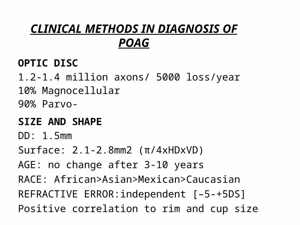
CLINICAL METHODS IN DIAGNOSIS OF POAG
OPTIC DISC
1.2-1.4 million axons/ 5000 loss/year10% Magnocellular90% Parvo-
SIZE AND SHAPE
DD: 1.5mm
Surface: 2.1-2.8mm2 (π/4xHDxVD)
AGE: no change after 3-10 years
RACE: African>Asian>Mexican>Caucasian
REFRACTIVE ERROR:independent [–5-+5DS]
Positive correlation to rim and cup size
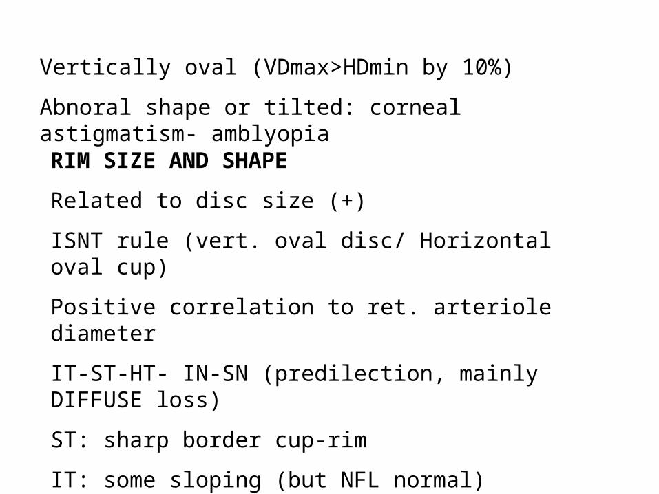
Vertically oval (VDmax>HDmin by 10%)
Abnoral shape or tilted: corneal astigmatism- amblyopia
RIM SIZE AND SHAPE
Related to disc size (+)
ISNT rule (vert. oval disc/ Horizontal oval cup)
Positive correlation to ret. arteriole diameter
IT-ST-HT- IN-SN (predilection, mainly DIFFUSE loss)
ST: sharp border cup-rim
IT: some sloping (but NFL normal)
Pallor: ? Non-glaucomatous (increased cup size)
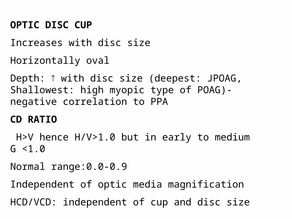
OPTIC DISC CUP
Increases with disc size
Horizontally oval
Depth: with disc size (deepest: JPOAG, Shallowest: high myopic type of POAG)- negative correlation to PPA
CD RATIO
H>V hence H/V>1.0 but in early to medium G <1.0
Normal range:0.0-0.9
Independent of optic media magnification
HCD/VCD: independent of cup and disc size
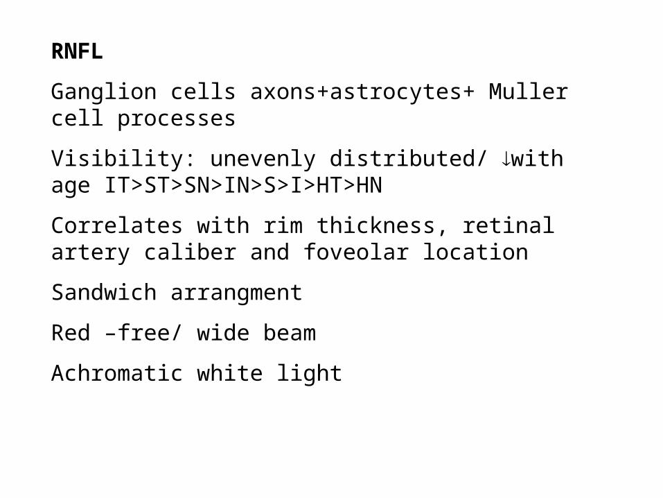
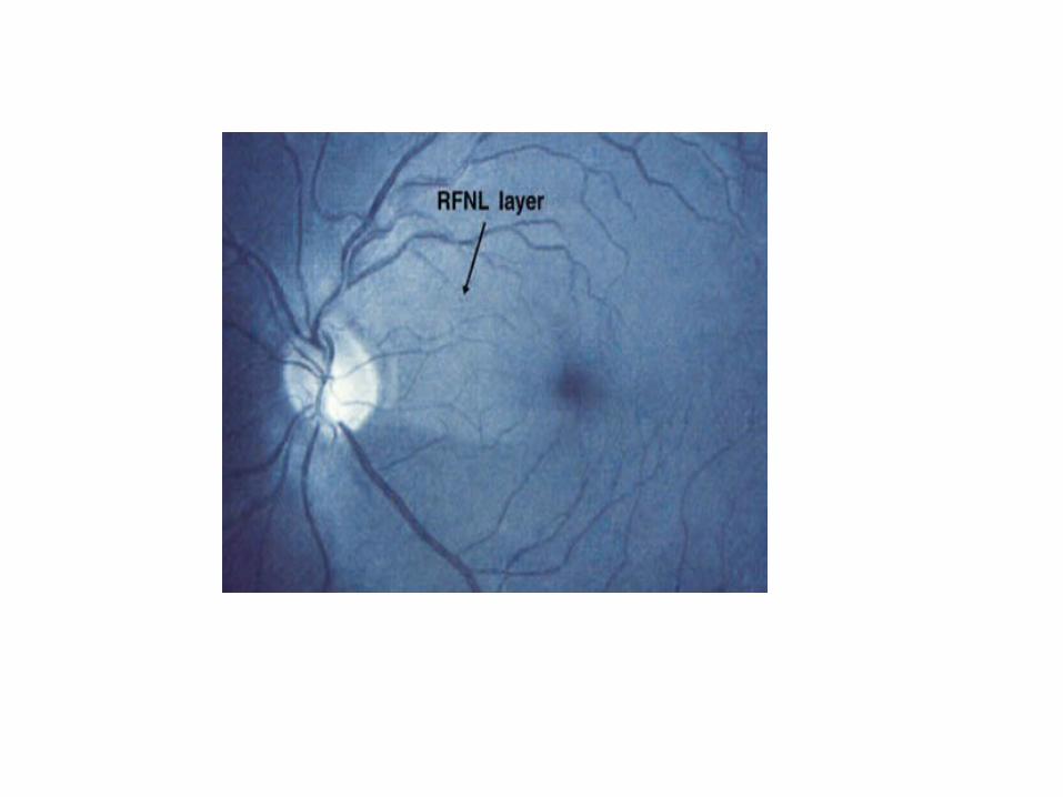
RNFL
Ganglion cells axons+astrocytes+ Muller cell processes
Visibility: unevenly distributed/ with age IT>ST>SN>IN>S>I>HT>HN
Correlates with rim thickness, retinal artery caliber and foveolar location
Sandwich arrangment
Red –free/ wide beam
Achromatic white light

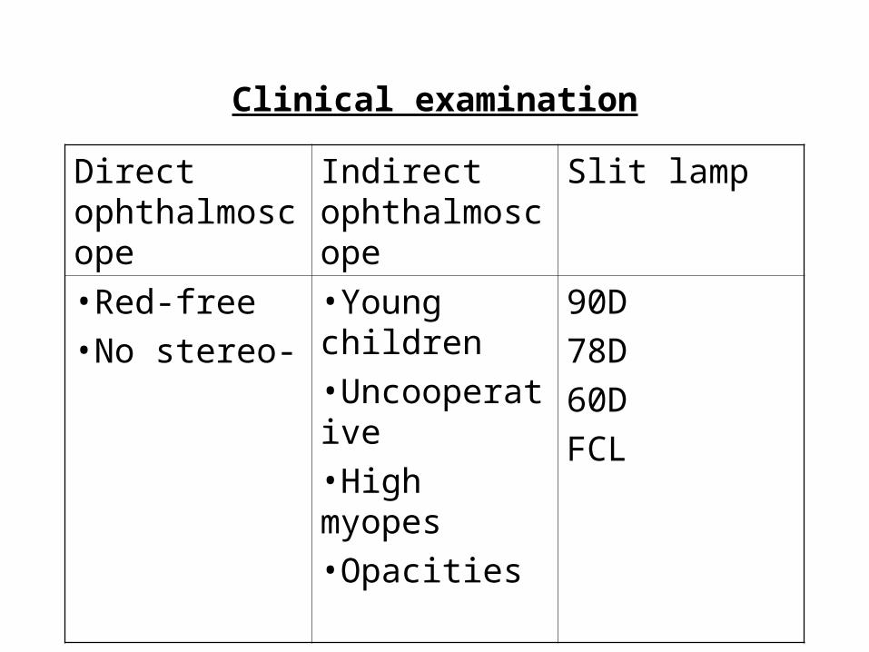
Clinical examination
Direct ophthalmoscope
Indirect ophthalmoscope
Slit lamp
•Red-free•No stereo-
•Young children•Uncooperative•High myopes•Opacities
90D
78D
60D
FCL
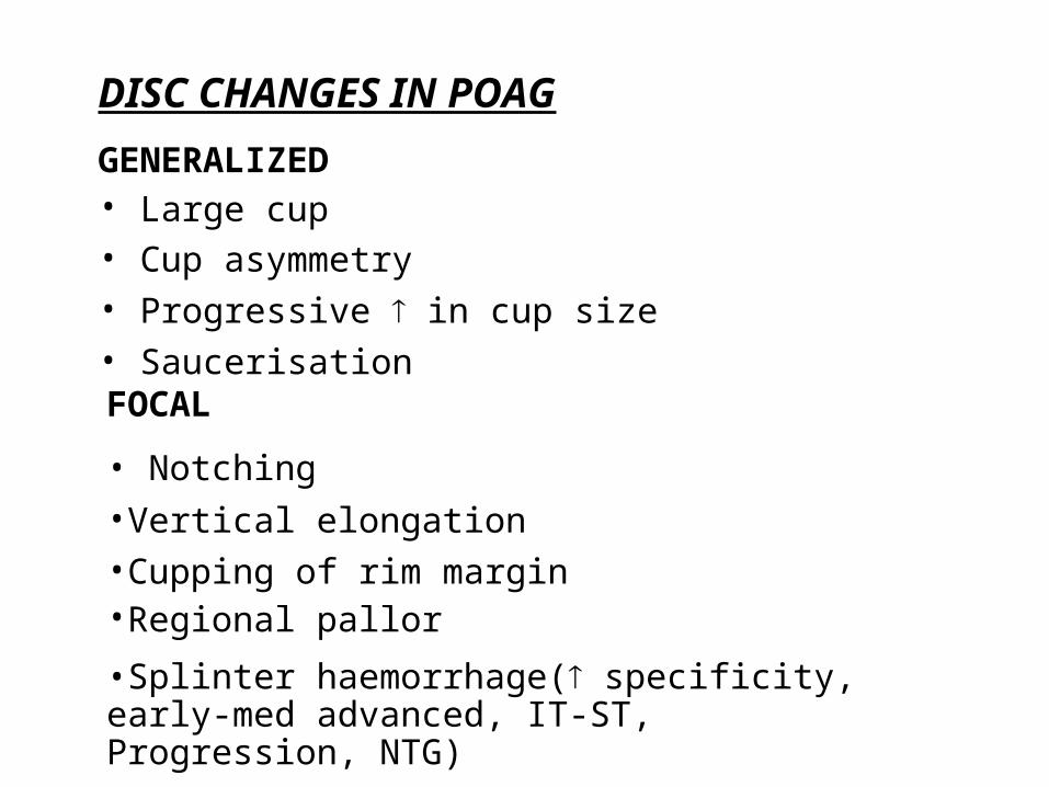
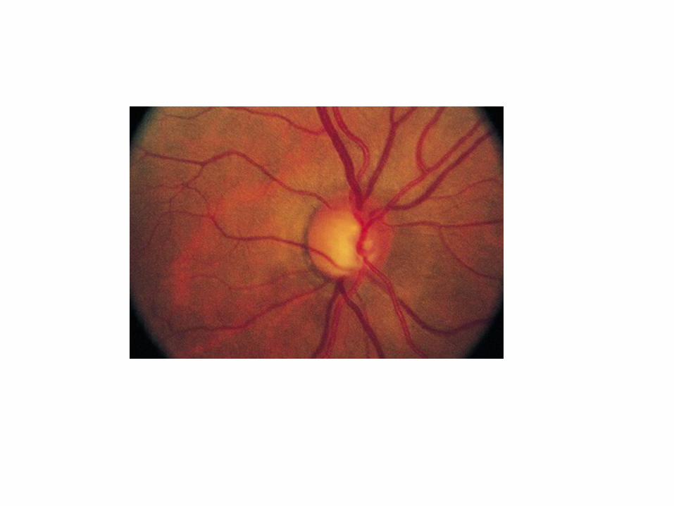
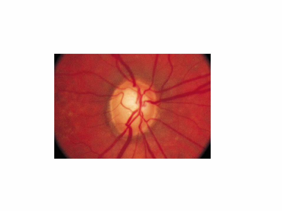
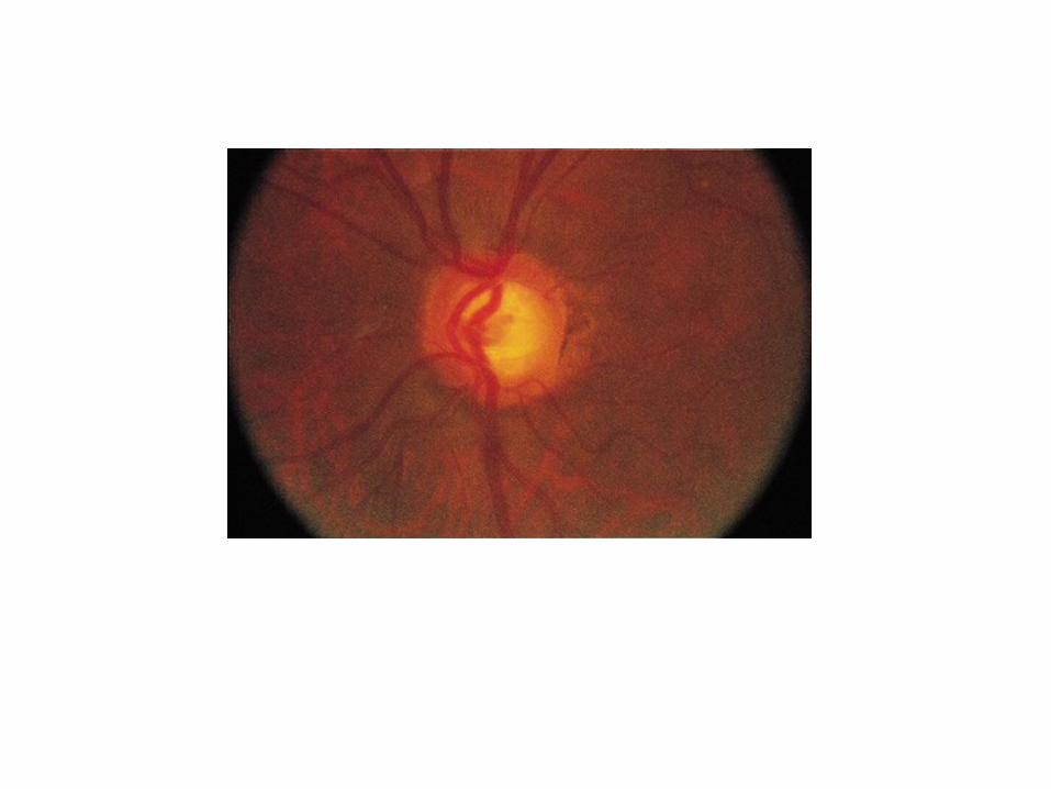
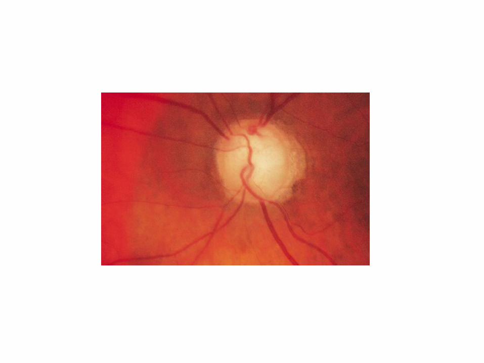
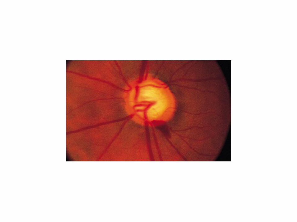
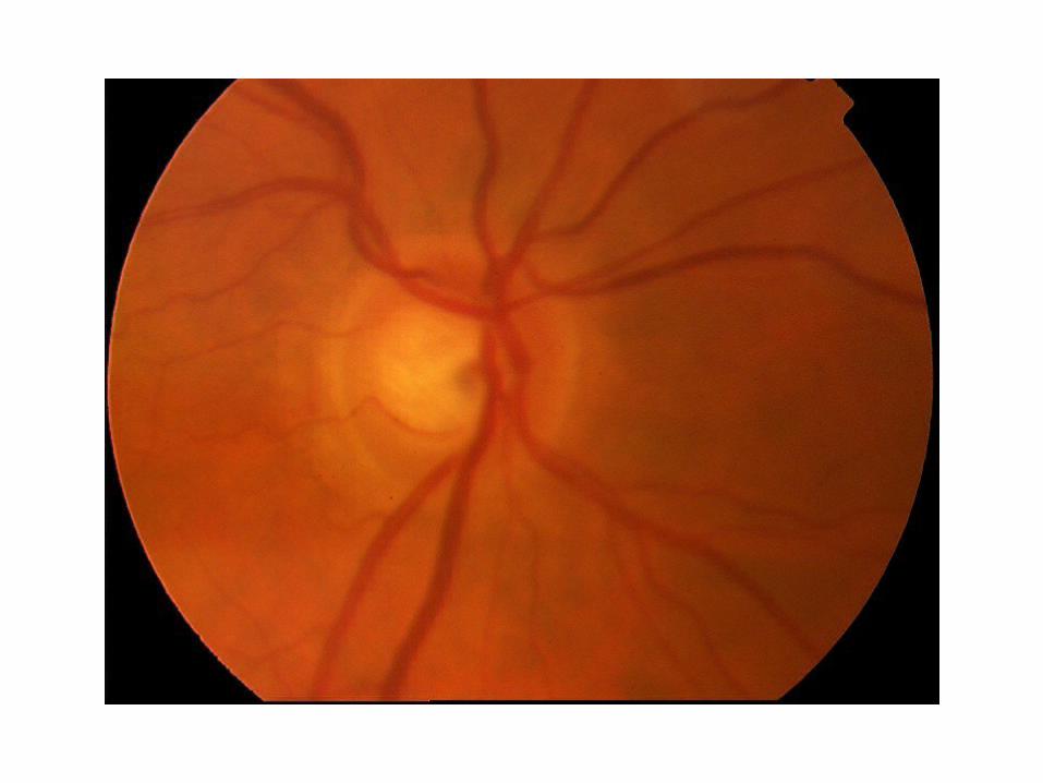
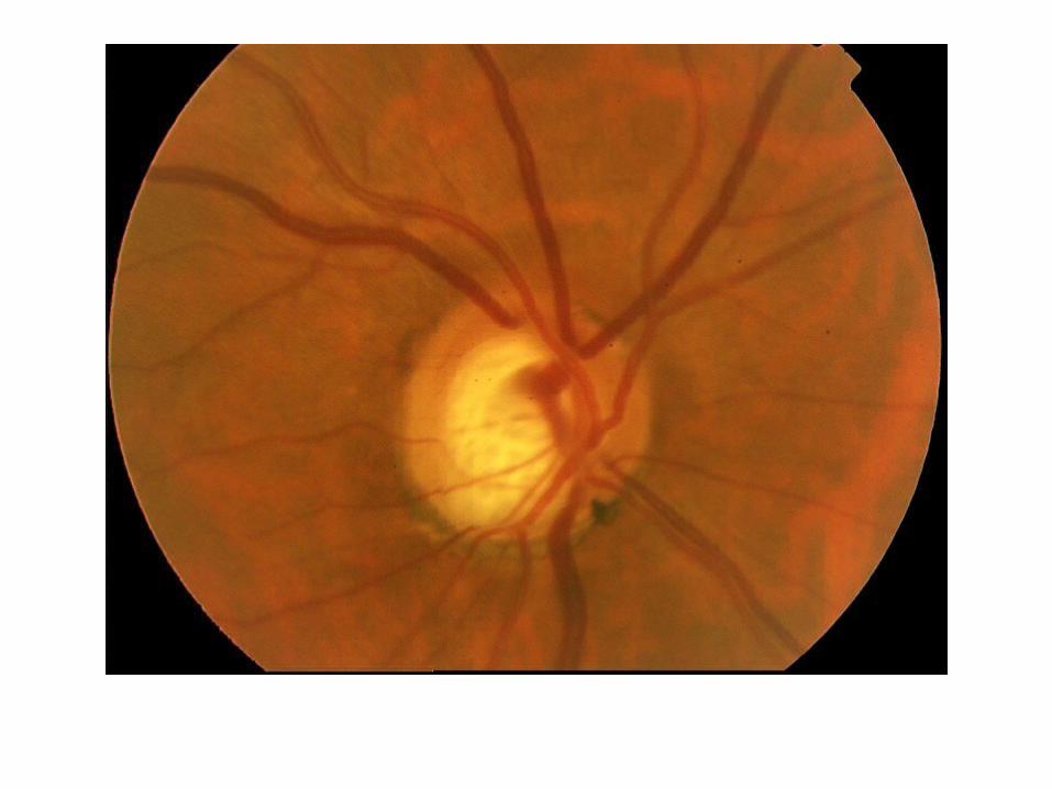
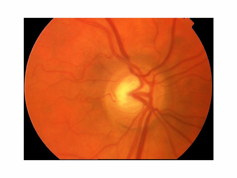
DISC CHANGES IN POAG
GENERALIZED• Large cup• Cup asymmetry• Progressive in cup size• SaucerisationFOCAL
• Notching
•Vertical elongation•Cupping of rim margin•Regional pallor
•Splinter haemorrhage( specificity, early-med advanced, IT-ST, Progression, NTG)
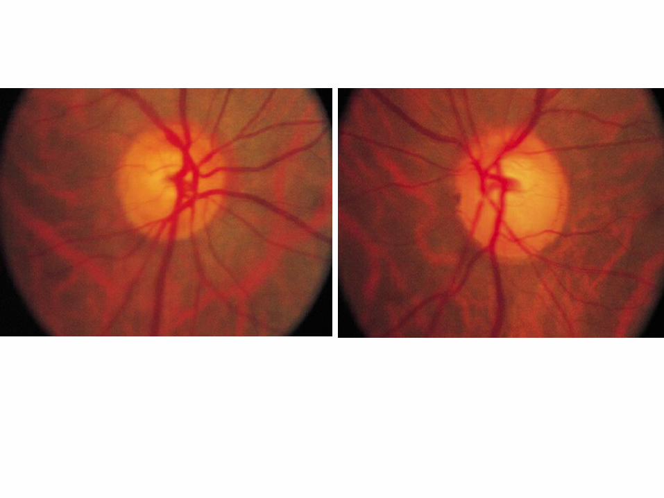
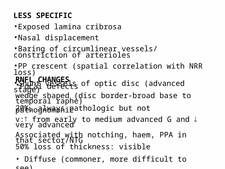
LESS SPECIFIC
•Exposed lamina cribrosa•Nasal displacement•Baring of circumlinear vessels/ constriction of arterioles•PP crescent (spatial correlation with NRR loss)•Shunt vessels of optic disc (advanced stage)RNFL CHANGES•Focal defects wedge shaped (disc border-broad base to temporal raphe)20%, always pathologic but not pathognomonicv: from early to medium advanced G and very advanced Associated with notching, haem, PPA in that sector/NTG50% loss of thickness: visible
• Diffuse (commoner, more difficult to see)Sequence of sectors regarding RNFL visibilityRetinal vessels( clearer- sharper)
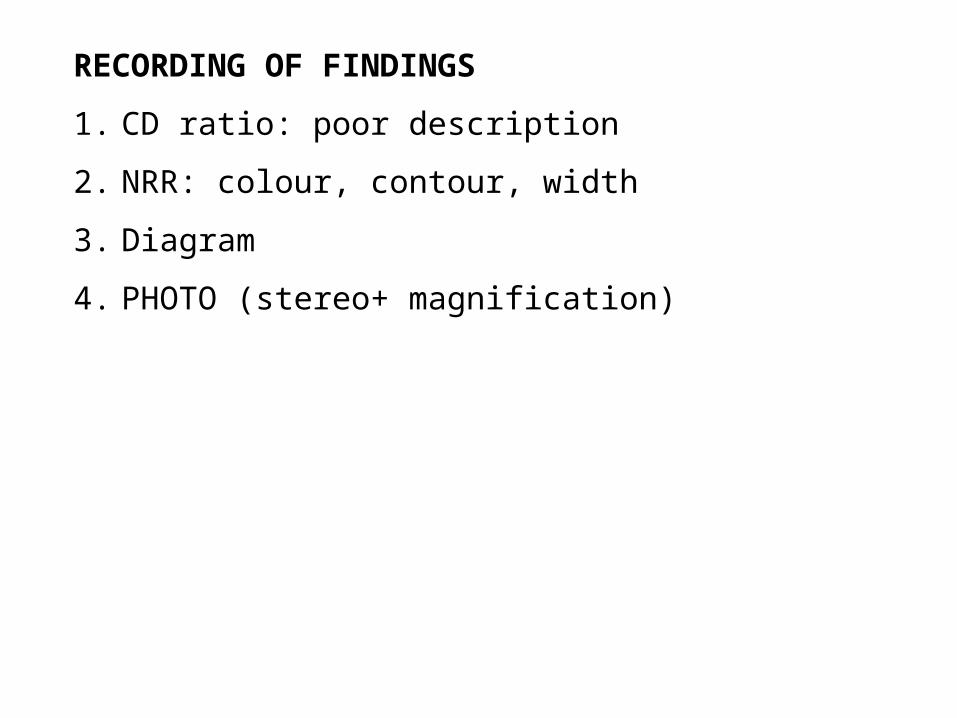
RECORDING OF FINDINGS
1. CD ratio: poor description
2. NRR: colour, contour, width
3. Diagram
4. PHOTO (stereo+ magnification)
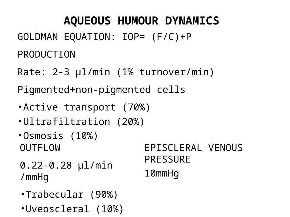
AQUEOUS HUMOUR DYNAMICSGOLDMAN EQUATION: IOP= (F/C)+P
PRODUCTION
Rate: 2-3 μl/min (1% turnover/min)
Pigmented+non-pigmented cells
•Active transport (70%)
•Ultrafiltration (20%)•Osmosis (10%)OUTFLOW
0.22-0.28 μl/min /mmHg
•Trabecular (90%)
•Uveoscleral (10%)
EPISCLERAL VENOUS PRESSURE
10mmHg
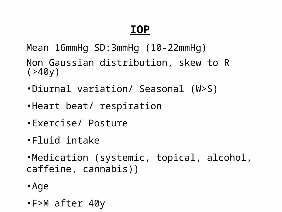
IOP
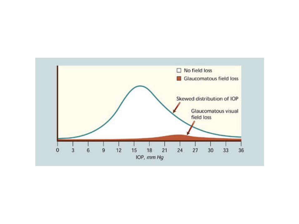
Mean 16mmHg SD:3mmHg (10-22mmHg)
Non Gaussian distribution, skew to R (>40y)
•Diurnal variation/ Seasonal (W>S)
•Heart beat/ respiration
•Exercise/ Posture
•Fluid intake
•Medication (systemic, topical, alcohol, caffeine, cannabis))
•Age
•F>M after 40y
•Genetically influenced
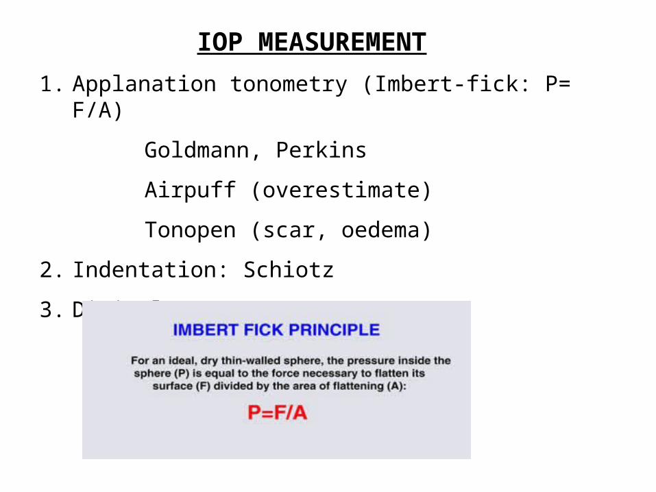
IOP MEASUREMENT
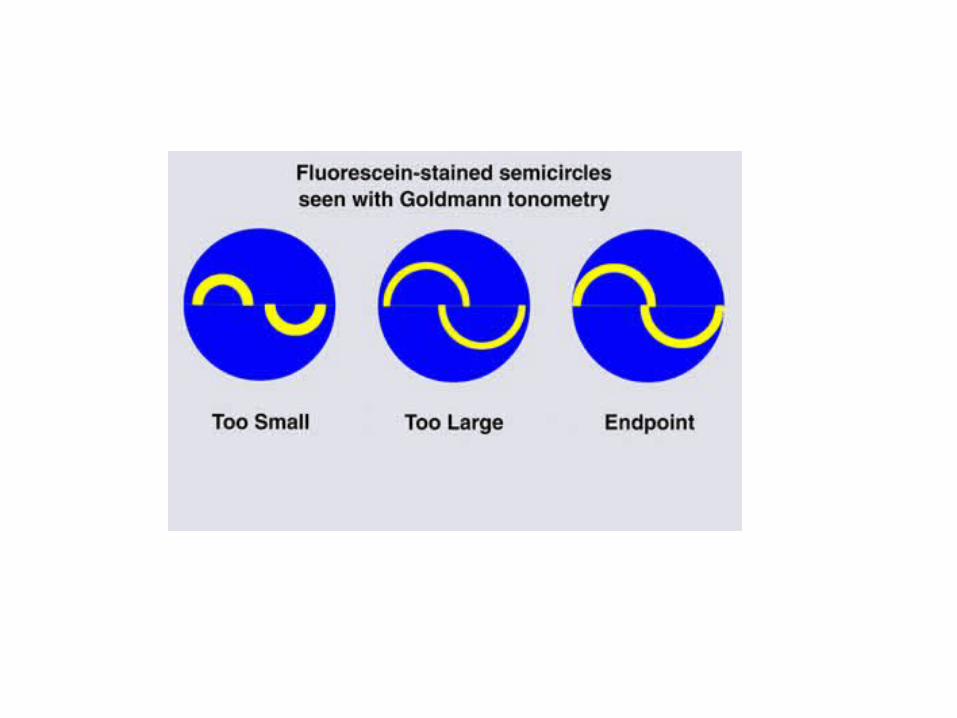
1. Applanation tonometry (Imbert-fick: P= F/A)
Goldmann, Perkins
Airpuff (overestimate)
Tonopen (scar, oedema)
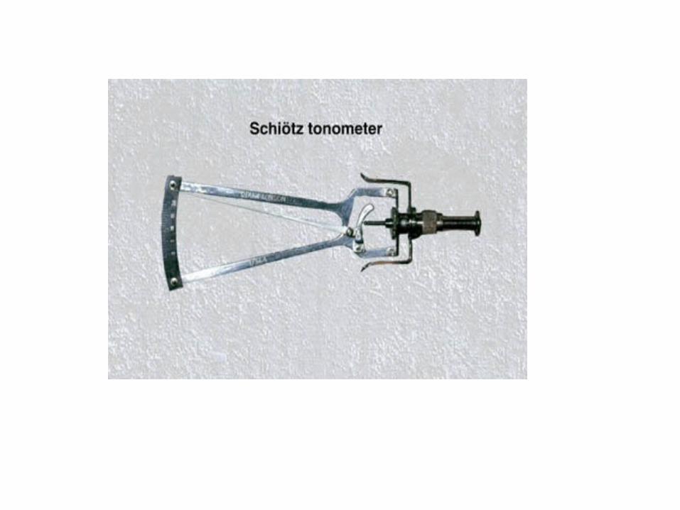
2. Indentation: Schiotz
3. Digital pressure
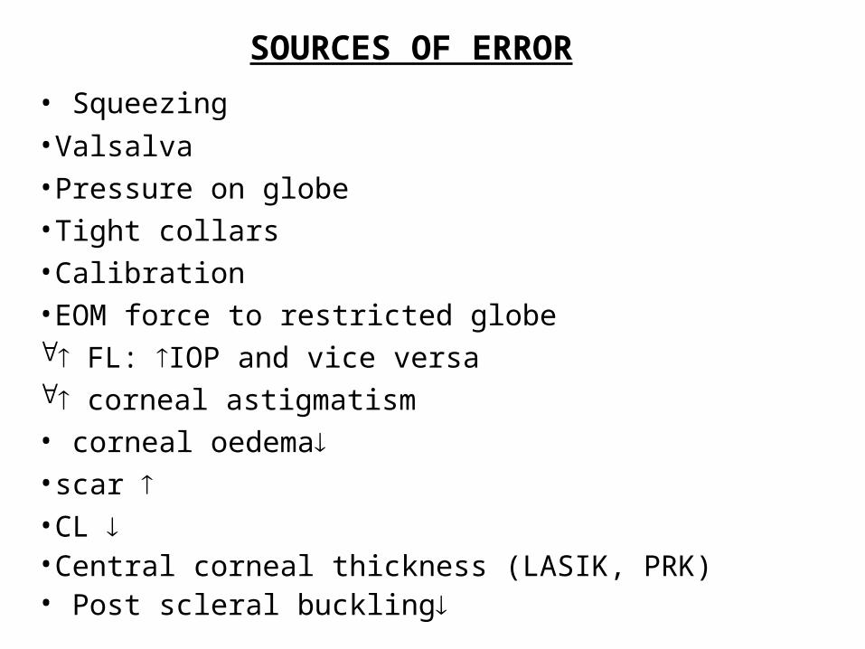
SOURCES OF ERROR
• Squeezing
•Valsalva•Pressure on globe•Tight collars•Calibration•EOM force to restricted globe FL: IOP and vice versa corneal astigmatism• corneal oedema•scar •CL •Central corneal thickness (LASIK, PRK)• Post scleral buckling