Cirrhosis and Coagulation A Paradigm of Awareness · Platelets Platelets function Factors XI, IX,...
61
Cirrhosis and Coagulation A Paradigm of Awareness 2014 Robert G. Gish MD
Transcript of Cirrhosis and Coagulation A Paradigm of Awareness · Platelets Platelets function Factors XI, IX,...
Cirrhosis and Coagulation
A Paradigm of Awareness
2014
Robert G. Gish MD
Disclosures:
• None relevant
The Clotting Process
• Initiation and formation of the platelet
plug
• Propagation of the clotting process by
the coagulation cascade
• Termination of clotting by
antithrombotic control mechanisms
• Removal of the clot by fibrinolysis
Coagulation Factors: comments
relevant to liver disease patients
• All coagulation factors except von
Willebrand’s factor/VIII, and Calcium are
produced in liver
– Vitamin K dependant factors: II, VII, IX, X
• Decreased vitamin K: liver disease
– Dietary deficiency, lack of absorption in cirrhosis
– Lack of bile salts – obstructive jaundice
– Decrease utilization of Vit K via liver dysfunction
• Decreased degradation of activated
coagulation factors
• Synthesis of abnormal coagulation factors
– Abnormal fibrinogen
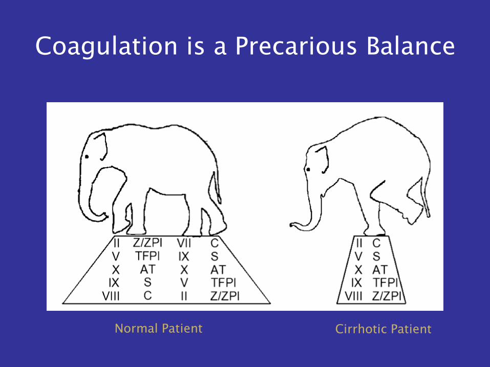
Coagulation is a Precarious Balance
Normal Patient Cirrhotic Patient
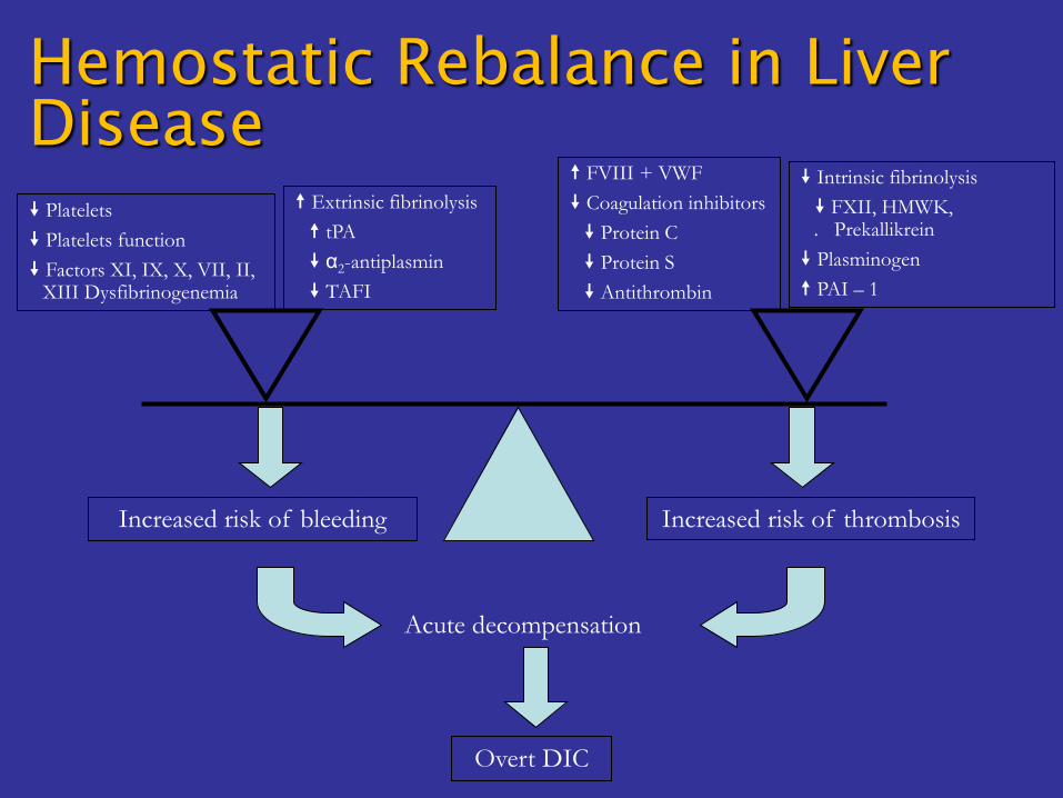
Platelets
Platelets function
Factors XI, IX, X, VII, II, XIII Dysfibrinogenemia
Extrinsic fibrinolysis
tPA
α2-antiplasmin
TAFI
FVIII + VWF
Coagulation inhibitors
Protein C
Protein S
Antithrombin
Intrinsic fibrinolysis
FXII, HMWK, . Prekallikrein
Plasminogen
PAI – 1
Increased risk of bleeding Increased risk of thrombosis
Acute decompensation
Overt DIC
Hemostatic Rebalance in Liver
Disease
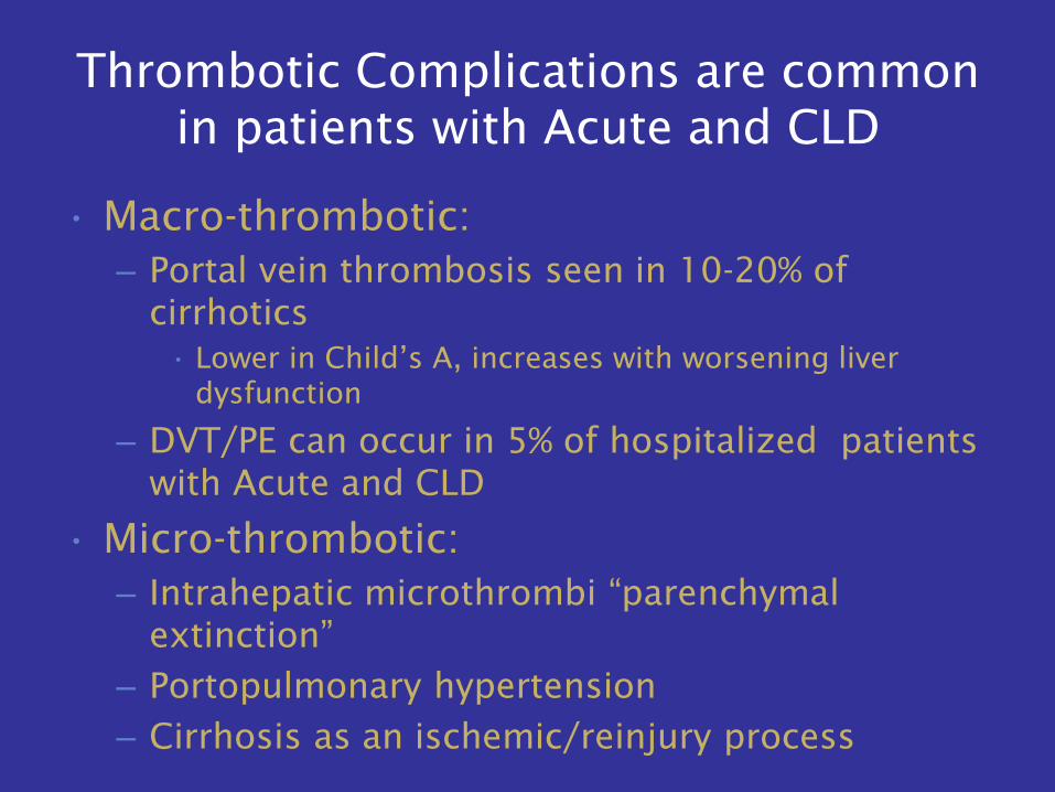
Thrombotic Complications are common
in patients with Acute and CLD
• Macro-thrombotic:
– Portal vein thrombosis seen in 10-20% of
cirrhotics
• Lower in Child’s A, increases with worsening liver
dysfunction
– DVT/PE can occur in 5% of hospitalized patients
with Acute and CLD
• Micro-thrombotic:
– Intrahepatic microthrombi “parenchymal
extinction”
– Portopulmonary hypertension
– Cirrhosis as an ischemic/reinjury process
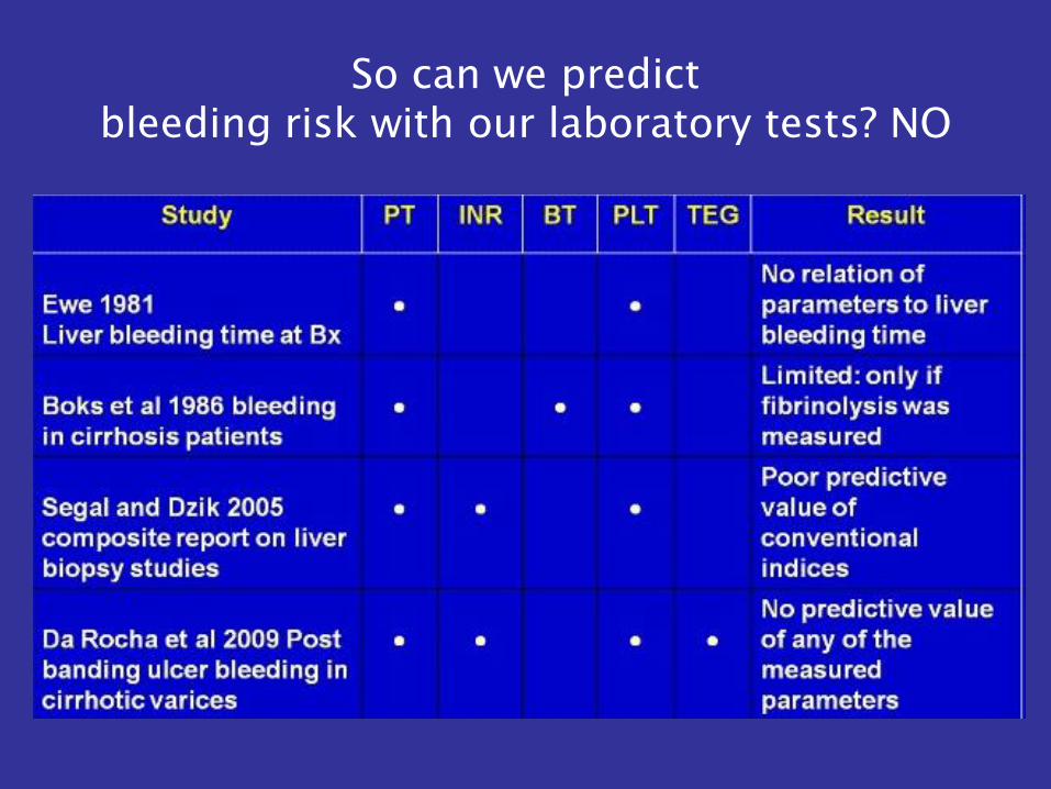
So can we predict
bleeding risk with our laboratory tests? NO
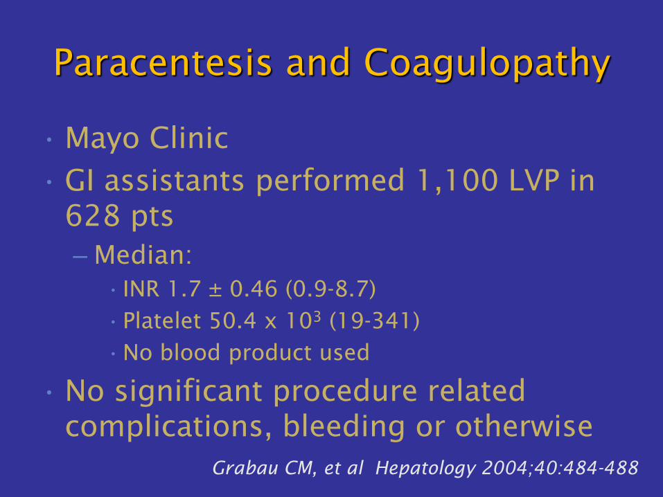
Paracentesis and Coagulopathy
• Mayo Clinic
• GI assistants performed 1,100 LVP in
628 pts
– Median:
• INR 1.7 ± 0.46 (0.9-8.7)
•Platelet 50.4 x 10³ (19-341)
•No blood product used
• No significant procedure related
complications, bleeding or otherwise
Grabau CM, et al Hepatology 2004;40:484-488
What are the detailed
changes we encounter in
cirrhosis?
The Yin and the Yang
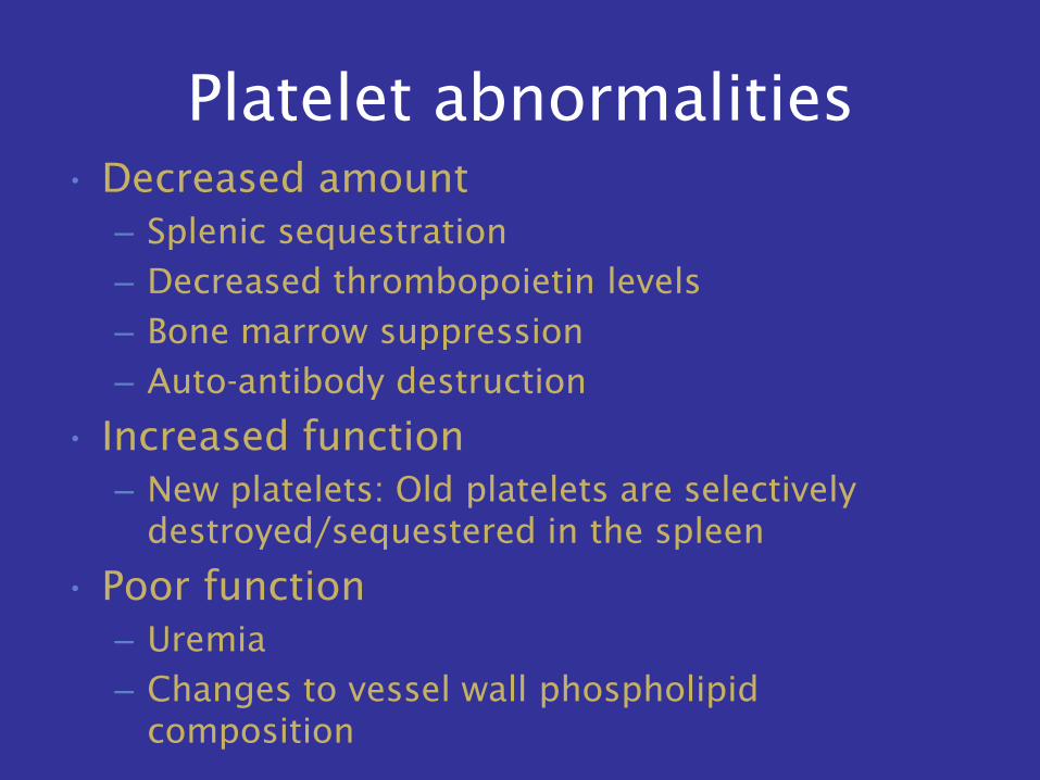
Platelet abnormalities
• Decreased amount
– Splenic sequestration
– Decreased thrombopoietin levels
– Bone marrow suppression
– Auto-antibody destruction
• Increased function
– New platelets: Old platelets are selectively
destroyed/sequestered in the spleen
• Poor function
– Uremia
– Changes to vessel wall phospholipid
composition
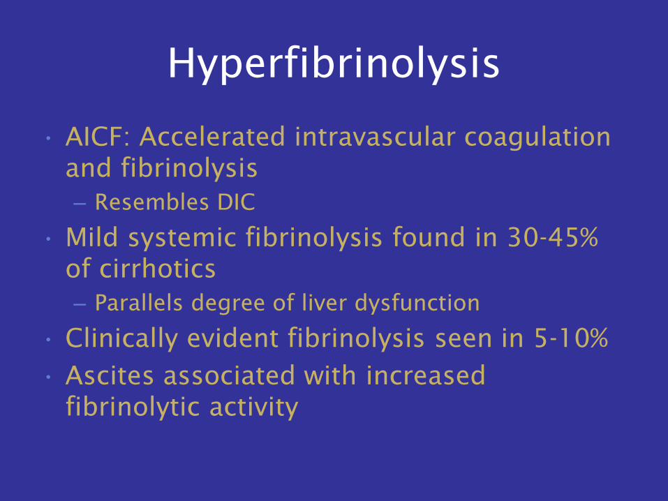
Hyperfibrinolysis
• AICF: Accelerated intravascular coagulation
and fibrinolysis
– Resembles DIC
• Mild systemic fibrinolysis found in 30-45%
of cirrhotics
– Parallels degree of liver dysfunction
• Clinically evident fibrinolysis seen in 5-10%
• Ascites associated with increased
fibrinolytic activity
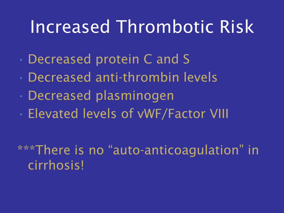
Increased Thrombotic Risk
• Decreased protein C and S
• Decreased anti-thrombin levels
• Decreased plasminogen
• Elevated levels of vWF/Factor VIII
***There is no “auto-anticoagulation” in
cirrhosis!
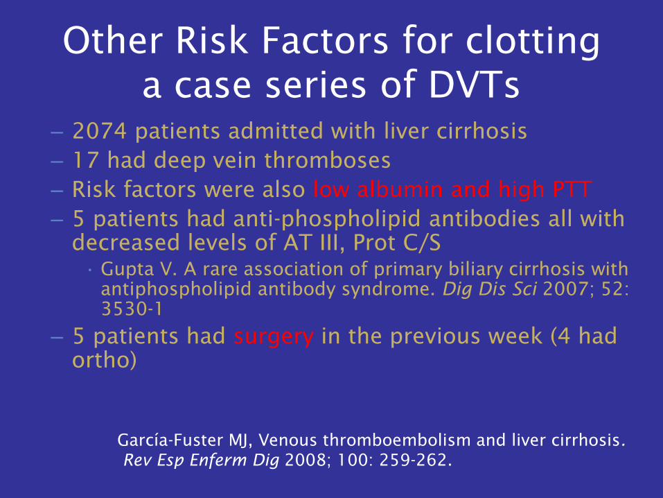
Other Risk Factors for clotting
a case series of DVTs
– 2074 patients admitted with liver cirrhosis
– 17 had deep vein thromboses
– Risk factors were also low albumin and high PTT
– 5 patients had anti-phospholipid antibodies all with
decreased levels of AT III, Prot C/S
• Gupta V. A rare association of primary biliary cirrhosis with
antiphospholipid antibody syndrome. Dig Dis Sci 2007; 52:
3530-1
– 5 patients had surgery in the previous week (4 had
ortho)
García-Fuster MJ, Venous thromboembolism and liver cirrhosis.
Rev Esp Enferm Dig 2008; 100: 259-262.
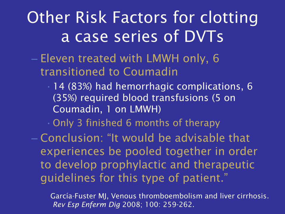
– Eleven treated with LMWH only, 6
transitioned to Coumadin
•14 (83%) had hemorrhagic complications, 6
(35%) required blood transfusions (5 on
Coumadin, 1 on LMWH)
•Only 3 finished 6 months of therapy
– Conclusion: “It would be advisable that
experiences be pooled together in order
to develop prophylactic and therapeutic
guidelines for this type of patient.”
García-Fuster MJ, Venous thromboembolism and liver cirrhosis.
Rev Esp Enferm Dig 2008; 100: 259-262.
Other Risk Factors for clotting
a case series of DVTs
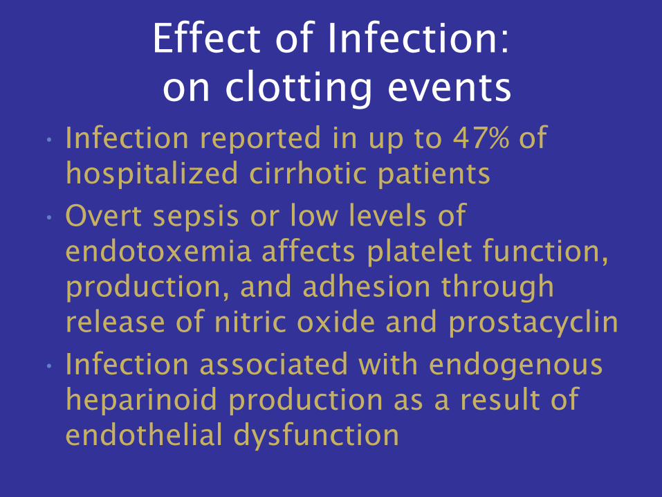
Effect of Infection:
on clotting events
• Infection reported in up to 47% of
hospitalized cirrhotic patients
• Overt sepsis or low levels of
endotoxemia affects platelet function,
production, and adhesion through
release of nitric oxide and prostacyclin
• Infection associated with endogenous
heparinoid production as a result of
endothelial dysfunction
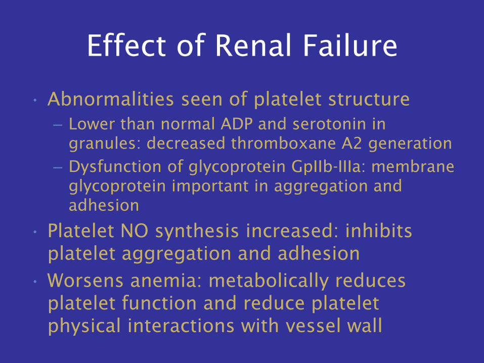
Effect of Renal Failure
• Abnormalities seen of platelet structure
– Lower than normal ADP and serotonin in
granules: decreased thromboxane A2 generation
– Dysfunction of glycoprotein GpIIb-IIIa: membrane
glycoprotein important in aggregation and
adhesion
• Platelet NO synthesis increased: inhibits
platelet aggregation and adhesion
• Worsens anemia: metabolically reduces
platelet function and reduce platelet
physical interactions with vessel wall
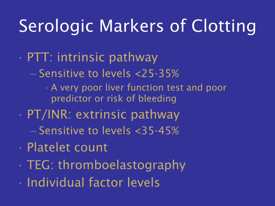
Serologic Markers of Clotting
• PTT: intrinsic pathway
– Sensitive to levels <25-35%
•A very poor liver function test and poor
predictor or risk of bleeding
• PT/INR: extrinsic pathway
– Sensitive to levels <35-45%
• Platelet count
• TEG: thromboelastography
• Individual factor levels
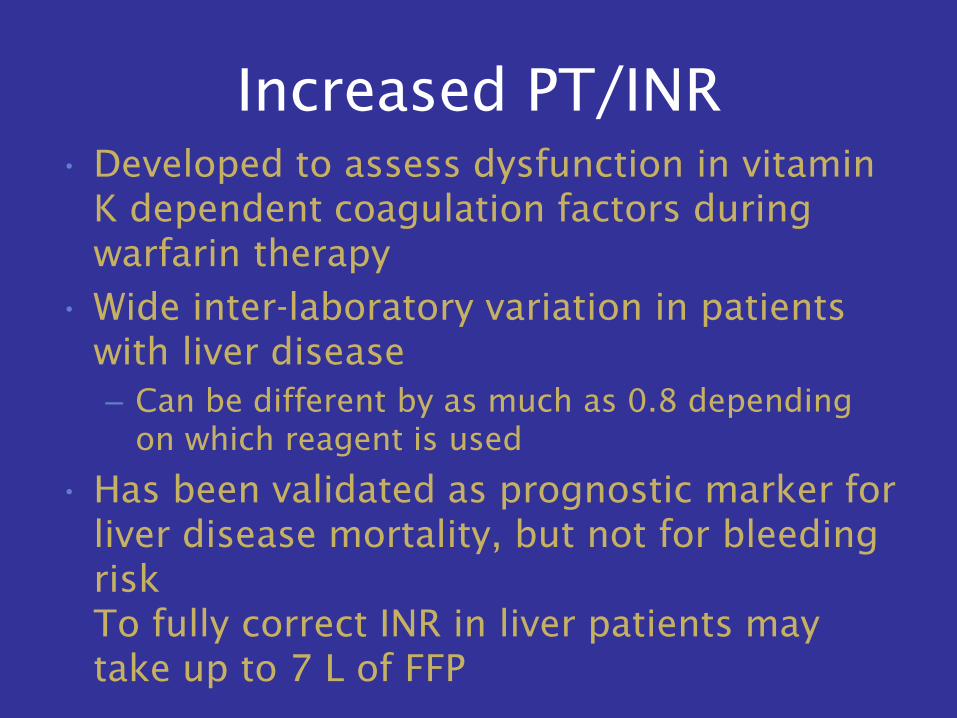
Increased PT/INR
• Developed to assess dysfunction in vitamin
K dependent coagulation factors during
warfarin therapy
• Wide inter-laboratory variation in patients
with liver disease
– Can be different by as much as 0.8 depending
on which reagent is used
• Has been validated as prognostic marker for
liver disease mortality, but not for bleeding
risk
To fully correct INR in liver patients may
take up to 7 L of FFP
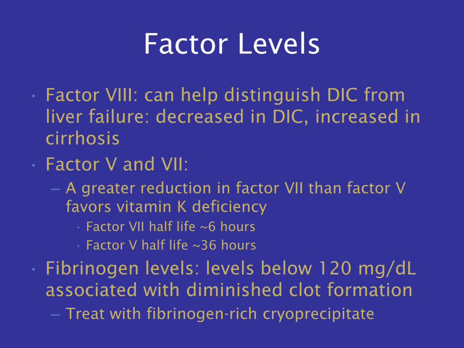
Factor Levels
• Factor VIII: can help distinguish DIC from
liver failure: decreased in DIC, increased in
cirrhosis
• Factor V and VII:
– A greater reduction in factor VII than factor V
favors vitamin K deficiency
• Factor VII half life ~6 hours
• Factor V half life ~36 hours
• Fibrinogen levels: levels below 120 mg/dL
associated with diminished clot formation
– Treat with fibrinogen-rich cryoprecipitate
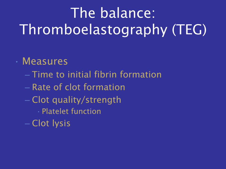
The balance:
Thromboelastography (TEG)
• Measures
– Time to initial fibrin formation
– Rate of clot formation
– Clot quality/strength
•Platelet function
– Clot lysis
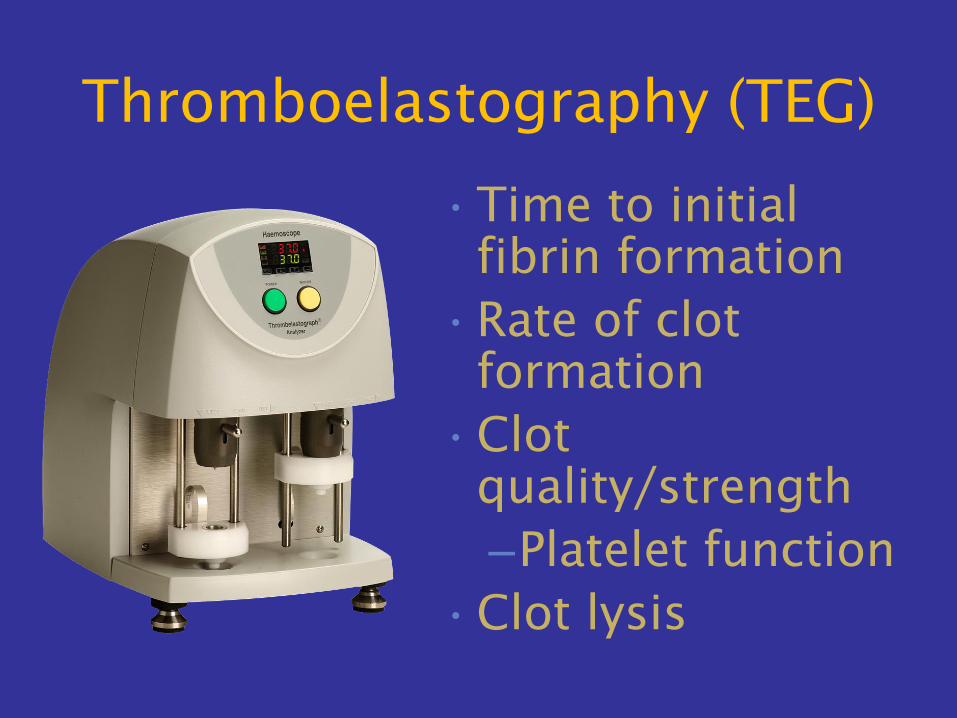
Thromboelastography (TEG)
•Time to initial
fibrin formation
•Rate of clot
formation
•Clot
quality/strength
–Platelet function
•Clot lysis
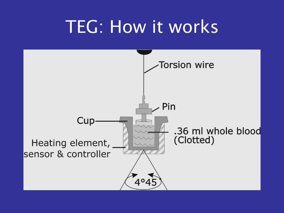
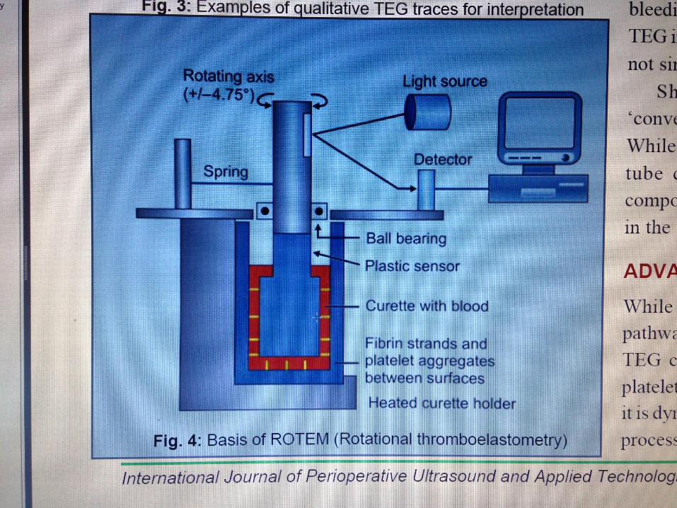
TEG: How it works
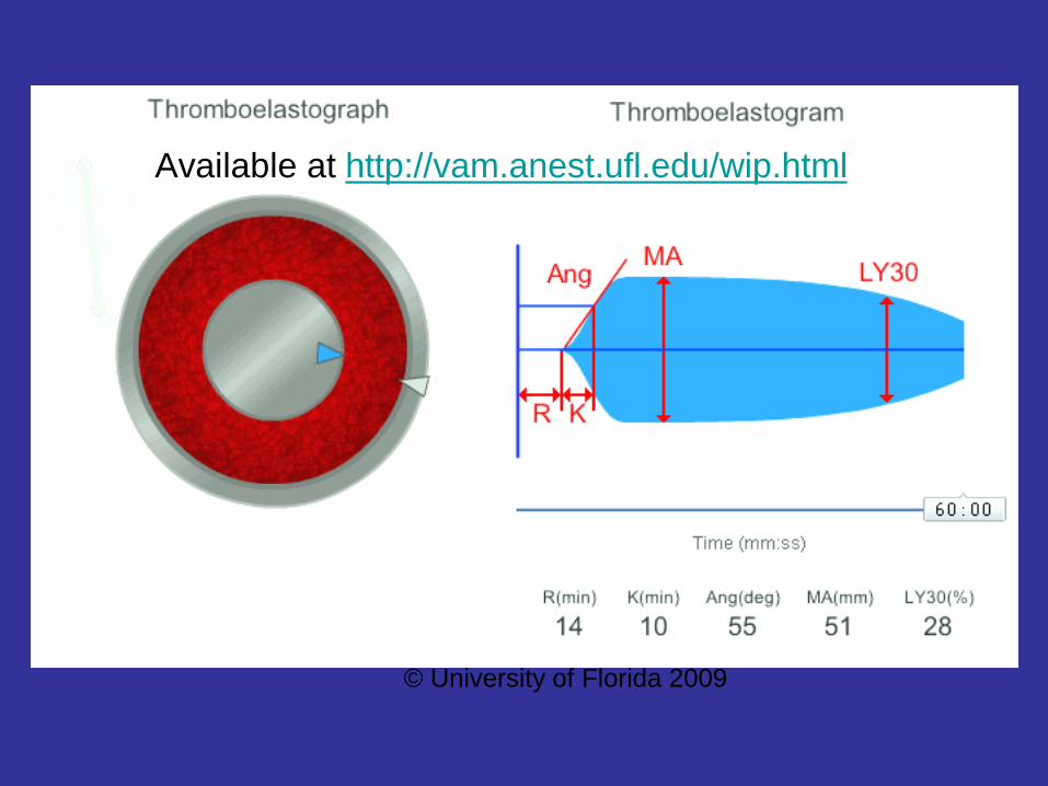
Available at http://vam.anest.ufl.edu/wip.html
© University of Florida 2009
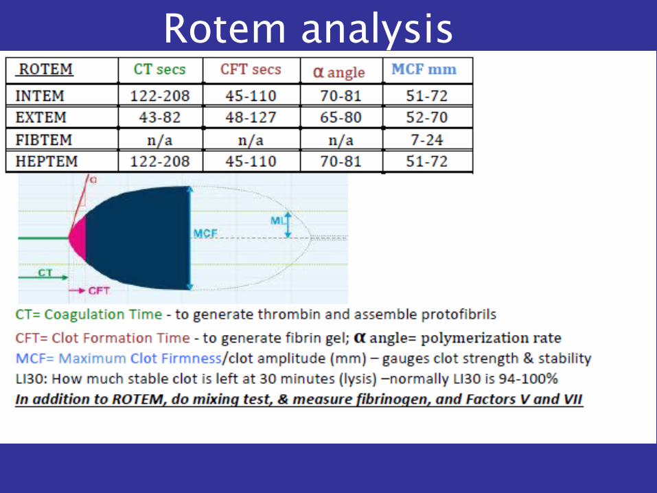
Rotem analysis
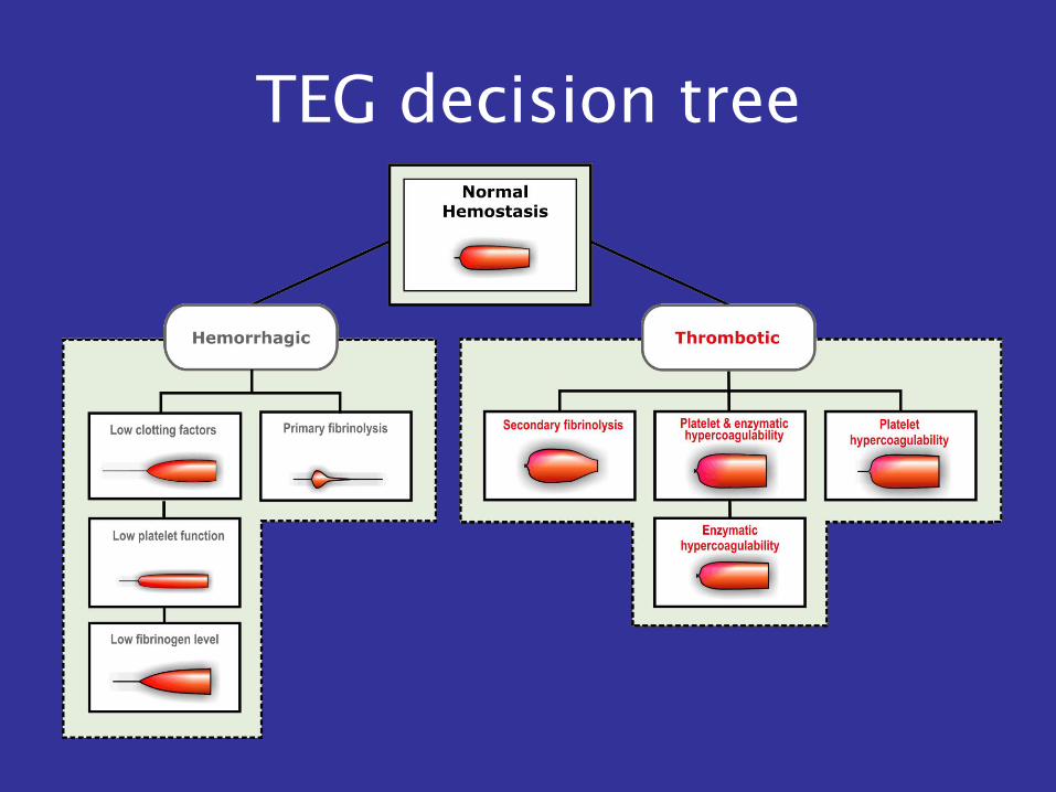
TEG decision tree
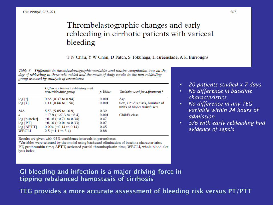
• 20 patients studied x 7 days
• No difference in baseline
characteristics
• No difference in any TEG
variable within 24 hours of
admission
• 5/6 with early rebleeding had
evidence of sepsis
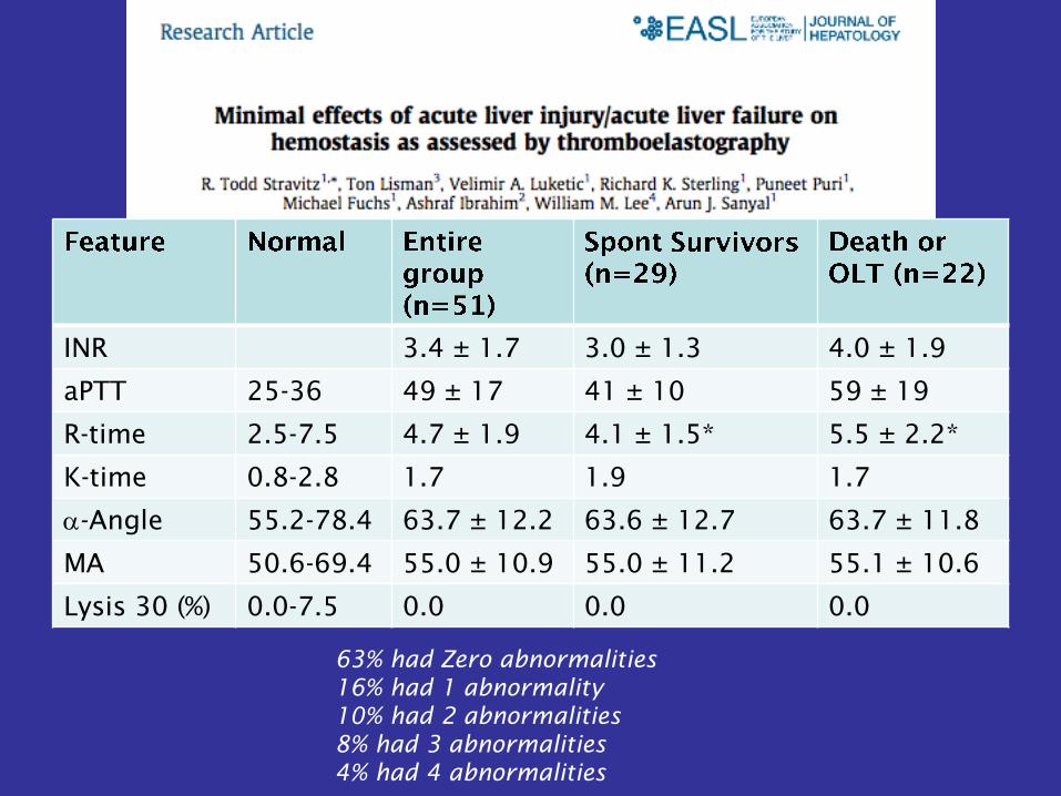
INR 3.4 ± 1.7 3.0 ± 1.3 4.0 ± 1.9
aPTT 25-36 49 ± 17 41 ± 10 59 ± 19
R-time 2.5-7.5 4.7 ± 1.9 4.1 ± 1.5* 5.5 ± 2.2*
K-time 0.8-2.8 1.7 1.9 1.7
a-Angle 55.2-78.4 63.7 ± 12.2 63.6 ± 12.7 63.7 ± 11.8
MA 50.6-69.4 55.0 ± 10.9 55.0 ± 11.2 55.1 ± 10.6
Lysis 30 (%) 0.0-7.5 0.0 0.0 0.0
63% had Zero abnormalities
16% had 1 abnormality
10% had 2 abnormalities
8% had 3 abnormalities
4% had 4 abnormalities
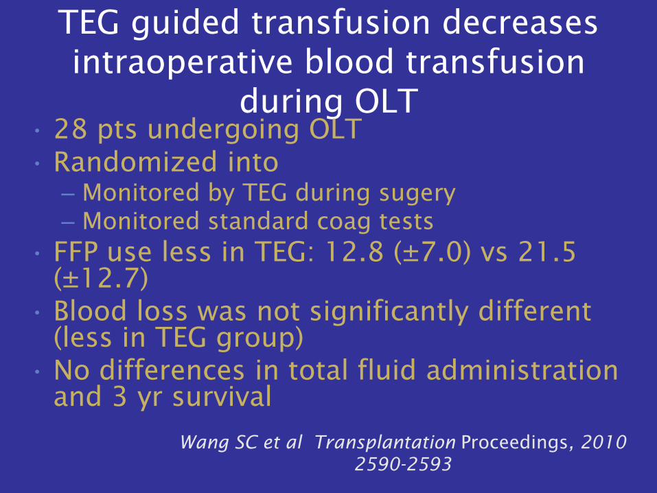
TEG guided transfusion decreases
intraoperative blood transfusion
during OLT
• 28 pts undergoing OLT
• Randomized into
– Monitored by TEG during sugery
– Monitored standard coag tests
• FFP use less in TEG: 12.8 (±7.0) vs 21.5
(±12.7)
• Blood loss was not significantly different
(less in TEG group)
• No differences in total fluid administration
and 3 yr survival
Wang SC et al Transplantation Proceedings, 2010
2590-2593
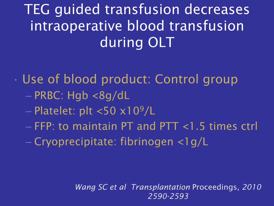
TEG guided transfusion decreases
intraoperative blood transfusion
during OLT
• Use of blood product: Control group
– PRBC: Hgb <8g/dL
– Platelet: plt <50 x109/L
– FFP: to maintain PT and PTT <1.5 times ctrl
– Cryoprecipitate: fibrinogen <1g/L
Wang SC et al Transplantation Proceedings, 2010
2590-2593
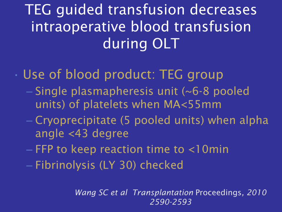
TEG guided transfusion decreases
intraoperative blood transfusion
during OLT
• Use of blood product: TEG group
– Single plasmapheresis unit (~6-8 pooled
units) of platelets when MA<55mm
– Cryoprecipitate (5 pooled units) when alpha
angle <43 degree
– FFP to keep reaction time to <10min
– Fibrinolysis (LY 30) checked
Wang SC et al Transplantation Proceedings, 2010
2590-2593
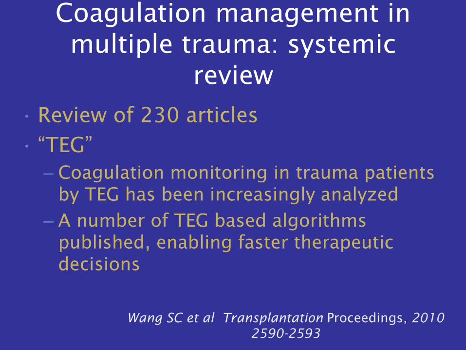
Coagulation management in
multiple trauma: systemic
review
• Review of 230 articles
• “TEG”
– Coagulation monitoring in trauma patients
by TEG has been increasingly analyzed
– A number of TEG based algorithms
published, enabling faster therapeutic
decisions
Wang SC et al Transplantation Proceedings, 2010
2590-2593
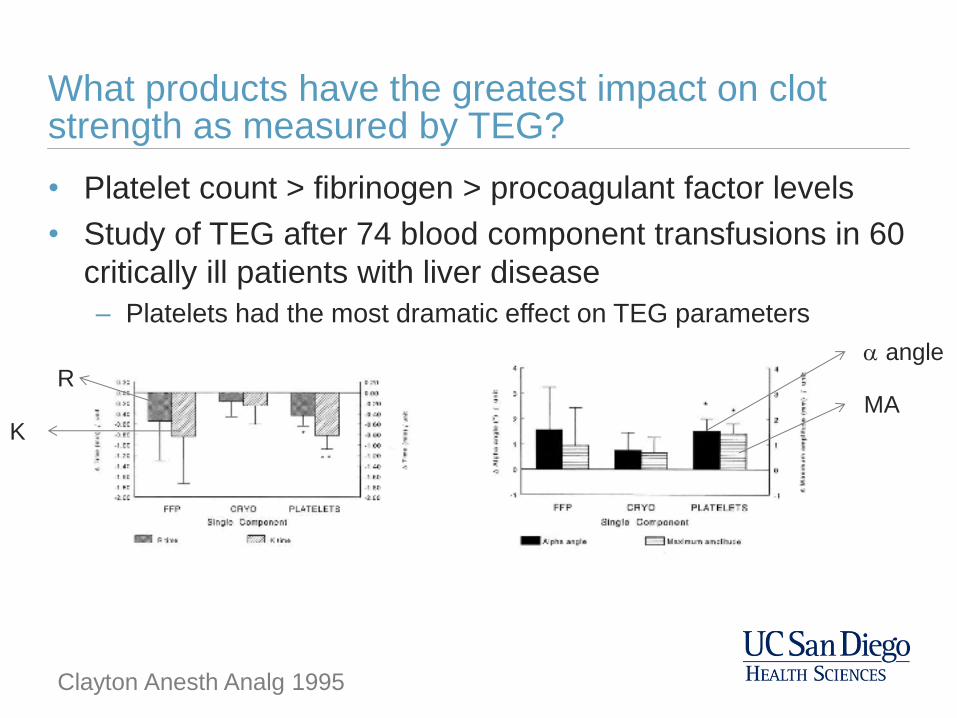
What products have the greatest impact on clot strength as measured by TEG?
• Platelet count > fibrinogen > procoagulant factor levels
• Study of TEG after 74 blood component transfusions in 60
critically ill patients with liver disease
– Platelets had the most dramatic effect on TEG parameters
MA
a angle R
K
Clayton Anesth Analg 1995
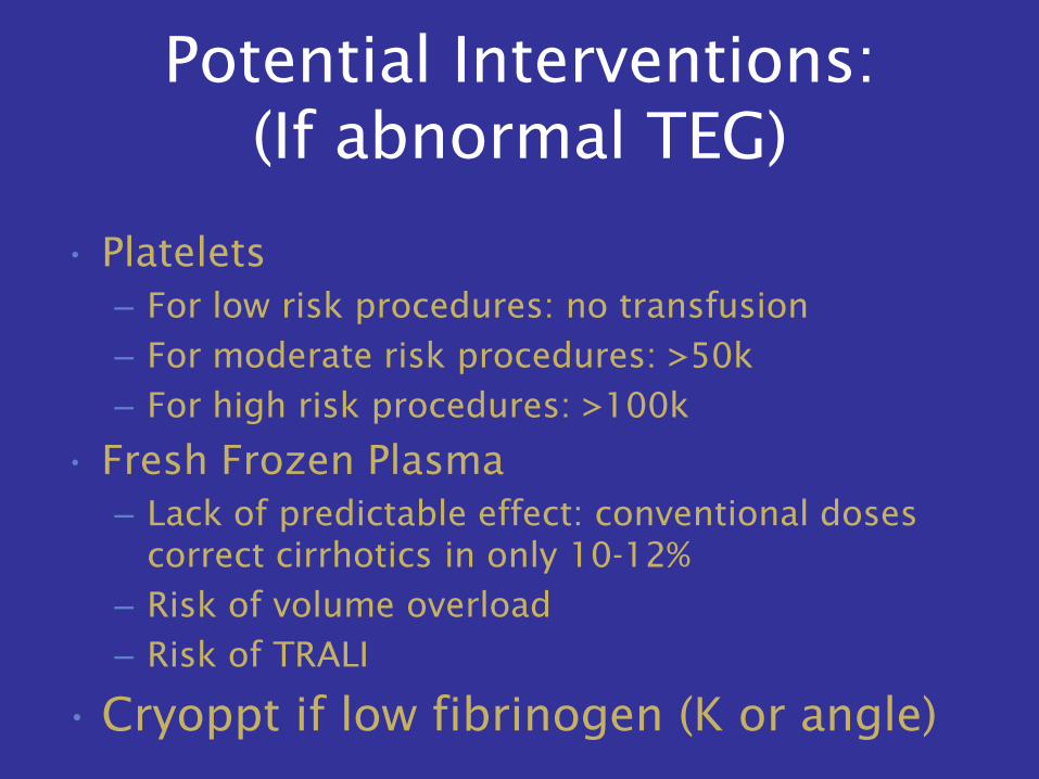
Potential Interventions:
(If abnormal TEG)
• Platelets
– For low risk procedures: no transfusion
– For moderate risk procedures: >50k
– For high risk procedures: >100k
• Fresh Frozen Plasma
– Lack of predictable effect: conventional doses
correct cirrhotics in only 10-12%
– Risk of volume overload
– Risk of TRALI
• Cryoppt if low fibrinogen (K or angle)
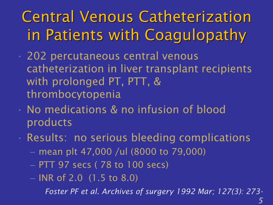
Central Venous Catheterization
in Patients with Coagulopathy
• 202 percutaneous central venous
catheterization in liver transplant recipients
with prolonged PT, PTT, &
thrombocytopenia
• No medications & no infusion of blood
products
• Results: no serious bleeding complications
– mean plt 47,000 /ul (8000 to 79,000)
– PTT 97 secs ( 78 to 100 secs)
– INR of 2.0 (1.5 to 8.0)
Foster PF et al. Archives of surgery 1992 Mar; 127(3): 273-
5
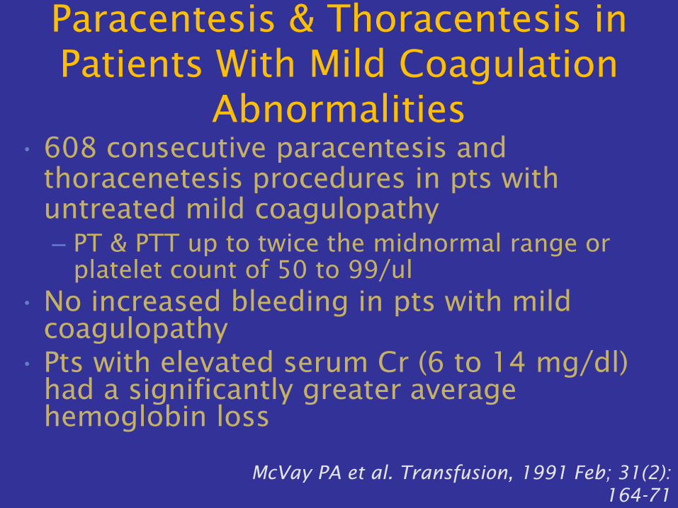
Paracentesis & Thoracentesis in
Patients With Mild Coagulation
Abnormalities
• 608 consecutive paracentesis and
thoracenetesis procedures in pts with
untreated mild coagulopathy
– PT & PTT up to twice the midnormal range or
platelet count of 50 to 99/ul
• No increased bleeding in pts with mild
coagulopathy
• Pts with elevated serum Cr (6 to 14 mg/dl)
had a significantly greater average
hemoglobin loss
McVay PA et al. Transfusion, 1991 Feb; 31(2):
164-71
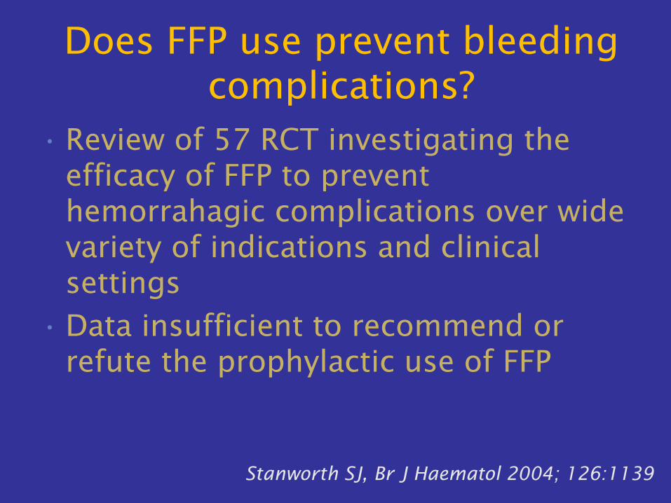
Does FFP use prevent bleeding
complications?
• Review of 57 RCT investigating the
efficacy of FFP to prevent
hemorrahagic complications over wide
variety of indications and clinical
settings
• Data insufficient to recommend or
refute the prophylactic use of FFP
Stanworth SJ, Br J Haematol 2004; 126:1139
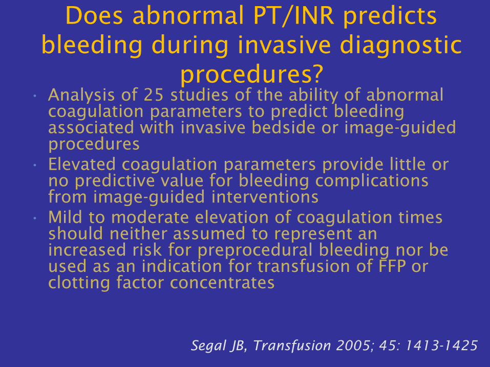
Does abnormal PT/INR predicts
bleeding during invasive diagnostic
procedures?
• Analysis of 25 studies of the ability of abnormal
coagulation parameters to predict bleeding
associated with invasive bedside or image-guided
procedures
• Elevated coagulation parameters provide little or
no predictive value for bleeding complications
from image-guided interventions
• Mild to moderate elevation of coagulation times
should neither assumed to represent an
increased risk for preprocedural bleeding nor be
used as an indication for transfusion of FFP or
clotting factor concentrates
Segal JB, Transfusion 2005; 45: 1413-1425
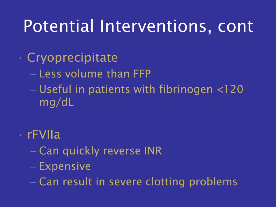
Potential Interventions, cont
• Cryoprecipitate
– Less volume than FFP
– Useful in patients with fibrinogen <120
mg/dL
• rFVIIa
– Can quickly reverse INR
– Expensive
– Can result in severe clotting problems
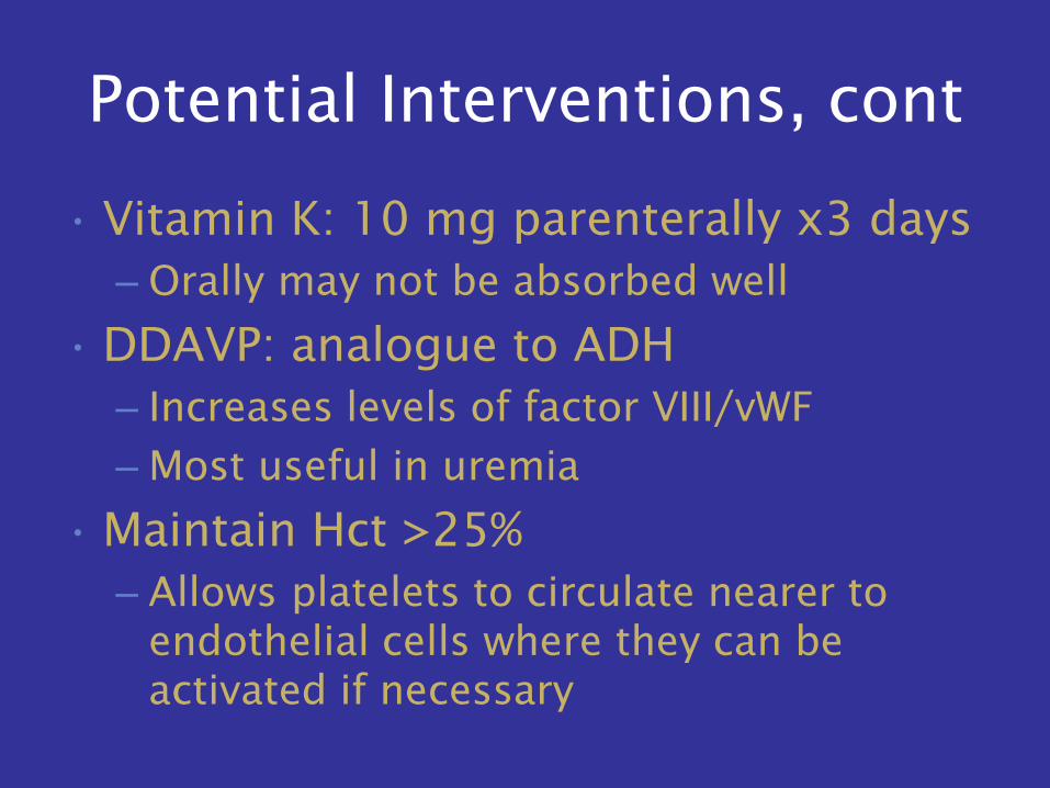
Potential Interventions, cont
• Vitamin K: 10 mg parenterally x3 days
– Orally may not be absorbed well
• DDAVP: analogue to ADH
– Increases levels of factor VIII/vWF
– Most useful in uremia
• Maintain Hct >25%
– Allows platelets to circulate nearer to
endothelial cells where they can be
activated if necessary
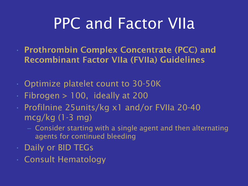
PPC and Factor VIIa
•
• Optimize platelet count to 30-50K
• Fibrogen > 100, ideally at 200
• Profilnine 25units/kg x1 and/or FVIIa 20-40
mcg/kg (1-3 mg)
– Consider starting with a single agent and then alternating
agents for continued bleeding
• Daily or BID TEGs
• Consult Hematology
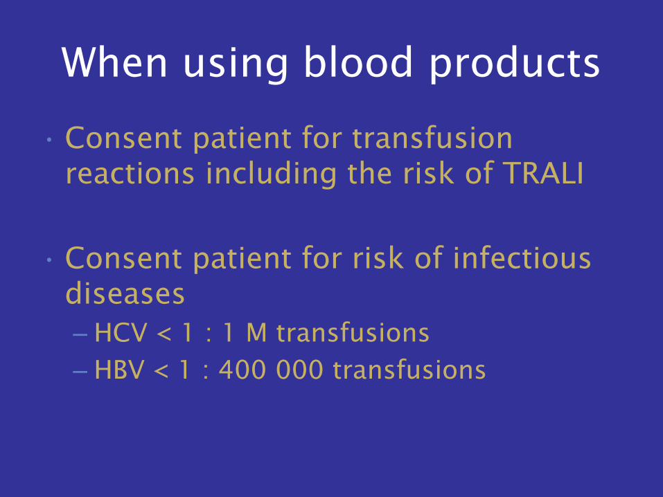
When using blood products
• Consent patient for transfusion
reactions including the risk of TRALI
• Consent patient for risk of infectious
diseases
– HCV < 1 : 1 M transfusions
– HBV < 1 : 400 000 transfusions
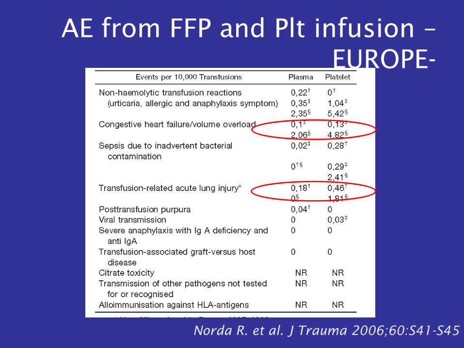
AE from FFP and Plt infusion –
EUROPE-
Norda R. et al. J Trauma 2006;60:S41-S45
Question
• Should we anti-coagulate patients with
cirrhosis?
Enoxaparin Prevents Portal Vein
Thrombosis and Liver
Decompensation in Patients With
Advanced Cirrhosis
GASTROENTEROLOGY 2012
Liver Journal Club
Shireena Desai
Nov 27, 2012
aims
• To evaluate the safety and efficacy of
enoxaparin in preventing PVT in
patients with advanced cirrhosis
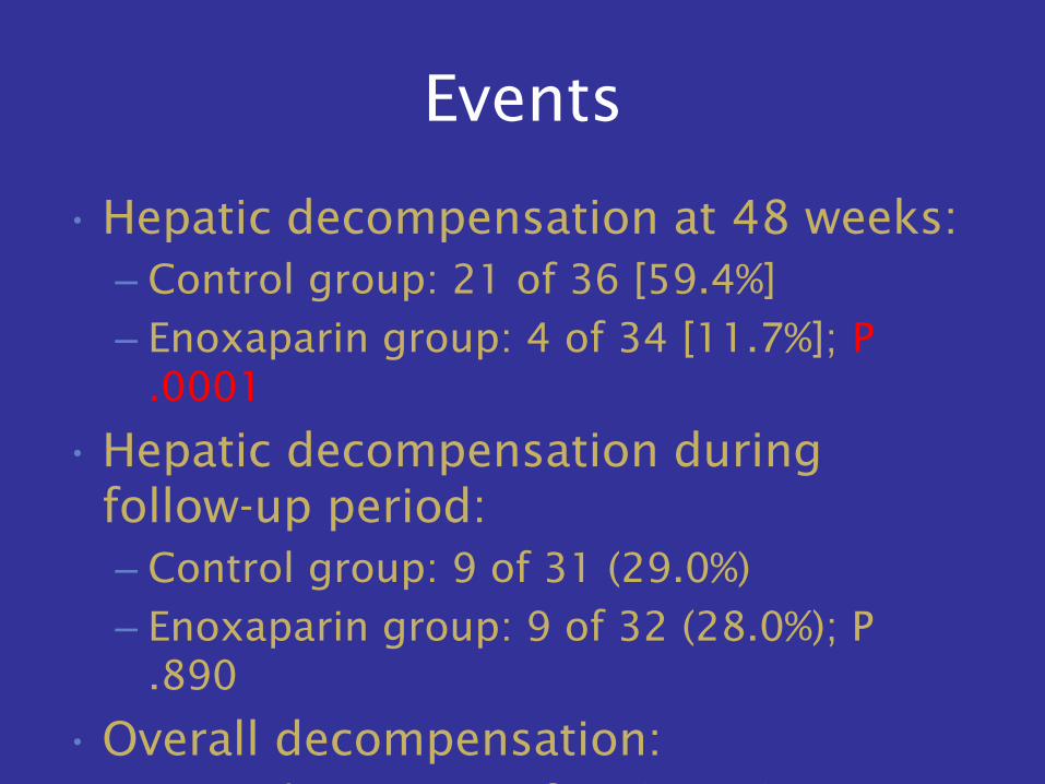
Events
• Hepatic decompensation at 48 weeks:
– Control group: 21 of 36 [59.4%]
– Enoxaparin group: 4 of 34 [11.7%]; P
.0001
• Hepatic decompensation during
follow-up period:
– Control group: 9 of 31 (29.0%)
– Enoxaparin group: 9 of 32 (28.0%); P
.890
• Overall decompensation:
– Control group: 30 of 36 (83.0%)
– Enoxaparin group: 13 of 34 (38.2%); P
.0001
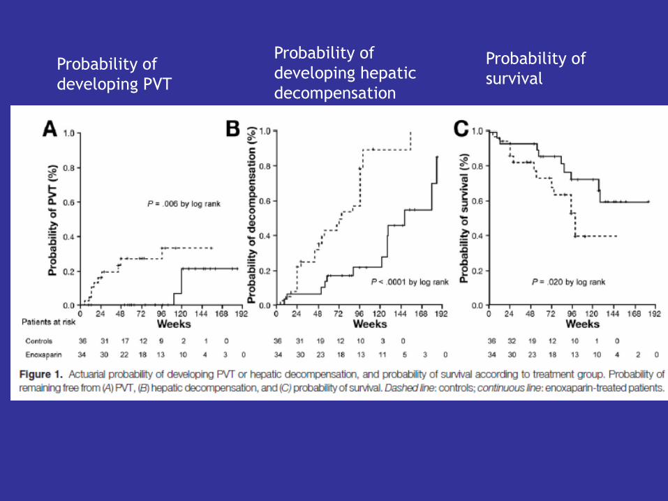
Probability of
developing PVT
Probability of
developing hepatic
decompensation
Probability of
survival
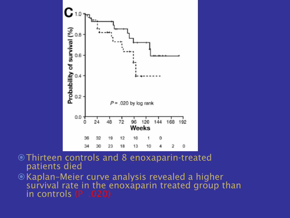
Thirteen controls and 8 enoxaparin-treated
patients died
Kaplan–Meier curve analysis revealed a higher
survival rate in the enoxaparin treated group than
in controls (P .020)
Conclusions: enoxaparin
• A 12-month course of enoxaparin was
safe and effective in preventing PVT in
patients with cirrhosis and a Child–
Pugh score of B7–C10.
• Enoxaparin appeared to significantly
reduce the risk of PVT development
and liver decompensation, improve
liver function and Child–Pugh score,
and increase overall survival.
Portal Vein Thrombosis
• Check first for cirrhosis, cancer of the abdominal organs, or
an inflammatory focus in the abdomen.
• Nontumorous and noncirrhotic patients: 30%-40% of patients
with PVT are affected with myeloproliferative diseases:
– polycythemia vera
– essential thrombocythemia
• Iron deficiency or portal hypertension can mask the expected
elevated Hgb or platelet count. Do not rule out a diagnosis of
myeloproliferative disease solely on the basis of normal/low
blood cell counts.
• Point mutation of the tyrosine kinase JAK2 is a marker for
myeloproliferative disease.
– In 5%-10% of patients with PVT, JAK2 was undetectable
whereas bone marrow biopsy provided evidence for a
myeloproliferative disorder.
Acute Portal Vein Thrombosis
• Sudden onset abdominal or lumbar pain
• When extensive thrombosis involves
distal mesenteric veins, intestinal
ischemia and infarction can occur,
leading to severe pain and bloody
diarrhea.
• The presence of ascites, thinning of the
intestinal wall, or the development of
multiorgan failure indicate that intestinal
infarction is likely and surgical
exploration should be considered
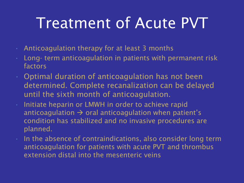
Treatment of Acute PVT
• Anticoagulation therapy for at least 3 months
• Long- term anticoagulation in patients with permanent risk
factors
• Optimal duration of anticoagulation has not been
determined. Complete recanalization can be delayed
until the sixth month of anticoagulation.
• Initiate heparin or LMWH in order to achieve rapid
anticoagulation oral anticoagulation when patient’s
condition has stabilized and no invasive procedures are
planned.
• In the absence of contraindications, also consider long term
anticoagulation for patients with acute PVT and thrombus
extension distal into the mesenteric veins
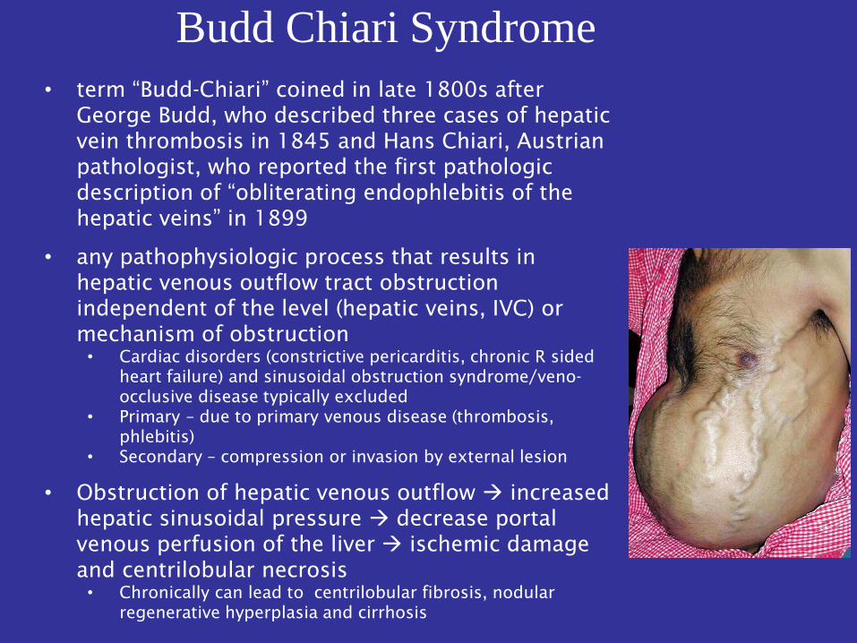
Budd Chiari Syndrome
• term “Budd-Chiari” coined in late 1800s after
George Budd, who described three cases of hepatic
vein thrombosis in 1845 and Hans Chiari, Austrian
pathologist, who reported the first pathologic
description of “obliterating endophlebitis of the
hepatic veins” in 1899
• any pathophysiologic process that results in
hepatic venous outflow tract obstruction
independent of the level (hepatic veins, IVC) or
mechanism of obstruction
• Cardiac disorders (constrictive pericarditis, chronic R sided
heart failure) and sinusoidal obstruction syndrome/veno-
occlusive disease typically excluded
• Primary – due to primary venous disease (thrombosis,
phlebitis)
• Secondary – compression or invasion by external lesion
• Obstruction of hepatic venous outflow increased
hepatic sinusoidal pressure decrease portal
venous perfusion of the liver ischemic damage
and centrilobular necrosis
• Chronically can lead to centrilobular fibrosis, nodular
regenerative hyperplasia and cirrhosis
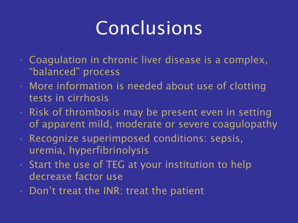
Conclusions
• Coagulation in chronic liver disease is a complex,
“balanced” process
• More information is needed about use of clotting
tests in cirrhosis
• Risk of thrombosis may be present even in setting
of apparent mild, moderate or severe coagulopathy
• Recognize superimposed conditions: sepsis,
uremia, hyperfibrinolysis
• Start the use of TEG at your institution to help
decrease factor use
• Don’t treat the INR: treat the patient

Thank you to:
• Todd Stravitz, Carrie Frenette, Patrick
Kamath and Yuko Kono for use of
their slides