CHARACTERIZATION OF THE β-D … · characterization of the β-d-glucopyranoside binding site of...
21
CHARACTERIZATION OF THE β-D-GLUCOPYRANOSIDE BINDING SITE OF THE HUMAN BITTER TASTE RECEPTOR hTAS2R16 Takanobu Sakurai 1, 2 , Takumi Misaka 2 , Masaji Ishiguro 3 , Katsuyoshi Masuda 3 , Taishi Sugawara 2 , Keisuke Ito 2, # , Takuya Kobayashi 4 , Shinji Matsuo 1 , Yoshiro Ishimaru 2 , Tomiko Asakura 2 , and Keiko Abe 2 From the 1 General Research Institute of Food Science and Technology, Nissin Foods Holdings Co., Ltd., Shiga 525-0058, Japan, 2 Department of Applied Biological Chemistry, Graduate School of Agricultural and Life Sciences, The University of Tokyo, Tokyo 113-8657, Japan, 3 Niigata University of Pharmacy and Applied Life Sciences, Niigata 956-8503, Japan, and 4 Department of Medical Chemistry, Kyoto University Faculty of Medicine, 606-8501, Japan Running head: β-D-Glucopyranoside binding site of hTAS2R16 Address correspondence to: Takumi Misaka, Department of Applied Biological Chemistry, Graduate School of Agricultural and Life Sciences, The University of Tokyo, 1-1-1 Yayoi, Bunkyo-ku, Tokyo 113-8657, Japan. Fax: +81-3-5841-8118; E-mail: [email protected] hTAS2R16 and β-D-glucopyranoside, and will also facilitate rational design of bitter blockers. G-protein-coupled receptors (GPCRs) mediate the senses of taste, smell, and vision in mammals. Humans recognize thousands of compounds as bitter, and this response is mediated by the hTAS2R family, which is one of the GPCR comprised of only 25 receptors. However, structural information on these receptors is limited. To address the molecular basis of bitter tastant discrimination by the hTAS2Rs, we performed ligand docking simulation and functional analysis using a series of point mutants of hTAS2R16 to identify its binding sites. The docking simulation predicted two candidate binding structures for a salicin-hTAS2R16 complex, and at least 7 amino acid residues in transmembrane (TM)3, TM5, and TM6 were shown to be involved in ligand recognition. We also identified the probable salicin-hTAS2R16 binding mode using a mutated receptor experiment. This study characterizes the molecular interaction between There are five basic and distinct taste sensations: sweetness, bitterness, saltiness, sourness, and umami. Recent progress in the molecular biology of taste has revealed that sweet, umami, and bitter tastes are mediated by G-protein-coupled receptors (GPCRs) (1). The TAS1R family of taste receptors mediates two taste sensations; TAS1R2/TAS1R3 responds to sweetness (2,3) and TAS1R1/TAS1R3 to umami taste (2,4), while the TAS2R family responds to bitterness (5-14). Bitterness is generally considered a sign of toxic substances; on the other hand, appropriate use of certain bitter food ingredients can provide a unique and favorable character to foods, as in the case of tea, coffee, and beer. It is thus important for the food industry to use bitter taste ingredients properly. Information on the ligand-binding 1 http://www.jbc.org/cgi/doi/10.1074/jbc.M110.144444 The latest version is at JBC Papers in Press. Published on July 6, 2010 as Manuscript M110.144444 Copyright 2010 by The American Society for Biochemistry and Molecular Biology, Inc. by guest on September 1, 2018 http://www.jbc.org/ Downloaded from
Transcript of CHARACTERIZATION OF THE β-D … · characterization of the β-d-glucopyranoside binding site of...
CHARACTERIZATION OF THE β-D-GLUCOPYRANOSIDE BINDING SITE OF THE HUMAN BITTER TASTE RECEPTOR hTAS2R16
Takanobu Sakurai1, 2, Takumi Misaka2, Masaji Ishiguro3, Katsuyoshi Masuda3, Taishi Sugawara2, Keisuke Ito2, #, Takuya Kobayashi4, Shinji Matsuo1, Yoshiro Ishimaru2, Tomiko Asakura2, and
Keiko Abe2 From the 1General Research Institute of Food Science and Technology, Nissin Foods Holdings Co., Ltd., Shiga 525-0058, Japan, 2Department of Applied Biological Chemistry, Graduate School of Agricultural and Life Sciences, The University of Tokyo, Tokyo 113-8657, Japan, 3Niigata University of Pharmacy and Applied Life Sciences, Niigata 956-8503, Japan, and 4Department of Medical Chemistry, Kyoto
University Faculty of Medicine, 606-8501, Japan Running head: β-D-Glucopyranoside binding site of hTAS2R16
Address correspondence to: Takumi Misaka, Department of Applied Biological Chemistry, Graduate School of Agricultural and Life Sciences, The University of Tokyo, 1-1-1 Yayoi, Bunkyo-ku, Tokyo
113-8657, Japan. Fax: +81-3-5841-8118; E-mail: [email protected]
hTAS2R16 and β-D-glucopyranoside, and will also facilitate rational design of bitter blockers.
G-protein-coupled receptors (GPCRs) mediate the senses of taste, smell, and vision in mammals. Humans recognize thousands of compounds as bitter, and this response is mediated by the hTAS2R family, which is one of the GPCR comprised of only 25 receptors. However, structural information on these receptors is limited. To address the molecular basis of bitter tastant discrimination by the hTAS2Rs, we performed ligand docking simulation and functional analysis using a series of point mutants of hTAS2R16 to identify its binding sites. The docking simulation predicted two candidate binding structures for a salicin-hTAS2R16 complex, and at least 7 amino acid residues in transmembrane (TM)3, TM5, and TM6 were shown to be involved in ligand recognition. We also identified the probable salicin-hTAS2R16 binding mode using a mutated receptor experiment. This study characterizes the molecular interaction between
There are five basic and distinct taste
sensations: sweetness, bitterness, saltiness, sourness, and umami. Recent progress in the molecular biology of taste has revealed that sweet, umami, and bitter tastes are mediated by G-protein-coupled receptors (GPCRs) (1). The TAS1R family of taste receptors mediates two taste sensations; TAS1R2/TAS1R3 responds to sweetness (2,3) and TAS1R1/TAS1R3 to umami taste (2,4), while the TAS2R family responds to bitterness (5-14).
Bitterness is generally considered a sign of toxic substances; on the other hand, appropriate use of certain bitter food ingredients can provide a unique and favorable character to foods, as in the case of tea, coffee, and beer. It is thus important for the food industry to use bitter taste ingredients properly. Information on the ligand-binding
1
http://www.jbc.org/cgi/doi/10.1074/jbc.M110.144444The latest version is at JBC Papers in Press. Published on July 6, 2010 as Manuscript M110.144444
Copyright 2010 by The American Society for Biochemistry and Molecular Biology, Inc.
by guest on September 1, 2018
http://ww
w.jbc.org/
Dow
nloaded from
structure of the bitter taste receptors with tastants is useful for controlling bitterness. Using the crystal structure of bovine rhodopsin as a template, several in silico computational modeling for hTAS2Rs have been demonstrated (15,16). In these reports, the 3D structure of hTAS2R38 was predicted, and docking simulations with 6-propyl-2-thiouracil (PROP) and N-phenylthiourea (PTC) were performed to clarify the differences in their sensitivity to the hTAS2R38 receptor variants (13). However, these previous analyses lacked functional studies to compensate for the ambiguity of the computational simulations and validate the accuracy of the predictions.
On the other hand, to clarify the ligand-binding sites in TAS1Rs, several studies have been conducted using chimeric and mutated receptor experiments. For example, the large extracellular amino terminal domain of TAS1R2 is required for recognition of aspartame and neotame as sweet tastants (17,18), and the hTAS1R3 amino terminal domain is required for response to neoculin, a sweet protein with taste-modifying activity (19-22). Molecular modeling is also effective for identifying binding sites, and combination of functional analysis along with molecular modeling has identified the neohesperidin dihydrochalcone-binding site in TAS1R3 and glutamate-binding site in the TAS1R1 venus flytrap domain (23,24).
In the present study, we performed both a precise ligand docking simulation and functional analysis of a series of point mutants to determine the molecular basis of bitter ligand discrimination by hTAS2R16, and have successfully identified
the putative salicin-binding site in hTAS2R16. This study provides new insights into bitter taste reception by hTAS2Rs.
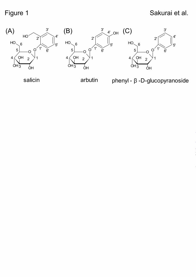
EXPERIMENTAL PROCEDURES Compounds – Salicin, arbutin, and phenyl-β-D-glucopyranoside were selected as test ligands for hTAS2R16 (Fig. 1) (7). Salicin was purchased from Nacalai Tesque, Inc. (Kyoto, Japan), arbutin from Tokyo Chemical Industry Co., Ltd. (Tokyo, Japan), and phenyl β-D-glucopyranoside from Sigma-Aldrich (Tokyo, Japan). Construction of a structural model of hTAS2R16 – Seven TM helical regions were deduced based on White and Wimley parameters (25), and a homology alignment between the amino acid sequences of bovine rhodopsin and hTAS2R16 was constructed. The structure of the TM helical regions and the extra- and intracellular loops were constructed using the Homology module installed in Insight II (Accelrys Inc., San Diego, CA, USA) and an alignment from a previously reported 3D structural model of a rhodopsin photointermediate, metarhodopsin (26). Insertion and deletion sites were selected in the loop regions and constructed using the loop structure database provided in Insight II.
Energy minimization was carried out using a molecular mechanics/molecular dynamics program, Discover 3 within Insight II, until the root mean square deviation became less than 0.1 Kcal/mol-Å2. Subsequent molecular dynamics calculations for the structural optimization were performed using Discover 3 at 300 K for 0.2 nsec
2
by guest on September 1, 2018
http://ww
w.jbc.org/
Dow
nloaded from
by sampling the structures every 2 psec. The resulting 100 structures were energy-minimized using Discover 3. Among the minimized structures, the lowest-energy structure was selected. During the calculations, the main-chain atoms in the TM regions were tethered at their original positions.
Construction of complex structural models of the TM region – The ligand-binding sites in the TM region were deduced using the Binding-Site module of Insight II. Salicin was docked in the binding site to simulate conformational changes in the receptor structure. These structures were energy-minimized using Discover 3, followed by structural optimization using molecular dynamics calculations.
Construction of expression plasmids for hTAS2R16 and the chimeric G protein – To construct an expression plasmid for hTAS2R16, DNA fragments encoding hTAS2R16 (NCBI refseq number: NM_016945) were subjected to polymerase chain reaction (PCR) amplification from human genomic DNA (BD Clontech, Mountain View, CA, USA). The coding region of hTAS2R16 (1–876) was tagged at the N terminus with the first 45 amino acids of rat somatostatin receptor type 3 (ssr3-) (27) and subcloned into the EcoR I-Not I site of the pEAK10 expression vector (Edge Biosystems, Gaithersburg, MD, USA). Expression plasmids for the chimeric G-protein subunit Gα16gust44, sub-cloned into pcDNA3.1 (Invitrogen, San Diego, CA, USA), were kindly gifted by Dr. Ueda and Dr. Shimada (Nagoya City University, Nagoya, Japan) (28). Site-directed mutagenesis – Mutations were introduced into ssr3-hTAS2R16 cDNA using the QuickChange site-directed mutagenesis kit
(Stratagene, La Jolla, CA, USA). Fragments containing the desired mutation product were digested with EcoR I and Not I, and ligated into the EcoR I–Not I site of the pEAK10 expression vector. All mutations were checked by DNA sequencing using the BigDye Terminator v3.1 Cycle Sequencing kit (Applied Biosystems, Foster City, CA, USA). Cell culture and transfection – Human embryonic kidney 293T (HEK293T) cells were cultured at 37 °C in Dulbecco’s modified Eagle's medium (DMEM; Sigma-Aldrich) supplemented with 10% fetal bovine serum (Invitrogen). Cells were seeded onto 35-mm dishes, and transiently transfected with the plasmid expressing the ssr3-hTAS2R16 or ssr3-hTAS2R16 mutants along with Gα16gust44 at a 4:1 ratio using Lipofectamine 2000 (Invitrogen). Cell-based assay – Transfected cells were transferred to a 96-well black, clear-bottomed CellBIND surface plate (Corning Inc., Bedford, MA, USA) 6 h after transfection. The cells were incubated for an additional 18–20 h, then rinsed with an assay buffer (130 mM NaCl, 10 mM glucose, 5 mM KCl, 2 mM CaCl2, 1.2 mM MgCl2, and 10 mM HEPES, pH 7.4), loaded with 5 µM Fluo-4 AM calcium indicator dye (Dojindo Laboratories, Kumamoto, Japan) diluted with the assay buffer, and incubated for an additional 30 min at 27 °C. The cells were then rinsed with the assay buffer and incubated in 100 µl of assay buffer for 10 min at 27 °C before the plate was loaded onto a FlexStation 3 (Molecular Devices, Inc., CA, USA) for fluorescence detection. Fluorescence (excitation at 485 nm, emission at 525 nm, and cutoff at 515 nm) was monitored at
3
by guest on September 1, 2018
http://ww
w.jbc.org/
Dow
nloaded from
2-sec intervals at 27 °C; 100 µl of assay buffer supplemented with 2× test compound solution was added at 20 sec and scanning was continued for an additional 100 sec. Fluorescence responses were measured from 20 to 30 sec after addition of the ligand, and corrected for background fluorescence measured prior to ligand application. Stimuli were tested at concentrations that did not elicit calcium responses from Gα16gust44-transfected cells (data not shown). Final concentrations of the test ligands (salicin, arbutin, and phenyl β-D-glucopyranoside) were each set at 0.01, 0.03, 0.1, 0.3, 1, 3, 10, and 30 mM. Data were collected from at least 3 independent experiments.
For calculation of EC50 values, plots of amplitude versus concentration were prepared in Clampfit ver. 9.2.0.09 (Molecular Devices, Sunnyvale, CA, USA). Nonlinear regression of the plots produced the function f(x) = Imin + (Imax – Imin) / (1 + (x / EC50)h), where x is the ligand concentration and h is the Hill coefficient, which was used to calculate the EC50 values for ligand-receptor interactions.
RESULTS
Molecular modeling of hTAS2R16 and ligand-receptor complexes. – We constructed a molecular model of hTAS2R16 based on the previously reported 3D structural model of a rhodopsin photointermediate, metarhodopsin (26). According to the homology alignment between bovine rhodopsin and hTAS2R16, the TM domains and extra- and intracellular loops of hTAS2R16 were constructed and the ligand-binding sites in the TM region were
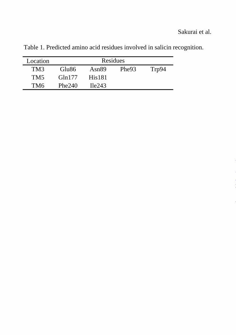
deduced. Salicin docked in the binding site was then obtained after structural optimization of the initial complexes. Salicin was bound in the putative ligand-binding pocket formed by TM3, TM5, and TM6 and interacted with eight amino acid residues (Table 1).
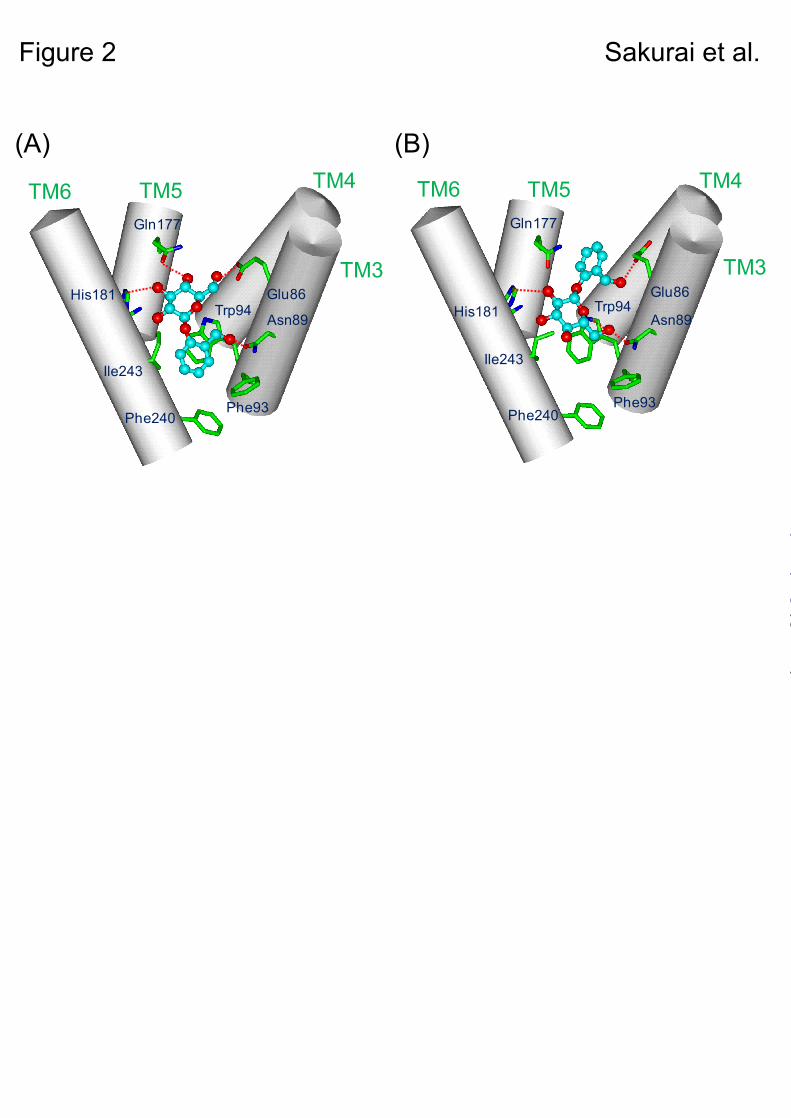
Through the above approach, two binding modes of salicin in the ligand-binding region were suggested as candidates for the salicin-receptor complex structure (Figs. 2A & B). In the both complex models, salicin was located in the same putative binding pocket with different binding modes. In one mode (model A), the aromatic ring of salicin was directed to the cytoplasmic site and the sugar moiety to the extracellular site of the putative ligand-binding pocket (Fig. 2A), whereas in the other mode (model B), it was located upside down in the binding site (Fig. 2B).
To clarify the salicin-hTAS2R16 complex structure, Glu86, Trp94, and Gln177 were selected as key amino acid residues in the constructed models. In model A, Glu86 and Gln177 formed hydrogen bonds with the sugar moiety, and Trp94 interacted with the aromatic ring of salicin (Fig. 2A). On the other hand, in model B, Glu86 hydrogen-bonded with the hydroxymethyl group on the aromatic ring, and Trp94 interacted with the sugar moiety of salicin, while Gln177 did not have any interaction with salicin (Fig. 2B).
Clarification of the salicin-hTAS2R16 binding mode using point mutations of key amino acid residues. – To clarify the salicin-hTAS2R16 binding mode, two key amino acid residues, Trp94 and Gln177, were targeted for point mutagenesis, and their responses to salicin were examined using a cell-based assay for HEK293T cells
4
by guest on September 1, 2018
http://ww
w.jbc.org/
Dow
nloaded from
cotransfected with the ssr3-hTAS2R16 mutants along with Gα16gust44.
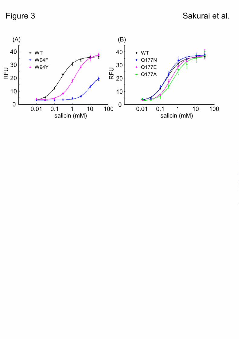
In model A, Trp94 predicted that salicin had aromatic-aromatic interactions only. In model B, Trp94 seemed to be involved in not only CH-π interactions but also in hydrogen bonding with the sugar moiety of salicin. To examine its interaction with salicin, Trp94 was mutated to a Phe (W94F) or Tyr (W94Y) residue. If salicin binding is mediated as in model A, the response of these mutants to salicin would not change, because both Phe and Tyr residues have aromatic rings to make hydrophobic interactions. However, if salicin binds as in model B, the response of W94F to salicin would be much more reduced drastically than W94Y mutants, because Phe residue has no hydroxyl group to make a hydrogen-bonding interaction with salicin.
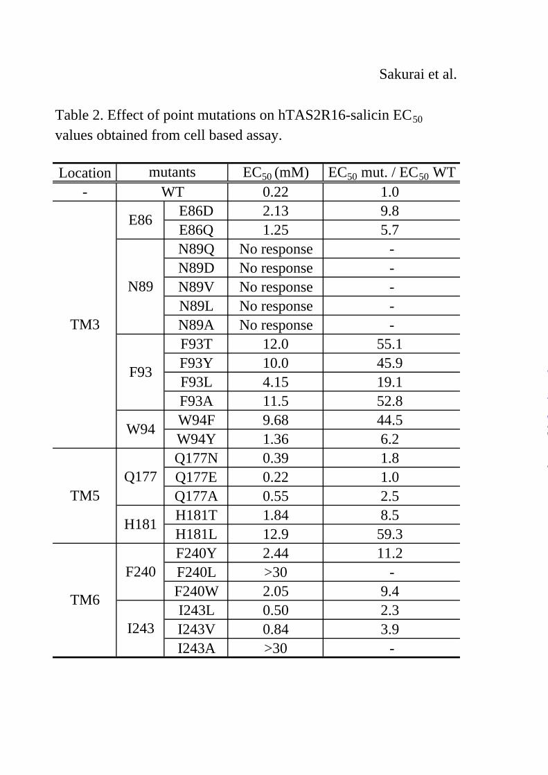
When these mutants were applied to the assay, the W94Y mutant resulted in a relatively small difference in the EC50 value to salicin (1.36 mM) compared with wild-type (WT) ssr3-hTAS2R16-salicin (0.22 mM), whereas W94F had a 40-fold higher EC50 value (9.68 mM) than the WT (Fig. 3A, Table 2). This result suggests that Trp94 interacts with salicin not only through hydrophobic interactions such as aromatic-aromatic or CH-π interactions, but also through hydrogen bonds.
Next, we observed the role of Gln177, which was predicted to form a hydrogen bond with the sugar moiety in model A, but not to form any hydrogen bonds with salicin in model B (Fig. 2). None of the Gln177 mutants tested (Q177N, Q177E, and Q177A) significantly affected the activity of salicin (Fig. 3B, Table 2). Thus, Gln177
is not predicted to be an important residue for interaction with salicin.
Based on the functional analysis of Trp94 and Gln177 mutants above, the binding mode for salicin in hTAS2R16 of model B is preferred over that of model A (Fig. 2B).
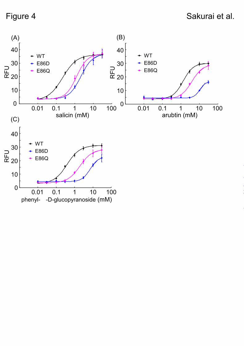
Functional analysis of Glu86 mutants with various β-glucopyranosides. – Next, we performed a functional analysis of the Glu86 mutants, E86D and E86Q using arbutin and phenyl-β-D-glucopyranoside as the ligands, to verify whether or not hTAS2R16-salicin binding mode accords with model B.
In model B, Glu86 was expected to interact with hydroxymethyl group on the aromatic ring of salicin. On the other hand, arbutin and phenyl-β-D-glucopyranoside could not interact with Glu86, because the para-hydroxy group on the phenyl group of arbutin would be too far from it, and phenyl-β-D-glucopyranoside has no hydroxy group on its aromatic ring (Fig. 1). From these reason, only salicin can form a hydrogen bond with Glu86 to facilitate the binding. In model A, Glu86 was predicted to interact with sugar moiety not hydroxy group on the aromatic ring, so there are no specific interactions between Glu86 and these phenyl-β-D-glucopyranosides to facilitate the binding.
E86D mutant was considered as feasible mutant to clarify the putative interaction, because Asp86 of E86D mutant was predicted to maintain hydrogen-bonding distance from the hydroxymethyl group on the aromatic ring of salicin. E86Q was designed to examine the effect of length of the side chain of Glu86.
The mutants did exhibit reduced responses to
5
by guest on September 1, 2018
http://ww
w.jbc.org/
Dow
nloaded from
salicin, and had similar EC50 values (E86D: 2.13 mM; E86Q: 1.25 mM) compared with the WT (0.22 mM) (Fig. 4A, Table 2). On the other hand, E86D had a large reduction in its response to arbutin and phenyl-β-D-glucopyranoside (arbutin, 10.6 mM; phenyl-β-D-glucopyranoside, 7.09 mM), whereas E86Q had a response similar to that of the WT (E86Q: arbutin, 3.89 mM, phenyl-β-D-glucopyranoside, 1.96 mM; WT: arbutin, 1.34 mM, phenyl-β-D-glucopyranoside, 0.38 mM) (Figs. 4B & C and supplementary Tables 1 & 2).
Our data obtained from the Glu86 mutant analysis suggested that the length of the side chain of the Glu86 amino acid residue is important for ligand recognition. In addition, only salicin forms a hydrogen bond with Glu86 to facilitate the binding. These results are also consistent with our putative model B.
Verification of the hTAS2R16-β-glucopyranoside binding mode. – Based on the functional analysis of Glu86, Trp94, and Gln177 mutants described above, model B was supported as the salicin-hTAS2R16 binding model (Fig. 2B). To further verify this model, we performed additional functional analyses using other amino acid residues expected to be involved in ligand recognition.
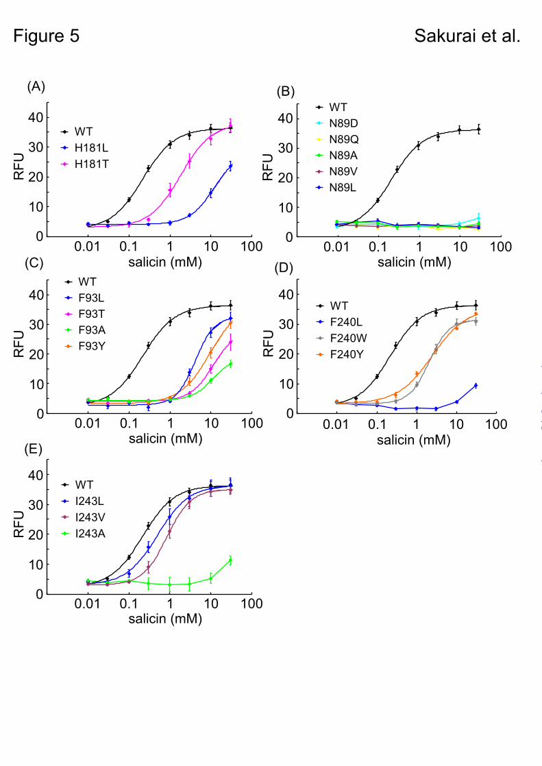
In model B, Asn89 and His181 formed hydrogen bonds with the sugar moiety, and were considered important for ligand recognition (Fig. 2B). To examine the putative hydrogen bond between His181 and the sugar moiety of salicin, we constructed two mutants, H181L and H181T. The H181T mutant responded to salicin with a small increase in EC50 value (1.84 mM) compared
with that of WT ssr3-hTAS2R16 (0.22 mM), whereas H181L had a 50-fold higher EC50 value (12.9 mM) than the WT (Fig. 5A, Table 2). These results suggest that His181 forms a hydrogen bond with salicin. Unfortunately, all of the mutations tested for Asn89 completely lost the ability to respond to salicin (Fig. 5B), and therefore it is unclear whether Asn89 is involved in ligand recognition.
Three other hydrophobic amino acid residues, Phe93, Phe240, and Ile243, were predicted to form the salicin binding site through hydrophobic interactions (Fig. 2B). All of the tested Phe93 mutants had a greatly reduced response to salicin (F93T: 12.0 mM, F93Y: 10.0 mM, F93L: 4.15 mM, F93A: 11.5 mM) compared with the WT (Fig. 5C, Table 2). We examined three Phe240 mutants, F240L, F240W, and F240Y. The F240L mutant had a substantially reduced response to salicin (F240L: > 30 mM) in comparison with the F240Y and F240W mutants (F240Y: 2.44 mM, F240W: 2.05 mM) (Fig. 5D, Table 2). Finally, the I243A mutant, with a change in the size of the amino acid residue, had a greatly reduced response to salicin (I243A: > 30 mM) in comparison with the I243L and I243V mutants (I243L: 0.50 mM, I243V:0.84 mM) (Fig. 5E, Table 2). These results indicate that at least these three amino acid residues play important roles in forming the salicin-binding pocket.
DISCUSSION
Humans recognize thousands of compounds as bitter, and response to bitterness is mediated by the hTAS2R family, which are GPCRs comprised of
6
by guest on September 1, 2018
http://ww
w.jbc.org/
Dow
nloaded from
only 25 receptors. Heterologous expression experiments have identified many of their cognate bitter ligands (29), and receptor-based assays have been developed as novel in vitro approaches for taste assessment (30). In principle, this method could be used to screen taste modulators such as bitter blockers that act on taste receptors. Using such assays, we found that Glu-Glu significantly reduced the response of several hTAS2Rs to these ligands, suggesting that acidic pH may be one of the critical factors responsible for the previously reported bitter-masking effect of Glu-Glu (31,32). Information on the ligand-binding structure of taste receptors with tastants is essential for rational design of taste modulators such as bitter-blockers. However, most GPCRs have not been crystallized, and to date only five independent X-ray crystal structures have been determined (33-38). In silico computational methods employing receptor-based modeling are useful tools for predicting ligand-receptor complex structures. However, because virtual docking simulations using homology-based structural models still have low predictive power, the accuracy of this approach is improved by combining it with functional studies using mutants of the receptor.
In this study, we experimentally identified the putative β-glucopyranoside-binding site in hTAS2R16 predicted from computational molecular modeling using functional analysis. Because salicin is an agonist of hTAS2R16, the structural model should be constructed from its activated form. Although crystal structures of GPCRs such as adrenergic receptors and adenosine receptor have been reported, all the structures are of inactive form (33-38). Thus, we
selected the previously reported, active form 3D structural model of a rhodopsin photointermediate, metarhodopsin (26), as a template.
The results of the docking model suggested two binding modes as candidates for the complex structure of salicin-hTAS2R16 (Fig. 2), and certain amino acid residues were deduced to be directly involved in salicin recognition (Table 1). Based on the two models, Glu86, Trp94, and Gln177 were selected as key amino acid residues for clarifying the salicin-hTAS2R16 complex structure (Fig. 2). In our functional analysis of these three mutants, interactions between Glu86 and the hydroxy methyl group on the aromatic ring, and between Trp94 and the sugar moiety of salicin were experimentally confirmed, with little interaction observed between Gln177 and salicin (Figs. 3A & 4). These results suggest that the salicin-hTAS2R16 complex structure is that of model B rather than model A (Fig. 6). Our functional analysis was able to experimentally confirm the salicin-hTAS2R16 binding mode, which could not be definitively determined based on the molecular modeling study alone.
We further identified at least five additional amino acid residues involved in salicin recognition through functional analysis using a series of hTAS2R16 point mutants. In model B, Asn89 forms a hydrogen bond with the sugar hydroxy group of salicin (Fig. 2B). Although all the Asn89 mutants tested abolished the Ca2+ response to salicin and other β-D-glucopyranosides (Fig. 5B, Supplementary Figs. 1 & 2A), from the modeling, Asn89 may be a critical amino acid residue for ligand recognition though the hydrogen bonds with the sugar hydroxyl group of salicin (Fig. 6).
7
by guest on September 1, 2018
http://ww
w.jbc.org/
Dow
nloaded from
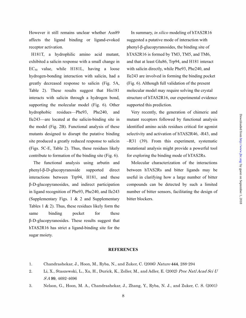
In summary, in silico modeling of hTAS2R16 suggested a putative mode of interaction with phenyl-β-glucopyranosides, the binding site of hTAS2R16 is formed by TM3, TM5, and TM6, and that at least Glu86, Trp94, and H181 interact with salicin directly, while Phe93, Phe240, and Ile243 are involved in forming the binding pocket (Fig. 6). Although full validation of the present molecular model may require solving the crystal structure of hTAS2R16, our experimental evidence supported this prediction.
However it still remains unclear whether Asn89 affects the ligand binding or ligand-evoked receptor activation. H181T, a hydrophilic amino acid mutant, exhibited a salicin response with a small change in EC50 value, while H181L, having a loose hydrogen-bonding interaction with salicin, had a greatly decreased response to salicin (Fig. 5A, Table 2). These results suggest that His181 interacts with salicin through a hydrogen bond, supporting the molecular model (Fig. 6). Other hydrophobic residues—Phe93, Phe240, and Ile243—are located at the salicin-binding site in the model (Fig. 2B). Functional analysis of these mutants designed to disrupt the putative binding site produced a greatly reduced response to salicin (Figs. 5C-E, Table 2). Thus, these residues likely contribute to formation of the binding site (Fig. 6).
Very recently, the generation of chimeric and mutant receptors followed by functional analysis identified amino acids residues critical for agonist selectivity and activation of hTAS2R46, -R43, and –R31 (39). From this experiment, systematic mutational analysis might provide a powerful tool for exploring the binding mode of hTAS2Rs.
The functional analysis using arbutin and phenyl-β-D-glucopyranoside supported direct interactions between Trp94, H181, and these β-D-glucopyranosides, and indirect participation in ligand recognition of Phe93, Phe240, and Ile243 (Supplementary Figs. 1 & 2 and Supplementary Tables 1 & 2). Thus, these residues likely form the same binding pocket for these β-D-glucopyranosides. These results suggest that hTAS2R16 has strict a ligand-binding site for the sugar moiety.
Molecular characterization of the interactions between hTAS2Rs and bitter ligands may be useful in clarifying how a large number of bitter compounds can be detected by such a limited number of bitter sensors, facilitating the design of bitter blockers.
REFERENCES
1. Chandrashekar, J., Hoon, M., Ryba, N., and Zuker, C. (2006) Nature 444, 288-294
2. Li, X., Staszewski, L., Xu, H., Durick, K., Zoller, M., and Adler, E. (2002) Proc Natl Acad Sci U S A 99, 4692-4696
3. Nelson, G., Hoon, M. A., Chandrashekar, J., Zhang, Y., Ryba, N. J., and Zuker, C. S. (2001)
8
by guest on September 1, 2018
http://ww
w.jbc.org/
Dow
nloaded from
Cell 106, 381-390
4. Nelson, G., Chandrashekar, J., Hoon, M., Feng, L., Zhao, G., Ryba, N., and Zuker, C. (2002)
Nature 416, 199-202
5. Maehashi, K., Matano, M., Wang, H., Vo, L. A., Yamamoto, Y., and Huang, L. (2008) Biochem Biophys Res Commun 365, 851-855
6. Behrens, M., Brockhoff, A., Kuhn, C., Bufe, B., Winnig, M., and Meyerhof, W. (2004) Biochem. Biophys. Res. Commun. 319, 479-485
7. Bufe, B., Hofmann, T., Krautwurst, D., Raguse, J., and Meyerhof, W. (2002) Nat. Genet. 32,
397-401
8. Kuhn, C., Bufe, B., Winnig, M., Hofmann, T., Frank, O., Behrens, M., Lewtschenko, T., Slack,
J., Ward, C., and Meyerhof, W. (2004) J. Neurosci. 24, 10260-10265
9. Pronin, A., Xu, H., Tang, H., Zhang, L., Li, Q., and Li, X. (2007) Curr. Biol. 17, 1403-1408
10. Brockhoff, A., Behrens, M., Massarotti, A., Appendino, G., and Meyerhof, W. (2007) J. Agric. Food Chem. 55, 6236-6243
11. Pronin, A. N., Tang, H., Connor, J., and Keung, W. (2004) Chem. Senses 29, 583-593
12. Sainz, E., Cavenagh, M. M., Gutierrez, J., Battey, J. F., Northup, J. K., and Sullivan, S. L.
(2007) Biochem. J. 403, 537-543
13. Bufe, B., Breslin, P. A., Kuhn, C., Reed, D. R., Tharp, C. D., Slack, J. P., Kim, U. K., Drayna,
D., and Meyerhof, W. (2005) Curr. Biol. 15, 322-327
14. Behrens, M., Brockhoff, A., Batram, C., Kuhn, C., Appendino, G., and Meyerhof, W. (2009) J Agric Food Chem 57, 9860-9866
15. Miguet, L., Zhang, Z., and Grigorov, M. G. (2006) J Recept Signal Transduct Res 26, 611-630
16. Floriano, W. B., Hall, S., Vaidehi, N., Kim, U., Drayna, D., and Goddard, W. A., 3rd. (2006) J Mol Model 12, 931-941
17. Jiang, P., Ji, Q., Liu, Z., Snyder, L. A., Benard, L. M., Margolskee, R. F., and Max, M. (2004) J Biol Chem 279, 45068-45075
18. Xu, H., Staszewski, L., Tang, H., Adler, E., Zoller, M., and Li, X. (2004) Proc Natl Acad Sci U S A 101, 14258-14263
19. Nakajima, K., Asakura, T., Oike, H., Morita, Y., Shimizu-Ibuka, A., Misaka, T., Sorimachi, H.,
Arai, S., and Abe, K. (2006) Neuroreport 17, 1241-1244
20. Koizumi, A., Nakajima, K., Asakura, T., Morita, Y., Ito, K., Shmizu-Ibuka, A., Misaka, T., and
Abe, K. (2007) Biochem Biophys Res Commun 358, 585-589
21. Shirasuka, Y., Nakajima, K., Asakura, T., Yamashita, H., Yamamoto, A., Hata, S., Nagata, S.,
Abo, M., Sorimachi, H., and Abe, K. (2004) Biosci Biotechnol Biochem 68, 1403-1407
22. Okubo, S., Asakura, T., Okubo, K., Abe, K., Misaka, T., and Akita, T. (2008) J Plant Physiol 165, 1964-1969
9
by guest on September 1, 2018
http://ww
w.jbc.org/
Dow
nloaded from
23. Winnig, M., Bufe, B., Kratochwil, N. A., Slack, J. P., and Meyerhof, W. (2007) BMC Struct Biol 7, 66
24. Zhang, F., Klebansky, B., Fine, R. M., Xu, H., Pronin, A., Liu, H., Tachdjian, C., and Li, X.
(2008) Proc Natl Acad Sci U S A 105, 20930-20934
25. White, S. H., and Wimley, W. C. (1999) Annu Rev Biophys Biomol Struct 28, 319-365
26. Ishiguro, M., Oyama, Y., and Hirano, T. (2004) Chembiochem 5, 298-310
27. Ammon, C., Schafer, J., Kreuzer, O. J., and Meyerhof, W. (2002) Arch Physiol Biochem 110,
137-145
28. Ueda, T., Ugawa, S., Yamamura, H., Imaizumi, Y., and Shimada, S. (2003) J. Neurosci. 23,
7376-7380
29. Meyerhof, W., Batram, C., Kuhn, C., Brockhoff, A., Chudoba, E., Bufe, B., Appendino, G., and
Behrens, M. (2010) Chem Senses 35, 157-170
30. Ruiz-Avila, L., Ming, D., and Margolskee, R. (2000) Chem. Senses 25, 361-368
31. Noguchi, M., Yamashita, M., Arai, S., and Fujimaki, M. (1975) J. Food Sci. 40, 367-369
32. Sakurai, T., Misaka, T., Nagai, T., Ishimaru, Y., Matsuo, S., Asakura, T., and Abe, K. (2009) J Agric Food Chem 57, 2508-2514
33. Palczewski, K., Kumasaka, T., Hori, T., Behnke, C. A., Motoshima, H., Fox, B. A., Le Trong, I.,
Teller, D. C., Okada, T., Stenkamp, R. E., Yamamoto, M., and Miyano, M. (2000) Science 289,
739-745
34. Murakami, M., and Kouyama, T. (2008) Nature 453, 363-367
35. Rasmussen, S. G., Choi, H. J., Rosenbaum, D. M., Kobilka, T. S., Thian, F. S., Edwards, P. C.,
Burghammer, M., Ratnala, V. R., Sanishvili, R., Fischetti, R. F., Schertler, G. F., Weis, W. I.,
and Kobilka, B. K. (2007) Nature 450, 383-387
36. Jaakola, V. P., Griffith, M. T., Hanson, M. A., Cherezov, V., Chien, E. Y., Lane, J. R., Ijzerman,
A. P., and Stevens, R. C. (2008) Science 322, 1211-1217
37. Warne, T., Serrano-Vega, M. J., Baker, J. G., Moukhametzianov, R., Edwards, P. C.,
Henderson, R., Leslie, A. G., Tate, C. G., and Schertler, G. F. (2008) Nature 454, 486-491
38. Shimamura, T., Hiraki, K., Takahashi, N., Hori, T., Ago, H., Masuda, K., Takio, K., Ishiguro,
M., and Miyano, M. (2008) J Biol Chem 283, 17753-17756
39. Brockhoff, A., Behrens, M., Niv, M. Y., and Meyerhof, W. (2010) Proc Natl Acad Sci U S A 107,
11110-11115
FOOTNOTES
This study was performed with a grant from the Research and Development Program for New Bio-industry Initiatives of Bio-oriented Technology Research Advancement Institution (BRAIN). This
10
by guest on September 1, 2018
http://ww
w.jbc.org/
Dow
nloaded from
work was also supported by a Grant-in-Aid for Scientific Research from the Ministry of Education, Culture, Sports, Science and Technology of Japan (19880008, and 20780089 to Y.I., and 18688005, 20688015, and 21658046 to T.M., 19300248 to T.A., and 20380183 to K.A.).
#: Present address: Department of Food and Nutritional Sciences, Graduate School of Nutritional and Environmental Sciences, University of Shizuoka, Shizuoka 422-8526, Japan
The abbreviations used are: GMP, guanosine-5’-monophosphate; GPCR, G protein-coupled receptor; HEK293T, human embryonic kidney 293T; IMP, inosine-5’-monophosphate; PCR, polymerase chain reaction; PROP, 6-propyl-2-thiouracil; PTC, N-phenylthiourea; TM, transmembrane; WT, wild type.
FIGURE LEGENDS
Fig. 1. The molecular structures of salicin (A), arbutin (B), and phenyl-β-D-glucopyranoside (C). Numbers denote the positions of carbon atoms.
Fig. 2. Two putative salicin-hTAS2R16 binding structures and a detailed map of the salicin-hTAS2R16 interaction. Numbered amino acid positions were predicted to be involved in salicin recognition. A. Side view of one proposed salicin-hTAS2R16 binding model (model A). Salicin is bound within the binding pocket with the aromatic ring of salicin oriented toward the cytoplasm. B. Side view of another proposed salicin-hTAS2R16 model (model B). Salicin is bound within the binding pocket with the aromatic ring of salicin oriented toward extracellular space.
Fig. 3. Point mutation analyses of the responses to salicin of key amino acid residues. The dose-response curves show the results of the analyses for W94 mutants (A) and Q177 mutants (B). The dose-response relationship to salicin was obtained using a cell-based assay of HEK293T cells cotransfected with ssr3-hTAS2R16 or ssr3-hTAS2R16 mutants along with Gα16gust44. Each point represents the mean ± SE from at least 3 independent measurements.
Fig. 4. Dose-response relationships for various phenyl-β-D-glucopyranosides obtained using a cell-based assay of HEK293T cells cotransfected with ssr3-hTAS2R16 or ssr3-hTAS2R16 Glu86 mutants along with Gα16gust44. Each point represents the mean ± SE from at least 3 independent measurements. Dose-response curves are shown for salicin (A), arbutin (B), and phenyl-β-D-glucopyranoside (C).
Fig. 5. Dose-response relationships for various hTAS2R16 mutants to salicin obtained using a cell-based assay of HEK293T cells cotransfected with ssr3-hTAS2R16 or ssr3-hTAS2R16 mutants along with
11
by guest on September 1, 2018
http://ww
w.jbc.org/
Dow
nloaded from
Gα16gust44. Each point represents the mean ± SE from at least 3 independent measurements. Dose-response curves are shown for His181 mutants (A), Asn89 mutants (B), Phe93 mutants (C), Phe240 mutants (D), and Ile243 mutants (E).
Fig. 6. Predicted salicin-binding structure of hTAS2R16. A. Side view of the predicted salicin-binding model of hTAS2R16. B. Close-up of the top view of the predicted salicin-binding model of hTAS2R16. The indicated amino acid positions are predicted to influence receptor activation by salicin. Hydrogen bonds are indicated by dashed lines.
12
by guest on September 1, 2018
http://ww
w.jbc.org/
Dow
nloaded from
Sakurai et al.
Table 1. Predicted amino acid residues involved in salicin recognition.
Location ResiduesTM3 Glu86 Asn89 Phe93 Trp94TM5 Gln177 His181TM6 Phe240 Ile243
by guest on September 1, 2018
http://ww
w.jbc.org/
Dow
nloaded from
Sakurai et al.
Table 2. Effect of point mutations on hTAS2R16-salicin EC50
values obtained from cell based assay.
Location mutants EC50 (mM) EC50 mut. / EC50 WT- WT 0.22 1.0
TM3
E86 E86D 2.13 9.8E86Q 1.25 5.7
N89
N89Q No response -N89D No response -N89V No response -N89L No response -N89A No response -
F93
F93T 12.0 55.1F93Y 10.0 45.9F93L 4.15 19.1F93A 11.5 52.8
W94 W94F 9.68 44.5W94Y 1.36 6.2
TM5Q177
Q177N 0.39 1.8Q177E 0.22 1.0Q177A 0.55 2.5
H181 H181T 1.84 8.5H181L 12.9 59.3
TM6
F240F240Y 2.44 11.2F240L >30 -F240W 2.05 9.4
I243I243L 0.50 2.3I243V 0.84 3.9I243A >30 -
by guest on September 1, 2018
http://ww
w.jbc.org/
Dow
nloaded from
O
OH
OH
OH
OH
O
OH
salicin
12
3
4
56
1'
2'
3'
4'
5'6' O
OH
OH
OH
OH
O
12
3
4
56
1'
2'
3'
4'
5'6'
phenyl - β -D-glucopyranoside
OH
O
OH
OH
OH
OH
O
arbutin
12
3
4
56
1'
2'
3'4'
5'6'
(A) (B) (C)
Figure 1 Sakurai et al.
by guest on September 1, 2018
http://ww
w.jbc.org/
Dow
nloaded from
TM3
TM6 TM5 TM4
Gln177
Glu86His181Trp94
Ile243
Phe93Phe240
Asn89
(A) (B)
TM3
TM6 TM5 TM4
Gln177
Glu86His181 Trp94
Ile243
Phe93Phe240
Asn89
Figure 2 Sakurai et al.
by guest on September 1, 2018
http://ww
w.jbc.org/
Dow
nloaded from
WTW94FW94Y
0
40
10
20
30
ΔR
FU
(A)
0.01 0.1 1 10 100salicin (mM) salicin (mM)
ΔR
FU
WTQ177NQ177EQ177A
(B)
0
40
10
20
30
0.01 0.1 1 10 100
Figure 3 Sakurai et al.
by guest on September 1, 2018
http://ww
w.jbc.org/
Dow
nloaded from
WTE86DE86Q
(A)
arubtin (mM)
WTE86DE86Q
(B)
0
40
10
20
30
ΔR
FU
0.01 0.1 1 10 100salicin (mM)
0
40
10
20
30
ΔR
FU0.01 0.1 1 10 100
phenyl-β-D-glucopyranoside (mM)
WTE86DE86Q
(C)
0
40
10
20
30
ΔR
FU
0.01 0.1 1 10 100
Figure 4 Sakurai et al.
by guest on September 1, 2018
http://ww
w.jbc.org/
Dow
nloaded from
(A)
(D)
WTF240LF240WF240Y
0
40
10
20
30
ΔR
FU
0.01 0.1 1 10 100salicin (mM)
WTI243LI243VI243A
0
40
10
20
30
0.01 0.1 1 10 100salicin (mM)
ΔR
FU
(E)
WTH181LH181T
0
40
10
20
30
ΔR
FU
0.01 0.1 1 10 100salicin (mM)
Figure 5 Sakurai et al.
(C)WTF93LF93TF93AF93Y
0
40
10
20
30
ΔR
FU
0.01 0.1 1 10 100salicin (mM)
WTN89DN89QN89AN89VN89L
1000
40
10
20
30
ΔR
FU0.01 0.1 1 10
salicin (mM)
(B)
by guest on September 1, 2018
http://ww
w.jbc.org/
Dow
nloaded from

TM3TM6
TM5TM4
Gln177
Glu86
His181Trp94
Ile243Phe93
Phe240
Asn89
TM7
(A)
(B)
Figure 6 Sakurai et al.
by guest on September 1, 2018
http://ww
w.jbc.org/
Dow
nloaded from
Keiko AbeKeisuke Ito, Takuya Kobayashi, Shinji Matsuo, Yoshiro Ishimaru, Tomiko Asakura and
Takanobu Sakurai, Takumi Misaka, Masaji Ishiguro, Katsuyoshi Masuda, Taishi Sugawara,receptor hTAS2R16
Characterization of the beta-D-glucopyranoside binding site of the human bitter taste
published online July 6, 2010J. Biol. Chem.
10.1074/jbc.M110.144444Access the most updated version of this article at doi:
Alerts:
When a correction for this article is posted•
When this article is cited•
to choose from all of JBC's e-mail alertsClick here
Supplemental material:
http://www.jbc.org/content/suppl/2010/07/06/M110.144444.DC1
by guest on September 1, 2018
http://ww
w.jbc.org/
Dow
nloaded from