Characterization of novel β1 integrin interactors involved ... Thesis Final - João...the openness,...
102
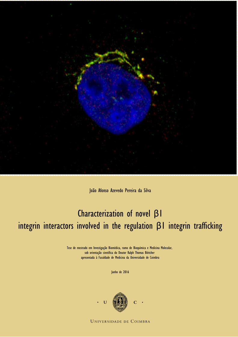
Imagem João Afonso Azevedo Pereira da Silva Characterization of novel β1 integrin interactors involved in the regulation β1 integrin trafficking Tese de mestrado em Investigação Biomédica, ramo de Bioquímica e Medicina Molecular, sob orientação científica do Doutor Ralph Thomas Böttcher apresentada à Faculdade de Medicina da Universidade de Coimbra Junho de 2016
Transcript of Characterization of novel β1 integrin interactors involved ... Thesis Final - João...the openness,...
Imagem
João Afonso Azevedo Pereira da Silva
Characterization of novel β1 integrin interactors involved in the regulation β1 integrin trafficking
Tese de mestrado em Investigação Biomédica, ramo de Bioquímica e Medicina Molecular,
sob orientação científica do Doutor Ralph Thomas Böttcher
apresentada à Faculdade de Medicina da Universidade de Coimbra
Junho de 2016
Imagem

Characterization of novel β1
integrin interactors involved in the regulation β1 integrin
trafficking
João Afonso Azevedo Pereira da Silva
Dissertação apresentada à Faculdade de Medicina da Universidade de Coimbra para cumprimento dos
requisitos necessários à obtenção do grau de Mestre em Investigação Biomédica. Trabalho realizado
no Departamento de Medicina Molecular do Instituto Max Planck de Bioquímica em Munique, sob a
orientação científica do Dr. Ralph Thomas Böttcher
Universidade de Coimbra
2016

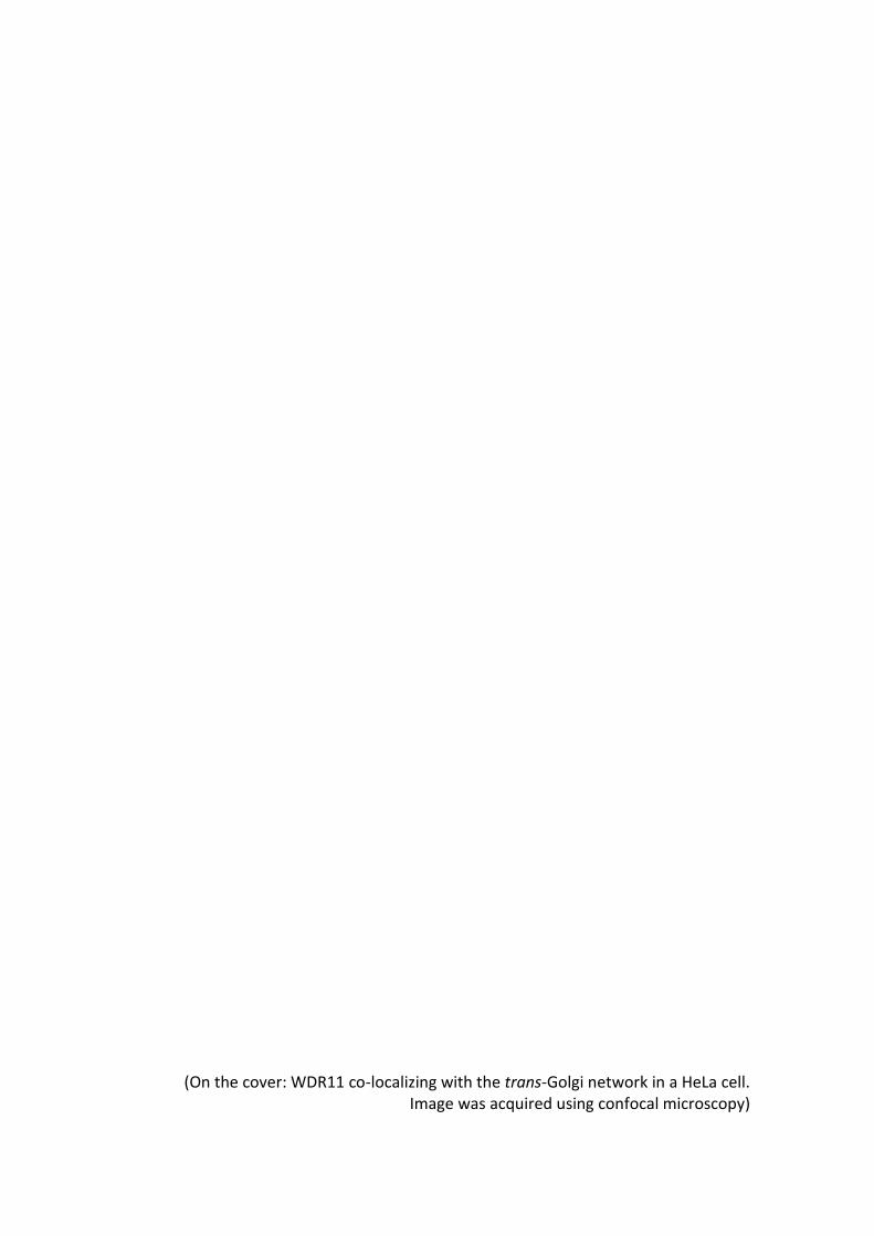
(On the cover: WDR11 co-localizing with the trans-Golgi network in a HeLa cell. Image was acquired using confocal microscopy)


“There's real poetry in the real world. Science is the poetry of reality”
Richard Dawkins

VII
Agradecimentos
Ao Ralph Böttcher por me ter proporcionado a oportunidade de desenvolver este
projecto de tese como membro do seu grupo de investigação, e na qualidade de
supervisor, agradecer por toda a experiência e conhecimento científico transmitida ao
longo deste ano e por promover em mim um espirito critico, de clareza e de organização
em todo o processo de “fazer ciência”. Quero também deixar um sincero agradecimento
ao Professor Reinhard Fässler por me ter dado a oportunidade de trabalhar no
Departamento de Medicina Molecular e no seu laboratório, assim como de poder fazer
parte do Instituto Max Planck de Bioquímica, sem o qual nada disto teria sido possível.
Um agradecimento muito especial à adorável Valeria Samarelli. Obrigado por
toda a abertura, disponibilide, simpatia e amabilidade com que desde o primeiro dia me
recebeste, e com que sempre me ouviste e prontamente esclareceste todas as minhas
dúvidas e perguntas, mesmo as às vezes um pouco menos pertinentes. Por todas as
pequenas coisas que fizeste e que me ajudaram fazer passar este primeiro ano fora de
casa e longe meu país de uma forma mais positiva e optimista. Ao Tilman Ziegler por
todo o input, paciência e conhecimento que me transmitiu ao longo do ano e por todos
os momentos mais relaxados e divertidos dentro e fora do lab. Agradecer também à
Mischa, à Sarah Schölze e ao Tom por toda a ajuda e orientação que sempre me deram
no lab. Não podia faltar o agradecimento à Marina, à Georgina, à Maria e à Lidia por
toda a diversão, ajuda e motivação para cada dia no laboratório. Aos meus colegas
portugueses, um muito obrigado à Ana Keating pela ajuda com a tese, pelos copos e
pelas pausas, ao Pedro Melo, e em especial também à Irene Ferreira por toda a
paciência, disponibilidade e abertura para me ajudar de forma indispensável a preparar
e guiar me para o meu futuro e por mostrar que existe mais desafios fora do lab. E claro,
agradecer a todos os outros colegas do Dept. de Medicina Molecular, em particular do
ao 1º andar por serem meus colegas de trabalho e nunca me negarem auxilio sempre
que precisei.
VIII
Ao Manuel Conde pela amizade criada entre 2 portugueses acabados de chegar
a Munique, pelas viagens, jantares, noites & co. e tudo o resto. E que no fundo foram
essenciais para que o ano de Erasmus fosse mais descomprimido e bem passado.
Aos meus colegas do MIB 2014, que apesar de a distancia nos ter separado este
ano, tiveram um papel preponderante no ano que viria a dar a este no qual espero, com
esta tese, terminar mais outro ciclo de estudos. Por todos os convívios, jantares, saídas,
trabalhos e ajuda partilhados no primeiro ano.
Por fim, mas de certeza não menos importante, agradecimento à minha família,
em especial aos meus pais, por todos os enormes sacrifícios dia após dia que permitiram
estar onde estou hoje e chegar aonde cheguei. Pela a constante preocupação e
dedicação em proporcionar-me uma vida com o melhor que têm para dar. Por me
ajudarem nos momentos mais difíceis não só neste ano de distância, mas ao longo de
toda a minha vida e fazerem de mim a pessoa que sou hoje. Um eterno obrigado!
IX
Acknowledgements
First and foremost, I want to thank Ralph Böttcher for providing this opportunity
to develop this thesis project as a member of his investigation group and as a supervisor,
thank all the experience and scientific knowledge transmitted during this year and for
promoting in me a more critical thinking and organization attitude in all the process of
“making science”. I also want to express my sincere gratitude to Professor Reinhard
Fässler for allowing me the chance to work in the Molecular Medicine Dept. and his lab,
as well as being able to be part of the Max Planck Institute for Biochemistry, without
whom none of this would have been possible.
A very much special thank you to the lovely Valeria Samarelli. Thank you for all
the openness, availability, kindness and heartfelt sympathy with which you received me
from the first day, and how with all of that you always readily listened to my questions
and doubts, sometimes even when they were less convenient and thought through. For
all the things you made that helped me carry on through this first year out of home and
far away from my country in a more optimistic and positive way. To Tilman Ziegler for
all the input, patience and knowledge he provided me during this year, and for all the
funny and relaxed moments we enjoyed in and outside of the lab. Also want to thank
Mischa, Sara Schölze and Tom for all the help and guidance always given in the lab. I
could not forget to thank Marina, Georgina, Maria and Lidia for all the amusement, help
and motivation for each day that went through in the lab.
To my Portuguese colleagues, a big thank you to Ana Keating for all the help with
the thesis, the drinks and the lab breaks, to Pedro Melo, and specially to Irene Ferreira
for all the patience, availability and readiness in helping me, for all the guidance and
support in preparing my future steps and for showing me there are more challenges
besides the ones in the lab. And of course, thank all my Molecular Medicine Dept.
colleagues, in particular the 1st floor por being my work colleagues and always help me
when I needed.

X
Many thanks to Manuel Conde for the friendship created between 2 Portuguese
just arrived in München, for the trips, dinners, nights out & co and everything else.
Which in the end were indispensable for this Erasmus year to ran smoothly and chilled
out.
To my MIB 2014 colleagues, despite distance separated us this year, still had a
remarkable role in the year before this one, in which with thesis, I hope to finish another
degree. Thank you for all the get togethers, dinners, night outs, works and help shared
in that first year.
Last, but definitely not the least, thank my family, specially my parents, for all the
sacrifices day after day that allowed me to be where I am today and accomplish what
I’ve accomplished. For the constant concerning and dedication in providing me a life
with the best they have to give. For helping me in all the hard moments not only in this
year apart, but through all my entire life and for making me the person I am today. An
everlasting Thank You!
XI
Table of Contents
List of figures…………………………………………………………………………………………………………………….. XIII
Abstract………………………………………………………………………………………………………………………..….. XVII
Resumo…………………………………………………………………………………………….……………………….……… XIX
INTRODUCTION ....................................................................................................................... 1
1. Integrin Overview ................................................................................................................ 3
1.1. Integrin Structure ....................................................................................................... 3
1.1.1. αβ Subunits ........................................................................................................... 4
1.1.2. Intracellular Domains - Cytoplasmic tails .............................................................. 5
1.1.3. Transmembrane Domains ..................................................................................... 5
1.1.4. Extracellular Domains ........................................................................................... 5
1.2. Integrin Signaling and Function .................................................................................. 5
1.3. α5β1 Integrin and Cell Migration ................................................................................ 6
2. Integrin Trafficking .............................................................................................................. 7
2.1. Canonical Trafficking .................................................................................................. 7
2.1.1. Clathrin-dependent endocytosis of integrins ........................................................ 7
2.1.2. Clathrin-independent endocytosis of integrins ..................................................... 8
2.1.3. Recycling: Short-loop pathway ............................................................................. 8
2.1.4. Recycling: Long-loop pathway .............................................................................. 9
3. Golgi-mediated Trafficking .................................................................................................. 9
3.1. Retrograde Trafficking and the trans-Golgi network ................................................ 10
3.1.1. Early endosome-to-TGN pathway ....................................................................... 11
3.1.2. Late endosome-to-TGN pathway ........................................................................ 12
3.1.3. Recycling endosome-to-TGN pathway ................................................................ 12
3.2. Retrograde Trafficking and Integrins ........................................................................ 13
3.3. Index of Trafficking mediating complexes ................................................................ 14
3.3.1. Retromer ............................................................................................................ 14
3.3.2. Rab GTPases Family ........................................................................................... 14
3.3.3. SNAREs ................................................................................................................ 15
3.3.4. GARP complex ..................................................................................................... 15
3.3.5. EARP complex ..................................................................................................... 15
3.3.6. Sorting Nexins ..................................................................................................... 16
4. Autophagy ........................................................................................................................ 16
XII
Aims ……………………………………….………………………………………………………..……………….……………. 19
Results …………………………………….……………………………………………………………………………….…… 23
1. Confirmation of a putative Golgi-associated trimeric protein complex of F.am91a1, WDR11
and C17orf75 ……………………………………………………………………………………………………….….…… 25
2. Fam91a1 and WDR11 KDs impaired retrograde trafficking of CI-M6Pr and TGN46….….. 29
3. Protein synthesis inhibition by cycloheximide shows increased degradation of TGN46 in
Fam91a1 and WDR11 KD HeLa cells …………………………………………………………….….…..…..…… 32
4. Cellular migration and directionality are compromised as α5β1 Integrins are misrouted
from FAs at the plasma membrane ……………………………………………………….….…..…..….....…. 35
5. Proteomic Mass-Spectrometry screening analysis of knock-downs and Biotin pull-downs of
the trimeric complex proteins ……………………………………………………………………..…………...….. 42
Discussion .............................................................................................................................. 49
Concluding Remarks ............................................................................................................. 59
Materials & Methods ............................................................................................................ 63
Bibliography ........................................................................................................................... 71
XIII
List of figures
Figure A: Representation of the integrin family……………………………..…………………… 4
Figure B: Steps of vesicle formation, transport and attaching in Golgi-mediated
trafficking events ……………………………………………………………………………………………… 10
Figure 1: Fam91a1, WDR11 and C17orf75 form a trimeric complex ………..………. 26
Figure 2: Confirmation of the Fam91a1, WDR11, C17orf75 complex and its
interaction with a5 integrins by BioID ………………………………………………………………. 28
Figure 3: A-T and H-Q GFP-WDR11 variants localize to the Golgi …………………..…. 31
Figure 4: Impaired retrograde CI-MP6r trafficking in Fam91a1 and WDR11 knock-
down cells …………………………………………………………………………….………………………….. 33
Figure 5: Fam91a1 and WDR11 knock-down reduce TGN46 protein levels and
cause an altered intracellular TGN46 localization similar to interfering with the
GARP complex absence condition ……………………………………………..…………………….. 34
Figure 6: Cycloheximide chase Fam91a1 and WDR11 knock-down cells suggest
increased degradation kinetics of TGN46 …………..……………………………………………..36
Figure 7: siRNA-mediated depletion of GARP and EARP subunits interfere with β1
integrin trafficking and recycling back.……………………………………………………………....37
Figure 8: Fam91a1 and WDR11 knock-downs interfere with proper α5 integrin
trafficking ………………………………………………………………………………………….…………..….39
Figure 9: Fam91a1 and WDR11 depletion impairs cell migration ……………………...40
Figure 10: GFP-Fam91a1 re-expression in Fam91a1 depletion HeLa cells does not
fully rescue the shFam91a1 migration defect ……………………………………………..…… 41
Figure 11: Whole Proteome Mass-Spectrometry on control and Fam91a1 and
WDR11-depleted HeLa cell lines ……………………………………………………………….…….…43
Figure 12: Mass-Spectrometry analysis of the C17orf75 interactome by C17orf75-
BioID ………………………………………………………………………………….…………………….………..45
Figure 13: Mass-Spectrometry analysis of the WDR11 interactome by WDR11-
BioID …….……………………………………………………..………………………………….…………………46

XIV

XV
Abstract/Resumo

XVI
XVII
Abstract
Integrin signaling is involved in many aspects of cell function including adhesion,
migration through tissues and signaling. Integrin signaling occurs when integrins are
extended and ligand bound, and is mediated through interaction of signaling and
adaptor proteins to the integrin cytoplasmic tails. The quality of integrin signaling is also
influenced by their trafficking through the endosomal system and by their stability.
Integrin cytoplasmic domain binding proteins are key to integrin function as integrins do
not possess enzymatic activity.
In the previous years the Fässler lab has set up different proteomic approaches
to identify proteins that bind to the cytoplasmic domains of α5β1 integrin including pull-
down experiments using the intracellular domains of α5β1 integrin, proximity-based
biotinylation in living cells or adhesome isolation. Among the candidate interactors are
proteins that localize to intracellular organelles involved in intracellular trafficking of
transmembrane proteins such as the Golgi apparatus and different endosomal proteins.
There is increasing evidence that integrin trafficking through the endosomal pathway
and retrograde route profoundly affects their function, their ‘polarized’ distribution on
the cell surface, the signaling properties of integrin-associated growth factor receptors
and the turnover of ECM proteins such as fibronectin (FN).
The aim of this master thesis is to confirm the interaction of these candidate
cytosolic α5β1 integrin interactors, determine their subcellular localization in the cell
and analyze integrin-mediated processes such as cell adhesion, spreading and migration
in a loss-of-function situation for these proteins. We started by confirming the existence
of a trimeric complex between Fam91a1, WDR11 and C17orf75 at the Golgi which
stability is Fam91a1-dependent. This trimeric complex is involved in retrograde
trafficking as its depletion impairs transport of CI-M6Pr and TGN46 to the trans-Golgi
network (TGN), reduces CI-M6Pr levels like retromer-depleted cells and compromises
TGN integrity. Importantly, the Fam91a1/WDR11/C17orf75 complex is essential for
intracellular trafficking of α5β1 integrin and for effective cell migration. We confirmed
the involvement of GARP and EARP complexes in retrograde transport. Finally,
proteomic screenings revealed a potential association between
XVIII
Fam91a1/WDR11/C17orf75 complex and autophagy, endocytic signaling and pubertal
development.
Overall the results obtained in this project show that an α5β1 integrin interacting
complex composed of Fam91a1/WDR11/C17orf75 plays a role on TGN-mediated
retrograde trafficking of α5β1 integrin, cell migration and possibly mediate other major
cellular processes like autophagy.
Key words:
Integrins; trans-Golgi network; retrograde trafficking; cell migration; Fam91a1; WDR11;
C17orf75; endosomal vesicles;
XIX
Resumo
O processo de sinalização via integrinas está envolvido em muitos aspetos da função
celular incluindo adesão, migração através de tecidos e a referida sinalização. Esta
sinalização via integrinas ocorre quando possuem uma conformação estendida e estão
vinculadas/ligadas a um ligando e essa mesma sinalização é mediada por interações
entre as caudas citoplasmáticas das integrinas e proteínas sinalizadoras ou adaptadoras.
A eficiência da sinalização por integrinas depende também do seu tráfico através do
sistema endossomal e pela sua estabilidade. As proteínas que se ligam ao domínio
citoplasmático das integrinas são essenciais para a função das integrinas, pois as
integrinas não possuem qualquer tipo de actividade enzimática.
Nos últimos anos passados no laboratório do Dr. Fässler, foram estabelecidas
diferentes abordagens de proteómica de forma a identificar poder identificar proteínas
que se liguem aos domínios citoplasmáticos da integrina α5β1, estes incluem técnicas
de pull-down com base nos domínios intracelulares da integrina α5β1, como biotinilação
de proximidade em células vivas ou isolamento do ‘adesoma’. De todos os candidatos
que interagem com a integrina α5β1, alguns são proteínas que estão localizadas em
organelos intracelulares envolvidos no tráfico retrógrado de proteínas
transmembranares, como o complexo de Golgi ou diferentes proteínas endossomais.
Torna-se cada mais evidente que o tráfego de integrinas pela via endossomal ou via
retrograda afetam significativamente a sua função, a sua distribuição polarizada à
superfície da célula, as propriedades sinalizadoras de fatores de crescimento associados
às integrinas e o turnover de proteínas da matriz extracelular (ECM) como a fibronectina
(FN).
O objectivo do trabalho desta tese de mestrado é confirmar as interacções de
possíveis proteínas citosólicas com a integrina α5β1, determinar a sua localização sub-
celular e analisar processos que sejam mediados por integrinas tais como, adesão
celular, espalhamento e migração em condições de perda-de-função destas mesmas
proteínas. Começamos por confirmar a existência de um complexo trimérico entre a
Fam91a1, WDR11 e C17orf75 no complexo de Golgi e cuja estabilidade está dependente
da Fam91a1. Este complexo trimérico está envolvido no tráfico retrógrado visto que a
sua ausência inibe o transporte de CI-M6Pr e TGN46 para a rede trans-Golgi (TGN), reduz

XX
os níveis de CI-M6Pr de forma semelhante a células sem retrómero e compromete a
integridade da rede trans-Golgi. De relevar que o complexo Fam91a1/WDR11/C17orf75
é essencial para o tráfego intracelular da integrina α5β1 e para uma migração celular
eficiente. Confirmamos o envolvimento dos complexos GARP e EARP no transporte
retrógrado. Por fim, screenings da proteómica revelaram uma potencial associação
entre o complexo Fam91a1/WDR11/C17orf75 e autofagia, sinalização endocítica e
desenvolvimento da puberdade.
No geral, os resultados obtidos neste projeto mostram que um complexo
composto por Fam91a1/WDR11/C17orf75 que interage com a integrina α5β1, tem um
papel muito importante no tráfego retrógrado mediado pela rede trans-Golgi da
integrina α5β1, na migração celular e possivelmente serve de mediador de outros
processos celulares chave, como a autofagia.
Palavras-chave:
Integrinas; rede trans-Golgi; tráfico retrógrado; migração celular; Fam91a1; WDR11;
C17orf75; vesiculas endossomais;

1
Introduction

2
3
1. Integrin Overview
Integrin proteins were first characterized 30 years ago as integral membrane proteins.
A high number of studies were published laying the foundations on the importance of
integrins in several areas of the life science fields. Integrins are specific to the metazoan
kingdom of living organisms, as they have not been discovered in any plants, fungi or
prokaryotes. They play crucial roles in development, immune responses, leukocyte
traffic, hemostasis, cancer cell development and tissue organization processes. As these
biological processes are cognate to a multitude of pathological conditions, integrins
have long been the target of study for different therapeutic approaches.
Integrins are a major family of cell adhesion receptors mediating adhesion of
cells to extracellular matrix (ECM) and to other cells. Additionally, integrins are
fundamental in underpinning transmembrane connections to the cell cytoskeleton and
particularly, in triggering intracellular signaling cascades through a myriad of different
networks.
1.1. Integrin Structure
Integrins are non-covalently bonded heterodimers of α and β subunits. In vertebrates,
an assembly of 24 different heterodimers are described from a combination between
18 distinct α and 8 β subunits, which differ in their ligand binding properties, functions
and distribution within cells and tissues1.
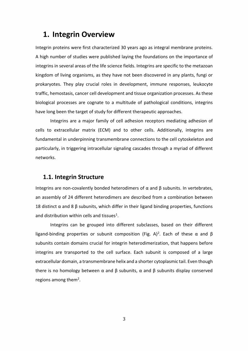
Integrins can be grouped into different subclasses, based on their different
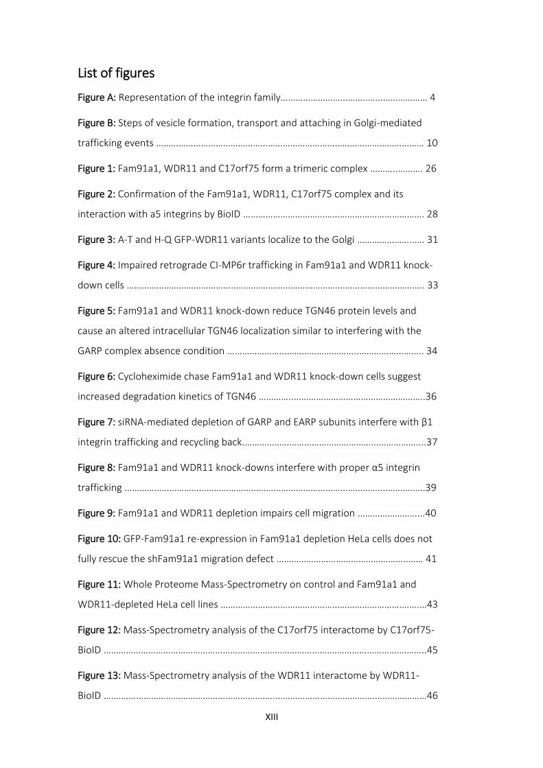
ligand-binding properties or subunit composition (Fig. A)2. Each of these α and β
subunits contain domains crucial for integrin heterodimerization, that happens before
integrins are transported to the cell surface. Each subunit is composed of a large
extracellular domain, a transmembrane helix and a shorter cytoplasmic tail. Even though
there is no homology between α and β subunits, α and β subunits display conserved
regions among them2.
4
1.1.1. αβ subunits
The α subunit is embodied by a seven-bladed β-propeller, connected to thigh and calf
domains that together form a ‘leg’ structure responsible for sustentating the integrin
head. These β-propeller blades provide integrins with the ability to bind Ca2+ and with
that to allosterically regulate ligand binding3. The α subunit is the one that grants ligand
specificity to integrins.
The β subunit is composed of several domains that, among others, give the
integrin heterodimer specific characteristics, including the ability to bind an inhibitory
Ca2+, an integrin activating Mn2+ or the cytoskeleton binding site2,3. Moreover, β integrin
chains have cytoplasmic tails responsible for intracellular ligand-binding, and
subsequent activation of downstream signaling pathways, due to a shared NPxY
homolog domain4.
Non-dimerized single α and β subunits are not present in the plasma membrane,
since α subunits are the limiting factor for integrin receptor formation as a result of
excess β subunits, thus only the dimerized form is present at the cell surface2.
Figure A Representation of the integrin family. 24 heterodimers are classified according to their ligand-
binding properties or subunit composition.
Adapted from: Barczyk, M., Carracedo, S. & Gullberg, D. Integrins. Cell Tissue Res. 339, 269–280 (2010).
5
1.1.2. Intracellular Domains - Cytoplasmic tails
Integrin cytoplasmic domains are usually short, contain about 70 amino acids, and while
β tails are remarkably homologous, this is opposed to the greatly distinct α subunit tails5.
It is thought that the cytoplasmic tails of α and β subunits form a salt bridge, between
the membrane-proximal regions of both chains. This interaction is believed to act as a
stabilizer of the integrin inactive and low-affinity state5.
The cytoplasmic β tails are comprised of a pair of well-defined motifs, the
membrane proximal NPxY and the distal NxxY motifs. These motifs are recognized by
proteins containing a phosphotyrosine-binding (PTB) domain including signaling
proteins, such as Talin and Kindlin6.
1.1.3. Transmembrane Domains
Integrins subunits have a single-spanning transmembrane (TM) domain, α-helical coiled
coil structures of about 25 amino acid residues. Evidence point to the highly conserved
GFF motif as critical structure mediating the transition from low-affinity inactive to
active states5.
1.1.4. Extracellular Domains
The extracellular domains of integrin subunits are composed of a spherical ligand-
binding head domain supported by two extended leg domains, that are linked with the
rest of the α and β integrin domains, respectively5. The extracellular domain provides
the ligand-binding site, that stems from the interaction between α-chain β-propeller
with the βI domain5,7.
Most common integrin ligands are components of the extracellular matrix (ECM).
Ligand binding elicits conformational changes in the integrin structure after, that leads
to integrin activation and signaling (integrin ‘outside-in’ signaling).
1.2. Integrin Signaling and Function
Contrary to most of other transmembrane receptors, integrins have the ability to signal
bidirectionally. This means that integrins transduce signals from the extracellular
6
environment following ligand binding to promote intracellular changes (‘outside-in
signaling’) but intracellular stimuli can also elicit extracellular responses (‘inside-out
signaling’)4.
During inside-out activation of integrins, intracellular signaling pathways lead to
the binding of talin and kindlin to the β integrin cytoplasmic tails inducing a
conformational change in the transmembrane and cytoplasmic integrin domains that is
transmitted to the extracellular domain. The extracellular domain changes from a bent,
low-affinity, inactive conformation to an extended high-affinity ligand-binding
conformation8.
Integrins do not have catalytic activity, integrin activation increases affinity for
ECM ligands, but to activate intracellular signaling pathways activated integrins are
needed to cluster and recruit cytoplasmic proteins to their tails. Nascent adhesions are
formed, that evolve to focal complexes, then larger focal adhesions (FA) and finally,
fibrillar adhesions4. Ligand-binding events to the extracellular head-domain of integrin,
initiate the previously described step-wise assembly of a dynamic multiprotein complex
(focal adhesion), that serve as the hub for transmission of intracellular signals (outside-
in signaling)4,5.
These mechanisms are essential for the cell-adhesion receptor properties of
integrins, that mediate cytoskeletal rearrangements, up- and downregulate cascade
pathways and change gene expression. All of this influences processes like survival,
growth and differentiation of cells4.
1.3. α5β1 Integrin and Cell Migration
α5β1 integrin, a major fibronectin (FN)-binding receptor, interacts with the tensin
adaptor protein to drive the formation of long and stable fibrillar adhesion complexes
9,10. Active α5β1 integrin is an endocytic FN receptor that plays a major role in regulating
FN fibrillar matrix11. The fibronectin binding to differently distributed α5β1 integrins at
the cell surface, stimulates RhoA-mediated organization of cell matrix adhesion12. The
activation of RhoA signaling is tightly linked to α5β1 recycling, as the recycling dictates
the availability of the heterodimer at the cell surface and subsequent random cell
migration4.
7
Cell migration is a highly complex process composed of a myriad of aggregated
and organized steps13. During migration, forces are generated to drive the protrusions
of the leading edge forward across the ECM. Followed by the formation of adhesion sites
at the protrusions, adhesions at the back of the cell will be disrupted to move of cell
body13,14.
Integrins, including α5β1, are primary components of the adhesion complexes that
serve as traction points that enable cell movement and also integrate signals that
regulate the migration process15.
2. Integrin Trafficking
As indicated above, integrin-mediated processes operate under tightly regulated
signaling events. Integrin availability at the plasma membrane is paramount for proper
physiological function and homeostasis.
Cell surface integrin availability is regulated by several trafficking mechanisms.
Simply put, cycles of endocytosis, re-exocytosis (recycling) or degradation, control the
pool of available integrins at the plasma membrane. For some integrins, their clearance
from the plasma membrane can be as fast as 30 minutes. Interestingly, the half-life of
an integrin is around 12-24 hours, which means that most of the integrins are
continuously being recycled back to the plasma membrane16. These mechanisms are
dependent on the function of several regulators like kinases, cytoskeleton modulators,
Rab family GTPases and Arf family GTPases.
2.1. Canonical Trafficking
Integrins are endocytosed by clathrin-dependent and -independent routes, as the latter
include macropinocytosis, actin-rich projections or clathrin-independent carriers17. The
internalization is followed by sorting mechanisms that decide if are to be either recycling
back to the cell surface or sorted into a degradative pathway. One integrin heterodimer
can, at different cell stages, follow different internalization paths or more than one
recycling route. For example, α5β1 can be endocytosed either in a clathrin- or caveolin-
dependent manner.
8
2.1.1. Clathrin-dependent endocytosis of integrins
Specific motifs at the integrin cytoplasmic β-subunit tails facilitate their recruitment of
to clathrin-coated structures through interactions with adaptor proteins like AP2 and
PTB-containing Dab and Numb, as well as bridging proteins such as HAX1 and cortactin18.
Dab2 was found to be an alternative clathrin adaptor of β1 integrins that regulates,
along with dynamin 2, microtubule-driven focal adhesion disassembly followed by
endocytosis.
It became evident the role of Dab2 in clathrin-dependent endocytosis, despite of
the unclear results of whether all conformation states of integrins are endocytosed or
just the active conformation. Recent literature also reported the requirement of a motor
protein, myosin VI, in the endocytosis of α5β1 integrin-NRP1 complexes from
fibronectin-rich fibrillar adhesions along F-actin structures18. Altogether, it was
established the role of clathrin-mediated endocytosis in focal adhesion disassembly19.
2.1.2. Clathrin-independent endocytosis of integrins
Some integrins including α5β1 can follow more than one route of internalization.
Overexpression of Rab21, led to an alternative endocytosis route using caveolin-1-
containing structures (caveolae). In these cells, caveolin-1 depletion resulted in an
accentuated reduction of β1 and fibronectin endocytosis indicating how clathrin-
independent endocytosis can regulate fibronectin matrix turnover and ECM
remodelling20. However, the molecular mechanisms of this route are still poorly defined,
and needs further analysis to enable a full understanding of the integrin endocytosis
process.
2.1.3. Recycling: Short-loop pathway
The Rab4 fast-recycling pathway (‘short-loop’) is one of two spatially and temporally
defined mechanisms of recycling21. Being associated to early endosomes, the entry to
this route is controlled by Rabex-5 (Rab5 guanine-exchange-factor (GEF)) that activates
Rab5, and by Rabaptin-5 that promotes a link between Rab5 and Rab4.
Phosphorylation of Rabaptin-5 by PKD1 controls Rabaptin-5 association to Rab4
endosomes which contain αvβ3 integrins, allowing the integrins to recycle back to the

9
plasma membrane. Thus, this route provides newly assembling adhesions with αvβ3
integrins, stimulating persistent cell migration on low fibronectin environments22.
Overall it is established that Rab4/5 early endosome-recycling of αvβ3 and inactive β1
integrins, allows cells to drive path persistent migration (in FN environments) and
invasion of cancer cells22.
2.1.4. Recycling: Long-loop pathway
The long-loop pathway is mainly characterized by Rab11-positive vesicles that go
through the perinuclear recycling compartment (PNRC) prior to returning to the cell
surface17.
Different signaling pathways promote selectivity in Rab11-mediated recycling of
a multitude of different integrin heterodimers, and therefore regulating cell migration21.
Evidences showed that PKB/Akt acts by phosphorylation of GSK-3β, to selectively recycle
α5β1 and αvβ3 via Rab11-positive compartments23. It was also reported that the ADP
ribosylation factor GTPase 6 (Arf6) present in the PNRC paires with Rab11 to regulate
the exit of β1, α5β1 and αvβ3 integrin from the PNRC, and cell migration towards
fibronectin21,24. Moreover, when Rab11 effector Rab-coupling protein (RCP) binds α5β1
integrin, signals cells for rapid and random cell migration25.
In summary, these studies demonstrate a step-wise controlled mechanism of
integrin transport through different recycling routes.
3. Golgi-mediated Trafficking
The Golgi apparatus is a central organelle of utmost importance when it comes to
protein sorting and trafficking in eukaryotic cells. The Golgi-mediated trafficking is
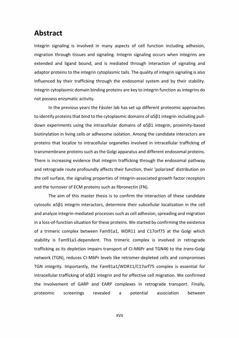
characterized by a bidirectional vesicular transport between different cellular
compartments26. This vesicular transport can be divided into the biosynthetic/secretory
pathway from the endoplasmic reticulum (ER) to the plasma membrane (anterograde
trafficking) or the reverse route, from the endosomal system/plasma membrane back
to early compartments (retrograde trafficking)27. These two trafficking routes can be
10
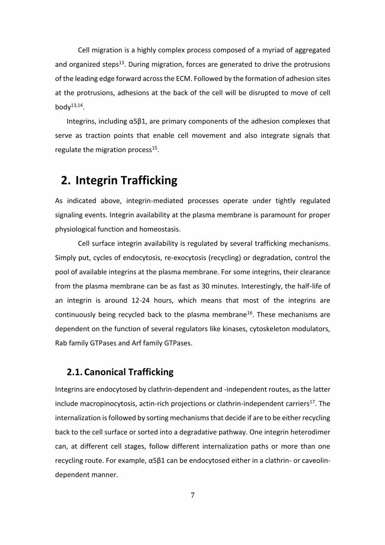
fragmented into five main steps: budding, movement, tethering, docking and fusion (Fig.
B)28.
Anterograde movement from the ER to the Golgi and retrograde movement from
the Golgi back to the ER, are mediated by coatomer coating protein-coated vesicles,
COPI and COPII respectively29. The pathways are connected and when COPII-dependent
retrograde transport is impaired, COPI-dependent anterograde transport is also
compromised due to the fact that tethering factors and cargo receptors are depleted
from the ER29. This demonstrates a tight relation between both trafficking pathways.
Figure B Steps of vesicle formation, transport and attaching in Golgi-mediated trafficking events.
Adapted from: Bonifacino, J. S. & Glick, B. S. The Mechanisms of Vesicle Budding and Fusion. Cell 116, 153–166 (2004).
3.1. Retrograde Trafficking and the trans-Golgi network
Transport of proteins from the endosomal system/plasma membrane to the trans-Golgi
network (TGN), Golgi membranes (cis-, medial- and trans-Golgi) or even to the
endoplasmic reticulum (ER) is known as retrograde transport27,30. This retrograde route
is also sometimes hijacked by pathogens and toxins in order to promote cytotoxicity or
gain control of the cells (Shiga toxin, cholera toxin, ricin and herpes virus)31–33. Thus,
understanding this trafficking route and the proteins involved will unveil new possible
outlets for upcoming therapeutic approaches and drug delivery strategies.
Amongst all of the framework in which retrograde trafficking takes place, the
trans-Golgi network has a central role as a traffic hub that regulates both the secretory
11
pathway and the retrograde route. As the anterograde transport depletes sorting
receptors, enzymes and vesicle-fate targeting factors from the Golgi these proteins need
to be retrieved to the TGN to allow for continuous cell function There are three main
retrograde transport routes characterized, referred to as early, recycling and late
endosome-to-TGN pathway. Each of these retrograde pathways is characterized by
different composing machineries and cargos, like the mannose 6-phospate receptors
(MPRs)34 or transmembrane proteins35.
Endosomes are a complex mosaic membrane system composed of tubules and
vesicles, that give rise to early/late endosomes, lysosomes and the endocytic recycling
compartment (recycling endosomes)36. Yet, all of these structures are part of a
continuum dependent on maturations stages, rather than defined singular structures,
sometimes distinguishable only by their morphology37. Therefore, classification of
endosome-to-TGN transport into three separate and distinct pathways is not a 100%
reliable approach but nonetheless still required to further our understanding of the
retrograde trafficking system.
3.1.1. Early endosome-to-TGN pathway
This pathway was first discovered as the pathway exploited by Shiga toxin (STxB)38, and
later found to be the route traversed by MPRs, which are dependent on AP1 and PACS-
1 (clathrin adaptors), clathrin coating and the retromer complex37 for their movement.
The retromer is a phylogenetically conserved multisubunit complex, whose best
characterized cargo is the cation-independent mannose 6-phosphate receptor (CI-
M6Pr). The CI-M6Pr binds freshly synthesized acid hydrolases at the TGN. These acid
hydrolases are then transported to the early endosomes/pre-lysosomal compartment
(part of the endosomal continuum), before pH acidic-induced release and proteolytical
cleavage into active form at the lysosomes30,34. Then, the retromer acts to retrieve these
CI-M6Prs back to the TGN, so they can re-engage in a new cycle of acid hydrolase sorting.
Conjointly, the Golgi-associated retrograde protein complex (GARP) was also
shown to be necessary for proper retrograde trafficking of CI-M6Pr, as well as STxB and
TGN46 (TGN transmembrane protein), to the trans-Golgi network39,40. Moreover, the
12
recruitment of the GARP complex at the TGN is promoted by Rab6 GTPase39 which in
turn enables SNARE complex formation.
The SNARE complexes are another set of proteins that are cycled between the
TGN and the endosomes. There are two distinct SNARE complexes, vesicle-membrane
SNAREs (v-SNAREs) which flow from the TGN to the early endosome in vesicles and
mediate the membrane fusion of these vesicles with the endosome compartments. The
fusion happens through assembly with the second type of SNAREs, the endosomal
target-membrane SNAREs (t-SNAREs)41. After the fusion process is complete, the v-
SNARE-t-SNARE complex (SNAREpin) is disassembled, and the v-SNAREs (Snc1) are
retrieved in retrograde transport vesicles back to the TGN in a GARP-dependent
fashion30,40.
3.1.2. Late endosome-to-TGN pathway
Late endosomes were also proposed to be a source of MPR as retrograde cargos for the
TGN. In this case, CI-M6Pr retrograde flow is mediated by a different complex of
proteins, the Rab9-TIP47 duplex.
Rab9 is a member of the large family of Ras-like GTPases that regulates vesicular
transport events at the level of membrane targeting, and localizes primarily at the
surface of late endosomes42 and vesicles that bud from them to fuse with the TGN30.
GTP-bound Rab9 forms pairs with TIP47, Rab9 serves as the membrane-recruitment
component, while TIP47 acts as a cargo-recognition component since it binds the
cytosolic domain of CI-M6Pr30. Interestingly, this Rab9-TIP47-duplex-derived retrograde
transport of CI-M6Pr is also dependent on a specific syntaxin 10 SNARE complex. Knock-
down of syntaxin 10 does not inhibit early transport of known endosome-to-TGN cargos
(TGN46/cholera toxin) but not MPRs, showing the specificity of certain factors for
different endosome-to-TGN pathways.
3.1.3. Recycling endosome-to-TGN pathway
Not many cargos have been reported to go through the recycling endosome-to-TGN, yet
CI-M6Pr and Shiga toxin need the recycling endosome-to-TGN pathway for efficient
transport, in addition to the early endosome-retromer-mediated transport43.
13
Furthermore, v-SNARE VAMP4 was shown be trafficked from early and recycling
endosomes, which goes in hand with the fact that VAMP4 forms a SNARE complex
(SNAREpin) with other known retrograde transport t-SNAREs: syntaxin 6 (Stx6), syntaxin
16 (Stx16) and Vtila44.
A recent paper characterized a newly found complex that operates at the level
of recycling endosomes. This complex called, the endosome-associated recycling protein
(EARP), is a multisubunit tethering complex with composition very similar to the TGN-
localized docking complex GARP45. While GARP complex is composed of Ang2 (Vps51),
Vps52, Vps53 and Vps5439, this new tethering complex EARP contains syndetin (Vps50)
instead of Vps54.
Although EARP complex was not directly shown to mediate a recycling
endosome-to-TGN pathway, multisubunit tethering complexes normally promote
SNARE-mediated vesicle fusion46, suggesting that indeed EARP (and to some extent
GARP) is mediating vesicle transport event from a distinct site to another. Adding that
shared subunits of EARP and GARP (Ang2, Vps52 and Vps53), were discovered to hold
interactions with Stx6–Stx16–Vti1a–VAMP4 complex45 (recycling endosome-to-TGN-
associated SNAREpin complex mentioned above), it is likely that EARP also mediates the
trafficking events of recycling endosomes-to-TGN. Sustaining this, is the observation of
a small population of GARP localized in the recycling endosomes, which like CORVET-to-
HOPS complex-mediated early-to-late endosome conversion, which suggests that EARP
might be a product of conversion after GARP reaches the recycling endosomes47.
3.2. Retrograde Trafficking and Integrins
The link between retrograde transport and integrin trafficking is still very much
unexplored. So far, the most commonly studied integrin trafficking pathways are the
canonical Rab4-dependent short-loop and Rab11-dependent long-loop pathways48.
However, a recent study found that cell adhesion, persistent cell migration,
polarized distribution of β1 integrin and focal adhesion disassembly dynamics depend
on a functional retrograde route49 indicating that TGN-mediated retrograde transport
of non-ligand-bound β1 integrin heterodimers acts as a complementary recycling
14
pathway. As a consequence, major cell functions that rely on integrin trafficking, might
also be dependent on retrograde trafficking.
3.3. Index of Trafficking mediating complexes
Here I give a concise insight about some of the most important and best studied
complexes involved in the retrograde trafficking network.
3.3.1. Retromer
The retromer is a multisubunit heteropentameric complex composed of sorting nexin
dimer (SNX1-SNX2) and vacuolar sorting protein trimer (VPS26 -VPS29-VPS35) in
humans. One of its most described functions is the retrieval of acid hydrolase receptors
(MPRs) to the trans-Golgi network, in retrograde transport30. The retromer associates
with the cytosolic interface of endosomes coated with clathrin. The SNX dimer is
responsible for the recruitment of the retromer to endosomes, while the VPS trimer
mediates the cargo-recognition process to recruit transmembrane proteins from
vacuole endosomes and channeling them for recycling at the TGN50,51.
3.3.2. Rab GTPases Family
Rab proteins compose one of the largest families of monomeric small GTPases, thought
to be more than 63 members in humans52. Their function as regulatory proteins are
based mainly on their ability to act molecular switches that shuffle between GTP- GDP-
bound conformations. While GTP-bound is the active form and the GDP-bound the
inactive one, their special feature lies in the ability to cycle between these
active/inactive states, enabling a temporal and spatial regulation of membrane
transport. They are characterized by a highly structural heterogeneity, thus endowing
them with very selective activity regarding their effectors. The diversity of Rab effectors
is obvious during intracellular transport, where they can mediate processes like budding,
movement, tethering, docking and fusion of vesicles into target compartments. This sets
up Rab proteins as pivotal factors in controlling a multitude of intracellular processes52.
15
3.3.3. SNAREs
SNAREs (soluble N-ethylmaleimide-sensitive factor attachment protein receptors), have
a crucial role in the docking and fusion events of vesicle-mediated transport into target
compartments. Regarding their function, they can be divided into v-SNAREs, associated
with vesicles being transported, and t-SNAREs which are associated with the targeted
receiving compartment. Binding of v-SNARE to a specific t-SNARE leads to the formation
of the trans-SNARE/SNAREpin complex. This SNARE complexes function are important
for secretory, endocytic and retrograde pathways as they mediate fusion events at the
TGN, early/recycling endosomes, or even mediate fusion of lysosomes with the plasma
membrane30,41.
3.3.4. GARP complex
The Golgi-associated retrograde protein (GARP) complex, is a tethering/docking
complex mainly localizing to the TGN, the sorting station of the cell composed of four
subunits (Ang2 (VPS51), VPS52, VPS53 and VPS54). Its recruitment to the TGN is
achieved by interactions with Rab6, where it coordinates tethering and fusion of
vesicles, due to interaction with t-SNARE Tlg153. The GARP complex is known for the
retrieval of receptors for lysosomal hydrolase precursors, such as the mannose 6-
phosphate receptors (MPRs) and interfering with GARP function results in impaired
retrograde transport to the TGN of transmembrane proteins (TGN46 and Kex2), SNAREs
(v-SNARE Snc1), toxins (Shiga toxin, ricin) and lysosomal dysfunction from the impaired
maturation of hydrolase percursors47,54. Hence, GARP plays a vital role in endosome to
TGN transport in a wide range of cellular functions.
3.3.5. EARP complex
The endosome-associated recycling protein (EARP) is a tethering complex with a similar
subunit composition as the GARP complex, with the exception of an exchange of
syndetin (VPS50) for VPS54. Syndetin is the key subunit that determines its distinct
localization to recycling endosomes instead of the TGN, like the GARP complex. Its main
function was described as a tethering factor for recycling proteins from the endosomes
to the plasma membrane. EARP promotes the recycling of transferrin receptor (TfR) back
to the cell surface, presumably through tethering of endosomal SNAREs. EARP also
16
associated Rab4-positive recycling endosomes (short-loop recycling pathway), and thus
established a requirement of multisubunit tethering complexes in the recycling
endocytic pathway29,45,47.
3.3.6. Sorting Nexins
Sorting nexins are a class of proteins containing a phox-homology (PX) domain, and up
to 33 have been identified in mammals. They bind phosphatidylinositol-3-
monophosphates (PtdIns3P) and function in processes such as endocytosis and
endosomal sorting/signaling55. Their main function is the orchestration of cargo sorting
in the membranous intricacy of the endosomal network. One of the best studied roles
of sorting nexins is within the retromer complex (SNX1-SNX2 dimer). After
internalization, early endosomes can sort their cargo either for lysosomal degradation,
to downregulate signaling receptors, or recycle it back to the plasma membrane. This
retrieval of the cargo is often coordinated by sortin nexins56. For instance, the binding
of SNX17 and SNX31 to the NPxY motif of the β1 subunit cytoplasmic tail of α5β1 integrin
at the early endosomes, prevented lysosomal degradation resulting in its recycling back
to the plasma membrane57,58.
4. Autophagy
Within all of the catabolic pathways available for a cell, autophagy is the most severe.
During autophagy, organelles and proteins are confined in acidic vacuoles for massive
lysosomal degradation. This pathway can be part of the response to nutrient deficit, cell
death, cell survival, development or tumor suppresion59.
Autophagy starts with the formation of a double-membrane vacuole or
autophagosome, which requires the function of a crucial family of autophagy-related
proteins (ATGs). A complex containing ATG14 and Beclin-1 is recruited to the
endoplasmic reticulum (ER) and acts upstream of the ATG12-ATG5-ATG16L1/LC3
elongation system. This complex along with ATG8 is responsible for elongating and
closing the autophagosomal membrane60,61.
17
Moreover, autophagy fulfils numerous functions and regulates a multitude of
physiological processes including cell migration, yet little is known how autophagy
affects integrin-mediated cell migration on a molecular level. There are apparent
contradictory observations of this phenomenon. For instance, one study showed that
increased cell migration is associated with low levels of autophagy, by reducing
endocytic recycling of integrins in HeLa cells62. While another study showed that
reduced levels of autophagy correlated with reduced tumor cell migration, due to
disruption of focal adhesion disassembly63. Further research is needed to fully uncover
the mechanisms by which autophagy regulates integrin-mediated processes.

18

19
Aims

20
21
Aims
The aim of this thesis was to characterize three novel candidate cytosolic α5β1 integrin
interactors for their roles in the regulation of α5β1 integrin stability and/or trafficking.
Specifically it was aimed to
- Confirm the interaction of candidate α5β1 integrin interactors.
- Determine the subcellular localization of candidate α5β1 integrin interactors
and their co-localization with α5β1 integrin.
- Analyze the knockdown cell lines for alterations in integrin-mediated
processes such as cell adhesion, spreading and migration.

22

23
Results

24
25
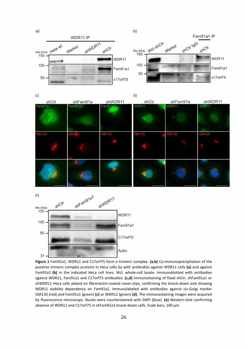
1. Confirmation of a putative Golgi-associated trimeric protein complex
of Fam91a1, WDR11 and C17orf75
Previous reports33,64 and previous proteomics in the Fässler department (unpublished
data) suggested the existence of a putative trimeric complex between Fam91a1, WDR11
and C17orf75. To further investigate this complex, we performed co-
immunoprecipitation (co-IP) assays of WDR11 and Fam91a1 in HeLa cells (Fig. 1a,b).
Immunoprecipitation of WDR11 co-immunoprecipitated both Fam91a1 and C17orf75 in
control shRNA (shCtr) HeLa cells, but not in the shRNA-mediated WDR11 knock-down
cells (Fig. 1a). Furthermore, both C17orf75 and WDR11 were co-immunoprecipitated
with an antibody against Fam91a1 but when a control IgG (Fig. 1b). These experiments
demonstrate the existence of a trimeric complex between Fam91a1, WDR11 and
C17orf75.
To assess the subcellular localizations of Fam91a1 and WDR11 with organelle
markers, knock-down cells for either Fam91a1 or WDR11 were used for
immunofluorescence assays. As expected from previous experiments, which showed a
localization of WDR11 to the TGN (unpublished data and ref.4), we observed both
Fam91a1 and WDR11 in the vicinity of the cis-Golgi marker GM13065 (Fig. 1c,d: shCtr).
Interestingly, the WDR11 signal was highly reduced in Fam91a1 knock-down cells
similarly to WDR11-depleted cells, and we did not observe a Golgi localization and
expression, while Fam91a1 localization and expression remained unaltered in WDR11
knock-down cells compared to control (Fig. 1 c,d: shFam91a1; shWDR11). These findings
were substantiated via western blot analysis, in which we observed that Fam91a1,
WDR11 and C17orf75 were all reduced in Fam91a1 knock-down cells, while the levels of
Fam91a1 and C17orf75 remained unchanged in WDR11 knock-down cells (Fig. 1e).
This suggests that the trimeric complex might be Fam91a1-dependent, and that
Fam91a1 knock-down leads to reduction of WDR11 protein rather than its
mislocalization from the Golgi apparatus.
To confirm the immunoprecipitation results with an independent method we
made use of BioID. BioID is a proximity-dependent biotin identification assay, based on
a promiscuous biotin ligase which labels proteins that are in its close proximity66. Cells
stably expressing either C17orf75- or α5-BioID-HA fusion proteins, previously reported
26
Figure 1 Fam91a1, WDR11 and C17orf75 form a trimeric complex. (a,b) Co-immunoprecipitation of the
putative trimeric complex proteins in HeLa cells by with antibodies against WDR11 cells (a) and against
Fam91a1 (b) in the indicated HeLa cell lines. Wcl, whole-cell lysate. Immunoblotted with antibodies
against WDR11, Fam91a1 and C17orf75 antibodies. (c,d) Immunostaining of fixed shCtr, shFam91a1 or
shWDR11 HeLa cells plated on fibronectin-coated cover-slips, confirming the knock-down and showing
WDR11 stability dependency on Fam91a1. Immunolabeled with antibodies against cis-Golgi marker
GM130 (red) and Fam91a1 (green) (c) or WDR11 (green) (d). The immunostaining images were acquired
by fluorescence microscopy. Nuclei were counterstained with DAPI (blue). (e) Western-blot confirming
absence of WDR11 and C17orf75 in shFam91a1 knock-down cells. Scale bars, 100 µm.
27
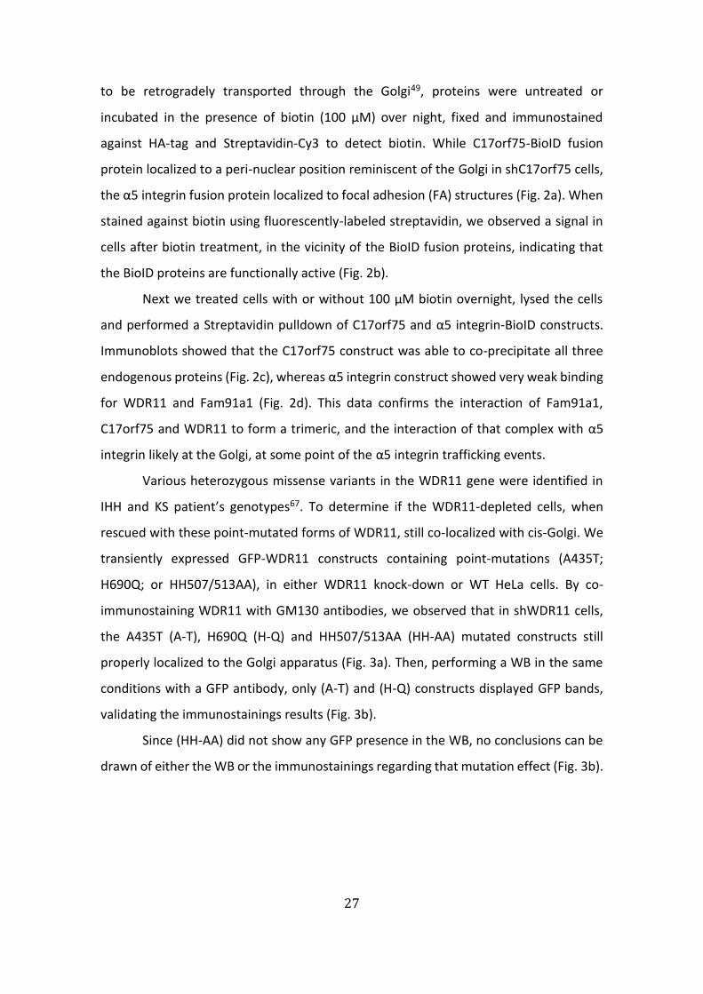
to be retrogradely transported through the Golgi49, proteins were untreated or
incubated in the presence of biotin (100 µM) over night, fixed and immunostained
against HA-tag and Streptavidin-Cy3 to detect biotin. While C17orf75-BioID fusion
protein localized to a peri-nuclear position reminiscent of the Golgi in shC17orf75 cells,
the α5 integrin fusion protein localized to focal adhesion (FA) structures (Fig. 2a). When
stained against biotin using fluorescently-labeled streptavidin, we observed a signal in
cells after biotin treatment, in the vicinity of the BioID fusion proteins, indicating that
the BioID proteins are functionally active (Fig. 2b).
Next we treated cells with or without 100 µM biotin overnight, lysed the cells
and performed a Streptavidin pulldown of C17orf75 and α5 integrin-BioID constructs.
Immunoblots showed that the C17orf75 construct was able to co-precipitate all three
endogenous proteins (Fig. 2c), whereas α5 integrin construct showed very weak binding
for WDR11 and Fam91a1 (Fig. 2d). This data confirms the interaction of Fam91a1,
C17orf75 and WDR11 to form a trimeric, and the interaction of that complex with α5
integrin likely at the Golgi, at some point of the α5 integrin trafficking events.
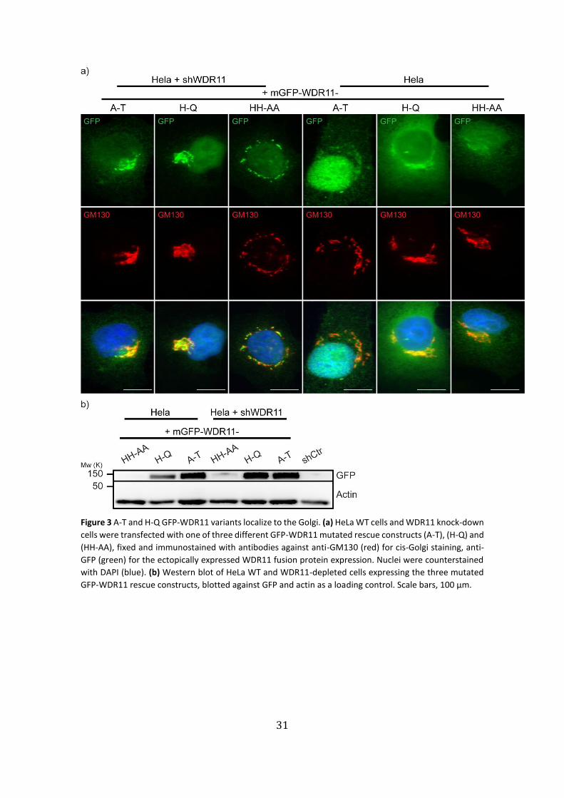
Various heterozygous missense variants in the WDR11 gene were identified in
IHH and KS patient’s genotypes67. To determine if the WDR11-depleted cells, when
rescued with these point-mutated forms of WDR11, still co-localized with cis-Golgi. We
transiently expressed GFP-WDR11 constructs containing point-mutations (A435T;
H690Q; or HH507/513AA), in either WDR11 knock-down or WT HeLa cells. By co-
immunostaining WDR11 with GM130 antibodies, we observed that in shWDR11 cells,
the A435T (A-T), H690Q (H-Q) and HH507/513AA (HH-AA) mutated constructs still
properly localized to the Golgi apparatus (Fig. 3a). Then, performing a WB in the same
conditions with a GFP antibody, only (A-T) and (H-Q) constructs displayed GFP bands,
validating the immunostainings results (Fig. 3b).
Since (HH-AA) did not show any GFP presence in the WB, no conclusions can be
drawn of either the WB or the immunostainings regarding that mutation effect (Fig. 3b).

28
Figure 2 Confirmation of the Fam91a1, WDR11, C17orf75 complex and its interaction with a5 integrins by
BioID (a,b) Co-localization of biotinylated proteins (labeled by Streptavidin-Cy3, red) with the BioID fusion
29
proteins in Hela cells stably expressing C17orf75-BioID-HA or α5-BioID-HA in the presence of biotin (100
µM). BioID-fusion proteins were detected with a HA-tag antibody (green) (a). To define the subcellular
localization cells in (b) we co-stained with antibodies against GM130 or Paxilin (green) in the presence or
absence of biotin. Scale bars, 200 µm (a) and 100 µm (b). Nuclei were counterstained with DAPI
(blue)..Scale bars, 200 µm (a) and 100 µm (b). Nuclei were counterstained with DAPI (blue) (c,d).
Formation and localization of the trimeric complex proteins in Golgi and focal-adhesions, its interaction
with a5 integrin was determined by BioID in biotinylation, followed by biotin pull down and western-blot
analysis of the neighboring partners of cells expressing the BioID fusion protein with Cc17orf75 (c) or α5
integrin (d).
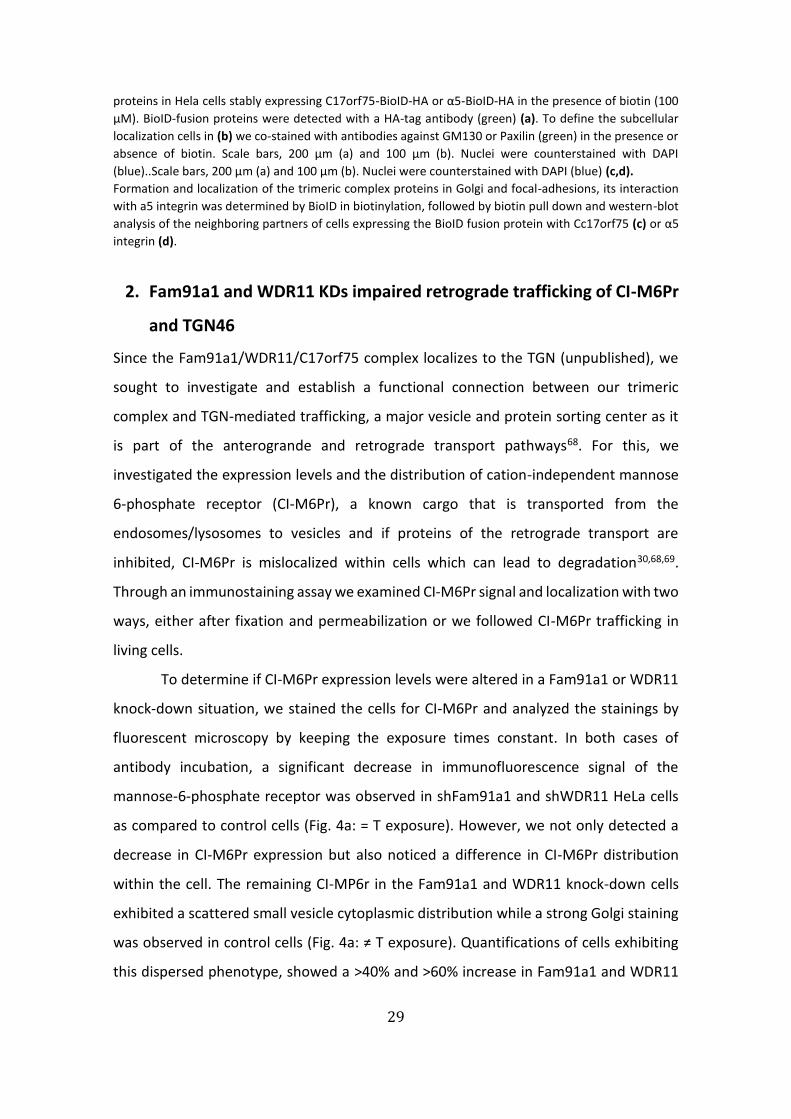
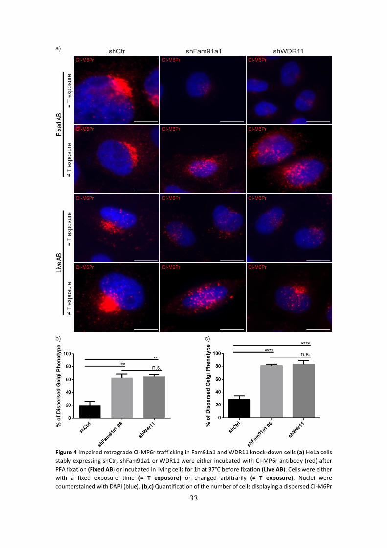
2. Fam91a1 and WDR11 KDs impaired retrograde trafficking of CI-M6Pr
and TGN46
Since the Fam91a1/WDR11/C17orf75 complex localizes to the TGN (unpublished), we
sought to investigate and establish a functional connection between our trimeric
complex and TGN-mediated trafficking, a major vesicle and protein sorting center as it
is part of the anterogrande and retrograde transport pathways68. For this, we
investigated the expression levels and the distribution of cation-independent mannose
6-phosphate receptor (CI-M6Pr), a known cargo that is transported from the
endosomes/lysosomes to vesicles and if proteins of the retrograde transport are
inhibited, CI-M6Pr is mislocalized within cells which can lead to degradation30,68,69.
Through an immunostaining assay we examined CI-M6Pr signal and localization with two
ways, either after fixation and permeabilization or we followed CI-M6Pr trafficking in
living cells.
To determine if CI-M6Pr expression levels were altered in a Fam91a1 or WDR11
knock-down situation, we stained the cells for CI-M6Pr and analyzed the stainings by
fluorescent microscopy by keeping the exposure times constant. In both cases of
antibody incubation, a significant decrease in immunofluorescence signal of the
mannose-6-phosphate receptor was observed in shFam91a1 and shWDR11 HeLa cells
as compared to control cells (Fig. 4a: = T exposure). However, we not only detected a
decrease in CI-M6Pr expression but also noticed a difference in CI-M6Pr distribution
within the cell. The remaining CI-MP6r in the Fam91a1 and WDR11 knock-down cells
exhibited a scattered small vesicle cytoplasmic distribution while a strong Golgi staining
was observed in control cells (Fig. 4a: ≠ T exposure). Quantifications of cells exhibiting
this dispersed phenotype, showed a >40% and >60% increase in Fam91a1 and WDR11
30
knock-downs versus shCtr in fixed and live antibody incubation conditions, respectively
(Fig. 4c,d).
This phenotype is reminiscent of a retromer-depleted phenotype69,70, and
suggests a defect in retrograde trafficking of the CI-M6Pr, which could lead to an
increased degradation of CI-M6Pr.
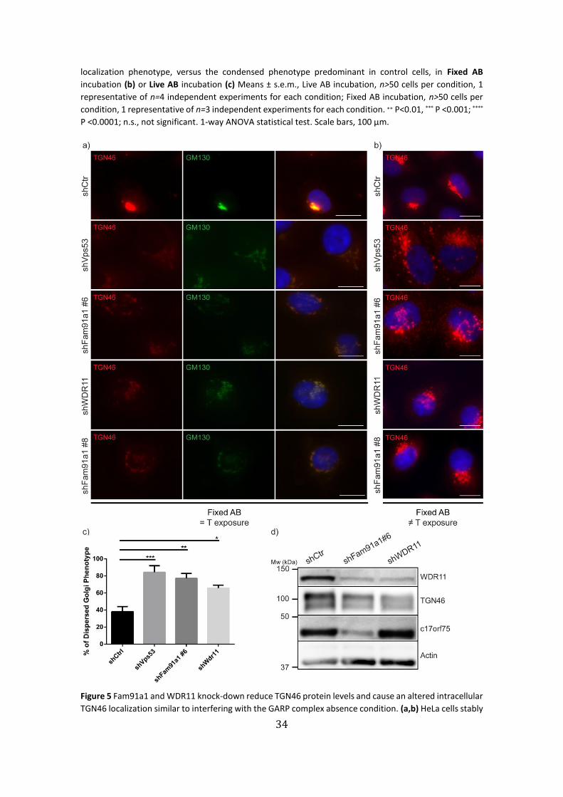
Similar to other vesicular transport pathways, retrograde transport from
endosomes is greatly intertwined with TGN integrity30,71. We therefore tested if and how
the TGN might also be compromised by absence of the Fam91a1 and WDR11. For this,
we immunostained two independent Fam91a1 knock-down cell lines (#6 and #8),
WDR11 knock-down cells and Vps53 knock-down HeLa cells as a positive control for TGN
destabilization by GARP complex depletion40,53,72,73, with antibodies against the TGN
marker protein TNG46. Microscopy images showed a significant decrease in signal
intensity of TGN46, when exposure was kept the same for all conditions (Fig. 5a). Both
Fam91a1 and WDR11 knock-downs display a similarly reduced TGN46 signal phenotype
as the positive control shVps53. Conjointly, they also show a dispersed, vesicular
localization of the remaining TGN46 indicating that TGN46 goes from an aggregated
juxtanuclear position in control HeLa cells to a scattered small vesicle morphology in the
knock-down conditions (Fig. 5b). Quantification of the number of cells displaying the
dispersed phenotype show a statistically significant increase of approximately 35% to
40% relative to control (Fig. 5c).
Moreover, we performed a western blot analysis of whole cell lysates of
Fam91a1 and WDR11 knock-down cell lines which confirmed the reduced levels of
TGN46 seen in the immunostainings (Fig. 5d). We speculate that this decrease might be
due to an increased turnover rate of the protein in the absence of our trimeric complex.
31
Figure 3 A-T and H-Q GFP-WDR11 variants localize to the Golgi. (a) HeLa WT cells and WDR11 knock-down
cells were transfected with one of three different GFP-WDR11 mutated rescue constructs (A-T), (H-Q) and
(HH-AA), fixed and immunostained with antibodies against anti-GM130 (red) for cis-Golgi staining, anti-
GFP (green) for the ectopically expressed WDR11 fusion protein expression. Nuclei were counterstained
with DAPI (blue). (b) Western blot of HeLa WT and WDR11-depleted cells expressing the three mutated
GFP-WDR11 rescue constructs, blotted against GFP and actin as a loading control. Scale bars, 100 µm.
32
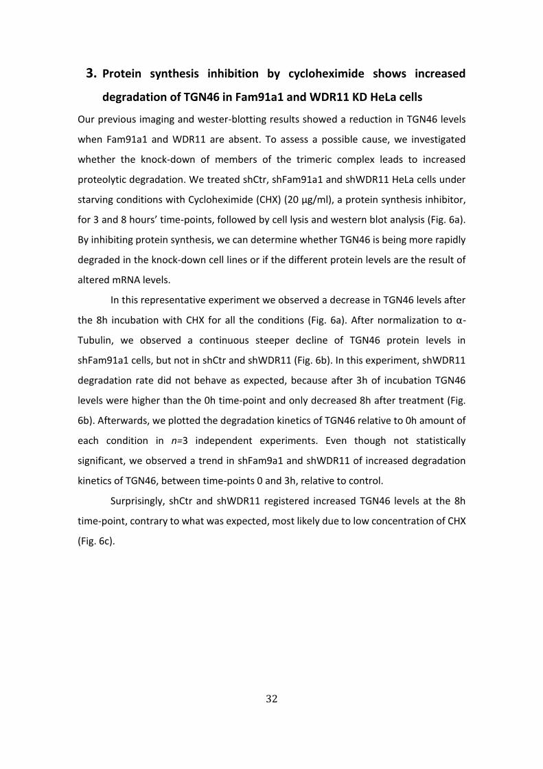
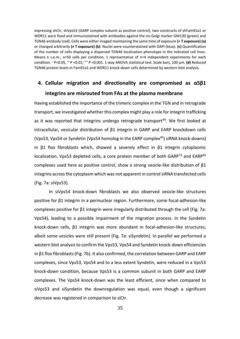
3. Protein synthesis inhibition by cycloheximide shows increased
degradation of TGN46 in Fam91a1 and WDR11 KD HeLa cells
Our previous imaging and wester-blotting results showed a reduction in TGN46 levels
when Fam91a1 and WDR11 are absent. To assess a possible cause, we investigated
whether the knock-down of members of the trimeric complex leads to increased
proteolytic degradation. We treated shCtr, shFam91a1 and shWDR11 HeLa cells under
starving conditions with Cycloheximide (CHX) (20 µg/ml), a protein synthesis inhibitor,
for 3 and 8 hours’ time-points, followed by cell lysis and western blot analysis (Fig. 6a).
By inhibiting protein synthesis, we can determine whether TGN46 is being more rapidly
degraded in the knock-down cell lines or if the different protein levels are the result of
altered mRNA levels.
In this representative experiment we observed a decrease in TGN46 levels after
the 8h incubation with CHX for all the conditions (Fig. 6a). After normalization to α-
Tubulin, we observed a continuous steeper decline of TGN46 protein levels in
shFam91a1 cells, but not in shCtr and shWDR11 (Fig. 6b). In this experiment, shWDR11
degradation rate did not behave as expected, because after 3h of incubation TGN46
levels were higher than the 0h time-point and only decreased 8h after treatment (Fig.
6b). Afterwards, we plotted the degradation kinetics of TGN46 relative to 0h amount of
each condition in n=3 independent experiments. Even though not statistically
significant, we observed a trend in shFam9a1 and shWDR11 of increased degradation
kinetics of TGN46, between time-points 0 and 3h, relative to control.
Surprisingly, shCtr and shWDR11 registered increased TGN46 levels at the 8h
time-point, contrary to what was expected, most likely due to low concentration of CHX
(Fig. 6c).
33
Figure 4 Impaired retrograde CI-MP6r trafficking in Fam91a1 and WDR11 knock-down cells (a) HeLa cells
stably expressing shCtr, shFam91a1 or WDR11 were either incubated with CI-MP6r antibody (red) after
PFA fixation (Fixed AB) or incubated in living cells for 1h at 37°C before fixation (Live AB). Cells were either
with a fixed exposure time (= T exposure) or changed arbitrarily (≠ T exposure). Nuclei were
counterstained with DAPI (blue). (b,c) Quantification of the number of cells displaying a dispersed CI-M6Pr
34
localization phenotype, versus the condensed phenotype predominant in control cells, in Fixed AB
incubation (b) or Live AB incubation (c) Means ± s.e.m., Live AB incubation, n>50 cells per condition, 1
representative of n=4 independent experiments for each condition; Fixed AB incubation, n>50 cells per
condition, 1 representative of n=3 independent experiments for each condition. ∗∗ P<0.01, *** P <0.001; ****
P <0.0001; n.s., not significant. 1-way ANOVA statistical test. Scale bars, 100 µm.
Figure 5 Fam91a1 and WDR11 knock-down reduce TGN46 protein levels and cause an altered intracellular
TGN46 localization similar to interfering with the GARP complex absence condition. (a,b) HeLa cells stably
35
expressing shCtr, shVps53 (GARP complex subunit as positive control), two constructs of shFam91a1 or
WDR11 were fixed and immunostained with antibodies against the cis-Golgi marker GM130 (green) and
TGN46 antibody (red). Cells were either imaged maintaining the same time of exposure (= T exposure) (a)
or changed arbitrarily (≠ T exposure) (b). Nuclei were counterstained with DAPI (blue). (c) Quantification
of the number of cells displaying a dispersed TGN46 localization phenotype in the indicated cell lines.
Means ± s.e.m., n>50 cells per condition, 1 representative of n=4 independent experiments for each
condition. ∗ P<0.05, ** P <0.01; *** P <0.001. 1-way ANOVA statistical test. Scale bars, 100 µm. (d) Reduced
TGN46 protein levels in Fam91a1 and WDR11 knock-down cells determined by western blot analysis.
4. Cellular migration and directionality are compromised as α5β1
integrins are misrouted from FAs at the plasma membrane
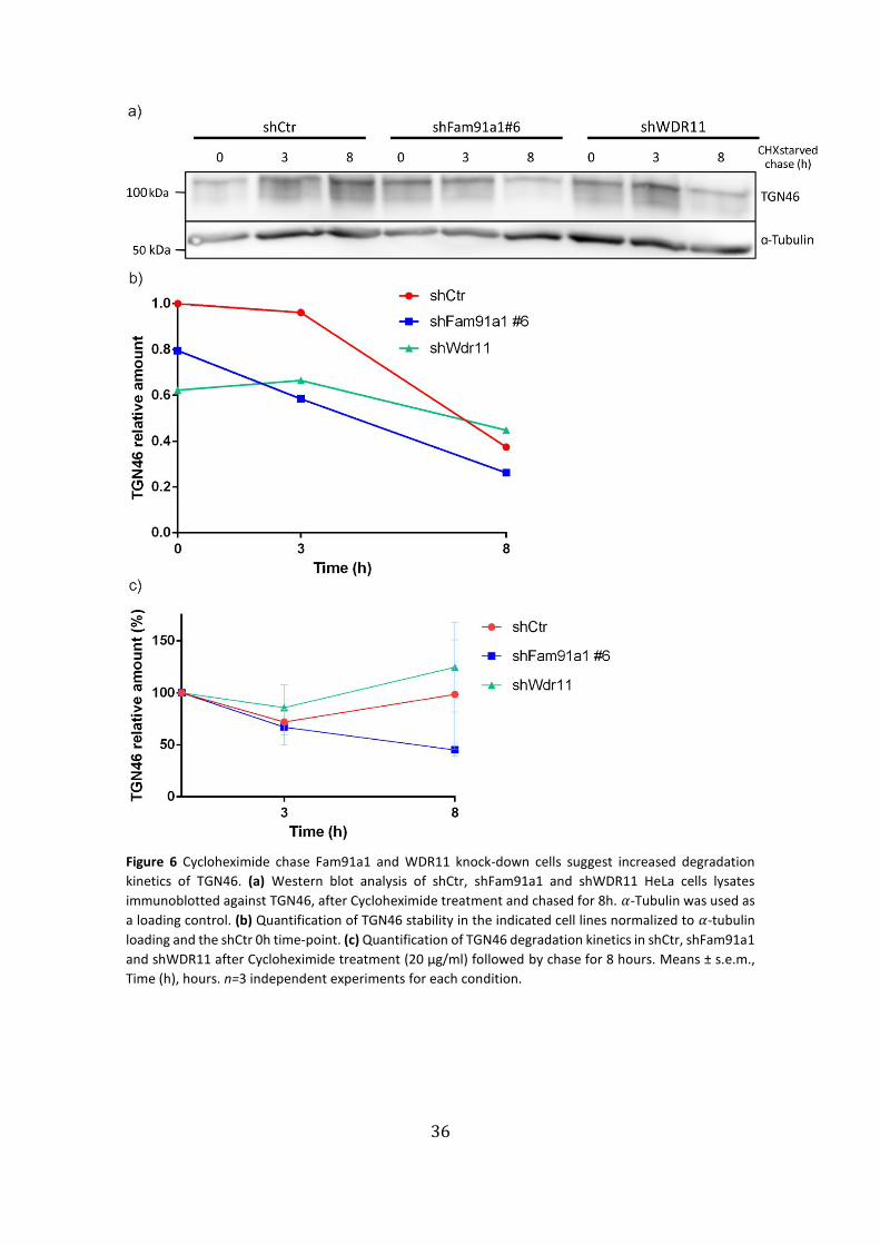
Having established the importance of the trimeric complex in the TGN and in retrograde
transport, we investigated whether this complex might play a role for integrin trafficking
as it was reported that integrins undergo retrograde transport49. We first looked at
intracellular, vesicular distribution of β1 integrin in GARP and EARP knockdown cells
(Vps53, Vps54 or Syndetin (Vps54 homolog in the EARP complex45) siRNA knock-downs)
in β1 flox fibroblasts which, showed a severely effect in β1 integrin cytoplasmic
localization. Vps53 depleted cells, a core protein member of both GARP73 and EARP45
complexes used here as positive control, show a strong vesicle-like distribution of β1
integrins across the cytoplasm which was not apparent in control siRNA transfected cells
(Fig. 7a: siVps53).
In siVps54 knock-down fibroblasts we also observed vesicle-like structures
positive for β1 integrin in a perinuclear region. Furthermore, some focal-adhesion-like
complexes positive for β1 integrin were irregularly distributed through the cell (Fig. 7a:
Vps54), leading to a possible impairment of the migration process. In the Syndetin
knock-down cells, β1 integrin was more abundant in focal-adhesion-like structures,
albeit some vesicles were still present (Fig. 7a: siSyndetin). In parallel we performed a
western blot analysis to confirm the Vps53, Vps54 and Syndetin knock-down efficiencies
in β1 flox fibroblasts (Fig. 7b). It also confirmed, the correlation between GARP and EARP
complexes, since Vps53, Vps54 and to a less extent Syndetin, were reduced in a Vps53
knock-down condition, because Vps53 is a common subunit in both GARP and EARP
complexes. The Vps54 knock-down was the least efficient, since when compared to
siVps53 and siSyndetin the downregulation was equal, even though a significant
decrease was registered in comparison to siCtr.
36
Figure 6 Cycloheximide chase Fam91a1 and WDR11 knock-down cells suggest increased degradation
kinetics of TGN46. (a) Western blot analysis of shCtr, shFam91a1 and shWDR11 HeLa cells lysates
immunoblotted against TGN46, after Cycloheximide treatment and chased for 8h. 𝛼-Tubulin was used as
a loading control. (b) Quantification of TGN46 stability in the indicated cell lines normalized to 𝛼-tubulin
loading and the shCtr 0h time-point. (c) Quantification of TGN46 degradation kinetics in shCtr, shFam91a1
and shWDR11 after Cycloheximide treatment (20 µg/ml) followed by chase for 8 hours. Means ± s.e.m.,
Time (h), hours. n=3 independent experiments for each condition.
37
Figure 7 siRNA-mediated depletion of GARP and EARP subunits interfere with β1 integrin trafficking and
recycling back. (a) β1 flox fibroblasts transiently transfected with siRNAs against Vps53, Vps54 (GARP-
specific subunit) and Syndetin (EARP-specific subunit) were fixed and stained with antibodies against β1
integrin antibody (red) and Phalloidin to visualize F-Actin marker (green). Cells were imaged in a
fluorescence microscopy. Nuclei were counterstained with DAPI (blue). Scale bars, 100 µm. (b) Western
blot analysis of the indicated siRNA-transfected β1 flox fibroblasts to determine the knock-down
efficiency. Actin was used as a loading control.
38
This means, either Vps54 presence is also affected by Vps53 and Syndetin or the siRNA
silencing was not completely efficient for Vps54 (Fig. 7b). The knock-down of Syndetin
also caused a moderate decrease in Vps53 protein when compared to the control,
suggesting a relation between Vps53 and Syndetin and not between Vps54, since knock-
down of the latter did not affect Vps53.
Overall, these results suggest a relation between the integrin trafficking and the
functionality of TGN and endosomes.
To investigate the involvement of the trimeric complex in the integrin trafficking,
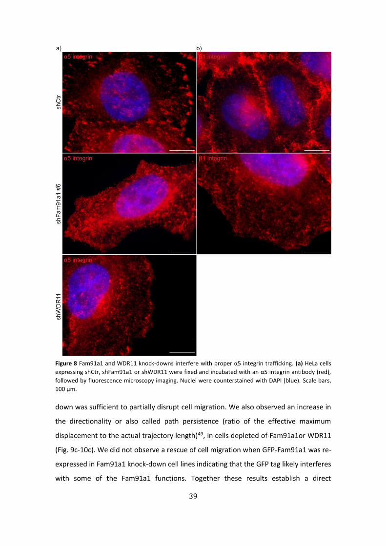
we performed α5 and β1 integrin immunostainings in Fam91a1 and WDR11 knock-down
HeLa cells. As expected, in control cells the integrin heterodimer localized to focal-
adhesion complexes at the plasma membrane (Fig. 8a,b: shCtr); while in Fam91a1 and
WDR11 knock-down cells, α5 and β1 integrin were mostly localized in scattered
cytoplasmic structures, particularly evident in a juxtanuclear position reminding of the
Golgi complex (Fig. 8a,b: shFam91a1/WDR11).
Collectively, these results suggest an involvement of the
Fam91a1/WDR11/C17orf75 in the trafficking of α5β1integrins.
α5β1 integrin is a major player in cell adhesion and migration and its precise
subcellular localization and trafficking is crucial for cell motility. Therefore, we tested for
a direct connection between the Fam91a1/WDR11/C17orf75 complex and cell
migration. We performed wound healing assays on confluent shCtr, shFam91a1 #6,
shWDR11, shFam91a1 #8 and GFP-Fam91a1 (rescue construct) HeLa cells, in starving
condition after treating the cells with mitomycin C, a cell division inhibitor (Fig. 9a-10a).
We recorded the cell migration by time lapse microscopy and quantified the cell
migration with respect to single-cell velocity and directionality. Images of three
representative time-points (0, 12 and 24h) for each cell lines were displayed, showing
how cells collectively migrated.
The shFam91a1 #6, shFam91a1 #8, shWDR11 and the GFP-Fam91a1 rescue cell
lines displayed reduced wound area closure (Fig. 9a-10a) as well as a reduced single-cell
migration velocity (Fig. 9b-10b). Pointing out that either Fam91a1 or WDR11 knock-
39
Figure 8 Fam91a1 and WDR11 knock-downs interfere with proper α5 integrin trafficking. (a) HeLa cells
expressing shCtr, shFam91a1 or shWDR11 were fixed and incubated with an α5 integrin antibody (red),
followed by fluorescence microscopy imaging. Nuclei were counterstained with DAPI (blue). Scale bars,
100 µm.
down was sufficient to partially disrupt cell migration. We also observed an increase in
the directionality or also called path persistence (ratio of the effective maximum
displacement to the actual trajectory length)49, in cells depleted of Fam91a1or WDR11
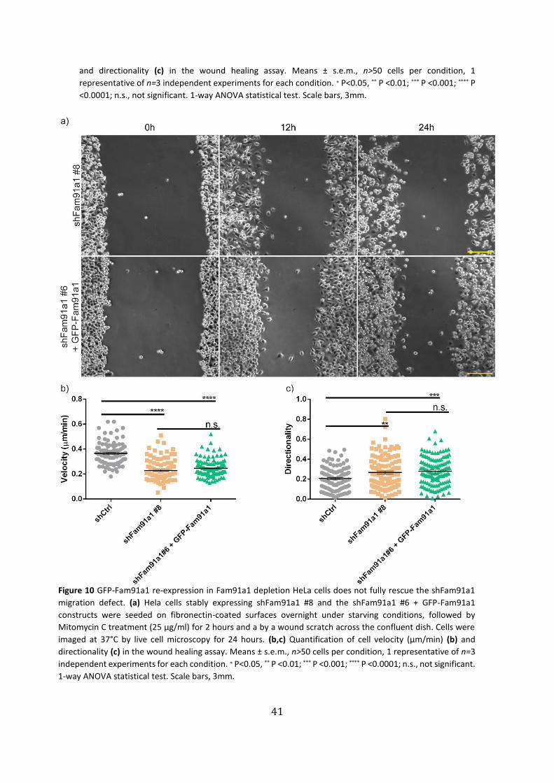
(Fig. 9c-10c). We did not observe a rescue of cell migration when GFP-Fam91a1 was re-
expressed in Fam91a1 knock-down cell lines indicating that the GFP tag likely interferes
with some of the Fam91a1 functions. Together these results establish a direct
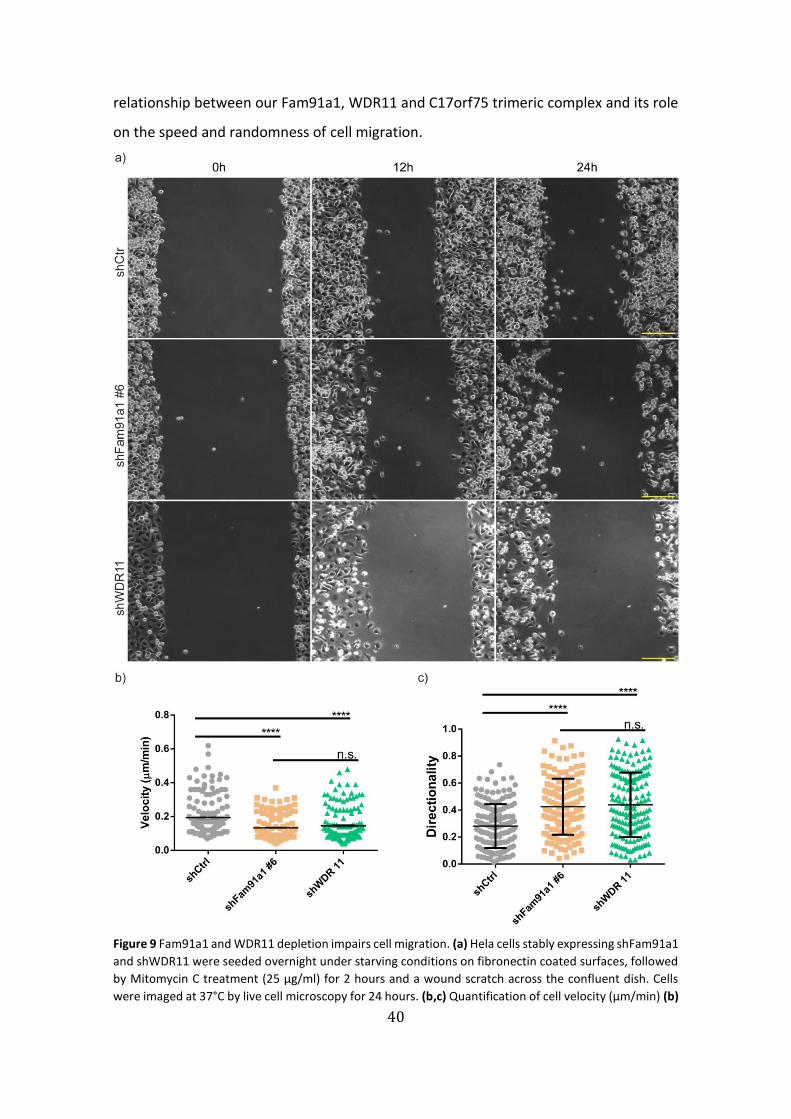
40
relationship between our Fam91a1, WDR11 and C17orf75 trimeric complex and its role
on the speed and randomness of cell migration.
Figure 9 Fam91a1 and WDR11 depletion impairs cell migration. (a) Hela cells stably expressing shFam91a1
and shWDR11 were seeded overnight under starving conditions on fibronectin coated surfaces, followed
by Mitomycin C treatment (25 µg/ml) for 2 hours and a wound scratch across the confluent dish. Cells
were imaged at 37°C by live cell microscopy for 24 hours. (b,c) Quantification of cell velocity (µm/min) (b)
41
and directionality (c) in the wound healing assay. Means ± s.e.m., n>50 cells per condition, 1
representative of n=3 independent experiments for each condition. ∗ P<0.05, ** P <0.01; *** P <0.001; **** P
<0.0001; n.s., not significant. 1-way ANOVA statistical test. Scale bars, 3mm.
Figure 10 GFP-Fam91a1 re-expression in Fam91a1 depletion HeLa cells does not fully rescue the shFam91a1
migration defect. (a) Hela cells stably expressing shFam91a1 #8 and the shFam91a1 #6 + GFP-Fam91a1
constructs were seeded on fibronectin-coated surfaces overnight under starving conditions, followed by
Mitomycin C treatment (25 µg/ml) for 2 hours and a by a wound scratch across the confluent dish. Cells were
imaged at 37°C by live cell microscopy for 24 hours. (b,c) Quantification of cell velocity (µm/min) (b) and
directionality (c) in the wound healing assay. Means ± s.e.m., n>50 cells per condition, 1 representative of n=3
independent experiments for each condition. ∗ P<0.05, ** P <0.01; *** P <0.001; **** P <0.0001; n.s., not significant.
1-way ANOVA statistical test. Scale bars, 3mm.
42
5. Proteomic Mass-Spectrometry screening analysis of knock-downs
and Biotin pull-downs of the trimeric complex proteins
In order to identify potential proteins interactors or regulators of the Fam91a1, WDR11,
C17orf75 complex and to determine pathways regulated by this complex a whole-
proteome mass-spectrometry (MS) screening of control HeLa cells and cells depleted of
Fam91a1 and WDR11 and was performed.
Firstly, we confirmed the knock-down efficiency of the different cells lines by
western blot analysis, which confirmed the efficiency of the protein expression silencing
(Fig. 11a). For the whole-proteome analysis 5x10⁶ cells of the three cell lines were lysed
and each condition analyzed in triplicates. The MS screening was carried out in the MPI
of Biochemistry mass spec core facility and the unfiltered results were analyzed with the
Perseus to highlight the statistically relevant changes in protein expression between
shFam91a1, shWDR11 and control. This filtering resulted in 1326 proteins with
statistically relevant deregulated expression out of 8159 initially found. Perseus
software allowed us to do a cluster heat map illustrating that the Fam91a1 deficiency is
responsible for the larger number of protein expression variations between Fam91a1
and WDR11 compared to shCtr (Fig. 11b). As a positive control for the experiment we
looked into both Fam91a1 and WDR11 expression profiles throughout the three
replicates of each of the three conditions. Since these proteins behaved as expected
(WDR11 downregulated in both knock-down conditions; Fam91a1 just on its knock-
down condition) more confidence can be attributed to the remaining results. (Fig.
11c,d).
Then, to find and characterize the body of altered proteins that constituted each
knock-down, we first set the knock-down expression profile as the reference to all other
proteins who might have a similar expression profile. Then, we searched for a number
of proteins that showed a similar profile, until an arbitrarily attributed signal-to-noise
ratio was reached, comparing the sum of the expression profiles of the unknown
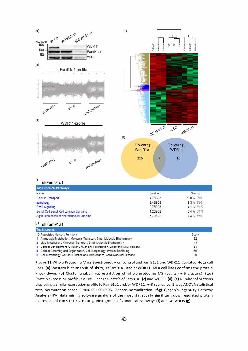
43
Figure 11 Whole Proteome Mass-Spectrometry on control and Fam91a1 and WDR11-depleted HeLa cell
lines. (a) Western blot analysis of shCtr, shFam91a1 and shWDR11 HeLa cell lines confirms the protein
knock-down. (b) Cluster analysis representation of whole-proteome MS results (n=5 clusters). (c,d)
Protein expression profile in all cell lines replicate’s of Fam91a1 (c) and WDR11 (d). (e) Number of proteins
displaying a similar expression profile to Fam91a1 and/or WDR11. n=3 replicates; 1-way ANOVA statistical
test, permutation-based FDR=0.05; S0=0.05. Z-score normalization. (f,g) Qiagen´s Ingenuity Pathway
Analysis (IPA) data mining software analysis of the most statistically significant downregulated protein
expression of Fam91a1 KD in categorical groups of Canonical Pathways (f) and Networks (g)

44
proteins and the similarities to the downregulated proteins in the knock-down
(shFam91a1/shWDR11) original expression profile. We registered only 7 proteins
commonly downregulated in both knock-downs, from a pool of 226 with a
downregulated in Fam91a1 knock-down profile and a pool of 10 with a downregulated
in WDR11 knock-down profile (Fig. 11e).
Among the seven downregulated proteins, in both knockdown cell lines, were
Perilipin 2 (PLIN2), ceramide synthase 1 (CERS1), desaturase-sphingolipid 1 (DEGS1),
autophagy-related protein 12 (ATG12), vacuolar protein sorting 13 (Vps13), which are
associated with spermatogenesis, germ cell differentiation or autophagy60,74.
In order to analyze the 226 proteins downregulated in shFam91a1 cells, we used
Qiagen’s Ingenuity Pathway Analysis® (IPA) data-mining software, which grouped the
proteins according to different categories based on different criteria, such as canonical
pathways involved, networks diseases, etc. We categorized the list of proteins
significantly downregulated in Fam91a1-depleted HeLa cells, because it was the
condition which showed most changes compared to control HeLa cells. We found
proteins involved in the canonical pathways of Sertoli-cell junction signaling, RhoA
signaling and autophagy (Fig. 11f). Interestingly, the literature showed a relevant
connection between retrograde transport machinery (Golgi-to-ER) and autophagy
regulation and function (autophagy regulators trafficking)75. In our MS screening we
found, Beclin-1 (BECN1), Autophagy related 9A (ATG9A), both associated with
autophagy and Vacuolar protein sorting 18 (Vps18), a member of endosomal tethering
complexes, to be downregulated in Fam91a1 knock-down. Through another
categorization of the IPA analysis, we found different altered networks in Fam91a1
knock-down, which confirmed the importance of the trimeric complex in the molecular
transport, protein trafficking, cellular growth and proliferation, lipid metabolism
(autophagy-related) and cell morphology (Fig. 11g).
In addition to the whole proteome analysis, interactome analysis of WDR11 and
C17orf75 was performed in the department. For this, streptavidin pull-downs of
C17orf75-BioID and WDR11-BioID expressing HeLa cells were carried out after
incubating the cells overnight in the absence and presence of 50 µM biotin (Fig. 12-13).
When cells were supplied with an excess of biotin, in both pull-down experiments a
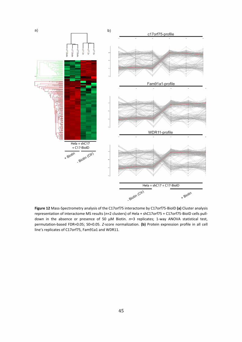
45
Figure 12 Mass-Spectrometry analysis of the C17orf75 interactome by C17orf75-BioID (a) Cluster analysis
representation of interactome MS results (n=2 clusters) of Hela + shC17orf75 + C17orf75-BioID cells pull-
down in the absence or presence of 50 µM Biotin. n=3 replicates; 1-way ANOVA statistical test,
permutation-based FDR=0.05; S0=0.05. Z-score normalization. (b) Protein expression profile in all cell
line’s replicates of C17orf75, Fam91a1 and WDR11.

46
Figure 13 Mass-Spectrometry analysis of the WDR11 interactome by WDR11-BioID (a) Cluster analysis
representation of interactome MS results (n=3 clusters) of Hela + shWDR11 + WDR11-BioID cells pull-
down in the absence or presence of 50 µM Biotin. n=2 replicates; 1-way ANOVA statistical test,
permutation-based FDR=0.05; S0=0.05. Z-score normalization. (b) Protein expression profile in all cell
line’s replicates of WDR11, Fam91a1 and C17orf75.
paramount change in the number of proteins baited was attained. The experiments
were carried out in replicates.
As expected Fam91a1, WDR11 and C17orf75 were highly enriched in pulldown
samples after biotin treatment, thus validating the experiment (Fig. 12b-13b). Finally,
the significant proteins found in the biotin pull-down were clustered, for the WDR11-
BioID the filtering and statistical analysis resulted in 640 altered proteins expression out
of 790 initially found. For the C17orf75-BioID pull-down the filtering and statistical
analysis resulted in 220 expression proteins altered proteins expression out of 615 (Fig.
12a-13a).
47
Proteins identified in both (the C17orf75 and WDR11) interactomes were
proteins from WASH complex subunits and related to Golgi apparatus, small GTP-
binding family and gonadal and gametes development. In an individual context,
C17orf75 biotin pull downs showed some interesting proteins, such as ATG9, CI-M6Pr,
Talin and Perilipin4. Whereas for WDR11 biotin pull downs, we found Vps complex
subunits: Vps53, 52, 51, 33b and 45; Integrin-linked protein kinase (Ilk) and Rab11
interacting protein.
Altogether, these data suggest the importance of the trimeric complex for the
regulation of cellular pathways and help to identify potential novel interactors which
could shed light onto the molecular mechanism of Fam91a1, WDR11, C17orf75 complex
function.

48

49
Discussion

50
51
This thesis work focused on the characterization of three poorly studied proteins,
Fam91a1, WDR11 and C17orf75, and their involvement in integrin trafficking. Most of
the studies that have been published, are about the WDR11 protein, a member of the
highly diverse WD repeat family of proteins, which are responsible for coordinating
multiprotein complexes31,76. WDR11 in particular, has been mostly known to bind
transcription factor EMX1, in the context of idiopathic hypogonadotropic hypogonadism
(IHH) disease and Kallman’s syndrome (KS)67. Another study characterized WDR11 as a
major key factor in promoting Herpes Simplex Virus (HSV) viral yields by interaction with
its virion components at the trans-Golgi network31. Other interactors of WDR11 were
reported, however, further studies are required to shed light on the biological
significance of the WDR11 interactions.
Fam91a1 and C17orf75 are two poorly characterized proteins; Fam91a1 and
WDR11 were reported to interact with dysbindin, a synapse-enriched schizophrenia-
associated protein64 and C17orf75 was shown to bind Wdr11 and being required to
sensitize cells to ricin toxin33.
Herein we demonstrate novel interactions between Fam91a1, WDR11 and
C17orf75 that form a trimeric complex at the Golgi apparatus and are involved in
retrograde trafficking and cell migration. In HeLa cells, loss of FAM91a1 results in a major
loss of WDR11 and C17orf75 protein levels while knock-down of WDR11 had not effect
on Fam91a1 and C17orf75 indicating that the stability of the trimeric complex is
Fam91a1-dependent.
Point mutations in WDR11 gene, present on the human chromosome 10q26,
were identified in male patients with IHH (idiopathic hypogonadotropic hypogonadism)
disease and Kallman’s syndrome67. We showed that expression of these WDR11 versions
(A435T; H690Q; HH507/513AA) in WDR11 knock-down HeLa cells resulted in no
observable change in the Golgi morphology and WDR11 localization which suggests that
these mutations, even though they have a been found in defective puberty development
and endocrine homeostasis of sex hormones77, they are not decisive for the integrity
and cellular localization of WDR11 or the Golgi apparatus in HeLa cells.
Adding to that, WDR11 was also found residing in the trans-Golgi network
(TGN)31 and interacting with the autophagy-related protein ATG861 which localizes in the
52
ER-Golgi vicinity78–81. Thus, we deemed relevant to further study how the trans-Golgi
network might be affected in the absence of the trimeric complex.
The trans-Golgi network is an important coordinating hub in several transport
routes within the cell, such as in the secretory, retrograde, recycling or endocytic
pathways30,47,82,83. We directed our focus to the retrograde trafficking route function of
the trans-Golgi network and could demonstrate that this function is compromised in the
absence of Fam91a1 or WDR11. The frafficking of the cation-independent mannose 6-
phosphate receptor (CI-MP6r), an acid hydrolase receptor and one of the most studied
retrograde transport cargo proteins, is impaired in the absence of the trimeric complex.
We observed reduced protein levels and an up-to-60% increase in a scattered vesicle
phenotype of CI-M6Pr similar to what has been observed in cells depleted of the
retromer complex69,70. This complex responsible for binding the cytosolic domain of CI-
M6Pr through Vps35, and retrieve the receptors from the early endosomes back to the
TGN84.
Our hypothesis is that the trimeric complex mediates the retrieval of CI-M6Pr
from the endosomes to the trans-Golgi network. A knock-down of Fam91a1 or WDR11
would therefore lead to inefficient endosome-to-TGN retrieval, a scattered CI-M6PR
subcellular localization and possibly an increased degradation of CI-M6Pr, due to an
increased transport of the CI-M6Pr to the lysosomes for degradation, similar to the
phenotype of the retromer knocked-down69. While we observed reduced CI-M6PR
levels indicative for increased lysosomal transport and degradation, no significant
change in CI-M6Pr plasma membrane levels was observed, although the early
endosomes and plasma membrane trafficking was reported to be an alternative CI-M6Pr
trafficking route in retromer-depleted cells70.
Another possibility was whether this disrupted localization of CI-M6Pr and its
decreased levels might be associated with the human homolog of the yeast Golgi-
associated retrograde protein tethering complex (GARP)47,73. Depletion of GARP
subunits results in inhibition of the retrograde transport of CI-M6Pr from the endosomes
to the TGN40 which then accumulate in endosome-resembling vesicles similar to the
phenotype we observed after Fam91a1 or WDR11 knock-down. Even though the
majority of the CI-M6Pr signal is dispersed through the cell in the small vesicles, a
53
partition still seems to aggregate to a juxtanuclear localization, suggesting that other
different routes might still be functioning in transporting CI-MP6r to the TGN through
other tethering factor complexes like, Rab9-TIP47 or PACS1-AP130,85.
In parallel to CI-M6Pr we examined the behavior of TGN46, another well-
characterized cargo of the trans-Golgi network retrograde pathway86. Similar to CI-
M6Pr, TGN46 also localized to small scattered vesicles when Fam91a1 and WDR11 were
knocked-down and the overall protein expression of TGN46 was reduced.
More experiments are needed to describe the role of the Fam91a1, WDR11,
C17orf75 complex in retrograde transport and to define the extend of its effect on
retrograde transport. For example, one could address if proper sorting of lysosomal
hydrolases to the lysosome (ex. Cathepsin D, cargo of CI-MP6r) is still occurring and
confirm the degradation rate kinetics of CI-M6Pr in the Fam91a1 and WDR11 knock-
down conditions. It would also be beneficial to identify the nature of the CI-M6Pr
positive vesicles in the scattered phenotype by co-staining with antibodies against
specific marker proteins and to assess if the remaining CI-M6Pr signal in the juxtanuclear
position is due to a parallel retrograde trafficking route or if the trimeric complex also
plays a role in the exiting of cargoes from the Golgi.
Fam91a1 and WDR11 depleted cells phenotype was very alike to the knock-
down of VPS53, a core subunit of the GARP tethering complex73. As previously reported,
GARP depletion induces the formation of dispersed small vesicles of TGN4640 that are
already past the point of sorting for lysosomal degradation. However, in our
experiments TGN46 expression was reduced indicating that sorting for lysosomal
degradation is still functioning. These data also indicate that the trimeric complex
regulates TGN46 retrograde transport differently from GARP complex. Accordingly, our
first cycloheximide chase assays suggested that indeed the trimeric complex absence
increases the degradation kinetics of TGN46Further experiments are needed to explain
the mechanistic differences between the function of the trimeric complex and GARP
complex in cargoes retrograde transport.
Until recently, the models of integrin trafficking did not consider retrograde
trafficking as an intracellular itinerary important for integrin-mediated cellular
functions. However, Shafaq-Zadah et al. recently shed light onto the role of TGN-
54
mediated retrograde trafficking of β1 integrin in persistent polarized cell migration49.
After establishing a functional connection between the Fam91a1/WDR11/C17orf75
trimeric complex and the TGN-associated retrograde transport, we examined α5β1
integrin subcellular localization in Fam91a1 and WDR11 knock-down HeLa cells. We
observed an altered α5β1 integrin subcellular localization similar to cells depleted for
GARP and EARP complexes subunits.
The GARP complex, as mentioned before, is a docking complex at the TGN53
crucial for endosome-to-Golgi retrieval of proteins, whilst the endosome-associated
recycling protein complex (EARP) is a tethering complex in the endocytic recycling
pathway that associates with Rab4-positive endosomes from the short-loop recycling
pathway45. Both complexes share the Ang2, Vps52 and Vps53 subunits29,45,87, but GARP
also contains Vps54 as the specific subunit, while the EARP complex includes Syndetin.
We show that the depletion of core subunits of these two complexes results in a
scattered vesicular distribution of β1 integrin opposed to its localization at the focal
adhesions in control cells. This might lead to impaired cell migration, as adhesion
complexes are needed in the plasma membrane to establish the traction points in which
cells migrate13,88.
Our results confirm the documented involvement of GARP and EARP complexes
in retrograde transport and endosome recycling, respectively, and the role of retrograde
trafficking on β1 integrin subcellular localization and transport49. These results served
as a positive control for the observed scattered vesicle-like subcellular distribution of
α5β1 integrin in the cytosol in Fam91a1- and WDR11-depleted cells. Similar to the GARP
and EARP complexes, the Fam91a1/WDR11/C17orf75 complex is responsible for the
proper trafficking of α5β1 integrin to focal adhesions as the interference with Fam91a1
or WDR11 function causes an accumulation of α5β1 integrin in endosomal vesicles. This
further supports the functional association between retrograde trafficking machinery
and integrin trafficking routes.
Again, further work is necessary to address the question of how these events,
mediated by Fam91a1/WDR11/C17orf75 complex, take place. We speculate that the
trimeric complex might play a role at one of the steps of budding, transport, tethering,
docking or fusion of the vesicles from the endosomal system to the trans-Golgi network,
55
as part of the overall process of retrograde trafficking of α5β1 integrins and other cargo
proteins.
Integrins play a central role in cell adhesion and migration4,22 and require defined
distribution, subcellular localization and trafficking to exert their functions. After
endocytosis into the endosomal system, the recycling of α5β1 integrin back to the
plasma membrane through the retrograde route is important for persistent cell
adhesion and migration of highly polarized cells49. Here we have uncovered another
subset of proteins, Fam91a1 and WDR11 that regulate random cell migration. When
Fam91a1 and WDR11 were depleted in HeLa cells, cell migration speed was significantly
reduced while the directionality (also called, path persistence) increased significantly. A
GFP-tagged Fam91a1 failed to rescue the cell migration speed and randomness,
suggesting that the GFP tag somehow interferes with Fam91a1 function.
Reduced directionality (increased randomness), is characteristic of an impaired
retrograde trafficking machinery, such as syntaxin 16 and Rab649 while increased
directionality (path persistency) and increased migration speed is characteristic of
impaired Rab11-mediated long-loop recycling of α5β1 integrin89. Our results suggest
that Fam91a1 and WDR11 might play a role in distinct trafficking pathways as it
promotes random cell migration contrary to the retrograde trafficking factors syntaxin
16 and Rab6 but on the other hand increases cell migration speed like Rab11-mediated
recycling. This might indicate that the Fam91a1/WDR11/C17orf75 complex might be a
central pivot in coordinating integrin trafficking events, including the ones involved in
Golgi-mediated retrograde trafficking and Rab11-mediated recycling.
To unravel more information about the cellular role(s) of the
Fam91a1/WDR11/C17orf75 complex future experiments might include assessments, of
integrin distribution at the plasma membrane, cell spreading, cell adhesion or Golgi
polarization in wound healing assays in control and Fam91a1 or WDR11-depleted cells.
Our findings prompted us to look for further new interactors and regulators of
the Fam91a1/WDR11/C17orf75 complex that might provide us with insights of the
extent to which this trimeric complex is regulating other major cellular pathways.
Through a whole-proteome mass-spectrometry screening of HeLa cells depleted of
either Fam91a1 or WDR11, and an interactome analysis of WDR11- and C17orf75-BioID
56
pull-downs we were able to identify several novel pathways potentially regulated by one
or more proteins of the Fam91a1/WDR11/C17orf75 complex.
By comparing the expression profiles of different proteins we assembled a set of
proteins downregulated in either Fam91a1 or WDR11 knock-down cells, and identified
seven proteins to be downregulated in both cell lines. These proteins were, Perilipin 2
(PLIN2) a lipid-droplet storage protein that has been involved in spermatogenesis in
Sertoli cells74; ceramide synthase 1 (CERS1) a protein that promotes autophagy,
particularly, phagophore induced mitophagy90; desaturase-sphingolipid 1 (DEGS1)
another protein involved in autophagy signaling pathway91; autophagy-related protein
12 (ATG12) a protein part of one ubiquitin-like conjugated system related to
autophagosome maturation62,92; and vacuolar protein sorting 13 (Vps13) involved in the
regulation of autophagy at different stages60. Thus, the proteomic analysis of the cell
lines point to autophagy as a common pathway to be intertwined with both Fam91a1
and WDR11 stability.
In addition, we used the Qiagen’s data-mining Ingenuity Pathway Analysis® (IPA)
software to analyze the proteomics data which confirmed autophagy as major canonical
pathway changed in the absence of Fam91a1, as well as Sertoli cell signaling (Fig. 11f).
The literature reported an interesting connection between retrograde transport
machinery, specifically Golgi-to-ER, and autophagy regulation and function (autophagy
regulators trafficking)75. In particular, Beclin-1 (BECN1), Autophagy related 9A (ATG9A)
and Vacuolar protein sorting 18 (Vps18) were discovered to be significantly
downregulated in Fam91a1 knock-down HeLa cells. Beclin-1 is a key factor of autophagy
which also plays different influential roles in retrograde trafficking, phagosome
maturation93,94 and endocytosis95. ATG9a, a multipass transmembrane ATG protein96
that cooperates in the formation and organization of autophagosomes and phagophore
assembly75 by a poorly described mechanism that involves cycling of ATG9a-containing
vesicles between the trans-Golgi network (TGN), plasma membrane, recycling
endosomes and autophagic membranes96,97. Vps18, plays a role in vesicle-mediated
protein trafficking to lysosomal compartments, as a part of the endo-lysosomal system.
In particular, part of the complexes HOPS and CORVET that coordinate tethering and
fusion events in endosomes and lysosomes. Both of these complexes are implicated in

57
the regulation of autophagic trafficking towards lysosomes98,99. WASH6, another protein
downregulated in Fam91a1 knock-down, is a WASH family protein. The WASH complex
is an endosome sorting hub important in recycling and trafficking of membrane proteins,
including α5β1 integrin100.
Altogether, literature illustrates a potential association between the
aforementioned proteins; for example, Vps35 (retromer complex subunit) mutations
impair WASH complex recruitment to endosomes which leads to inhibition of
autophagy, due to deficient trafficking of ATG9a, and result in a role of WASH complex
in autophagosome formation101. Allied with a study showing that Vps13 depletion (one
of the proteins already mentioned to be commonly downregulated in Fam91a1 and
WDR11) was associated with an accumulation of autophagy markers and impaired
autophagy flux60. Also, a Beclin-1-binding autophagic factor (UVRAG), along with the
vesicle fusion complex (SNARE), mediate ER tethering and fusion events. When these
UVRAG-mediated tethering and fusion events are impaired, it affects Golgi-to-ER
retrograde trafficking, disrupting Golgi structure75. Such disruption was reported to
hinder the exit of ATG9-positive vesicles from the Golgi, thus blocking membrane
expansion from autophagosomes75.
Moreover, induction of autophagy can require the activation of PIK3C3/Vps34
within a multiprotein complex that contains Beclin-1. When depleted of Beclin-1, cells
cannot form phagophores and autophagosomes because of the inhibition of a cis-
unsaturated fatty acid producing enzyme102. Another study reported the high
dependency of the functional TGN membranes for the correct autophagosome
formation, in a mechanism mediated by Beclin-1/Vps34 complex and proper ATG9
trafficking103.
Besides autophagy, Sertoli cell signaling (Sertoli cells assist the process of
spermatogenesis) was another pathway identified by proteomics to be affected in the
Fam91a1 knock-down. Spectrin beta 1 (SPB1), Y-Box binding protein 3 (YXB3), Plastin 1
(PLS1) and the small GTPase Rab 8 (Rab8b), as well as PLIN2 (downregulated in
shWDR11), are worth mentioning in this context due to the tight link between WDR11
gene mutations and idiopathic hypogonadotropic hypogonadism (IHH) and Kallmann’s
58
syndrome (KS), both related to deficient pubertal development caused by low levels of
sex steroid production67.
Although not yet fully analyzed and interpreted, the initial reviewing of the
interactomes of WDR11- and C17orf75-BioID biotin pull down partially showed
concordant results with the whole-proteome screening and thorough analysis of the
proteins downregulated in Fam91a1 knock-down. Surprisingly, C17orf75-BioID
interactome was the one which showed most similar classes of protein interactors,
compared to the ones found downregulated in Fam91a1 knock-down, like ATG9 and
Perilipin 4. It is also important to notice that CI-M6Pr and Talin, a major binding
interactor of integrins, were also biotinylated and pulled-down in C17orf75-BioID cells.
Regarding WDR11-BioID biotinylated pull-down interactome, the most apparently
interesting proteins captured were mostly Vps complex subunits, like Vps 51, Vps52 and
Vps53 (GARP subunits), suggesting a possible connection between the GARP complex
and the Fam91a1/WDR11/C17orf75 complex, Vps33a and Vps45.
In summary, this mass-spectrometry screening both of whole-proteome or
interactome proteins, revealed a remarkable association between the
Fam91a1/WDR11/C17orf75 complex and autophagy, endocytic signaling and pubertal
development. Further studies will be required to establish the link between proteins
downregulated in Fam91a1 knock-down and the discovered pathways.

59
Concluding Remarks

60
61
Concluding Remarks
Up to now retrograde transport was a largely unexplored intracellular route for integrin
trafficking. In the present study we identified a trimeric complex composed of Fam91a1,
WDR11 and C17orf75 that plays a role in TGN-mediated retrograde trafficking and
localization of β1 integrins.
Previous proteomic screenings identified candidate cytoplasmic α5β1 integrin
interactors which localize to intracellular organelles involved in retrograde trafficking.
By co-immunoprecipitation and biotinylation experiments we were able to characterize
a Golgi-localized complex composed of Fam91a1, WDR11 andC17orf75, whose stability
is Fam91a1-dependent confirming previously reported interactions between some of
the proteins. Depletion of members of this complex in HeLa cells resulted in impaired
trafficking of known TGN cargos, CI-M6Pr and TGN46, and increased localization to
dispersed vesicles, similar to retromer and GARP complexes depleted cells.
A recent study discovered that α5β1 integrin traffics via a retrograde transport
and we also observed altered α5β1 integrin subcellular localization upon depletion of
the Fam91a1/WDR11/C17orf75 complex. As α5β1 integrins were misrouted from focal
adhesions, wound healing assays showed that cell migration speed and randomness
were reduced.
Proteomic screenings suggested that Fam91a1/WDR11/C17orf75 complex might
be essentially associated with other processes such as autophagy, endocytic signaling
and pubertal development.
Nevertheless, future research on this trimeric complex and overall retrograde
trafficking of integrins is important to fully understand how integrin availability at the
cell surface is regulated which has a direct effect on integrin function and the integrin-
mediated cellular processes.

62

63
Materials & Methods

64
65
1. Antibodies
The following antibodies were used for western blotting (WB) and immunofluorescence
(IF): actin (A-2066, Sigma; 1:3000 for WB), α5 integrin (4705, Cell Signaling; for IF), α-
tubulin (2144, Cell Signaling; 1:5000 for WB), β1 integrin (Chemicon; 1:400 for IF),
C17orf75 (SAB1405246, Sigma; 1:400 for WB), CI-M6Pr (ABCam; 1:250 [live incubation]
1:500 [fixed incubation] for IF), Fam91a1 (PA5-24948, Thermo, Sta. Cruz; 1:400 for WB;
1:1000 for IF), GFP (A11122, Invitrogren; 1:1000 for WB; 1:1500 for IF), GM130 (610822,
BD; 1:1000 for IF) HA-tag (H6908, Sigma; for IF), Paxilin (ab32084, ABCam; 1:300 for IF),
Phalloidin (A12379, Alexa488, Molecular Probes; 1:600 for IF), Streptavidin (016-160-
084, cy3, Jackson; 1:300 for IF), Syndetin (FLJ20097, ABnova; for WB and IF), TGN46
(AHP500G, AbD Serotec; 1:1500 for WB and IF), WDR11 (AB93871, ABCam; 1:1000 for
WB and IF). DAPI (Sigma) was used to stain nuclei.
2. Constructs and Transfections
All the constructs and respective stable transfections/cell lines of HeLa cells or
fibroblasts used in this study were designed, produced and performed by Dr. Ralph
Thomas Böttcher: Silencing small-hairpin RNAs shFam91a1 #6/#8, shC17orf75,
shWDR11. HA-tagged Biotin ligase constructs for BioID experiments1 C17orf75-BioID-HA
and α5-BioID-HA. Point mutations of GFP-tagged WDR11 (A435T, H690Q,
HH507/513AA). shRNA-resistant GFP-tagged Fam91a1. Constructs of siVps53, siVps54
and siSyndetin were bought from GE Healthcare.
Cells were transiently transfected with Lipofectamine 2000 (Invitrogen) in Opti-
MeM medium for 6 hours, according to the manufacturer's protocol.
3. Cell Culture
The cell lines used in this study are immortalized cervical cancer standard HeLa cells
stably expressing shFam91a1, shC17orf75 or shWDR11 knock-downs. Or β1 flox
fibroblasts transiently expressing siVps53, siVps54 or siSyndetin. All cells cultured were
detached with trypsin-EDTA before each passage to a new plate. Were cultured under
standard cell culture conditions using Dulbecco’s modified Eagle’s medium (DMEM;
Invitrogen) supplemented with 10% (v/v) fetal bovine serum (FBS) and, unless stated
66
otherwise, 1% Penicillin/Streptomycin (P/S; Invitrogen) at 37ºC and 5% CO2. Cells were
not subjected to mycoplasma contamination testing.
4. Cell lysis and Western Blotting
For cell lysis, 10 cm dishes were collected, washed 3x times with PBS and added with β1
lysis buffer (modified RIPA buffer) (0.05% (w/v) sodium deoxycholate, 0.1% SDS (w/v),
1% Triton X-100, 50 mM Tris-HCl [pH 8.0], 150 mM NaCl, 10 mM EDTA, ddH2O) with
protease and phosphatase inhibitors (Roche) for 10 minutes at 4 ºC. Lysates were
sonicated for 10s and centrifuged at 13,200g for 10 min at 4ºC and stored with 4x SDS-
loading buffer (1% SDS, 100 mM Tris-HCl pH 6.8, 10% Glycerol, 0.001% Bromophenol
Blue, 4% β-mercaptoethanol) at -20ºC. Protein quantification was performed according
to standard BCA assay.
Supernatants were separated by 8% gel SDS-PAGE. After transfer to
polyvinylidene fluoride (PVDF) membrane for 2 hours at 4 ºC, membranes were blocked
in milk- or 3% BSA-TBS-Tween (0.1%) and incubated at 4ºC overnight with primary
antibody. Detection was performed by enhanced chemiluminescence after 1-hr
incubation with horseradish-peroxidase (HRP)-conjugated secondary antibodies and
image acquisition with peroxide/luminal (ECF) solution. Scanning densitometry was
performed using the ImageJ software.
5. Immunoprecipitations and Biotin pull-downs
Selective isolation of Fam91a1 and WDR11, as well as the rest of the trimeric complex
components, was achieved by co-immunoprecipitation. Cells were lysed as previously
described, and incubated with either rabbit IgG, anti-WDR11 or anti-Fam91a1
antibodies, for 1 hour at 4 ºC. G Sepharose (Sigma) beads coated with protein A and G
were washed 3x times with 500 µl lysis buffer.
The trimeric complex (Fam91a1/WDR11/C17orf75) was pulled down by
incubating lysate samples with 50 µl of beads incubated for 2 hours at 4 ºC subjected to
gentle rotation. Beads-protein complexes were washed 3x times with lysis buffer for 2
minutes at 4000 rpms, followed SDS-PAGE and western blot analysis as described.
67
For the biotinylation assay, HeLa + shC17orf75 + C17orf75-BioID-HA and HeLa
α5-BioID-HA cell lines were incubated with 100 µM of biotin overnight (24 hours). Cell
plates were washed 2x times with PBS, incubated for 1 hour 37 ºC with normal
DMEM/FBS/PS medium and then lysed as described before. After determining
concentration, a small aliquot was removed for whole cell lysates. 1000 µg of lysate were
then mixed with lysis buffer + protease inhibitor and 1 µl of 1 M DTT. 50 µl of 3x times
washed Streptavidin-Sepharose beads were added to the lysates and incubated
overnight at 4 ºC rotating. The next day beads were washed 3x times with PBS + 0.5 M
NaCl + 1% Triton-X100, then to break beads-protein link, beads were mixed with 80 µl
of 4x SDS-loading buffer and cooked for 7 min at 95 ºC. After centrifugation, supernatant
was collected and stored at -20 ºC. Lysates were separated by 8% gel SDS-PAGE and
western blot analysis as described before.
6. Immunofluorescence Microscopy
For immunostainings, cells were cultured on glass coated with 10 µg ml-1 fibronectin
(Calbiochem). For following CI-M6Pr trafficking in live cells, CI-M6Pr (1:300) Cells were
fixed with 4% (w/v) PFA/PBS for 15 min at room temperature, washed with pre-warmed
PBS (37ºC) and permeabilized with either 0.1% Triton X/PBS or 0.01% Saponin/PBS for
10 min at room temperature or ice. Cells were blocked with 3 % BSA/PBS for 1 hour at
room temperature followed by incubation with the primary antibody in 3% BSA/PBS
overnight at 4ºC and secondary antibodies for 1 hour at room temperature in the dark.
Coverslips were mounted with Evanol (Mowiol and Dabco, Sigma) and DAPI (Sigma) to
counterstain nuclei.
Fluorescent images were acquired with a AxioVision Apotome fluorescence
microscopy (Carl Zeiss, Germany) equipped with 63x/oil objective, using multidimension
acquisition feature. Contrast adjustment, editing and further image analysis were
performed in ImageJ (v1.47) and Adobe Illustrator software.
7. Cycloheximide chase assay
1x10⁶ HeLa cells stably expressing shFam91a1 and shWDR11 were seeded on 10 cm
dishes with full DMEM medium, after attachment to the base of the plate medium was
68
changed to starving 0.2% FBS DMEM/PS and incubated overnight. Dishes were washed
with PBS and incubated with 20 µg/ml of Cycloheximide, to inhibit protein synthesis, in
the same starving medium conditions at 37 ºC and TGN46 chased for the different time-
points (0, 3 and 8 hours) followed lysis and storage of lysates at -20ºC. Lysates were
separated by 8% gel SDS-PAGE and western blot analysis as described before.
8. Scratch wound healing assays
Scratch wound healing assays were performed with 6-well dishes coated with 10 µg ml-
1 fibronectin (Calbiochem) and confluent monolayers of cells-serum starved overnight
at 37 ºC. Afterwards, to limit cell proliferation cells were treated with Mitomycin C
(Sigma, 25 µg ml-1) for 2 hours in serum-free Penicillin/Streptomycin-free DMEM
medium at 37 ºC before wounds were applied with a 200 µl plastic micropipette
followed by thorough washing with PBS and imaging.
9. Live-cell imaging by time-lapse video microscopy
Wound closure was imaged in serum-free DMEM medium at 20 minute intervals
overnight (24 hours). Images of live cells were recorded at 37 ºC and 5% CO2 on a Zeiss
Axiovert 200M (Carl Zeiss, Germany) equipped with x10/.3 x16/.3 and x20/.4 objectives,
a motorized stage (Märzhäuser, Germany) and an environment chamber (EMBL
Precision Engineering) with a cooled CCD (charged-coupled device) camera (Roper
Scientific, Princeton, NJ). Image acquisition and microscope control were carried out
with MetaMorph software (Molecular Devices).
From time-lapse videos, a total of approximately 50 migrating cells per condition
(shCtr, shFam91a1 #6/#8, shWDR11, shFam91a1 #6 + GFP-Fam91a1) from 3
independent experiments were analyzed using the manual tracking plugin of ImageJ and
the Chemotaxis and Migration Tool (v2.0) of the QWT project.
10. Mass Spectrometry and Analysis
For whole-proteome mass spectrometry (MS) screening of Hela shCtr, shFam91a1,
shWDR11, 3x 10 cm dishes of each condition were collected, thoroughly washed 5x
times with PBS and lysed with 100 µl of MS-buffer (6 M GnHCl, 10 mM TCEP, 40 mM 2-
69
Chloroacetamide), spin-down and supernatants were taken to in-house core facility for
MS screening. The proximity biotinylation interactome MS screening of HeLa +
shC17orf75 + C17orf75-BioID-HA and HeLa + shWDR11 + WDR11-BioID-HA was
performed by Dr. Ralph Thomas Böttcher.
To analyze the results from both whole-proteome and interactome MS
screenings, a Perseus software (v1.5.2.11) adjusted analysis protocol was used. Briefly,
‘ProteinGroups.txt’ files containing the raw results from the MS screenings were loaded
into the software, and 8159 hits were registered for the whole-proteome screening of
Fam91a1 and WDR11 knock-downs, 790 for the WDR-BioID and 615 for C17orf75-BioID.
Analysis was based on the different LFQ intensities registered for each protein
compared to the control. Protein hits without LFQ intensity values on the 3 replicates
on at least one condition were discarded and missing values were replaced by a normal
distribution. 1-way ANOVA (S0 = 0.5, Permutation-based FDR = 0.05) statistical test and
Z-Score transformation were applied. Final filtered results were displayed using
Hierarchical clustering and Profile plots. Special thanks to Dr. Nagarjuna Nagaraj (head
of Mass-Spec and Proteomics MPI of Biochemistry Core facility) for the insights and help
on developing the protocol.
Qiagen Ingenuity Pathway Analysis® (IPA) data mining software analysis of the
most statistically significant downregulated protein expression of Fam91a1 KD in
categorical groups of Canonical Pathways and Networks was kindly provided by
Scientific Information Services of the MPI of Biochemistry.
11. Statistical Analysis
Statistical analysis was performed using the GraphPad Prism software (version
5.00, GraphPad Software). Statistical significance was determined by 1-way ANOVA test.
Results are expressed as means ± standard error of mean (S.E.M.). Values were
considered significant at P (P-value) < 0.05.

70

71
Bibliography

72

73
1. Hynes, R. O. Integrins : Bidirectional , Allosteric Signaling Machines In their roles
as major adhesion receptors , integrins. Cell 110, 673–687 (2002).
2. Barczyk, M., Carracedo, S. & Gullberg, D. Integrins. Cell Tissue Res. 339, 269–280
(2010).
3. Humphries, M. J., Symonds, E. J. H. & Mould, A. P. Mapping functional residues
onto integrin crystal structures. Curr. Opin. Struct. Biol. 13, 236–243 (2003).
4. Legate, K. R., Wickström, S. a, Fässler, R., Fa, R. & Wickstro, S. a. Genetic and cell
biological analysis of integrin outside-in signaling Genetic and cell biological
analysis of integrin outside-in signaling. 397–418 (2009).
doi:10.1101/gad.1758709
5. Zent, R. & Pozzi, A. Cell-extracellular matrix interactions in cancer. Cell-
Extracellular Matrix Interact. Cancer 1–314 (2010). doi:10.1007/978-1-4419-
0814-8
6. Calderwood, D. a et al. Integrin beta cytoplasmic domain interactions with
phosphotyrosine-binding domains: a structural prototype for diversity in integrin
signaling. Proc. Natl. Acad. Sci. U. S. A. 100, 2272–2277 (2003).
7. Luo, B. H. & Springer, T. A. Integrin structures and conformational signaling. Curr.
Opin. Cell Biol. 18, 579–586 (2006).
8. Moser, M., Legate, K. R., Zent, R. & Fassler, R. The Tail of Integrins, Talin, and
Kindlins. Science (80-. ). 324, 895–899 (2009).
9. Pankov, R. et al. Integrin dynamics and matrix assembly: Tensin-dependent
translocation of ??5??1 integrins promotes early fibronectin fibrillogenesis. J. Cell
Biol. 148, 1075–1090 (2000).
10. Clark, K. et al. A specific alpha5beta1-integrin conformation promotes directional
integrin translocation and fibronectin matrix formation. J. Cell Sci. 118, 291–300
(2005).
11. Valdembri, D. & Serini, G. Regulation of adhesion site dynamics by integrin traffic.
Curr. Opin. Cell Biol. 24, 582–591 (2012).
74
12. Danen, E. H. J., Sonneveld, P., Brakebusch, C., Fässler, R. & Sonnenberg, A. The
fibronectin-binding integrins α5β1 and αvβ3 differentially modulate RhoA-GTP
loading, organization of cell matrix adhesions, and fibronectin fibrillogenesis. J.
Cell Biol. 159, 1071–1086 (2002).
13. Chen, L., Vicente-Manzanares, M., Potvin-Trottier, L., Wiseman, P. W. & Horwitz,
A. R. The integrin-ligand interaction regulates adhesion and migration through a
molecular clutch. PLoS One 7, (2012).
14. Ponti, A., Machacek, M., Gupton, S. L., Waterman-Storer, C. M. & Danuser, G. Two
distinct actin networks drive the protrusion of migrating cells. Science 305, 1782–
6 (2004).
15. Parsons, J. T., Horwitz, A. R. & Schwartz, M. a. Cell adhesion: integrating
cytoskeletal dynamics and cellular tension. Nat. Rev. Mol. Cell Biol. 11, 633–43
(2010).
16. Pellinen, T. & Ivaska, J. Integrin traffic. J. Cell Sci. 119, 3723–3731 (2006).
17. De Franceschi, N., Hamidi, H., Alanko, J., Sahgal, P. & Ivaska, J. Integrin traffic - the
update. J. Cell Sci. 128, 839–52 (2015).
18. Caswell, P. T., Vadrevu, S. & Norman, J. C. Integrins: masters and slaves of
endocytic transport. Nat. Rev. Mol. Cell Biol. 10, 843–853 (2009).
19. Chao, W. T. & Kunz, J. Focal adhesion disassembly requires clathrin-dependent
endocytosis of integrins. FEBS Lett. 583, 1337–1343 (2009).
20. Shi, F. & Sottile, J. Caveolin-1-dependent beta1 integrin endocytosis is a critical
regulator of fibronectin turnover. J. Cell Sci. 121, 2360–71 (2008).
21. Caswell, P. T. & Norman, J. C. Integrin trafficking and the control of cell migration.
Traffic 7, 14–21 (2006).
22. Paul, N. R., Jacquemet, G. & Caswell, P. T. Endocytic Trafficking of Integrins in Cell
Migration. Curr. Biol. 25, R1092–R1105 (2015).
23. Roberts, M. S., Woods, A. J., Dale, T. C., Van Der Sluijs, P. & Norman, J. C. Protein
kinase B/Akt acts via glycogen synthase kinase 3 to regulate recycling of alpha v
beta 3 and alpha 5 beta 1 integrins. Mol. Cell. Biol. 24, 1505–1515 (2004).
24. Powelka, A. M. et al. Stimulation-Dependent Recycling of Integrin b1 Regulated
by ARF6 and Rab11. Traffic 1–18 (2004). doi:10.1046/j.1600-0854.2003.00150.x
75
25. Caswell, P. T. et al. Rab-coupling protein coordinates recycling of ??5??1 integrin
and EGFR1 to promote cell migration in 3D microenvironments. J. Cell Biol. 183,
143–155 (2008).
26. Brandizzi, F. & Barlowe, C. Organization of the ER-Golgi interface for membrane
traffic control. Nat. Rev. Mol. Cell Biol. 14, 382–92 (2013).
27. Johannes, L. & Popoff, V. Tracing the Retrograde Route in Protein Trafficking. Cell
135, 1175–1187 (2008).
28. Bonifacino, J. S. & Glick, B. S. The Mechanisms of Vesicle Budding and Fusion. Cell
116, 153–166 (2004).
29. Hirata, T. et al. Post-Golgi anterograde transport requires GARP-dependent
endosome-to-TGN retrograde transport. Mol. Biol. Cell 26, 3071–3084 (2015).
30. Bonifacino, J. S. & Rojas, R. Retrograde transport from endosomes to the trans-
Golgi network. Nat. Rev. Mol. Cell Biol. 7, 568–579 (2006).
31. Taylor, K. E. & Mossman, K. L. Cellular Protein WDR11 Interacts with Specific
Herpes Simplex Virus Proteins at the trans -Golgi Network To Promote Virus
Replication. J. Virol. 89, 9841–9852 (2015).
32. Sandvig, K. & van Deurs, B. Delivery into cells: lessons learned from plant and
bacterial toxins. Gene Ther. 12, 865–872 (2005).
33. Bassik, M. C. et al. A systematic mammalian genetic interaction map reveals
pathways underlying ricin susceptibility. Cell 152, 909–922 (2013).
34. Rojas, R. et al. Regulation of retromer recruitment to endosomes by sequential
action of Rab5 and Rab7. J. Cell Biol. 183, 513–26 (2008).
35. Bonifacino, J. S. & Traub, L. M. Signals for Sorting of Transmembrane Proteins to
Endosomes and Lysosomes*. Annu. Rev. Biochem. 72, 395–447 (2003).
36. Gruenberg, J. & Stenmark, H. The biogenesis of multivesicular endosomes. Nat.
Rev. Mol. Cell Biol. 5, 317–323 (2004).
37. Chia, P. Z. C., Gunn, P. & Gleeson, P. A. Cargo trafficking between endosomes and
the trans-Golgi network. Histochem. Cell Biol. 140, 307–315 (2013).
38. Mallard, F. et al. Direct pathway from early/recycling endosomes to the Golgi
apparatus revealed through the study of shiga toxin B-fragment transport. J. Cell
Biol. 143, 973–90 (1998).
76
39. Liewen, H. et al. Characterization of the human GARP (Golgi associated retrograde
protein) complex. Exp. Cell Res. 306, 24–34 (2005).
40. Perez-Victoria, F. J., Mardones, G. A. & Bonifacino, J. S. Requirement of the
Human GARP Complex for Mannose 6-phosphate-receptor-dependent Sorting of
Cathepsin D to Lysosomes. Mol. Biol. Cell 19, 2350–2362 (2008).
41. Hong, W. SNAREs and traffic. Biochim. Biophys. Acta - Mol. Cell Res. 1744, 120–
144 (2005).
42. Lombardi, D. et al. Rab9 functions in transport between late endosomes and the
trans Golgi network. EMBO J. 12, 677–82 (1993).
43. Mckenzie, J. E. et al. Retromer Guides STxB and CD8-M6PR from Early to Recycling
Endosomes, EHD1 Guides STxB from Recycling Endosome to Golgi. Traffic 13,
1140–1159 (2012).
44. Mallard, F. et al. Early/recycling endosomes-to-TGN transport involves two SNARE
complexes and a Rab6 isoform. J. Cell Biol. 156, 653–664 (2002).
45. Schindler, C., Chen, Y., Pu, J., Guo, X. & Bonifacino, J. S. EARP is a multisubunit
tethering complex involved in endocytic recycling. Nat. Cell Biol. 17, 639–650
(2015).
46. Hong, W. & Lev, S. Tethering the assembly of SNARE complexes. Trends Cell Biol.
24, 35–43 (2014).
47. Spang, A. Membrane Tethering Complexes in the Endosomal System. Front. Cell
Dev. Biol. 4, 1–7 (2016).
48. Arjonen, A., Alanko, J., Veltel, S. & Ivaska, J. Distinct Recycling of Active and
Inactive ??1 Integrins. Traffic 13, 610–625 (2012).
49. Shafaq-Zadah, M. et al. Persistent cell migration and adhesion rely on retrograde
transport of β1 integrin. Nat. Cell Biol. 18, 54–64 (2016).
50. Bonifacino, J. S. & Hurley, J. H. Retromer. Curr. Opin. Cell Biol. 20, 427–436 (2008).
51. Seaman, M. N. J. The retromer complex - endosomal protein recycling and
beyond. J. Cell Sci. 125, 4693–702 (2012).
52. Zerial, M. & McBride, H. Rab proteins as membrane organizers. Nat. Rev. Mol. Cell
Biol. 2, 107–117 (2001).
77
53. Conibear, E., Cleck, J. N. & Stevens, T. H. Vps51p mediates the association of the
GARP (Vps52/53/54) complex with the late Golgi t-SNARE Tlg1p. Mol. Biol. Cell 14,
1610–23 (2003).
54. Bonifacino, J. S. & Hierro, A. Transport according to GARP: Receiving retrograde
cargo at the trans-Golgi network. Trends Cell Biol. 21, 159–167 (2011).
55. Cullen, P. J. Endosomal sorting and signalling: an emerging role for sorting nexins.
Nat. Rev. Mol. Cell Biol. 9, 574–82 (2008).
56. Cullen, P. J. & Korswagen, H. C. Sorting nexins provide diversity for retromer-
dependent trafficking events. Nat. Cell Biol. 14, 29–37 (2012).
57. Böttcher, R. T. et al. Sorting nexin 17 prevents lysosomal degradation of β1
integrins by binding to the β1-integrin tail. Nat. Cell Biol. 14, 584–92 (2012).
58. Tseng, H. Y. et al. Sorting nexin 31 binds multiple β integrin cytoplasmic domains
and regulates β1 integrin surface levels and stability. J. Mol. Biol. 426, 3180–3194
(2014).
59. Lum, J. J., DeBerardinis, R. J. & Thompson, C. B. Autophagy in metazoans: cell
survival in the land of plenty. Nat. Rev. Mol. Cell Biol. 6, 439–48 (2005).
60. Mu??oz-Braceras, S., Calvo, R. & Escalante, R. TipC and the chorea-acanthocytosis
protein VPS13A regulate autophagy in Dictyostelium and human HeLa cells.
Autophagy 11, 918–927 (2015).
61. Behrends, C., Sowa, M. E., Gygi, S. P. & Harper, J. W. Network organization of the
human autophagy system. Nature 466, 68–76 (2010).
62. Tuloup-Minguez, V. et al. Autophagy modulates cell migration and ??1 integrin
membrane recycling. Cell Cycle 12, 3317–3328 (2013).
63. Sharifi, M. N. et al. Autophagy Promotes Focal Adhesion Disassembly and Cell
Motility of Metastatic Tumor Cells through the Direct Interaction of Paxillin with
LC3. Cell Rep. 15, 1660–1672 (2016).
64. Han, M. H. J. et al. Dysbindin-associated proteome in the P2 synaptosome fraction
of mouse brain. J. Proteome Res. 13, 4567–4580 (2014).
65. Nakamura, N. et al. Characterization of a. 131, 1715–1726 (1995).
78
66. Roux, K. J., Kim, D. I., Raida, M. & Burke, B. A promiscuous biotin ligase fusion
protein identifies proximal and interacting proteins in mammalian cells. J. Cell
Biol. 196, 801–810 (2012).
67. Kim, H. G. et al. WDR11, a WD protein that interacts with transcription factor
EMX1, is mutated in idiopathic hypogonadotropic hypogonadism and Kallmann
syndrome. Am. J. Hum. Genet. 87, 465–479 (2010).
68. Klinger, S. C., Siupka, P. & Nielsen, M. S. Retromer-mediated trafficking of
transmembrane receptors and transporters. Membranes (Basel). 5, 288–306
(2015).
69. Arighi, C. N., Harmell, L. M., Aguilar, R. C., Haft, C. R. & Bonifacino, J. S. Role of the
mammalian retromer in sorting of the cation-independent mannose 6-phosphate
receptor. J. Cell Biol. 165, 123–133 (2004).
70. Seaman, M. N. J. Cargo-selective endosomal sorting for retrieval to the Golgi
requires retromer. J. Cell Biol. 165, 111–122 (2004).
71. Wang, S. et al. A role of Rab29 in the integrity of the trans-golgi network and
retrograde trafficking of mannose-6-phosphate receptor. PLoS One 9, 1–12
(2014).
72. Pérez-Victoria, F. J. & Bonifacino, J. S. Dual roles of the mammalian GARP complex
in tethering and SNARE complex assembly at the trans-golgi network. Mol. Cell.
Biol. 29, 5251–63 (2009).
73. Conibear, E. & Stevens, T. H. Vps52p, Vps53p, and Vps54p form a novel
multisubunit complex required for protein sorting at the yeast late Golgi. Mol.
Biol. Cell 11, 305–323 (2000).
74. Aveldan, M. I. & Oresti, G. M. Cell-type-specific regulation of genes involved in
testicular lipid metabolism: fatty acid-binding proteins, diacylglycerol
acyltransferases, and perilipin 2 ´. (2010). doi:10.1530/REP-13-0199
75. He, S. et al. PtdIns(3)P-bound UVRAG coordinates Golgi-ER retrograde and Atg9
transport by differential interactions with the ER tether and the beclin 1 complex.
Nat. Cell Biol. 15, 1206–19 (2013).
76. Smith, T. F., Gaitatzes, C., Saxena, K. & Neer, E. J. The WD repeat: A common
architecture for diverse functions. Trends Biochem. Sci. 24, 181–185 (1999).
79
77. Izumi, Y., Suzuki, E., Kanzaki, S., Yatsuga, S. & Kinjo, S. Genome-wide copy number
analysis and systematic mutation screening in 58 patients with hypogonadotropic
hypogonadism. FNS 102, 1130–1136.e3 (2014).
78. Weidberg, H. et al. LC3 and GATE-16 / GABARAP subfamilies are both essential
yet act differently in autophagosome biogenesis. EMBO J. 29, 1792–1802 (2010).
79. Nakamura, T. et al. PX-RICS mediates ER-to-Golgi transport of the N-cadherin / β
-catenin complex. 1244–1256 (2008). doi:10.1101/gad.1632308.mic
80. Hayashi-nishino, M., Fujita, N., Noda, T., Yamaguchi, A. & Yoshimori, T. A
subdomain of the endoplasmic reticulum forms a cradle for autophagosome
formation. Nat. Cell Biol. 11, 1433–1437 (2009).
81. Ylä-anttila, P., Vihinen, H., Jokitalo, E. & Eskelinen, E. phagophore and
endoplasmic reticulum 3D tomography reveals connections between the
phagophore and endoplasmic reticulum. 8627, (2016).
82. Griffiths, G. & Simons, K. A. I. trans Golgi Network : Sorting at the Exit Site of the
Golgi Complex The TGN-the Exit Compartment of the.
83. Gadila, S. K. G. & Kim, K. Cargo trafficking from the trans-Golgi network towards
the endosome. Biol. Cell 108, 1–14 (2016).
84. Liu, J. J. Retromer-Mediated Protein Sorting and Vesicular Trafficking. J. Genet.
Genomics 43, 165–177 (2015).
85. Wan, L. et al. PACS-1 Defines a Novel Gene Family of Cytosolic Sorting Proteins
Required for trans -Golgi Network Localization. 94, 205–216 (1998).
86. Nonnenmacher, M. E., Cintrat, J.-C., Gillet, D. & Weber, T. Syntaxin 5-dependent
retrograde transport to the trans-Golgi network is required for adeno-associated
virus transduction. J. Virol. 89, 1673–1687 (2015).
87. Topalidou, I. et al. The EARP Complex and Its Interactor EIPR-1 Are Required for
Cargo Sorting to Dense-Core Vesicles. PLOS Genet. 12, e1006074 (2016).
88. Rossier, O. et al. Integrins β1 and β3 exhibit distinct dynamic nanoscale
organizations inside focal adhesions. Nat. Cell Biol. 14, 1057–1067 (2012).
89. White, D. P., Caswell, P. T. & Norman, J. C. ??v??3 and ??5??1 integrin recycling
pathways dictate downstream Rho kinase signaling to regulate persistent cell
migration. J. Cell Biol. 177, 515–525 (2007).
80
90. Huang, S. et al. A new microRNA signal pathway regulated by long noncoding RNA
TGFB2-OT1 in autophagy and inflammation of vascular endothelial cells.
Autophagy 8627, 2172–2183 (2015).
91. Ordoñez, Y. F. et al. 3-Ketosphinganine provokes the accumulation of
dihydroshingolipids and induces autophagy in cancer cells. Mol. BioSyst. 12,
1166–1173 (2016).
92. Moreau, K. & Rubinsztein, D. C. The plasma membrane as a control center for
autophagy. Autophagy 8, 861–863 (2012).
93. Liang, X. H. et al. Induction of autophagy and inhibition of tumorigenesis by beclin
1. Nature 402, 672–676 (1999).
94. Matsunaga, K. et al. Two Beclin 1-binding proteins, Atg14L and Rubicon,
reciprocally regulate autophagy at different stages. Nat. Cell Biol. 11, 385–396
(2009).
95. McKnight, N. C. et al. Beclin 1 Is Required for Neuron Viability and Regulates
Endosome Pathways via the UVRAG-VPS34 Complex. PLoS Genet. 10, 1–18
(2014).
96. Corcelle-Termeau, E. et al. Excess sphingomyelin disturbs ATG9A trafficking and
autophagosome closure. Autophagy 8627, 1–17 (2016).
97. Puri, C., Renna, M., Bento, C. F., Moreau, K. & Rubinsztein, D. C. ATG16L1 meets
ATG9 in recycling endosomes additional roles for the plasma membrane and
endocytosis in autophagosome biogenesis. Autophagy 10, 182–184 (2014).
98. Toh, W. H. & Gleeson, P. A. Emerging Insights into the Roles of Membrane Tethers
from Analysis of Whole Organisms : The Tip of an Iceberg ? 4, (2016).
99. Wartosch, L., Gunesdogan, U., Graham, S. C. & Luzio, J. P. Recruitment of VPS33A
to HOPS by VPS16 Is Required for Lysosome Fusion with Endosomes and
Autophagosomes. Traffic 727–742 (2015). doi:10.1111/tra.12283
100. Zech, T. et al. The Arp2/3 activator WASH regulates α5β1-integrin-mediated
invasive migration. J. Cell Sci. 124, 1–7 (2011).
101. Zavodszky, E. et al. Mutation in VPS35 associated with Parkinson’s disease impairs
WASH complex association and inhibits autophagy. Nat. Commun. 5, 3828 (2014).
81
102. Niso-Santano, M. et al. Novel inducers of BECN1-independent autophagy: Cis-
unsaturated fatty acids. Autophagy 11, 575–577 (2015).
103. Guo, Y. et al. AP1 is essential for generation of autophagosomes from the trans-
Golgi network. J. Cell Sci. 125, 1706–15 (2012).