Characterization of Halomonas sp. Strain H11 α ...aem.asm.org/content/78/6/1836.full.pdf ·...
10
Characterization of Halomonas sp. Strain H11 -Glucosidase Activated by Monovalent Cations and Its Application for Efficient Synthesis of -D-Glucosylglycerol Teruyo Ojima, a Wataru Saburi, a,b Takeshi Yamamoto, a and Toshiaki Kudo c Nihon Shokuhin Kako Co., Ltd., Shizuoka, Japan a ; Research Faculty of Agriculture, Hokkaido University, Sapporo, Japan b ; and Graduate School of Fisheries Science and Environmental Studies, Nagasaki University, Nagasaki, Japan c An -glucosidase (HaG) with the following unique properties was isolated from Halomonas sp. strain H11: (i) high transglu- cosylation activity, (ii) activation by monovalent cations, and (iii) very narrow substrate specificity. The molecular mass of the purified HaG was estimated to be 58 kDa by sodium dodecyl sulfate-polyacrylamide gel electrophoresis (SDS-PAGE). HaG showed high hydrolytic activities toward maltose, sucrose, and p-nitrophenyl -D-glucoside (pNPG) but to almost no other di- saccharides or malto-oligosaccharides higher than trisaccharides. HaG showed optimum activity to maltose at 30°C and pH 6.5. Monovalent cations such as K , Rb , Cs , and NH 4 increased the enzymatic activity to 2- to 9-fold of the original activity. These ions shifted the activity-pH profile to the alkaline side. The optimum temperature rose to 40°C in the presence of 10 mM NH 4 , although temperature stability was not affected. The apparent K m and k cat values for maltose and pNPG were significantly improved by monovalent cations. Surprisingly, k cat /K m for pNPG increased 372- to 969-fold in their presence. HaG used some alcohols as acceptor substrates in transglucosylation and was useful for efficient synthesis of -D-glucosylglycerol. The efficiency of the production level was superior to that of the previously reported enzyme Aspergillus niger -glucosidase in terms of small amounts of by-products. Sequence analysis of HaG revealed that it was classified in glycoside hydrolase family 13. Its amino acid sequence showed high identities, 60%, 58%, 57%, and 56%, to Xanthomonas campestris WU-9701 -glucosidase, Xanthomonas campestris pv. raphani 756C oligo-1,6-glucosidase, Pseudomonas stutzeri DSM 4166 oligo-1,6-glucosidase, and Agrobacterium tumefaciens F2 -glucosidase, respectively. -Glucosidase (EC 3.2.1.20, -D-glucoside glucohydrolase) is a ubiquitous enzyme widely distributed in microorganisms, plants, and animals. It catalyzes liberation of -glucose from the nonreducing ends of -glucosides such as malto-oligosaccharides and sucrose. Its substrate specificity is significantly diverse, de- pending on the enzyme origins, and the enzymes are classified into three groups on the basis of their substrate specificities (10). Group I enzymes are more active toward heterogeneous sub- strates, represented by sucrose and synthetic glycosides such as p-nitrophenyl -D-glucoside (pNPG) and phenyl -D-glucoside, than toward homogeneous substrates such as maltose. Group II enzymes, known as maltases, prefer homogeneous substrates to heterogeneous substrates. Like group II enzymes, group III en- zymes mainly hydrolyze homogeneous substrates but have high activity toward long-chain substrates like glycogen and soluble starch. According to the sequence-based classification of glycoside hy- drolase (GH), -glucosidases are generally classified into two fam- ilies, GH families 13 (subfamily 30) and 31 (9). Enzymes from bacteria, some yeast such as Saccharomyces cerevisiae, and insects are mainly classified into the former family, and those from plants, animals, some yeast such as Schizosaccharomyces pombe, and fungi are in the latter. Enzymes of both families catalyze hydrolysis of -glucosides, with net retention of the anomeric configuration, through a double dis- placement mechanism, where two acidic amino acid residues act as a catalytic nucleophile and a general acid/base catalyst (31). The general acid/base catalyst donates a proton to the glucosidic oxygen, and the catalytic nucleophile simultaneously attacks the anomeric carbon to form a -glucosyl enzyme intermediate. Consequently, the general acid/base catalyst activates a water molecule as a general base catalyst to stimulate nucleophilic attack of the water molecule at the anomeric carbon of the glucosyl-enzyme intermediate. Transglucosylation can occur if a free hydroxy group of a sugar or alcohol, rather than water, attacks nucleophilically. In the food and pharmaceutical industries, -glucosidases play essential roles in the production of isomalto-oligosaccharides, nigero-oligosaccharides (48, 57), and some glucosides such as -D-glucosyl vitamin E (33) and -D-glucosylglycerol (GG) (50). Glucosylation of nonsugar compounds is a useful technique for improving their physicochemical properties, such as stability and water solubility (26, 33, 47, 56). GG, a collective term for 2-O--D-glucosylglycerol (2-O- GG), (2R)-1-O--D-glucosylglycerol [(2R)-1-O-GG], and (2S)-1-O--D-glucosylglycerol [(2S-1-O-GG], is a glucosylated compound observed in some osmotolerant bacteria and plants (18) and is also found in traditional Japanese liquors produced from rice (49). GG has been reported to have some beneficial functions as a moisturizing agent in cosmetics (58) and potential as a health food material and therapeutic agent because it stimu- Received 10 November 2011 Accepted 23 December 2011 Published ahead of print 6 January 2012 Address correspondence to Teruyo Ojima, [email protected]. Supplemental material for this article may be found at http://aem.asm.org/. Copyright © 2012, American Society for Microbiology. All Rights Reserved. doi:10.1128/AEM.07514-11 1836 aem.asm.org 0099-2240/12/$12.00 Applied and Environmental Microbiology p. 1836 –1845 on September 8, 2018 by guest http://aem.asm.org/ Downloaded from
Transcript of Characterization of Halomonas sp. Strain H11 α ...aem.asm.org/content/78/6/1836.full.pdf ·...
Characterization of Halomonas sp. Strain H11 �-Glucosidase Activatedby Monovalent Cations and Its Application for Efficient Synthesis of�-D-Glucosylglycerol
Teruyo Ojima,a Wataru Saburi,a,b Takeshi Yamamoto,a and Toshiaki Kudoc
Nihon Shokuhin Kako Co., Ltd., Shizuoka, Japana; Research Faculty of Agriculture, Hokkaido University, Sapporo, Japanb; and Graduate School of Fisheries Science andEnvironmental Studies, Nagasaki University, Nagasaki, Japanc
An �-glucosidase (HaG) with the following unique properties was isolated from Halomonas sp. strain H11: (i) high transglu-cosylation activity, (ii) activation by monovalent cations, and (iii) very narrow substrate specificity. The molecular mass of thepurified HaG was estimated to be 58 kDa by sodium dodecyl sulfate-polyacrylamide gel electrophoresis (SDS-PAGE). HaGshowed high hydrolytic activities toward maltose, sucrose, and p-nitrophenyl �-D-glucoside (pNPG) but to almost no other di-saccharides or malto-oligosaccharides higher than trisaccharides. HaG showed optimum activity to maltose at 30°C and pH 6.5.Monovalent cations such as K�, Rb�, Cs�, and NH4
� increased the enzymatic activity to 2- to 9-fold of the original activity.These ions shifted the activity-pH profile to the alkaline side. The optimum temperature rose to 40°C in the presence of 10 mMNH4
�, although temperature stability was not affected. The apparent Km and kcat values for maltose and pNPG were significantlyimproved by monovalent cations. Surprisingly, kcat/Km for pNPG increased 372- to 969-fold in their presence. HaG used somealcohols as acceptor substrates in transglucosylation and was useful for efficient synthesis of �-D-glucosylglycerol. The efficiencyof the production level was superior to that of the previously reported enzyme Aspergillus niger �-glucosidase in terms of smallamounts of by-products. Sequence analysis of HaG revealed that it was classified in glycoside hydrolase family 13. Its amino acidsequence showed high identities, 60%, 58%, 57%, and 56%, to Xanthomonas campestris WU-9701 �-glucosidase, Xanthomonascampestris pv. raphani 756C oligo-1,6-glucosidase, Pseudomonas stutzeri DSM 4166 oligo-1,6-glucosidase, and Agrobacteriumtumefaciens F2 �-glucosidase, respectively.
�-Glucosidase (EC 3.2.1.20, �-D-glucoside glucohydrolase) isa ubiquitous enzyme widely distributed in microorganisms,
plants, and animals. It catalyzes liberation of �-glucose from thenonreducing ends of �-glucosides such as malto-oligosaccharidesand sucrose. Its substrate specificity is significantly diverse, de-pending on the enzyme origins, and the enzymes are classified intothree groups on the basis of their substrate specificities (10).Group I enzymes are more active toward heterogeneous sub-strates, represented by sucrose and synthetic glycosides such asp-nitrophenyl �-D-glucoside (pNPG) and phenyl �-D-glucoside,than toward homogeneous substrates such as maltose. Group IIenzymes, known as maltases, prefer homogeneous substrates toheterogeneous substrates. Like group II enzymes, group III en-zymes mainly hydrolyze homogeneous substrates but have highactivity toward long-chain substrates like glycogen and solublestarch.
According to the sequence-based classification of glycoside hy-drolase (GH), �-glucosidases are generally classified into two fam-ilies, GH families 13 (subfamily 30) and 31 (9). Enzymes frombacteria, some yeast such as Saccharomyces cerevisiae, and insectsare mainly classified into the former family, and those from plants,animals, some yeast such as Schizosaccharomyces pombe, and fungiare in the latter.
Enzymes of both families catalyze hydrolysis of �-glucosides, withnet retention of the anomeric configuration, through a double dis-placement mechanism, where two acidic amino acid residues act as acatalytic nucleophile and a general acid/base catalyst (31). The generalacid/base catalyst donates a proton to the glucosidic oxygen, and thecatalytic nucleophile simultaneously attacks the anomeric carbon toform a �-glucosyl enzyme intermediate. Consequently, the general
acid/base catalyst activates a water molecule as a general base catalystto stimulate nucleophilic attack of the water molecule at the anomericcarbon of the glucosyl-enzyme intermediate. Transglucosylation canoccur if a free hydroxy group of a sugar or alcohol, rather than water,attacks nucleophilically.
In the food and pharmaceutical industries, �-glucosidases playessential roles in the production of isomalto-oligosaccharides,nigero-oligosaccharides (48, 57), and some glucosides such as�-D-glucosyl vitamin E (33) and �-D-glucosylglycerol (�GG)(50). Glucosylation of nonsugar compounds is a useful techniquefor improving their physicochemical properties, such as stabilityand water solubility (26, 33, 47, 56).
�GG, a collective term for 2-O-�-D-glucosylglycerol (2-O-�GG), (2R)-1-O-�-D-glucosylglycerol [(2R)-1-O-�GG], and(2S)-1-O-�-D-glucosylglycerol [(2S-1-O-�GG], is a glucosylatedcompound observed in some osmotolerant bacteria and plants(18) and is also found in traditional Japanese liquors producedfrom rice (49). �GG has been reported to have some beneficialfunctions as a moisturizing agent in cosmetics (58) and potentialas a health food material and therapeutic agent because it stimu-
Received 10 November 2011 Accepted 23 December 2011
Published ahead of print 6 January 2012
Address correspondence to Teruyo Ojima, [email protected].
Supplemental material for this article may be found at http://aem.asm.org/.
Copyright © 2012, American Society for Microbiology. All Rights Reserved.
doi:10.1128/AEM.07514-11
1836 aem.asm.org 0099-2240/12/$12.00 Applied and Environmental Microbiology p. 1836–1845
on Septem
ber 8, 2018 by guesthttp://aem
.asm.org/
Dow
nloaded from
lates insulin-like growth factor behavior (17) and has antitumoreffects (37).
�GG can be synthesized enzymatically from malto-oligosaccharidesand glycerol with fungal �-glucosidase (50) or bacterial cyclodex-trin glucanotransferase (EC. 2.4.1.19) (35). However, the produc-tion yield has been restricted because accumulation of by-products such as malto-oligosaccharides and di- or higherglucosylated glycerols was unavoidable as a result of the nature ofthe enzyme. In contrast, sucrose phosphorylase (EC. 2.4.1.7) hasbeen reported to regioselectively produce 2-O-�GG from sucroseand glycerol in high yield (15). Although sucrose phosphorylase isan attractive biocatalyst for glucosides (29), not all types of gluco-sides can be synthesized using it, and research on its use for theproduction of various kinds of glucosides is still under develop-ment (1).
In this work, we focused on �-glucosidase as a biocatalystfor the production of glucosides because its substrate, malto-oligosaccharides, can be obtained by degradation of starch at lowcost. We screened �-glucosidases showing high transglucosyla-tion activity, and an enzyme with the desired properties was suc-cessfully obtained from a halophilic marine bacterium, Halomo-nas sp. strain H11, isolated from the surface of coral. In addition toits high transglucosylation activity, the enzyme has several uniqueproperties: first, significant stimulation by monovalent cationssuch as NH4
�, K�, Rb�, and Cs�, and second, very narrow spec-ificity for substrate chain length and glucosidic bonds. In this ar-ticle, the enzymatic properties and applications of �-glucosidasefrom Halomonas sp. H11 (HaG) are described.
MATERIALS AND METHODSIsolation and identification of bacterial strains. Bacterial strains fromcoral, collected in the sea near Okinawa, Japan, were propagated with aculture medium consisting of 1% (wt/vol) sodium arginate (Wako PureChemical Industries, Osaka, Japan), 0.5% (wt/vol) peptone (Becton Dick-inson, Franklin Lakes, NJ), 0.5% (wt/vol) yeast extract (Becton Dickin-son), 0.1% (wt/vol) KH2PO4 (Kanto Chemical, Tokyo, Japan), 0.02%(wt/vol) MgSO4 · 7H2O (Kanto Chemical), and 10% (wt/vol) NaCl; thepH was adjusted to 10 with separately sterilized 1% (wt/vol) Na2CO3
(Kanto Chemical). The culture medium was incubated at 30°C for 2weeks, and a single colony was isolated from the resulting culture fluid onthe same medium containing 2% (wt/vol) agar. Chromosomal DNA ofthe obtained bacterium was extracted using an Instragene matrix (Bio-Rad, Hercules, CA) and used as the template in PCR. Primestar HS DNApolymerase (Takara Bio, Shiga, Japan) and a primer set of 9F and 1510R(34) were used to amplify a region of 16S rRNA (see Table S1 in thesupplemental material). A BigDye Terminator (version 3.1) cycle se-quencing kit (Applied Biosystems, Foster City, CA) and primers 9F, 785F,802R, and 1510R (34) were used in sequence analysis (see Table S1 in thesupplemental material). An automatic DNA sequencer, the ABI Prism3130xl genetic analyzer system (Applied Biosystems), and the softwareChromaspro (version 1.4; Technelysium Pty., Tewantin, Australia) wereused. Sequence similarity searches and phylogenetic classifications wereperformed using the software Aporon (version 2.0; Techno Suruga Lab-oratory, Shizuoka, Japan), the database Aporon DB-BA (version 6.0;Techno Suruga Laboratory), GenBank, the DNA Data Bank of Japan(DDBJ), and the EMBL Nucleotide Sequence Database. Cells grown onMarine Broth 2216 (Becton Dickinson) containing 2% (wt/vol) agar wereused for the following tests. Morphological observations were carried outusing an optical microscope, BX50F4 (Olympus, Tokyo, Japan). Cellswere stained with Feyber G Nissui (Nissui Seiyaku, Tokyo, Japan) in aGram-staining test. Other physiological tests were performed by themethods described by Barrow and Feltham (6) and using a kit, API 20NE(bioMérieux, Lyon, France).
Purification of �-glucosidase from Halomonas sp. strain H11. Ha-lomonas sp. H11 was cultured aerobically at 37°C for 24 h with rotaryshaking at 150 rpm in 2 liters of a medium consisting of 1% (wt/vol)soluble starch (Kanto Chemical), 0.5% (wt/vol) polypeptone (Wako PureChemical Industries), 0.5% (wt/vol) yeast extract, 3.5% (wt/vol) NaCl,0.01% (wt/vol) KH2PO4, and 0.02% (wt/vol) MgSO4 · 7H2O; the pH wasadjusted to 7.0 with 2 N NaOH. Bacterial cells were harvested by centrif-ugation and disrupted by sonication using an Astrason ultrasonic proces-sor (XL; Misonix, Farmingdale, NY) in 50 ml of 20 mM HEPES-NaOHbuffer (pH 7.0). The resulting cell debris was removed by centrifugation,and the supernatant was regarded as the crude extract. The pH of thecrude extract was adjusted to 5.0 with 20 mM sodium acetate buffer (pH5.0) and applied to a Toyopearl DEAE-650 M column (16 by 170 mm;Tosoh, Tokyo, Japan) equilibrated with 20 mM sodium acetate buffer (pH5.0). After washing with the same buffer, the adsorbed protein was elutedusing a linear gradient of NaCl (0 to 0.5 M; total volume, 60 ml; flow rate,2 ml/min). The active fractions that eluted at about 0.1 M NaCl werecollected, and ammonium sulfate was added to the solution to give a finalconcentration of 1.5 M. This was loaded on a Toyopearl Butyl-650 Mcolumn (16 by 200 mm; Tosoh) equilibrated with 20 mM HEPES-NaOHbuffer containing 1.5 M ammonium sulfate (pH 7.0). The adsorbed pro-tein was eluted using a descending linear gradient of ammonium sulfate(1.5 to 0 M; total volume, 120 ml; flow rate, 2 ml/min). The active frac-tions that eluted at about 1.0 M ammonium sulfate were collected anddesalted by repeated dilution and concentration. The sample was concen-trated by ultracentrifugation using an Amicon Ultra-15 centrifugal unit(nominal molecular weight limit, 10,000; Millipore, Billerica, MA), andthe resulting concentrate was diluted with 20 mM HEPES-NaOH buffer(pH 7.0). Finally, the sample was concentrated to about 5 ml and appliedto a Resource Q column (1 ml; GE Healthcare, Uppsala, Sweden) equili-brated with 20 mM HEPES-NaOH buffer (pH 7.0). After washing with 4ml of the same buffer, the adsorbed protein was eluted using a lineargradient of NaCl (0 to 1.0 M; total volume, 30 ml; flow rate, 3 ml/min).The active fractions that eluted at about 0.1 M NaCl were collected anddesalted as described above. To confirm the purity of the enzyme prepa-ration, sodium dodecyl sulfate-polyacrylamide gel electrophoresis (SDS-PAGE) was carried out on a 12.5% polyacrylamide gel, as described byLaemmli (27). A molecular size marker, Mark12 unstained standard (In-vitrogen, Carlsbad, CA), was used. The gel was stained with Rapid CBBKanto (Kanto Chemical). Protein concentration was determined with aDC protein assay kit (Bio-Rad) in all procedures. Bovine serum albumin(Bio-Rad) was used as a standard protein. Since the enzyme activity wasnot affected by freeze-thawing, the enzyme preparation was frozen forlong-term storage.
Enzyme activity assay. For the standard assay, maltase activity wasmeasured. A reaction mixture (0.1 ml) consisting of an appropriate con-centration of enzyme, 0.2% (wt/vol) maltose (Nihon Shokuhin Kako,Tokyo, Japan), and 40 mM HEPES-NaOH buffer (pH 7.0) was incubatedat 30°C for 10 min. The reaction was terminated by adding twice thevolume of 2 M Tris-HCl buffer (pH 7.0), and the liberated glucose wasmeasured using the glucose oxidase-peroxidase method (32) with a Glu-cose C II test kit (Wako Pure Chemical Industries). One unit of�-glucosidase activity was defined as the amount of enzyme that hydro-lyzes 1 �mol of maltose per minute.
Effects of pH and temperature on activity and stability of HaG. Theoptimum pH and optimum temperature were evaluated by measuringenzyme activities at given pH values and temperatures, respectively. Thereaction pH was adjusted with 40 mM Britton-Robinson buffer (pH 3.0 to12.0; composed of a mixture of 40 mM acetic acid, 40 mM phosphoricacid, and 40 mM glycine; the pH value was adjusted with NaOH) as thereaction buffer. The pH stability was determined on the basis of the resid-ual activity after incubation of 0.36 �g/ml of enzyme solutions in 10 mMBritton-Robinson buffer at various pH values at 4°C for 24 h. The tem-perature stability was evaluated from the residual activity after incubatingthe enzyme in 20 mM HEPES-NaOH (pH 7.0) at given temperatures for
�-Glucosidase from Halomonas sp. Strain H11
March 2012 Volume 78 Number 6 aem.asm.org 1837
on Septem
ber 8, 2018 by guesthttp://aem
.asm.org/
Dow
nloaded from
15 min. The activity before treatment was considered 100% of the residualactivity.
Substrate specificity. The initial hydrolytic rates for the followingsubstrates were measured: maltose (Nihon Shokuhin Kako), maltotriose(Nihon Shokuhin Kako), maltotetraose (Seikagaku, Tokyo, Japan),maltopentaose (Seikagaku), maltohexaose (Seikagaku), isomaltose (To-kyo Chemical Industry, Tokyo, Japan), trehalose (Wako Pure ChemicalIndustries), nigerose (Wako Pure Chemical Industries), kojibiose (WakoPure Chemical Industries), sucrose (Kanto Chemical), methyl �-D-glucoside (Taoka Chemical, Osaka, Japan), and soluble starch (NacalaiTesque, Kyoto, Japan). A reaction mixture (0.1 ml) consisting of 7.2�g/ml enzyme, 4 mM substrate (soluble starch, 0.2% [wt/vol]), and 40mM HEPES-NaOH buffer (pH 7.0) was incubated at 30°C for 10 min. Theliberated glucose was determined as described above. The enzyme reac-tion toward pNPG (Sigma, St. Louis, MO) was performed by incubating0.1 ml of a mixture consisting of 4 mM substrate and 7.2 �g/ml enzyme inthe same buffer. The reaction was stopped by adding twice the volume of1 M Na2CO3. The absorbance at 405 nm was measured to determine theamount of p-nitrophenol (pNP) released. pNP (Wako Pure ChemicalIndustries) was used as a standard.
Effects of various salts on enzyme activity and stability. Enzyme ac-tivities in the presence of 10 mM solutions of various salts (LiCl, NaCl,MgCl, KCl, CaCl2, MnCl2, FeCl2, CoCl2, CuCl, ZnCl2, AgNO3, CsCl, andNH4Cl) and 5 mM (NH4)2SO4 were assayed. The effects of ammoniumions on the temperature-activity profile were investigated by measuringthe activity at various temperatures in the presence or absence of 10 mMNH4Cl. The thermal stability of HaG in the presence of 10 mM NH4Cl wasevaluated as described above.
Kinetic parameters for maltose and pNPG in the presence of mon-ovalent cations. To evaluate the effects of monovalent cations on thekinetic parameters for maltose and pNPG, the apparent kinetic parame-ters for these substrates in the presence of 10 mM concentration of acti-vators were determined from the initial rates of hydrolysis at various sub-strate concentrations (2 to 20 mM for maltose and 0.001 to 8 mM forpNPG) by regressing to the Michaelis-Menten equation.
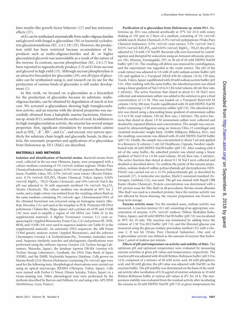
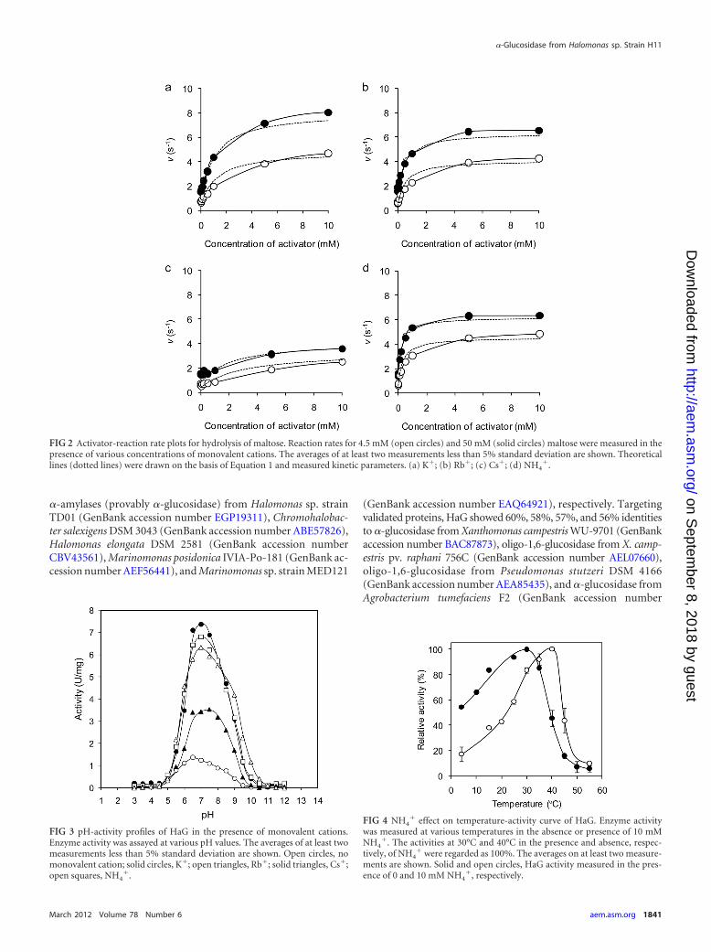
Activations for hydrolysis of maltose by monovalent cations were ki-netically analyzed in detail, based on a nonessential activator model (12).The reaction scheme for a reaction activated by a nonessential activator isshown in Fig. 1. In this model, rate equations 1 and 2 below were givenunder the assumption of rapid equilibration:
v �KA�kcat1 · KAS � kcat2 · A�S
Km · KAS�KA � A� � KA�KAS � A�S(1)
where v is the initial hydrolysis rate, and A and S indicate the concentra-tions of the activator and substrate, respectively. The four dissociationconstants in this equation are related as shown in Equation 2:
KmA · KA � KAS · Km (2)
We determined the individual constants as follows. First, the initial hy-drolytic rates were measured at various concentrations of maltose (2 to 20mM), without an activator, to obtain kcat1 and Km. The values were calcu-
lated by fitting the experimental data to Equation 1 at A equal to 0 bynonlinear regression. Next, the hydrolytic rates were measured at a sub-strate concentration much higher than that for Km in the presence ofvarious concentrations of activators (0.001 to 100 mM). The kcat2 and KAS
values were calculated from the concentration in the activator-rate plot.Finally, the enzyme assay was carried out with maltose at the same con-centration as that for Km in the presence of various concentrations ofactivators (0.001 to 100 mM). KA was determined from the concentrationof the activator-hydrolytic rate plot as described above. Then KmA wasobtained by substituting the other parameters for equation 2. To verify thecalculated parameters, the experimental hydrolytic rates for 4.5 mM or 50mM maltose in the presence of various concentrations of activators werecompared with the theoretical values calculated from the obtained param-eters.
Transglucosylation activity toward various alcohols. The trans-glucosylation activities toward ethanol, glycerol, propylene glycol,1-propanol, and 2-propanol (all reagents were purchased from KantoChemical) were qualitatively analyzed by thin-layer chromatography. Re-action mixtures (10 �l) containing 5% (vol/vol) alcohols, 10% (wt/vol)maltose, 50 mM HEPES-NaOH buffer (pH 7.0), 5 mM (NH4)2SO4, and0.18 �g/�l of HaG were incubated at 37°C for 24 h. A 10-fold-dilutedsample (1 �l) was analyzed by thin-layer chromatography (Silica Gel 60F254; Merck, Darmstadt, Germany) with a solvent system consisting of2-propanol–1-butanol–water at 2:2:1 (vol/vol/vol). Glucose and maltose(each 1% [wt/vol]) were used as standards. The chromatograms werevisualized by spraying with a solvent consisting of 10% (vol/vol) sulfuricacid in methanol and heating.
Enzymatic synthesis of �GG. The production efficiencies of �GG byHaG and Aspergillus niger �-glucosidase (AnG) were compared; AnG hasbeen reported to produce �GG efficiently (50). A reaction mixture (1 ml)containing 20% (wt/vol) maltose, 20% (wt/vol) glycerol, 0.04 U/ml ofpurified HaG or AnG, and 40 mM reaction buffer (HEPES-NaOH buffercontaining 5 mM ammonium sulfate, pH 7.0, for HaG and sodium acetatebuffer, pH 5.0, for AnG) was incubated at 37°C. AnG was purchased fromAmano Enzymes (Nagoya, Japan), and the activity was assayed using thesame method as that used for HaG except for the reaction buffer, whichwas 40 mM sodium acetate buffer (pH 5.0). The total �GG was quantifiedby high-performance liquid chromatography (HPLC) under the follow-ing conditions: column, Ultron PS-80N.L (8.0 by 500 mm; Shinwa Chem-ical Industries, Kyoto, Japan); eluant, water; flow rate, 0.9 ml/min; col-umn temperature, 50°C; detection, refractive index. To determine theratio of stereoisomers of the product, �GG was purified with HPLC. �GG(5 mg) was trimethylsilylated using TMSI-C reagent (GL Sciences, Tokyo,Japan) and analyzed by gas chromatography (49).
N-terminal amino acid sequence analysis. Purified �-glucosidasewas transferred to a polyvinylidene difluoride membrane (Immobilon-PSQ, Millipore) using a semidry electroblotting apparatus (Bio-Rad). Theprotein band detected with Coomassie brilliant blue R-250 (Wako PureChemical Industries) was cut off and analyzed using a protein sequencer(Procise 492cLC; Applied Biosystems). To determine the internal aminoacid sequence, purified HaG was digested by lysyl endopeptidase, and theresulting peptides were sequenced as follows. The purified enzyme (55�g) was dissolved in 100 �l of 20 mM Tris-HCl (pH 8.0) containing 8 Murea. Then 3 �l of 0.02% (wt/vol) lysyl endopeptidase (Wako Pure Chem-ical Industries) was added, followed by incubation at 37°C overnight. Theresultant peptides were separated by reverse-phase HPLC under the fol-lowing conditions: column, ODS AS-303 (4.6 by 250 mm; YMC, Kyoto,Japan); flow rate, 1 ml/min; column temperature, 40°C; detection, UV215 nm; elution, linear gradient of 0 to 100% acetonitrile in 0.1% trifluo-roacetic acid for 60 min.
Cloning of HaG gene. Chromosomal DNA of Halomonas sp. H11 wasprepared using a previously described method (41) and digested with SalIor PstI (Takara Bio) to connect cassette DNA fragments supplied by an LAPCR in vitro cloning kit (Takara Bio). The resulting DNA fragments wereused as a template in the PCR to amplify the HaG gene. All the primers
FIG 1 Kinetic model for the reaction activated by a nonessential activator. E,S, P, and A indicate enzyme, substrate, product, and activator, respectively. Km
and KmA denote the dissociation constants for the ES complex and ESA ternarycomplex, respectively, to EA. KA and KAS are the dissociation constants for theEA complex and ESA ternary complex, respectively, to ES. kcat1 and kcat2 arerate constants in the absence and presence, respectively, of an activator.
Ojima et al.
1838 aem.asm.org Applied and Environmental Microbiology
on Septem
ber 8, 2018 by guesthttp://aem
.asm.org/
Dow
nloaded from
used in this research are listed in Table S1 in the supplemental material. ExTaq polymerase (Takara Bio) and a set of primers, HAG-N1, designed onthe basis of the N-terminal amino acid sequence of HaG, and primer CS1,corresponding to the cassette sequence, were used. To amplify the desiredregion specifically, nested PCR was performed with primers HAG-N2 andCS2. The amplified DNA fragment was cloned into a pGEM-T Easy vector(Promega, Madison, WI) and sequenced with primers HAG-F=3, HAG-F=4, HAG-F=5, HAG-F=6, HAG-R=1, HAG-R=4, pGEM-up-F, and pGEM-dn-R. Sequence analyses were performed using a BigDye Terminator cyclesequencing kit (Beckman Coulter, Inc., Brea, CA) and a DNA sequencer,CEQ2000XL (Beckman Coulter). To obtain the remaining regions of theHaG gene, PCR was performed with two primer sets designed on the basisof the analyzed DNA sequence, HAG-I1 and CS1 and HAG-I2 and CSI.Nested PCR was followed by PCR with primers HAG-F=1 and CS2 andprimers HAG-I3 and CS2, respectively. Amplified DNA fragments werecloned into the pGEM-T Easy vector and sequenced. A homology searchwas performed using the Basic Local Alignment Search Tool (BLAST)program (5).
Nucleotide sequence accession number. The sequence data for HaGare available from DDBJ with accession number AB663683.
RESULTSIdentification of bacterial strain H11 isolated from coral. A bac-terial strain, H11, isolated from coral was selected as the enzymesource because it appeared to produce an enzyme harboring strongtransglucosylation activity toward glycerol when it was cultivatedwith the medium containing soluble starch. A phylogenetic tree con-structed with its 16S rRNA sequence showed that the H11 strain isclosely related to Halomonas aquamarina, Halomonas meridiana, andHalomonas axialensis and is likely to be categorized in the Halomonasgenus (see Fig. S1 in the supplemental material). Its 16S rRNA se-quence showed 99.5% identities to the 16S rRNA sequences of bothH. aquamarina DMS 30161 and H. meridiana DMS 5425. Physiolog-ical tests revealed that the H11 strain is rod shaped (0.7 to 0.8 by 1.2 to2.0 �m), pale yellow, smooth surfaced, Gram negative, mobile, andcapable of growing at 4 to 45°C. It showed catalase, oxidase, urease,and cytochrome oxidase activities. Otherwise, all of the followingwere negative: nitrate reduction; production of indole; glucose oxi-dation; arginine dihydrolase and �-galactosidase activities; lipaseactivity for Tween 80; hydrolysis of esculin, gelatin, starch, andcasein; and use of glucose, L-arabinose, D-mannose, D-mannitol,N-acetylglucosamine, maltose, potassium gluconate, n-capric acid,adipic acid, DL-malic acid, sodium citrate, and phenyl acetate. Thesefeatures were not totally identical to those of any Halomonas speciesreported so far (23, 53). Therefore, the H11 strain was named Halo-monas sp. H11 and deposited at the National Biological ResourceCenter (Chiba, Japan) as NBRC 108813.
Purification of HaG. HaG was purified from a cell extract tohomogeneity by several types of column chromatography (see Ta-ble S2 in the supplemental material). After hydrophobic columnchromatography with Toyopearl Butyl-650 M, the total enzymeactivity significantly increased because of activation by NH4
�, de-scribed in detail later. The purified enzyme showed a single bandin SDS-PAGE (see Fig. S2 in the supplemental material), and itsmolecular mass was estimated to be 58 kDa. It showed 50.9-foldhigher specific activity than that of the crude extract.
Effects of pH and temperature. The effects of pH and temper-ature on enzyme activity and stability were investigated, and theseare summarized in Fig. S3 in the supplemental material. The max-imum activity was observed at pH 6.5 when the buffer was 20 mMBritton-Robinson buffer. In 40 mM HEPES-NaOH (pH 7.0), theoptimum temperature for HaG was 30°C, and interestingly, it
exhibited more than 50% of the maximum activity even at 4°C. Aswill be mentioned in detail later, the monovalent cation in thebuffer used was important for both pH activity and temperatureactivity. HaG was stable in the range from medium pH to thealkaline side at 4°C for 24 h. After treatment at 4 to 60°C for 15min, the original activity was retained below 45°C but abruptlydecreased above 50°C.
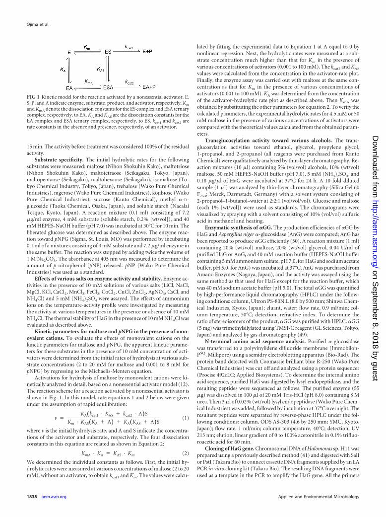
Substrate specificity. The hydrolytic rates for various sub-strates were measured (Table 1). HaG showed high activity formaltose, pNPG, and sucrose, in this order. The hydrolysis rates forisomaltose, trehalose, nigerose, and maltotriose were negligible,and kojibiose was not hydrolyzed at all. Malto-oligosaccharideslonger than maltotriose, starch, and methyl �-D-glucoside wererarely hydrolyzed.
Effects of various salts on HaG activity. The effects of varioussalts on HaG activity were investigated. The relative activities inthe presence of a 10 mM salt concentration, where the activitywith no salt was regarded as 100%, were as follows: LiCl, 151%;NaCl, 116%; KCl, 601%; RbCl, 579%; CsCl, 332%; NH4Cl, 548%;MgCl2, 30%; CaCl2, 20%; CoCl2, 1%; and ZnCl2, MnCl2, FeCl2,CuCl, and AgNO3, not detectable. (NH4)2SO4 also caused an in-crease in HaG activity to 545% when the molar concentration ofNH4
� was equal to that of NH4Cl. The results indicate that HaGactivity was obviously activated by monovalent cations. On theother hand, HaG activity was inhibited by divalent cations such asMg2� and Ca2�.
Kinetic analysis for activation of HaG by monovalent cat-ions. The results described above showed that monovalent cat-ions, particularly K�, Rb�, NH4
�, and Cs�, were strong activatorsfor HaG. To clarify the effects of these monovalent cations, theapparent kinetic parameters for maltose and pNPG in the absenceor presence of a 10 mM concentration of activators were measured(Table 2). The apparent Km (Kmapp) and apparent kcat (kcatapp)values for maltose in the absence of monovalent cations were 4.50mM and 0.80 s�1, respectively. These values were most improvedto 1.45 mM and 7.43 s�1 by NH4
� and K�, respectively. Thekcatapp/Kmapp value was increased 22.4-fold by NH4
� relative tothat without any monovalent cations. For pNPG, Kmapp and kcatapp
were 4.64 mM and 1.00 s�1, respectively, in the absence of anactivator. They were almost the same as those for maltose. On the
TABLE 1 Hydrolytic rates for various substrates
SubstrateHydrolytic rate(s�1)
Relative hydrolysisactivitya (%)
Maltose 0.763 100Isomaltose 0.016 1.1Trehalose 0.024 2.6Nigerose 0.032 4.2Kojibiose NDb NDMaltotriose 0.040 5.3Maltotetraose ND NDMaltopentaose ND NDMaltohexaose ND NDSucrose 0.253 33Methyl-�-D-glucoside ND NDSoluble starch ND NDpNPG 0.439 57.5a Reaction rates on 4 mM each substrate (expect soluble starch was 0.2% [wt/vol]) areshown, where the activity to maltose is 100 for calculating relative activity.b ND, not detectable.
�-Glucosidase from Halomonas sp. Strain H11
March 2012 Volume 78 Number 6 aem.asm.org 1839
on Septem
ber 8, 2018 by guesthttp://aem
.asm.org/
Dow
nloaded from
other hand, Kmapp was significantly improved to about 0.02 mM inthe presence of all the monovalent cations tested. Although thechanges were less, kcatapp values were also improved, to 4.36 s�1,3.29 s�1, 1.55 s�1, and 3.59 s�1 by K�, Rb�, Cs�, and NH4
�,respectively. The kcatapp/Kmapp values were increased 969-fold,893-fold, 372-fold, and 929-fold by K�, Rb�, Cs�, and NH4
�,respectively.
Next, the kinetic parameters for maltose hydrolysis in a non-essential activation model were measured to evaluate the activa-tion by K�, Rb�, NH4
�, and Cs� (Table 3). The measured reac-tion rates for 4.5 mM or 50 mM maltose with variousconcentrations of activators fitted well to the theoretical lines,indicating that activation by monovalent cations was explainedwell by a nonessential activation model (Fig. 2). The activationdegree reached almost maximum at 10 mM each activator andeven over the range of 10 to 100 mM. The actual hydrolytic ratesfor 4.5 mM and 50 mM maltose in the absence of an activator were0.71 s�1 and 1.53 s�1, respectively, whereas those for 4.5 mMmaltose increased 8.3-fold, 5.9-fold, 5.0-fold, and 7.1-fold in thepresence of 50 mM K�, Rb�, Cs�, and NH4
�, respectively. Simi-larly, the rates for 50 mM maltose were increased 5.7-fold, 4.0-fold, 2.7-fold, and 4.3-fold by the same concentration of the re-spective monovalent cations.
The KA and KAS values for NH4�, which mirror the affinity of
the activator with the enzyme and the enzyme-substrate complex,respectively, were the lowest among the monovalent cationstested, followed by Rb�, K�, and Cs�, in this order. The KmA
values were as follows: Cs�, 1.19 mM; NH4�, 1.86 mM; Rb�, 2.88
mM; and K�, 3.52 mM. The kcat2 value for the hydrolysis of malt-ose in the presence of K� was the best of those for the monovalentcations tested, and those with Rb�, NH4
�, and Cs� followed, inthis order.
Effects of monovalent cations on pH-activity and temperature-activity. The effects of NH4
� on enzyme activity at various pHvalues and temperatures were evaluated. As shown in Fig. 3, theactivities at all pH values were raised by monovalent cations in
the following order: K�, Rb�, NH4�, and Cs�. These ions shifted
the bell-shaped pH-activity curve to the alkaline side. The opti-mum pH in the absence of monovalent cations was 6.5, that in thepresence of 10 mM K�, Rb�, and NH4
� was 7.0, and that in thepresence of Cs� was 7.5. NH4
� was most effective in increasingthe activity around the alkaline side. Interestingly, the optimumtemperature for HaG activity was shifted from 30°C to 40°C in thepresence of 10 mM NH4
� (Fig. 4), although stability was not af-fected (data not shown). The actual activity in the presence ofNH4
� at 40°C was 11.6-fold higher than that in its absence.Transglucosylation toward various alcohols and enzymatic
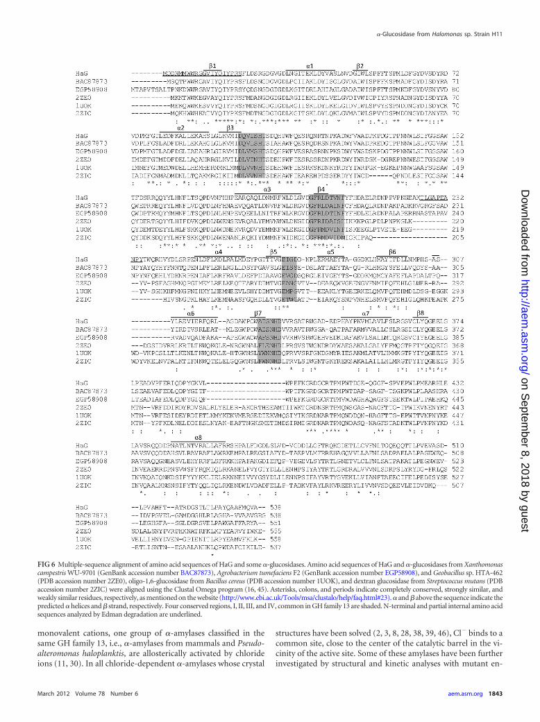
synthesis of �-D-glucosylglycerol. The transglucosylation activi-ties toward various alcohols, i.e., ethanol, glycerol, propylene gly-col, 1-propanol, and 2-propanol, were investigated. Transglu-cosylation products were detected in all the reactions tested (datanot shown). As expected from the results on substrate specificity,no oligosaccharides were produced by transglucosylation. The effi-ciencies of the synthesis of �GG by HaG and AnG were comparedin the absence and presence of 10 mM NH4
�. The time course of�GG production is shown in Fig. 5a. The production rate andyield of �GG by HaG in the presence of NH4
� were the highestamong the four cases tested. The carbohydrate contents (percent,wt/wt) of the reaction mixture at 816 h are summarized in Fig. 5b.In the reaction of HaG in the presence of NH4
�, the content of�GG represented 29.4% (wt/wt) of the total carbohydrates (themolar yield of �GG from converted maltose was 92.7%), and the�GG content for the reaction of AnG was about 8% (wt/wt), in-dependent of NH4
�. The accumulations of glucose and trisaccha-rides (G3) were considerably less in the reaction with HaG than inthat with AnG. The compositions of the stereoisomers of �GG at816 h were analyzed by gas chromatography, revealing that theratios of the constituent isomers 2-O-�GG/(2R)-1-O-�GG/(2S)-1-O-�GG were 10:52:38 for HaG and 7:73:20 for AnG.
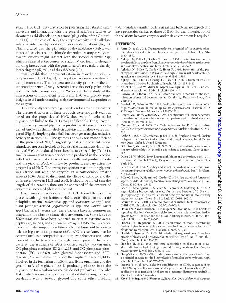
Sequence analysis of HaG. The N-terminal amino acid se-quence of purified HaG was MQDNMMWWRGGVIYQIYPRS.Primers were designed on the basis of this sequence, and the HaGgene was obtained by the cassette PCR method (22). The HaGgene consisted of 1,617 bases and encoded 538 amino acids (Fig.6). The theoretical molecular mass and pI were 61,139 Da and4.75, respectively. The partial internal peptide sequence TLGAPEANPY was found in this sequence. Four regions, I, II, III, and IV,conserved in the GH family 13 enzymes, were found in the aminoacid sequence of HaG, namely, DQVISH, GFRLDTVNF, EIGD,and ATSNHD. HaG was therefore obviously classified in GH fam-ily 13. The amino acid sequence of HaG exhibited high identities,of the order of 87%, 79%, 78%, 69%, and 68%, to those of putative
TABLE 2 Apparent kinetic parameters for maltose and pNPG in the absence or presence of monovalent cationsa
Activator
Maltose pNPG
kcatapp
(s�1)Kmspp
(mM)kcatapp/Kmapp
(s�1 mM�1)kcatapp/Kmapp
(fold)kcatapp
(s�1)Kmspp
(mM)kcatapp/Kmapp
(s�1 mM�1)kcatapp/Kmapp
(fold)
None 0.80 4.50 0.18 1 1.00 4.64 0.22 1K� 7.43 2.06 3.61 20.3 4.36 0.021 209 969Rb� 6.73 1.92 3.51 19.7 3.29 0.017 193 893Cs� 5.23 2.88 1.82 10.2 1.55 0.019 80.2 372NH4
� 5.77 1.45 3.98 22.4 3.59 0.018 200 929a The hydrolysis rate was assayed in the presence of 10 mM activator (cation) at various substrate concentrations. Parameters were calculated by regressing the experimental data tothe Michaelis-Menten equation.
TABLE 3 Kinetic parameters for hydrolysis of maltose in nonessentialactivator model
Activatorkcat1
(s�1)kcat2
(s�1)Km
(mM)KmA
(mM)KA
(mM)KAS
(mM)
K� 0.8 8.6 4.5 3.52 1.35 1.06Rb� 0.8 6.7 4.5 2.88 0.66 0.42Cs� 0.8 4.2 4.5 1.19 7.14 1.88NH4
� 0.8 6.4 4.5 1.86 0.54 0.22
Ojima et al.
1840 aem.asm.org Applied and Environmental Microbiology
on Septem
ber 8, 2018 by guesthttp://aem
.asm.org/
Dow
nloaded from
�-amylases (provably �-glucosidase) from Halomonas sp. strainTD01 (GenBank accession number EGP19311), Chromohalobac-ter salexigens DSM 3043 (GenBank accession number ABE57826),Halomonas elongata DSM 2581 (GenBank accession numberCBV43561), Marinomonas posidonica IVIA-Po-181 (GenBank ac-cession number AEF56441), and Marinomonas sp. strain MED121
(GenBank accession number EAQ64921), respectively. Targetingvalidated proteins, HaG showed 60%, 58%, 57%, and 56% identitiesto �-glucosidase from Xanthomonas campestris WU-9701 (GenBankaccession number BAC87873), oligo-1,6-glucosidase from X. camp-estris pv. raphani 756C (GenBank accession number AEL07660),oligo-1,6-glucosidase from Pseudomonas stutzeri DSM 4166(GenBank accession number AEA85435), and �-glucosidase fromAgrobacterium tumefaciens F2 (GenBank accession number
FIG 2 Activator-reaction rate plots for hydrolysis of maltose. Reaction rates for 4.5 mM (open circles) and 50 mM (solid circles) maltose were measured in thepresence of various concentrations of monovalent cations. The averages of at least two measurements less than 5% standard deviation are shown. Theoreticallines (dotted lines) were drawn on the basis of Equation 1 and measured kinetic parameters. (a) K�; (b) Rb�; (c) Cs�; (d) NH4
�.
FIG 3 pH-activity profiles of HaG in the presence of monovalent cations.Enzyme activity was assayed at various pH values. The averages of at least twomeasurements less than 5% standard deviation are shown. Open circles, nomonovalent cation; solid circles, K�; open triangles, Rb�; solid triangles, Cs�;open squares, NH4
�.
FIG 4 NH4� effect on temperature-activity curve of HaG. Enzyme activity
was measured at various temperatures in the absence or presence of 10 mMNH4
�. The activities at 30°C and 40°C in the presence and absence, respec-tively, of NH4
� were regarded as 100%. The averages on at least two measure-ments are shown. Solid and open circles, HaG activity measured in the pres-ence of 0 and 10 mM NH4
�, respectively.
�-Glucosidase from Halomonas sp. Strain H11
March 2012 Volume 78 Number 6 aem.asm.org 1841
on Septem
ber 8, 2018 by guesthttp://aem
.asm.org/
Dow
nloaded from
EGP58908), respectively. Identities to �-glucosidase from Geoba-cillus sp. strain HTA-462 (Protein Data Bank [PDB] accessionnumber 2ZE0) (43), trehalulose synthase from Pseudomonasmesoacidophia (PDB accession number 2PWH), oligo-1,6-glucosidase from Bacillus cereus (PDB accession number 1UOK)(54), and dextran glucosidase from Streptococcus mutans (PDBaccession number 2ZIC) (20), all of whose crystal structures havebeen solved, were 36%, 35%, 33%, and 31%, respectively.
DISCUSSION
In this work, we describe a unique �-glucosidase which exhibitsstrong transglucosylation activity toward alcohols and glyceroland has additional remarkable features, namely, stimulation bymonovalent cations and narrow substrate specificity. The enzymeis probably psychrophilic and prefers a moderate pH (Fig. 3; seeFig. S3 in the supplemental material). These conditions are wellsuited to the marine environment where Halomonas sp. H11 wasisolated. Halomonas sp. H11 was negative for the hydrolysis ofstarch and utilization of maltose or glucose under the conditionsof the physiological tests. However, it could utilize starch whensoluble starch was contained in the culture medium and producedHaG. This suggests that expression of enzymes that are related toglycoside hydrolysis is probably induced by derivatives of starch.
HaG was found to have specific activity toward maltose, sucrose,
and pNPG but rarely had activity toward longer malto-oligosaccharides or disaccharides other than maltose (Table 1). Sucha narrow specificity for the substrate chain length by HaG is remark-able because bacterial �-glucosidases generally hydrolyze not onlymaltose but also longer malto-oligosaccharides (21, 24, 36).
On the basis of multiple-sequence alignment, HaG was pre-dicted to have a longer � ¡ � loop 4, which connects �-strand 4and �-helix 4, than other enzymes (Fig. 6). The length of � ¡ �loop 4 is the determinant of the specificity for substrate chainlength in GH family 13 �-glucosidases and related enzymes, suchas oligo-1,6-glucosidase and dextran glucosidase (40). The shorterloop of dextran glucosidase, which forms open substrate bindingsites to accommodate long-chain substrates, has been shown to beassociated with a high preference for long-chain substrates. Com-pared with �-glucosidases and oligo-1,6-glucosidase, HaG has aneven longer � ¡ � loop 4. The length of the putative � ¡ � loop4 of �-glucosidase from X. campestris WU-9701 (GenBank acces-sion number BAC87873), which hydrolyzes almost only maltoseamong the malto-oligosaccharides, as does HaG (52), is the sameas that of HaG. We expect that such an unusually long � ¡ � loop4 might contribute to the narrow specificity for substrate chainlength in HaG.
HaG was greatly stimulated by monovalent cations, namely, K�,Rb�, NH4
�, and Cs�. Although numerous enzymes have been doc-umented to be activated by monovalent cations in the plant and an-imal world (44), most types of known �-glucosidases in GH family 13are independent of monovalent cations. There have been only tworeports documenting stimulation of �-glucosidase activity by mon-ovalent cations. Crude �-glucosidases from fast-growing rhizobiaand A. tumefaciens were reported to be stimulated by K�, NH4
�, andRb� (19), and purified �-glucosidase from Rhizobium sp. strainUSDA 4280 was reported to be stimulated to 141% and 207% byaddition of 10 mM K� and NH4
�, respectively (7). However, such�-glucosidases have as yet rarely been investigated in detail. HaGexhibited high sequence identity (56%) to �-glucosidase from A. tu-mefaciens F2 (GenBank accession number EGP58908), implying thatthese enzymes share a monovalent cation binding site structure andactivation mechanism.
We determined the activation kinetics and enzymologicalproperties of HaG. The monovalent cation activators K�, Rb�,NH4
�, and Cs� for HaG have similar ionic diameters. In particu-lar, the ionic diameters of Rb� and NH4
� are almost the same(Rb�, 1.48 Å; NH4
�, 1.43 Å). These ions showed almost the sameKA and KAS values (Table 3). In addition, the kcat2 values for malt-ose hydrolysis in the presence of these ions were also similar. TheKA and KAS values for Cs�, which has an ionic diameter of 1.70 Å,were higher than those for the other ions. The kcat2 value washighest with the smallest ion, K�, whose diameter is 1.33 Å. Theseresults indicate that the activator binding site of HaG is suitable forbinding to monovalent ions with ionic diameters of about 1.4 Å.
The Kmapp and kcatapp values for maltose and pNPG were im-proved by the addition of K�, Rb�, Cs�, and NH4
� (Table 2). Inparticular, the Kmapp values for pNPG were significantly im-proved, regardless of the type of monovalent cation. Enzyme-pNPG complexes might be stabilized by interaction between thenegative charge of the nitrate group of pNP and a monovalentcation bound to the substrate binding site of the enzyme. Mon-ovalent cation activators may therefore be involved in the forma-tion of the substrate binding sites of HaG.
Unlike the minority of �-glucosidases, which are stimulated by
FIG 5 Enzymatic synthesis of �GG. (a) Time course of production of �GG.�GG (percent, wt/vol) produced in the reaction mixture with 20% (wt/vol)maltose, 20% (wt/vol) glycerol, and 0.04 U/ml enzymes. Open and solid cir-cles, reaction with purified HaG in the absence and presence of 10 mM NH4
�,respectively; open and solid triangles, reaction with purified HaG in the ab-sence and presence of �-glucosidase from Aspergillus niger, respectively. (b)Content (percent, wt/wt) of carbohydrates at a reaction time of 816 h. Plus andminus signs, presence and absence of NH4
�, respectively. The carbohydratecontent is equivalent to the area percentage of the detector, i.e., the refractiveindex response, in HPLC analysis. G1, glucose; �GG, �-glucosylglycerol; G2,disaccharides; G3, trisaccharides.
Ojima et al.
1842 aem.asm.org Applied and Environmental Microbiology
on Septem
ber 8, 2018 by guesthttp://aem
.asm.org/
Dow
nloaded from
monovalent cations, one group of �-amylases classified in thesame GH family 13, i.e., �-amylases from mammals and Pseudo-alteromonas haloplanktis, are allosterically activated by chlorideions (11, 30). In all chloride-dependent �-amylases whose crystal
structures have been solved (2, 3, 8, 28, 38, 39, 46), Cl� binds to acommon site, close to the center of the catalytic barrel in the vi-cinity of the active site. Some of these amylases have been furtherinvestigated by structural and kinetic analyses with mutant en-
FIG 6 Multiple-sequence alignment of amino acid sequences of HaG and some �-glucosidases. Amino acid sequences of HaG and �-glucosidases from Xanthomonascampestris WU-9701 (GenBank accession number BAC87873), Agrobacterium tumefaciens F2 (GenBank accession number EGP58908), and Geobacillus sp. HTA-462(PDB accession number 2ZE0), oligo-1,6-glucosidase from Bacillus cereus (PDB accession number 1UOK), and dextran glucosidase from Streptococcus mutans (PDBaccession number 2ZIC) were aligned using the Clustal Omega program (16, 45). Asterisks, colons, and periods indicate completely conserved, strongly similar, andweakly similar residues, respectively, as mentioned on the website (http://www.ebi.ac.uk/Tools/msa/clustalo/help/faq.html#23). � and � above the sequence indicate thepredicted � helices and � strand, respectively. Four conserved regions, I, II, III, and IV, common in GH family 13 are shaded. N-terminal and partial internal amino acidsequences analyzed by Edman degradation are underlined.
�-Glucosidase from Halomonas sp. Strain H11
March 2012 Volume 78 Number 6 aem.asm.org 1843
on Septem
ber 8, 2018 by guesthttp://aem
.asm.org/
Dow
nloaded from
zymes (4, 30); Cl� may play a role by polarizing the catalytic watermolecule and interacting with the general acid/base catalyst toelevate the acid dissociation constant (pKa) value of the Glu resi-due (14). In the case of HaG, the enzyme activity at the alkalineside was enhanced by addition of monovalent cations (Fig. 3).This indicated that the pKa value of the acid/base catalyst wasincreased, as observed in chloride-dependent �-amylases. Mon-ovalent cations might interact with the second catalytic Asp,which is situated at the conserved region IV and forms hydrogen-bonding interactions with the general acid/base catalyst, therebyincreasing the pKa value of the acid/base (30).
It was notable that monovalent cations increased the optimumtemperature of HaG (Fig. 4), but as yet we have no explanation forthis phenomenon. The temperature-activity profiles in the ab-sence and presence of NH4
� were similar to those of psychrophilicand mesophilic �-amylases (13). We expect that a study of theinteractions of monovalent cations and HaG will provide someinsights to aid understanding of the environmental adaptation ofthe enzyme.
HaG efficiently transferred glucosyl residues to some alcohols.The precise structures of these products were not analyzed, butbased on the properties of HaG, they were thought to be�-glucosides linked to the OH groups of alcohols. The glucosyla-tion efficiency toward glycerol to produce �GG was superior tothat of AnG when their hydrolysis activities for maltose were com-pared (Fig. 5), implying that HaG has stronger transglucosylationactivity than does AnG. The synthesis of �GG was more efficientin the presence of NH4
�, suggesting that a monovalent cationstimulated not only hydrolysis but also the transglucosylation ac-tivity of HaG. As deduced from the substrate specificity (Table 1),smaller amounts of trisaccharides were produced in the reactionwith HaG than in that with AnG. Such an efficient production rateand the yield of �GG, with few by-products, are very attractiveproperties of HaG. The transglucosylation reaction for glycerolwas carried out with the enzymes in a considerably smalleramount (0.04 U/ml) to distinguish the effects of activator and thedifferences between HaG and AnG. It should be noted that thelength of the reaction time can be shortened if the amount ofenzymes is increased (data not shown).
A sequence similarity search by BLAST showed that putativeproteins with high similarities to HaG are distributed in moderatehalophilic, marine (Halomonas spp. and Marinomonas spp.), andplant pathogen-related (Agrobacterium spp. and Xanthomonasspp.) bacteria. It seems that these bacteria have in common anadaptation to saline or nitrate-rich environments. Some kinds ofHalomonas spp. have been reported to exist at extreme oceandepths (23, 42, 51), and Halomonas elongata DMS 3043 is knownto accumulate compatible solutes such as ectoine and betaine tobalance high osmotic pressure (55). �GG is also known to beaccumulated as a compatible solute in cyanobacteria and someosmotolerant bacteria to adopt a high osmotic pressure. In cyano-bacteria, the synthesis of �GG is carried out by two enzymes,GG-phosphate synthase (EC 2.4.1.213) and GG-phosphate phos-phatase (EC. 3.1.3.69), with glycerol 3-phosphate and ADP-glucose (25). As there is no report that �-glucosidases might beinvolved in the formation of �GG in any living organisms and thegeneral task of �-glucosidase is producing glucose from the�-glucoside for a carbon source, we do not yet have an idea whyHaG hydrolyzes maltose specifically and exhibits strong transglu-cosylation activity toward glycerol and some other alcohols.
�-Glucosidases similar to HaG in marine bacteria are expected tohave properties similar to those of HaG. Further investigation ofthe relations between enzymes and their environment is required.
REFERENCES1. Aerts D, et al. 2011. Transglucosylation potential of six sucrose phos-
phorylases toward different classes of acceptors. Carbohydr. Res. 346:1860 –1867.
2. Aghajari N, Feller G, Gerday C, Haser R. 1998. Crystal structures of thepsychrophilic �-amylase from Alteromonas haloplanctis in its native formand complexed with an inhibitor. Protein Sci. 7:564 –572.
3. Aghajari N, Feller G, Gerday C, Haser R. 1998. Structures of the psy-chrophilic Alteromonas haloplanctis �-amylase give insights into cold ad-aptation at a molecular level. Structure 6:1503–1516.
4. Aghajari N, Feller G, Gerday C, Haser R. 2002. Structural basis of�-amylase activation by chloride. Protein Sci. 11:1435–1441.
5. Altschul SF, Gish W, Miller W, Myers EW, Lipman DJ. 1990. Basic localalignment search tool. J. Mol. Biol. 215:403– 410.
6. Barrow GI, Feltham RKA. 1993. Cowan and Steel’s manual for the iden-tification of medical bacteria, 3rd ed. Cambridge University Press, NewYork, NY.
7. Berthelot K, Delmotte FM. 1999. Purification and characterization of an�-glucosidase from Rhizobium sp. (Robinia pseudoacacia L.) strain USDA4280. Appl. Environ. Microbiol. 65:2907–2911.
8. Brayer GD, Luo Y, Withers SG. 1995. The structure of human pancreatic�-amylase at 1.8 Å resolution and comparisons with related enzymes.Protein Sci. 4:1730 –1742.
9. Cantarel BL, et al. 2009. The Carbohydrate-Active EnZymes database(CAZy): an expert resource for glycogenomics. Nucleic Acids Res. 37:233–238.
10. Chiba S. 1988. �-Glucosidases, p 104 –116. In Amylase Research Societyof Japan (ed), Handbook of amylases and related enzymes, 1st ed. Perga-mon Press, Oxford, United Kingdom.
11. D’Amico S, Gerbay C, Feller G. 2000. Structural similarities and evolu-tionary relationships in chloride-dependent �-amylases. Gene 253:95–105.
12. Dixon M, Webb EC. 1979. Enzyme inhibition and activation, p 389 –391.In Dixon M, Webb EC (ed), Enzymes, 3rd ed. Academic Press, NewYork, NY.
13. Feller G, et al. 1994. Stability and structural analysis of �-amylase fromthe Antarctic psychrophile Alteromonas haloplanctis A23. Eur. J. Biochem.222:441– 447.
14. Feller G, Bussy O, Houssier C, Gerday C. 1996. Structural and functionalaspects of chloride binding to Alteromonas haloplanctis �-amylase. J. Biol.Chem. 271:23836 –23841.
15. Goedl C, Sawangwan T, Mueller M, Schwarz A, Nidetzky B. 2008. Ahigh-yielding biocatalytic process for the production of 2-O-(�-D-glucopyranosyl)-sn-glycerol, a natural osmolyte and useful moisturizingingredient. Angew. Chem. Int. Ed. Engl. 47:10086 –10089.
16. Goujon M, et al. 2010. A new bioinformatics analysis tools framework atEMBL-EBI. Nucleic Acids Res. 38:W695–W699.
17. Harada N, Zhao J, Kurihara H, Nakagata N, Okajima K. 2010. Effects oftopical application of �-D-glucosylglycerol on dermal levels of insulin-likegrowth factor-I in mice and facial skin elasticity in humans. Biosci. Bio-technol. Biochem. 74:759 –765.
18. Hincha DK, Hagemann M. 2004. Stabilization of model membranesduring drying by compatible solutes involved in the stress tolerance ofplants and microorganisms. Biochem. J. 383:277–283.
19. Hoelzle I, Streeter JG. 1989. Stimulation of �-glucosidases from fast-growing rhizobia and Agrobacterium tumefaciens by K�, NH4
�, and Rb�.Can. J. Microbiol. 36:223–227.
20. Hondoh H, et al. 2008. Substrate recognition mechanism of �-1,6-glucosidic linkage hydrolyzing enzyme, dextran glucosidase from Strepto-coccus mutans. J. Mol. Biol. 378:911–920.
21. Hung VS, et al. 2005. �-Glucosidase from a strain of deep-sea Geobacillus:a potential enzyme for the biosynthesis of complex carbohydrates. Appl.Microbiol. Biotechnol. 68:757–765.
22. Isegawa Y, et al. 1992. Selective amplification of cDNA sequence fromtotal RNA by cassette-ligation mediated polymerase chain reaction (PCR):application to sequencing 6.5 kb genome segment of hantavirus strain B-1.Mol. Cell. Probes 6:467– 475.
23. Kaye JZ, Márquez MC, Ventosa A, Baross JA. 2004. Halomonas neptunia
Ojima et al.
1844 aem.asm.org Applied and Environmental Microbiology
on Septem
ber 8, 2018 by guesthttp://aem
.asm.org/
Dow
nloaded from
sp. nov., Halomonas sulfidaeris sp. nov., Halomonas axialensis sp. nov. andHalomonas hydrothermalis sp nov.: halophilic bacteria isolated from deep-sea hydrothermal-vent environments. Int. J. Syst. Evol. Microbiol. 54:499 –511.
24. Kelly CT, Fogarty W. 1983. Microbial �-glucosidases. Process Biochem.18:6 –12.
25. Klähn S, Hagemann M. 2011. Compatible solute biosynthesis in cyano-bacteria. Environ. Microbiol. 13:551–562.
26. Kurosu J, et al. 2002. Enzymatic synthesis of �-arbutin by �-anomer-selective glucosylation of hydroquinone using lyophilized cells of Xan-thomonas campestris WU-9701. J. Biosci. Bioeng. 93:328 –330.
27. Laemmli UK. 1970. Cleavage of structural proteins during the assembly ofthe head of bacteriophage T4. Nature 227:680 – 685.
28. Larson SB, Greenwood A, Cascio D, Day J, McPherson A. 1994. Refinedmolecular structure of pig pancreatic �-amylase at 2.1 Å resolution. J. Mol.Biol. 235:1560 –1584.
29. Luley-Goedl C, Nidetzky B. 2010. Carbohydrate synthesis by disaccha-ride phosphorylases: reactions, catalytic mechanisms and application inthe glycosciences. Biotechnol. J. 5:1324 –1338.
30. Maurus R, et al. 2005. Structural and mechanistic studies of chlorideinduced activation of human pancreatic �-amylase. Protein Sci. 14:743–755.
31. McCarter JD, Withers SG. 1996. Unequivocal identification of Asp-214as the catalytic nucleophile of Saccharomyces cerevisiae �-glucosidase us-ing 5-fluoro glycosyl fluorides. J. Biol. Chem. 271:6889 – 6894.
32. Miwa I, Okuda J, Maeda K, Okuda G. 1972. Mutarotase effect oncolorimetric determination of blood glucose with �-D-glucose oxidase.Clin. Chim. Acta 37:538 –540.
33. Murase H, Yamauchi R, Kato K, Kunieda T, Terao J. 1997. Synthesis ofa novel vitamin E derivative, 2-(�-D-glucopyranosyl) methyl-2,5,7,8-tetramethylchroman-6-ol, by �-glucosidase-catalyzed transglycosylation.Lipids 32:73–78.
34. Nakagawa K, Kawasaki H. 2001. DNA sequence analysis, p 83–117. InSociety for Actinomycetes Japan (ed), Identification manual of Actinomy-cetes. Business Center for Academic Societies Japan, Tokyo, Japan.
35. Nakano H, et al. 2003. Synthesis of glycosyl glycerol by cyclodextringlucanotransferases. J. Biosci. Bioeng. 95:583–588.
36. Nakao M, et al. 1994. Structure and expression of gene coding for ther-mostable �-glucosidase with broad substrate specificity from Bacillus. sp.SAM1606. Eur. J. Biochem. 220:293–300.
37. Nitta A, Takenaka F, Iki M, Matsumura E, Sakaguchi M. October 2007.Antitumor agents containing a-D-glucosylglycerols and food and cosmet-ics containing them. JP patent 2007-262023.
38. Qian M, Haser R, Payan F. 1993. Structure and molecular model refine-ment of pig pancreatic �-amylase at 2.1 Å resolution. J. Mol. Biol. 231:785–799.
39. Ramasubbu N, Paloth V, Luo Y, Brayer GD, Levine MJ. 1996. Structureof human salivary �-amylase at 1.6 Å resolution: implication for its role inthe oral cavity. Acta Crystallogr. D Biol. Crystallogr. 52:435– 446.
40. Saburi W, Mori H, Saito S, Okuyama M, Kimura A. 2006. Structural
elements in dextran glucosidase responsible for high specificity to longchain substrate. Biochim. Biophys. Acta 1764:688 – 698.
41. Sambrook J, Russell DW. 2001. Molecular cloning: a laboratory manual,3rd ed. Cold Spring Harbor Laboratory Press, Cold Spring Harbor, NY.
42. Sánchez-Porro C, Kaur B, Mann H, Ventosa A. 2010. Halomonas titani-cae sp. nov., a halophilic bacterium isolated from the RMS Titanic. Int. J.Syst. Evol. Microbiol. 60:2768 –2774.
43. Shirai T, Hung VS, Morinaka K, Kobayashi T, Ito S. 2008. Crystalstructure of GH13 �-glucosidase GSJ from one of the deepest sea bacteria.Proteins 73:126 –133.
44. Shulter CH. 1970. Enzymes activated by monovalent cations. Science168:789 –795.
45. Sievers F, et al. 2011. Fast, scalable generation of high-quality proteinmultiple sequence alignments using Clustal Omega. Mol. Syst. Biol. 7:539.
46. Strobl S, et al. 1998. Crystal structure of yellow meal worm �-amylase at1.64 Å resolution. J. Mol. Biol. 278:617– 628.
47. Suzuki Y, Suzuki K. 1991. Enzymatic formation of 4G-�-D-glucopyranosyl-rutin. Agric. Biol. Chem. 55:181–187.
48. Takaku H. 1988. Manufacture of oligosaccharides, p 212–222. In AmylaseResearch Society of Japan (ed), Handbook of amylases and related en-zymes, 1st ed. Pergamon Press, Oxford, United Kingdom.
49. Takenaka F, Uchiyama H, Imamura T. 2000. Identification of �-D-glucosylglycerol in sake. Biosci. Biotechnol. Biochem. 64:378 –385.
50. Takenaka F, Uchiyama H. 2000. Synthesis of �-D-glucosyl glycerol by�-glucosidase and some its characteristics. Biosci. Biotechnol. Biochem.64:1821–1826.
51. Tamegai H, et al. 2006. Halomonas sp. strain DT-W, a halophile from the11,000 m depth of the Mariana Trench. Extremophiles 5:27–33.
52. Usami S, et al. February 2001. Production of glycoside with �-glucosidaseand new �-glucosidase and its production. JP patent 2001-46096.
53. Vreeland RH. 2005. Genus I. Halomonas, p 300 –313. In Garrity GM, et al.(ed), Bergey’s manual of systematic bacteriology, 2nd ed, vol 2, part B.Springer, New York, NY.
54. Watanabe K, Hata Y, Kizaki H, Katsube Y, Suzuki Y. 1997. The refinedcrystal structure of Bacillus cereus oligo-1,6-glucosidase at 2.0 Å resolu-tion: structural characterization of proline-substitution sites for proteinthermostabilization. J. Mol. Biol. 269:142–153.
55. Wohlfarth A, Severin J, Galinski EA. 1990. The spectrum of compatiblesolutes in heterotrophic halophilic eubacteria of the family Halomon-adaceae. J. Gen. Microbiol. 135:705–712.
56. Yamamoto I, Muto N. 1992. Bioavailability and biological activity ofL-ascorbic acid 2-O-alpha-glucoside. J. Nutr. Sci. Vitaminol. Spec. No.1992:161–164.
57. Yamamoto T, Unno T, Sugawara M, Goda T. 1999. Properties of anigeose and nigerosylmaltooligosaccharides-suppplemented syrup. J.Appl. Glycosci. 46:475– 482.
58. Yoshida K, Takenaka F, Nitta A, Iki M. June 2007. �-D-Glucopyranosylglycerol derivatives as antiallergic agents, health foods, and cosmetics. JPpatent 2007-137862.
�-Glucosidase from Halomonas sp. Strain H11
March 2012 Volume 78 Number 6 aem.asm.org 1845
on Septem
ber 8, 2018 by guesthttp://aem
.asm.org/
Dow
nloaded from


![3D characterization and modelling of small fatigue cracks ...irsp2016.malab.com/wp-content/uploads/2016/07/AT13... · Small strain crystal plasticity [Meric and Cailletaud, 1991]](https://static.fdocument.org/doc/165x107/5ec1f4b393e4a025a2723ab5/3d-characterization-and-modelling-of-small-fatigue-cracks-small-strain-crystal.jpg)