CHAPTER-I ARRHYTHMIA - SRM · PDF fileCHAPTER-I . ARRHYTHMIA. by: j. jayasutha. lecturer....
55
CHAPTER-I ARRHYTHMIA by: j. jayasutha lecturer department of pharmacy practice Srm college of pharmacy srm university
-
Upload
hoangquynh -
Category
Documents
-
view
227 -
download
4
Transcript of CHAPTER-I ARRHYTHMIA - SRM · PDF fileCHAPTER-I . ARRHYTHMIA. by: j. jayasutha. lecturer....
CHAPTER-I
ARRHYTHMIAby:
j. jayasuthalecturer
department of pharmacy practiceSrm college of pharmacy
srm university
D���������
Irregular heart beat (or) loss of cardiac rhythm
Cardiac arrhythmia is the disturbance of normal
rhythm of the heart may be due to
alterations in impulse generation or
disturbances in impulse conduction or
a combination of both these factors
Normal range 70-80 beats/min
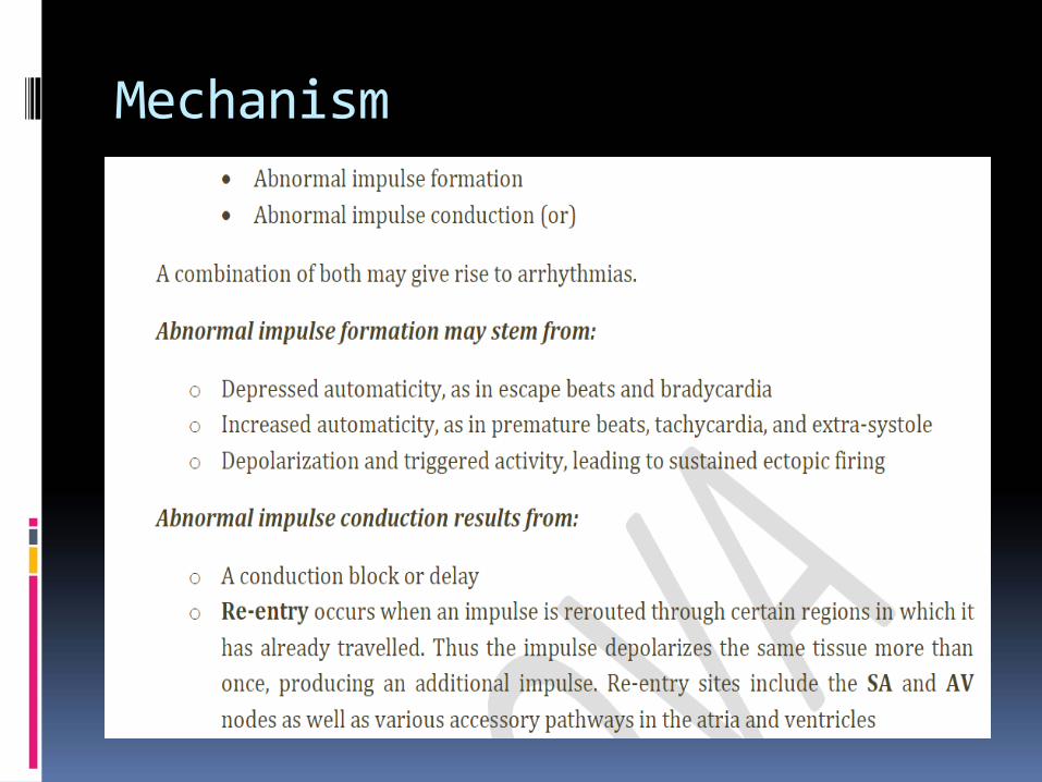
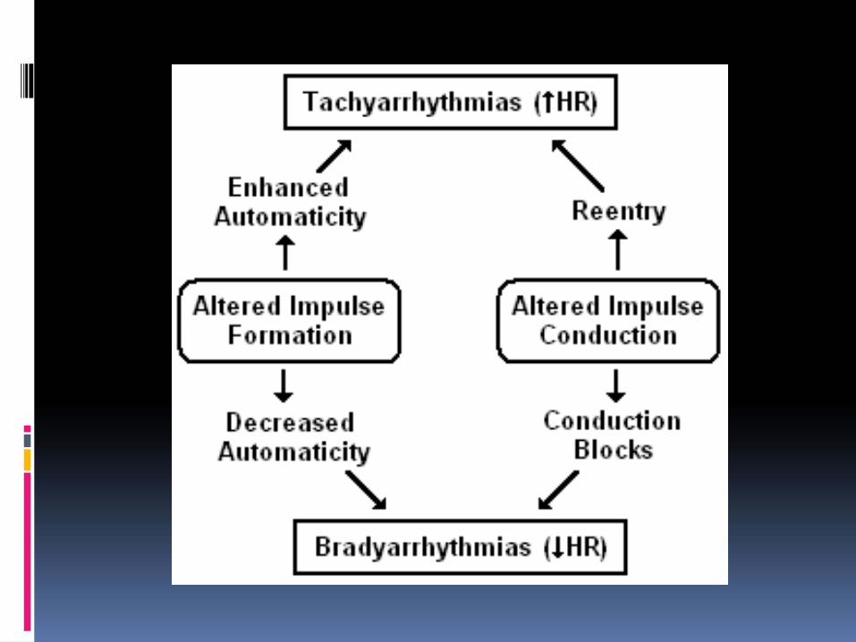
Mechanism
Mechanism of arrhythmiaAlteration in Automaticity
Abnormal automaticityThe sinus node contains pacemaker cells that havespontaneous firing capacity. This is called normalautomaticity. Abnormal automaticity occurs when othercells start firing spontaneously, resulting in prematureheartbeats.Increased sinus node activity is normally due to thesympathetic nervous system.
Sympathetic stimulation or an increase in circulatingcatecholamine acting via the B1-adrenergic receptorswhich increase the rate of phase 4 depolarization.
Triggered activity
During triggered activity heart cells contract twice,although they only have been activated once. This isoften caused by so called afterdepolarizations (early ordelayed afterdepolarizations EADs / DADs) caused byelectrical instability in themyocardial cell membrane
Altered impulse conduction
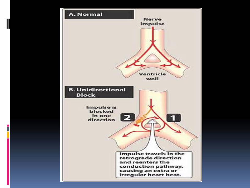
Re-entry pathwayConduction defect impulse recirculate in
the heart repetive activation without needfor further any new impulse to be generatedConduction blockEven under physiological conditions, conductionthrough SA node and AV node is slow. It may befurther slowed by ischemia or myocardialinfarction causing partial to complete A-V block.
Classification of Arrhythmia
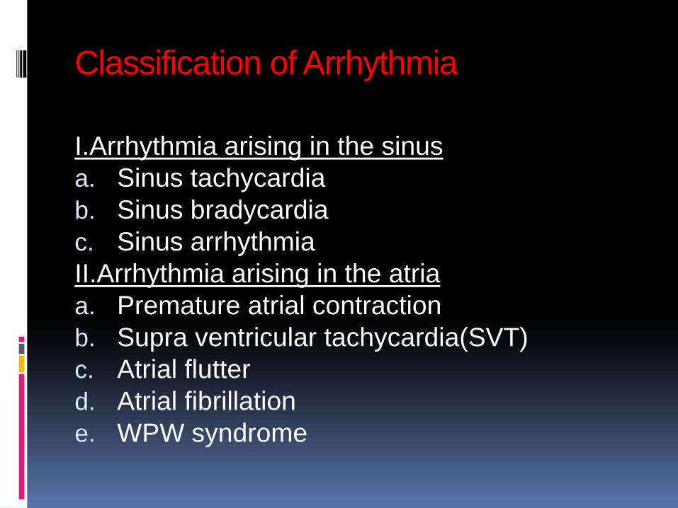
I.Arrhythmia arising in the sinusa. Sinus tachycardiab. Sinus bradycardiac. Sinus arrhythmiaII.Arrhythmia arising in the atriaa. Premature atrial contractionb. Supra ventricular tachycardia(SVT)c. Atrial flutterd. Atrial fibrillatione. WPW syndrome
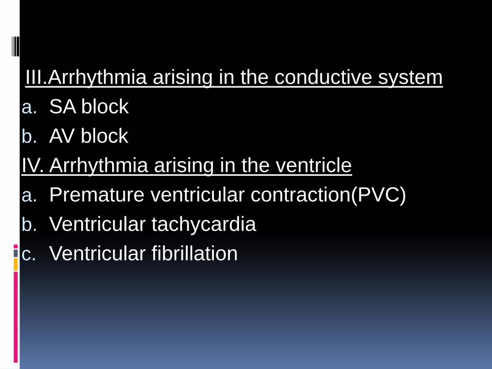
III.Arrhythmia arising in the conductive systema. SA blockb. AV blockIV. Arrhythmia arising in the ventriclea. Premature ventricular contraction(PVC)b. Ventricular tachycardiac. Ventricular fibrillation
Sinus bradycardia
Reduction in the discharge of impulses from SA node Heart rate less than 60/minCauses: disease in SA node, hypothermia, hypothyroidism, congenital heart disease, atherosclerosis, drugs like beta-blocker, digitalis.Symptoms : fatigue,dizziness,shortness of breath,lack of concentration,difficulty in exercising
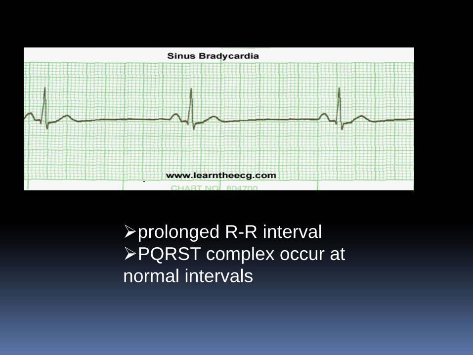
prolonged R-R intervalPQRST complex occur at
normal intervals
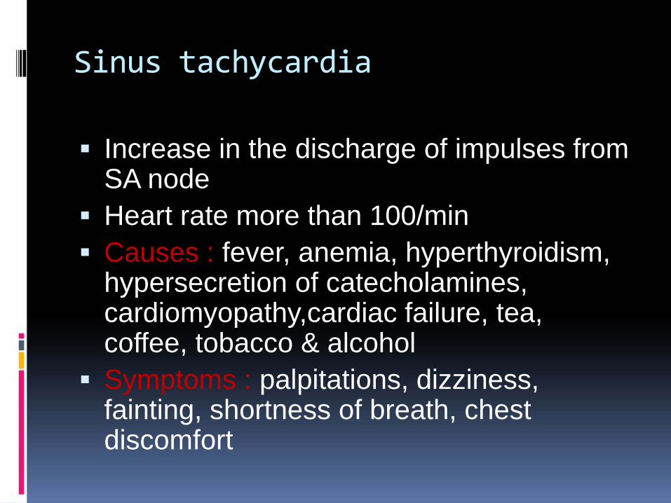
Sinus tachycardia
Increase in the discharge of impulses from SA nodeHeart rate more than 100/minCauses : fever, anemia, hyperthyroidism, hypersecretion of catecholamines, cardiomyopathy,cardiac failure, tea, coffee, tobacco & alcoholSymptoms : palpitations, dizziness, fainting, shortness of breath, chest discomfort
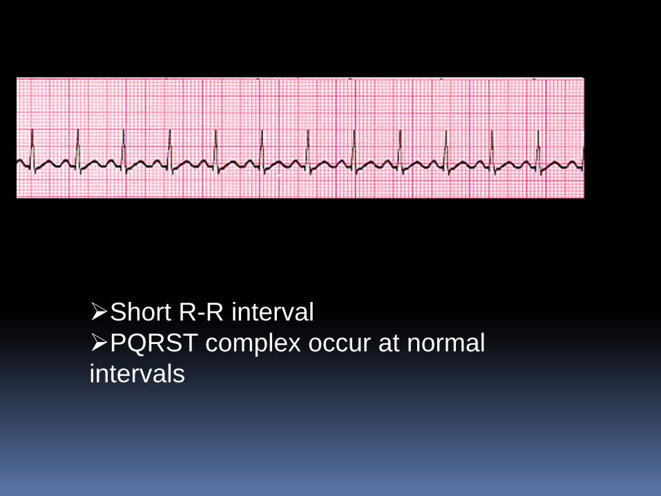
Short R-R intervalPQRST complex occur at normal
intervals
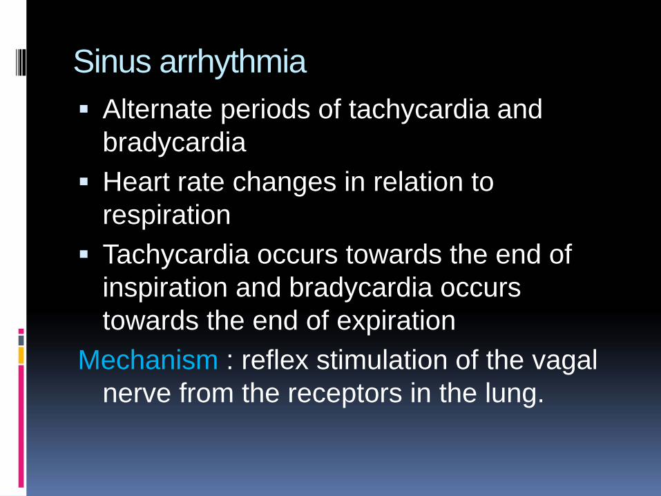
Sinus arrhythmiaAlternate periods of tachycardia and bradycardiaHeart rate changes in relation to respirationTachycardia occurs towards the end of inspiration and bradycardia occurs towards the end of expiration
Mechanism : reflex stimulation of the vagalnerve from the receptors in the lung.
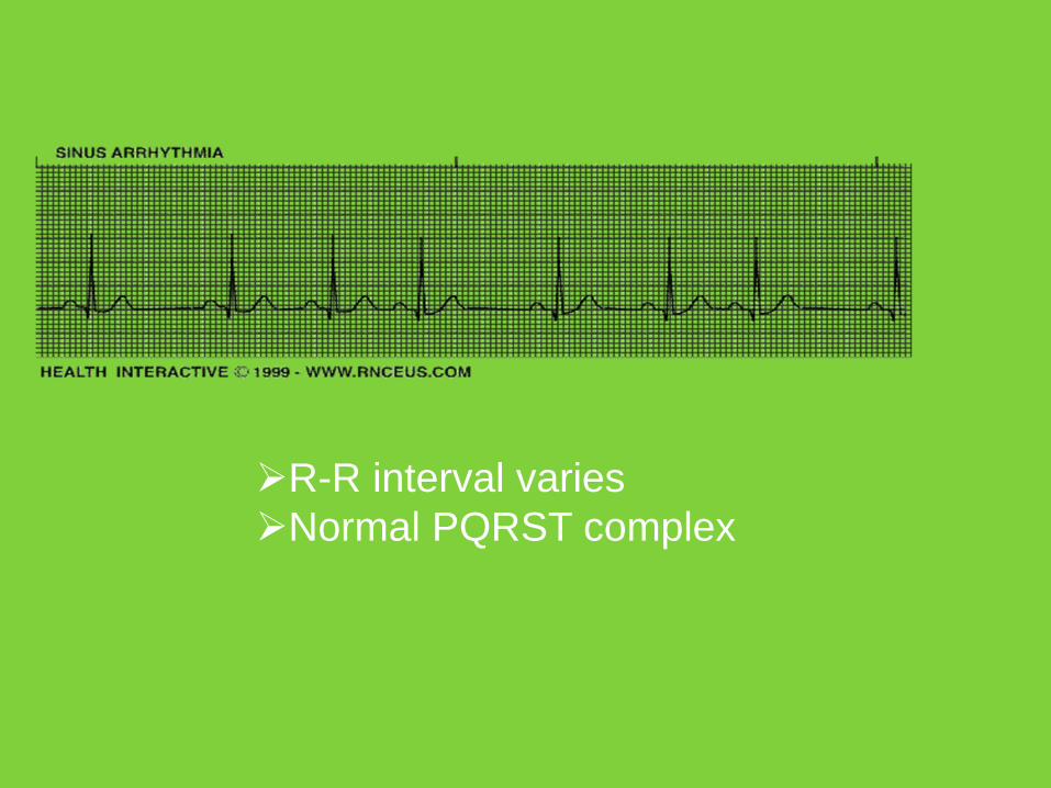
R-R interval variesNormal PQRST complex
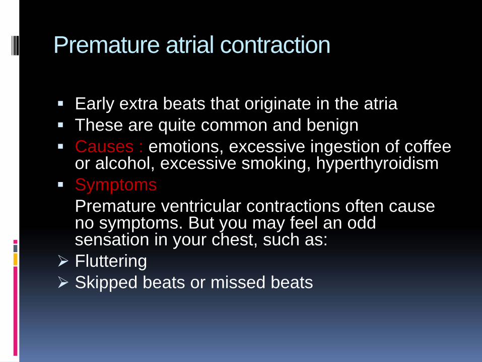
Premature atrial contraction
Early extra beats that originate in the atriaThese are quite common and benignCauses : emotions, excessive ingestion of coffee or alcohol, excessive smoking, hyperthyroidismSymptomsPremature ventricular contractions often cause no symptoms. But you may feel an odd sensation in your chest, such as: FlutteringSkipped beats or missed beats
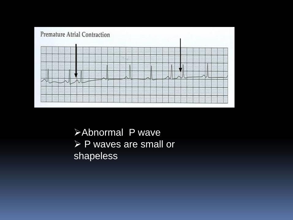
Abnormal P waveP waves are small or
shapeless
Supra ventricular tachycardia
SVT is a series of three or more PACWhich may occur for a few beats or continuously for several hours or daysIncrease in heart rate 150-250/min
Causes: emotions, excessive ingestion of coffee or alcohol, excessive smoking, hyperthyroidismMechanism: AV nodal re-entry ,SA nodal re-entry.Persistence of SVT in a patient lead to cardiac failure.
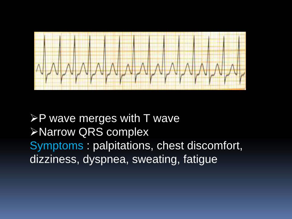
P wave merges with T waveNarrow QRS complex
Symptoms : palpitations, chest discomfort, dizziness, dyspnea, sweating, fatigue
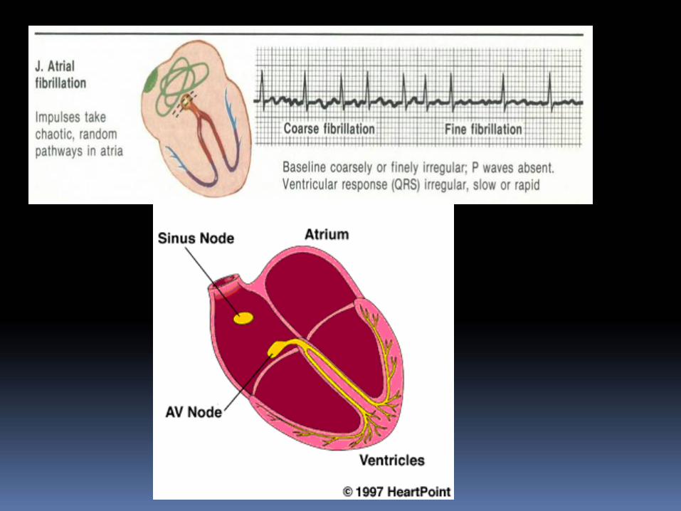
Atrial fibrillation Atrial fibrillation is characterized as an extremely rapid (400 to600 atrial beats/min) and disorganized atrial activation. There is aloss of atrial contraction (atrial kick), and supraventricularimpulses penetrate the atrioventricular (AV) conduction system invariable degrees, resulting in irregular ventricular activation andirregularly irregular pulse (120 to 180 beats/min).Electricity travelling in a chaotic fashion, causing upper chambersto quiver(like bag of worms) and contract inefficientlyMost common in elderly people those with heart diseasesMechanism : Due to circus movement of impulses within atrialmusculatureCauses : IHD,MV disease, cardiac surgery, pericarditisSymptoms : palpitations, light headness,chestpain, dyspnea,weakness
Atrial flutter
The atria are stimulated quickly that they cannot contract or squeezeHeart rate 220-350/minThe maximum rate of conduction by AV node is about 230-240/min so during atrial flutter the second degree heart block occurs.The ratio between atrial beats and ventricular beats is 2:1 or sometimes 3:1Causes : IHD,MV disease, cardiac surgery, pericarditis, cardiomyopathy, ASD
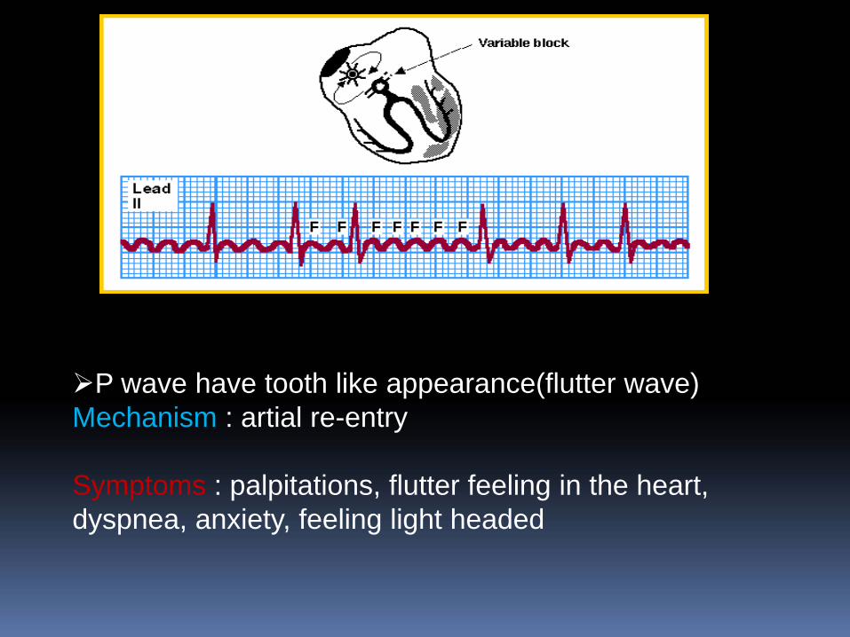
P wave have tooth like appearance(flutter wave)Mechanism : artial re-entry
Symptoms : palpitations, flutter feeling in the heart, dyspnea, anxiety, feeling light headed
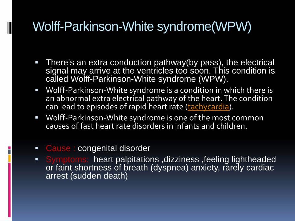
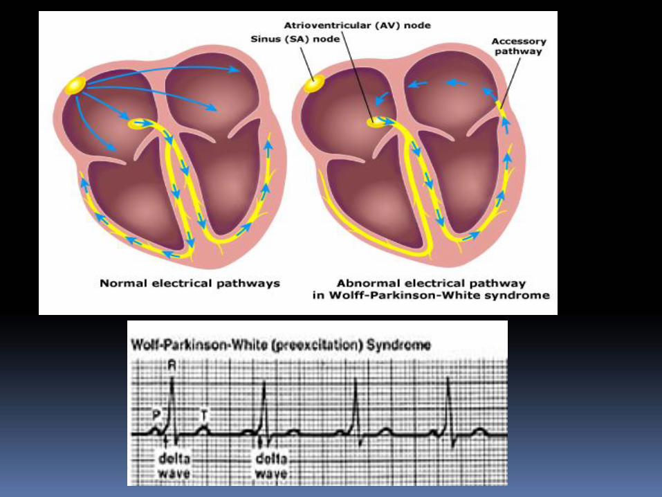
Wolff-Parkinson-White syndrome(WPW)
There's an extra conduction pathway(by pass), the electrical signal may arrive at the ventricles too soon. This condition is called Wolff-Parkinson-White syndrome (WPW). Wolff‐Parkinson‐White syndrome is a condition in which there is an abnormal extra electrical pathway of the heart. The condition can lead to episodes of rapid heart rate (tachycardia).Wolff‐Parkinson‐White syndrome is one of the most common causes of fast heart rate disorders in infants and children.
Cause : congenital disorder Symptoms: heart palpitations ,dizziness ,feeling lightheaded or faint shortness of breath (dyspnea) anxiety, rarely cardiac arrest (sudden death)
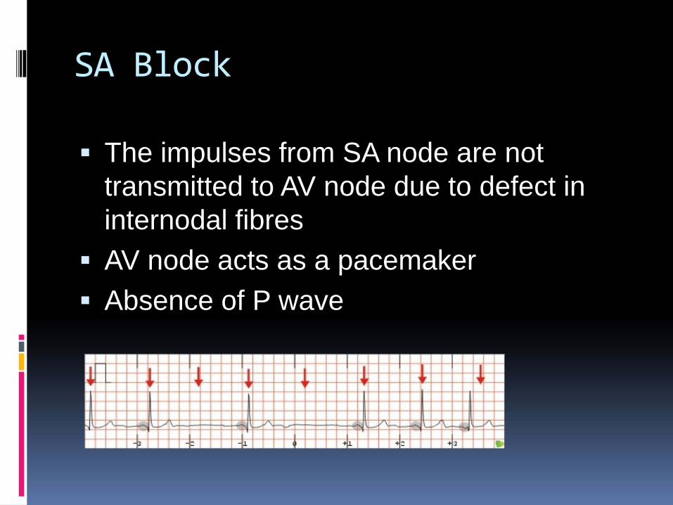
SA Block
The impulses from SA node are not transmitted to AV node due to defect in internodal fibresAV node acts as a pacemakerAbsence of P wave
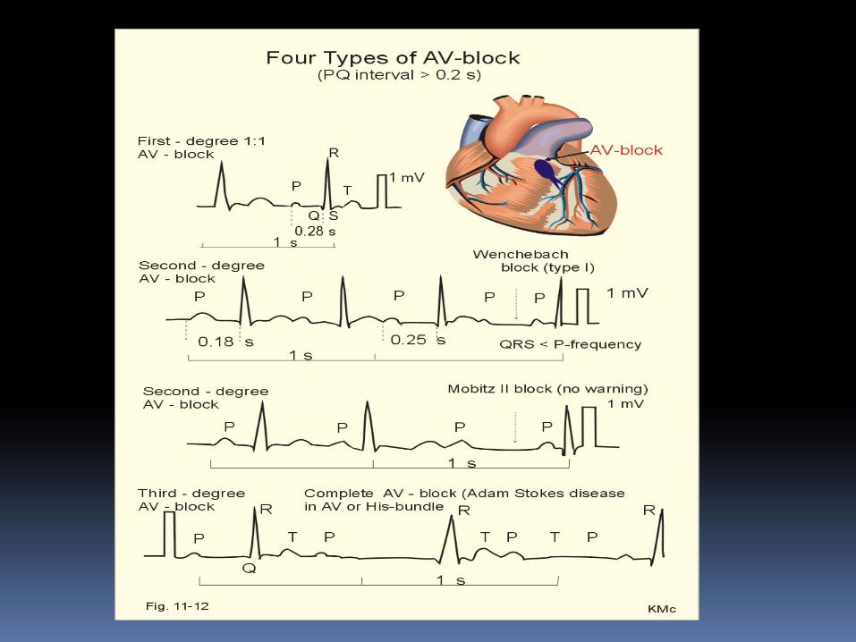
AV Block
AV block is disturbances in the conduction of the atrial impulses through the AV conductive system
A. Incomplete blocki)First degree AV blockii)Second degree AV blockB. Complete or third degree AV conductionFirst degree AV block
There is a delay in conduction of every impulse passing through AV node
Rhythm is regular and no beat is dropped ECG shows PR interval prolongation
Causes of 10 block
CAD, rheumatic fever, acute infectious diseases. Congenital heart disease, ASD, digitalis,propranolol
20 Heart blockIntermittent interruption of AV conduction so that some of the impulses are conducted to the ventricles and others are blocked
i.Mobitz type1Missing of one complete blockGradual increase in PR interval
ii.Mobitz type 2
The ventricle fails to respond to the atrial contraction PeriodicallyECG: for 5 QRS complexes there are 6 P
waves(6:5block)Causes: acute rheumatic carditis,CAD,dipherial carditisB.Complete block(30 block)
There is a permanent interruption of AV conduction so that all supraventricular impulses are blockedThe rate QRS complex is almost half that of P wave
Causes: inferior wall MI,congenital complete AV Block, CAD,ASD,VSD
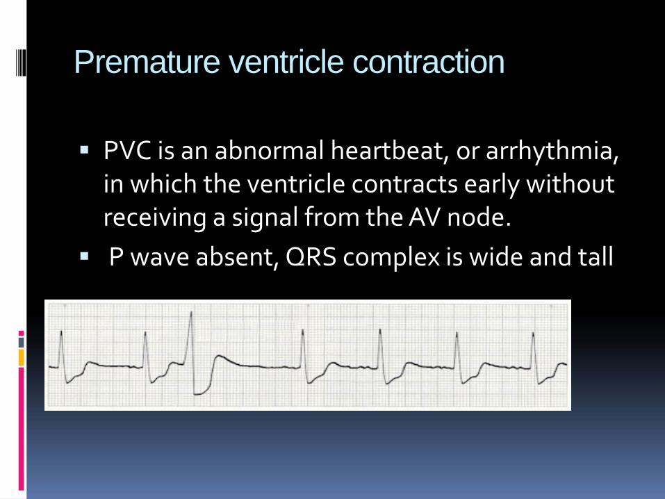
Premature ventricle contraction
PVC is an abnormal heartbeat, or arrhythmia, in which the ventricle contracts early without receiving a signal from the AV node.P wave absent, QRS complex is wide and tall
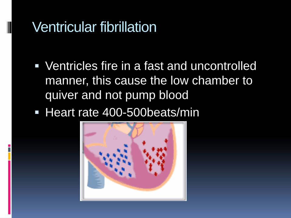
Ventricular fibrillation
Ventricles fire in a fast and uncontrolled manner, this cause the low chamber to quiver and not pump bloodHeart rate 400-500beats/min
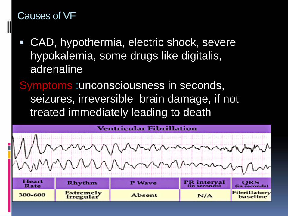
Causes of VF
CAD, hypothermia, electric shock, severe hypokalemia, some drugs like digitalis, adrenaline
Symptoms :unconsciousness in seconds, seizures, irreversible brain damage, if not treated immediately leading to death
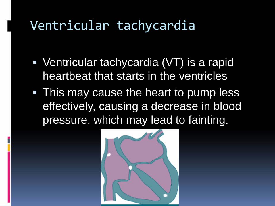
Ventricular tachycardia
Ventricular tachycardia (VT) is a rapid heartbeat that starts in the ventriclesThis may cause the heart to pump less effectively, causing a decrease in blood pressure, which may lead to fainting.
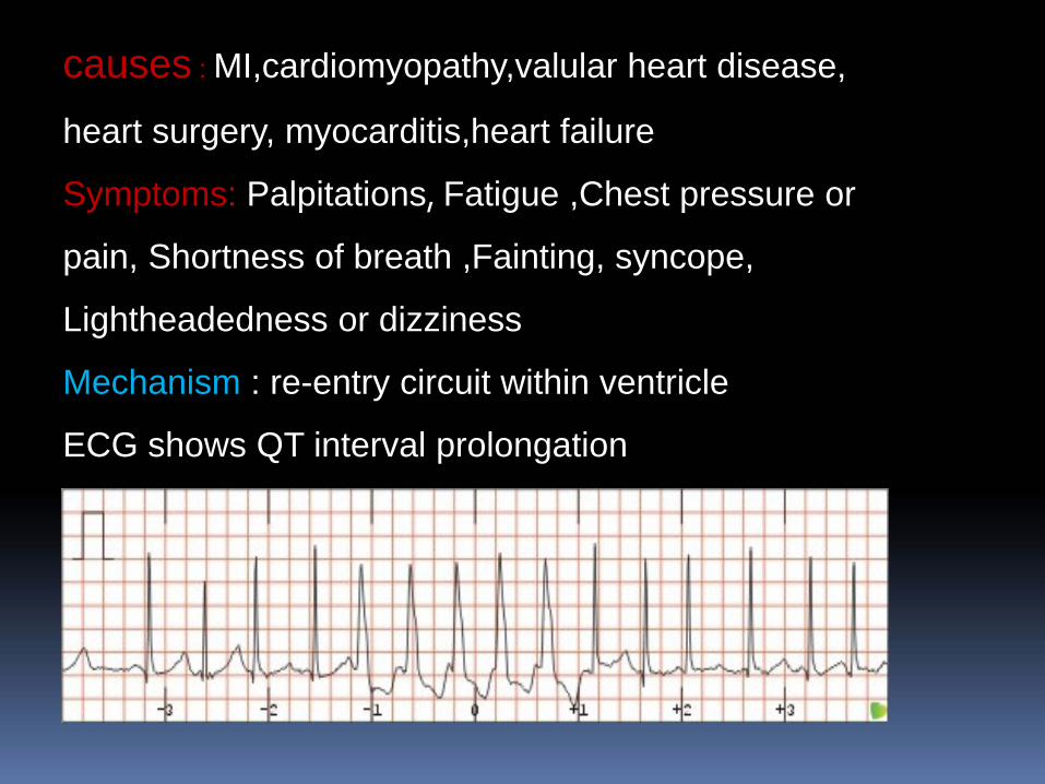
causes : MI,cardiomyopathy,valular heart disease,
heart surgery, myocarditis,heart failure
Symptoms: Palpitations, Fatigue ,Chest pressure or
pain, Shortness of breath ,Fainting, syncope,
Lightheadedness or dizziness
Mechanism : re-entry circuit within ventricle
ECG shows QT interval prolongation
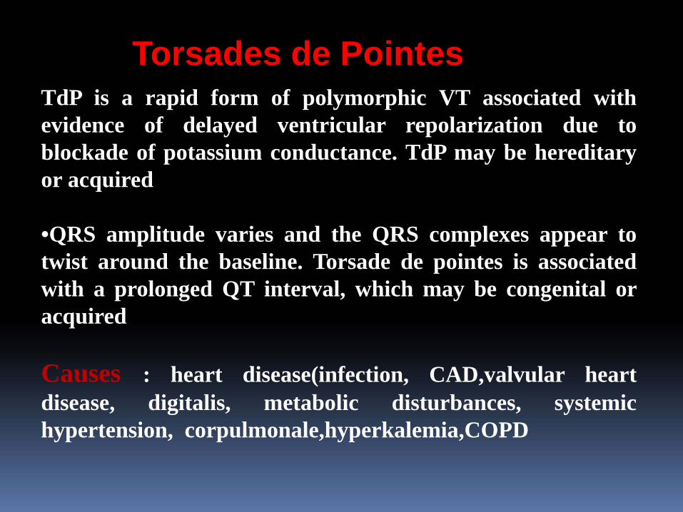
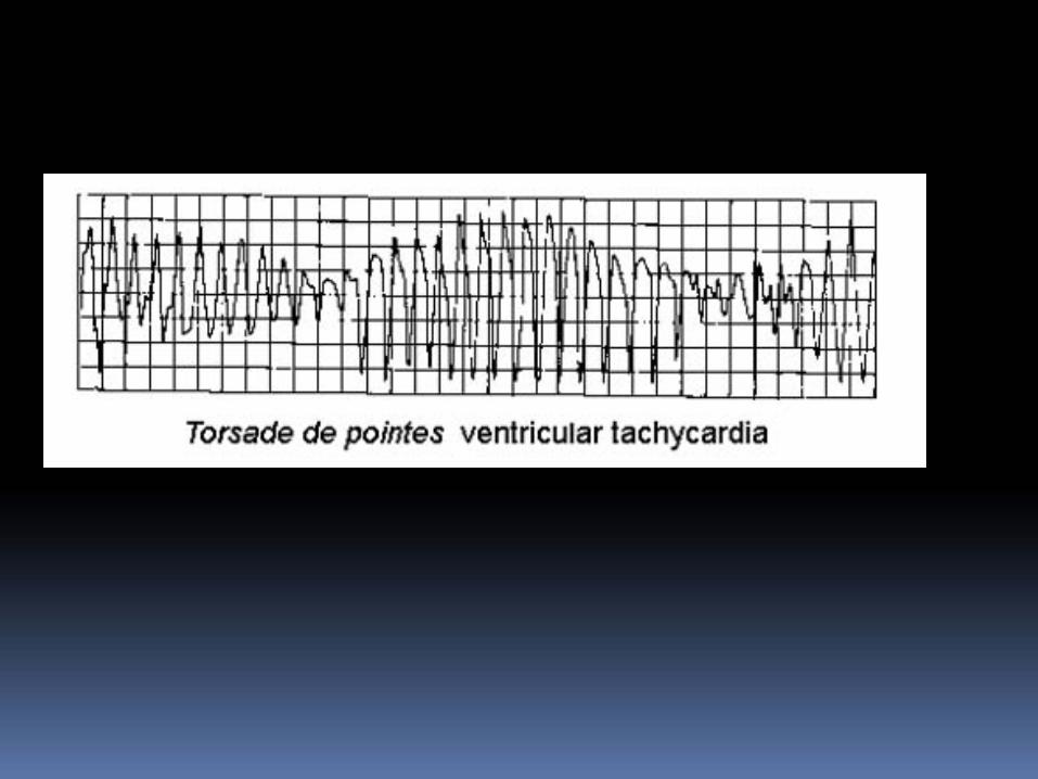
TdP is a rapid form of polymorphic VT associated withevidence of delayed ventricular repolarization due toblockade of potassium conductance. TdP may be hereditaryor acquired
•QRS amplitude varies and the QRS complexes appear totwist around the baseline. Torsade de pointes is associatedwith a prolonged QT interval, which may be congenital oracquired
Causes : heart disease(infection, CAD,valvular heartdisease, digitalis, metabolic disturbances, systemichypertension, corpulmonale,hyperkalemia,COPD
Torsades de Pointes
Ventricular Proarrhythmia
Proarrhythmia refers to development of a significant new arrhythmia (such as VT,
ventricular fibrillation [VF], or TdP) or worsening of an existing arrhythmia.
Proarrhythmia results from the same mechanisms that cause other arrhythmias or
from an alteration in the underlying substrate due to the antiarrhythmic agent.
Incessant MonomorphicVentricularTachycardia
Although the proarrhythmia associated with type Ic agents was initially thought to
occur within several days of drug initiation, risk may persist throughout treatment.
Factors that predispose patients to this type of proarrhythmia include underlying
ventricular arrhythmias, ischemic heart disease, and poor left ventricular function.
TREATMENT
Increasing or decreasing the APD and ERP can either increase or
decrease arrhythmogenesis, depending on the underlying cause of the
arrhythmia. Increasing the ERP, for example, can interrupt tachycardia
caused by reentry mechanisms by prolonging the duration that normal
tissue is unexcitable (its refractory period). This can prevent reentry
currents from re‐exciting the tissue. On the other hand, increasing the
APD can precipitate torsades de pointes, a type of ventricular
tachycardia caused by after depolarizations.
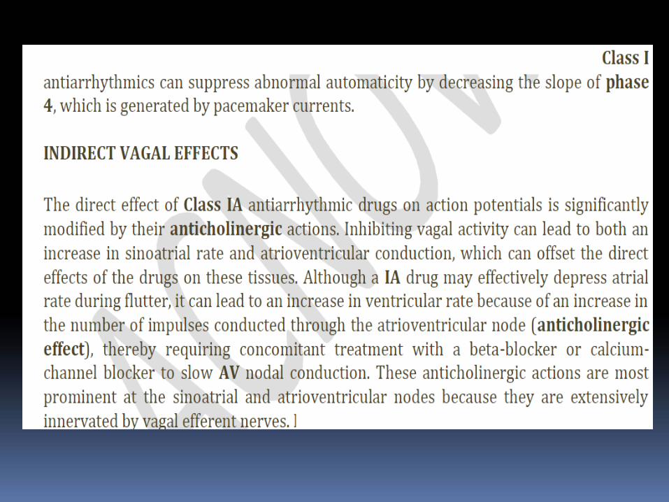
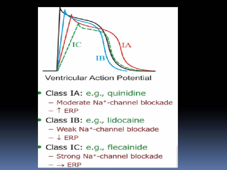
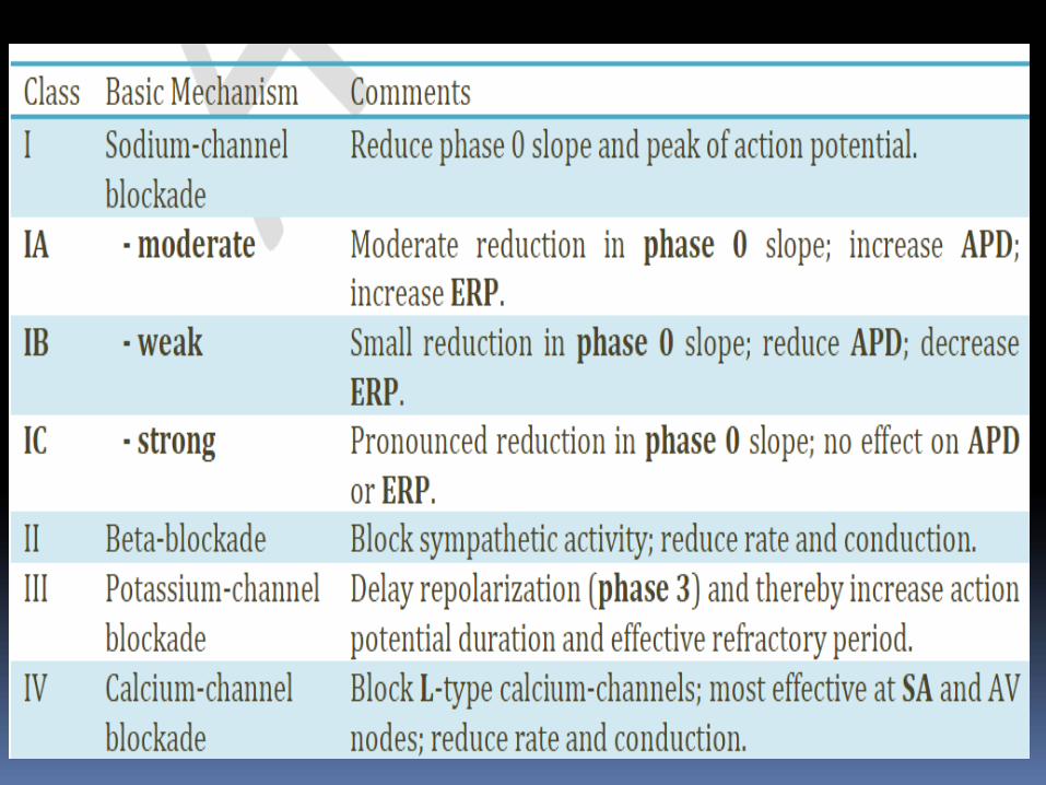
MECHANISM OF ACTION As a class I, all of the agents work by
blocking the rapid inward sodium current and thereby slow down
the rate of rise of the cardiac tissue’s action potential. However,
though this is a similar effect for all class I agents, differences in ERP
effects had led to a sub classification of the class I agents into three
subsets (IA, IB, and IC), based on these EP effects.
•Class II antiarrhythmics reduce sympathetic stimulation of
the heart, decreasing impulse conduction through the AV
node and lengthening the refractory period.
• Additionally, this class of antiarrhythmics slow the sinus
rhythm without significantly changing the QT or QRS
intervals, resulting in a reduced heart rate and a decrease in
myocardial oxygen demand.
Class III antiarrhythmic drugs prolong the refractory period and
action potential; they have no effect on myocardial contractility
or conduction time.
Class IV antiarrhythmics are calcium‐channel blockers.
They inhibit AV node conduction by depressing the SA
andAV nodes, where calcium channels predominate.
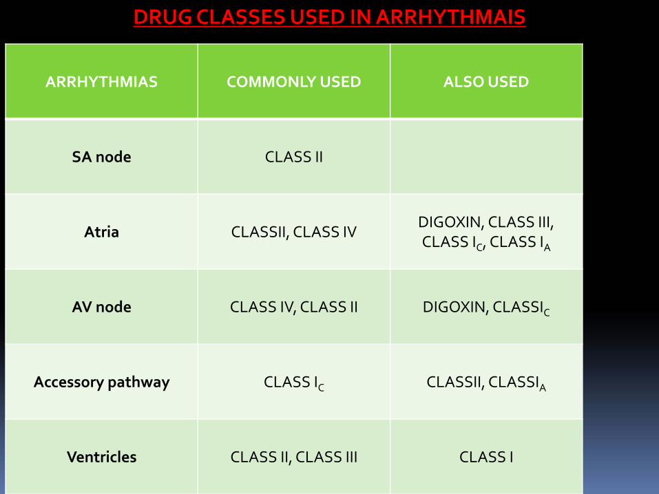
ARRHYTHMIAS COMMONLY USED ALSO USED
SA node CLASS II
Atria CLASSII, CLASS IVDIGOXIN, CLASS III, CLASS IC, CLASS IA
AV node CLASS IV, CLASS II DIGOXIN, CLASSIC
Accessory pathway CLASS IC CLASSII, CLASSIA
Ventricles CLASS II, CLASS III CLASS I
DRUG CLASSES USED IN ARRHYTHMAIS
CLASS DRUG CARDIAC NON‐CARDIAC CAUTION
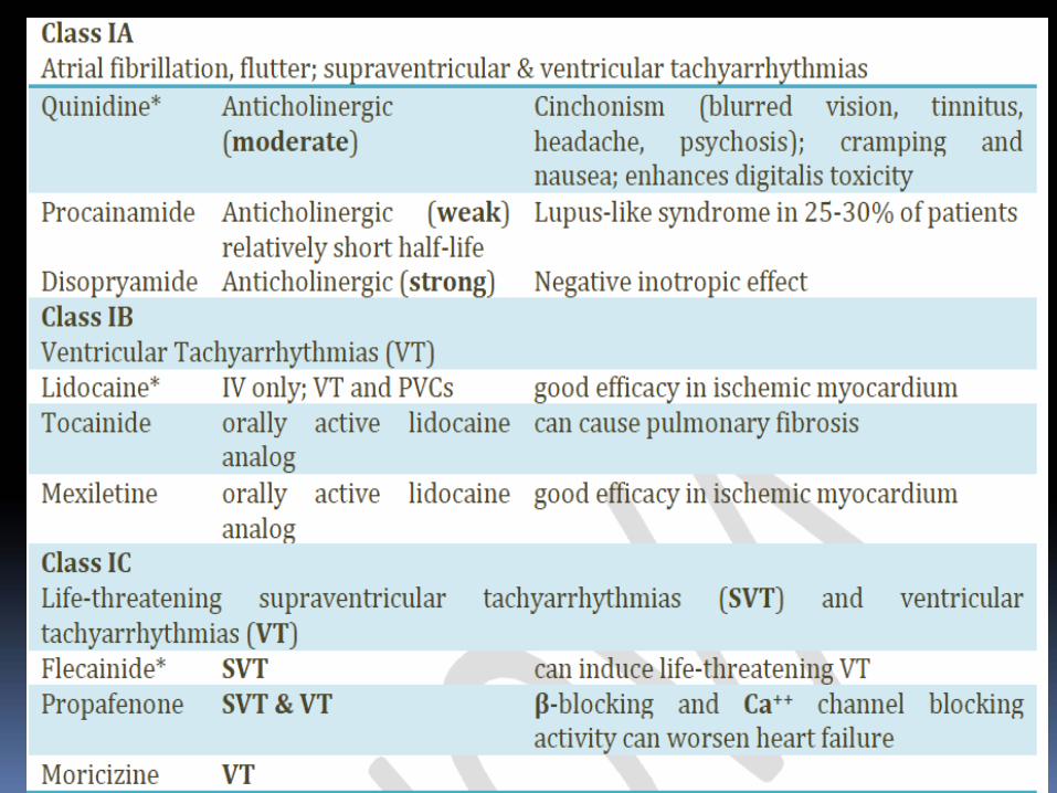
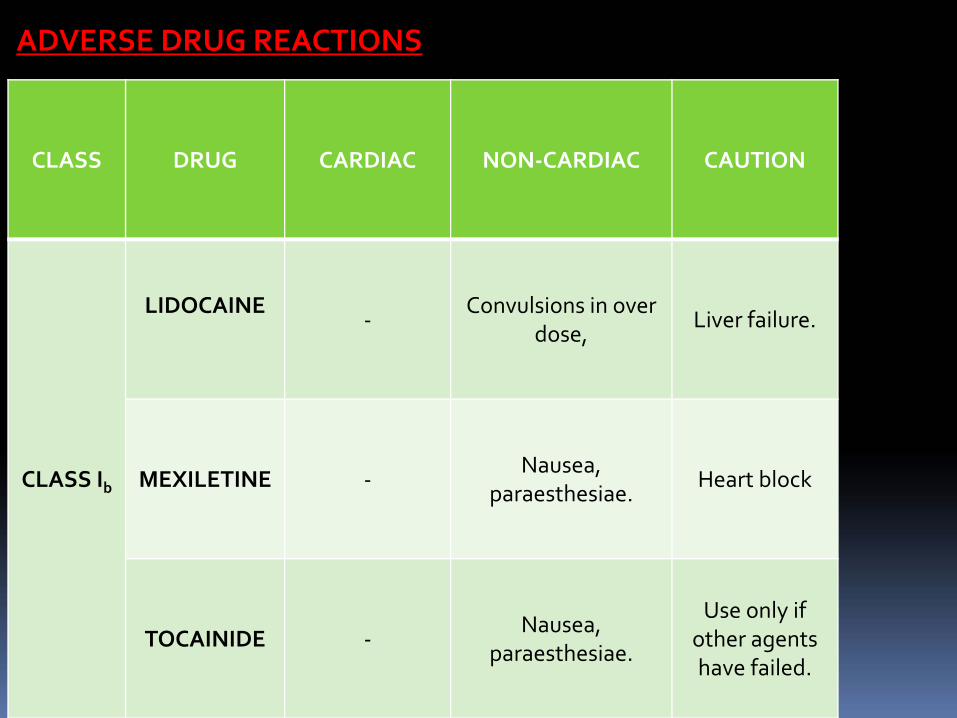
CLASS Ib
LIDOCAINE‐
Convulsions in over dose,
Liver failure.
MEXILETINE ‐Nausea,
paraesthesiae.Heart block
TOCAINIDE ‐Nausea,
paraesthesiae.
Use only if other agents have failed.
ADVERSE DRUG REACTIONS
CLASS DRUG CARDIAC NON‐CARDIAC CAUTION
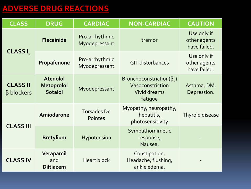
CLASS Ic
FlecainidePro‐arrhythmicMyodepressant
tremorUse only if
other agents have failed.
PropafenonePro‐arrhythmicMyodepressant
GIT disturbancesUse only if
other agents have failed.
CLASS IIβ blockers
AtenololMetoprololSotalol
Myodepressant
Bronchoconstriction(β2)VasoconstrictionVivid dreams
fatigue
Asthma, DM,Depression.
CLASS III
AmiodaroneTorsades De
Pointes
Myopathy, neuropathy, hepatitis,
photosensitivityThyroid disease
Bretylium HypotensionSympathomimetic
response, Nausea.
‐
CLASS IVVerapamil
andDiltiazem
Heart blockConstipation,
Headache, flushing, ankle edema.
‐
ADVERSE DRUG REACTIONS
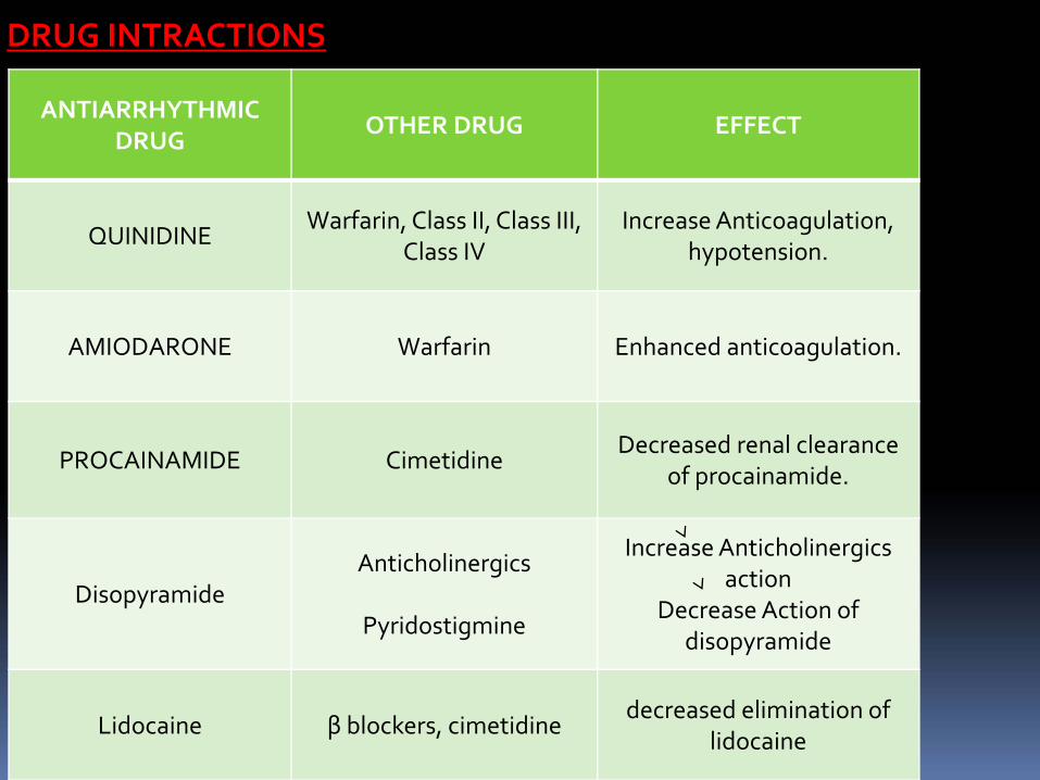
ANTIARRHYTHMIC DRUG
OTHER DRUG EFFECT
QUINIDINEWarfarin, Class II, Class III,
Class IVIncrease Anticoagulation,
hypotension.
AMIODARONE Warfarin Enhanced anticoagulation.
PROCAINAMIDE CimetidineDecreased renal clearance
of procainamide.
DisopyramideAnticholinergics
Pyridostigmine
Increase Anticholinergics action
Decrease Action of disopyramide
Lidocaine β blockers, cimetidinedecreased elimination of
lidocaine
DRUG INTRACTIONS
Thank you