Chapter 30 Capillary electrophoresis, Capillary Electro-...
48
Chapter 30 Capillary electrophoresis, Capillary Electro- chromatography, and Field-Flow Fractionation 30A-1 Types of Electrophoresis Electrophoretic separations are currently performed in two quite different formats: one is called slab electrophoresis and the other capillary electrophoresis. 30A-2 The basis for electrophoretic separations The migration rate ν of an ion (cm/s) in an electric field is equal to the product of the field strength E(V cm -1 ) and the electrophoretic mobility μ e (cm 2 V -1 s -1 ). ν= μ e E
Transcript of Chapter 30 Capillary electrophoresis, Capillary Electro-...
Chapter 30 Capillary electrophoresis, Capillary Electro-chromatography, and Field-Flow Fractionation
30A-1 Types of Electrophoresis Electrophoretic separations are currently performed in two quite different formats: one is called slab electrophoresis and the other capillary electrophoresis. 30A-2 The basis for electrophoretic separations The migration rate ν of an ion (cm/s) in an electric field is equal to the product of the field strength E(V cm-1) and the electrophoretic mobility μe(cm2 V-1 s-1).
ν= μeE
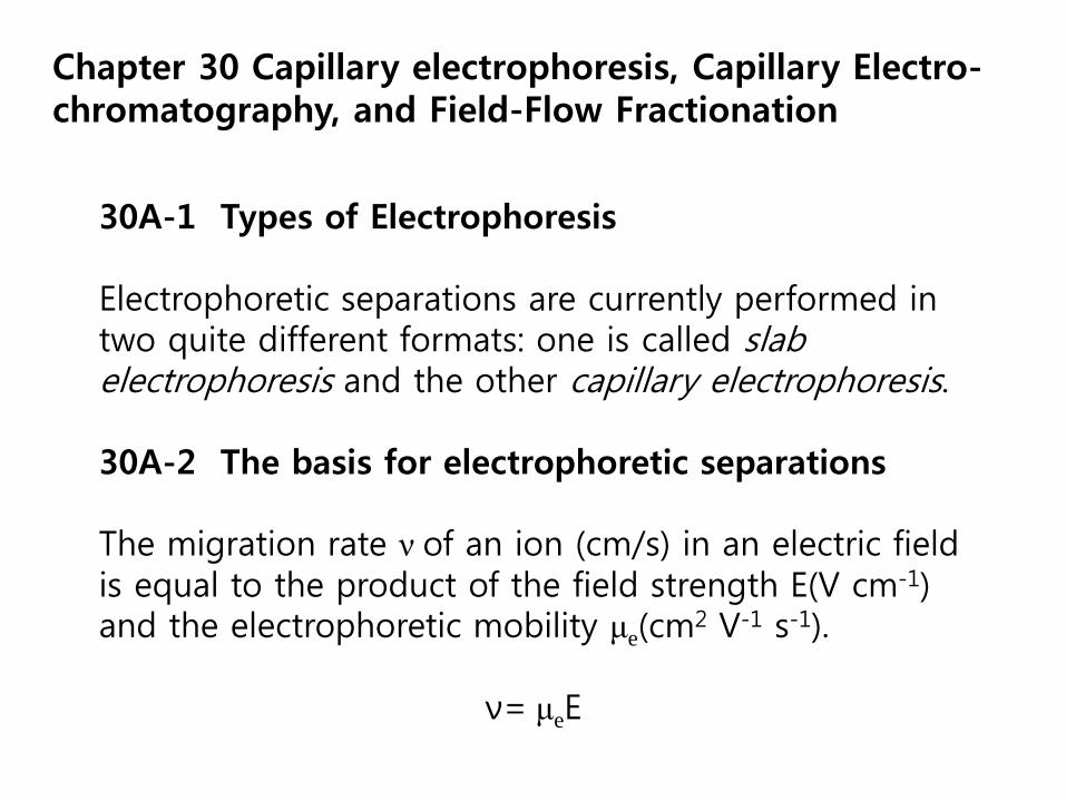
Horizontal electrophoresis.
http://ocw.mit.edu/courses/biological-engineering/20-109-laboratory-fundamentals-in-biological-engineering-fall-2007/labs/mod1_2_photo.jpg
2
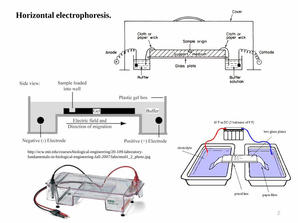
Vertical gel (or paper) electrophoresis.
3
Electrical field 의 영향 하에서 용액을 통한 입자의 migration 으로 분리.
Free soln. method :
buffered liquid 로 채워진 U-tube 바닥에 시료를 introduce.
단점 :
Thermal , density gradient의 결과 convection 발생 mixing 현상
Fig. 30-1
◉ Stabilizing medium 사용
Paper , a layer of finely divided solid.
Column packed with a suitable solid.
Electrochromatography
Zone electrophoresis
Electromigration ionophoresis 등으로 불리워짐
Solid media : paper , cellulose, acetate membranes, cellulose powders,
starch gel, ion-exchange resin, glass powders, agar-gels.
전압 : 100~1000 V 정도의 AC
전극 반응 방지를 위해 전극 을 media 로부터 충분히 격리
전류 크기 : mA 단위
V‥ arrangement :
Two - dimensional . electrochromatograph
전기장이 용액의 흐름에 수직으로 작용
*임상화학과 생화학에 반드시 필요
Type: Slab electrophoresis and capillary electrophoresis
Electrophoretic Separations:
v = ueE
* Neutral species are not separated.
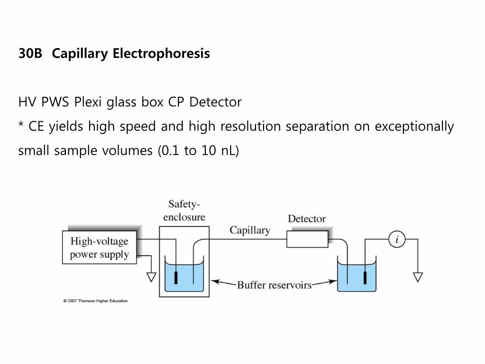
30B Capillary Electrophoresis
HV PWS Plexi glass box CP Detector
* CE yields high speed and high resolution separation on exceptionally
small sample volumes (0.1 to 10 nL)
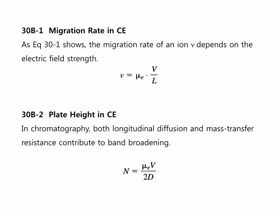
30B-1 Migration Rate in CE
As Eq 30-1 shows, the migration rate of an ion ν depends on the
electric field strength.
30B-2 Plate Height in CE
In chromatography, both longitudinal diffusion and mass-transfer
resistance contribute to band broadening.
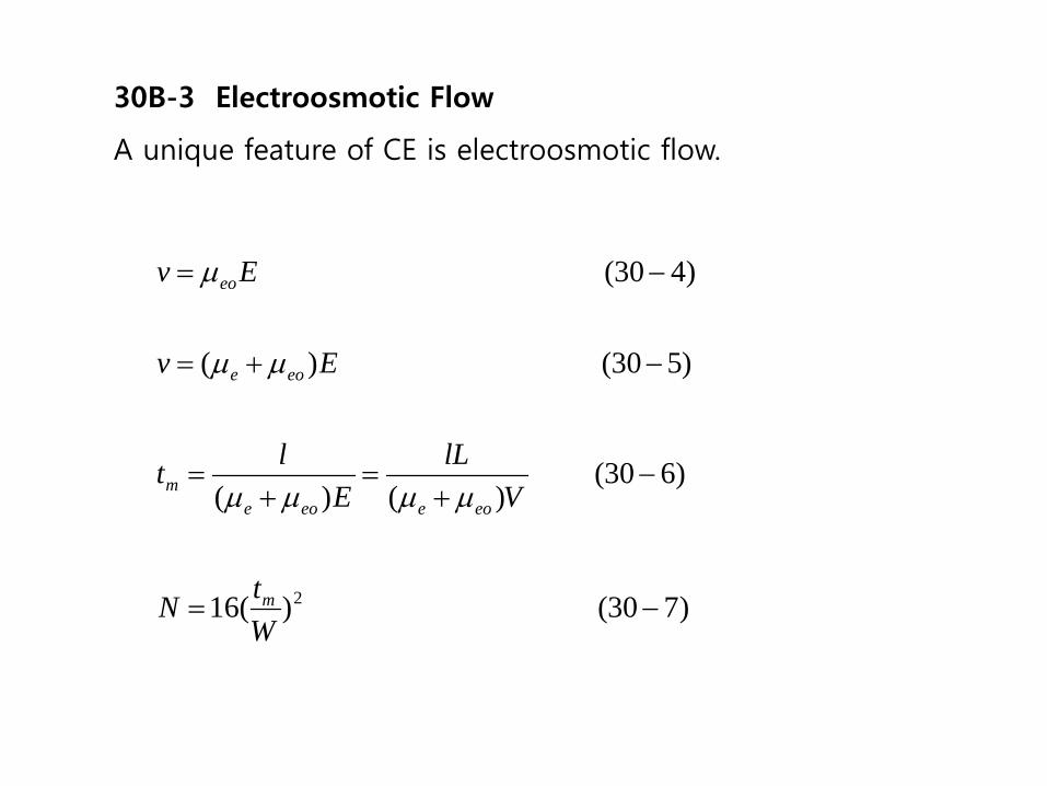
30B-3 Electroosmotic Flow
A unique feature of CE is electroosmotic flow.
)730( )(16
)630( )()(
)530( )(
)430(
2 −=
−+
=+
=
−+=
−=
WtN
VlL
Elt
Ev
Ev
m
eoeeoem
eoe
eo
µµµµ
µµ
µ
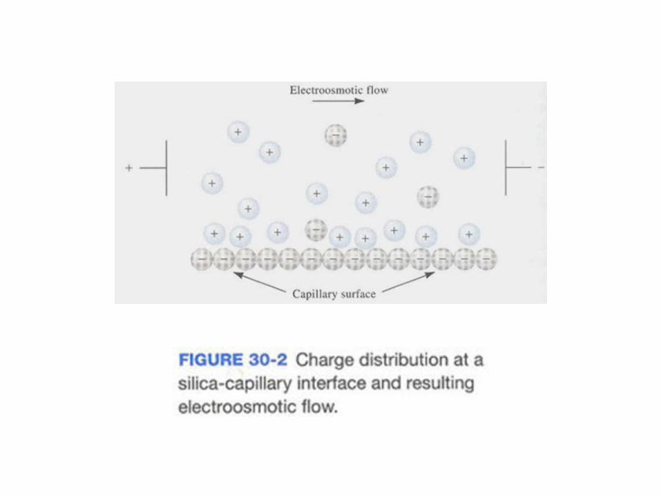
Fig. 30-3 Flow profiles for liquids under
(a) electroosmotic flow and
(b) pressure-induced flow.
Fig. 30-4 Velocities in the presence of electroosmotic flow
30B-4 Instrumentation for CE
As shown in Fig 30-1, the instrumentation for CE is
relatively simple.
▪ Sample introduction
The most common sample-introduction methods are
electrokinetic injection and pressure injection.
▪ Detection
∙ Absorption methods
Fig 30-5 b shows a second way to increase the absorption
path length.
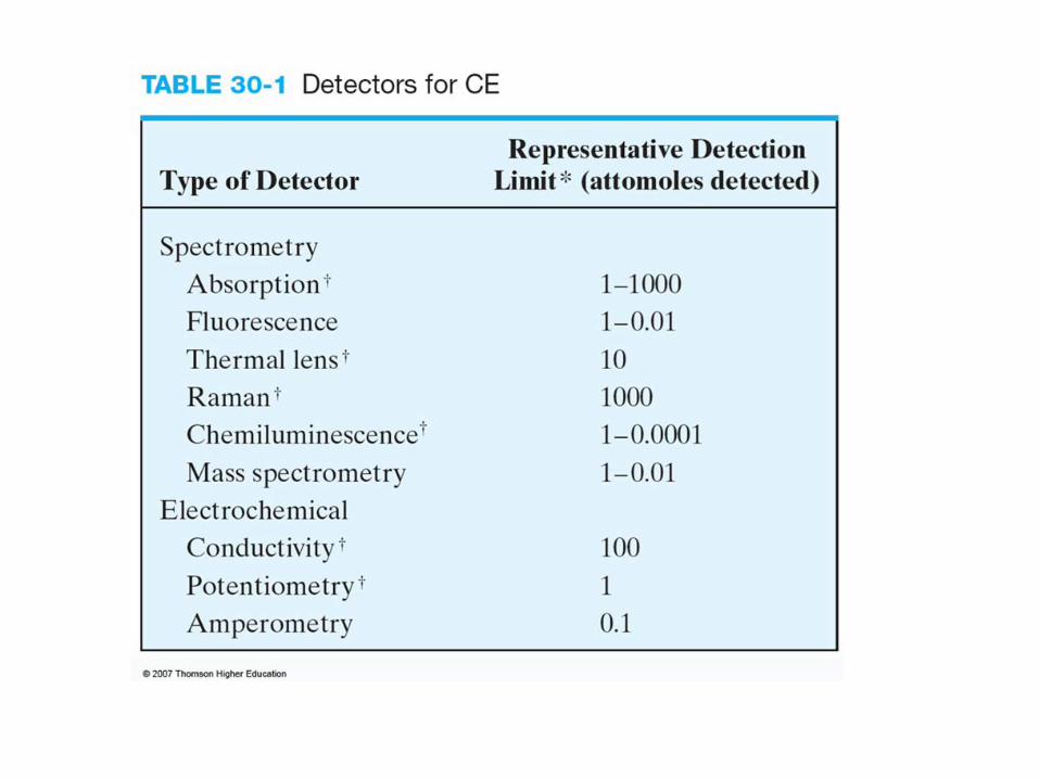
Fig. 30-5 Detection cells of absorbance measurements
∙ Indirect Detection
Indirect absorbance detection has been used for
species of low molar absorptivity that are difficult
to detect without derivatization.
∙ Fluorescence
Just as in HPLC, fluorescence detection yields in
creased sensitivity and selectivity for fluorescent
analytes or fluoresecent derevatives.
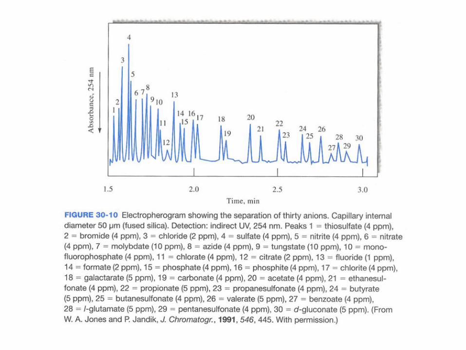
Fig. 30-6 Electropherogram of a six-anion mixture by indirect detection with 4-mM chromate ion at 254 nm.
∙ Electrochemical Detection
Two types of electrochemical detection have been
used with CE: conductivity and amperometry.
∙ Mass Spectrometric Detection
Fig 30-7 is scahematic of a typical eletrospray interface
coupled to a quadrupole mass spectrometer.
▪ Commericial CE systems
Currently, fewer than companies worldwide
manufacture CE in struments.
Fig. 30-7 An instrument for CE/MS
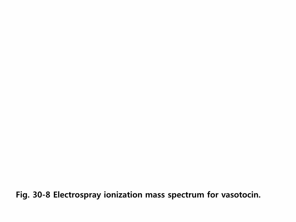
Fig. 30-8 Electrospray ionization mass spectrum for vasotocin.
30C Applications of CE(capillary electrophoresis)
a) Capillary zone electrophoresis (CZE)
b) Capillary gel electrophoresis (CGE)
c) Capillary isoeletric focusing (CIEF)
d) Capillary isotachophoresis (CITP)
a) CZE: buffer composition is constant through the regions of the
separation.
b) CGE: is generally performed in a porous gel matrix.
c) CITP: all analyte bands ultimately migrate at the same velocity.
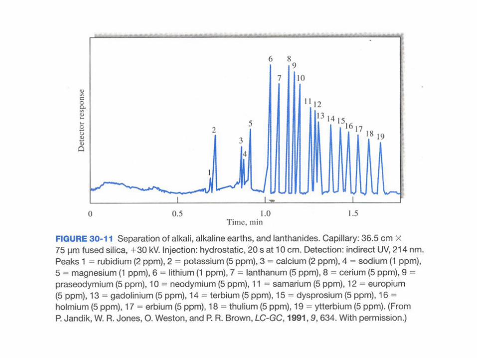
30C-1 Capillary Zone Electrophoresis
▪ Separation of Small Ions
Fig 30-10 illustrates the speed at which separations can
be carried out.
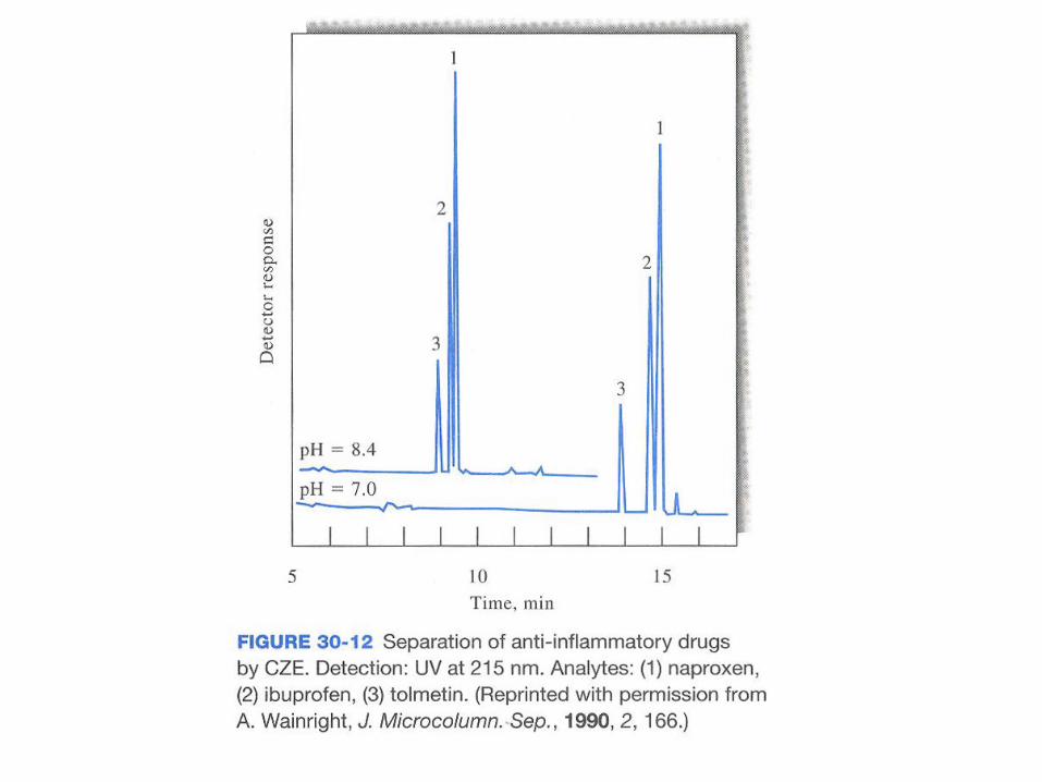
▪ Separation of Molecular Species
A variety of synthetic herbicides, pesticides, and
pharmaceuticals that are ions or can be derivatized to
yield ions have been separated and analyzed by CZE.
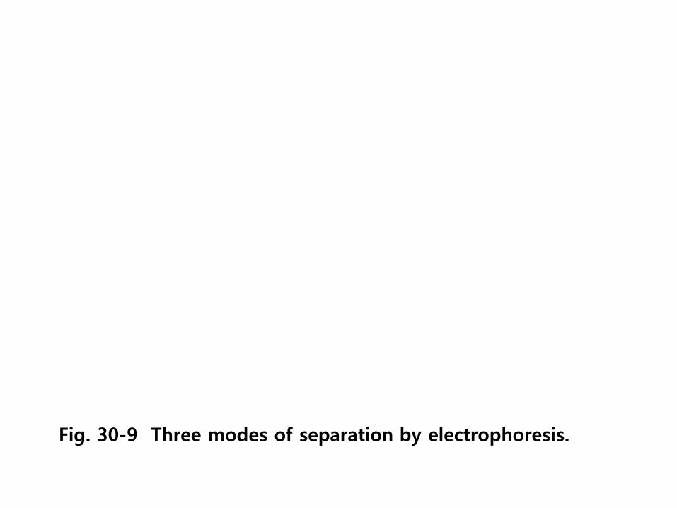
Fig. 30-9 Three modes of separation by electrophoresis.
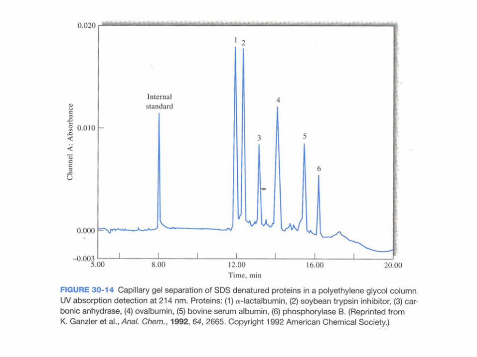
30C-2 Capillary Gel Electrophoresis
▪ Types of Gels
The most common type of gel used in electrophoresis is
a polyacrylamide polymer formed by polymerizing
acrylamide ( CH2=CH-CO-NH2 ) in the presence of a
cross-linking agent.
▪ CE in DNA Sequencing
In sequencing, DNA extracted from cells is fragmented by
various approaches.
30C-3 Capillary Isotachophoresis
In capillary isotachophoresis all analyte bands
ultimately migrate at the same velocity; hance, the
name from iso for same and tach for speed.
30C-4 Capillary Isoelectric Focusing
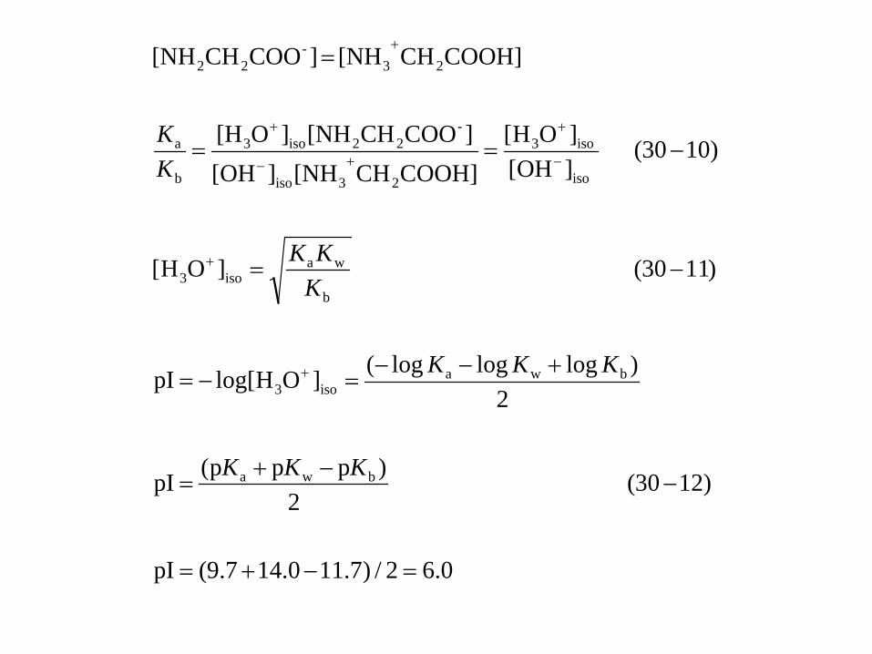
▪ Properties of Amphiprotic Compounds
An amphiprotic conpound is a species that in solution
is capable of both donating and accepting a proton.
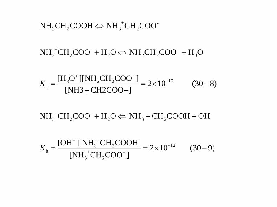
)930( 102]COOCH[NH
COOH]CH][NH[OH
OH COOHCHNHOH COOCHNH
)830( 102]CH2COO[NH3
]COOCH][NHO[H
OH COOCHNHOH COOCHNH
COOCHNHCOOHCHNH
12
23
23b
-232
-23
10223a
3 -
222-
23
-2322
−×==
++⇔+
−×=−+
=
+⇔+
⇔
−−+
+−
+
−−+
++
+
K
K
0.62/)7.110.147.9(pI
)1230( 2
)ppp(pI
2)logloglog(]OHlog[pI
)1130( ]OH[
)1030( ]OH[]OH[
COOH]CH[NH][OH]COOCH[NH]O[H
COOH]CH[NH ]COOCH[NH
bwa
bwaiso3
b
waiso3
iso
iso3
23iso
-22iso3
b
a
23-
22
=−+=
−−+
=
+−−=−=
−=
−==
=
+
+
−
+
+−
+
+
KKK
KKK
KKK
KK
▪ Separation of Amphiprotic Species
Ampholytes are anphoteric compounds usually
containing carboxylic and amino groups.
▪ Mobilization of Focused Bands
Fig 30-16 shows an electropherogram for the
separation of several proteins by capillary
isoelectric focusing.
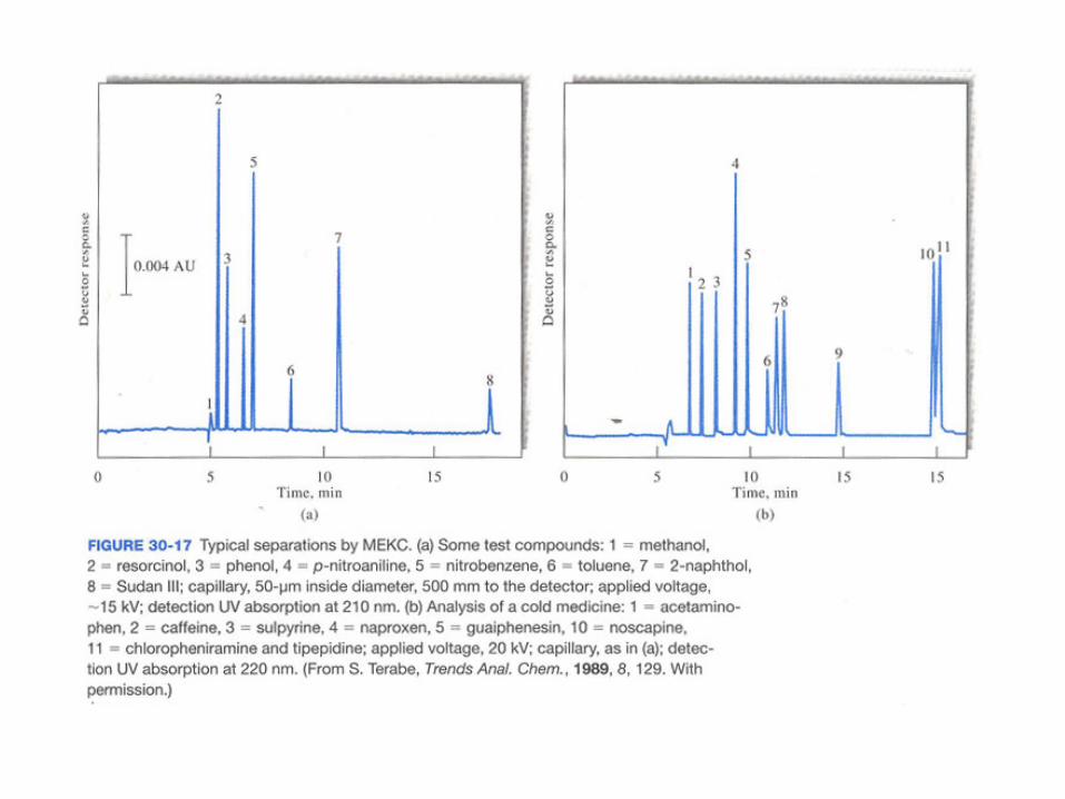
30C-5 Micellar Electrokinetic Chromatography
In 1984, Terabe and collaborators described a
modification of CE that permitted the separation
of low-molecular-mass aromatic phenols and nitro
compounds with equipment such as shown in Fig
30-1.
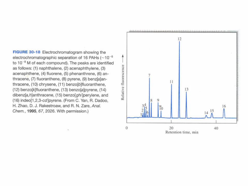
30D Packed Column Electrochromatography
Electrochromatography is a hybrid of HPLC and CE
that offers some of the best features of the two
methods.
Fig. 30-16 Capillary isoelectric focusing of proteins.
30E Packed Column Electrochromatography
30E-1 Separation Mechanisms
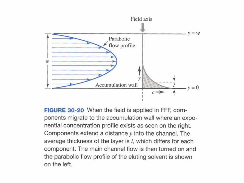
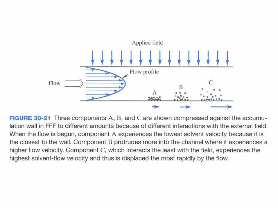
Separations in FFF occur in a thin ribbon-like flow
channel such as that shown in Fig 30-19.
30E-2 FFF Methods
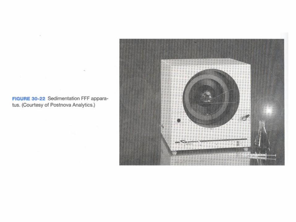
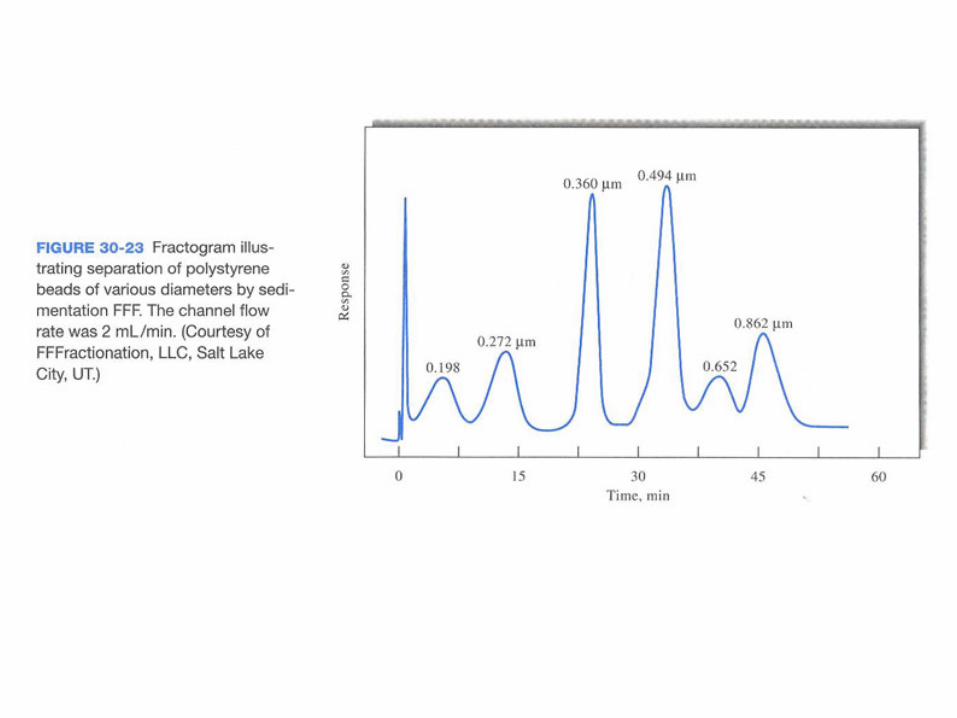
▪ Sedimentation FFF
Sedimentation FFF is by far the most widely used form.
▪ Electrical FFF
In electrical FFF, an electric field is applied perpendicular
to the flow direction.
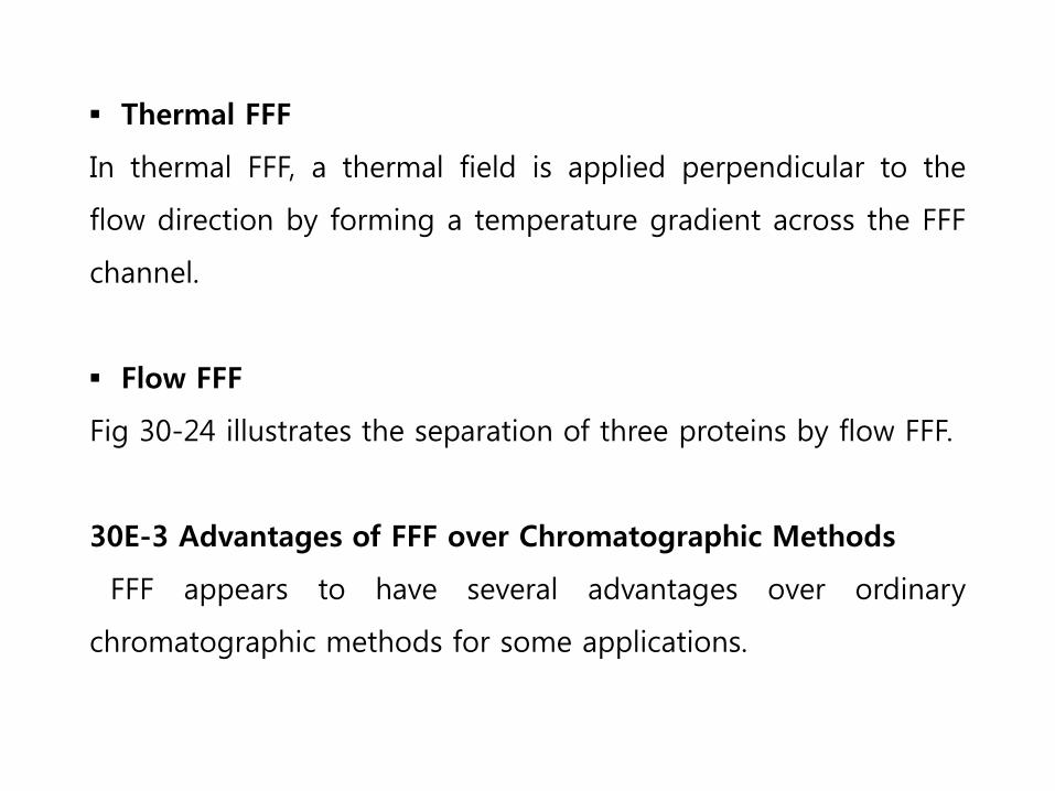
▪ Thermal FFF
In thermal FFF, a thermal field is applied perpendicular to the
flow direction by forming a temperature gradient across the FFF
channel.
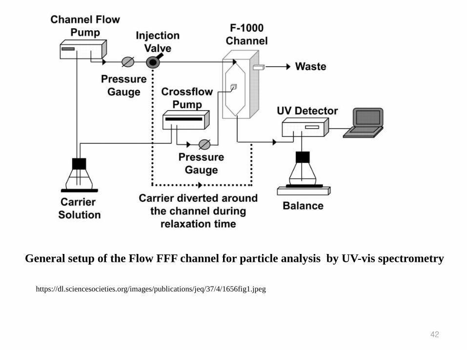
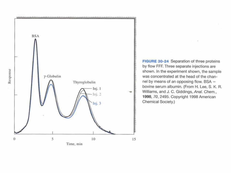
▪ Flow FFF
Fig 30-24 illustrates the separation of three proteins by flow FFF.
30E-3 Advantages of FFF over Chromatographic Methods
FFF appears to have several advantages over ordinary
chromatographic methods for some applications.
41
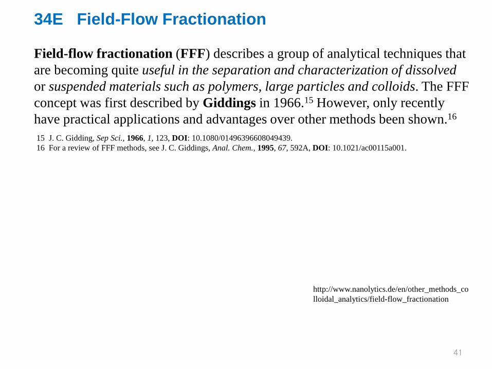
34E Field-Flow Fractionation Field-flow fractionation (FFF) describes a group of analytical techniques that are becoming quite useful in the separation and characterization of dissolved or suspended materials such as polymers, large particles and colloids. The FFF concept was first described by Giddings in 1966.15 However, only recently have practical applications and advantages over other methods been shown.16
15 J. C. Gidding, Sep Sci., 1966, 1, 123, DOI: 10.1080/01496396608049439. 16 For a review of FFF methods, see J. C. Giddings, Anal. Chem., 1995, 67, 592A, DOI: 10.1021/ac00115a001.
http://www.nanolytics.de/en/other_methods_colloidal_analytics/field-flow_fractionation
42
https://dl.sciencesocieties.org/images/publications/jeq/37/4/1656fig1.jpeg
General setup of the Flow FFF channel for particle analysis by UV-vis spectrometry
Fig. 30-19 Schematic diagram of FFF flow channel sandwiched between two walls.
![METHODOLOGY Open Access Development and … · amination, and then separated by polyacrylamide gel electrophoresis [11]. ... of APTS labelled hydrolysed dextran and β-1,4-xylo oligosaccharides](https://static.fdocument.org/doc/165x107/5adeff457f8b9ab4688b939a/methodology-open-access-development-and-and-then-separated-by-polyacrylamide.jpg)