
Chapter 3. Infrared spectroscopy - Concordia...
14
vibrational transitions • which transitions? wavelength and intensity, ν • how many vibrations • what happens if we change the molecule? substituent effects on ν • prediction of ν Chapter 3. Infrared spectroscopy O Reading: Pavia Chapter 2
Transcript of Chapter 3. Infrared spectroscopy - Concordia...
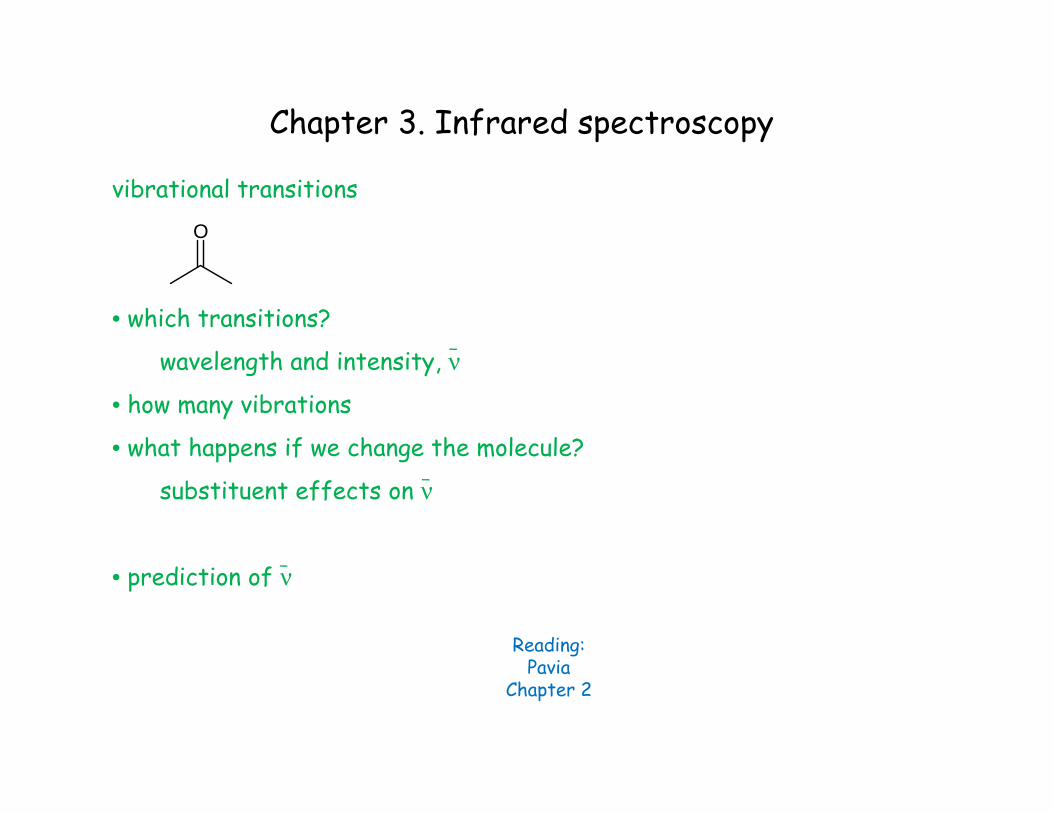
vibrational transitions
• which transitions?
wavelength and intensity, ν
• how many vibrations
• what happens if we change the molecule?
substituent effects on ν
• prediction of ν
Chapter 3. Infrared spectroscopy
O
Reading:Pavia
Chapter 2
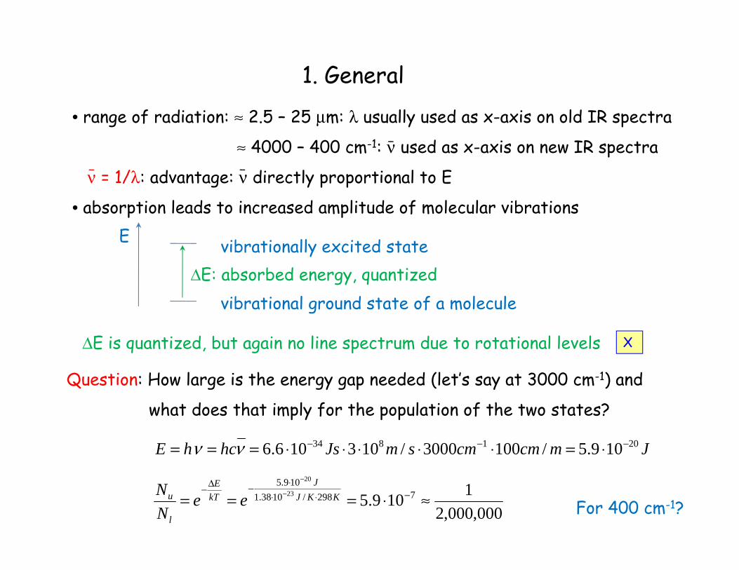
• range of radiation: ≈ 2.5 – 25 μm: λ usually used as x-axis on old IR spectra
≈ 4000 – 400 cm-1: ν used as x-axis on new IR spectra
ν = 1/λ: advantage: ν directly proportional to E
• absorption leads to increased amplitude of molecular vibrations
1. General
ΔE: absorbed energy, quantized
Question: How large is the energy gap needed (let’s say at 3000 cm-1) and
what does that imply for the population of the two states?
E vibrationally excited state
vibrational ground state of a molecule
JmcmcmsmJshchE 201834 109.5/1003000/103106.6 −−− ⋅=⋅⋅⋅⋅⋅=== νν
000,000,21109.5 7298/1038.1
109.523
20
≈⋅=== −⋅⋅⋅−Δ− −
−
KKJJ
kTE
l
u eeNN
ΔE is quantized, but again no line spectrum due to rotational levels
For 400 cm-1?
X
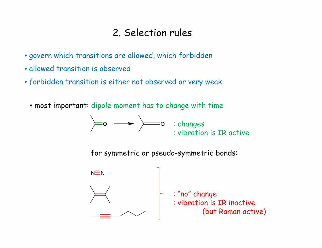
2. Selection rules
• govern which transitions are allowed, which forbidden
• allowed transition is observed
• forbidden transition is either not observed or very weak
• most important: dipole moment has to change with time
O O : changes: vibration is IR active
for symmetric or pseudo-symmetric bonds:
N N
: “no” change: vibration is IR inactive
(but Raman active)
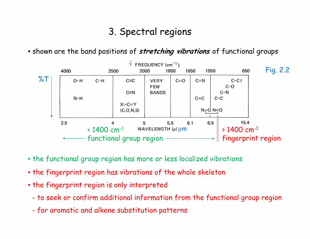
3. Spectral regions
• shown are the band positions of stretching vibrations of functional groups
> 1400 cm-1
fingerprint region
• the fingerprint region has vibrations of the whole skeleton
• the fingerprint region is only interpreted
- to seek or confirm additional information from the functional group region
- for aromatic and alkene substitution patterns
μm
%T
• the functional group region has more or less localized vibrations
< 1400 cm-1
functional group region
νFig. 2.2
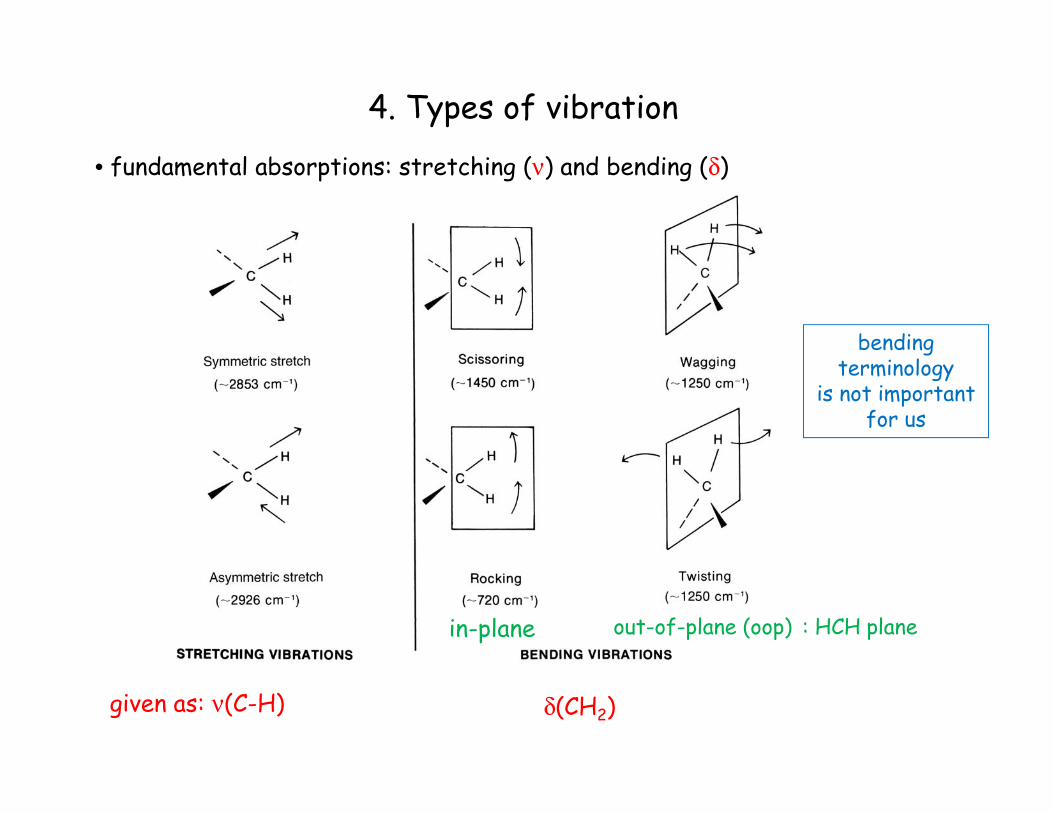
4. Types of vibration
• fundamental absorptions: stretching (ν) and bending (δ)
given as: ν(C-H) δ(CH2)
in-plane out-of-plane (oop) : HCH plane
bendingterminology
is not importantfor us
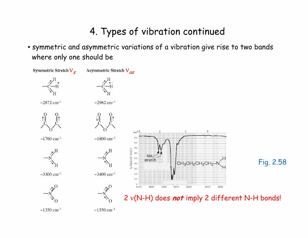
4. Types of vibration continued• symmetric and asymmetric variations of a vibration give rise to two bandswhere only one should be
2 ν(N-H) does not imply 2 different N-H bonds!
νs νas
Fig. 2.58
4. Types of vibration continued
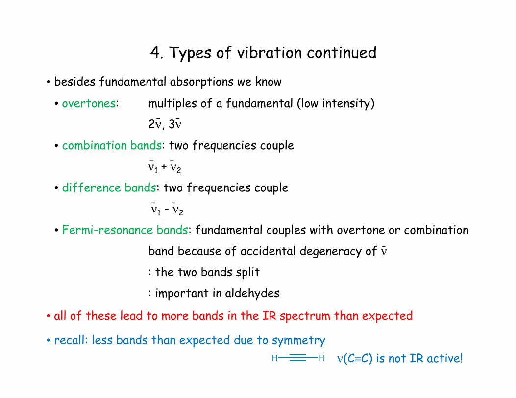
• besides fundamental absorptions we know
• overtones: multiples of a fundamental (low intensity)
2ν, 3ν
• combination bands: two frequencies couple
ν1 + ν2
• difference bands: two frequencies couple
ν1 - ν2
• Fermi-resonance bands: fundamental couples with overtone or combination
band because of accidental degeneracy of ν
: the two bands split
: important in aldehydes
• all of these lead to more bands in the IR spectrum than expected
• recall: less bands than expected due to symmetryH H ν(C≡C) is not IR active!
4. Types of vibration continued
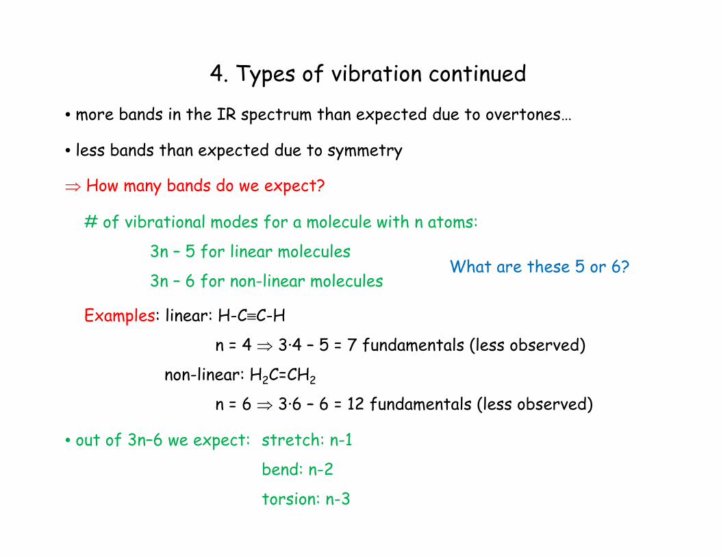
• more bands in the IR spectrum than expected due to overtones…
• less bands than expected due to symmetry
⇒ How many bands do we expect?
# of vibrational modes for a molecule with n atoms:
3n – 5 for linear molecules
3n – 6 for non-linear molecules
Examples: linear: H-C≡C-H
n = 4 ⇒ 3·4 – 5 = 7 fundamentals (less observed)
non-linear: H2C=CH2
n = 6 ⇒ 3·6 – 6 = 12 fundamentals (less observed)
• out of 3n–6 we expect: stretch: n-1
bend: n-2
torsion: n-3
What are these 5 or 6?
4. Types of vibration continued
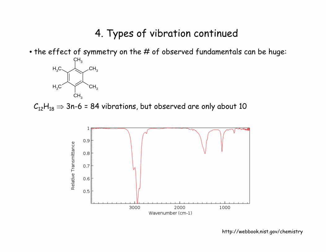
• the effect of symmetry on the # of observed fundamentals can be huge:
C12H18 ⇒ 3n-6 = 84 vibrations, but observed are only about 10
CH3
CH3
CH3
CH3
CH3
CH3
http://webbook.nist.gov/chemistry
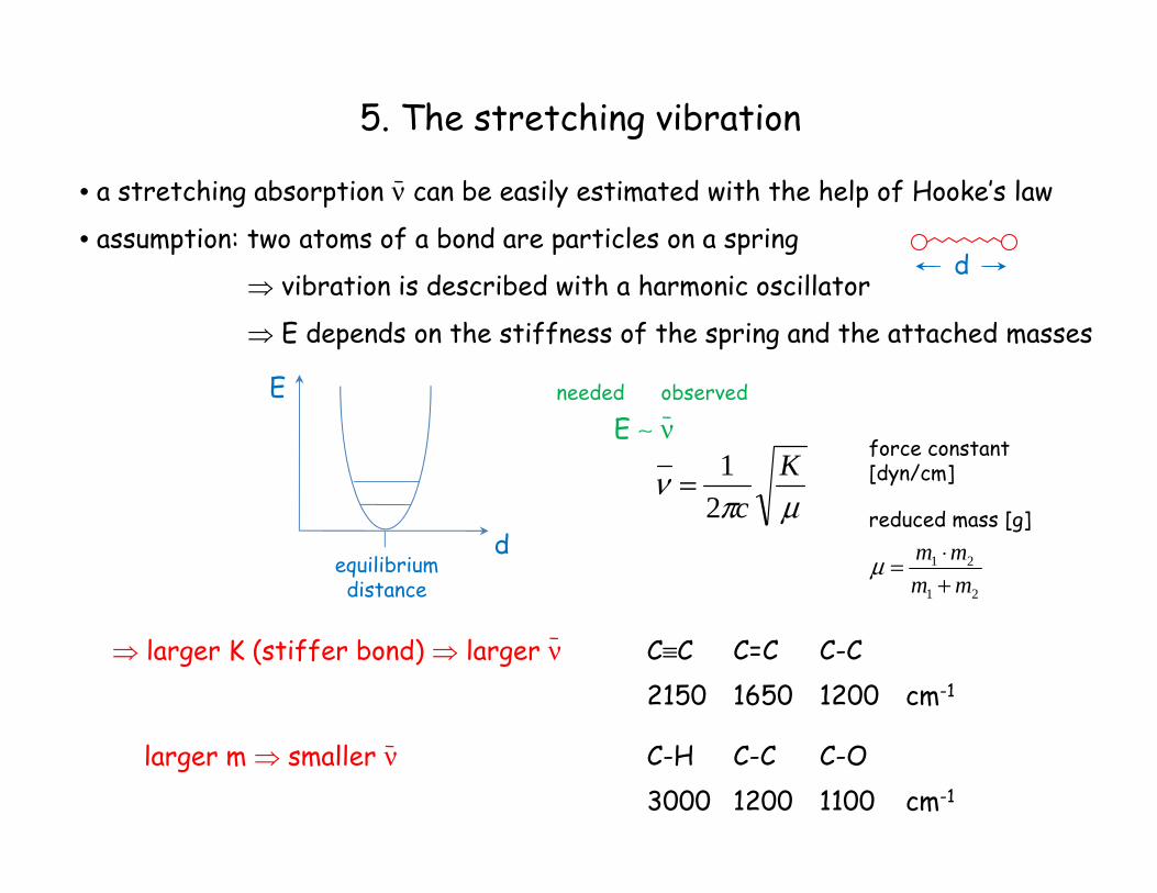
5. The stretching vibration
• a stretching absorption ν can be easily estimated with the help of Hooke’s law
• assumption: two atoms of a bond are particles on a spring
⇒ vibration is described with a harmonic oscillator
⇒ E depends on the stiffness of the spring and the attached masses
d
E
dequilibriumdistance
E ∼ νneeded observed
μπν K
c21=
force constant[dyn/cm]
reduced mass [g]
21
21
mmmm
+⋅=μ
⇒ larger K (stiffer bond) ⇒ larger ν
larger m ⇒ smaller ν
C≡C C=C C-C2150 1650 1200 cm-1
C-H C-C C-O3000 1200 1100 cm-1
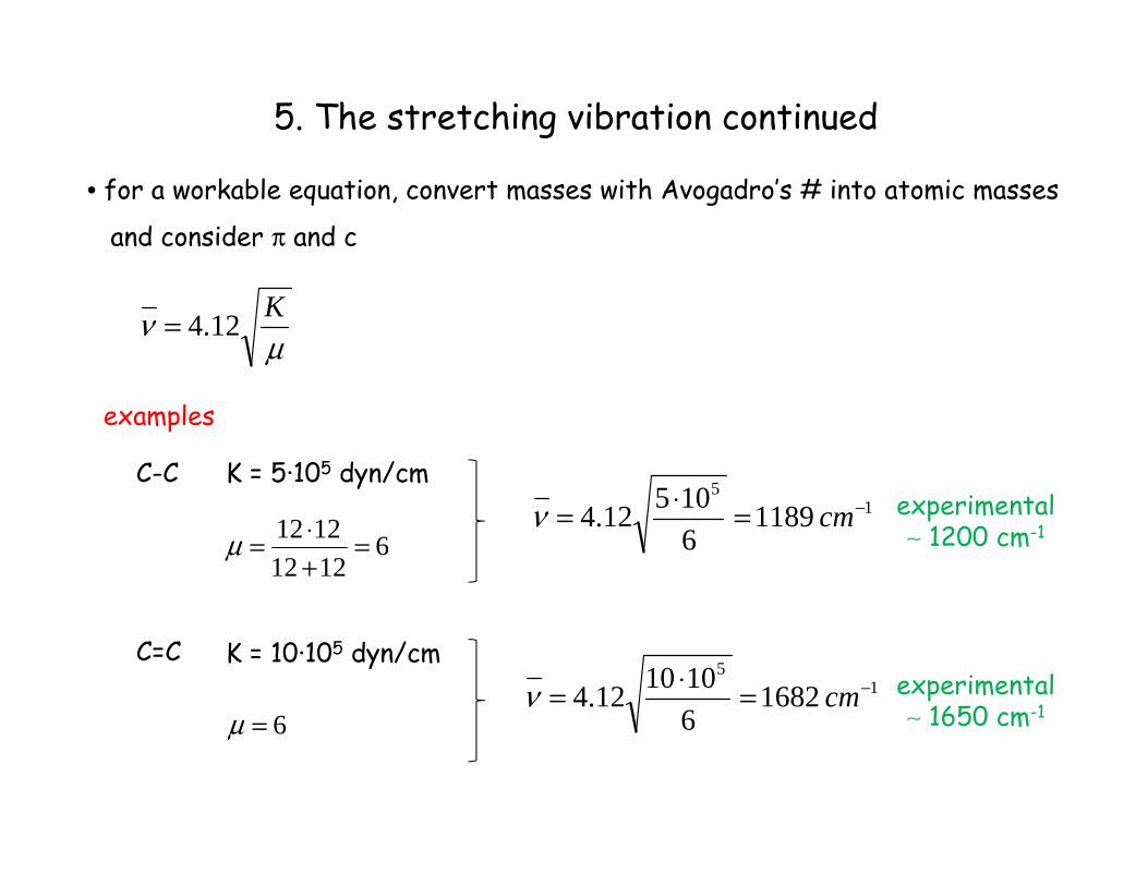
5. The stretching vibration continued
• for a workable equation, convert masses with Avogadro’s # into atomic masses
and consider π and c
μν K12.4=
examples
C-C
C=C
K = 5·105 dyn/cm
612121212 =
+⋅=μ
15
1189610512.4 −=⋅= cmν
K = 10·105 dyn/cm
6=μ1
5
16826101012.4 −=⋅= cmν
experimental∼ 1200 cm-1
experimental∼ 1650 cm-1
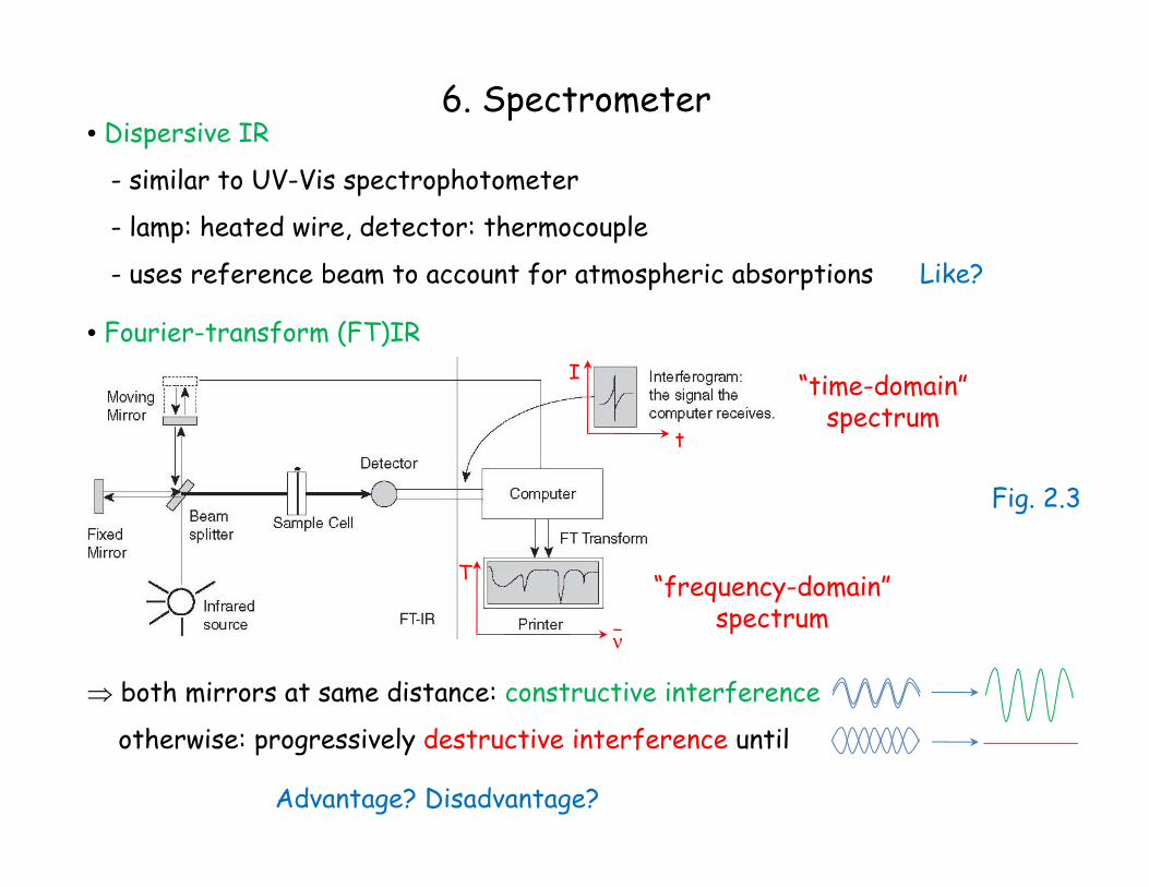
6. Spectrometer• Dispersive IR
- similar to UV-Vis spectrophotometer
- lamp: heated wire, detector: thermocouple
- uses reference beam to account for atmospheric absorptions Like?
• Fourier-transform (FT)IRI
t
“time-domain”spectrum
T
ν
“frequency-domain”spectrum
⇒ both mirrors at same distance: constructive interference
otherwise: progressively destructive interference until
Advantage? Disadvantage?
Fig. 2.3
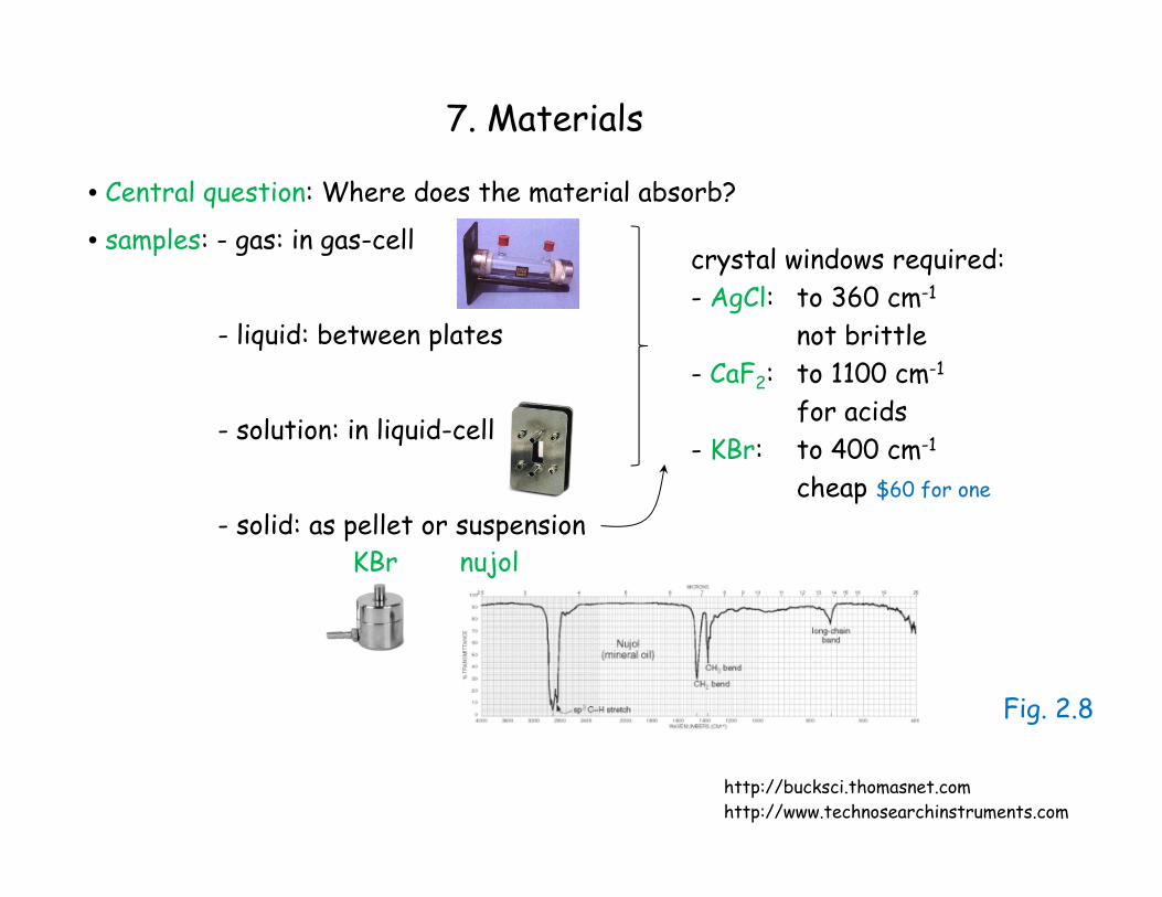
7. Materials
• Central question: Where does the material absorb?
• samples: - gas: in gas-cell
- liquid: between plates
- solution: in liquid-cell
- solid: as pellet or suspension
crystal windows required:- AgCl: to 360 cm-1
not brittle- CaF2: to 1100 cm-1
for acids- KBr: to 400 cm-1
cheap $60 for one
KBr nujol
http://www.technosearchinstruments.comhttp://bucksci.thomasnet.com
Fig. 2.8
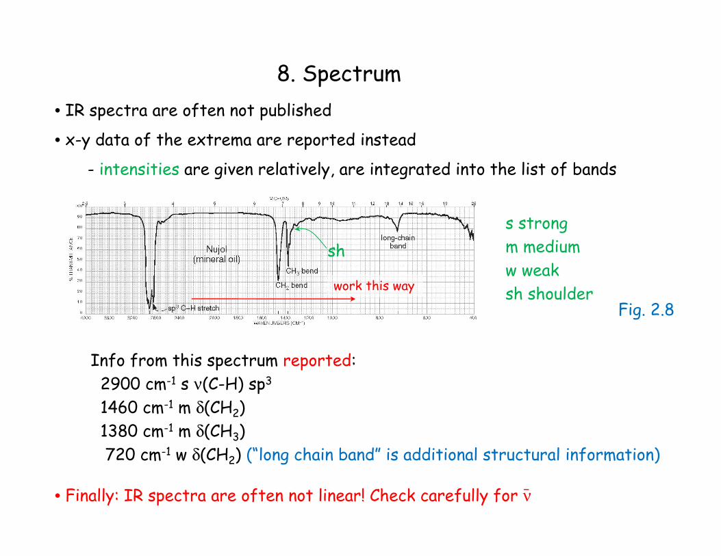
8. Spectrum• IR spectra are often not published
• x-y data of the extrema are reported instead
- intensities are given relatively, are integrated into the list of bands
Info from this spectrum reported:2900 cm-1 s ν(C-H) sp3
1460 cm-1 m δ(CH2)1380 cm-1 m δ(CH3)720 cm-1 w δ(CH2) (“long chain band” is additional structural information)
s strongm mediumw weaksh shoulder
sh
• Finally: IR spectra are often not linear! Check carefully for ν
work this way
Fig. 2.8