ChangesintheTh1:Th2CytokineBiasinPregnancyandthe...
13
Hindawi Publishing Corporation Mediators of Inflammation Volume 2012, Article ID 416739, 12 pages doi:10.1155/2012/416739 Research Article Changes in the Th1 : Th2 Cytokine Bias in Pregnancy and the Effects of the Anti-Inflammatory Cyclopentenone Prostaglandin 15-Deoxy-Δ 12,14 -Prostaglandin J 2 Lynne Sykes, 1 David A. MacIntyre, 1 Xiao J. Yap, 1 Sathana Ponnampalam, 1 Tiong Ghee Teoh, 2 and Phillip R. Bennett 1 1 Parturition Research Group, Department of Surgery and Cancer, Institute of Reproduction and Developmental Biology, Imperial College London, W120NN London, UK 2 St Mary’s Hospital, Imperial College Healthcare NHS Trust, W21NY London, UK Correspondence should be addressed to Phillip R. Bennett, [email protected] Received 13 January 2012; Revised 6 March 2012; Accepted 2 April 2012 Academic Editor: Noboru Uchide Copyright © 2012 Lynne Sykes et al. This is an open access article distributed under the Creative Commons Attribution License, which permits unrestricted use, distribution, and reproduction in any medium, provided the original work is properly cited. Pregnancy is a complex immunological state in which a bias towards T helper 2 (Th2) protects the fetus. Evidence suggests that proinflammatory cytokines increase the risk of poor neonatal outcome, independently of the direct effect of preterm labour. The anti-inflammatory prostaglandin 15-deoxy-Δ 12,14 -Prostaglandin J 2 (15dPGJ 2 ) inhibits nuclear factor Kappa B (NF-κB) in amniocytes and myocytes in vitro and is a ligand for the chemoattractant receptor-homologous molecule expressed on Th2 cells (CRTH2) receptor. Here we examine the Th1:Th2 cytokine bias in pregnancy and whether 15dPGJ 2 could be used to inhibit the production of the proinflammatory cytokines through inhibition of NF-κB while simultaneously promoting Th2 interleukin 4 (IL-4) synthesis via CRTH2 in T helper cells. Peripheral blood mononuclear cells (PBMCs) from women at 28 weeks, term pre-labour, term labour as well as non-pregnant female controls were cultured with 15dPGJ 2 or vehicle control and stimulated with phorbol myristyl acetate (PMA)/ionomycin. The percentage of CD4 + cells producing interferon gamma (IFN-γ) and tumor necrosis factor alpha (TNF-α) in response to PMA/ionomycin was significantly reduced in pregnancy. 15dPGJ 2 reduced IFN-γ and TNF-α production in stimulated T helper cells, but did not alter IL-4 production in CRTH2 +ve cells. 15dPGJ 2 also reduced phospho-p65 in stimulated PBMCs. In summary, 15dPGJ 2 suppresses the Th1 response of PBMCs during pregnancy and active labour whilst maintaining the Th2 response suggesting a therapeutic benefit in reducing neonatal morbidity in inflammation- induced PTL. 1. Introduction Preterm labour <34 weeks occurs in about 4% of pregnancies [1]. In 80–85% of cases of spontaneous preterm labour (PTL) <28 weeks, there is evidence of intrauterine infection [2]. Despite increased awareness of the association between inflammation and preterm labour [3], there have been no major advances in the prevention of preterm labour which have been shown to improve long-term neonatal outcomes. During normal term labour, the uterus and cervix become infiltrated with leukocytes and undergo changes in response to local secretion of proinflammatory cytokines. A similar pattern of biochemical events occur during PTL; however, the triggers for this premature activation are still not fully understood. Regardless, the presence of increased proinflam- matory cytokines during pregnancy is clearly associated with a poor prognosis for babies born preterm [4]. The immune system can generally be divided into the innate and adaptive immune system. The former is a nonspecific system providing immediate defence against pathogens, while the latter is more targeted, characterised by T and B lymphocytes. Although cross-talk between these lymphocytes exist, B cells and their antibodies mainly give rise to humoral immunity, whereas T cells primarily provide cell mediated immunity [5]. T helper cells (CD4 + ) form a subset of T cells and can be further subdivided into T helper 1 cells (Th1) and T helper 2 cells (Th2) depending on their pattern of cytokine production. Th1 cells secrete
Transcript of ChangesintheTh1:Th2CytokineBiasinPregnancyandthe...
Hindawi Publishing CorporationMediators of InflammationVolume 2012, Article ID 416739, 12 pagesdoi:10.1155/2012/416739
Research Article
Changes in the Th1 : Th2 Cytokine Bias in Pregnancy and theEffects of the Anti-Inflammatory Cyclopentenone Prostaglandin15-Deoxy-Δ12,14-Prostaglandin J2
Lynne Sykes,1 David A. MacIntyre,1 Xiao J. Yap,1 Sathana Ponnampalam,1
Tiong Ghee Teoh,2 and Phillip R. Bennett1
1 Parturition Research Group, Department of Surgery and Cancer, Institute of Reproduction and Developmental Biology,Imperial College London, W120NN London, UK
2 St Mary’s Hospital, Imperial College Healthcare NHS Trust, W21NY London, UK
Correspondence should be addressed to Phillip R. Bennett, [email protected]
Received 13 January 2012; Revised 6 March 2012; Accepted 2 April 2012
Academic Editor: Noboru Uchide
Copyright © 2012 Lynne Sykes et al. This is an open access article distributed under the Creative Commons Attribution License,which permits unrestricted use, distribution, and reproduction in any medium, provided the original work is properly cited.
Pregnancy is a complex immunological state in which a bias towards T helper 2 (Th2) protects the fetus. Evidence suggests thatproinflammatory cytokines increase the risk of poor neonatal outcome, independently of the direct effect of preterm labour.The anti-inflammatory prostaglandin 15-deoxy-Δ12,14-Prostaglandin J2 (15dPGJ2) inhibits nuclear factor Kappa B (NF-κB) inamniocytes and myocytes in vitro and is a ligand for the chemoattractant receptor-homologous molecule expressed on Th2 cells(CRTH2) receptor. Here we examine the Th1:Th2 cytokine bias in pregnancy and whether 15dPGJ2 could be used to inhibitthe production of the proinflammatory cytokines through inhibition of NF-κB while simultaneously promoting Th2 interleukin4 (IL-4) synthesis via CRTH2 in T helper cells. Peripheral blood mononuclear cells (PBMCs) from women at 28 weeks, termpre-labour, term labour as well as non-pregnant female controls were cultured with 15dPGJ2 or vehicle control and stimulatedwith phorbol myristyl acetate (PMA)/ionomycin. The percentage of CD4+ cells producing interferon gamma (IFN-γ) and tumornecrosis factor alpha (TNF-α) in response to PMA/ionomycin was significantly reduced in pregnancy. 15dPGJ2 reduced IFN-γand TNF-α production in stimulated T helper cells, but did not alter IL-4 production in CRTH2+ve cells. 15dPGJ2 also reducedphospho-p65 in stimulated PBMCs. In summary, 15dPGJ2 suppresses the Th1 response of PBMCs during pregnancy and activelabour whilst maintaining the Th2 response suggesting a therapeutic benefit in reducing neonatal morbidity in inflammation-induced PTL.
1. Introduction
Preterm labour <34 weeks occurs in about 4% of pregnancies[1]. In 80–85% of cases of spontaneous preterm labour(PTL) <28 weeks, there is evidence of intrauterine infection[2]. Despite increased awareness of the association betweeninflammation and preterm labour [3], there have been nomajor advances in the prevention of preterm labour whichhave been shown to improve long-term neonatal outcomes.During normal term labour, the uterus and cervix becomeinfiltrated with leukocytes and undergo changes in responseto local secretion of proinflammatory cytokines. A similarpattern of biochemical events occur during PTL; however,the triggers for this premature activation are still not fully
understood. Regardless, the presence of increased proinflam-matory cytokines during pregnancy is clearly associated witha poor prognosis for babies born preterm [4].
The immune system can generally be divided intothe innate and adaptive immune system. The former isa nonspecific system providing immediate defence againstpathogens, while the latter is more targeted, characterisedby T and B lymphocytes. Although cross-talk between theselymphocytes exist, B cells and their antibodies mainly giverise to humoral immunity, whereas T cells primarily providecell mediated immunity [5]. T helper cells (CD4+) forma subset of T cells and can be further subdivided into Thelper 1 cells (Th1) and T helper 2 cells (Th2) dependingon their pattern of cytokine production. Th1 cells secrete
2 Mediators of Inflammation
pro inflammatory cytokines such as interferon gamma (IFN-γ) and tumor necrosis factor alpha (TNF-α), whereas theTh2 cells secrete anti-inflammatory cytokines such as inter-leukin 4 (IL-4), IL-10, and IL-13 [6]. A mutually exclusiveinteraction exists between the Th1 interleukin, IFN-γ, andthe Th2 interleukin, IL-4. IL-4 is the dominant factor forpromoting growth and differentiation from the Th0 to theTh2 subtype, and directly inhibits the development of theTh1 cells [7]. IFN-γ indirectly promotes Th1 differentiationby upregulating the IL-12 receptor whilst inhibiting thegrowth of Th2 cells [8, 9].
Wegmann and colleagues first developed the conceptthat during pregnancy there is a shift from a T helper 1(Th1) response to a T helper 2 (Th2) bias during pregnancythat functionally induces maternal tolerance and suppression[10]. Consistent with this notion, administration of theTh1 interleukins IFN-γ [11] and IL-2 [12] leads to fetalloss and preterm labour in the mouse. Similarly, CBA ×DBA/2 mice that have placentas deficient in IL-4 and IL-10are prone to fetal resorption. Treatment of these mice viaintraperitoneal injection of IL-10 protects the fetuses fromresorption [13]. Several human studies have shown a Th2bias in the ratio of circulating T helper cytokine profilein normal pregnancy; and an increase in the Th1 ratio incases of recurrent miscarriage [14] and in preeclampsia [15].Peripheral blood lymphocytes taken from women in thefirst trimester show an increased production of IL-4 andIL-10 and less IFN-γ compared to nonpregnant controls invitro [16]. There is in vivo evidence in pregnant womenof increased expression of IL-4 messenger ribonucleic acid(mRNA) and lower expression of the IFN-γ mRNA [17].
Not all studies support the requirement of the shift fromTh1 to Th2 for successful pregnancy outcome [18, 19].Despite showing a suppression of IFN-γ and an increase inIL-10 during pregnancy compared to nonpregnant controls,Bates et al. showed no difference in IFN-γ, IL-10, or IL-4secretion in women who subsequently miscarried comparedwith those who went on to complete their pregnancy [18].However, contrary to this study, women with recurrentmiscarriage have been shown to have increased IFN-γ andTNF-α levels compared with women that go on to havesuccessful pregnancies [20]. While the mechanism regulatingthe Th1 : Th2 ratio is yet to be fully elucidated the importanceof maternal immune tolerance during pregnancy is unques-tionable. Several pregnancy-related proteins are known topromote Th2 bias such as leukemia inhibitory factor [21],progesterone, progesterone-induced blocking factor [22],and estradiol [23]. Prostaglandin D2 (PGD2) promotes IL-4,IL-13, IL-5, and IL-10 production in T helper 2 cells in vitrovia the second PGD2 receptor; chemoattractant receptor-homologous molecule expressed on Th2 cells (CRTH2) [24].PGD2 is produced by the placenta and may play a role in thechemoattraction of Th2 cells via the CRTH2 receptor to thematernal fetal interface to produce a localised Th2 bias [25].
15-deoxy-Δ12,14-Prostaglandin J2 (15dPGJ2), an endoge-nous prostaglandin (PG), is a product of PGD2 via a seriesof dehydration reactions [26]. As well as being a ligand forPeroxisome proliferator-activated receptor gamma (PPARγ),it is also an agonist of the CRTH2 receptor [27]. We have
previously demonstrated that this anti-inflammatory PGinhibits nuclear factor Kappa B (NF-κB) in amniocytes andmyocytes in vitro [28] and delays infection induced pretermlabour and increases pup survival in the mouse via NF-κBinhibition [29]. Moreover, recent work has shown that thefirst trimester of pregnancy is associated with the suppressionof available T cell NF-κB and reduced levels of IFN-γcompared to nonpregnant controls [30, 31]. Collectivelythese data suggest that the Th1 : Th2 ratio is modulatedthrough the regulatory interplay of both Th1 suppressionand Th2 promotion.
In this study, we examined the expression of the dom-inant Th1 interleukins IFN-γ and TNF-α, and the Th2interleukin IL-4 in T helper cells obtained from nonpregnantwomen, women in the early and late third trimester, andduring labour. We examined the effect of the CRTH2 agonist,15dPGJ2, on these interleukins and on NF-κB activation atdifferent gestational time points to determine its potentialrole in suppressing the Th1 response via NF-κB whilstsimultaneously promoting the Th2 response via CRTH2.
2. Materials and Methods
2.1. Subjects. Pregnant women at 28 weeks and term(prelabour and in labour) and nonpregnant women ofchild bearing age were included in the study in accordancewith ethical approval from the South East London EthicalCommittee Ref. 10/H0805/54. Exclusion criteria includedwomen with Asthma and HIV, women not of childbearingage, and women with a fever. After obtaining informedconsent, 5 mL of peripheral venous blood was collectedin sodium citrate tubes, and processed within 30 mins ofcollection. Unless otherwise stated, a minimum of 4 subjectswere included for each experimental sample group.
2.2. Isolation of Peripheral Blood Mononuclear Cells (PBMCs).Blood was diluted 1 : 1 with phosphate-buffered saline(PBS) and carefully layered onto Ficoll-Paque PLUS (GEHealthcare, Uppsala, Sweden) before centrifuging at 400×gfor 40 mins at room temperature. After centrifugation, thehalo containing PBMCs was carefully transferred into aclean centrifuge tube and washed twice with 7 mL of PBS.After centrifugation (400×g for 10 mins), the cell pellet wasresuspended in either PBS or RPMI 1640 (Invitrogen LifeTechnologies, Grand Island, NY, USA) culture medium.
2.3. CRTH2 Expression Studies. The PBMC pellet was resus-pended PBS, and cells were counted with a Neubauerhaemocytometer and then resuspended in staining buffer(1% fetal calf serum; 0.09% sodium azide in PBS) toobtain roughly 3.5 × 10⁶ cells per sample. Preparationswere incubated in the dark for 1 h at 37◦C with 20 μLof CRTH2-phycoerythrin (CRTH2-PE) (Beckman Coulter,High Wycombe, UK) and 3 μL of CD4-Allophycocyanin(CD4-APC) (BD Pharminogen, Oxford, UK) or the relevantisotype controls; Rat Immunoglobulin (Ig)G2a-PE (BeckmanCoulter) and Mouse IgG1κ-APC (BD Pharminogen). Afterincubation, the PBMC suspension was washed twice in
Mediators of Inflammation 3
1 mL of PBS and then resuspended in PBS for analysis.Flow cytometry settings were as follows: forward scatter E0voltage, 1.00 Amp gain Lin, and side scatter of 329 voltage,1.00 Amp gain Lin. A total of 50,000 cells were counted andgating was on lymphocytes based on the forward and sidescatter.
2.4. Intracellular Cytokine Studies (IL-4, IFN-γ, and TNF-α). For intracellular cytokine studies, cells were resuspendedin RPMI 1640 (Invitrogen) media supplemented with 10%fetal calf serum, 2 mM/L L-glutamine, 100 U/mL penicillin,and 100 μg/mL of streptomycin (Sigma, St. Louis, MO, USA)before being plated in 24-well plates and incubated for 18 hin 5% CO2/humidified air at 37◦C. Following this, 10 μg ofbrefeldin A (Sigma) was added to each well to immobilizethe interleukins in the golgi apparatus. Cells were pretreatedwith 32 μM of 15dPGJ2 (Cayman Chemicals, Ann Arbor, MI,USA) or dimethyl sulfoxide (DMSO) (Sigma) vehicle controlfor 2 h in the case of IFN-γ and TNF-α experiments or 1 h forIL-4. Following this 50 ng/mL of phorbol myristate acetate(PMA) and 0.5 μg/mL of ionomycin were added for 4 h inthe case of IFN-γ and TNF-α experiments or 5 h for IL-4.Prior to intracellular staining, cell surface staining with eitherCRTH2 and CD4, or CD4 antibodies alone, was performedas described in Section 2.3. Cells were then fixed with 2%paraformaldehyde (PFA) and incubated at 37◦C for 15 minsin the dark. Cells were washed and resuspended in 0.5%saponin and incubated for 30 mins on ice in the dark topermeabilize the cells. After incubation, cells were washedand resuspended in 0.5% saponin for intracellular stainingwith the relevant antibody; IL-4 PE/Cy7 (BioLegend, SanDiego, CA, USA), IFN-γ-(fluorescein isothiocyanate (FITC)or TNF-α-FITC (BD Biosciences, Franklin Lakes, NJ, USA).Appropriate isotype controls were used; PE/Cy7 Rat IgG1κ
(Biolegend), or FITC Mouse IgG1κ (BD Biosciences, Oxford,UK). Cells were incubated in the dark for 1 h at roomtemperature for IL-4 staining or 20 mins on ice for IFN-γ andTNF-α staining. Finally, cell suspensions were washed with0.5% saponin and resuspended in PBS for flow cytometryanalysis as follows: Forward scatter E0 voltage; 1.00 Amp gainLin; side scatter of 329 Voltage; 1.00 Amp gain Lin.
2.5. Flow Cytometry. Flow cytometry of lymphocytes wascarried out using FACSCalibur flow cytometer (BD Bio-sciences) equipped with FACSCalibur software for analysis.The lymphocyte population was gated on the scatter plotas determined by the characteristic forward scatter (FS) andside scatter (SC) which indicates the cell size and shape. Theanalysis was of CD4+, CRTH2+, and CRTH2+/CD4+ in theCRTH2 expression studies and IL-4+, IL-4+/CRTH2+, TNF-α+/CD4+, and IFN-γ+/CD4+ for the intracellular cytokines.The expression levels of CRTH2, CD4, and intracellularcytokines were evaluated by the percentage of cells expressingthe protein of interest or by mean fluorescence intensity(MFI).
2.6. Sodium Dodecyl Sulfate Polyacrylamide Gel Electrophore-sis (SDS-PAGE) and Western Blotting. Following culture
and treatments, cells were collected using a 1 mL pipetteand incubated on ice in whole cell lysis buffer (CellSignalling, Beverly, MA, USA) with 5 μL/mL of proteaseinhibitor (Sigma) lysis buffer for 5 mins and centrifuged for20 mins at 13,000 rpm at 4◦C. The supernatant was stored at−80◦C until use. Prior to SDS-PAGE, protein concentrationswere determined using the BIORAD quantification assaymeasuring absorbance at 655 nm. Approximately 15 μg ofextracted protein per sample was resolved by SDS-PAGE andsubsequently transferred onto polyvinyl difluoride (PVDF)membranes (GE Healthcare) 100 V constant at 4◦C. Fol-lowing transfer, the membrane was then blocked in 5%(wt/vol) milk in tris-buffered saline (TBS) supplementedwith 0.1% Tween 20 (TBST) for 1 h. The membrane wasthen probed with phospho-p65 (Ser 536) (Cell signalling)primary antibody (1 : 1000 in tris-buffered saline) overnightat 4◦C followed by the appropriate secondary antibody(1 : 2000 in 1% milk/TBS) for 1 h at room temperature.Chemiluminescence detection was then carried out with ECLPlus (GE Healthcare), and the membranes developed using ahigh-performance chemiluminescence film (GE Healthcare).Blots were scanned, and densitometry was performed withImageJ (v1.44p).
2.7. Statistical Analysis. Experimental sample groups con-sisted of at least 4 biological replicates unless otherwisestated. Statistical analysis was performed with Graph-PadPrism (v4.0; GraphPad Software, San Diego, CA, USA).Paired Student’s t-test or repeated measures ANOVA wasconducted where appropriate. Samples with P < 0.05 wasconsidered to be statistically significant.
3. Results
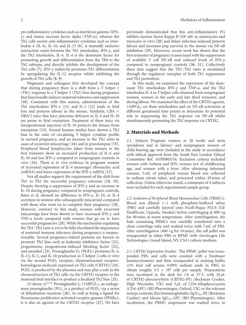
3.1. Gestational Effect on Th1 and Th2 Cytokines. A change inthe production of the Th1 and Th2 cytokines in pregnancyhas previously been described. In this study, we employedflow cytometry to examine the effect of stimulation bythe mitogen PMA/ionomycin on cytokine production atdifferent gestational stages of pregnancy and during labour(see Figure 1). Th1 cytokine profiles of CD4 positive cellswere assessed for intracellular IFN-γ and TNF-α (Figures1(a)–1(d)) and compared to levels of nonpregnant controls.The percentage of peripheral T cells producing IFN-γ inresponse to stimulation reduced in pregnancy from 10.7%in nonpregnant women to 6.7% at 28 weeks, 5.1% atterm (P < 0.05), and 5.6% at term in labour (P <0.05). Similarly, the proportion of TNF-α producing cellswas reduced, although not significantly, from 20.6% innonpregnant women to 14.5% at 28 weeks, 15.8% at termand 13.3% at term in labour. Overall levels of Th1 cytokineproduction (expressed as mean fluorescence intensity), in theCD4+/IFN-γ+ or CD4+/TNF-α+ cells remained consistentthroughout gestation and unchanged compared to nonpreg-nant controls. The Th2 cytokine, IL-4, was similarly assessedin CRTH2 positive cells (Figures 1(e) and 1(f)). WhilePMA/ionomycin stimulation did not increase the percentageof IL-4 expressing cells, the mean fluorescence intensity of
4 Mediators of Inflammation
15
10
5
0NP 28 weeks TNL TL
∗ ∗Po
siti
ve c
ells
for
IFN
-γ(%
)
(a)
NP 28 weeks TNL TL
150
100
50
0
MFI
IFN
-γ
(b)
25
20
15
10
5
0Posi
tive
cel
ls fo
r T
NF-α
(%)
NP 28 weeks TNL TL
(c)
NP 28 weeks TNL TL
100
80
40
20
0
MFI
TN
F-α
(d)
NP 28 weeks TNL TL
3
2
1
0Posi
tive
cel
ls fo
r IL
-4 (
%)
Nonstimulated
PMA/ionomycin
(e)
NP 28 weeks TNL TL
50
40
30
20
10
0
MFI
IL-
4
Nonstimulated
PMA/ionomycin
(f)
NP 28 weeks TNL TL
12.5
10
7.5
5
2.5
0
IFN
-γ: I
L-4
PMA/ionomycin
(g)
Figure 1: Th1 and Th2 cytokine production in peripheral T cells from nonpregnant and pregnant women. Peripheral blood mononuclearcells were isolated and stimulated with PMA/ionomycin. The percentage of CD4+/IFN-γ+, CD4+/TNF-α+, and CRTH2+/IL-4 positive cellswere detected by flow cytometry in samples derived from nonpregnant (NP), 28-week pregnant, term no labour (TNL), and term labour(TL) samples (a), (c), and (e). A reduction in the percentage of peripheral CD4 positive cells secreting the Th1 cytokines IFN-γ and TNF-αwas observed, whereas the percentage of cells secreting the Th2 cytokine, IL-4, remained consistent throughout gestation. Mean fluorescenceintensity (MFI) was also assessed as a measure of total cytokine production (b), (d), and (f). PMA/ionomycin stimulation induced both Th1and Th2 cytokine production in all sample groups compared to NL controls. No differences in gestation-dependent responses were detected.(g) a ratio of IFN-γ : IL-4 revealed a decrease in the Th1 : Th2 ratio during pregnancy, which was increased prior to the onset of labour. Forstatistical analysis ANOVA with Dunnett’s multiple comparison test with NP as a control was used; ∗∗P < 0.01 and ∗P < 0.05.
Mediators of Inflammation 5
104
103
102
101
00 101 102 103 104
0.34% 1.21%
36.22% 29.56%
CD4-APC
CR
TH
2-P
ECRTH2+/CD4+
(a)
4
3
2
1
0
NP 28 weeks TNL TL
Posi
tive
cel
ls fo
r C
RT
H2
(%)
(b)
NP 28 weeks TNL TL
400
300
200
100
0
MFI
CR
TH
2
(c)
Figure 2: CRTH2 expression in lymphocytes derived from nonpregnant and pregnant subjects. Lymphocytes were gated according toforward scatter and side scatter and T helper cells were identified using CD4 as a cell surface marker (n = 8 for each group). A representativecytogram from a woman of 28-week gestation is presented with the right upper quadrant showing CRTH2+/CD4+ lymphocytes (a). Thepercentage of CRTH2 positive T helper cells (presented as a percentage of positive CD4 positive cells) increased slightly in the third trimester,although statistical significance was not reached (b). No change in mean fluorescence intensity was detected in either the nonpregnant orpregnant samples (c). For statistical analysis ANOVA with Dunnett’s multiple comparison test with NP as a control was used.
IL-4 was significantly increased in samples collected fromwomen at 28 weeks (39.3, P < 0.01) and at term (39.4,P < 0.05) compared to levels of nonpregnant controls (37.1).Levels of IL-4 in term labouring samples were consistentwith nonlabouring samples (37.1). The ratio of the IFN-γ : IL-4 producing cells reduces during pregnancy, due to thesuppression of the Th1 rather than the promotion of the Th2cytokine production (Figure 1(g)).
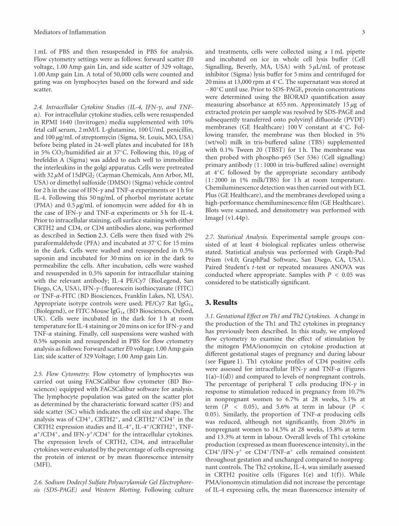
3.2. CRTH2 Expression. To determine whether the Th2response in pregnancy is reflected by an increase in thepercentage of CRTH2 positive cells, we used CD4 as a markerof T helper cells and calculated the percentage these cellsthat express CRTH2 (Figure 2(a)). The percentage of CRTH2positive cells in the CD4 positive population was observed toincrease in the second trimester of pregnancy from 2.24% innonpregnant women to 3.12% at 28 weeks before reducing to2.75% at term and 2.4% in term labour, although statisticalsignificance was not reached (Figure 2(b)). There was nosignificant increase in the mean fluorescence intensity ofCRTH2 in T helper cells between nonpregnant and pregnant
subjects (Figure 2(c)), although a slight increase in CRTH2production was noted in term labouring samples (P = 0.15).
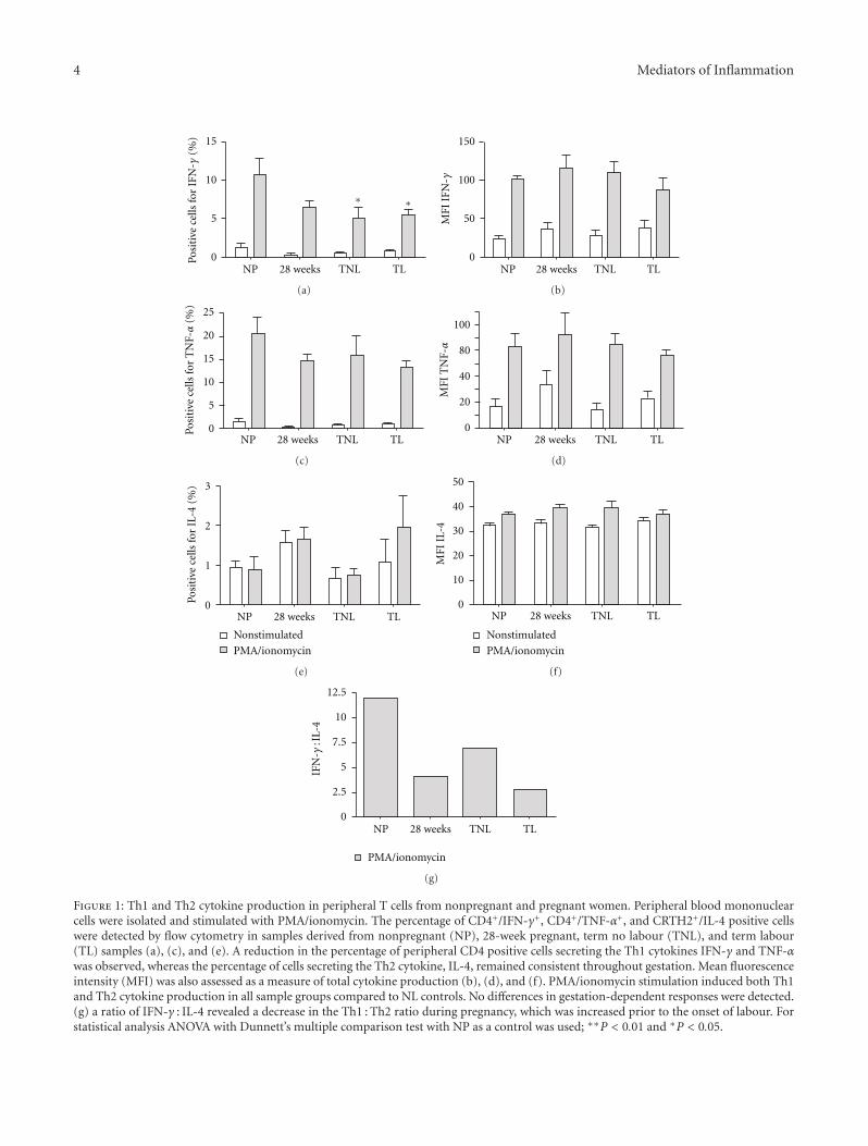
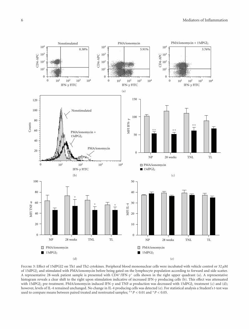
3.3. The Effect of 15dPGJ2 on the Th1 and Th2 Cytokines.15dPGJ2 is an anti-inflammatory PG and a ligand forCRTH2. We therefore explored the effect of 15dPGJ2 onthe Th1 cytokines, IFN-γ and TNF-α. To do this, cells werepreincubated with 32 μM of 15dPGJ2 or vehicle controlprior to the addition of PMA/ionomycin before intracellularcytokines were detected by flow cytometry (Figures 3(a) and3(b)). 15dPGJ2 reduced the production of PMA/ionomycinstimulated IFN-γ production (measured as mean fluores-cence intensity) in T helper cells from 101.9 to 53 (P <0.01) in nonpregnant controls, 115.7 to 52.4 (P < 0.01)in 28 week samples and from 110 to 60.7 in term (P <0.01) samples. There was a nonsignificant decrease observedin term labouring samples also (88.3 to 66.3, Figure 3(c)).Similarly, a significant decrease in PMA/ionomycin-inducedTNF-α production following treatment with 15dPGJ2 wasdetected in all pregnant samples assessed (Figure 3(d)).
The percentage of T helper cells producing IFN-γ wasalso reduced from 10.7% to 5.13% in nonpregnant women
6 Mediators of Inflammation
104
103
102
101
00 101 102 103 104
CD
4-A
PC
IFN-γ FITC
0.38%
Nonstimulated104
103
102
101
00 101 102 103 104
CD
4-A
PC
IFN-γ FITC
104
103
102
101
00 101 102 103 104
CD
4-A
PC
IFN-γ FITC
5.91%
PMA/ionomycin
3.76%
PMA/ionomycin + 15dPGJ2
(a)
120
100
80
60
40
20
0 101 102 103 104
Cou
nts
IFN-γ FITC
Nonstimulated
PMA/ionomycin +15dPGJ2
PMA/ionomycin
(b)
150
100
50
0
NP 28 weeks TNL TL
∗∗
PMA/ionomycin15dPGJ2
∗∗∗∗
MFI
IFN
-γ
(c)
100
80
60
40
20
0
∗
MFI
TN
F-α ∗ ∗
NP 28 weeks TNL TL
PMA/ionomycin
15dPGJ2
(d)
PMA/ionomycin
15dPGJ2
50
40
30
20
10
0
NP 28 weeks TNL TL
MFI
IL-
4
(e)
Figure 3: Effect of 15dPGJ2 on Th1 and Th2 cytokines. Peripheral blood mononuclear cells were incubated with vehicle control or 32 μMof 15dPGJ2 and stimulated with PMA/ionomycin before being gated on the lymphocyte population according to forward and side scatter.A representative 28-week patient sample is presented with CD4+/IFN-γ+ cells shown in the right upper quadrant (a). A representativehistogram reveals a clear shift to the right upon stimulation indicative of increased IFN-γ producing cells (b). This effect was attenuatedwith 15dPGJ2 pre-treatment. PMA/ionomycin induced IFN-γ and TNF-α production was decreased with 15dPGJ2 treatment (c) and (d);however, levels of IL-4 remained unchanged. No change in IL-4 producing cells was detected (e). For statistical analysis a Student’s t-test wasused to compare means between paired treated and nontreated samples; ∗∗P < 0.01 and ∗P < 0.05.
Mediators of Inflammation 7
(P < 0.01), 6.6% to 3.4% at 28 weeks (P < 0.01), 5.1% to2.5% at term (P < 0.05), and 5.6% to 2.9% (P < 0.01). Areduction in the percentage of T cells producing TNF-α wasalso seen with 15dPGJ2 from 20.6% to 16.7% in nonpregnantwomen, 14.5% to 9.0% at 28 weeks (P < 0.05), 15.8% to12.9% at term (P < 0.01), and 13.2% to 8.93% at term inlabour (P = 0.05) (data not shown).
Considering its anti-inflammatory properties, it washypothesized that 15dPGJ2 would induce an increase inIL-4 production via its action on the CRTH2 receptor.Contrary to what was anticipated, 15dPGJ2 had no effect onthe production of IL-4 (Figure 3(e)). Consistent with this,the percentage of CRTH2+/IL-4+ lymphocytes remainedconstant following 15dPGJ2 treatment except for samplescollected at 28 weeks gestation (1.65% to 1.16%, P < 0.05;data not shown).
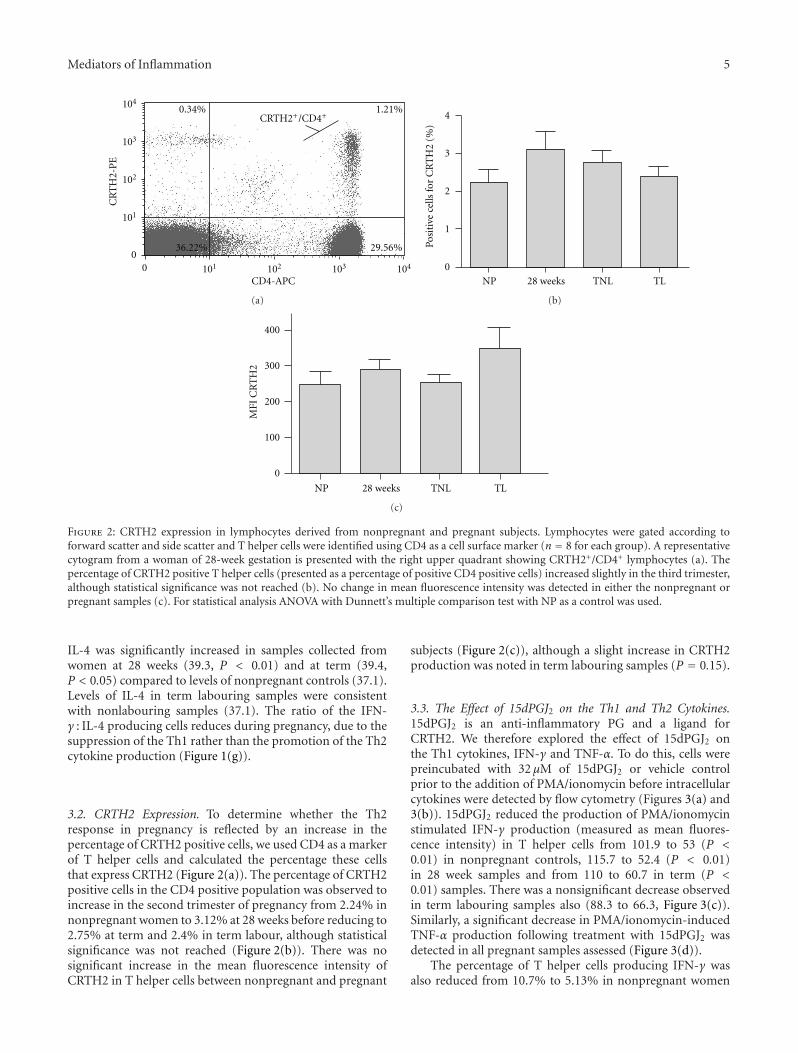
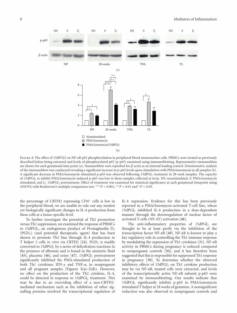
3.4. 15dPGJ2 Inhibits PMA/Ionomycin-Stimulated NF-κBActivation in Peripheral Blood Mononuclear Cells. Peripheralblood mononuclear cells were incubated with vehicle controlor 32 μM 15dPGJ2 and stimulated with PMA/ionomycinfor 10 min before being extracted and assessed for levels ofphosphorylated p65, the transcriptionally active subunit ofNF-κB. PMA/ionomycin treatment stimulated NF-κB phos-phorylation in all samples and at all gestational time points(Figure 4). Pretreatment of cells with 15dPGJ2 significantlyreduced levels of p-p65 in samples collected from womenat 28-week gestation. A clear trend of decreased p-p65following 15dPGJ2 was observed in the remaining samplesyet they did not return to basal levels of p-p65.
4. Discussion
The immunological paradox of pregnancy relies on a carefulbalance of both immune tolerance and immune suppression.Work by Medawar, and later Wegmann and colleagues, hasled to the proposal that this balance is determined by a func-tional immune suppression via a shift from a Th1 responseto a Th2 bias during pregnancy [10, 20, 32]. Although notall human and murine studies support the importance or thesimplification of the Th1 : Th2 paradigm [18, 19, 33], furtherunderstanding of this hypothesis to aid the development ofnew therapeutic strategies is still required. In this study, wehave explored the Th1 cytokines IFN-γ and TNF-α, andthe Th2 cytokine IL-4 throughout pregnancy, and examinedthe potential of the cyclopentenone PG CRTH2 agonist,15dPGJ2, to act as a therapeutic agent to modulate theTh1 : Th2 response.
Consistent with the notion of suppressed Th1 cytokineresponse throughout gestation [16, 17, 34, 35], our resultsrevealed a reduction in the percentage of IFN-γ and TNF-α-secreting T helper cells during pregnancy in responseto PMA/ionomycin stimulation (Figures 1(a) and 1(c)).However, mean production of IFN-γ and TNF-α remainedconstant within the CD4+/IFN-γ+ and CD4+/TNF-α+ cellpopulations (Figures 1(b) and 1(d)). This indicates thatwhile fewer cells maintain the capacity to initiate a Th1response to inflammatory stimuli with advancing gestation,
a compensatory mechanism facilitates increased IFN-γ andTNF-α production. These results suggest that Th1 responseduring gestation is likely to be regulated at both an individualcellular level as well as at the level of the cell population.
As normal human labour reflects a proinflammatorystate, we anticipated that the percentage of IFN-γ and TNF-α producing T helper cells would increase with the onset oflabour. However, no difference between term nonlabouringand term labouring samples was detected. This may be dueto the proinflammatory state of labour being a localizedresponse in the uterus and fetal membranes, rather thanan extending to peripheral inflammation. This would beconsistent with the localised increase in the DNA bindingactivity of NF-κB in amnion cells postlabour compared toprelabour [36] and in myometrium during labour comparedto pre-labour [37] as well as the reported increase in labourof IFN-γ in amnion, choriodecidua, and placenta [38], andTNF-α in the amniotic fluid, myometrium, and cervix [39,40] rather than in the peripheral blood.
A number of studies have shown that mitogen stimula-tion can induce IL-4 production in PBMCs of the secondand third trimesters of pregnancy in vitro [16, 35, 41].Intrinsic IL-4 production by PBMCs also shows an increasein pregnancy compared with nonpregnant controls [17, 42].We therefore hypothesized that PMA/ionomycin stimulationof PMBCs would drive IL-4 production during pregnancy,and that this phenomenon would subside at term concurrentwith the activation of inflammatory pathways involved innormal labour pathways. To examine any evidence of ashift towards the Th2 profile in peripheral blood duringpregnancy, PBMCs were gated for CRTH2 (a marker forTh2 cells) and the level of IL-4 assessed (Figures 1(e) and1(f)). The percentage of CRTH2+/IL-4+ secreting cells, aswell as mean production levels of IL-4 remained consistentthroughout pregnancy and advancing gestation (Figures 1(f)and 1(e)) although a slight increase in the percentage of IL-4 secreting cells was observed in both nonstimulated andstimulated 28-week samples. Between 0.4–6.5% of peripheralblood CD4+ cells of nonpregnant women express CRTH2and secrete IL-4, but not IFN-γ, in response to PMAand ionomycin and are thus typical of Th2 cells [43].Consistent with this, we found that between 2–4% of CD4+
PMBCs express CRTH2 (Figure 2(a)). This percentage wasmaintained throughout gestation with a minor, statisticallynonsignificant, increase observed at 28 weeks (Figure 2(b)).Tsuda et al. have previously reported an increase in CRTH2expression in CD4+ cells between nonpregnant (2.08%)and 7-week gestation (3.43%, P = 0.02) [44], but it ispossible that this effect is diminished with advancing ges-tation. Mean levels of CRTH2 expressed in CD4+ cells werealso unchanged through gestation and labour (Figure 2(c)).Taken together with the fact that we were unable to detectany difference in IL-4 secretion levels in these cells uponmitogen stimulation, our results collectively suggest that theTh2 response in PMBCs is maximised during pregnancy andcannot be further stimulated. This would imply that anybeneficial effects of therapeutic agents intended to push theTh2 : Th1 ratio towards Th2 would need to do so throughinhibition of Th1, rather than upregulation of Th2. Although
8 Mediators of Inflammation
p-p65
β-actin
NS S J2
NP 28 weeks TNL TL
NS S J2 NS S J2 NS S J2
(a)
8
6
4
2
0
NP 28 weeks TNL TL
∗
∗∗
∗∗∗
NonstimulatedPMA/ionomycin
∗
∗∗
∗∗
∗
PMA/ionomycin/15dPGJ2
(b)
Figure 4: The effect of 15dPGJ2 on NF-κB p65 phosphorylation in peripheral blood mononuclear cells. PBMCs were treated as previouslydescribed before being extracted and levels of phosphorylated p65 (p-p65) examined using immunoblotting. Representative immunoblotsare shown for each gestational time point (a). Immunoblots were reprobed for β-actin as an internal loading control. Densitometric analysisof the immunoblots was conducted revealing a significant increase in p-p65 levels upon stimulation with PMA/ionomycin in all samples (b).A significant decrease in PMA/ionomycin stimulated p-p65 was observed following 15dPGJ2 treatment in 28-week samples. The capacityof 15dPGJ2 to inhibit PMA/ionomycin-induced p-p65 was lost in those samples collected at term. NS: nonstimulated, S: PMA/ionomycinstimulated, and J2: 15dPGJ2 pretreatment. Effect of treatment was examined for statistical significance at each gestational timepoint usingANOVA with Bonferroni’s multiple comparison test; ∗∗∗P < 0.001, ∗∗P < 0.01 and ∗P < 0.05.
the percentage of CRTH2 expressing CD4+ cells is low inthe peripheral blood, we are unable to rule out any modestyet biologically significant changes in IL-4 production fromthese cells at a tissue-specific level.
To further investigate the potential of Th2 promotionversus Th1 suppression, we examined the response of PBMCsto 15dPGJ2, an endogenous product of Prostaglandin D2
(PGD2) (and potential therapeutic agent) that has beenshown to promote Th2 bias through IL-4 production inT helper 2 cells in vitro via CRTH2 [24]. PGD2 is readilyconverted to 15dPGJ2 by a series of dehydration reactions inthe presence of albumin and is found in the amniotic fluid[45], placenta [46], and urine [47]. 15dPGJ2 pretreatmentsignificantly inhibited the PMA-stimulated production ofboth Th1 cytokines: IFN-γ and TNF-α, in nonpregnantand all pregnant samples (Figures 3(a)–3(d)). However,no effect on the production of the Th2 cytokine, IL-4,could be detected in response to 15dPGJ2 treatment. Thismay be due to an overriding effect of a non-CRTH2-mediated mechanism such as the inhibition of other sig-nalling proteins involved the transcriptional regulation of
IL-4 expression. Evidence for this has been previouslyreported in a PMA/Ionomycin-activated T-cell line, where15dPGJ2 inhibited IL-4 production in a dose-dependentmanner through the downregulation of nuclear factor ofactivated T-cells (NF-AT) activation [48].
The anti-inflammatory properties of 15dPGJ2 arethought to be at least partly via the inhibition of thetranscription factor NF-κB [49]. NF-κB is known to play akey regulatory role in controlling the Th1 immune responseby modulating the expression of Th1 cytokines [31]. NF-κBactivity in PBMCs during pregnancy is reduced comparedto nonpregnant controls [50], and it has therefore beensuggested that this is responsible for suppressed Th1 responsein pregnancy [30]. To determine whether the observedinhibitory effects of 15dPGJ2 on Th1 cytokine productionmay be via NF-κB, treated cells were extracted, and levelsof the transcriptionally active NF-κB subunit p-p65 wereexamined by immunoblotting. Our results indicate that15dPGJ2 significantly inhibits p-p65 in PMA/ionomycinstimulated T helper at 28 weeks of gestation. A nonsignificantreduction was also observed in nonpregnant controls and
Mediators of Inflammation 9
Th0
Th1 Th2
Infection Progesterone/oestrogen
IFN-γ
IFN-γIL-4
IL-4+
−+
+
−
−−
IL-4IFN-γ
PGD2CRTH2
15dPGJ2
TNF-α
Pregnancy loss/preterm labour Successful pregnancy
NFATNF-κB
Receptor?
Receptor?
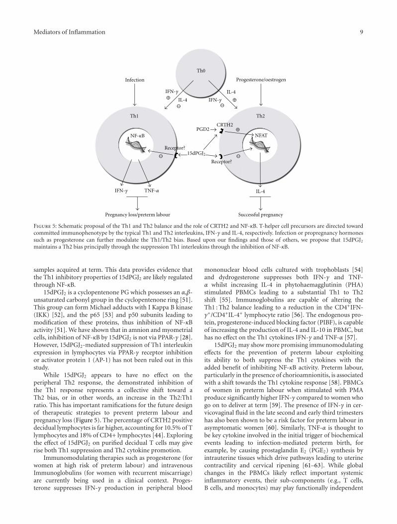
Figure 5: Schematic proposal of the Th1 and Th2 balance and the role of CRTH2 and NF-κB. T-helper cell precursors are directed towardcommitted immunophenotype by the typical Th1 and Th2 interleukins, IFN-γ and IL-4, respectively. Infection or propregnancy hormonessuch as progesterone can further modulate the Th1/Th2 bias. Based upon our findings and those of others, we propose that 15dPGJ2
maintains a Th2 bias principally through the suppression Th1 interleukins through the inhibition of NF-κB.
samples acquired at term. This data provides evidence thatthe Th1 inhibitory properties of 15dPGJ2 are likely regulatedthrough NF-κB.
15dPGJ2 is a cyclopentenone PG which possesses an α,β-unsaturated carbonyl group in the cyclopentenone ring [51].This group can form Michael adducts with I Kappa B kinase(IKK) [52], and the p65 [53] and p50 subunits leading tomodification of these proteins, thus inhibition of NF-κBactivity [51]. We have shown that in amnion and myometrialcells, inhibition of NF-κB by 15dPGJ2 is not via PPAR-γ [28].However, 15dPGJ2-mediated suppression of Th1 interleukinexpression in lymphocytes via PPAR-γ receptor inhibitionor activator protein 1 (AP-1) has not been ruled out in thisstudy.
While 15dPGJ2 appears to have no effect on theperipheral Th2 response, the demonstrated inhibition ofthe Th1 response represents a collective shift toward aTh2 bias, or in other words, an increase in the Th2:Th1ratio. This has important ramifications for the future designof therapeutic strategies to prevent preterm labour andpregnancy loss (Figure 5). The percentage of CRTH2 positivedecidual lymphocytes is far higher, accounting for 10.5% of Tlymphocytes and 18% of CD4+ lymphocytes [44]. Exploringthe effect of 15dPGJ2 on purified decidual T cells may giverise both Th1 suppression and Th2 cytokine promotion.
Immunomodulating therapies such as progesterone (forwomen at high risk of preterm labour) and intravenousImmunoglobulins (for women with recurrent miscarriage)are currently being used in a clinical context. Proges-terone suppresses IFN-γ production in peripheral blood
mononuclear blood cells cultured with trophoblasts [54]and dydrogesterone suppresses both IFN-γ and TNF-α whilst increasing IL-4 in phytohaemagglutinin (PHA)stimulated PBMCs leading to a substantial Th1 to Th2shift [55]. Immunoglobulins are capable of altering theTh1 : Th2 balance leading to a reduction in the CD4+IFN-γ+/CD4+IL-4+ lymphocyte ratio [56]. The endogenous pro-tein, progesterone-induced blocking factor (PIBF), is capableof increasing the production of IL-4 and IL-10 in PBMC, buthas no effect on the Th1 cytokines IFN-γ and TNF-α [57].
15dPGJ2 may show more promising immunomodulatingeffects for the prevention of preterm labour exploitingits ability to both suppress the Th1 cytokines with theadded benefit of inhibiting NF-κB activity. Preterm labour,particularly in the presence of chorioamnionitis, is associatedwith a shift towards the Th1 cytokine response [58]. PBMCsof women in preterm labour when stimulated with PMAproduce significantly higher IFN-γ compared to women whogo on to deliver at term [59]. The presence of IFN-γ in cer-vicovaginal fluid in the late second and early third trimestershas also been shown to be a risk factor for preterm labour inasymptomatic women [60]. Similarly, TNF-α is thought tobe key cytokine involved in the initial trigger of biochemicalevents leading to infection-mediated preterm birth, forexample, by causing prostaglandin E2 (PGE2) synthesis byintrauterine tissues which drive pathways leading to uterinecontractility and cervical ripening [61–63]. While globalchanges in the PBMCs likely reflect important systemicinflammatory events, their sub-components (e.g., T cells,B cells, and monocytes) may play functionally independent
10 Mediators of Inflammation
roles at the maternal fetal interface. Future work using anin vivo model examining effects of 15dPGJ2 and CRTH2agonists on Th1 and Th2 interleukins at local sites (e.g.,decidua and myometrium) would allow us to extrapolatehow immune cells influence nonimmune cell function.
Prophylactic administration of 15dPGJ2 to women athigh risk of preterm birth may lead to the inability ofinfection to activate NF-κB and thus preventing transcrip-tion of labour associated genes, and the inability to alterthe Th1 : Th2 bias to favour Th1, thus preventing the proinflammatory detrimental environment of the fetus. Consid-eration for the route of administration should be supportedby further studies of the potential local effect of 15dPGJ2
on interleukin production from the cervix, myometrium,and fetal membranes. The anti-inflammatory effect on NF-κB and the Th1 interleukins may not be replicated in thecervix, for example, which would serve as the least invasiveroute of administration of 15dPGJ2 in the form of a pessary.Absorption at the local level is also unlikely to providesystemic levels high enough to effect peripheral cytokineproduction, likewise, the effect of peripheral administrationon the local maternal fetal interface is not guaranteed toresult in a physiological effect. Nevertheless, this study showspromising potential for the future application of 15dPGJ2
as an immunomodulating therapy for infection-mediatedpreterm labour, and thus further studies examining its effecton gestational tissues should be pursued.
5. Conclusion
This study presents evidence that the Th1 : Th2 balance ischanged in pregnancy through Th1 suppression rather thanTh2 promotion. We have also demonstrated the potentialanti-inflammatory effects of 15dPGJ2 through the suppres-sion of NF-κB and the Th1 proinflammatory cytokinesIFN-γ and TNF-α. These properties may be of therapeuticbenefit for the prevention of infection-mediated pretermlabour and the reduction of inflammation induced neonatalmorbidity, where an aberrant profile favouring the Th1response is seen. Thus, future work in this area may be bestdirected toward designing therapeutic strategies aimed tomanipulate the Th1 response during pregnancy as a rationalstrategy for the prevention of preterm labour and pregnancyloss/miscarriage.
Conflict of Interests
The authors report that no conflict of interests exist.
Acknowledgment
This study was funded by Wellbeing of Women.
References
[1] H. Honest, C. A. Forbes, K. H. Duree et al., “Screeningto prevent spontaneous preterm birth: systematic reviews of
accuracy and effectiveness literature with economic mod-elling,” Health Technology Assessment, vol. 13, no. 43, pp. 1–627, 2009.
[2] R. L. Goldenberg, W. W. Andrews, and J. C. Hauth, “Choriode-cidual infection and preterm birth,” Nutrition Reviews, vol. 60,no. 5, pp. S19–S25, 2002.
[3] L. Sykes, D. A. MacIntyre, T. G. Teoh, and P. R. Bennett,“Targeting immune activation in the prevention of pre-termlabour,” European Obstetrics & Gynaecology, vol. 6, no. 2, pp.100–106, 2011.
[4] B. H. Yoon, R. Romero, J. S. Park et al., “Fetal exposure to anintra-amniotic inflammation and the development of cerebralpalsy at the age of three years,” American Journal of Obstetrics& Gynecology, vol. 182, no. 3, pp. 675–681, 2000.
[5] E. T. Abrams and E. M. Miller, “The roles of the immunesystem in Women’s reproduction: evolutionary constraintsand life history trade-offs,” American Journal of PhysicalAnthropology, vol. 146, supplement 53, pp. 134–154, 2011.
[6] T. R. Mosmann and S. Sad, “The expanding universe of T-cellsubsets: Th1, Th2 and more,” Immunology Today, vol. 17, no.3, pp. 138–146, 1996.
[7] A. O’Garra and N. Arai, “The molecular basis of T helper 1and T helper 2 cell differentiation,” Trends in Cell Biology, vol.10, no. 12, pp. 542–550, 2000.
[8] A. O’Garra, “Cytokines induce the development of function-ally heterogeneous T helper cell subsets,” Immunity, vol. 8, no.3, pp. 275–283, 1998.
[9] K. M. Murphy, W. Ouyang, S. J. Szabo et al., “T helper differ-entiation proceeds through Stat1-dependent, Stat4-dependentand Stat4-independent phases,” Current Topics in Microbiologyand Immunology, vol. 238, pp. 23–26, 1999.
[10] T. G. Wegmann, H. Lin, L. Guilbert, and T. R. Mosmann,“Bidirectional cytokine interactions in the maternal-fetalrelationship: is successful pregnancy a TH2 phenomenon?”Immunology Today, vol. 14, no. 7, pp. 353–356, 1993.
[11] R. Mattsson, R. Holmdahl, A. Scheynius, F. Bernadotte, A.Mattsson, and P. H. van der Meide, “Placental MHC classI antigen expression is induced in mice following in vivotreatment with recombinant interferon-gamma,” Journal ofReproductive Immunology, vol. 19, no. 2, pp. 115–129, 1991.
[12] B. U. Tezabwala, P. M. Johnson, and R. C. Rees, “Inhibitionof pregnancy viability in mice following IL-2 administration,”Immunology, vol. 67, no. 1, pp. 115–119, 1989.
[13] G. Chaouat, A. A. Meliani, J. Martal et al., “IL-10 preventsnaturally occuring fetal loss in the CBA x DBA/2 matingcombination, and local defect in IL-10 production in thisabortion-prone combination is corrected by in vivo injectionof IFN-τ,” Journal of Immunology, vol. 154, no. 9, pp. 4261–4268, 1995.
[14] R. Druckmann and M. A. Druckmann, “Progesterone and theimmunology of pregnancy,” Journal of Steroid Biochemistryand Molecular Biology, vol. 97, no. 5, pp. 389–396, 2005.
[15] H. Lin, T. R. Mosmann, L. Guilbert, S. Tuntipopipat, and T.G. Wegmann, “Synthesis of T helper 2-type cytokines at thematernal-fetal interface,” Journal of Immunology, vol. 151, no.9, pp. 4562–4573, 1993.
[16] M. Marzi, A. Vigano, D. Trabattoni et al., “Characterization oftype 1 and type 2 cytokine production profile in physiologicand pathologic human pregnancy,” Clinical and ExperimentalImmunology, vol. 106, no. 1, pp. 127–133, 1996.
[17] J. Tranchot-Diallo, G. Gras, F. Parnet-Mathieu et al., “Modu-lations of cytokine expression in pregnant women,” AmericanJournal of Reproductive Immunology, vol. 37, no. 3, pp. 215–226, 1997.
Mediators of Inflammation 11
[18] M. D. Bates, S. Quenby, K. Takakuwa, P. M. Johnson, andG. S. Vince, “Aberrant cytokine production by peripheralblood mononuclear cells in recurrent pregnancy loss?” HumanReproduction, vol. 17, no. 9, pp. 2439–2444, 2002.
[19] S. Shimada, K. Iwabuchi, E. H. Kato et al., “No difference innatural-killer-T cell population, but Th2/Tc2 predominancein peripheral blood of recurrent aborters,” American Journalof Reproductive Immunology, vol. 50, no. 4, pp. 334–339, 2003.
[20] R. Raghupathy, M. Makhseed, R. Azizieh, A. Omu, M. Gupta,and R. Farhat, “Cytokine production by maternal lymphocytesduring normal human pregnancy and in unexplained recur-rent spontaneous abortion,” Human Reproduction, vol. 15, no.3, pp. 713–718, 2000.
[21] J. Trowsdale and A. G. Betz, “Mother’s little helpers: mecha-nisms of maternal-fetal tolerance,” Nature Immunology, vol. 7,no. 3, pp. 241–246, 2006.
[22] J. Szekeres-Bartho, A. Barakonyi, G. Par, B. Polgar, T.Palkovics, and L. Szereday, “Progesterone as an immunomod-ulatory molecule,” International Immunopharmacology, vol. 1,no. 6, pp. 1037–1048, 2001.
[23] S. A. Huber, J. Kupperman, and M. K. Newell, “Estradiolprevents and testosterone promotes Fas-dependent apoptosisin CD4+ Th2 cells by altering Bcl 2 expression,” Lupus, vol. 8,no. 5, pp. 384–387, 1999.
[24] L. Xue, S. L. Gyles, F. R. Wettey et al., “ProstaglandinD2 causes preferential induction of proinflammatory Th2cytokine production through an action on chemoattractantreceptor-like molecule expressed on Th2 cells,” Journal ofImmunology, vol. 175, no. 10, pp. 6531–6536, 2005.
[25] T. Michimata, H. Tsuda, M. Sakai et al., “Accumulationof CRTH2-positive T-helper 2 and T-cytotoxic 2 cells atimplantation sites of human decidua in a prostaglandin D2-mediated manner,” Molecular Human Reproduction, vol. 8, no.2, pp. 181–187, 2002.
[26] T. Shibata, M. Kondo, T. Osawa, N. Shibata, M. Kobayashi, andK. Uchida, “15-DeoxyΔ12,14-prostaglandin J2. A prostaglandinD2 metabolite generated during inflammatory processes,”Journal of Biological Chemistry, vol. 277, no. 12, pp. 10459–10466, 2002.
[27] K. Nagata and H. Hirai, “The second PGD2 receptorCRTH2: structure, properties, and functions in leukocytes,”Prostaglandins Leukotrienes and Essential Fatty Acids, vol. 69,no. 2-3, pp. 169–177, 2003.
[28] T. M. Lindstrom and P. R. Bennett, “15-Deoxy-Δ12,14-prostaglandin J2 inhibits interleukin-1β-induced nuclearfactor-κB in human amnion and myometrial cells: mecha-nisms and implications,” Journal of Clinical Endocrinology andMetabolism, vol. 90, no. 6, pp. 3534–3543, 2005.
[29] G. Pirianov, S. N. Waddington, T. M. Lindstrom, V. Terzi-dou, H. Mehmet, and P. R. Bennett, “The cyclopentenone15-deoxy-Δ12,14-prostaglandin J2 delays lipopolysaccharide-induced preterm delivery and reduces mortality in the new-born mouse,” Endocrinology, vol. 150, no. 2, pp. 699–706,2009.
[30] S. A. McCracken, E. Gallery, and J. M. Morris, “Pregnancy-specific down-regulation of NF-κB expression in T cells inhumans is essential for the maintenance of the cytokine profilerequired for pregnancy success,” Journal of Immunology, vol.172, no. 7, pp. 4583–4591, 2004.
[31] K. A. Hadfield, S. A. McCracken, A. W. Ashton, T. G.Nguyen, and J. M. Morris, “Regulated suppression of NF-κB throughout pregnancy maintains a favourable cytokineenvironment necessary for pregnancy success,” Journal ofReproductive Immunology, vol. 89, no. 1, pp. 1–9, 2011.
[32] P. B. Medawar, “Some immunological and endocrinologicalproblems raised by the evolution of viviparity in vertebrates,”Symposia of the Society for Experimental Biology, vol. 7, pp.320–338, 1953.
[33] S. Zourbas, S. Dubanchet, J. Martal, and G. Chaouat,“Localization of pro-inflammatory (IL-12, IL-15) and anti-inflammatory (IL-11, IL-13) cytokines at the foetomaternalinterface during murine pregnancy,” Clinical and ExperimentalImmunology, vol. 126, no. 3, pp. 519–528, 2001.
[34] L. Keski-Nisula, M. R. Hirvonen, M. Roponen, S. Heinonen,and J. Pekkanen, “Maternal and neonatal IL-4 and IFN-gamma production at delivery and 3 months after birth,”Journal of Reproductive Immunology, vol. 60, no. 1, pp. 25–33,2003.
[35] G. Reinhard, A. Noll, H. Schlebusch, P. Mallmann, and A.V. Ruecker, “Shifts in the TH1/TH2 balance during humanpregnancy correlate with apoptotic changes,” Biochemical andBiophysical Research Communications, vol. 245, no. 3, pp. 933–938, 1998.
[36] V. C. Allport, D. Pieber, D. M. Slater, R. Newton, J. O.White, and P. R. Bennett, “Human labour is associated withnuclear factor-κB activity which mediates cyclo-oxygenase-2expression and is involved with the ’functional progesteronewithdrawal’,” Molecular Human Reproduction, vol. 7, no. 6, pp.581–586, 2001.
[37] N. R. Chapman, G. N. Europe-Finner, and S. C. Robson,“Expression and deoxyribonucleic acid-binding activity of thenuclear factor κB family in the human myometrium duringpregnancy and labor,” Journal of Clinical Endocrinology andMetabolism, vol. 89, no. 11, pp. 5683–5693, 2004.
[38] G. L. Veith and G. E. Rice, “Interferon gamma expressionduring human pregnancy and in association with labour,”Gynecologic and Obstetric Investigation, vol. 48, no. 3, pp. 163–167, 1999.
[39] A. Young, A. J. Thomson, M. Ledingham, F. Jordan, I. A. Greer,and J. E. Norman, “Immunolocalization of proinflammatorycytokines in myometrium, cervix, and fetal membranesduring human parturition at term,” Biology of Reproduction,vol. 66, no. 2, pp. 445–449, 2002.
[40] S. J. Fortunato, R. P. Menon, K. F. Swan, and R. Menon,“Inflammatory cytokine (interleukins 1, 6, and 8 and tumornecrosis factor-α) release from cultured human fetal mem-branes in response to endotoxic lipopolysaccharide mirrorsamniotic fluid concentrations,” American Journal of Obstetrics& Gynecology, vol. 174, no. 6, pp. 1855–1862, 1996, discussion1861-1862.
[41] L. Matthiesen, C. Ekerfelt, G. Berg, and J. Ernerudh,“Increased numbers of circulating interferon-γ- andinterleukin-4-secreting cells during normal pregnancy,”American Journal of Reproductive Immunology, vol. 39, no. 6,pp. 362–367, 1998.
[42] L. Matthiesen, M. Khademi, C. Ekerfelt et al., “In-situ detec-tion of both inflammatory and anti-inflammatory cytokinesin resting peripheral blood mononuclear cells during preg-nancy,” Journal of Reproductive Immunology, vol. 58, no. 1, pp.49–59, 2003.
[43] K. Nagata, K. Tanaka, K. Ogawa et al., “Selective expression ofa novel surface molecule by human Th2 cells in vivo,” Journalof Immunology, vol. 162, no. 3, pp. 1278–1286, 1999.
[44] H. Tsuda, T. Michimata, M. Sakai, K. Nagata, M. Nakamura,and S. Saito, “A novel surface molecule of Th2- and Tc2-typecells, CRTH2 expression on human peripheral and decidualCD4+ and CD8+ T cells during the early stage of pregnancy,”
12 Mediators of Inflammation
Clinical and Experimental Immunology, vol. 123, no. 1, pp.105–111, 2001.
[45] R. J. A. Helliwell, J. A. Keelan, K. W. Marvin et al., “Gestationalage-dependent up-regulation of prostaglandin D synthase(PGDS) and production of PGDS-derived antiinflamma-tory prostaglandins in human placenta,” Journal of ClinicalEndocrinology and Metabolism, vol. 91, no. 2, pp. 597–606,2006.
[46] A. Jawerbaum, E. Capobianco, C. Pustovrh et al., “Influenceof peroxisome proliferator-activated receptor γ activation byits endogenous ligand 15-deoxy Δ12,14 prostaglandin J2 onnitric oxide production in term placental tissues from diabeticwomen,” Molecular Human Reproduction, vol. 10, no. 9, pp.671–676, 2004.
[47] M. Fukushima, “Prostaglandin J2—anti-tumour and anti-viral activities and the mechanisms involved,” Eicosanoids, vol.3, no. 4, pp. 189–199, 1990.
[48] S. W. Chung, B. Y. Kang, and T. S. Kim, “Inhibition ofinterleukin-4 production in CD4+ T cells by peroxisomeproliferator-activated receptor-γ (PPAR-γ) ligands: involve-ment of physical association between PPAR-γ and the nuclearfactor of activated T cells transcription factor,” MolecularPharmacology, vol. 64, no. 5, pp. 1169–1179, 2003.
[49] M. Ricote, A. C. Li, T. M. Willson, C. J. Kelly, and C. K.Glass, “The peroxisome proliferator-activated receptor-γ is anegative regulator of macrophage activation,” Nature, vol. 391,no. 6662, pp. 79–82, 1998.
[50] S. A. McCracken, C. L. Drury, H. S. Lee, and J. M. Morris,“Pregnancy is associated with suppression of the nuclear factorκB/I κB activation pathway in peripheral blood mononuclearcells,” Journal of Reproductive Immunology, vol. 58, no. 1, pp.27–47, 2003.
[51] E. Cernuda-Morollon, E. Pineda-Molina, F. Javier Canada,and D. Perez-Sala, “15-Deoxy-Δ12,14-prostaglandin J2 inhibi-tion of NF-κB-DNA binding through covalent modification ofthe p50 subunit,” Journal of Biological Chemistry, vol. 276, no.38, pp. 35530–35536, 2001.
[52] A. Rossi, P. Kapahi, G. Natoli et al., “Anti-inflammatorycyclopentenone prostaglandins are direct inhibitors of IκBkinase,” Nature, vol. 403, no. 6765, pp. 103–108, 2000.
[53] D. S. Straus, G. Pascual, M. Li et al., “15-deoxy-Δ12,14-prostaglandin J2 inhibits multiple steps in the NF-κB signalingpathway,” Proceedings of the National Academy of Sciences of theUnited States of America, vol. 97, no. 9, pp. 4844–4849, 2000.
[54] B. C. Choi, K. Polgar, L. Xiao, and J. A. Hill, “Progesteroneinhibits in-vitro embryotoxic Th1 cytokine production totrophoblast in women with recurrent pregnancy loss,” HumanReproduction, vol. 15, supplement 1, pp. 46–59, 2000.
[55] R. Raghupathy, E. Al Mutawa, M. Makhseed, F. Azizieh, andJ. Szekeres-Bartho, “Modulation of cytokine production bydydrogesterone in lymphocytes from women with recurrentmiscarriage,” BJOG, vol. 112, no. 8, pp. 1096–1101, 2005.
[56] H. Yamada, M. Morikawa, I. Furuta et al., “Intravenousimmunoglobulin treatment in women with recurrent abor-tions: increased cytokine levels and reduced Th1/Th2 lym-phocyte ratio in peripheral blood,” American Journal ofReproductive Immunology, vol. 49, no. 2, pp. 84–89, 2003.
[57] R. Raghupathy, E. Al-Mutawa, M. Al-Azemi, M. Makhseed,F. Azizieh, and J. Szekeres-Bartho, “Progesterone-inducedblocking factor (PIBF) modulates cytokine production bylymphocytes from women with recurrent miscarriage orpreterm delivery,” Journal of Reproductive Immunology, vol. 80,no. 1-2, pp. 91–99, 2009.
[58] J. R. Wilczynski, “Th1/Th2 cytokines balance—yin and yangof reproductive immunology,” European Journal of Obstetrics& Gynecology and Reproductive Biology, vol. 122, no. 2, pp.136–143, 2005.
[59] M. Makhseed, R. Raghupathy, S. El-Shazly, F. Azizieh, J. A. Al-Harmi, and M. M. K. Al-Azemi, “Pro-inflammatory maternalcytokine profile in preterm delivery,” American Journal ofReproductive Immunology, vol. 49, no. 5, pp. 308–318, 2003.
[60] F. Ni Chuileannain and S. Brennecke, “Prediction of pretermlabour in multiple pregnancies,” Bailliere’s Clinical Obstetricsand Gynaecology, vol. 12, no. 1, pp. 53–66, 1998.
[61] H. A. Alexander, S. R. Sooranna, L. Myatt, and M. R. Johnson,“Myometrial tumor necrosis factor-α receptors increase withgestation and labor and modulate gene expression throughmitogen-activated kinase and nuclear factor-κB,” ReproductiveSciences, vol. 19, no. 1, pp. 43–54, 2012.
[62] R. Romero, D. T. Brody, E. Oyarzun et al., “Infection andlabor. III. Interleukin-1: a signal for the onset of parturition,”American Journal of Obstetrics & Gynecology, vol. 160, no. 5,pp. 1117–1123, 1989.
[63] D. W. Sadowsky, K. M. Adams, M. G. Gravett, S. S. Witkin,and M. J. Novy, “Preterm labor is induced by intraamnioticinfusions of interleukin-1β and tumor necrosis factor-α butnot by interleukin-6 or interleukin-8 in a nonhuman primatemodel,” American Journal of Obstetrics & Gynecology, vol. 195,no. 6, pp. 1578–1589, 2006.

Submit your manuscripts athttp://www.hindawi.com
Stem CellsInternational
Hindawi Publishing Corporationhttp://www.hindawi.com Volume 2014
Hindawi Publishing Corporationhttp://www.hindawi.com Volume 2014
MEDIATORSINFLAMMATION
of
Hindawi Publishing Corporationhttp://www.hindawi.com Volume 2014
Behavioural Neurology
EndocrinologyInternational Journal of
Hindawi Publishing Corporationhttp://www.hindawi.com Volume 2014
Hindawi Publishing Corporationhttp://www.hindawi.com Volume 2014
Disease Markers
Hindawi Publishing Corporationhttp://www.hindawi.com Volume 2014
BioMed Research International
OncologyJournal of
Hindawi Publishing Corporationhttp://www.hindawi.com Volume 2014
Hindawi Publishing Corporationhttp://www.hindawi.com Volume 2014
Oxidative Medicine and Cellular Longevity
Hindawi Publishing Corporationhttp://www.hindawi.com Volume 2014
PPAR Research
The Scientific World JournalHindawi Publishing Corporation http://www.hindawi.com Volume 2014
Immunology ResearchHindawi Publishing Corporationhttp://www.hindawi.com Volume 2014
Journal of
ObesityJournal of
Hindawi Publishing Corporationhttp://www.hindawi.com Volume 2014
Hindawi Publishing Corporationhttp://www.hindawi.com Volume 2014
Computational and Mathematical Methods in Medicine
OphthalmologyJournal of
Hindawi Publishing Corporationhttp://www.hindawi.com Volume 2014
Diabetes ResearchJournal of
Hindawi Publishing Corporationhttp://www.hindawi.com Volume 2014
Hindawi Publishing Corporationhttp://www.hindawi.com Volume 2014
Research and TreatmentAIDS
Hindawi Publishing Corporationhttp://www.hindawi.com Volume 2014
Gastroenterology Research and Practice
Hindawi Publishing Corporationhttp://www.hindawi.com Volume 2014
Parkinson’s Disease
Evidence-Based Complementary and Alternative Medicine
Volume 2014Hindawi Publishing Corporationhttp://www.hindawi.com

![Secretion of IFN- Associated with Galectin-9 Production by ......Int. J. Mol. Sci. 2017, 18, 1382 3 of 16 cells (PFCs) compared to PBMCs [22]. More importantly, significantly higher](https://static.fdocument.org/doc/165x107/5fa9c08c241dac4876102678/secretion-of-ifn-associated-with-galectin-9-production-by-int-j-mol.jpg)