CELL-MEDIATED AND HUMORAL IMMUNITY IN WEST SYNDROME … · cell-mediated and humoral immunity in...
12
CELL-MEDIATED AND HUMORAL IMMUNITY IN WEST SYNDROME TEREZINHA C. B. MONTELLI * MARIA ΤEREΖA R. IWA880 ** MARIA TEREZINHA S. PERAQOLI ** NORMA GERUSA 8. MOTA** The underlying mechanisms of cerebral insult producing infantile spasms and hypsarrhythmia in young children is unknow. The basic etiology of this disorder was reported as "biochemical abnormality of genetic or acquired origiin, or a failure of maturation of certain enzymes, and the character of the seizure pattern, a reflection of the immaturity of the nervous system at the onset of the disorder" by Millichap and Bickford (1962). Others, as Low (1958) referred to the hypothesis of an allergic mechanisms. In children with the West syndrome, Gastaut et al (1978) based on compu- terized tomography, reported diffuse cerebral atrophy predominant in the fronto-temporal region. They concluded that "an unknown factor, therefore, must be assumed to be responsible for the eletro-clinicat symptoms, and perhaps for the atrophy as well". It seems that factors of cerebral insults so diverse may act a highly unes- pecific manner. In relation to immunological studies in hypsarrhythmia, it is important to remember Reinskov's report (1963) on the presence of antibody to brain tissue in the sera of four children with this disorder. Unfortunately, systematic studies were not performed. In this preliminary report, we have studied the immunological status in patients with West syndrome, in order to investigate the possible role of humoral and cell mediated immunity in this neurological disorder. MATERIAL AND METHODS The test group consisted of five children, aged between 4 months and 3 1/2 years, with EEG and clinical West syndrome features. They were all submitted to clinical, neurological, electroencephalographic and immunological evaluation. MATERIAL AND METHODS The test group consisted of five children, aged between 4 months and 3 1/2 years, with EEG and clinical West syndrome features. They were all submitted to clinical, neurological, electroencephalographic and immunological evaluation. * Department of Neurology, Faculdade de Medicina de Botucatu; **Department of Immunology, I.Β.Β.Μ.Α., Botucatu, Brasil.
-
Upload
nguyenduong -
Category
Documents
-
view
219 -
download
0
Transcript of CELL-MEDIATED AND HUMORAL IMMUNITY IN WEST SYNDROME … · cell-mediated and humoral immunity in...
CELL-MEDIATED AND HUMORAL IMMUNITY IN WEST SYNDROME
TEREZINHA C. B. MONTELLI * MARIA Τ ERE Ζ A R. IWA880 ** MARIA TEREZINHA S. PERAQOLI ** NORMA GERUSA 8. MOTA**
The underlying mechanisms of cerebral insult producing infantile spasms and hypsarrhythmia in young children is unknow. The basic etiology of this disorder was reported as "biochemical abnormality of genetic or acquired origiin, or a failure of maturation of certain enzymes, and the character of the seizure pattern, a reflection of the immaturity of the nervous system at the onset of the disorder" by Millichap and Bickford (1962). Others, as Low (1958) referred to the hypothesis of an allergic mechanisms.
In children with the West syndrome, Gastaut et al (1978) based on computerized tomography, reported diffuse cerebral atrophy predominant in the fronto-temporal region. They concluded that "an unknown factor, therefore, must be assumed to be responsible for the eletro-clinicat symptoms, and perhaps for the atrophy as well".
It seems that factors of cerebral insults so diverse may act a highly unes-pecific manner.
In relation to immunological studies in hypsarrhythmia, it is important to remember Reinskov's report (1963) on the presence of antibody to brain tissue in the sera of four children with this disorder. Unfortunately, systematic studies were not performed.
In this preliminary report, we have studied the immunological status in patients with West syndrome, in order to investigate the possible role of humoral and cell mediated immunity in this neurological disorder.
MATERIAL AND METHODS
The test group consisted of five children, aged between 4 months and 3 1/2 years, with EEG and clinical West syndrome features. They were all submitted to clinical, neurological, electroencephalographic and immunological evaluation.
MATERIAL AND METHODS
The test group consisted of five children, aged between 4 months and 3 1/2 years, with EEG and clinical West syndrome features. They were all submitted to clinical, neurological, electroencephalographic and immunological evaluation.
* Department of Neurology, Faculdade de Medicina de Botucatu; **Department of Immunology, I.Β.Β.Μ.Α., Botucatu, Brasil.
Neurological evaluation — EEGs were recorded on a Elema Shoenander instrument, using the 10-20 international system of electrode placement and both bipolar and unipolar derivations. Records were obtained while the patients were awake whenever possible and during spontaneous or barbituric induced sleep.
Urine qualitative tests for inborn metabolic errors were performed in every children (clinistrix, fenistix, clinitest, 2 — 4 dinitrophenylhydrazine and azure Α.) .
Immunological evaluation — Quantitative analysis of serum IgG, IgA and IgM were performed by radial immunodifusion (Mancini et al, 1956), using "Partigen Plates" (Hoechst, Behringwerke, Marburg). The Ig values were classified as normal, depressed or elevated, by comparison with our laboratory data for healthy person, in accordance with standard values for Brazilian population in different age ranges, as described by Irulegui et al. (1975).
Intracutaneous test with 2 ^g of Ρ HA (purified phytohaemagglutinin for clinical use, Wellcome reagents, England), were carried out according to Bonforte et al. (1972). The arithmetic mean between the two largest diameters of the area of induration was calculated in millemeters at 24 hs and nodules equal or larger than 5 mm, were considered as an indication of a positive reaction.
Sensitization with dinitrochlorobenzene (DNCB) was accomplished according to Mendes et al. 1974. Sensitizing doses of 2,000 ^g of DNCB dissolved in 0,1 ml of acetone were spread and maintained over a 2 sq. cm. area of skin on the patient's back for 48 hours. Eighteen or more days later, excluding the ACTH therapy days, a patch text was made with 100 μg of DNCB in acetone on a skin site different from the one used for sensitization. Skin reactions consisting of erythema and induration at 48 hours, were accepted as evidence of sensitization.
Τ and Β peripheral blood lymphocytes were studied by rosette formation, a& described by Mendes et al. (1974b). By this method, the lower values of our laboratory, obtained from 20 healthy children over 1 year of age, are 13% for Β cells and 35% for Τ cells. Values below this limit, where considered depressed.
Inhibition of peripheral leucocyte migration in the presence of PHA (10 ^g/ml) was performed according to Morison (1974). In our experience (more than 50 cases), leucocyte migration index (mean area of migration without PHA) were always lower than 0.55 for normal persons.
Lymphocyte cultures were performed according to Mussati et al. (1976). The stimulation agent employed was PHA-P (Difco, 1:100 dilution) and the results were expressed as blastogenic index, which is the ratio: cpm of stimulated tubes/cpm of control tubes. Prior experience with 50 cases of PHA lymphocyte cultures in normal subjects indicated that subjects with blastogenic indices lower than 10.0 are depressed and those with indices between 10.0 and 20.0 are sightly depressed.
CASE REPORTS
V.C.P. — Is a boy, born of a 38 weeks pregnancy. He weighed 1,700 g. at birth and his Apgar score at first minute was 4. After seven hours, he developed respiratory distress syndrome and bronchopneumonia at five days of life. He had normal
psychomotor development up to seven months of age, when he got bacterial meningitis and a few days after recovery, chickenpox. Then, the child began to show neuromotor development arrest, so that 15 days after chickenpox recovery, he had lost all his neuromotor acquisition. At one year of age, the child was first seen in neuropediatric clinic. He was in an almost permanent infectious disease state (bronchopneumoniae, otitis, diarrheas and tonsillitis). On clinical examination he was a well nourished microcephallic boy (CP == 42 cm) with normal facies and grossly normal hair. No cutaneous abnormalities were noticed. There was an abundant bronchial secretion anl a toe nail mycosis. On neurological examination the child had normal fundi, impaired active movements, hypertonia in all extremities, exaggerated deep tendon reflexes with knee and ankle clonus and a positive Babinski sign in both feet. No spasms or seizures were reported. Results of laboratory tests for inborn metabolic errors screening were all normal. In the first immunological study performed, he had negative response to DNCB sensitization and impaired lymphocyte transformation induced by PHA in autologous plasma. Skull X Rays and cerebrospinal fluid examination were normal.
At two years of age his parents first noticed spasms, characterized by upward, leftward or rightward gaze, followed by flexion of the arms and head, several times a day. EEG, performed in barbituric induced sleep, showed a pattern of hypsarrhytmia, with synchronous burst supression activity. The child received a course of ACTH 25 units per day, nitrazepan 0,5 mg/kg/day and phenobarbital 50 mg per day. ACTH was withdrawn within five days due to a new infection. At this time, immunological evaluation showed a fall in Τ cell number, in the peripheral blood. A second EEG at this time was unchanged. Therapy with nitrazepan was continued, due to improvement of seizures. At 2 years and 7 months of age athetoid movements in both arms were first noticed, with a neurological examination otherwise unchanged. Several EEGs performed later during barbituric induced sleep and resting, showed generalized, bilateral, and synchronous sharp and slow wave complexes, bursts of high voltage fast spikes discharges, mixed with a hypsarrhythmic pattern. Computerized axial tomography showed minimal diffuse enlargement of the ventricles.
After the age of 3 years and 6 months, the resting and sleep EEGs showed slow delta waves background actvity with posterior predominance, bursts of desynchro-nization of background activity, bursts of recruiting high voltage fast rhythm and generalized and synchronous sharp ana slow wave complexes.
In his last immunological examination, depressed PHA lymphocyte stimulation in autologous plasma, positive sensitization to DNCB and increased IgA serum levels, were still present.
The child is now 4 years old. There has been no significant improvement in his psychomotor performance, he has brief periods of cessation of movements, with upward gaze and smiles, several times a day. He has had no serious infections since the age of 2 years and 10 months.
S.A.B. — Is a girl, born of normal pregnancy and delivery. She weighed 3,000 g at birth and her Apgar score in the first minute was 8. Wassermann test performed in the umbilical cord blood showed a positive result and the child was properly treated with penicilin.
At 1 1/2 month of age she developed bacterial meningitis. Despite this fact she had a normal psychomotor development until 4 months of age, when there was abrupt onset of multiple brief spasms, characterized by flexion of arms, legs and head. She stopped to follow objects, to smile, to sit with support and to support her head. Examination at admission (4 months and 10 days of age) disclosed a well nourished girl, with normal facies and normally sized head; hair was grossly normal; no cutaneous abnormalities were noticed.
There was an intense bronchial secretion. On neurological examination the child showed normal fundi, hypertonia in lower extremities, exaggerated deep tendon reflexes in legs and feet. Deep tendon reflexes in arms and face were normal and symetrical.
Skull X-Rays and cerebrospinal fluid examination were normal. Blood Wassermann test was negative, inborn metabolic errors screening was negative. EEG, performed in barbituric induced sleep showed a pattern of hypsarrhythmia, with synchronous burst supression activity. Immunological study showed a negative response to sensitization with DNCB and a reduced percentage of Τ lymphocytes. She was given clorazepam 12 mg per day, phenobarbital 50 mg per day and ACTH, starting with 25 units per day, until 75 units per day. With this therapy, there was disappearance of the hypsarrhythmia.
The last EEG showed normal background activity and a central focus in the left hemisphere. The child is now 9 months old. There has been improvement of her psychomotor development.
A.D.P. — Is a boy, born of a normal pregnancy and delivery. He weighed 2,850 g at birth. At 2 months of age he developed brief spasms of reflexion of arms and head with eye blinks several times a day. Also, he had seizures with hypertonia, cyanosis and crying, for 30 minutes until 7 months of age. There was arrest of psychomotor development: he never supported his head only smiled and followed objects. The child was not medically treated. He had also one episode of bronchopneumonia. He was first seen at this hospital at 1 year and 1 month of age.
Examination revealed a well nourished microcephalic boy (CP == 42 cm) with generalized hypertonia and impairment of movements. There was exaggerated deep reflexes, with knee and ankle clonus and a positive Babinski sign in both feet.
The child had normal fundi. Several flexion spasms were observed and also brief periods of cessation of movements; no cutaneous abnormalities were noticed.
Results of laboratory tests for inborn metabolic errors, skull X-Rays and cerebrospinal fluid examination were normal.
EEG, (performed in barbituric induced sleep, showed generalized synchronous sharp and slow wave complexes discharges, mixed with hypsarrhythmic pattern and synchronous burst supression activity. There was also sharp and slow wave complexes occurring only in the right temporal region.
Immunological study showed impairment of cell-mediated immunity, detected by decrease in the following tests: DNCB sensitization, PHA lymphocyte stimulation and leucocyte migration inhibition.
The child received a course of clonazepan 12 mg per day, phenobarbital 5 mg/kg/day and ACTH 25 units per day, during 20 days.
A second resting and spontaneous sleep EEG showed slow background activity and generalized synchronous sharp and slow wave complexes discharges.
The child is now 1 year and 10 months old, he is free of flexion spasms seizures, but there has been no improvement of his psychomotor development.
F.D.S. — Is a boy born of normal pregnancy. Delivery was complicated: the umbilical cord was around his neck. The child did not cry immediately after delivery. He weighed 3,800 g. at birth. He had no signs of psychomotor development at all. At 3 months of age, there was onset of seizures with generalized hypertonia, cyanosis and clonus of extremities, lasting for 5 minutes, several times a day. The child was treated with phenobarbital, with slight improvement.
He was first admitted at 11 months of age. Examination revealed a well nourished microcephalic boy (CP *= 45 cm), with no cutaneous or hair abnormalities and normal fundi. There was an abundant bronchial secretion.
The child had generalized hipotonia and normal deep tendon reflexes. Several spasms, characterized by flexion of the arms and eye blinks, were noticed. Inborn metabolic errors screening was negative. Skull X-Rays and cerebrospinal fluid examination were normal.
EEG, performed in barbituric induced sleep showed a pattern of hypsarrhythmia; with sychronous burst supression activity.
Immunological study showed a negative response to sensitization with DNCB. The child began a course of ACTH, 25 units per day, nitrazepan 0.5 mg/kg and phenobarbital 50 mg per day on an outpatient basis. He had several episodes of fever and respiratory infections.
We lost contact with the patient after two consultations, during which it was observed that his parents were not giving him the medication.
E.A.P. — Is a boy born of a normal pregnancy and delivery. He had a normal development up to 3 months of age, when presented with diarrhea, bronchopneumonia and urinary infection. He was admited to the pediatric clinic where he remained for 3 months. He had also bacterial meningitis at the hospital.
A few days after discharge, his parents moticed the occurrence of brief spasms characterized by flexion of trunk, legs and arms, with upward gaze, several times a day. There was an arrest of psychomotor development.
Examination at 7 months of age showed an undernourished, microcephalic child (weight 4,520 g; CP 39 cm) in opisthotonus.
He had normal fundi, generalized hypertonia, exaggerated deep tendon reflexes, normal facies and grossly normal hair. There was an abundant bronchial secretion. Several flexion and extension spasms seizures were reported. There was also periods of cessation of movements with upward gaze and eye blinks.
EEG, performed in barbituric induced sleep showed hypsarrhythmia, with synchronous burst supression activity. Inborn metabolic errors screening showed normal results. Skull X-Rays and cerebrospinal fluid examination were normal. Bone-marrow
examination for storage cells was negative. Therapy with nitrazepan 0.5 mg/kg. phenobarbital 50 mg per day and ACTH 25 units per day was introduced, but ACTH was withdrawn within 3 days because of new infections (otitis and bronchopneumonia). Nitrazepan and phenobarbital were continued. There was no improvement in the frequency of seizures, but there was evidence of inadequate care of the child, with discontinued intake of medication. He returned only at 2 years and 3 months of age. He presented with a 3 id degree protein-caloric undernutrition (weight: 8,150 g) , with unchanged seizures.
He had athetoid movements of extremities, tongue and neck, hypotonia, depressed deep tendon reflexes, ankle jerk and Babinski sign.
EEG performed in spontaneous sleep, showed generalized, synchronous sharp and slow wave complexes, mixed with hypsarrhythmic pattern and synchronous burst supression activity.
Immunological tests performed at 2 years and 9 months of age showed a negative response to sensitization with DNCB, impaired lymphocyte transformation induced by PHA and elevated serum levels of IgG and IgA.
Therapy was changed to clonazepam, 9 mg per day. The child is now 4 years old. He has had until now moniliasis, several urinary infections and seven episodes of bronchopneumonia. There has been no improvement of his psychomotor development nor of his seizures.
RESULTS
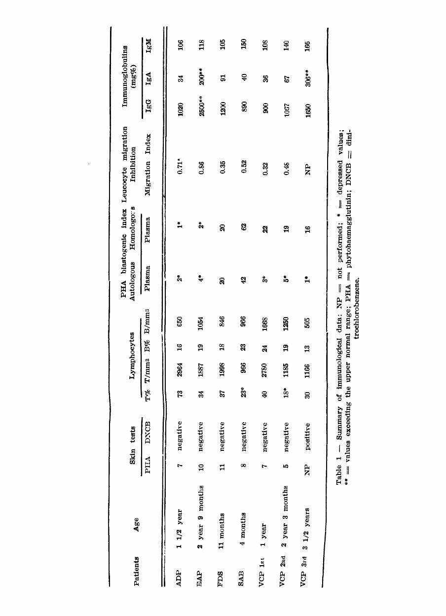
Results of immunological study are shown in table 1.
DISCUSSION
The first case described (V.C.P.) had his immunological deficiency diagnosed prior to the onset of the flexion spasms.
All the children had probably pre-existing cerebral lesions; three had bacterial meningitis, one was born with the umbilical cord around his neck and one had microcephalia of unknown cause.
EEG examinations showed that three children are changing from West to Lennox-Gastaut syndrome.
All children had had frequent episodes of infectious diseases particularly serious in two of them (VCP and ΕΑΡ). The child in better conditions had one bronchopneumonia.
Great incidence of infections in patients with West syndrome has been reported. Beaussart (1960) related recorrent infections interfering with ACTH therapy. Lison (1968) comments that infections of so great frequency can not be atributed only to the physical conditions of these children and considered therapy with benzodiazepines as related with respiratory infectious episodes. Millichap and Ortiz (1966), Hagberg (1967) and Volzke et al (1967) also have refered the frequency of infections in these children. Friedman and Pampiglione (1971) in a report about the clinical outcome of children with hypsarrhythmia,

found a high early mortality (1:4) and refered that most of them succumbed to bronchopneumonia or other infections. Recently, Riikonen (1978) reported that 8 of the 11 children who had infantile spasms and cytomegalovirus infection, had severe recurrent respiratory infections.
The infectious diseases may be related to defects in cell mediated immunity, since all our children examined showed alterations in immune response. All showed a negative response to DNCB sensitization, that is a method used to investigate cell-mediated immunity "in vivo", since 95% of normal individuals give a positive response. (Convit et al, 1971; Mendes and Raphael, 1971; Lawlor et al, 1973). This may be interpreted as a inability of these children in being sensitized with a new antigen. Our children had a positive response to intracutaneous PHA, showing that the mechanisms involved in this response were preserved. Since PHA stimulates lymphocytes nonspecifically, prior exposure is not required for elicit a cell-mediated immune response "in vivo" (Burgio et al, 1971; Bonforte et al, 1972).
There was variability in the responses of lymphocyte blastic stimulation with PHA. Two children (F.D.S. and S.A.B.) had normal responses and three showed depressed values (A.D.P., E.A.P. and V.C.P.). The responses of A.D.P. and E.A.P. suggest a functional deficiency of thymus dependent lymphocytes in undergoing blastic transformation induced by this mytogen. The three evaluations performed in V.C.P. have shown that cell-mediated immunity depression is due to the presence of an autologous plasmatic blocking factor of blastic transformation.
One child (A.D.P.) showed deficiency in leucocyte migration inhibition test with PHA. There was no correlation in response of PHA lymphocyte blastic stimulation with the leucocyte migration inhibition in the presence of PHA. Both tests appraise Τ lymphocyte functional activity "in vitro", but disagreement of responses has been refered. Curtis and Hersh (1973) in normal persons immunized with haemocyanin, and Valdimarsson et al (1973) in patients with disseminated mucocutaneous candidiasis found similar results.
There was also disagreement in PHA reactivity "in vivo" and "in vitro". This finding was described before, by Airo et al (1967) in patients with chronic lymphocytic leukemia and by Bonforte et al (1972) in patients with sarcoidosis. They found strong positive responses to intracutaneous PHA and depressed responses to lymphocyte blastic stimulation "in vitro".
Serum levels of IgA, IgG, IgM were within normal values. The high values in one children may be atributed to infectious diseases.
Peripheral Τ and Β blood lymphocytes were within normal values, except for the low Τ lymphocyte percentual observed in S.A.B. and V.C.P. (2 n d evaluation). This latter result was obtained during ACTH therapy, that seems to affect Τ cell sub-population production (Claman & Moorhead, 1972). The interpretation of S.A.B. Τ lymphocyte percentual value is difficult since the values for normal young children are unknown. Diaz-JouAnen et al (1975), and Fleischer et al (1979) have found that newborn infants had lower Τ cell percentual values in relation to adult normal values.
The immunological evaluations of three children (S.A.B., F.D.S. and V.C.P. 1 s t ) were performed without any medication; in two (A.D.P. and E.A.P.) and in the 3 r d of V.C.P., evaluations were made during therapy with phenobarbital and benzodiazepines.
Changes of immunological behaviour induced by phenobarbital do not seem to occur (Crob and Herold, 1972; Fontana et al, 1976). Benzodiazepine effects on immunological function have not been studied.
One of these children (E.A.P.) presented a 3 r d degree protein calorie undernutrition when submitted to the immunological tests. The abnormalities in the immune response detected by a negative sensitization with DNCB, impaired lymphocyte blastic transformation, Τ cell values slightly depressed and high levels of serum IgG, may be associated to his nutritional deficit. Other authors have found depressed cell-mediated immunity together with a raising in the Ig levels in patients with malnutrition (Faulk et al., 1976; Mullen et al., 1978, Chandra, 1979).
The demonstration of a precipitating antibody to extract of brain tissues in four patients with hypsarrhythmia, reported by Reinskov (1963) supports the hypothesis of a autoimmune mechanism in West syndrome. Our study was not planned to investigate the relationship between autoimmunity and West Syndrome, however the depressed cell-mediated immunity observed in our children could be a predisposing factor to the development of an autoimmune disorder. Some authors have pointed out the association between immuno-deciciency state and autoimmunity. Allisson et al (1971), Stiller et al (1975) have suggested that failure in the thymus dependent system may be an important factor in the releasing of autoantibody production.
In this preliminary study, we have found immunological abnormalities associated with secondary West syndrome. Further investigations are needed in order to clarify the extent of this association and to investigate a possible role of an autoimmune process in this neurological disorder.
SUMMARY
The immunological status of five children with West syndrome consequent to previous cerebral lesions was investigated. Three children had West syndrome and two were in transition from West to Lennox-Gastaut syndrome. All of them showed cellular immunological deficiencies in the following tests: sensitization to DNCB, intracutaneous reaction to PHA, inhibition of leucocyte migration, blastic transformation of lymphocytes, Τ and Β lymphocytes in peripheric blood and levels of serum immunoglobulins. These immunological deficiencies, of different degrees of severity, were associated to frequent infections in these children. A possible association between the immunological deficiencies and autoimmunity is discussed.
RESUMO
Imunidade celular e humoral na sindrome de West
Realizamos avaliações de imunidade celular e humoral em 5 crianças com
sindrome de West secundária a lesões cerebrais. Todas apresentaram deficiência
de imunidade celular, detectada pelas seguintes provas: sensibilização ao DNCB,
reação intracutânea à PHA, inibição da migração de leucócitos, transformação
blástica de linfócitos, determinação de linfócitos Τ e Β no sangue periférico e
dosagem de imunoglobulinas séricas. Estes exames foram realizados durante a
síndrome de West em três crianças e durante evolução de síndrome de West
para síndrome de Lennox-Gastaut em duas. Estas alterações imunitárias, de
graus variáveis, associam-se a infecções frequentes nessas crianças. Discute-se
uma possível associação desses defeitos imunitários com auto-imunidade.
Imunidade celular e humoral na sindrome de West Realizamos avaliações de imunidade celular e humoral em 5 crianças com
sindrome de West secundária a lesões cerebrais. Todas apresentaram deficiência
de imunidade celular, detectada pelas seguintes provas: sensibilização ao DNCB,
reação intracutânea à PHA, inibição da migração de leucócitos, transformação
blãstica de linfócitos, determinação de linfócitos Τ e Β no sangue periférico e
dosagem de imunoglobulinas séricas. Estes exames foram realizados durante a
sindrome de West em três crianças e durante evolução de sindrome de West
para sindrome de Lennox-Gastaut em duas. Estas alterações imunitárias, de
graus variáveis, associam-se a infecções freqüentes nessas crianças. Discute-se
uma possível associação desses defeitos imunitários com auto-imunidade. REFERENCES
1. AIRÔ, R.; MIHAILESCU, E.; ASTALDI, G. & MEARDI, G. — Skin reactions to phytohemagglutinin. Lancet 1:899, 1967.
2. ALLISON, Α. Α.; DENMAN, Α. Μ. & BARNES, R. D. — Cooperating and controllling functions of thymus: derived lymphocytes in relation to autoimmunity. Lancet 2:135, 1971.
3. BEAUSSART, M. — Encephalopathie myocloniqt.e du nourrison avec hypsarrhythmia: étude EEG. avant et après traitement par ACTH Rev. Neurol. (Paris) 103:243, 1960.
4. BONFORTE, R. J.; TOPILSKY, M.; SILTZBACH, L. E. & GLADE, P. R. Phytohemagglutinin skin test: a possible in vivo measure of cell-mediated immunity J. Pediatrics. 81:775, 1972.
5. BURGIO, G. R.; CURTONI, E.; GENOVA, R. & MAGRINI, V. — Skin reactivity in childhood: phytohaemagglutinin (PHA) skin test and streptokinase (STK) skin test. Pediatric Research 5:88. 1971.
6. CLAMAN, Η. N. & MOORHEAD, I. W. — Heterogeneity of thymus-dependent lymphoid cell functions in the mouse. In Proc. 3 rd. Lepetit Colloquium, North Holland Publ. Co., Amsterdam, pp. 133, 1972.
7. CONVIT, I.; PINARDI, Μ. E. & ROSAS ) P. A. — Some considerations regarding the immunology of leprosy. Internat J. Leprosy 39:566, 1971.
8. CURTIS, J. E. & HERSH, E. — Cellular immunity in man: correlation of leukocyte migration inhibition factor formation and delayed hypersensitivity. Cellular Immunology, 8:55. 1973.
9. DIAZ-JOU ANEN, E.; STRICKLAND, R. G. & WILLIAMS, R. C. — Studies on human lymphocytes in the newborn and the aged. Amer. J. Medicine 58:620, 1975.
10. FLEISHER, J.; LUCKASEN, JR.; SABAD, Α.; GERTZ, R. C. & KERSEY, J. H. — Τ and Β lymphocyte subpopulation in children. Pediatrics 55:162, 1975.
11. FONTANA, Α.; GROB. P. J.; SAUTER, R. & JOLLER, H. — IgA deficiency epilepsy and hydantoin medication. Lancet 2:228, 1979.
12. FRIEDMAN, E. & PAMPIGLIONE, E.; - Prognostic implications of EEG. findings of hypsarrhytmia in first year of life. British Med. J. 4;323, 1971.
13. GASTAUT, H. J. L.; GASTAUT, H.; BERNARD, H. R. R.; PINSARD, N.; SAINT-JEAN, M.; ROGER, J. & DRAVET, C. — Computerized Tomography in the study of West's syndrome. Develop. Med. Child Neurol 20:21, 1978.
14. GROB, P. J. & HEROLD, G. E. — Immunological abnormalities and hydantoins. Bristish Me J. J. 2:561, 1972.
15. HAGBERG, B. — The libriun analogue Mogadon in the treatment of epilepsy children. In Proceedings of the eightenth Congress of Scandinavian Neurologists. Helsinki. Acta Neurol. Scandnavica supl. 31:167, 1967.
16. IRULEGUI, I.; FERRI, R. G.; BARBIERI, D.; QUARENTEI, G.; GONZALES, C. Η.; PENNA, H. A. O.; CORRADINI, Η. B.; PENNAPHIM, D. & LAGÔA, N. — Immunoglobulins in normal children by Immunoelectrophoresis and quantitative radial immunodiffusion: values from birth to three years. Rev. do Hospital das Clinicas da Faculdade de Medicina de São Paulo 30:198, 1976.
17. LAWLOR, G. J. JR.; STIEHM, E. R.; KAPLAN, Μ. S.; SENGAR, D. P. S. & ΤERASARI, P. I. — Phytohemagglutinin (PHA) skin test in the diagnosis os cellular immunodeficiency. J. of Allergy and Clinical Immunol. 52:31, 1973.
18. LISON, M. P. — Contribuição ao estudo da ação de derivados benzoadiazepínicos na epilepsia. Tese j Faculdade de Medicina de Ribeirão Preto, USP, 1968.
19. LOW, Ν. L. — Infantile spasms with mental retardation: treatment with cortisone and adrenocorticotropin. Pediatrics 22:1165, 1958.
20. MANCINI, G.; CARBONARA, A. O. & HEREMANS, J. F. — Immunochemical quantitation of antigens by single radial immunodiffusion. Immunochemistry 2: 235, 1965.
21. MENDES, Ε. & RAPHAEL, A. — Impaired delayed hypersensitivity in patients with South American blastomycosis. J. of Allergy and Clinical Immunol. 47:17, 1971.
22. MENDES, Ε.; RAPHAEL, Α.; MOTA, Ν. G. S. & MENDES, Ν. F. — Cell-mediated immunity in leprosy and transfer of delayed hypersensitivity reactions. J. of Allergy and Clinical Immunol. 53:233, 1974 a.
23. MENDES, Ν. F.; MIKI, S. S. & PEIXINHO, Ζ. F. — Combined detection of human Τ and Β lymphocyte by rosette formation with sheep erythrocytes and zymosan-C3 complexes. J. of Immunol. 113:531 1974 b.
24. MILLICHAP, J. G. & BICKFORD, R. G. — Infantile spasms, hypsarrhythmia and mental retardation. J. of American Medical Association. 182:523, 1962.
25. MILLICHAP, J. G. & ORTIZ, W. R. — Nitrazepan in mioclonic epilepsies. Amer. J. Dis. Children 112:242, 1966.
26. MORISON, W. L. — Effect of phytohaemagglutinin in the leucocyte migration inhibition test as a measure of cell-mediated immunity. J. of Clinical Pathol. 27:113, 1974.
27. MUSSATTI, C. C ; REZKALLAH, Μ. T.; MENDES, Ε. & MENDES, Ν. P. — In vivo and in vitro evaluation of cell-mediated immunity in patients with paracoccidioidomycosis. Cellular Immunology 24:365, 1976.
28. NASPITZ, C. K. & RITCHER, M. — The action of phytohemagglutinin in vivo and in vitro: a review. Progress in Allergy 12:1, 1968.
29. REINSKOV, T. — Demonstration of precipitating antibody to extract of brain tissue in patients with hypsarrhythmia. Acta Paediatrica, suppl. 140:73, 1963.
30. RIIKONEN, R. — Cytomegalovirus infection and infantile spasms. Develop. Med. Child Neurol. 20:570, 1978.
31. STILLER, C. R.; RUSSEL, A. S. & DOSSETOR, J. B. — Auto immunity: present concepts. Ann. Internal Medicine 82:405, 1975.
32. VALDIMARSSON, H.; HIGGS, I. M.; WILLS, R. S.; YAMAMURA, M.; HOBBS, J. R. & HOLT, P. J. L. — Immune abnormalities associated with chronic mucocutaneous candidiasis. Cellular Immunology 6:248, 1973.
33. VOLZKE, E.; DOOSE, H.; STEPHAN, E. — The treatment of infantile spasms and by hypsarrhythmia with Mogadon. Epilepsia 8:64, 1967.
Faculdade de Medicina de Botucatu — Departamento de Neurologia e Psiquitria — 18600 Rubião Junior — Botucatu, SP — Brasil.


![Clinical Characteristics to Differentiate · Asthma-COPD overlap syndrome (ACOS) [a description] Asthma-COPD overlap syndrome (ACOS) is characterized by persistent airflow limitation](https://static.fdocument.org/doc/165x107/5f0914d17e708231d4252460/clinical-characteristics-to-differentiate-asthma-copd-overlap-syndrome-acos-a.jpg)