Cel9D, an Atypical 1,4--D-Glucan Glucohydrolase from Fibrobacter
9
JOURNAL OF BACTERIOLOGY, Mar. 2008, p. 1976–1984 Vol. 190, No. 6 0021-9193/08/$08.000 doi:10.1128/JB.01667-07 Copyright © 2008, American Society for Microbiology. All Rights Reserved. Cel9D, an Atypical 1,4- - D-Glucan Glucohydrolase from Fibrobacter succinogenes: Characteristics, Catalytic Residues, and Synergistic Interactions with Other Cellulases † Meng Qi, Hyun-Sik Jun,‡ and Cecil W. Forsberg* Department of Molecular and Cellular Biology, University of Guelph, Guelph, Ontario, Canada Received 15 October 2007/Accepted 3 January 2008 The increasing demands of renewable energy have led to the critical emphasis on novel enzymes to enhance cellulose biodegradation for biomass conversion. To identify new cellulases in the ruminal bacterium Fibrobacter succinogenes, a cell extract of cellulose-grown cells was separated by ion-exchange chromatography and cellulases were located by zymogram analysis and identified by peptide mass fingerprinting. An atypical family 9 glycoside hydrolase (GH9), Cel9D, with less than 20% identity to typical GH9 cellulases, was identified. Purified recombinant Cel9D enhanced the production of reducing sugar from acid swollen cellulose (ASC) and Avicel by 1.5- to 4-fold when mixed separately with each of four other glucanases, although it had low activity on these substrates. Cel9D degraded ASC and cellodextrins with a degree of polymerization higher than 2 to glucose with no apparent endoglucanase activity, and its activity was restricted to -134-linked glucose residues. It catalyzed the hydrolysis of cellulose by an inverting mode of reaction, releasing glucose from the nonreducing end. Unlike many GH9 cellulases, calcium ions were not required for its function. Cel9D had increased k cat /K m values for cello-oligosaccharides with higher degrees of polymerization. The k cat /K m value for cellohexaose was 2,300 times higher than that on cellobiose. This result indicates that Cel9D is a 1,4-- D-glucan glucohydrolase (EC 3.2.1.74) in the GH9 family. Site-directed mutagenesis of Cel9D identified Asp166 and Glu612 as the candidate catalytic residues, while Ser168, which is not present in typical GH9 cellulases, has a crucial structural role. This enzyme has an important role in crystalline cellulose digestion by releasing glucose from accessible cello-oligosaccharides. Plant material is now viewed as the bioenergy feedstock of the future. Cellulose, which accounts for the bulk of plant materials, is recalcitrant, which necessitates a combination of the classes of cellulases for biodegradation. These include en- doglucanases (EC 3.2.1.4) that cut randomly at internal amor- phous sites in the cellulose chain; exoglucanases (EC 3.2.1.91 and EC 3.2.1.74) that act processively on the reducing or non- reducing ends of cellulose chains, releasing either cellobiose or glucose as major products; and -glucosidases (EC 3.2.1.21) that hydrolyze soluble cellodextrins and cellobiose to glucose (18). Fibrobacter succinogenes is a highly cellulolytic bacterium that is closely related to the Cytophaga-Flavobacterium-Bacte- roides group (8). It is commonly found in the rumen of rumi- nant animals and appears to be one of the most active rumen cellulose degraders (4). Recently, the genome was sequenced (21) and a number of cellulases (24) and cellulose binding proteins (12) were identified. We reported synergistic interac- tions among five cellulases of this organism (24). Although these enzymes had degrees of synergism (DoS) of up to 3.7 and the combination of Cel9B, Ce51A, and Cel8B gave the highest activity, the extent of hydrolysis of cellulose was low. In addi- tion, Cel10A, known as the exo-acting, Cl-stimulated cellobio- sidase, had limited synergistic interaction with other enzymes. In the following study, a novel cellulase gene, cel9D, was cloned and the protein encoded was purified and character- ized. An investigation of the synergistic effect of Cel9D with four other cellulases, Cel9B, Cel51A, Cel8B, and Cel45C, from F. succinogenes revealed that Cel9D acted synergistically with these glucanases. Our data also document the fact that this novel enzyme is the first glucan 1,4--D-glucohydrolase in gly- coside hydrolase family 9 (GH9) with enhanced activity on long-chain cello-oligosaccharides. MATERIALS AND METHODS Bacterial strains and plasmid. Fibrobacter succinogenes S85 was used as the source of genomic DNA. It was maintained and cultured in chemically defined medium with either glucose or microcrystalline cellulose (Avicel) PH105 (FMC BioPolymer) as the carbon source as described previously (30). The Escherichia coli strain DH10B (Invitrogen) was used as the host for genetic manipulation. E. coli BL21(DE3) was the host for protein production. Cultures were grown at 37°C or 18°C in Luria-Bertani medium or Luria-Bertani medium supplied with 1 M sorbitol and 2.5 mM betaine glycine for enhanced solubility of the recombi- nant proteins (29). When required, media were supplemented with kanamycin at 34 g/ml. Solid media contained 1.6% (wt/vol) agar. Identification of Cel9D. F. succinogenes S85 was grown in 300 ml chemically defined medium with 0.3% (wt/vol) Avicel cellulose PH105 as the sole carbon source at 37°C for 24 h with reciprocating shaking at 150 rpm. After the removal * Corresponding author. Mailing address: Department of Molecular and Cellular Biology, University of Guelph, Guelph, Ontario N1G 2W1, Canada. Phone: (519) 824-4120, ext. 53433. Fax: (519) 837-1802. E-mail: [email protected]. † Supplemental material for this article may be found at http://jb .asm.org/. ‡ Present address: Section on Cellular Differentiation, NICHD, Na- tional Institute of Health, Bldg. 10, Room 10N325, 9000 Rockville Pike, Bethesda, MD 20892. Published ahead of print on 18 January 2008. 1976 Downloaded from https://journals.asm.org/journal/jb on 02 February 2022 by 14.34.130.23.
Transcript of Cel9D, an Atypical 1,4--D-Glucan Glucohydrolase from Fibrobacter
JOURNAL OF BACTERIOLOGY, Mar. 2008, p. 1976–1984 Vol. 190, No. 60021-9193/08/$08.00�0 doi:10.1128/JB.01667-07Copyright © 2008, American Society for Microbiology. All Rights Reserved.
Cel9D, an Atypical 1,4-�-D-Glucan Glucohydrolase fromFibrobacter succinogenes: Characteristics, Catalytic
Residues, and Synergistic Interactions withOther Cellulases�†
Meng Qi, Hyun-Sik Jun,‡ and Cecil W. Forsberg*Department of Molecular and Cellular Biology, University of Guelph, Guelph, Ontario, Canada
Received 15 October 2007/Accepted 3 January 2008
The increasing demands of renewable energy have led to the critical emphasis on novel enzymes to enhancecellulose biodegradation for biomass conversion. To identify new cellulases in the ruminal bacteriumFibrobacter succinogenes, a cell extract of cellulose-grown cells was separated by ion-exchange chromatographyand cellulases were located by zymogram analysis and identified by peptide mass fingerprinting. An atypicalfamily 9 glycoside hydrolase (GH9), Cel9D, with less than 20% identity to typical GH9 cellulases, wasidentified. Purified recombinant Cel9D enhanced the production of reducing sugar from acid swollen cellulose(ASC) and Avicel by 1.5- to 4-fold when mixed separately with each of four other glucanases, although it hadlow activity on these substrates. Cel9D degraded ASC and cellodextrins with a degree of polymerization higherthan 2 to glucose with no apparent endoglucanase activity, and its activity was restricted to �-134-linkedglucose residues. It catalyzed the hydrolysis of cellulose by an inverting mode of reaction, releasing glucosefrom the nonreducing end. Unlike many GH9 cellulases, calcium ions were not required for its function. Cel9Dhad increased kcat/Km values for cello-oligosaccharides with higher degrees of polymerization. The kcat/Km valuefor cellohexaose was 2,300 times higher than that on cellobiose. This result indicates that Cel9D is a 1,4-�-D-glucan glucohydrolase (EC 3.2.1.74) in the GH9 family. Site-directed mutagenesis of Cel9D identified Asp166and Glu612 as the candidate catalytic residues, while Ser168, which is not present in typical GH9 cellulases,has a crucial structural role. This enzyme has an important role in crystalline cellulose digestion by releasingglucose from accessible cello-oligosaccharides.
Plant material is now viewed as the bioenergy feedstock ofthe future. Cellulose, which accounts for the bulk of plantmaterials, is recalcitrant, which necessitates a combination ofthe classes of cellulases for biodegradation. These include en-doglucanases (EC 3.2.1.4) that cut randomly at internal amor-phous sites in the cellulose chain; exoglucanases (EC 3.2.1.91and EC 3.2.1.74) that act processively on the reducing or non-reducing ends of cellulose chains, releasing either cellobiose orglucose as major products; and �-glucosidases (EC 3.2.1.21)that hydrolyze soluble cellodextrins and cellobiose to glucose(18).
Fibrobacter succinogenes is a highly cellulolytic bacteriumthat is closely related to the Cytophaga-Flavobacterium-Bacte-roides group (8). It is commonly found in the rumen of rumi-nant animals and appears to be one of the most active rumencellulose degraders (4). Recently, the genome was sequenced(21) and a number of cellulases (24) and cellulose bindingproteins (12) were identified. We reported synergistic interac-
tions among five cellulases of this organism (24). Althoughthese enzymes had degrees of synergism (DoS) of up to 3.7 andthe combination of Cel9B, Ce51A, and Cel8B gave the highestactivity, the extent of hydrolysis of cellulose was low. In addi-tion, Cel10A, known as the exo-acting, Cl-stimulated cellobio-sidase, had limited synergistic interaction with other enzymes.
In the following study, a novel cellulase gene, cel9D, wascloned and the protein encoded was purified and character-ized. An investigation of the synergistic effect of Cel9D withfour other cellulases, Cel9B, Cel51A, Cel8B, and Cel45C, fromF. succinogenes revealed that Cel9D acted synergistically withthese glucanases. Our data also document the fact that thisnovel enzyme is the first glucan 1,4-�-D-glucohydrolase in gly-coside hydrolase family 9 (GH9) with enhanced activity onlong-chain cello-oligosaccharides.
MATERIALS AND METHODS
Bacterial strains and plasmid. Fibrobacter succinogenes S85 was used as thesource of genomic DNA. It was maintained and cultured in chemically definedmedium with either glucose or microcrystalline cellulose (Avicel) PH105 (FMCBioPolymer) as the carbon source as described previously (30). The Escherichiacoli strain DH10B (Invitrogen) was used as the host for genetic manipulation. E.coli BL21(DE3) was the host for protein production. Cultures were grown at37°C or 18°C in Luria-Bertani medium or Luria-Bertani medium supplied with 1M sorbitol and 2.5 mM betaine glycine for enhanced solubility of the recombi-nant proteins (29). When required, media were supplemented with kanamycin at34 �g/ml. Solid media contained 1.6% (wt/vol) agar.
Identification of Cel9D. F. succinogenes S85 was grown in 300 ml chemicallydefined medium with 0.3% (wt/vol) Avicel cellulose PH105 as the sole carbonsource at 37°C for 24 h with reciprocating shaking at 150 rpm. After the removal
* Corresponding author. Mailing address: Department of Molecularand Cellular Biology, University of Guelph, Guelph, Ontario N1G2W1, Canada. Phone: (519) 824-4120, ext. 53433. Fax: (519) 837-1802.E-mail: [email protected].
† Supplemental material for this article may be found at http://jb.asm.org/.
‡ Present address: Section on Cellular Differentiation, NICHD, Na-tional Institute of Health, Bldg. 10, Room 10N325, 9000 RockvillePike, Bethesda, MD 20892.
� Published ahead of print on 18 January 2008.
1976
Dow
nloa
ded
from
http
s://j
ourn
als.
asm
.org
/jour
nal/j
b on
02
Febr
uary
202
2 by
14.
34.1
30.2
3.
of residual cellulose by centrifugation at 300 rpm for 10 min, cells were harvestedby centrifugation at 5,000 � g and then resuspended in 10 ml of 30 timesconcentrated culture fluid prepared as described before (20). The cells were thenbroken by three passes through a French pressure cell at 1,200 lb/in2. The celllysate was centrifuged at 12,000 � g for 30 min. The clarified supernatantcontaining 25 mg of protein was separated by a 2.5 � 10-cm column of DEAE-Sepharose CL-6B as described previously (20), except that imidazole buffer (20mM) was used instead of potassium phosphate buffer. Approximately 40% of theactivity eluted in the application buffer before NaCl gradient was applied, andthese fractions were kept for further study. Four hundred microliters of eachfraction eluted by the NaCl gradient buffer was mixed with 100 �l 2.5% AvicelPH105 suspension and incubated at 37°C for 20 h. Reducing sugar produced wasdetermined by the p-hydroxybenzoic acid hydrazide methods (16).
Fractions eluting in the buffer gradient exhibiting Avicelase activities wereloaded to a 7.5% (wt/vol) polyacrylamide gel without sodium dodecyl sulfate andsubjected to electrophoresis at 100 V for 2 h. The gel was then laid on a platecontaining 20 ml 0.1% (wt/vol) carboxymethyl cellulose (CMC) solidified with0.7% agarose and incubated at 37°C for 4 h. The gel was then removed andstained with colloidal Coomassie blue, and the agar layer on the plate was stainedby 0.1% Congo red for 30 min and destained by 1 M NaCl solution. The bandson the gel that corresponded to clear zones on the CMC plate were excised andsubjected to matrix-assisted laser desorption ionization–time of flight mass spec-trometry analysis, and Cel9D was identified by matching the mass spectra ofpeptides to the F. succinogenes protein database as described previously (14).
Cloning, expression, and purification of Cel9D and its mutants. GenomicDNA was prepared from F. succinogenes S85 by using the cetyltrimethylammo-nium bromide method as described before (1). The intact cel9D gene was am-plified from the genomic DNA by using Cel9D_1 and Cel9D_2C primers (Table1) and Pwo polymerase (Roche). The restriction endonuclease sites NdeI andXhoI in the 5� primer and 3� primer, respectively, allowed for cloning of the geneinto the expression vector pET30a (Novagen). The coding sequence of Cel9Dwas fused in frame with a downstream sequence of the vector encoding six-histidine residues. The mutants of Cel9D, D166A, E612A, S168A, and �S168,were generated by the PCR overlap extension method (27) using the primerslisted (Table 1) to replace amino acid residues as indicated and to remove theSer168. The wild-type and mutant genes were sequenced to ensure the correctnucleotide changes and that there were no PCR-induced errors. Cel9D and itsmutants were produced in E. coli BL21(DE3) and purified by immobilized metalaffinity chromatography. Wild-type Cel9D was further purified by molecularsieve chromatography as described previously (14). Cel8B, Cel9B, and Cel51Awere produced and purified as described previously (24).
Enzyme assays. Glycoside hydrolase activity was assayed with various sub-strates as described previously (24). The standard reaction period for Cel9D was16 h for Avicel PH105 and 2 h for all other substrates in 0.05 M sodiumphosphate buffer, pH 6.5. The reducing sugar produced was measured. Thesubstrate used was either Avicel PH105 (0.5%, wt/vol) or phosphoric acid swol-len cellulose (ASC; 0.25%, wt/vol). The DoS was calculated as follows: (observedactivity of a mixture of enzymes)/(additive activities of the individual enzymesmaking up the mixture).
To determine the kinetics parameters for hydrolysis of cello-oligosaccharides,
0.1 �g purified Cel9D enzyme was mixed with different concentrations of cello-oligosaccharide substrates in 0.05 mM phosphate buffer, pH 6.5. Samples weretaken at given intervals and appropriately diluted in 0.15 M sodium hydroxide tostop the reaction and hydrolysis products were analyzed by high-performanceliquid chromatography (HPLC).
Analysis of degradation products. Cello-oligosaccharides and their hydrolysisproducts as well as products from synthetic substrates p-nitrophenol (pNP)glucopyranoside (pNPG), pNP �-D-cellobioside (pNPC), and ASC were ana-lyzed by high-performance anion-exchange chromatography coupled with a Wa-ters model 464-pulsed amperimetric detector system with a base-resistant refer-ence electrode as described before (14).
1H NMR spectroscopy. 1H nuclear magnetic resonance (NMR) spectra weregenerated on a Bruker Avance-400 NMR spectrometer in 5-mm-diameter sam-ple tubes. Samples (0.6 ml) were analyzed at 27°C in 50 mM potassium phos-phate buffer, pH 6.5, containing 20 mM pNPC, or 30 mM cellotetraose as thesubstrate. Chemicals and substrates were dissolved in D2O. Cel9D enzyme wastransferred to buffer containing D2O by ultrafiltration by using a PM-10 (Ami-con) membrane. Spectra were collected in 32-k data points using a spectral widthof 5 KHz and a relaxation delay of 2 s. The spectra were referenced against anexternal standard of trimethylsilyl-2,2,3,3-tetradeuteropropionic acid (sodiumsalt) at 27°C. One hundred micrograms of purified Cel9D was added to eachreaction mixture.
Nucleotide sequence accession numbers. The nucleotide sequences of thegene cel9D from F. succinogenes S85 have been deposited in the GenBankdatabase under accession number EU352748.
RESULTS
Identification and sequence analysis of a novel family 9enzyme from the genome of F. succinogenes. When the av-icelases present in the whole-cell extract from cellulose-grownF. succinogenes were separated on DEAE-Sepharose, a sharpsingle peak of activity was eluted in the buffer gradient at 0.15to 0.25 M NaCl, followed by several minor peaks at 0.35 M and0.45 M NaCl (see Fig. S1 in the supplemental material). Thefractions (numbers 15, 16, and 17) corresponding to the majorpeak were subjected to native polyacrylamide gel electrophore-sis (PAGE) and zymogram analysis (see Fig. S2 in the supple-mental material). The three fractions yielded similar profiles,with activity zones at about 62, 75, and 80 kDa, respectively.Proteins in the corresponding bands of fraction 16 on thenative PAGE were extracted and analyzed by peptide massfingerprinting. No proteins involved in cellulose degradationwere identified from the two smaller bands, but in the 80-kDaband, a cellulase with an amino acid sequence corresponding
TABLE 1. Oligonucleotide primers used in this study
Primer Sequencea Locationb Purpose
Cel9D_1 5�-GTCCTCCATATGCGTATCTATAAGCTTTC-3� 1–20 Cloning with NdeICel9D_2C 5�-ATATGC CTCGAGCTTCACAACAAACTTCTT-3� 2143–2160 Cloning with XhoI
D166A_1 5�-CGGTTGGTATGCTGCGAGCGGCG-3� 487–509 MutagenesisD166A_2C 5�-CGCCGCTCGCAGCATACCAACCG-3� 487–509 (reverse) Mutagenesis
S168A_1 5�-GGTTGGTATGATGCGGCCGGCGATGTCAGTAAG-3� 487–519 MutagenesisS168A_2C 5�-CTTACTGACATCGCCGGCCGCATCATACCAACC-3� 487–519 (reverse) Mutagenesis
�S168_1 5�-GGTTGGTATGATGCG�GGCGATGTCAGTAAG-3� 487–519 Mutagenesis�S168_2C 5�-CTTACTGACATCGCC�CGCATCATACCAACC-3� 487–519 (reverse) Mutagenesis
E612A_1 5�-GCGCTGGGATGCACAGTGGCTGC-3� 1824–1846 MutagenesisE612A_2C 5�-GCAGCCACTGTGCATCCCAGCGC-3� 1824–1846 (reverse) Mutagenesis
a Underlining indicates restriction sites for cloning. Bold and tilde denote the substituted nucleotides for amino acid replacement and deletion, respectively.b Values shown are nucleotide positions.
VOL. 190, 2008 Cel9D IN FIBROBACTER SUCCINOGENES 1977
Dow
nloa
ded
from
http
s://j
ourn
als.
asm
.org
/jour
nal/j
b on
02
Febr
uary
202
2 by
14.
34.1
30.2
3.
to FSU2558, known as cel9D, was identified in the genome ofF. succinogenes S85. Matrix-assisted laser desorption ioniza-tion–time of flight mass spectrometry analysis of tryptic pep-tides of the 80-kDa bands accounted for 22% of the Cel9Dprotein and the peptide sequences were dispersed throughoutthe protein (see Fig. S3 in the supplemental material).
The gene cel9D encoded a protein that had an N-terminalsignal peptide and a putative cleavage site located betweenAla23 and Glu24. The mature protein contained 697 aminoacid residues and had a theoretical mass of 77.0 kDa and atheoretical isoelectric point of 5.27. Following the signal pep-tide, there was a 100-amino-acid region that had low similarityto an immunoglobulin-like module, followed by a putative fam-ily 9 catalytic domain and a 69-amino-acid basic C-terminaldomain with a theoretical pI of 10.5. The protein sequence hadthe highest sequence similarities to several putative endoglu-canases or hypothetical proteins mainly from the genera ofVibrio, Photobacterium, and Cytophaga, but had low similaritiesto nearly all endo- or exoglucanases (“classical GH9s”). Phy-logenetic analysis showed that the group of GHs in whichCel9D resides is a unique branch of the GH9 family (see Fig.S4 in the supplemental material). Multiple sequence alignment(Fig. 1) showed that the putative catalytic residues of Cel9D(D166 and D170) were separated by three other amino acids,while in all other characterized cellulases, they were separatedby two. The extra residue (S168) seems to be conserved in theCel9D group (data not shown). E612 was identified as a puta-tive catalytic base; however, the surrounding amino acid resi-dues, which are conserved in classical GH9s, were not alignedin the Cel9D group.
Production and purification of recombinant Cel9D. cel9Dgene was cloned, and protein Cel9D was produced in E. coliBL21(DE3). Most of the recombinant Cel9D enzyme was as-sociated with the membrane fraction perhaps in inclusion bod-ies. However, a small amount present in the nonsedimentablefraction was purified by immobilized metal affinity chromatog-raphy, followed by molecular sieve chromatography. About 1mg of pure protein was obtained from 1 liter of culture. Therecombinant protein had a mass of 76 kDa, as determined bysodium dodecyl sulfate-PAGE (see Fig. S5 in the supplementalmaterial), which is in good agreement with the theoretical massof 77 kDa based on the primary sequence.
Characterization of Cel9D. As with most other GHs identi-fied in F. succinogenes S85, Cel9D had an optimum pH of 6.5and retained 80% activity between pH 6.0 and 8.0. The tem-perature for maximum activity was 37°C. Detailed substratespecificity assays (Table 2) on various types of polysaccharidesshowed that Cel9D had activity on all polymers with a �-1,4linkage, but not on laminarin, which has a �-1,3 linkage. Bymeasuring the release of pNP, no activity was detected onpNP-�-l-arabinofuranoside, pNP-�-D-glucoside, pNP-�-D-cel-lobioside, pNP-�-D-fucoside, pNP-�-D-galactoside, pNP-N-acetyl-�-D-glucosaminide, pNP-�-D-glucoside, pNP-�-D-glucu-ronoside, pNP-�-D-lactoside, or pNP-�-D-maltoside. Cel9Dhad 27% lower activity on medium-viscosity (average Mw,�250,000) CMC compared to the activity on low-viscosityCMC (average Mw, �90,000) (Table 2). This property ofCel9D was quite different from that of the Cel9B enzyme,which had high activity on both medium- and low-viscosityCMC (24). The hydrolytic activities of Cel9D on Avicel cellu-
lose, ASC, and CMC were tested and compared to those ofCel8B, Cel9B, and Cel51A from F. succinogenes (Table 3). Ithad less activity on CMC than on other enzymes.
Although Cel9D did not release pNP from any of the aryl-glycosides tested, it did efficiently cleave pNPC to pNPG andglucose, as indicated by HPLC analysis (see Fig. S6A in thesupplemental material). This result documented the fact thatCel9D acts on the nonreducing end. Cel9D did not degradesophorose (�1-2), laminaribiose (�1-3), or gentiobiose (�1-6),as assayed by HPLC analysis.
Hydrolytic activities on either CMC or cellopentaose of pu-rified Cel9D were not enhanced by 1 mM CaCl2 or 1 mMMgCl2. EDTA (10 mM) did not inhibit the activities.
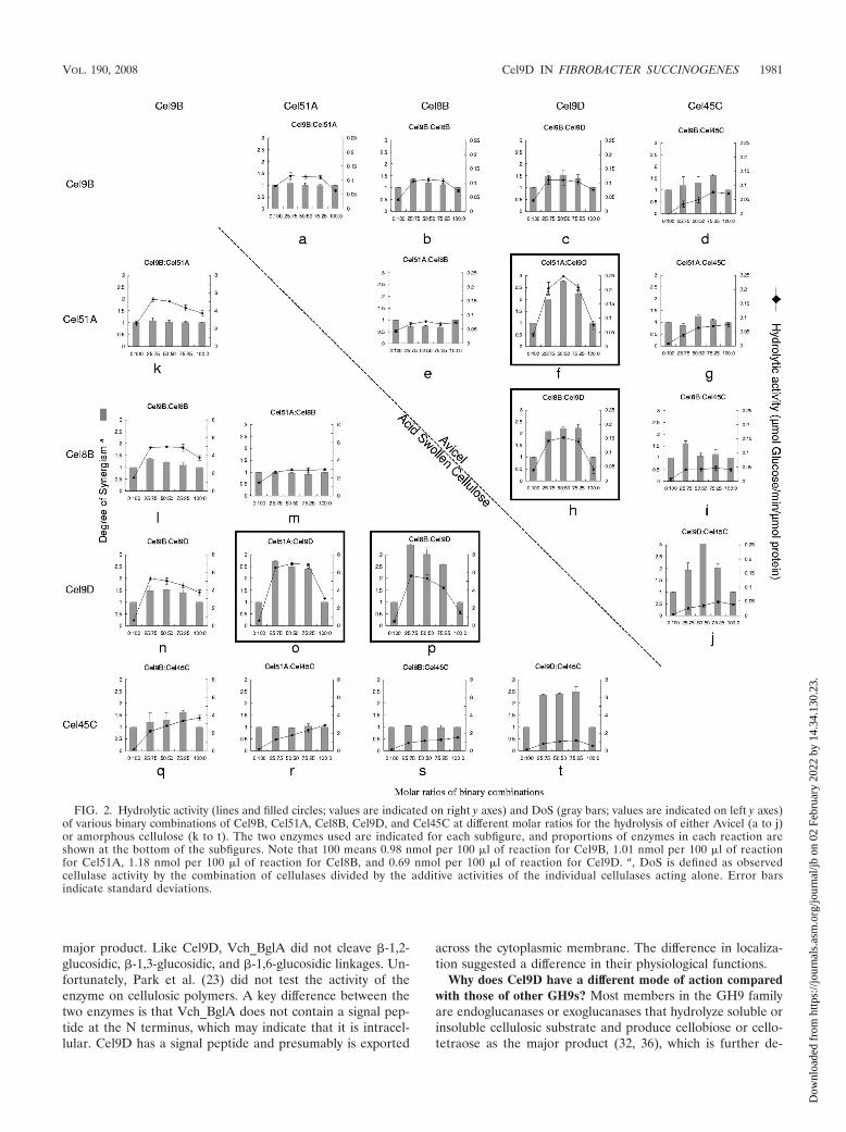
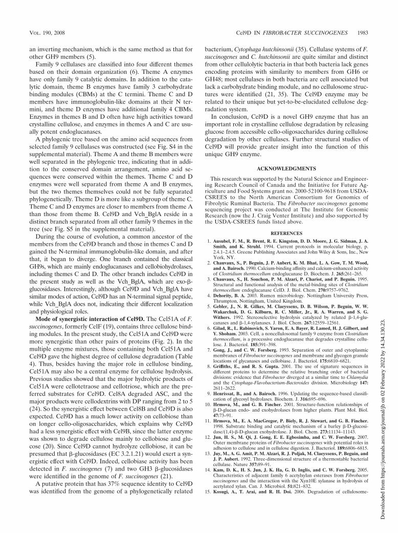
Synergism of Cel9D with other cellulases from F. succino-genes. To identify possible synergistic interactions among dif-ferent GH families of cellulases synthesized by F. succinogenes,ASC and Avicel cellulose hydrolysis by different combinationsof cellulases was assessed. Reducing sugars released by differ-ent combinations of enzymes are presented in Fig. 2 and Table4. For all the binary mixtures, the greatest DoS was obtainedfrom the reaction containing 1.2 nmol of Cel9D and 1.2 nmolof Cel51 on Avicel (Fig. 2f), in which the actual activity wasfour times the sum of individual enzyme activities (Table 4).The highest DoS on the ASC was from the combination ofCel9D and Cel8B, with a synergism value of 3.4 (Fig. 2p). Thecombinations of the two family 9 enzymes, Cel9D and Cel9B,gave a moderate DoS of 1.5, which indicates that there aredifferences between the two cellulases within the same family.Cellulose degradation by mixtures of up to four cellulases wasalso investigated (Table 4). The enzyme mixtures containingCel9D generally yielded a higher DoS as well as a higheroverall level of reducing sugar production. By incubation of theenzyme mixture containing all four enzymes for 60 h, theamount of reducing sugar released corresponded to the hydro-lysis of 2.8% of the initial Avicel substrate present.
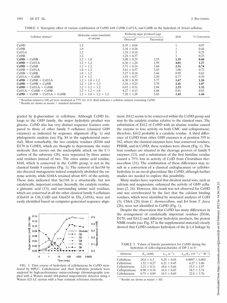
Hydrolysis products of Cel9D. Glucose was the only productreleased from ASC by Cel9D, as detected by HPLC analysis (seeFig. S6B in the supplemental material). This result indicated thatCel9D has �-1,4-glucosidase activity. Cellohexaose hydrolysis ini-tially yielded cellopentaose and glucose (Fig. 3). No cellotetraosewas detected (at time zero) until a larger amount of cellopentaoseaccumulated (at 0.5 min). Similarly, no cellotriose was detected(at 0, 0.5, and 1 min) until a larger amount of cellotetraoseaccumulated (at 2 min) and no cellobiose was detected (at 0, 0.5,1, 2, and 4 min) until there was a larger amount of cellotrioseaccumulated (at 8 min). These results clearly showed that Cel9Dcatalyzed an exotype of hydrolysis reaction. Cellobiose was de-graded very slowly such that less than 5% of the cellobiose accu-mulated at 120 min was digested to glucose after 3,000 min (Fig.3). In contrast, for the same amount of cello-oligosaccharides withthree or more glucose residues, complete hydrolysis was achievedwithin 2 h. The detailed relationships between the kinetic param-eters (Km, kcat, and kcat/Km) and the cello-oligosaccharide sub-strates of Cel9D are shown in Table 5. The Km decreased withincreasing chain length of substrate. The kcats for cellotetraose,cellopentaose, and cellohexaose were similar, while the kcats forcellotriose and cellobiose hydrolysis were substantially smaller.Catalytic efficiency factors kcat/Km increased steadily with increas-ing degrees of polymerization (DP). The kcat/Km value for cello-biose was 2,300-fold lower than that for cellohexaose. The obser-
1978 QI ET AL. J. BACTERIOL.
Dow
nloa
ded
from
http
s://j
ourn
als.
asm
.org
/jour
nal/j
b on
02
Febr
uary
202
2 by
14.
34.1
30.2
3.
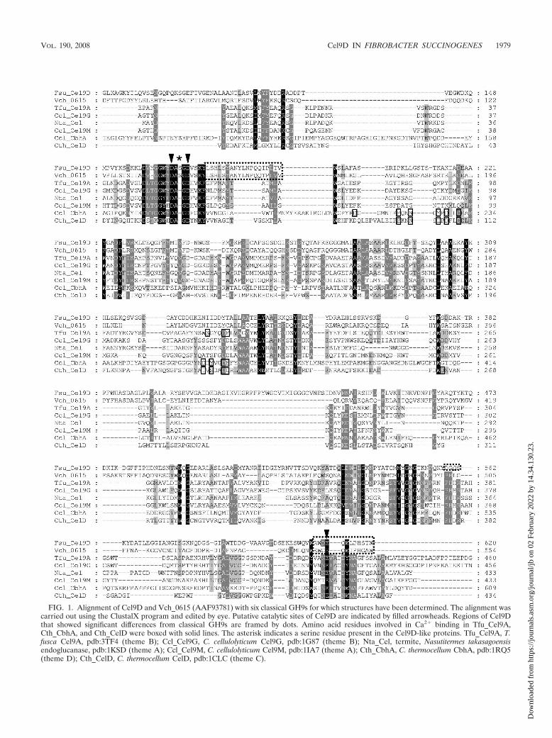
FIG. 1. Alignment of Cel9D and Vch_0615 (AAF93781) with six classical GH9s for which structures have been determined. The alignment wascarried out using the ClustalX program and edited by eye. Putative catalytic sites of Cel9D are indicated by filled arrowheads. Regions of Cel9Dthat showed significant differences from classical GH9s are framed by dots. Amino acid residues involved in Ca2� binding in Tfu_Cel9A,Cth_CbhA, and Cth_CelD were boxed with solid lines. The asterisk indicates a serine residue present in the Cel9D-like proteins. Tfu_Cel9A, T.fusca Cel9A, pdb:3TF4 (theme B); Ccl_Cel9G, C. cellulolyticum Cel9G, pdb:1G87 (theme B); Nta_Cel, termite, Nasutitermes takasagoensisendoglucanase, pdb:1KSD (theme A); Ccl_Cel9M, C. cellulolyticum Cel9M, pdb:1IA7 (theme A); Cth_CbhA, C. thermocellum CbhA, pdb:1RQ5(theme D); Cth_CelD, C. thermocellum CelD, pdb:1CLC (theme C).
VOL. 190, 2008 Cel9D IN FIBROBACTER SUCCINOGENES 1979
Dow
nloa
ded
from
http
s://j
ourn
als.
asm
.org
/jour
nal/j
b on
02
Febr
uary
202
2 by
14.
34.1
30.2
3.
vation that cello-oligosaccharides with longer DP were degradedfaster than those with shorter chain lengths documented a pref-erence for longer cello-oligosaccharides.
After we mixed Cel9D with up to 100 mM glucose and/orcellobiose and incubated it at 37°C for 24 h, cello-oligosaccharideswith longer chain lengths were not detected, indicating thatCel9D does not catalyze the reverse transglycosylation reaction.
Anomeric configuration of hydrolysis products. Proton-NMR was used to investigate the anomeric configuration ofoligosaccharides released during the hydrolysis of pNP-cello-bioside by Cel9D (see Fig. S7 in the supplemental material).The results indicate that Cel9D catalyzes the hydrolysis of the�-(1,4) linkage with inversion of configuration.
Site-directed mutagenesis of Cel9D. To investigate whetherD166 and E612 were the catalytic residues, the two amino acidresidues were replaced with alanine. The mutant proteins(D166A and E612A) were produced and purified by immobi-lized metal ion affinity chromatography. No activity was de-tected on either 1% (wt/vol) CMC after 4 h of incubation or 5mM cellopentaose after 24 h of incubation, as determinedeither by reducing sugar analysis or by HPLC. Thus, D166 andE612 are good candidates for catalytic residues.
To investigate whether the serine residue at position 168 had arole in catalysis, two Cel9D mutants were made. �S168, con-structed with the Ser168 removed, had no activity on CMC orcellopentaose when assayed under the same conditions withCel9D as the parallel positive control, although when this serineresidue was replaced by an alanine residue (S168A), the mutantsretained about 40% percent of activity compared to the wild type.
DISCUSSION
Cel9D is a glucan 1,4-�-glucohydrolase. The present exper-iments showed that Cel9D hydrolyzed cello-oligosaccharidesfrom the nonreducing end, releasing glucose units. There issimilarity between �-glucosidase (EC 3.2.1.21) and glucan 1,4-�-glucohydrolase (EC 3.2.1.74), as both cleave the nonreduc-
ing terminal glycosyl residues from cello-oligosaccharide sub-strates. Cel9D hydrolyzed cello-oligosaccharides with a DPgreater than 2 or hydrolyzed pNPC at much higher rates thanthose for cellobiose or pNPG, showing that the rate of hydro-lysis increases with the DP of the cello-oligosaccharides. Cel9Dreleased glucose from the nonreducing end of the substrate,and its activity was restricted to �-1,4 linkages. Cel9D also hadactivity on soluble and insoluble �-glucans, including CMC,ASC, and Avicel. This capability is distinct from that of �-glu-cosidases (EC 3.2.1.21), which have a decreased rate of hydro-lysis with increasing DP (11, 25). Thus, Cel9D is classified as aglucan 1,4-�-glucohydrolase (34).
Since HPLC showed that the enzyme removed glucose res-idues from the nonreducing ends of cello-oligosaccharides, thesubsites for binding substrate were designated 1, �1, �2, �3,etc., where the “1” subsite binds to the nonreducing terminalglucose and hydrolysis occurs between 1 and �1. Subsiteaffinities of Cel9D for glucose residues in cello-oligosacchar-ides were estimated (see Table S1 in the supplemental mate-rial). Highest affinity was seen at the �2 subsite, which was 15.8kJ/mol. Due to the difference in the subsite affinities between1 (5.7 kJ/mol) and �2 (15.8 kJ/mol), cellobiose and pNPGwould prefer to stay across sites �1 and �2 or �2 and �3rather than the “productive” sites 1 and �1, which explainsthe low rate of hydrolysis of cellobiose and pNPG.
Glucan 1,4-�-D-glucohydrolase is a type of exoglucanase thatreleases glucose from �-134-linked glucan. This type of en-zyme has been identified in many organisms, including bacteria(15, 34), fungi (33), and plants (10, 22). Previously, all glucan1,4-�-glucohydrolases and most �-glucosidases were classifiedas GH1 or GH3 (www.cazy.org/fam/GH1.html and www.cazy.org/fam/GH3.html [9]), which not only catalyzed hydrolysiswith retention of anomeric configuration but also catalyzed thereverse transglycosylation reaction (17). Our data showed thatCel9D could not produce long-chain cello-oligosaccharidefrom glucose or cellobiose, indicating that it does not catalyzethe reverse transglycosylation reaction. Thus, Cel9D is aunique inverting glucan 1,4-�-glucohydrolase.
Cel9D exhibits less than 20% identity to classical GH9s,while it shows about 35% similarity to a series of putativeendoglucanases mainly from the genus Vibrio. Among its manyrelatives, Vch_BglA (Vch0615) from Vibrio cholerae was pre-viously characterized and was subsequently classified as a�-glucosidase (EC 3.2.1.21) (www.cazy.org/fam/GH9.html)that releases pNP from pNPG and hydrolyzed cellobiose effi-ciently (23). In a manner similar to that of Cel9D, Vch_BglAdegraded cello-oligosaccharides and produced glucose as the
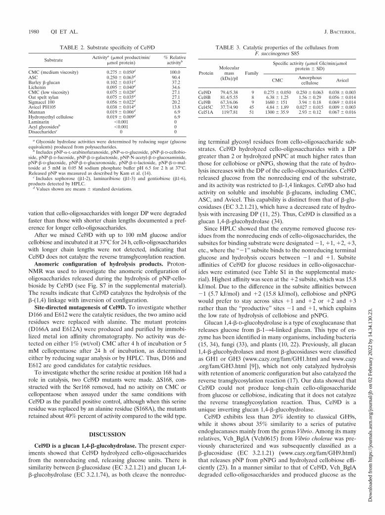
TABLE 2. Substrate specificity of Cel9D
Substrate Activitya (�mol product/min/�mol protein)
% Relativeactivityb
CMC (medium viscosity) 0.275 0.050d 100.0ASC 0.250 0.063d 90.4Barley �-glucan 0.102 0.031d 37.2Lichenin 0.095 0.040d 34.6CMC (low viscosity) 0.075 0.028d 27.1Oat spelt xylan 0.075 0.035d 27.1Sigmacel 100 0.056 0.022d 20.2Avicel PH105 0.038 0.014d 13.8Mannan 0.019 0.006d 6.9Hydroxyethyl cellulose 0.019 0.009d 6.9Laminarin �0.001 0Acyl glycosidesb �0.001 0Disaccharidesc 0 0
a Glycoside hydrolase activities were determined by reducing sugar (glucoseequivalents) produced from polysaccharides.
b Includes pNP-�-L-arabinofuranoside, pNP-�-D-glucoside, pNP-�-D-cellobio-side, pNP-�-D-fucoside, pNP-�-D-galactoside, pNP-N-acetyl-�-D-glucosaminide,pNP-�-D-glucoside, pNP-�-D-glucuronoside, pNP-�-D-lactoside, pNP-�-D-mal-toside at 5 mM in 0.05 M sodium phosphate buffer pH 6.5 for 2 h at 37oC.Released pNP was measured as described by Kam et al. (14).
c Includes sophorose (�1-2), laminaribiose (�1-3) and gentiobiose (�1-6),products detected by HPLC.
d Values shown are means standard deviations.
TABLE 3. Catalytic properties of the cellulases fromF. succinogenes S85
ProteinMolecular
mass(kDa)/pI
Family
Specific activity (�mol Glc/min/�molprotein SD)
CMC Amorphouscellulose Avicel
Cel9D 79.4/5.38 9 0.275 0.050 0.250 0.063 0.038 0.003Cel8B 81.4/5.55 8 6.38 1.25 1.56 0.29 0.056 0.014Cel9B 67.3/6.06 9 1680 151 3.94 0.18 0.069 0.014Cel45C 37.7/4.90 45 4.84 1.89 0.027 0.015 0.009 0.003Cel51A 119/7.81 51 1300 35.9 2.93 0.12 0.067 0.016
1980 QI ET AL. J. BACTERIOL.
Dow
nloa
ded
from
http
s://j
ourn
als.
asm
.org
/jour
nal/j
b on
02
Febr
uary
202
2 by
14.
34.1
30.2
3.
major product. Like Cel9D, Vch_BglA did not cleave �-1,2-glucosidic, �-1,3-glucosidic, and �-1,6-glucosidic linkages. Un-fortunately, Park et al. (23) did not test the activity of theenzyme on cellulosic polymers. A key difference between thetwo enzymes is that Vch_BglA does not contain a signal pep-tide at the N terminus, which may indicate that it is intracel-lular. Cel9D has a signal peptide and presumably is exported
across the cytoplasmic membrane. The difference in localiza-tion suggested a difference in their physiological functions.
Why does Cel9D have a different mode of action comparedwith those of other GH9s? Most members in the GH9 familyare endoglucanases or exoglucanases that hydrolyze soluble orinsoluble cellulosic substrate and produce cellobiose or cello-tetraose as the major product (32, 36), which is further de-
FIG. 2. Hydrolytic activity (lines and filled circles; values are indicated on right y axes) and DoS (gray bars; values are indicated on left y axes)of various binary combinations of Cel9B, Cel51A, Cel8B, Cel9D, and Cel45C at different molar ratios for the hydrolysis of either Avicel (a to j)or amorphous cellulose (k to t). The two enzymes used are indicated for each subfigure, and proportions of enzymes in each reaction areshown at the bottom of the subfigures. Note that 100 means 0.98 nmol per 100 �l of reaction for Cel9B, 1.01 nmol per 100 �l of reactionfor Cel51A, 1.18 nmol per 100 �l of reaction for Cel8B, and 0.69 nmol per 100 �l of reaction for Cel9D. a, DoS is defined as observedcellulase activity by the combination of cellulases divided by the additive activities of the individual cellulases acting alone. Error barsindicate standard deviations.
VOL. 190, 2008 Cel9D IN FIBROBACTER SUCCINOGENES 1981
Dow
nloa
ded
from
http
s://j
ourn
als.
asm
.org
/jour
nal/j
b on
02
Febr
uary
202
2 by
14.
34.1
30.2
3.
graded by �-glucosidase or cellobiase. Although Cel9D be-longs to the GH9 family, the major hydrolytic product wasglucose. Cel9D also has very distinct sequence features com-pared to those of other family 9 cellulases (classical GH9enzymes) as indicated by sequence alignment (Fig. 1) andphylogenetic analysis (see Fig. S4 in the supplemental mate-rial). Most remarkably, the two catalytic residues (D166 andD170 in Cel9D), which are thought to deprotonate the watermolecule that carries out the nucleophilic attack on the C-1carbon of the substrate (28), were separated by three aminoacid residues instead of two. The extra amino acid residue,S168, which is conserved in the Cel9D group, is not in theclassical family 9 enzymes (Fig. 1). The removal of Ser168 bysite-directed mutagenesis indeed completely abolished the en-zyme activity, while S168A retained about 40% of the activity.These data indicated that Ser168 is a structurally, but notcatalytically, important residue. Secondly, the catalytic residue,a glutamic acid (13), and surrounding amino acid residues,which are conserved in all the other classical family 9 cellulases(Glu418 in Cth_CelD and Glu424 in Tfu_Cel9A), were noteasily identified based on computer-generated sequence align-
ment. E612 seems to be conserved within the Cel9D group andmay be the catalytic residue relative to the classical ones. Thesubstitution of E612 of Cel9D with an alanine residue causedthe enzyme to lose activity on both CMC and cellopentaose;therefore, E612 probably is a catalytic residue. A third differ-ence of Cel9D from other GH9 enzymes is at position 559 to562, where the classical enzymes have four conserved residues,PHHR, and in Cel9D, these residues were absent (Fig. 1). Thefour residues are situated in the cleavage grooves of family 9enzymes (13), and a substitution of the first histidine residuecaused a 75% loss in activity of CelD from Clostridium ther-mocellum (31). The combination of these differences may re-sult in a conversion of a classical endoglucanase or cellobio-hydrolase to an exo-�-glucosidase like Cel9D, although furtherstudies are needed to explore this possibility.
Many studies have reported that divalent metal ions, such ascalcium and magnesium, enhanced the activity of GH9 cellu-lases (2, 24). However, this result was not observed for Cel9Dand was corroborated by the fact that the calcium bindingresidues, which were identified by structural analyses of CelD(3), CbhA (28) from C. thermocellum, and E4 from T. fusca(26), were not identified in Cel9D (Fig. 1).
Despite the observation that Cel9D has many differences inthe arrangement of catalytically important residues (D166,D170, and E612) and different hydrolytic products, the protonNMR results (see Fig. S7 in the supplemental material) clearlyshowed that Cel9D catalyzes hydrolysis of the �-1,4 linkage by
FIG. 3. Time course of hydrolysis of cellohexaose by Cel9D mon-itored by HPLC. Cellohexaose and their hydrolysis products wereanalyzed by high-performance anion-exchange chromatography cou-pled with a Waters model 464-pulsed amperimetric detector using aWaters 625 LC system with a base resistant reference electrode.
TABLE 4. Synergistic effect of various combination of Cel9D with Cel9B, Cel51A, and Cel8B on the hydrolysis of Avicel cellulose
Cellulase mixturea Molecular concn (nmol/ml)of enzyme
Reducing sugar produced (�g)DoS % Conversion
Observedb Theoretical
Cel9D 1.2 0.35 0.04 1 0.07Cel9B 1.8 2.18 0.36 1 0.44Cel51A 1.2 1.24 0.18 1 0.25Cel8B 1.2 1.26 0.37 1 0.25Cel9D � Cel9B 1.2 � 1.8 3.00 0.25 2.53 1.19 0.60Cel9D � Cel51A 1.2 � 1.2 6.36 1.24 1.59 4.01 1.27Cel9D � Cel8B 1.2 � 1.2 3.71 0.24 1.60 2.31 0.74Cel9B � Cel51A 1.8 � 1.2 3.61 0.57 3.43 1.06 0.72Cel9B � Cel8B 1.8 � 1.2 3.27 0.18 3.44 0.95 0.65Cel51A � Cel8B 1.2 � 1.2 1.93 0.57 2.50 0.77 0.39Cel9D � Cel9B � Cel51A 1.2 � 1.8 � 1.2 6.30 0.39 3.77 1.67 1.26Cel9D � Cel9B � Cel8B 1.2 � 1.8 � 1.2 5.34 0.23 3.79 1.41 1.07Cel9D � Cel51A � Cel8B 1.2 � 1.2 � 1.2 6.62 0.31 2.84 2.33 1.32Cel51A � Cel8B � Cel9B 1.2 � 1.2 � 1.8 4.27 0.13 4.68 0.91 0.85Cel9D � Cel9B � Cel51A � Cel8B 1.2 � 1.8 � 1.2 � 1.2 7.20 1.28 5.03 1.43 1.44
a Reaction mixtures (100 �l) were incubated at 37°C for 16 h. Bold indicates a cellulose mixture containing Cel9D.b Results are shown as means standard deviations.
TABLE 5. Values of kinetic parameters for Cel9D during thehydrolysis of cello-oligosaccharides of DP 2 to 6a
Substrate Km (mM) kcat (s1) kcat/Km (103 s1 M1)
Cellobiose 28.0 6.3 0.25 0.03 0.0097 0.003Cellotriose 1.52 0.27 6.21 0.45 4.27 1.06Cellotetraose 1.21 0.11 18.6 0.66 15.5 1.97Cellopentaose 0.90 0.10 16.4 0.65 18.5 2.74Cellohexaose 0.73 0.09 16.3 0.69 22.8 3.76
a Results are shown as means SD.
1982 QI ET AL. J. BACTERIOL.
Dow
nloa
ded
from
http
s://j
ourn
als.
asm
.org
/jour
nal/j
b on
02
Febr
uary
202
2 by
14.
34.1
30.2
3.
an inverting mechanism, which is the same method as that forother GH9 members (5).
Family 9 cellulases are classified into four different themesbased on their domain organization (6). Theme A enzymeshave only family 9 catalytic domains. In addition to the cata-lytic domain, theme B enzymes have family 3 carbohydratebinding modules (CBMs) at the C termini. Theme C and Dmembers have immunoglobulin-like domains at their N ter-mini, and theme D enzymes have additional family 4 CBMs.Enzymes in themes B and D often have high activities towardcrystalline cellulose, and enzymes in themes A and C are usu-ally potent endoglucanases.
A phylogenic tree based on the amino acid sequences fromselected family 9 cellulases was constructed (see Fig. S4 in thesupplemental material). Theme A and theme B members werewell separated in the phylogenic tree, indicating that in addi-tion to the conserved domain arrangement, amino acid se-quences were conserved within the themes. Theme C and Denzymes were well separated from theme A and B enzymes,but the two themes themselves could not be fully separatedphylogenetically. Theme D is more like a subgroup of theme C.Theme C and D enzymes are closer to members from theme Athan those from theme B. Cel9D and Vch_BglA reside in adistinct branch separated from all other family 9 themes in thetree (see Fig. S5 in the supplemental material).
During the course of evolution, a common ancestor of themembers from the Cel9D branch and those in themes C and Dgained the N-terminal immunoglobulin-like domain, and afterthat, it began to diverge. One branch contained the classicalGH9s, which are mainly endoglucanases and cellobiohydrolases,including themes C and D. The other branch includes Cel9D inthe present study as well as the Vch_BglA, which are exo-�-glucosidases. Interestingly, although Cel9D and Vch_BglA havesimilar modes of action, Cel9D has an N-terminal signal peptide,while Vch_BglA does not, indicating their different localizationand physiological roles.
Mode of synergistic interaction of Cel9D. The Cel51A of F.succinogenes, formerly CelF (19), contains three cellulose bind-ing modules. In the present study, the Cel51A and Cel9D weremore synergistic than other pairs of proteins (Fig. 2). In themultiple enzyme mixtures, those containing both Cel51A andCel9D gave the highest degree of cellulose degradation (Table4). Thus, besides having the major role in cellulose binding,Cel51A may also be a central enzyme for cellulose hydrolysis.Previous studies showed that the major hydrolytic products ofCel51A were cellotetraose and cellotriose, which are the pre-ferred substrates for Cel9D. Cel8A degraded ASC, and themajor products were cellodextrins with DP ranging from 2 to 5(24). So the synergistic effect between Cel8B and Cel9D is alsoexpected. Cel9D has a much lower activity on cellobiose thanon longer cello-oligosaccharides, which explains why Cel9Dhad a less synergistic effect with Cel9B, since the latter enzymewas shown to degrade cellulose mainly to cellobiose and glu-cose (20). Since Cel9D cannot hydrolyze cellobiose, it can bepresumed that �-glucosidases (EC 3.2.1.21) would exert a syn-ergistic effect with Cel9D. Indeed, cellobiase activity has beendetected in F. succinogenes (7) and two GH3 �-glucosidaseswere identified in the genome of F. succinogenes (21).
A putative protein that has 37% sequence identity to Cel9Dwas identified from the genome of a phylogenetically related
bacterium, Cytophaga hutchinsonii (35). Cellulase systems of F.succinogenes and C. hutchinsonii are quite similar and distinctfrom other cellulolytic bacteria in that both bacteria lack genesencoding proteins with similarity to members from GH6 orGH48; most cellulases in both bacteria are cell associated butlack a carbohydrate binding module, and no cellulosome struc-tures were identified (21, 35). The Cel9D enzyme may berelated to their unique but yet-to-be-elucidated cellulose deg-radation system.
In conclusion, Cel9D is a novel GH9 enzyme that has animportant role in crystalline cellulose degradation by releasingglucose from accessible cello-oligosaccharides during cellulosedegradation by other cellulases. Further structural studies ofCel9D will provide greater insight into the function of thisunique GH9 enzyme.
ACKNOWLEDGMENTS
This research was supported by the Natural Science and Engineer-ing Research Council of Canada and the Initiative for Future Ag-riculture and Food Systems grant no. 2000-52100-9618 from USDA-CSREES to the North American Consortium for Genomics ofFibrolytic Ruminal Bacteria. The Fibrobacter succinogenes genomesequencing project was conducted at The Institute for GenomicResearch (now the J. Craig Venter Institute) and also supported bythe USDA-CSREES funds listed above.
REFERENCES
1. Ausubel, F. M., R. Brent, R. E. Kingston, D. D. Moore, J. G. Sidman, J. A.Smith, and K. Struhl. 1994. Current protocols in molecular biology, p.2.4.1–2.4.5. Greene Publishing Associates and John Wiley & Sons, Inc., NewYork, NY.
2. Chauvaux, S., P. Beguin, J. P. Aubert, K. M. Bhat, L. A. Gow, T. M. Wood,and A. Bairoch. 1990. Calcium-binding affinity and calcium-enhanced activityof Clostridium thermocellum endoglucanase D. Biochem. J. 265:261–265.
3. Chauvaux, S., H. Souchon, P. M. Alzari, P. Chariot, and P. Beguin. 1995.Structural and functional analysis of the metal-binding sites of Clostridiumthermocellum endoglucanase CelD. J. Biol. Chem. 270:9757–9762.
4. Dehority, B. A. 2003. Rumen microbiology. Nottingham University Press,Thrumpton, Nottingham, United Kingdom.
5. Gebler, J., N. R. Gilkes, M. Claeyssens, D. B. Wilson, P. Beguin, W. W.Wakarchuk, D. G. Kilburn, R. C. Miller, Jr., R. A. Warren, and S. G.Withers. 1992. Stereoselective hydrolysis catalyzed by related �-1,4-glu-canases and �-1,4-xylanases. J. Biol. Chem. 267:12559–12561.
6. Gilad, R., L. Rabinovich, S. Yaron, E. A. Bayer, R. Lamed, H. J. Gilbert, andY. Shoham. 2003. CelI, a noncellulosomal family 9 enzyme from Clostridiumthermocellum, is a processive endoglucanase that degrades crystalline cellu-lose. J. Bacteriol. 185:391–398.
7. Gong, J., and C. W. Forsberg. 1993. Separation of outer and cytoplasmicmembranes of Fibrobacter succinogenes and membrane and glycogen granulelocations of glycanases and cellobiase. J. Bacteriol. 175:6810–6821.
8. Griffiths, E., and R. S. Gupta. 2001. The use of signature sequences indifferent proteins to determine the relative branching order of bacterialdivisions: evidence that Fibrobacter diverged at a similar time to Chlamydiaand the Cytophaga-Flavobacterium-Bacteroides division. Microbiology 147:2611–2622.
9. Henrissat, B., and A. Bairoch. 1996. Updating the sequence-based classifi-cation of glycosyl hydrolases. Biochem. J. 316:695–696.
10. Hrmova, M., and G. B. Fincher. 2001. Structure-function relationships of�-D-glucan endo- and exohydrolases from higher plants. Plant Mol. Biol.47:73–91.
11. Hrmova, M., E. A. MacGregor, P. Biely, R. J. Stewart, and G. B. Fincher.1998. Substrate binding and catalytic mechanism of a barley �-D-glucosi-dase/(1,4)-�-D-glucan exohydrolase. J. Biol. Chem. 273:11134–11143.
12. Jun, H. S., M. Qi, J. Gong, E. E. Egbosimba, and C. W. Forsberg. 2007.Outer membrane proteins of Fibrobacter succinogenes with potential roles inadhesion to cellulose and in cellulose digestion. J. Bacteriol. 189:6806–6815.
13. Juy, M., A. G. Amit, P. M. Alzari, R. J. Poljak, M. Claeyssens, P. Beguin, andJ. P. Aubert. 1992. Three-dimensional structure of a thermostable bacterialcellulase. Nature 357:89–91.
14. Kam, D. K., H. S. Jun, J. K. Ha, G. D. Inglis, and C. W. Forsberg. 2005.Characteristics of adjacent family 6 acetylxylan esterases from Fibrobactersuccinogenes and the interaction with the Xyn10E xylanase in hydrolysis ofacetylated xylan. Can. J. Microbiol. 51:821–832.
15. Kosugi, A., T. Arai, and R. H. Doi. 2006. Degradation of cellulosome-
VOL. 190, 2008 Cel9D IN FIBROBACTER SUCCINOGENES 1983
Dow
nloa
ded
from
http
s://j
ourn
als.
asm
.org
/jour
nal/j
b on
02
Febr
uary
202
2 by
14.
34.1
30.2
3.
produced cello-oligosaccharides by an extracellular non-cellulosomal �-glu-can glucohydrolase, BglA, from Clostridium cellulovorans. Biochem. Biophys.Res. Commun. 349:20–23.
16. Lever, M. 1972. A new reaction for colorimetric determination of carbohy-drates. Anal. Biochem. 47:273–279.
17. Ly, H. D., and S. G. Withers. 1999. Mutagenesis of glycosidases. Annu. Rev.Biochem. 68:487–522.
18. Lynd, L. R., P. J. Weimer, W. H. van Zyl, and I. S. Pretorius. 2002. Microbialcellulose utilization: fundamentals and biotechnology. Microbiol. Mol. Biol.Rev. 66:506–577.
19. Malburg, S. R., L. M. Malburg, Jr., T. Liu, A. H. Iyo, and C. W. Forsberg.1997. Catalytic properties of the cellulose-binding endoglucanase F fromFibrobacter succinogenes S85. Appl. Environ. Microbiol. 63:2449–2453.
20. McGavin, M., and C. W. Forsberg. 1988. Isolation and characterization ofendoglucanases 1 and 2 from Bacteroides succinogenes S85. J. Bacteriol.170:2914–2922.
21. Morrison, M., K. E. Nelson, I. K. O. Cann, C. W. Forsberg, R. I. Mackie,J. B. Russell, B. A. White, K. Amava, B. Cheng, M. Qi, H. Jun, S. Mulligan,K. Tran, H. A. Carty, H. Khouri, W. Nelson, S. Daugherty, and C. M. Fraser.2003. The Fibrobacter succinogenes strain S85 genome sequencing project,p. 33. Abstr. 3rd ASM-TIGR Conf. Microb. Genomes, p. 33.
22. Opassiri, R., Y. Hua, O. Wara-Aswapati, T. Akiyama, J. Svasti, A. Esen, andC. Ketudat, Jr. 2004. �-Glucosidase, exo-�-glucanase and pyridoxine trans-glucosylase activities of rice BGlu1. Biochem. J. 379:125–131.
23. Park, J. K., L. X. Wang, H. V. Patel, and S. Roseman. 2002. Molecularcloning and characterization of a unique �-glucosidase from Vibrio cholerae.J. Biol. Chem. 277:29555–29560.
24. Qi, M., H. S. Jun, and C. W. Forsberg. 2007. Characterization and synergisticinteractions of Fibrobacter succinogenes glycoside hydrolases. Appl. Environ.Microbiol. 73:6098–6105.
25. Reese, E. T., A. H. Maguire, and F. W. Parrish. 1968. Glucosidases andexo-glucanases. Can. J. Biochem. 46:25–34.
26. Sakon, J., D. Irwin, D. B. Wilson, and P. A. Karplus. 1997. Structure and
mechanism of endo/exocellulase E4 from Thermomonospora fusca. Nat.Struct. Biol. 4:810–818.
27. Sambrook, J., and D. W. Russell. 2001. Molecular cloning: a laboratorymanual, 3rd ed. Cold Spring Harbor Laboratory Press, Cold Spring Harbor,New York, NY.
28. Schubot, F. D., I. A. Kataeva, J. Chang, A. K. Shah, L. G. Ljungdahl, J. P.Rose, and B. C. Wang. 2004. Structural basis for the exocellulase activity ofthe cellobiohydrolase CbhA from Clostridium thermocellum. Biochemistry43:1163–1170.
29. Schumann, W., and L. C. S. Ferreira. 2004. Production of recombinantproteins in Escherichia coli. Genet. Mol. Biol. 27:442–453.
30. Scott, H. W., and B. A. Dehority. 1965. Vitamin requirements of severalcellulolytic rumen bacteria. J. Bacteriol. 89:1169–1175.
31. Tomme, P., S. Chauvaux, P. Beguin, J. Millet, J. P. Aubert, and M. Claey-ssens. 1991. Identification of a histidyl residue in the active center of endo-glucanase D from Clostridium thermocellum. J. Biol. Chem. 266:10313–10318.
32. Tomme, P., R. A. Warren, and N. R. Gilkes. 1995. Cellulose hydrolysis bybacteria and fungi. Adv. Microb. Physiol. 37:1–81.
33. Wood, T. M., and S. I. McCrae. 1982. Purification and some properties of A(1–4)-�-D-glucan glucohydrolase associated with the cellulase from the fun-gus Penicillium funiculosum. Carbohydr. Res. 110:291–303.
34. Wulff-Strobel, C. R., and D. B. Wilson. 1995. Cloning, sequencing, andcharacterization of a membrane-associated Prevotella ruminicola B(1)4�-glucosidase with cellodextrinase and cyanoglycosidase activities. J. Bacte-riol. 177:5884–5890.
35. Xie, G., D. C. Bruce, J. F. Challacombe, O. Chertkov, J. C. Detter, P. Gilna,C. S. Han, S. Lucas, M. Misra, G. L. Myers, P. Richardson, R. Tapia, N.Thayer, L. S. Thompson, T. S. Brettin, B. Henrissat, D. B. Wilson, and M. J.McBride. 2007. Genome sequence of the cellulolytic gliding bacteriumCytophaga hutchinsonii. Appl. Environ. Microbiol. 73:3536–3546.
36. Zverlov, V. V., N. Schantz, and W. H. Schwarz. 2005. A major new compo-nent in the cellulosome of Clostridium thermocellum is a processive endo-�-1,4-glucanase producing cellotetraose. FEMS Microbiol. Lett. 249:353–358.
1984 QI ET AL. J. BACTERIOL.
Dow
nloa
ded
from
http
s://j
ourn
als.
asm
.org
/jour
nal/j
b on
02
Febr
uary
202
2 by
14.
34.1
30.2
3.