The copper centers of tyramine β-monooxygenase and its catalytic ...
CATALYTIC ROLE OF THE METAL ION IN THE METALLO-β-LACTAMASE GOB María Natalia Lisa ‡, Lars Hemmingsen § and Alejandro J. Vila ‡
‡ From Departamento de Química Biológica and Instituto de Biología Molecular y Celular de Rosario (IBR, CONICET-UNR), Facultad de Ciencias Bioquímicas y Farmacéuticas, Universidad Nacional de
Rosario, Suipacha 531, S2002LRK Rosario, Argentina. § From Department of Basic Sciences and Environment, University of Copenhagen, Thorvaldsensvej
40, DK-1871 Frederiksberg C, Denmark.
Running head: GOB stabilizes an anionic reaction intermediate
Address correspondence to: Alejandro J. Vila, Departamento de Química Biológica, Instituto de Biología Molecular y Celular de Rosario (IBR, CONICET-UNR), Facultad de Ciencias Bioquímicas y Farmacéuticas, Universidad Nacional de Rosario, Suipacha 531, S2002LRK Rosario, Argentina, Phone: +54-341-4351235 ext 108; Fax: +54-341-4390465; E-mail: [email protected]
Metallo-β-lactamases (MβLs)1 stand as one
of the main mechanisms of bacterial resistance toward carbapenems. The rational design of an inhibitor for MβLs has been limited by an incomplete knowledge of their catalytic mechanism and by the structural diversity of their active sites. Here we show that the MβL GOB from Elizabethkingia meningoseptica is active as a mono-metallic enzyme, by using different divalent transition metal ions as surrogates of the native Zn(II) ion. Of the metal derivatives in which Zn(II) is replaced, Co(II) and Cd(II) give rise to the most active enzymes, and are shown to occupy the same binding site as the native ion. However, Zn(II) is the only metal ion capable of stabilizing an anionic intermediate which accumulates during nitrocefin hydrolysis, in which the C-N bond has already been cleaved. This finding demonstrates that the catalytic role of the metal ion in GOB is to stabilize the formation of this intermediate, prior to nitrogen protonation. This role may be general to all MβLs, while nucleophile activation by a Zn(II) ion is not a conserved mechanistic feature. 1 Abbreviations: MβL, Metallo-β-lactamase; NMR, Nuclear Magnetic Resonance; PAC, Perturbed Angular Correlation Spectroscopy of γ Rays; Hepes, N-(2-hydroxy-ethyl)piperazine-N'-2-ethanesulfonic acid; EDTA, ethylenediaminetetraacetic acid; Tris, tris(hidroximetil)aminometano.
The expression of β-lactam degrading enzymes (β-lactamases) is the most common mechanism of antibiotic resistance among bacteria (1,2). These enzymes have been grouped into four classes (A-D) according to sequence homology (3,4). Class A, C and D enzymes use an active site serine residue as a nucleophile, whereas class B lactamases (generically termed metallo-β-lactamases, MβLs) employ one or two Zn(II) ions to cleave the β-lactam ring (1-10).
MβLs have particular importance in the clinical setting in that they can hydrolyze a broader spectrum of β-lactam substrates (including carbapenems) than the serine-type enzymes and are resistant to most clinically employed inhibitors (5-10). The design of an efficient pan-MβL inhibitor has been mostly limited by a striking diversity in the active site structures, catalytic profiles and metal ion requirements for activity among different enzymes. Based on this heterogeneity, MβLs have been classified into three subclasses: B1, B2, and B3 (3). Subclass B1 includes several chromosomally encoded enzymes such as BcII from Bacillus cereus (11-13), CcrA from Bacteroides fragilis (14-17), BlaB from Elizabethkingia meningoseptica (18), as well as the transferable VIM (19), IMP (20,21), SPM (22) and GIM-type enzymes (23). Subclass B2 includes the CphA (24,25) and ImiS (26) lactamases from Aeromonas species, and Sfh-I from Serratia fonticola (27). Subclass B3, originally represented only by L1 from Stenotrophomonas maltophilia (28-30), now includes enzymes from other opportunistic pathogens like FEZ-1 from
1
http://www.jbc.org/cgi/doi/10.1074/jbc.M109.063743The latest version is at JBC Papers in Press. Published on December 10, 2009 as Manuscript M109.063743
Copyright 2009 by The American Society for Biochemistry and Molecular Biology, Inc.
by guest on April 11, 2018
http://ww
w.jbc.org/
Dow
nloaded from
Legionella gormanii (31) and GOB from Elizabethkingia meningoseptica (32,33), as well as from environmental bacteria such as CAU-1 from Caulobacter crescentus (34) and THIN-B from Janthinobacterium lividum (35).
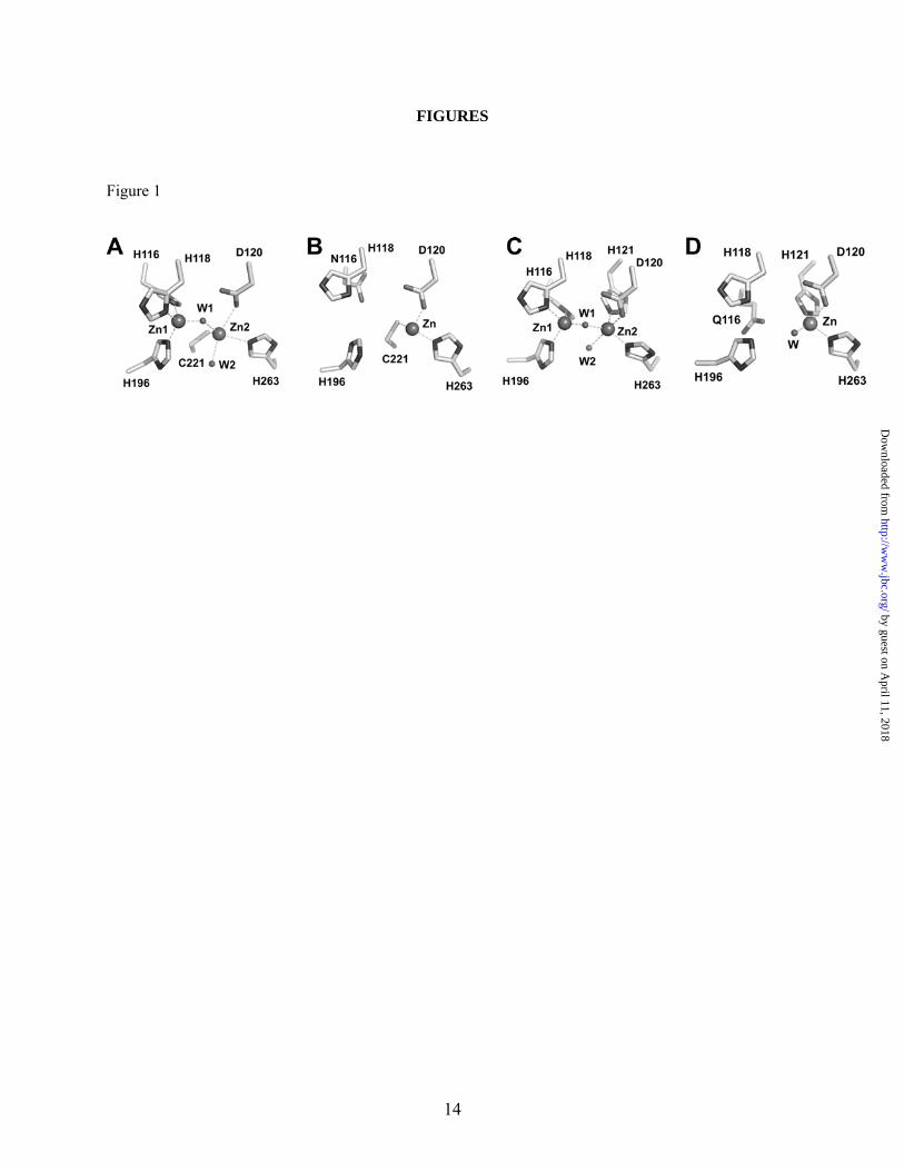
Molecular structures of MβLs from the three subclasses have been solved by X-ray crystallography (11,13,14,24,28,31). Comparison of their structures reveals a common αβ/βα sandwich fold, in which diverse insertions and deletions have resulted in different loop topologies and, ultimately, in different zinc coordination environments and metal site occupancies (Figure 1). MβLs bind up to two metal ions in their active sites. In B1 and B3 enzymes, Zn1 is tetrahedrally coordinated to three histidine ligands (His116, His118 and His196, in Figure 1A and 1C) and a water/OH- molecule (3H site), which is the attacking nucleophile (13,14,28,31). The coordination polyhedron of Zn2 in B1 enzymes is provided by Asp120, Cys221, His263, and one or two water molecules (DCH site) (Figure 1A) (13,14). Notably, this site constitutes the active species in mono-Zn(II) B2 enzymes (Figure 1B) (24). Instead, two mutations (Cys221Ser and Arg121His) affect the Zn2 coordination geometry in B3 MβLs, and the metal ion is now bound to Asp120, His121, His263, and one or two water molecules while Ser221 is no longer a metal ligand (DHH site) (Figure 1C) (28,31). A remarkable exception is constituted by the deepest branching member of the MβL B3 subclass, GOB from Elizabethkingia meningoseptica (4). In all reported GOB sequences, His116 and Ser221 are substituted by Gln and Met, respectively, suggesting the presence of an unusually perturbed metal binding site within this subclass.
We have recently reported a biochemical and biophysical characterization of GOB-18 (33). In contrast to all known MβLs, GOB-18 is fully active against a broad range of β-lactam substrates using a single Zn(II) ion. Based on spectroscopic, mutagenesis and modeling experiments, we already proposed that the Zn(II) ion is bound to Asp120, His121, His263 and one or two solvent molecule(s), i.e., in the canonical Zn2 site of dinuclear MβLs (Figure 1D).
These findings are puzzling, since while it is accepted that B2 lactamases may work without a metal-activated nucleophile (24,36), this is not the
case for B3 lactamases (37). A crystal structure of mono-Zn(II) L1 has revealed that the single metal ion is located in the 3H site, i.e., the one involved in nucleophile activation (38). A recent kinetic and spectroscopic study in L1 has also indicated that mono–Zn(II) L1 in solution is active with the metal ion localized in the 3H site (37). Thus, there is no precedent of a B3 lactamase without a metal-activated nucleophile. The absence of a crystal structure for GOB has precluded a possible description of the role of the metal site in this enzyme, which is still unclear. Here we report a series of experiments that allow us to propose that the metal site in GOB is involved in stabilization of an anionic intermediate that accumulates during catalysis, thus favoring C-N bond cleavage after the nucleophilic attack. These results are unprecedented, since these intermediates have been only identified in catalysis by dinuclear lactamases (16,17,37,39), and disclose the existence of a common catalytic feature in MβLs from all subclasses.
EXPERIMENTAL PROCEDURES
Chemicals – All reagents were purchased from Sigma-Aldrich with the exception of NAP-10 gel filtration column purchased from Amersham Pharmacia Biotech. Metal-free buffers were prepared adding Chelex 100 (Sigma) to normal buffers and stirring for half an hour.
GOB-18 and Asp120Ser GOB-18 expression and purification – It was performed as previously described (33), employing plasmid pET9a-GOB-18 or pET9a-Asp120Ser GOB-18 and E. coli BL21 (DE3) Codon Plus RIL as the bacterial host. Protein purity was higher than 95 % as determined by sodium dodecyl sulfate polyacrylamide gel electrophoresis and Coomassie staining. Protein concentration was determined spectrophotometrically employing ε280 = 32,200 M–1 cm–1.
Metal substitution – Apo-GOB-18 was generated as previously described (33) and metal removal was corroborated by atomic absorption spectroscopy. Zn(II)-GOB-18 was prepared by dialyzing 50-100 μM apo-GOB-18 against 300 volumes of buffer 15 mM Hepes at pH 7.5, 200 mM NaCl containing ZnSO4 at a concentration equal to the protein concentration in the dialysis
2
by guest on April 11, 2018
http://ww
w.jbc.org/
Dow
nloaded from
bag. Zn(II)-Asp120Ser GOB-18, Co(II)-GOB-18, Co(II)-Asp120Ser GOB-18, Ni(II)-GOB-18, Ni(II)-Asp120Ser GOB-18, Cd(II)-GOB-18 and Cd(II)-Asp120Ser GOB-18 were instead generated by dialyzing 50-100 μM apo-GOB-18 or apo- Asp120Ser GOB-18 against 300 volumes of buffer 15 mM Hepes at pH 7.5, 200 mM NaCl containing ZnSO4, CoSO4, NiCl2 or CdCl2, respectively, at a concentration equivalent to five times the protein concentration in the dialysis bag. Excess metal was then removed by a second dialysis with 500 volumes of metal free buffer. All dialysis steps (8-12 hours each) were run at 4ºC.
Metal content determination - Metal content determinations were performed by atomic absorption spectroscopy using a Metrolab 250 instrument operating in the flame mode.
Non-steady state kinetics – Nitrocefin hydrolysis catalyzed by different GOB-18 metal variants was followed employing a Jasco V-550 spectrophotometer for slow reactions and an SX.18-MVR stopped-flow spectrometer associated to a PD.1 photodiode array (Applied Photophysics, Surrey, U.K.) or an absorbance photomultiplier for fast reactions. All measurements were performed in metal free buffer 100 mM Hepes at pH 7.5, 200 mM NaCl. Reaction temperature was 30ºC for all GOB-18 metal variants with the exception of Zn(II)-GOB-18, where the reaction temperature was 4ºC. Enzyme and substrate concentrations were adjusted to 10-30 μM and 5-15 μM respectively, to attain single turnover conditions. Nonlinear regression analysis was used to fit single-wavelength absorbance changes with the program Dynafit (40). Groups of data corresponding to hydrolysis of nitrocefin were fitted simultaneously. The molar extinction coefficients of nitrocefin used were: substrate ε390 = 18,400 M-1 cm-1; product ε390 = 6,300 M-1 cm-1, ε490 = 17,400 M-1 cm-1, ε665 = 500 M-1 cm-1. Steady state kinetic parameters for the hydrolysis of nitrocefin were calculated as kcat = k2 k3 / (k2 + k3) and Km = k3 (k-1 + k2) / k1 (k2 + k3) for Zn(II)-GOB-18 and kcat = k2 and Km = (k-1 + k2) / k1 for all other GOB-18 metal variants.
Steady state kinetics – All reactions were performed at 30 ºC in buffer 15 mM Hepes at pH 7.5, 200 mM NaCl. Antibiotic hydrolysis was monitored following the absorbance variation resulting from the hydrolysis of the β-lactam ring,
using the following extinction coefficients: imipenem, Δε300 = –9,000 M–1 cm–1; penicillin G, Δε235 = –800 M–1 cm–1 and cefotaxime Δε260 = –7,500 M–1 cm–1. The kinetic parameters kcat and Km were derived from nonlinear fit of Michaelis-Menten equation to initial rate measurements recorded on a Jasco V-550 spectrophotometer. In each case, kcat values have been calculated taking into account the concentration of metallated mononuclear enzyme.
Circular Dichroism – Measurements were performed at 25 ºC, using a Jasco J-715 spectropolarimeter flushed with N2. Samples were prepared dialyzing the corresponding protein solution against 300 volumes of buffer 10 mM Tris-HCl at pH 7, 50 mM NaCl, twice for 8-12 hours, at 4ºC.
NMR spectroscopy – NMR spectra were recorded at 298 K on a Bruker Avance II 600 spectrometer operating at 600.13 MHz. 1H NMR paramagnetic spectra were acquired under conditions to optimize detection of the fast relaxing isotropically shifted resonances, either using the superWEFT pulse sequence (41) or water presaturation. These spectra were recorded over large spectral widths with acquisition times ranging from 16 to 80 ms and intermediate delays from 2 to 35 ms. 1H NMR spectra in the diamagnetic envelope were obtained employing a selective shaped pulse for solvent saturation (42). Hydrodynamic radius determination by Pulsed Field Gradient 1H NMR was performed according to reference (43). 113Cd NMR experiment was performed at 133 MHz, with a 5 mm reverse broad-band probe. About 260,000 free induction decays were recorded, acquiring 32,768 complex points in 100 ms, with a 30° observe pulse (4s) and a spectral width of 160000 Hz (1200 ppm), and with a relaxation delay of 0.5 s. The external reference was Cd(ClO4)2.
Electronic absorption spectroscopy – UV-vis spectra were recorded on a Jasco V-550 spectrophotometer, at 25ºC. Samples were in buffer 15 mM Hepes at pH 7.5, 200 mM NaCl.
111mCd(II) PAC spectroscopy – The experiment was performed employing a setup with six BaF2 detectors. Radioactive 111mCd was produced on the day of the experiments by the Cyclotron Department at the University Hospital in Copenhagen. Preparation and purification of 111mCd were as previously described (44). A 25 μL
3
by guest on April 11, 2018
http://ww
w.jbc.org/
Dow
nloaded from

111mCd containing solution was mixed with non-radioactive cadmium acetate and buffer (Hepes and NaCl, see final concentrations below). pH was adjusted to 7.5 at room temperature. Apo-GOB-18 in 15 mM Hepes at pH 7.5, 200 mM NaCl was added at a final enzyme concentration of 40 μM and pH was adjusted to 7.5 at room temperature. The total volume of the sample was 750 μl. Sample was then left 2 hours at room temperature to allow binding of Cd(II) to the protein and subsequently run through a gelfiltration column (NAP10), using 15 mM Hepes at pH 7.5, 200 mM NaCl as eluent. 111mCd containing protein fractions were pooled (4 fractions with a volume of 250 μL each). The sample was frozen in liquid nitrogen and placed in the PAC instrument, where the temperature was set to -20 ºC and controlled by a Peltier element. Fit to PAC data was carried out with 300 data points, disregarding the 5 first points due to systematic errors in these. The analytical expression for the perturbation function is known, and five parameters are fitted for each NQI (45). In radiotracer (109Cd) experiment condition were the same as for PAC experiments.
RESULTS
Nitrocefin hydrolysis by Zn(II)-GOB-18 proceeds by means of an anionic intermediate
Nitrocefin, a synthetic cephalosporin, has been extensively employed as a chromogenic probe of the mechanism of MβLs. Benkovic and coworkers reported for the first time the accumulation of a reaction intermediate in the hydrolysis of nitrocefin by the B1 enzyme CcrA from Bacteroides fragilis (16,17). The same intermediate was also reported for the B3 enzyme L1 (37,46), and for an evolved mutant of the B1 lactamase BcII (39). In all cases, this intermediate was stabilized exclusively by the dinuclear forms of these enzymes.
We studied the hydrolysis of nitrocefin catalyzed by mono Zn(II)-GOB-18 employing a stopped flow equipment coupled to a photodiode array. Besides the decay of the substrate absorption at 390 nm and the rise of the product absorption at 490 nm, the sequence of electronic absorption spectra obtained clearly shows the accumulation and the decay of a species with an intense absorption band centered at 665 nm, identical to the one reported for the anionic
intermediate previously observed for dinuclear enzymes (Figure 2A).
In order to obtain a minimal kinetic mechanism for the UV-vis data we performed similar experiments with different amounts of nitrocefin and enzyme measuring the evolution of the absorbance at 390, 490 and 665 nm (Figure 2B). Data were then subjected to simultaneous global fit. The kinetic model which fits the experimental data best is: k+1 k2 k3
E + S ⇔ ES → EI → E + P k-1
Scheme 1
The individual kinetic constants obtained were employed to calculate kcat = (4.4 ± 0.1) s-1 and Km = (18 ± 3) μM as described in the Experimental Procedures section. These values compare very well with the steady state kinetic parameters determined under the same experimental conditions: kcat = (4.0 ± 0.3) s-1 and Km = (25 ± 5) μM, strongly supporting the proposed model.
The finding of a reaction intermediate in mononuclear Zn(II)-GOB-18 similar to that reported for dinuclear enzymes suggests that the Zn2 ion is involved in its stabilization in all cases. To further explore the role of this metal ion, we decided to probe the mechanism of nitrocefin hydrolysis by GOB-18 substituted with a variety of transition divalent metal ions.
Nitrocefin hydrolysis by M(II)-GOB-18 derivatives
Cytoplasmic overexpression of GOB-18 in E.coli gives rise to a mixture of the Fe(III) and Zn(II) variants (33). We have already optimized a procedure for metal depletion (33). The apo-protein can then be fully loaded with metal ions by dialysis (see section of Experimental Procedures). We have now prepared different metal derivatives (M(II)-GOB-18 hereafter) using Co(II), Ni(II) and Cd(II) as surrogates of the native Zn(II) ion. The metal content of each M(II)-GOB-18 variant was determined by atomic absorption spectroscopy. Similar values were obtained for several protein preparations. The metal content of the different M(II)-GOB-18 derivatives never exceeded one equivalent per protein molecule in agreement with the metal content of the native enzyme (Table 1).
4
by guest on April 11, 2018
http://ww
w.jbc.org/
Dow
nloaded from
In order to determine the essentiality of the Zn(II) ion in the stabilization of the anionic intermediate in the hydrolysis of nitrocefin by GOB-18 and to explore the mechanistic changes induced by the metal replacements, reactions catalyzed by the different M(II)-GOB-18 derivatives were studied similarly to those above described for the native Zn(II) enzyme. Two major facts result from these experiments: (1) the intermediate is not accumulated in the reaction catalyzed by M(II)-GOB-18 derivatives as detected by Zn(II)-GOB-18 (direct conversion of substrate to product was observed in all cases, see Figure S1 provided as Supplemental Data); and (2) all M(II)-GOB-18 derivatives are less active than Zn(II)-GOB-18, among these the Co(II) variant displays higher levels of nitrocefinase activity than the Cd(II) and Ni(II) derivatives.
In each case, single wavelength traces corresponding to substrate depletion and product formation were globally fit to the model presented in Scheme 2 (Figure S1 provided as Supplemental Data). Kinetic constants obtained are summarized in Table S1 provided as Supplemental Data.
k+1 k2
E + S ⇔ ES → E + P k-1
Scheme 2
The fact that there is no accumulation of the anionic intermediate in the hydrolysis of nitrocefin catalyzed by GOB-18 metal variants other than the Zn(II) native one, may be interpreted as an inherent inability of the metal ions to stabilize the intermediate, or as a different binding of these ions to the protein. To explore these hypotheses we employed different spectroscopic techniques to ascertain the coordination sphere of the M(II)-GOB-18 derivatives.
Spectroscopic and functional study of
M(II)-GOB-18 derivatives We exploited the features of the employed
metal ions as spectroscopic probes of their coordination spheres. High spin Co(II) and high spin Ni(II) can be employed as paramagnetic probes to identify the metal ligands by following their effect on the 1H NMR spectrum of the protein. Cd(II) can be directly interrogated by NMR and PAC spectroscopy.
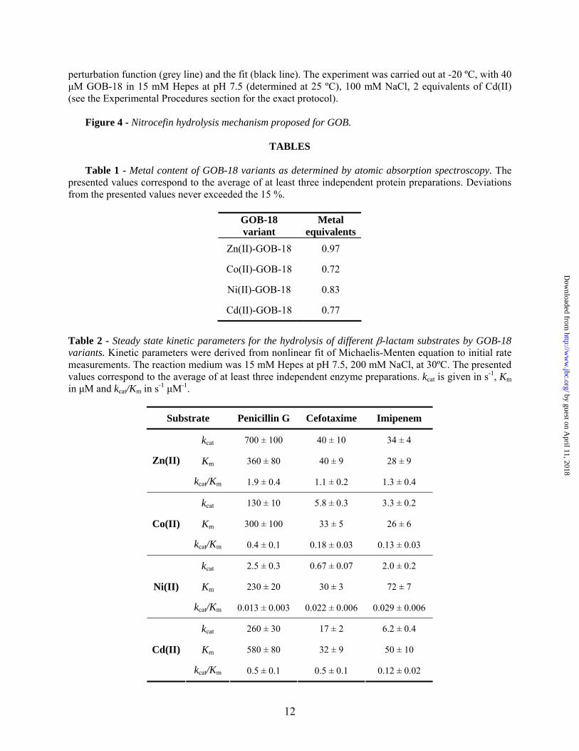
We tested the activity of Co(II)-GOB-18, Ni(II)-GOB-18 and Cd(II)-GOB-18 against a series of clinically relevant substrates (penicillin G, cefotaxime and imipenem). The corresponding steady state kinetic parameters are summarized and compared to those of Zn(II)-GOB-18 in Table 2. The general activity trend is Zn(II) > Cd(II), Co(II) > Ni(II) (in all cases addition of 20 μM metal ion to the reaction medium does not improve the catalytic efficiency). Remarkably, the catalytic efficiencies of M(II)-GOB-18 derivatives are mainly affected in the kcat values as compared to the native enzyme. The kcat values for Cd(II), Co(II) and Ni(II)-GOB-18 have decreased by ca. 3 fold, 10 fold and 100 fold, respectively, compared to those corresponding to Zn(II)-GOB-18. Instead, Km values for each substrate are within the same order of magnitude for the different assayed metal ions.
We recorded circular dichroism and 1H NMR spectra of M(II)-GOB-18 derivatives as well as those of apo and Zn(II)-GOB-18 in order to probe their correct folding (see Figure S2 provided as Supplemental Data). CD spectra in the far UV range are similar with some minor differences in the case of apo-GOB-18. However, the CD spectra in the near UV region suggest a reduced level of tertiary structure in apo-GOB-18 as compared to the metallated forms. Also, the signals in the 1H NMR spectrum of apo-GOB-18 show less dispersion than for any of the metallated variants (see Figure S1 provided as Supplemental Data), suggesting that (despite showing secondary structure) apo-GOB-18 does not adopt the native conformation. The hydrodynamic radii of apo-GOB-18 and Zn(II)-GOB-18 were determined by Pulse Field Gradient 1H NMR spectroscopy, resulting in values of 29 ± 1 and 23,1 ± 0.5 Å, respectively, confirming that apo-GOB-18 adopts a more extended conformation than the metallated protein.
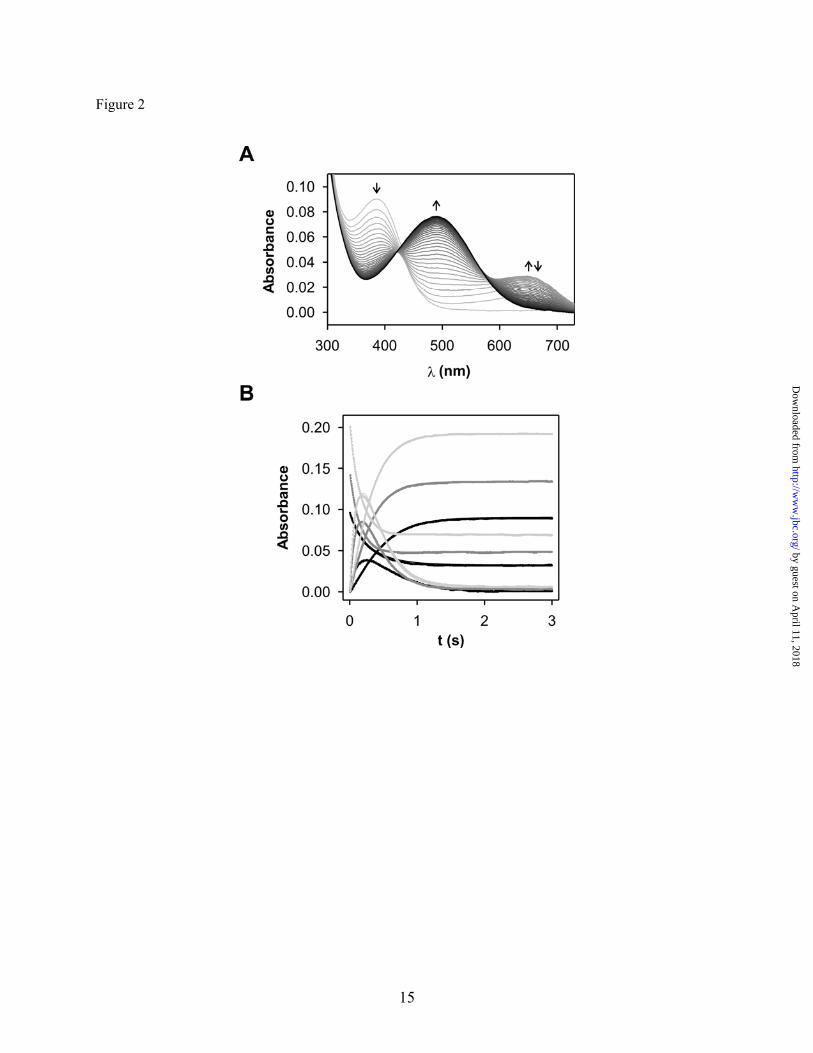
Figure 3A shows the electronic absorption spectrum of Co(II)-GOB-18. The transitions observed in the visible range correspond to Laporte forbidden d-d (or ligand field) electronic transitions of the Co(II) ion bound to the protein. The molar extinction coefficient of these bands (50-60 M-1 cm-1) is consistent with a pentacoordinated Co(II) ion (47). The spectral features observed resemble those of mononuclear Co(II)-L1 with the metal ion localized in the DHH
5
by guest on April 11, 2018
http://ww
w.jbc.org/
Dow
nloaded from
site (37). A 1H NMR spectrum of Co(II)-GOB-18 recorded under conditions that allow the detection of paramagnetic signals reveals a set of isotropically shifted resonances spanning from 80 to 20 ppm (Figure 3B). When the spectrum was recorded in D2O, signals at 60 and 47 ppm were absent, indicating the presence of two solvent exchangeable resonances which can be attributed to two His ligands. In addition, the low number of signals observed in the paramagnetic region of the spectrum and their dispersion in the chemical shift range is also consistent with a pentacoordinated Co(II) ion (47,48). The present spectrum shows a larger signal dispersion than that of mononuclear Co(II)-L1 (37).
Figure 3C shows the electronic absorption spectrum of Ni(II)-GOB-18. The signals observed in the visible range corresponds to ligand field electronic transitions of the Ni(II) ion bound to the protein. The spectrum resembles that of Ni(II)-Carboxypeptidase A, which contains a single hexacoordinated metal ion (49). The 1H NMR spectrum of Ni(II)-GOB-18 reveals a set of isotropically shifted resonances spanning from 70 to 50 ppm (Figure 3D). When the spectrum was recorded in D2O, the signals at 70 and 67 ppm were no longer detected, indicating the presence of two exchangeable resonances which can be attributed to two His ligands. The chemical shifts of this variant resemble those reported for Ni(II)-Carboxypeptidase A (50). This observation, together with the absence of signals experiencing pseudocontact shifts, supports the hypothesis of a pseudo octahedral Ni(II) site (50).
The 113Cd NMR spectrum of 113Cd(II)-GOB-18 shows only one signal (Figure 3E) at 115 ppm, revealing only one metal binding site. Besides, the chemical shift of the observed signal is consistent with a coordination sphere containing N and O donor atoms. 113Cd(II) resonances in proteins with a mixed N/O coordination sphere are usually found between 300 and 40 ppm, being O the most shielding donor atom (51). On the other hand, each sulfur donor present at a 113Cd(II) binding site is so deshielding that it is possible to use chemical shifts to determine the number of sulfur donor atoms at an unknown site (51). Resonances of 113Cd(II)-BcII have been reported at 262 and 142 ppm, which have been attributed to the metal ion bound to sites 3H and DCH, respectively, differing significantly from the resonance here reported
(52,53). The 115 ppm resonance observed for 113Cd(II)-GOB-18 agrees well with experimental 113Cd NMR data giving a resonance at 120 ppm for Carboxypeptidase A (54), presumably with a N2O3 coordination sphere (two histidines, one water and a bidentate carboxylate), and also with theoretical predictions (55).
Figure 3F shows the 111mCd(II) PAC spectroscopic data recorded for Cd(II)-GOB-18. With 2 equivalents of Cd(II) added to the protein and an incubation time of 2 hours the PAC signal is found at relatively low frequency. It can be fit with just one NQI which would agree with the single resonance observed by 113Cd NMR. Also, only one Cd(II) ion was observed to bind per protein molecule, as demonstrated by a radiotracer experiment using 109Cd(II), where 2 equivalents of 109Cd(II) were incubated with the protein under the same conditions as applied for the PAC experiment, and a subsequent gelfiltration displayed very close to 50 % of the radioactivity in the same fractions as the protein. All data from 113Cd NMR, 111mCd PAC, and atomic absorption spectroscopy for the fully metal loaded protein are thus consistent with one binding site.
The NQI fall in the spectral range expected for ligands coordinating with nitrogen or oxygen atoms. The PAC spectrum obtained from a mixture of 111mCd(II) but not protein in the buffer is very different from the one described above (not shown), indicating that in that case, no free 111mCd(II) was present. Fitted parameters are shown in Table S2 provided as Supplemental Data.
M(II)-Asp120Ser GOB-18 derivatives
Spectroscopic data on Co(II), Ni(II) and Cd(II) substituted GOB-18 unequivocally point to metal binding to a single site in all M(II)-GOB-18 derivatives. However these data could be compatible with binding to a His2Gln or to His2Asp site. In order to unequivocally determine the metal binding site in GOB-18 metal derivatives, we obtained the metal substituted mutant Asp120Ser GOB-18, where one of the possible metal binding sites is drastically perturbed. Asp120Ser GOB-18 as isolated contained no metal bound and displayed no activity against nitrocefin. Although exhaustive dialysis of this mutant against buffer containing excess of ZnSO4, CoSO4, NiCl2, or CdCl2 resulted
6
by guest on April 11, 2018
http://ww
w.jbc.org/
Dow
nloaded from
in some extent of metal binding (Table 3), all metal derivatives displayed non detectable activity against nitrocefin. All Asp120Ser GOB-18 metal derivatives were interrogated by CD showing similar features to those presented for the wild type enzyme (not shown), thus indicating comparable secondary and tertiary structure. These results allow us to unambiguously assign site DHH as the metal binding site in all GOB-18 derivatives.
DISCUSSION
Metallo-β-lactamases (MβLs) represent the
largest group of carbapenemases (5-10). The lack of a pan-MβL inhibitor is mostly due to the failure in identifying common structural and mechanistic features in enzymes from different organisms, which show a striking diversity in terms of substrate spectrum, active site structure and metal ion requirements. The Zn(II) ions in MβLs are required for substrate binding and hydrolysis, but the specific role and essentiality of each metal binding site are still subject of intense debate (5-10).
Zn(II) is ubiquitous in nature, being the only metal ion present in enzymes from all six groups in the EC nomenclature (56). This fact highlights its amazing chemical versatility. In the case of MβLs, the Zn(II) ion is able to contribute to β-lactam hydrolysis by (a) lowering the pKa of a bound water molecule, which may act as a nucleophile, providing a high local concentration of hydroxide ions at neutral pH (37,57); (b) as a Lewis acid, polarizing the C=O bond and therefore augmenting the electrophilic nature of the carbonyl carbon (57) and (c) by stabilizing a negative charge in the bridging nitrogen of the lactam moiety, after C-N bond cleavage (16,17,24,36,58,59). These three roles have been invoked for the two metal binding sites in MβLs, but it is not clear yet which of them are essential for catalysis.
Substrate docking studies to the active site of B1 and B3 lactamases suggest that the attacking nucleophile in the dinuclear forms is the bridging water/OH- ligand (13,14,28,36,60). This moiety is asymmetrically positioned with respect to the two metal ions, lying closer (1.9-2.1 Å) to the metal ion in the 3H site than to the Zn(II) ion in the DCH
or DHH sites (2.1-3.1 Å) (13,14,28). The shorter bond length in the former case is consistent with this ligand being a hydroxide, and with the idea that the 3H site is responsible of lowering the pKa of a water molecule, thus being responsible of nucleophile activation (57). The role of C=O polarization by this same zinc ion is not supported by QM/MM calculations, and by different docking studies, which reveal that the β-lactam bond may not directly bind to the metal ion (60,61).
The availability of crystal structures of mono-Zn(II) BcII (a B1 enzyme) (11) and L1 (B3) (38) disclosing the presence of one metal ion localized in the preserved 3H site, as well as theoretical studies of substrate binding to mononuclear BcII (62-64), has supported the hypothesis that only this metal site would be essential, delivering the attacking nucleophile. The structure of the mono-Zn(II) B2 enzyme CphA revealed that the only metal ion in this case is bound to the DCH site, since one of the His of the 3H binding site is replaced by an Asn residue (24). In this case, the attacking nucleophile has been proposed to be a water molecule which is not activated by a metal ion (24,36). However, B2 enzymes have been considered as an exception, being limited to few bacteria, and performing as exclusive carbapenemases.
The recent report of GOB, a B3 enzyme, which is active as a mononuclear enzyme and is not inhibited by excess Zn(II) (33,36), provides an excellent opportunity to examine the role of the metal ion in a broad spectrum MβL. Here we have shown that nitrocefin hydrolysis mediated by mono-Zn(II) GOB proceeds via accumulation of an anionic intermediate identical to the one reported by Benkovic and coworkers for a dinuclear B1 enzyme (16,17), and by Crowder and colleagues for a dinuclear B3 lactamase (37,46). Replacement of the native Zn(II) ion by other metal ions, such as Co(II), Ni(II) and Cd(II), gives rise to metal derivatives with disparate catalytic efficiencies and for which accumulation of such an intermediate can not be detected. We attribute the reduced catalytic efficiencies in Co(II), Ni(II) and Cd(II)-GOB to a less efficient metal-nitrogen interaction in the first step of the reaction, where the C-N bond cleavage is concerted with the nucleophilic attack. QM/MM calculations simulating nitrocefin hydrolysis by a dinuclear B1 enzyme have shown that the metal ion in the Zn2
7
by guest on April 11, 2018
http://ww
w.jbc.org/
Dow
nloaded from
site plays a significant role in lowering the energetic barrier for C-N bond scission (65), in agreement with this proposal. Different spectroscopic techniques unequivocally show that metal replacement does not affect the identity of the metal binding site (the DHH site), revealing that the Zn(II) ion in the DHH site is essential for the stabilization of this intermediate, which does not require formation of a dinuclear center. Steady state kinetic data reveal similar Km values for the different GOB-18 variants, suggesting that the metal ions used here for Zn(II) replacement are equally well suited to provide an anchoring site for the substrate carboxylate. These results are in agreement with the finding that Zn(II) is essential for substrate binding in MβLs (66).
The kinetic data of nitrocefin hydrolysis by Co(II), Cd(II) and Ni(II) GOB can also be fit to Scheme 1 by assuming k3 >> k2. In this model, k2 reflects the nucleophilic attack concerted with the C-N bond cleavage, the latter being triggered by a metal-nitrogen interaction. We therefore conclude that the reduced k2 values can also be accounted for by a less efficient interaction with the N atom of the substrate. Another possibility that we cannot fully discard at this point is that the metal site is also involved in nucleophile activation. Nevertheless QM/MM calculations on B2 enzymes do not support this hypothesis (36).
We have recently shown that second shell mutations in the B1 lactamase BcII obtained by in vitro evolution are able to fine tune the position of the metal ion in the DCH site of this enzyme, stabilizing the nitrocefin intermediate which is not accumulated in hydrolysis mediated by wild type BcII (39). Here we observe the same phenomenon: subtle changes (as those which may be induced by metal substitution) determine the stability of this intermediate.
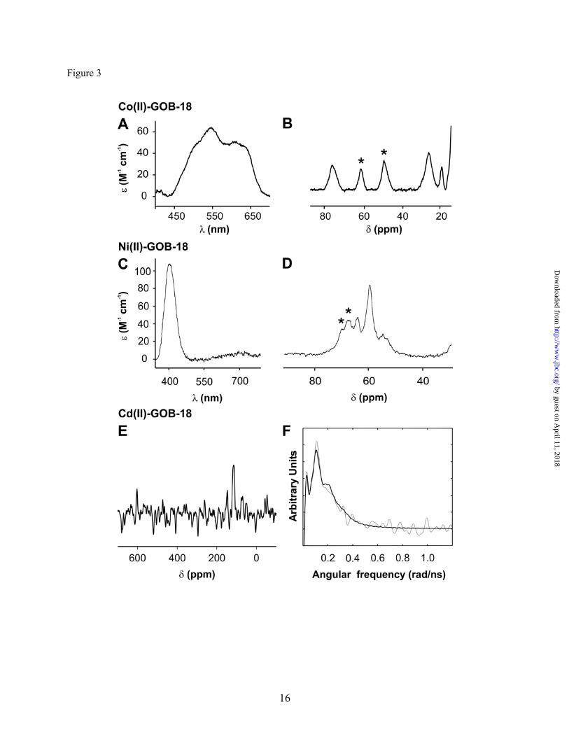
Based on these observations, we propose a β-lactam hydrolysis mechanism for GOB, which does not require a metal-activated nucleophile (Figure 4). Instead, the role of the metal ion is to steer substrate binding and to provide electrostatic stabilization of the anionic intermediate. This mechanism is in agreement with the proposal that BcII could be active as a mononuclear enzyme with an empty 3H site (67), i.e., with a requirement of a Zn(II) ion for intermediate stabilization rather than for nucleophile activation (58), which is formed by shifting of an equilibrium
between the two sites (52) upon substrate binding (67).
This conclusion suggests that the positioning of the metal ion at the DCH/DHH site is crucial to define a catalytically active lactamase. This is in excellent agreement with the finding that engineering a more buried position for this metal center seriously impairs the lactamase activity (68), while rendering a more substrate-accessible Zn(II) at this site enhances the catalytic efficiency (39). Despite nitrocefin is not a clinically useful antibiotic, its usefulness as a probe of the catalytic mechanism of MβLs cannot be underscored. It has been shown recently that carbapenem hydrolysis by a B1 MβL also proceeds by means of an anionic intermediate, after C-N bond cleavage (58). These evidences point to a general role of the Zn(II) ion in the DCH/DHH site of MβL from all subclasses, which may be targeted as a common mechanistic element for inhibitor design.
8
by guest on April 11, 2018
http://ww
w.jbc.org/
Dow
nloaded from
REFERENCES 1. Fisher, J. F., Meroueh, S. O., and Mobashery, S. (2005) Chem. Rev. 105, 395-424 2. Perez, F., Endimiani, A., Hujer, K. M., and Bonomo, R. A. (2007) Curr. Opin. Pharmacol. 7,
459-469 3. Frere, J. M., Galleni, M., Bush, K., and Dideberg, O. (2005) J. Antimicrob. Chemother. 55, 1051-
1053 4. Hall, B. G., Salipante, S. J., and Barlow, M. (2003) J. Mol. Evol. 57, 249-254 5. Crowder, M. W., Spencer, J., and Vila, A. J. (2006) Acc. Chem. Res. 39, 721-728 6. Bebrone, C. (2007) Biochem. Pharmacol. 74, 1686-1701 7. Galleni, M., Lamotte-Brasseur, J., Rossolini, G. M., Spencer, J., Dideberg, O., and Frere, J. M.
(2001) Antimicrob. Agents Chemother. 45, 660-663 8. Walsh, T. R., Toleman, M. A., Poirel, L., and Nordmann, P. (2005) Clin. Microbiol. Rev. 18, 306-
325 9. Cricco, J. A., Rasia, R. M., Orellano, E. G., Ceccarelli, E. A., and Vila, A. J. (1999) Coord.
Chem. Rev. 190-192, 519-535 10. Cricco, J. A. and Vila, A. J. (1999) Curr. Pharm. Des 5, 915-927 11. Carfi, A., Pares, S., Duee, E., Galleni, M., Duez, C., Frere, J. M., and Dideberg, O. (1995) EMBO
J. 14, 4914-4921 12. Orellano, E. G., Girardini, J. E., Cricco, J. A., Ceccarelli, E. A., and Vila, A. J. (1998)
Biochemistry 37, 10173-10180 13. Fabiane, S. M., Sohi, M. K., Wan, T., Payne, D. J., Bateson, J. H., Mitchell, T., and Sutton, B. J.
(1998) Biochemistry 37, 12404-12411 14. Concha, N. O., Rasmussen, B. A., Bush, K., and Herzberg, O. (1996) Structure. 4, 823-836 15. Yanchak, M. P., Taylor, R. A., and Crowder, M. W. (2000) Biochemistry 39, 11330-11339 16. Wang, Z., Fast, W., and Benkovic, S. J. (1998) J. Am. Chem. Soc. 120, 10788-10789 17. Wang, Z., Fast, W., and Benkovic, S. J. (1999) Biochemistry 38, 10013-10023 18. Garcia-Saez, I., Hopkins, J., Papamicael, C., Franceschini, N., Amicosante, G., Rossolini, G. M.,
Galleni, M., Frere, J. M., and Dideberg, O. (2003) J. Biol. Chem. 278, 23868-23873 19. Docquier, J. D., Lamotte-Brasseur, J., Galleni, M., Amicosante, G., Frere, J. M., and Rossolini,
G. M. (2003) J. Antimicrob. Chemother. 51, 257-266 20. Materon, I. C., Beharry, Z., Huang, W., Perez, C., and Palzkill, T. (2004) J. Mol. Biol. 344, 653-
663 21. Toney, J. H., Hammond, G. G., Fitzgerald, P. M., Sharma, N., Balkovec, J. M., Rouen, G. P.,
Olson, S. H., Hammond, M. L., Greenlee, M. L., and Gao, Y. D. (2001) J. Biol. Chem 276, 31913-31918
22. Murphy, T. A., Catto, L. E., Halford, S. E., Hadfield, A. T., Minor, W., Walsh, T. R., and Spencer, J. (2006) J. Mol. Biol. 357, 890-903
23. Castanheira, M., Toleman, M. A., Jones, R. N., Schmidt, F. J., and Walsh, T. R. (2004) Antimicrob. Agents Chemother. 48, 4654-4661
24. Garau, G., Bebrone, C., Anne, C., Galleni, M., Frere, J. M., and Dideberg, O. (2005) J. Mol. Biol. 345, 785-795
25. Hernández Valladares, M., Felici, A., Weber, G., Adolph, H. W., Zeppezauer, M., Rossolini, G. M., Amicosante, G., Frère, J. M., and Galleni, M. (1997) Biochemistry 36, 11534-11541
26. Crawford, P. A., Yang, K. W., Sharma, N., Bennett, B., and Crowder, M. W. (2005) Biochemistry 44, 5168-5176
27. Saavedra, M. J., Peixe, L., Sousa, J. C., Henriques, I., Alves, A., and Correia, A. (2003) Antimicrob. Agents Chemother. 47, 2330-2333
28. Ullah, J. H., Walsh, T. R., Taylor, I. A., Emery, D. C., Verma, C. S., Gamblin, S. J., and Spencer, J. (1998) J. Mol. Biol. 284, 125-136
29. Spencer, J., Clarke, A. R., and Walsh, T. R. (2001) J. Biol. Chem 276, 33638-33644
9
by guest on April 11, 2018
http://ww
w.jbc.org/
Dow
nloaded from
30. Garrity, J. D., Carenbauer, A. L., Herron, L. R., and Crowder, M. W. (2004) J. Biol. Chem 279, 920-927
31. Garcia-Saez, I., Mercuri, P. S., Papamicael, C., Kahn, R., Frere, J. M., Galleni, M., Rossolini, G. M., and Dideberg, O. (2003) J. Mol. Biol. 325, 651-660
32. Bellais, S., Aubert, D., Naas, T., and Nordmann, P. (2000) Antimicrob. Agents Chemother. 44, 1878-1886
33. Moran-Barrio, J., Gonzalez, J. M., Lisa, M. N., Costello, A. L., Peraro, M. D., Carloni, P., Bennett, B., Tierney, D. L., Limansky, A. S., Viale, A. M., and Vila, A. J. (2007) J Biol. Chem 282, 18286-18293
34. Docquier, J. D., Pantanella, F., Giuliani, F., Thaller, M. C., Amicosante, G., Galleni, M., Frere, J. M., Bush, K., and Rossolini, G. M. (2002) Antimicrob. Agents Chemother. 46, 1823-1830
35. Docquier, J. D., Lopizzo, T., Liberatori, S., Prenna, M., Thaller, M. C., Frere, J. M., and Rossolini, G. M. (2004) Antimicrob. Agents Chemother. 48, 4778-4783
36. Simona, F., Magistrato, A., Dal Peraro, M., Cavalli, A., Vila, A. J., and Carloni, P. (2009) J Biol. Chem 284, 28164-28171
37. Hu, Z., Periyannan, G., Bennett, B., and Crowder, M. W. (2008) J. Am. Chem. Soc. 130, 14207-14216
38. Nauton, L., Kahn, R., Garau, G., Hernandez, J. F., and Dideberg, O. (2008) J. Mol. Biol. 375, 257-269
39. Tomatis, P. E., Fabiane, S. M., Simona, F., Carloni, P., Sutton, B. J., and Vila, A. J. (2008) Proc. Natl. Acad. Sci. U. S. A 105, 20605-20610
40. Kuzmic, P. (1996) Anal. Biochem. 237, 260-273 41. Inubushi, T. and Becker, E. D. (1983) J. Magn. Reson. 51, 128-133 42. Hwang, T. L. and Shaka, A. J. (1995) J. Magn. Reson. A 112, 275-279 43. Wilkins, D. K., Grimshaw, S. B., Receveur, V., Dobson, C. M., Jones, J. A., and Smith, L. J.
(1999) Biochemistry 38, 16424-16431 44. Hemmingsen, L., Bauer, R., Bjerrum, M. J., Zeppezauer, M., Adolph, H. W., Formicka, G., and
Cedergren-Zeppezauer, E. (1995) Biochemistry 34, 7145-7153 45. Hemmingsen, L., Sas, K. N., and Danielsen, E. (2004) Chem. Rev. 104, 4027-4062 46. McManus-Muñoz, S. and Crowder, M. W. (1999) Biochemistry 38, 1547-1553 47. Bertini, I. and Luchinat, C. (1985) Adv. Inorg. Biochem. 6, 71-111 48. Bertini, I., Turano, P., and Vila, A. J. (1993) Chem. Rev. 93, 2833-2932 49. Rosenberg, R. C., Root, C. A., and Gray, H. B. (1975) J. Am. Chem. Soc. 97, 21-26 50. Bertini, I., Donaire, A., Monnanni, R., Moratal Mascarell, J. M., and Salgado, J. (1992) J. Chem.
Soc. Dalton Trans. 1443-1447 51. Coleman, J. E. (1993) Methods Enzymol. 227, 16-43 52. Hemmingsen, L., Damblon, C., Antony, J., Jensen, M., Adolph, H. W., Wommer, S., Roberts, G.
C., and Bauer, R. (2001) J. Am. Chem. Soc. 123, 10329-10335 53. Damblon, C., Jensen, M., Ababou, A., Barsukov, I., Papamicael, C., Schofield, C. J., Olsen, L.,
Bauer, R., and Roberts, G. C. (2003) J. Biol. Chem. 278, 29240-29251 54. Gettins, P. (1986) J Biol. Chem 261, 15513-15518 55. Hemmingsen, L., Olsen, L., Antony, J., and Sauer, S. P. (2004) J Biol. Inorg. Chem 9, 591-599 56. Lipscomb, W. N. and Strater, N. (1996) Chem. Rev. 96, 2375-2434 57. Bounaga, S., Laws, A. P., Galleni, M., and Page, M. I. (1998) Biochem. J. 31, 703-711 58. Tioni, M. F., Llarrull, L. I., Poeylaut-Palena, A. A., Marti, M. A., Saggu, M., Periyannan, G. R.,
Mata, E. G., Bennett, B., Murgida, D. H., and Vila, A. J. (2008) J. Am. Chem. Soc. 130, 15852-15863
59. Spencer, J., Read, J., Sessions, R. B., Howell, S., Blackburn, G. M., and Gamblin, S. J. (2005) J. Am. Chem. Soc. 127, 14439-14444
60. Dal Peraro M., Vila, A. J., Carloni, P., and Klein, M. L. (2007) J Am. Chem Soc. 129, 2808-2816
10
by guest on April 11, 2018
http://ww
w.jbc.org/
Dow
nloaded from
61. Dal Peraro, M., Llarrull, L. I., Rothlisberger, U., Vila, A. J., and Carloni, P. (2004) J. Am. Chem. Soc. 126, 12661-12668
62. Dal Peraro, M., Vila, A. J., and Carloni, P. (2004) Proteins 54, 412-423 63. Diaz, N., Suarez, D., and Merz, K. M., Jr. (2001) J. Am. Chem. Soc. 123, 9867-9879 64. Olsen, L., Rasmussen, T., Hemmingsen, L., and Ryde, U. (2004) J. Phys. Chem. B 108, 17639-
17648 65. Park, H., Brothers, E. N., and Merz, K. M., Jr. (2005) J. Am. Chem. Soc. 127, 4232-4241 66. Rasia, R. M. and Vila, A. J. (2004) J. Biol. Chem 279, 26046-26051 67. Llarrull, L. I., Tioni, M. F., and Vila, A. J. (2008) J. Am. Chem. Soc. 130, 15842-15851 68. Gonzalez, J. M., Medrano Martin, F. J., Costello, A. L., Tierney, D. L., and Vila, A. J. (2007) J
Mol. Biol. 373, 1141-1156
FOOTNOTES M.N.L. is recipient of a doctoral fellowship from CONICET. A.J.V. is a Staff member from
CONICET and an International Research Scholar of the Howard Hughes Medical Institute. This work was supported by grants from ANPCyT and HHMI to AJV and from the Danish research
Council for nature and Universe to LH. The Bruker Avance II 600 MHz NMR spectrometer was purchased with funds from ANPCyT (PME2003-0026) and CONICET.
FIGURE LEGENDS
Figure 1 - Metallo-β-lactamase metal binding sites. (A) Dinuclear B1 MβL BcII from Bacillus
cereus (PDB 1bc2) (13); (B) Mononuclear B2 MβL CphA from Aeromonas hydrophila (PDB 1x8g) (24); (C) Dinuclear B3 MβL L1 from Stenotrophomonas maltophilia (PDB 1sml) (28); (D): Molecular model for the mononuclear B3 MβL GOB-18 from Elizabethkingia meningoseptica (33). Zinc atoms are shown as large grey spheres, and water molecules (W) are shown as small grey spheres. Coordination bonds are shown as dashed lines.
Figure 2 - Non steady state nitrocefin hydrolysis catalyzed by Zn(II)-GOB-18. (A) Sequence of
electronic absorption spectra of 4.5 μM Nitrocefin upon the reaction with 9.3 μM Zn(II)-GOB-18 obtained employing a stopped flow equipment coupled to a photo diode array. Reaction progresses from gray to black spectra. (B) Time evolution of absorbance at 390, 490 and 665 nm upon reaction of: a) 5.2 μM nitrocefin with 14.1 μM Zn(II)-GOB-18 (black); b) 7.7 μM nitrocefin with 28.9 μM Zn(II)-GOB-18 (dark gray) and c) 11 μM nitrocefin with 28.9 μM Zn(II)-GOB-18 (light gray). In each case, 600 points were recorded on a linear time scale over 3 seconds. Fits to the kinetic model in Scheme 1 overlay experimental points. Parameters obtained from the global fit of data: k+1 = (4.8 ± 0.4) s-1 μM-1; k-1 = (260 ± 20) s-1; k2 = (14.1 ± 0.2) s-1; k3 = (6.43 ± 0.02) s-1; εEI = (35,000 ± 2,000) M-1 cm-1. Measurements were performed in 100 mM Hepes at pH 7.5, 200 mM NaCl, at 4ºC.
Figure 3 - Co(II)-GOB-18, Ni(II)-GOB-18 and Cd(II)-GOB-18 spectroscopy characterization. (A)
and (C) Differential electronic absorption spectrum of Co(II)-GOB-18 and Ni-GOB-18, respectively, minus apo-GOB-18. Samples were in 15 mM Hepes at pH 7.5, 200 mM NaCl. Measurements were performed at 25ºC. (B) and (D) Co(II)-GOB-18 and Ni(II)-GOB-18 1H NMR spectra. Protein concentration was 1 mM. Samples were in 15 mM Hepes at pH 7.5, 200 mM NaCl, 10% D2O. Measurements were performed at 298 K. Signals (*) were no longer detected in D2O medium. Spectra were processed employing a 120 Hz line broadening. (E) 113Cd NMR spectrum of Cd(II)-GOB-18. Protein concentration was 1 mM. The sample was in 15 mM Hepes at pH 7.5, 200 M NaCl, 10% D2O. The measurement was performed at 298 K. The spectrum was processed employing a 800 Hz line broadening. (F) 111mCd PAC spectrum of Cd(II)-GOB-18. Fourier transform of the experimental
11
by guest on April 11, 2018
http://ww
w.jbc.org/
Dow
nloaded from
perturbation function (grey line) and the fit (black line). The experiment was carried out at -20 ºC, with 40 μM GOB-18 in 15 mM Hepes at pH 7.5 (determined at 25 ºC), 100 mM NaCl, 2 equivalents of Cd(II) (see the Experimental Procedures section for the exact protocol).
Figure 4 - Nitrocefin hydrolysis mechanism proposed for GOB.
TABLES
Table 1 - Metal content of GOB-18 variants as determined by atomic absorption spectroscopy. The presented values correspond to the average of at least three independent protein preparations. Deviations from the presented values never exceeded the 15 %.
GOB-18 variant
Metal equivalents
Zn(II)-GOB-18 0.97
Co(II)-GOB-18 0.72
Ni(II)-GOB-18 0.83
Cd(II)-GOB-18 0.77
Table 2 - Steady state kinetic parameters for the hydrolysis of different β-lactam substrates by GOB-18 variants. Kinetic parameters were derived from nonlinear fit of Michaelis-Menten equation to initial rate measurements. The reaction medium was 15 mM Hepes at pH 7.5, 200 mM NaCl, at 30ºC. The presented values correspond to the average of at least three independent enzyme preparations. kcat is given in s-1, Km in μM and kcat/Km in s-1 μM-1.
Substrate Penicillin G Cefotaxime Imipenem
kcat 700 ± 100 40 ± 10 34 ± 4
Km 360 ± 80 40 ± 9 28 ± 9 Zn(II)
kcat/Km 1.9 ± 0.4 1.1 ± 0.2 1.3 ± 0.4
kcat 130 ± 10 5.8 ± 0.3 3.3 ± 0.2
Km 300 ± 100 33 ± 5 26 ± 6 Co(II)
kcat/Km 0.4 ± 0.1 0.18 ± 0.03 0.13 ± 0.03
kcat 2.5 ± 0.3 0.67 ± 0.07 2.0 ± 0.2
Km 230 ± 20 30 ± 3 72 ± 7 Ni(II)
kcat/Km 0.013 ± 0.003 0.022 ± 0.006 0.029 ± 0.006
kcat 260 ± 30 17 ± 2 6.2 ± 0.4
Km 580 ± 80 32 ± 9 50 ± 10 Cd(II)
kcat/Km 0.5 ± 0.1 0.5 ± 0.1 0.12 ± 0.02
12
by guest on April 11, 2018
http://ww
w.jbc.org/
Dow
nloaded from
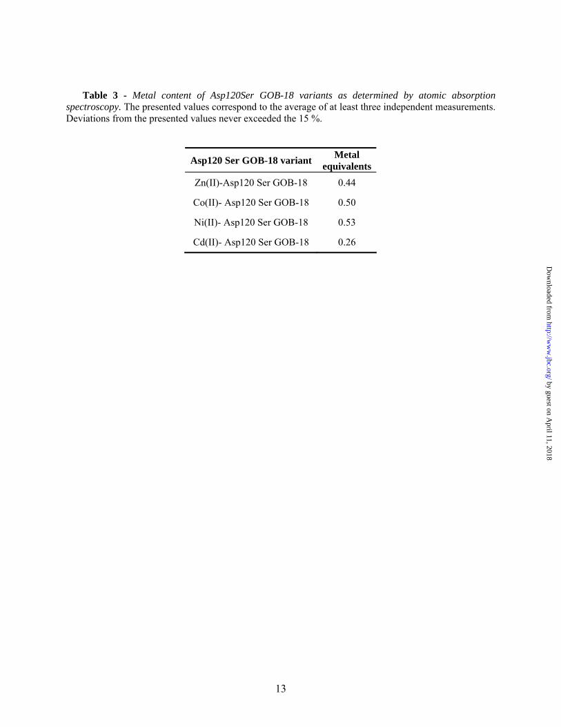
Table 3 - Metal content of Asp120Ser GOB-18 variants as determined by atomic absorption spectroscopy. The presented values correspond to the average of at least three independent measurements. Deviations from the presented values never exceeded the 15 %.
Asp120 Ser GOB-18 variant Metal equivalents
Zn(II)-Asp120 Ser GOB-18 0.44
Co(II)- Asp120 Ser GOB-18 0.50
Ni(II)- Asp120 Ser GOB-18 0.53
Cd(II)- Asp120 Ser GOB-18 0.26
13
by guest on April 11, 2018
http://ww
w.jbc.org/
Dow
nloaded from
FIGURES
Figure 1
14
by guest on April 11, 2018
http://ww
w.jbc.org/
Dow
nloaded from
Maria Natalia Lisa, Lars Hemmingsen and Alejandro J. Vila-lactamase GOBβCatalytic role of the metal ION in the metallo-
published online December 10, 2009J. Biol. Chem.
10.1074/jbc.M109.063743Access the most updated version of this article at doi:
Alerts:
When a correction for this article is posted•
When this article is cited•
to choose from all of JBC's e-mail alertsClick here
Supplemental material:
http://www.jbc.org/content/suppl/2009/12/10/M109.063743.DC1
by guest on April 11, 2018
http://ww
w.jbc.org/
Dow
nloaded from