Cardiac contractility
34
Kiran Goushika
-
Upload
kiran-goushika -
Category
Education
-
view
487 -
download
2
Transcript of Cardiac contractility
Kiran Goushika
Cardiac Electrophysiology
Cardiac Contractility
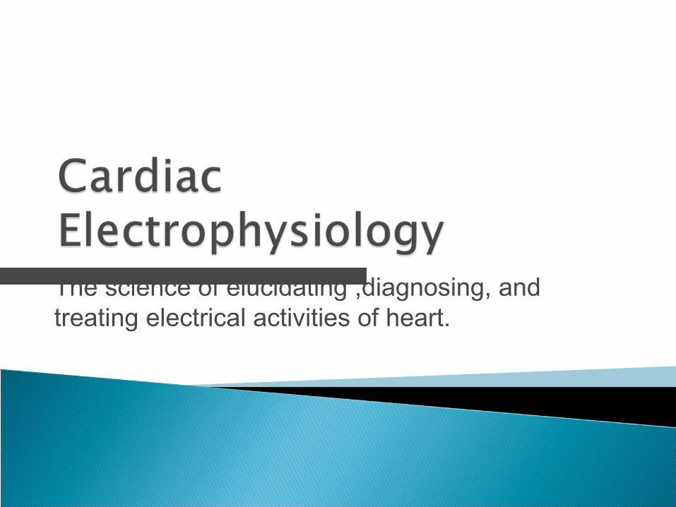
The science of elucidating ,diagnosing, and treating electrical activities of heart.

Actions potentials◦ SA node◦ Cardiac muscle
(atria, ventricles & Purkinje fibers)
Channels◦ Ca2+ channel◦ β-adrenergic receptor◦ Na+/K+-ATPase
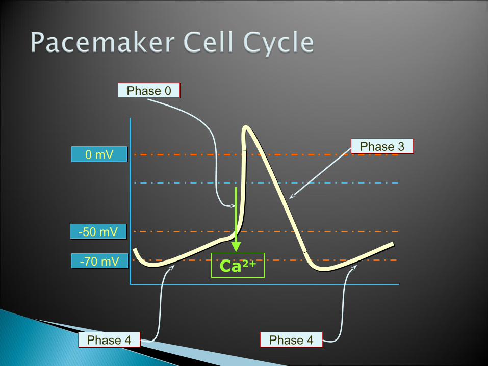
0 mV0 mV
-70 mV-70 mV
-50 mV-50 mV
Phase 0Phase 0
Phase 3Phase 3
Phase 4Phase 4 Phase 4Phase 4
Ca2+
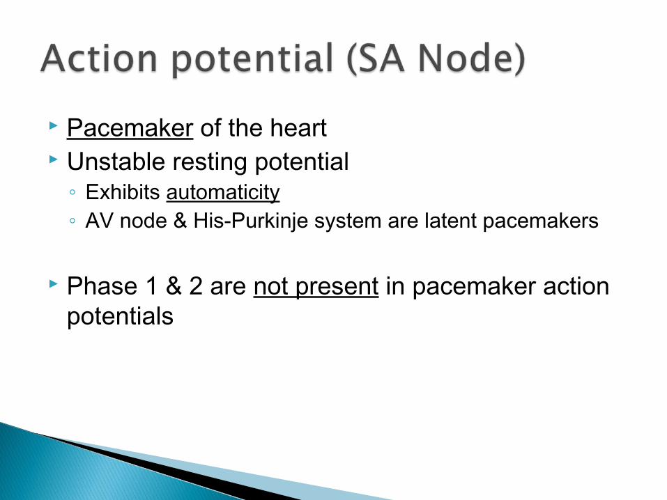
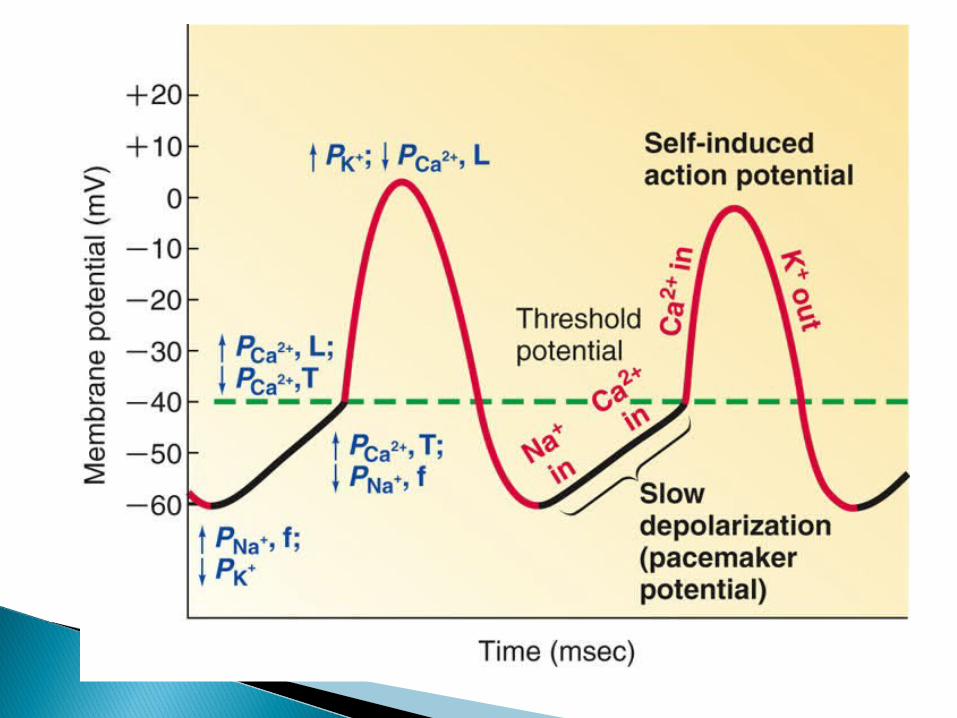
Pacemaker of the heart Unstable resting potential◦ Exhibits automaticity◦ AV node & His-Purkinje system are latent pacemakers
Phase 1 & 2 are not present in pacemaker action potentials
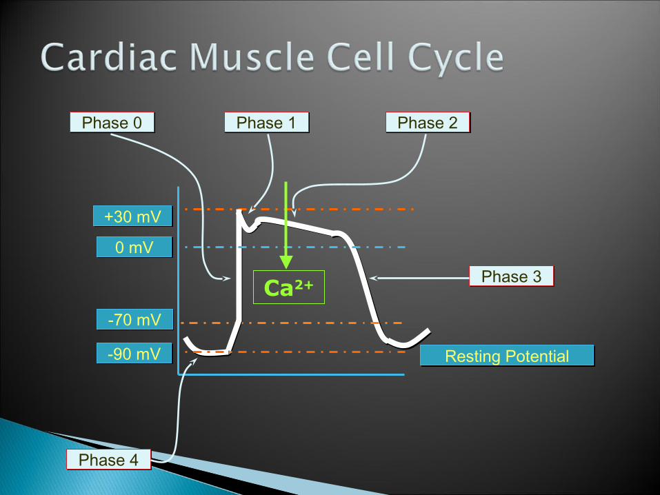
Resting PotentialResting Potential-90 mV-90 mV
0 mV0 mV
+30 mV+30 mV
-70 mV-70 mV
Phase 0Phase 0 Phase 1Phase 1 Phase 2Phase 2
Phase 3Phase 3
Phase 4Phase 4
Ca2+
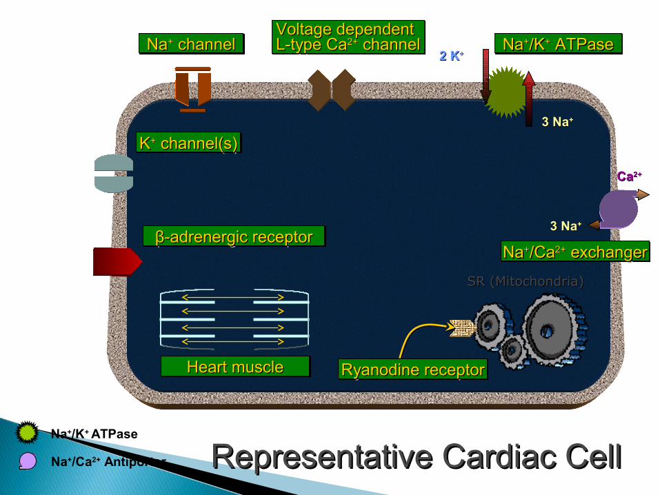
Na+/K+ ATPase
3 Na3 Na++
2 K2 K++
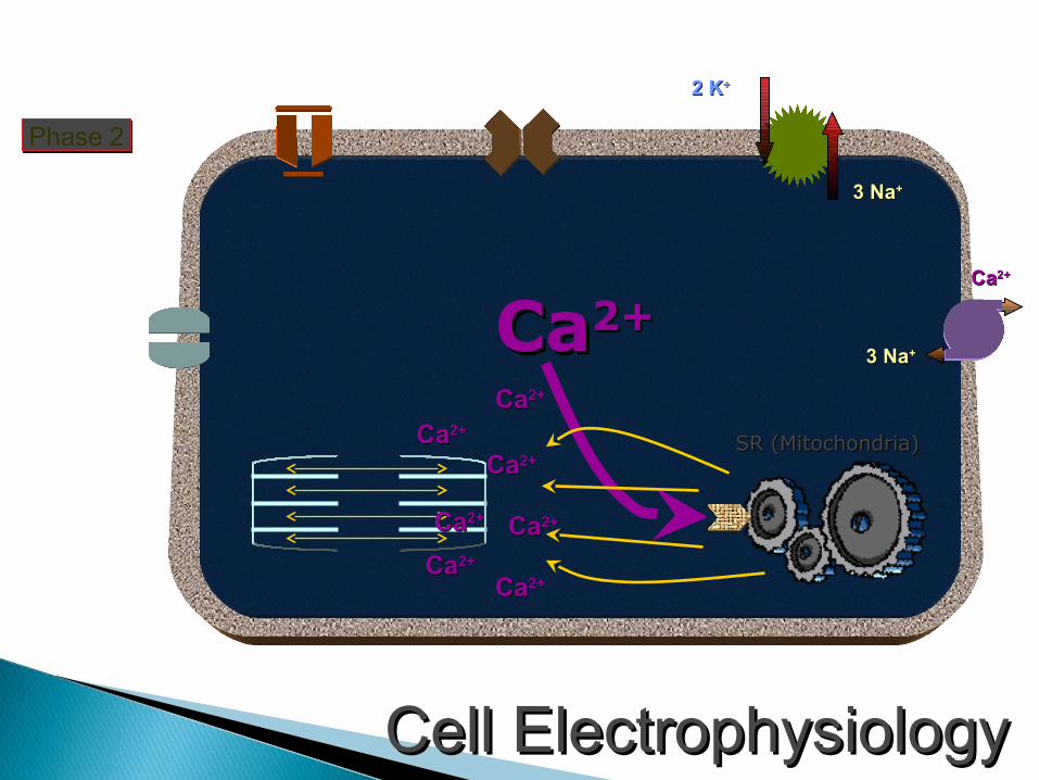
Representative Cardiac CellRepresentative Cardiac Cell
NaNa++ channel channelNaNa++ channel channelVoltage dependentVoltage dependentL-typeL-type CaCa2+2+ channel channelVoltage dependentVoltage dependentL-typeL-type CaCa2+2+ channel channel NaNa++/K/K++ ATPase ATPaseNaNa++/K/K++ ATPase ATPase
NaNa++/Ca/Ca2+2+ exchanger exchangerNaNa++/Ca/Ca2+2+ exchanger exchanger
SR (Mitochondria)SR (Mitochondria)
Heart muscleHeart muscleHeart muscleHeart muscle
KK++ channel(s) channel(s)KK++ channel(s) channel(s)
Na+/Ca2+ Antiporter
Ryanodine receptorRyanodine receptorRyanodine receptorRyanodine receptor
3 Na3 Na++
CaCa2+2+
ββ-adrenergic receptor-adrenergic receptorββ-adrenergic receptor-adrenergic receptor
The intrinsic ability of heart to contract
Intrinsic ability of cardiac muscle
Also called ‘inotropism’ or ‘inotropy’
Related to the intracellular [Ca2+]
Inotropic agents◦ positive: increase contractility◦ negative: decrease contractility
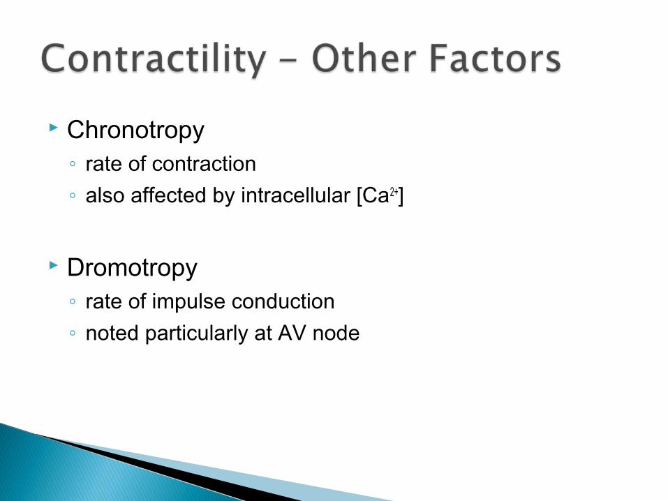
Chronotropy◦ rate of contraction◦ also affected by intracellular [Ca2+]
Dromotropy◦ rate of impulse conduction◦ noted particularly at AV node

Increased intracellular [Ca2+]◦ increased heart rate◦ cardiac glycosides (e.g. digoxin)
Stimulation of β1-adrenergic receptor◦ sympathomimetic agents◦ catecholamines
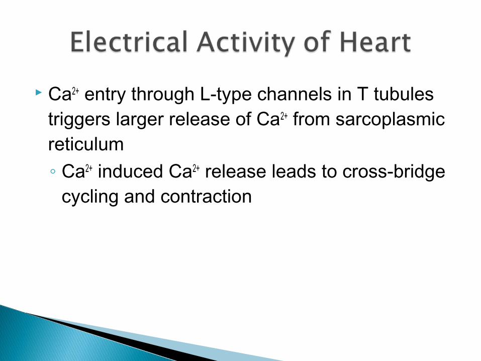
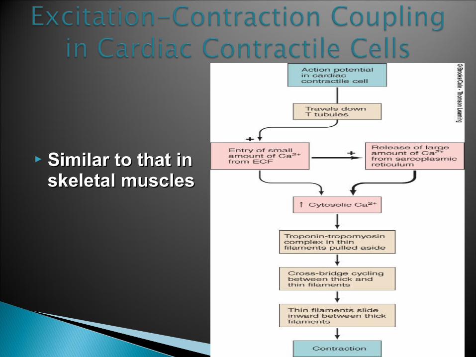
Ca2+ entry through L-type channels in T tubules triggers larger release of Ca2+ from sarcoplasmic reticulum◦ Ca2+ induced Ca2+ release leads to cross-bridge
cycling and contraction
3 Na3 Na++
2 K2 K++
Cell ElectrophysiologyCell Electrophysiology
SR (Mitochondria)SR (Mitochondria)
CaCa2+2+
Phase 2Phase 2
CaCa2+2+
CaCa2+2+
CaCa2+2+
CaCa2+2+
CaCa2+2+
CaCa2+2+
CaCa2+2+
3 Na3 Na++
CaCa2+2+
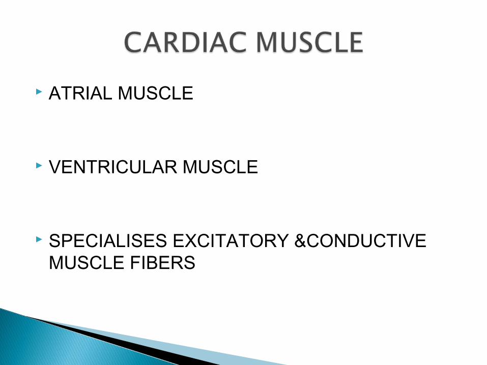
ATRIAL MUSCLE
VENTRICULAR MUSCLE
SPECIALISES EXCITATORY &CONDUCTIVE MUSCLE FIBERS
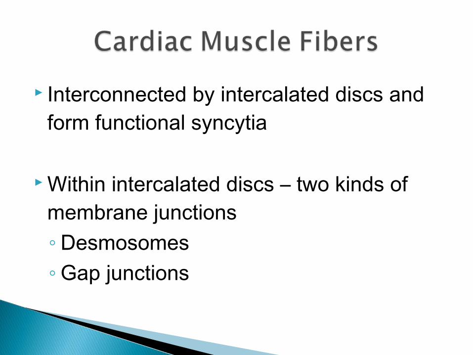
Interconnected by intercalated discs and form functional syncytia
Within intercalated discs – two kinds of membrane junctions◦ Desmosomes◦ Gap junctions
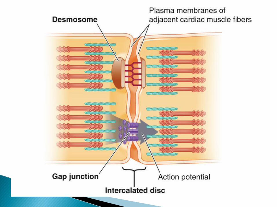
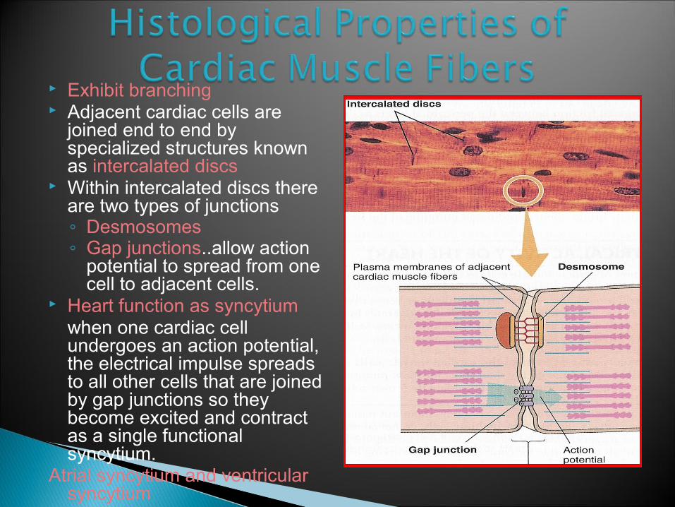
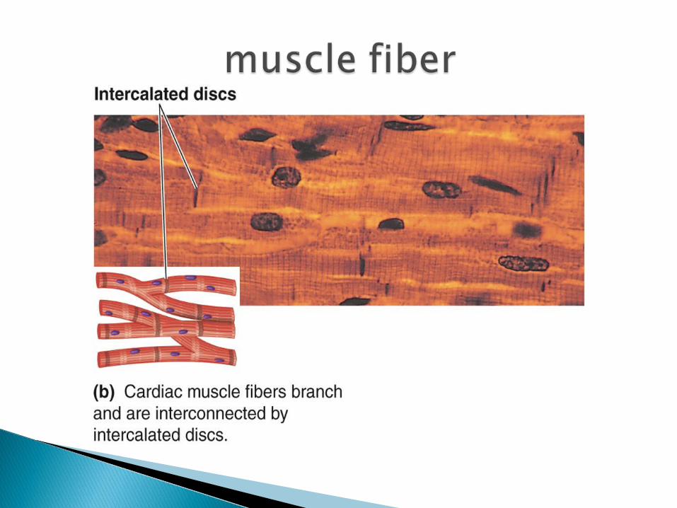
Exhibit branching Adjacent cardiac cells are
joined end to end by specialized structures known as intercalated discs
Within intercalated discs there are two types of junctions◦ Desmosomes◦ Gap junctions..allow action
potential to spread from one cell to adjacent cells.
Heart function as syncytiumwhen one cardiac cell undergoes an action potential, the electrical impulse spreads to all other cells that are joined by gap junctions so they become excited and contract as a single functional syncytium.
Atrial syncytium and ventricular syncytium
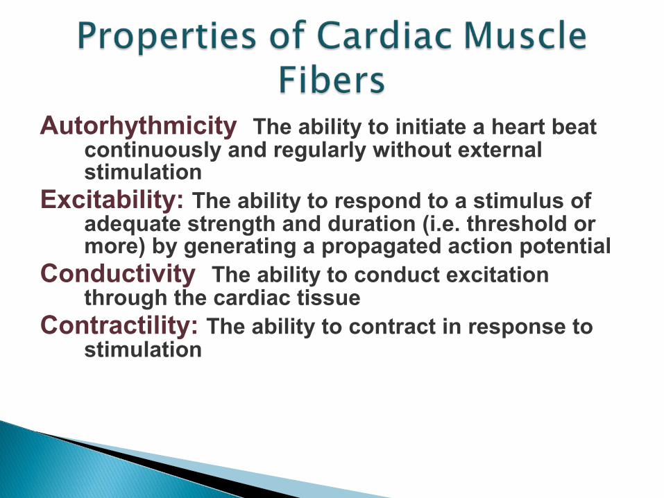
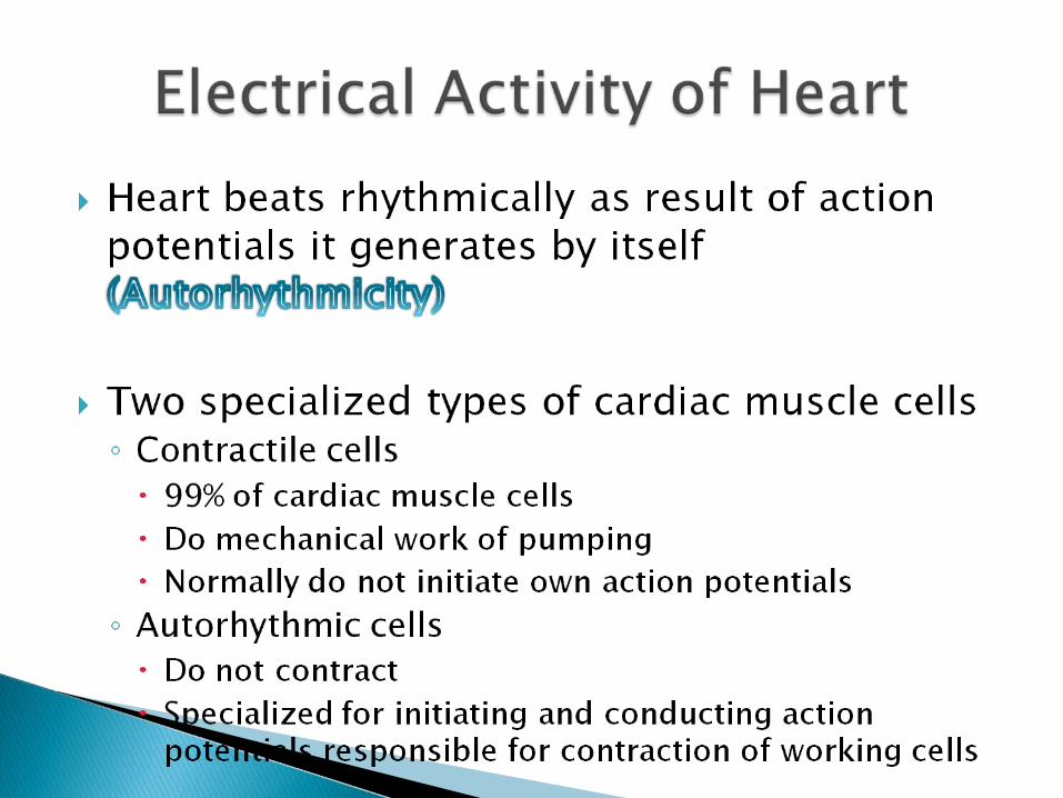
Autorhythmicity: The ability to initiate a heart beat continuously and regularly without external stimulation
Excitability: The ability to respond to a stimulus of adequate strength and duration (i.e. threshold or more) by generating a propagated action potential
Conductivity: The ability to conduct excitation through the cardiac tissue
Contractility: The ability to contract in response to stimulation
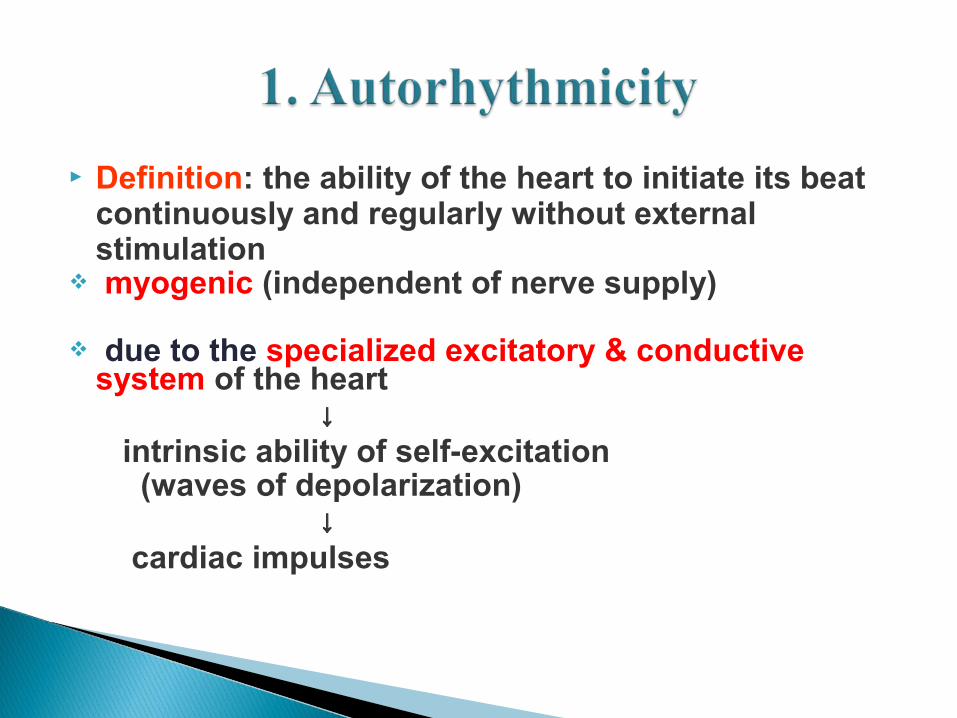
Definition: the ability of the heart to initiate its beat continuously and regularly without external stimulation
myogenic (independent of nerve supply)
due to the specialized excitatory & conductive system of the heart
↓ intrinsic ability of self-excitation (waves of depolarization) ↓ cardiac impulses
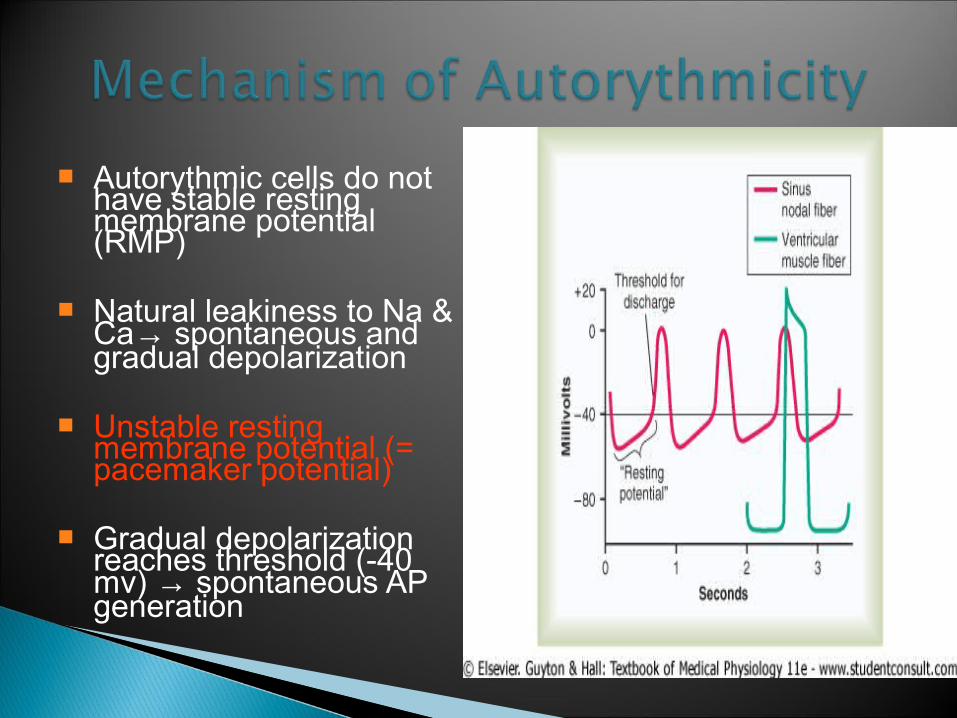
Autorythmic cells do not have stable resting membrane potential (RMP)
Natural leakiness to Na & Ca→ spontaneous and gradual depolarization
Unstable resting membrane potential (= pacemaker potential)
Gradual depolarization reaches threshold (-40 mv) → spontaneous AP generation
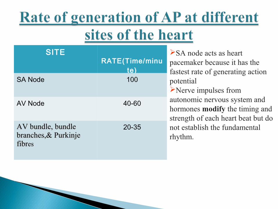
SITE RATE(Time/minu
te)SA Node 100
AV Node 40-60
AV bundle, bundle AV bundle, bundle branches,& Purkinje branches,& Purkinje fibrfibreses
20-35
SA node acts as heart pacemaker because it has the fastest rate of generating action potentialNerve impulses from autonomic nervous system and hormones modify the timing and strength of each heart beat but do not establish the fundamental rhythm.
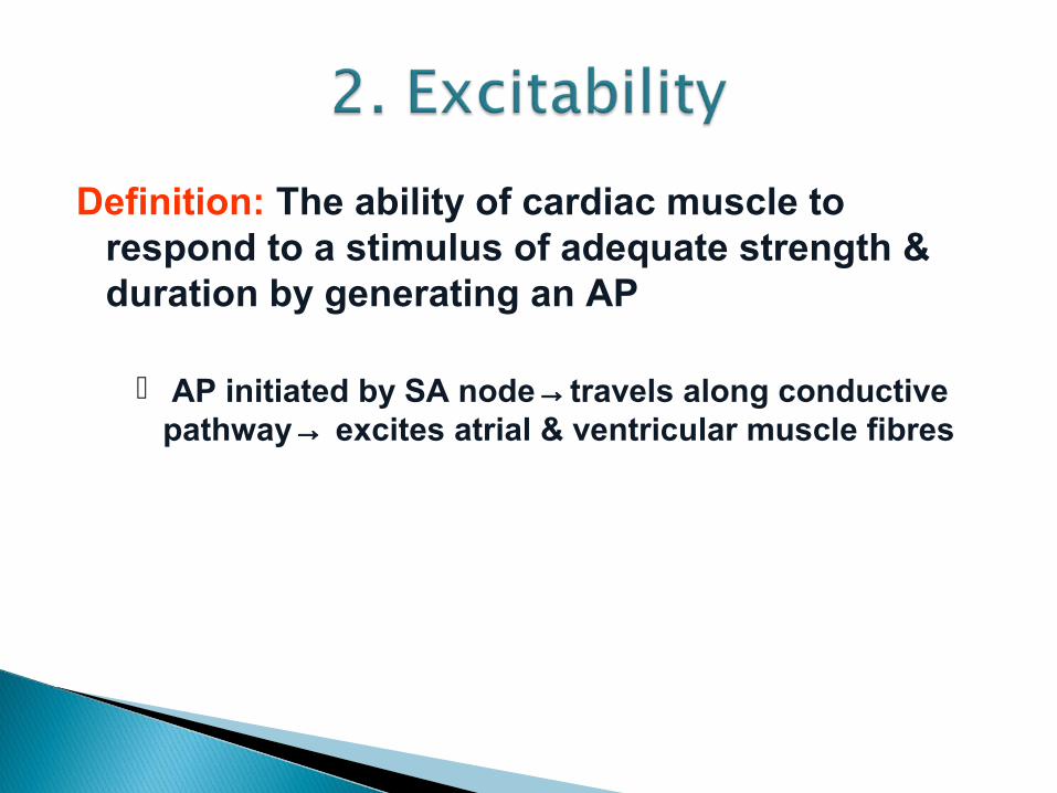
Definition: The ability of cardiac muscle to respond to a stimulus of adequate strength & duration by generating an AP
AP initiated by SA node→travels along conductive pathway→ excites atrial & ventricular muscle fibres
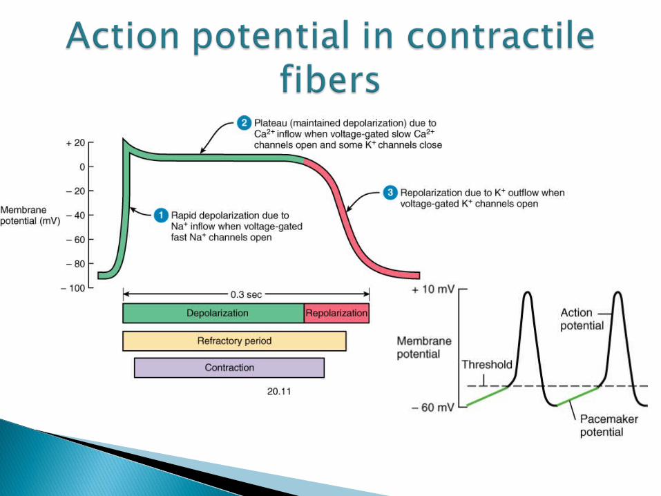
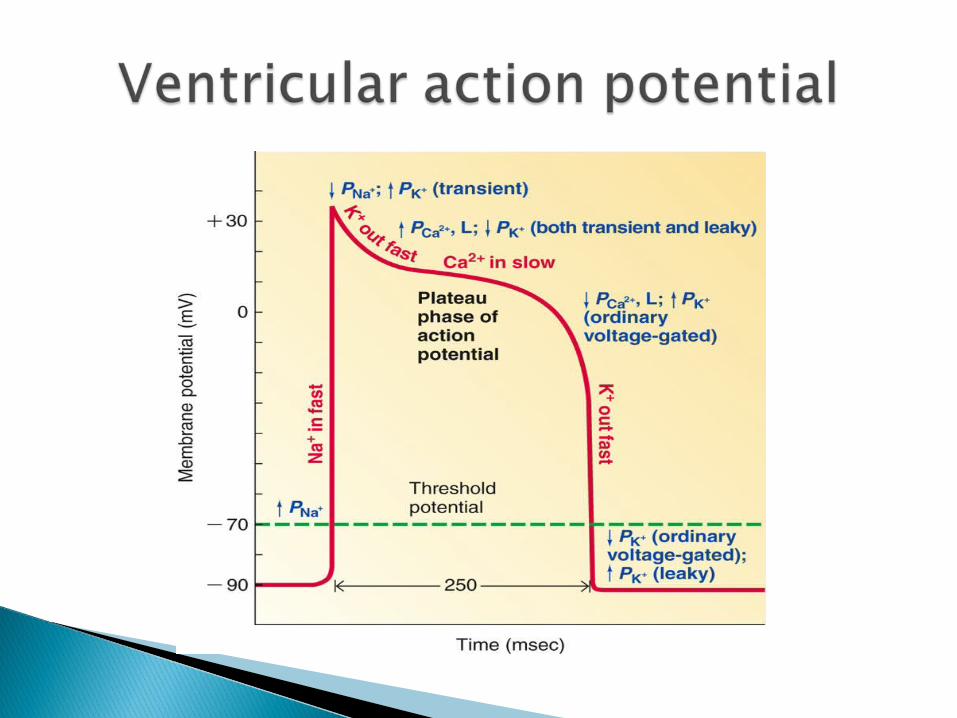
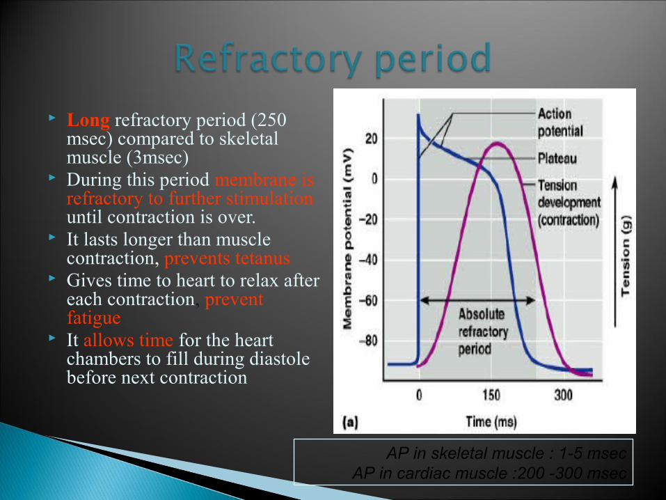
Long refractory period (250 msec) compared to skeletal muscle (3msec)
During this period membrane is refractory to further stimulation until contraction is over.
It lasts longer than muscle contraction, prevents tetanus
Gives time to heart to relax after each contraction, prevent fatigue
It allows time for the heart chambers to fill during diastole before next contraction
AP in skeletal muscle : 1-5 msecAP in cardiac muscle :200 -300 msec
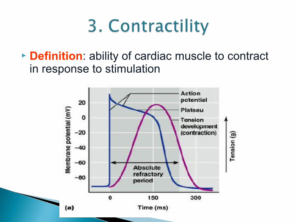
Definition: ability of cardiac muscle to contract in response to stimulation
Similar to that in Similar to that in skeletal musclesskeletal muscles
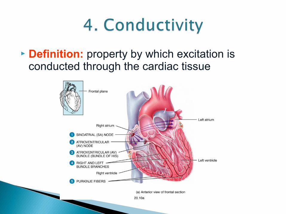
Definition: property by which excitation is conducted through the cardiac tissue
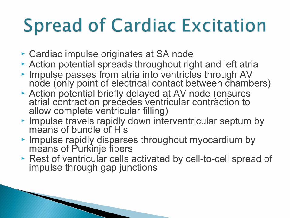
Cardiac impulse originates at SA node Action potential spreads throughout right and left atria Impulse passes from atria into ventricles through AV
node (only point of electrical contact between chambers) Action potential briefly delayed at AV node (ensures
atrial contraction precedes ventricular contraction to allow complete ventricular filling)
Impulse travels rapidly down interventricular septum by means of bundle of His
Impulse rapidly disperses throughout myocardium by means of Purkinje fibers
Rest of ventricular cells activated by cell-to-cell spread of impulse through gap junctions

PROFESSOR: Maka


![Ca2+ Entry (SOCE) Contributes to Muscle Contractility in ... · physiological role in young and aged skeletal muscle. We found that reagents that prevent [Ca2+] o entry reduce contractile](https://static.fdocument.org/doc/165x107/5fbbf98d4e86af3f2a7e3a76/ca2-entry-soce-contributes-to-muscle-contractility-in-physiological-role.jpg)