Bulletin - Sigma-Aldrich
6
Cor.At CL-i Cardiomyocytes (Mouse iPS cell derived) Catalog Numbers AXIO0002 and AXIO0006 TECHNICAL BULLETIN Product Description Cor.At cells are cardiomyocytes derived from transgenic mouse induced pluripotent stem (miPS). These cells have puromycin resistance controlled by the promoter of Myh6, which encodes α-myosin heavy chain (αMHC). 1 Cor.At cells are produced by in vitro differentiation of mouse induced pluripotent stem (iPS) cells and puromycin selection of cardiomyocytes. Similar to primary cells, they have limited proliferative capacity, demonstrated by BrdU incorporation assays. These cells express cardiac-specific connexin-43, which demonstrates the potential for electric coupling. Patch clamp analyses, as well as multi-electrode array recordings, demonstrate normal electrophysiological properties. Further, transplantation of stem cell-derived cardiomyocytes into infarcted heart resulted in teratoma-free engraftment. 2 These cardiomyocytes have many advantages over primary cells and cell lines. These cells are highly standardized and are 100% pure when cultured with puromycin. The culture and maintenance of the cells require minimal laboratory time when compared to the culture and maintenance of rat neonatal cells. Reproducible results can be expected for every assay. Features of the cells: • Standardized from lot-to-lot • 100% pure with fully functional cardiomyocyte phenotype • Frozen, stored, and thawed with complete recovery of functionality • Part of an entire in vitro based system • Absolutely no fibroblast contamination • Express all relevant cardiac ion channels (K + , Ca 2+ , and Na + ) and show predictive response to cardiotoxic compounds when compared to clinical data Cor.At cells can be used for: • Safety pharmacology • Toxicological analysis of compounds (cardiac-specific cytotoxicity) • Screening for pharmacological effects • Drug development • Molecular and cell biology • Research & development • Electrophysiology, patch clamp, and MEAs • Tissue engineering and transplantation Component Cor.At CL-i Cardiomyocytes 1 vial (Mouse iPS cell derived) 1M (Cor.At CL-iC-1M) 1 × 10 6 mouse iPS cell derived cells Catalog Number AXIO0002 Or (Mouse iPS cell derived) 5M (Cor.At CL-iC-5M) 5 × 10 6 mouse iPS cell derived cells Catalog Number AXIO0006 Reagents and Equipment Required but Not Provided. • Cor.At Culture Medium, Catalog Number AXIO0076 • Puromycin solution, Catalog Number AXIO0078 • PBS without Ca 2+ and Mg 2+ • Disposable Neubauer Hemacytometer • 0.4% Trypan Blue solution, Catalog Number 93595 • Biological safety cabinet certified for Level I handling of biological materials • Incubator with humidity and gas control to maintain 37 °C and 95% humidity in an atmosphere of 5% CO 2 in air • 37 °C water bath • Sterile 50 mL polypropylene (PP) tubes • Centrifuge with rotor for 50 mL PP tubes (not required for 96 well plate format) • 8 channel or 12 channel micropipette • Sterile pipette tips
Transcript of Bulletin - Sigma-Aldrich
Cor.At CL-i Cardiomyocytes (Mouse iPS cell derived)
Catalog Numbers AXIO0002 and AXIO0006
TECHNICAL BULLETIN
Product DescriptionCor.At cells are cardiomyocytes derived from transgenic mouse induced pluripotent stem (miPS). These cells have puromycin resistance controlled by the promoter of Myh6, which encodes α-myosin heavy chain (αMHC).1
Cor.At cells are produced by in vitro differentiation of mouse induced pluripotent stem (iPS) cells and puromycin selection of cardiomyocytes. Similar to primary cells, they have limited proliferative capacity, demonstrated by BrdU incorporation assays. These cells express cardiac-specific connexin-43, which demonstrates the potential for electric coupling. Patch clamp analyses, as well as multi-electrode array recordings, demonstrate normal electrophysiological properties. Further, transplantation of stem cell-derived cardiomyocytes into infarcted heart resulted in teratoma-free engraftment.2
These cardiomyocytes have many advantages over primary cells and cell lines. These cells are highly standardized and are 100% pure when cultured with puromycin. The culture and maintenance of the cells require minimal laboratory time when compared to the culture and maintenance of rat neonatal cells. Reproducible results can be expected for every assay.
Features of the cells:• Standardized from lot-to-lot• 100% pure with fully functional cardiomyocyte
phenotype• Frozen, stored, and thawed with complete recovery
of functionality• Part of an entire in vitro based system• Absolutely no fibroblast contamination • Express all relevant cardiac ion channels (K+, Ca2+,
and Na+) and show predictive response to cardiotoxic compounds when compared to clinical data
Cor.At cells can be used for:• Safety pharmacology• Toxicological analysis of compounds
(cardiac-specific cytotoxicity)• Screening for pharmacological effects• Drug development• Molecular and cell biology• Research & development• Electrophysiology, patch clamp, and MEAs• Tissue engineering and transplantation
ComponentCor.At CL-i Cardiomyocytes 1 vial
(Mouse iPS cell derived) 1M(Cor.At CL-iC-1M)1 × 10⁶ mouse iPS cell derived cellsCatalog Number AXIO0002
Or
(Mouse iPS cell derived) 5M(Cor.At CL-iC-5M)5 × 10⁶ mouse iPS cell derived cellsCatalog Number AXIO0006
Reagents and Equipment Required but Not Provided.• Cor.At Culture Medium, Catalog Number AXIO0076• Puromycin solution, Catalog Number AXIO0078• PBS without Ca2+ and Mg2+
• Disposable Neubauer Hemacytometer• 0.4% Trypan Blue solution, Catalog Number 93595• Biological safety cabinet certified for Level I
handling of biological materials• Incubator with humidity and gas control to maintain
37 °C and 95% humidity in an atmosphere of 5% CO2 in air
• 37 °C water bath• Sterile 50 mL polypropylene (PP) tubes• Centrifuge with rotor for 50 mL PP tubes (not
required for 96 well plate format)• 8 channel or 12 channel micropipette• Sterile pipette tips
2
• BD BioCoat Fibronectin-coated dishes (Catalog Number 354457 for 35 mm dishes or Catalog Number 354403 for 60 mm dishes) not required for 96 well plate format
• Fibronectin from bovine plasma (1 mg/mL solution, Sigma Catalog Number F1141) for coating alternate sized dishes and plates
Precautions and Disclaimer This product is for R&D use only, not for drug, household, or other uses. Please consult the Material Safety Data Sheet for information regarding hazards and safe handling practices.
Storage/Stability AXIO0002 and AXIO0006 are shipped in liquid nitrogen dry shippers and stored at –196 °C.
Procedure Cor.At cells are genetically modified mouse cells and should be handled according to local directives (Typically Biosafety level 1).
The cardiomyocytes should be cultured using sterile cell culture techniques and good laboratory practices.
Cells can be inactivated by autoclaving at 121 °C for 20 minutes.
Day 0 – Media Preparation1. Thaw the Cor.At Culture Medium (Catalog Number
AXIO0076) and the Puromycin solution (Catalog Number AXIO0078) at 4 °C overnight or at room temperature (15–25 °C). Notes: Do not attempt to thaw the medium at 37 °Cbecause precipitation of the proteins in the medium may occur.
The culture medium contains light sensitive compounds and should be kept in the dark while thawing.
2. Prepare Culture Medium containing puromycin –Pipette 50 mL of the thawed Culture Medium (Catalog Number AXIO0076) into a sterile 50 mL PP tube and add 50 µL of the Puromycin solution(Catalog Number AXIO0078). Mix well and store at 4 °C and warm to 37 °C before use. Note: This medium is used for the medium changeat 24 hours of culture.
3. Pipette another 8 mL of thawed Culture Medium into a second sterile 50 mL PP tube. Do not add puromycin to this tube. Note: This medium is used to wash the cells once the cells are thawed.
4. Store the remaining Culture Medium (Catalog Number AXIO0076) without puromycin at 4 °C. The remaining medium is used for medium changes after 48 hours of culture and can be kept at 4 °Cfor up to three weeks.Note: It is highly recommended to note the date when the Culture Medium (Catalog Number AXIO0076) bottle was opened to ensure the medium is not used if the bottle has been opened for more than three weeks.
Day 0 – Fibronectin-Coated DishesBD BioCoat Fibronectin-coated dishes (Catalog Number 354457 for 35 mm dishes or Catalog Number354403 for 60 mm dishes) are recommended for the culture of the cardiomyocytes. Prior to plating the cells onto the BD BioCoat fibronectin-coated dishes, hydrate the dish by adding the appropriate volume (see Table 1) of the Culture Medium (Catalog Number AXIO0076) without puromycin into the dish and place the dish into the 37 °C incubator for 1 hour.
For coating your own 35 mm or 60 mm dishes, or alternate sized dishes and plates (96 well or other multiwell plates, use of Fibronectin from bovine plasma (1 mg/mL solution, Sigma Catalog Number F1141) is recommended. 1. Dilute the 1 mg/mL fibronectin solution 1:100 with
PBS without Ca2+ and Mg2+.
2. Add the appropriate volume of fibronectin solution into the cultureware according to Table 1.
3. Place the freshly coated cultureware into the 37 °Cincubator for at least 3 hours. If the dishes are not immediately used, they can be stored for up to 3 days at 4 °C.
3
4. Remove the remaining fibronectin solution immediately before seeding the cardiomyocytes using a slow-running suction pump or a multichannel pipette. Notes: Do not allow the surface of the cultureware to become dry after fibronectin coating. Do notremove the fibronectin solution until ready to plate the cells.
Fibronectin is highly susceptible to shear stress. Avoid vigorous pipetting and do not vortex the fibronectin solution. For additional handling instructions, consult the Product Display page on the Sigma website.
Table 1.Plate Parameters
Dish/Multiwell Plate Surface Area (cm2)
Volume of Fibronectin Solution for Coating
(µL)
Cardiomyocytes per Well (× 106)*
Volume (Medium Change or Cell
Suspension, mL)35 mm dish 11.78 750 1 360 mm dish 21.29 1,500 2 5
1 well of a 6 well plate 9.62 750 1 3–41 well of a 12 well plate 3.80 300 0.35 21 well of a 24 well plate 2.00 200 0.2 11 well of a 48 well plate 0.75 100 0.075 0.51 well of a 96 well plate 0.32 50 0.02 0.2–0.25
* ∼1 × 10⁵ viable cells per cm2 of the fibronectin-coated cultureware
Day 0 – Washing & Counting Cells1. Add 50 µL of Trypan Blue solution to 40 µL of PBS
without Ca2+ and Mg2+ in a microcentrifuge tube.
2. Prepare the hemacytometer by cleaning the chambers and coverslip with isopropanol. Wipe the hemacytometer dry using lint-free tissue.
3. Remove the vial(s) containing the clearcardiomyocytes from the shipment box or storage. Immediately place the vial(s) in a 37 °C water bath for two minutes. Remove the vial(s) from the water bath and wipe with 70% isopropanol or ethanol to sterilize.
4. Immediately pipette the cell suspension into the second 50 mL tube containing 8 mL of thawed Culture Medium without puromycin to wash the cells.
5. Use 1 mL of thawed Culture Medium without puromycin to rinse the vial, ensuring any cells remaining in the vial are also recovered. Add the 1 mL of cells to the 50 mL tube (there should now be ∼9 mL of cell suspension in the tube).
6. Centrifuge cells at 200 × g for 5 minutes at room temperature.
7. Aspirate the supernatant, leaving the cell pellet intact. In preparation for cell counting, resuspend the cells in 500 µL of Culture Medium containing puromycin.
8. Pipette 10 µL of the cell suspension into the prepared Trypan Blue/PBS solution, mix well, and incubate for 5 minutes at 37 °C (i.e., place tube into the incubator). This is a 10-fold dilution of the cells.
9. After 5 minutes, fill the Neubauer hemacytometer with the cell suspension (step 8), ensuring the chambers are not underfilled or overfilled (use 10 µL of cells/Trypan Blue/PBS suspension). Count both the clear, refractile cells and the blue colored cells in the 4 large outer quadrants. This is the total number of cells (N) in the four large, outer quadrants (see Figure 1).
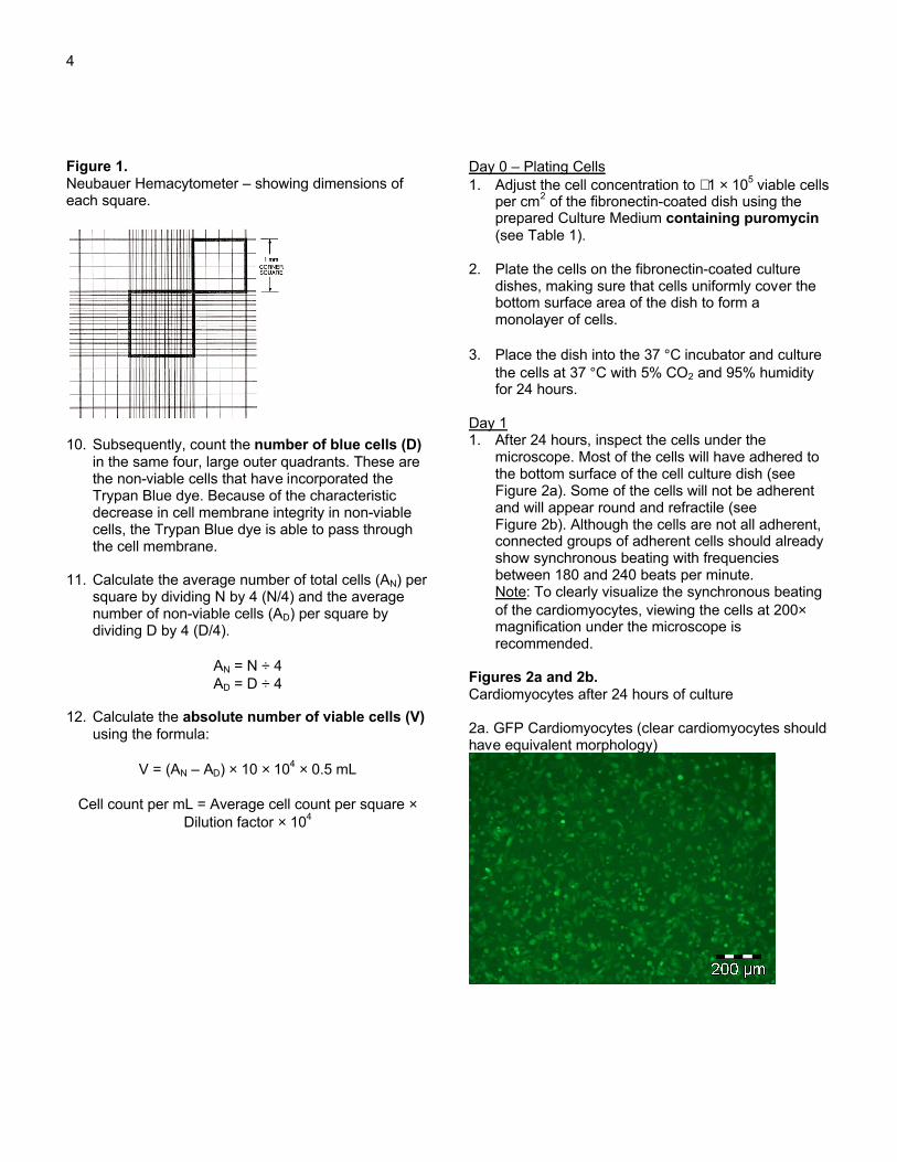
4
Figure 1.Neubauer Hemacytometer – showing dimensions of each square.
10. Subsequently, count the number of blue cells (D) in the same four, large outer quadrants. These are the non-viable cells that have incorporated the Trypan Blue dye. Because of the characteristic decrease in cell membrane integrity in non-viable cells, the Trypan Blue dye is able to pass through the cell membrane.
11. Calculate the average number of total cells (AN) per square by dividing N by 4 (N/4) and the average number of non-viable cells (AD) per square by dividing D by 4 (D/4).
AN = N ÷ 4AD = D ÷ 4
12. Calculate the absolute number of viable cells (V)using the formula:
V = (AN – AD) × 10 × 104 × 0.5 mL
Cell count per mL = Average cell count per square ×Dilution factor × 104
Day 0 – Plating Cells1. Adjust the cell concentration to ∼1 × 10⁵ viable cells
per cm2 of the fibronectin-coated dish using the prepared Culture Medium containing puromycin(see Table 1).
2. Plate the cells on the fibronectin-coated culture dishes, making sure that cells uniformly cover the bottom surface area of the dish to form a monolayer of cells.
3. Place the dish into the 37 °C incubator and culture the cells at 37 °C with 5% CO2 and 95% humidity for 24 hours.
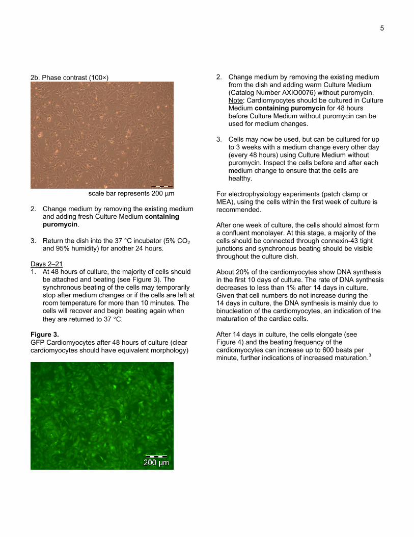
Day 11. After 24 hours, inspect the cells under the
microscope. Most of the cells will have adhered to the bottom surface of the cell culture dish (see Figure 2a). Some of the cells will not be adherent and will appear round and refractile (see Figure 2b). Although the cells are not all adherent, connected groups of adherent cells should already show synchronous beating with frequencies between 180 and 240 beats per minute.Note: To clearly visualize the synchronous beating of the cardiomyocytes, viewing the cells at 200×magnification under the microscope is recommended.
Figures 2a and 2b.Cardiomyocytes after 24 hours of culture
2a. GFP Cardiomyocytes (clear cardiomyocytes should have equivalent morphology)
5
2b. Phase contrast (100×)
scale bar represents 200 µm
2. Change medium by removing the existing medium and adding fresh Culture Medium containing puromycin.
3. Return the dish into the 37 °C incubator (5% CO2and 95% humidity) for another 24 hours.
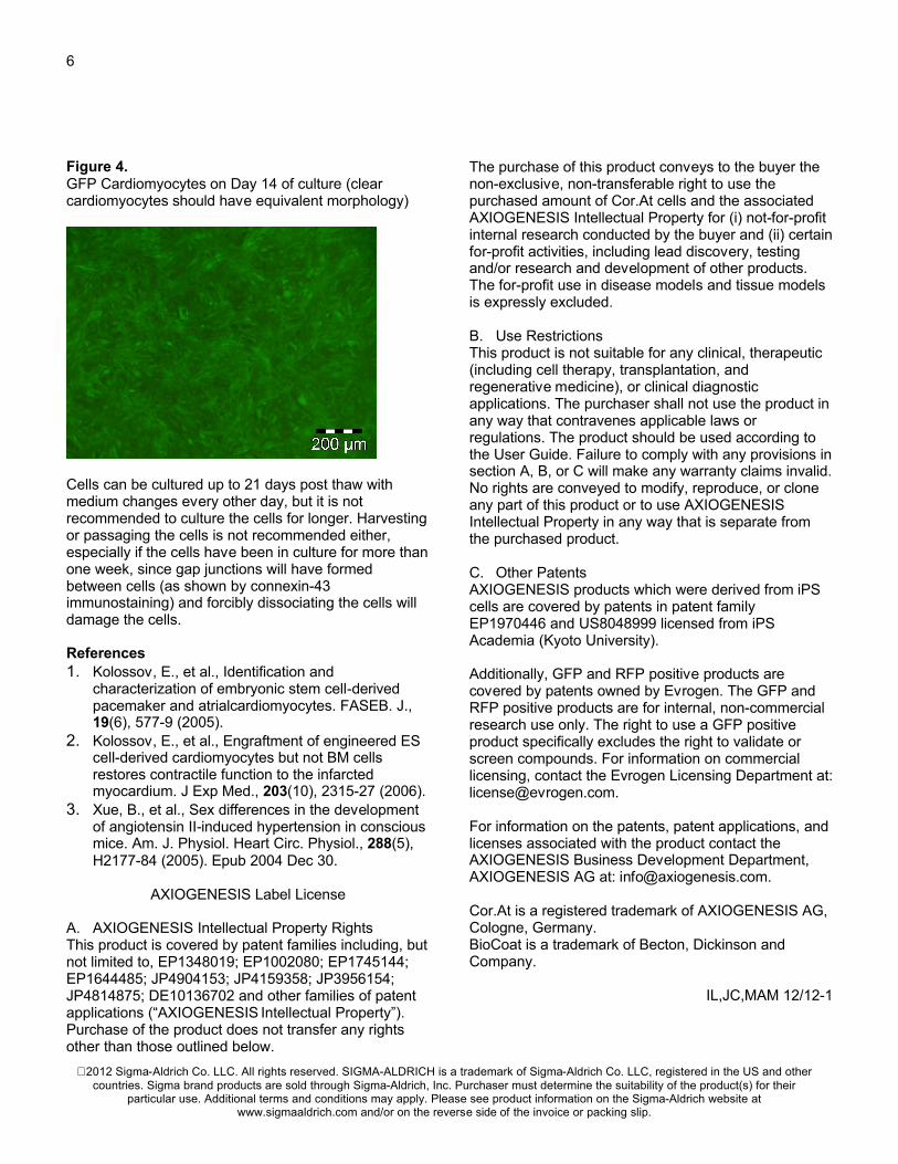
Days 2–211. At 48 hours of culture, the majority of cells should
be attached and beating (see Figure 3). The synchronous beating of the cells may temporarily stop after medium changes or if the cells are left at room temperature for more than 10 minutes. The cells will recover and begin beating again when they are returned to 37 °C.
Figure 3.GFP Cardiomyocytes after 48 hours of culture (clear cardiomyocytes should have equivalent morphology)
2. Change medium by removing the existing medium from the dish and adding warm Culture Medium (Catalog Number AXIO0076) without puromycin.Note: Cardiomyocytes should be cultured in Culture Medium containing puromycin for 48 hours before Culture Medium without puromycin can be used for medium changes.
3. Cells may now be used, but can be cultured for up to 3 weeks with a medium change every other day (every 48 hours) using Culture Medium without puromycin. Inspect the cells before and after each medium change to ensure that the cells are healthy.
For electrophysiology experiments (patch clamp or MEA), using the cells within the first week of culture is recommended.
After one week of culture, the cells should almost form a confluent monolayer. At this stage, a majority of the cells should be connected through connexin-43 tight junctions and synchronous beating should be visible throughout the culture dish.
About 20% of the cardiomyocytes show DNA synthesis in the first 10 days of culture. The rate of DNA synthesis decreases to less than 1% after 14 days in culture. Given that cell numbers do not increase during the 14 days in culture, the DNA synthesis is mainly due to binucleation of the cardiomyocytes, an indication of the maturation of the cardiac cells.
After 14 days in culture, the cells elongate (see Figure 4) and the beating frequency of the cardiomyocytes can increase up to 600 beats per minute, further indications of increased maturation.3
6
Figure 4.GFP Cardiomyocytes on Day 14 of culture (clear cardiomyocytes should have equivalent morphology)
Cells can be cultured up to 21 days post thaw with medium changes every other day, but it is not recommended to culture the cells for longer. Harvesting or passaging the cells is not recommended either, especially if the cells have been in culture for more than one week, since gap junctions will have formed between cells (as shown by connexin-43 immunostaining) and forcibly dissociating the cells will damage the cells.
References1. Kolossov, E., et al., Identification and
characterization of embryonic stem cell-derived pacemaker and atrialcardiomyocytes. FASEB. J., 19(6), 577-9 (2005).
2. Kolossov, E., et al., Engraftment of engineered ES cell-derived cardiomyocytes but not BM cells restores contractile function to the infarcted myocardium. J Exp Med., 203(10), 2315-27 (2006).
3. Xue, B., et al., Sex differences in the development of angiotensin II-induced hypertension in conscious mice. Am. J. Physiol. Heart Circ. Physiol., 288(5), H2177-84 (2005). Epub 2004 Dec 30.
AXIOGENESIS Label License
A. AXIOGENESIS Intellectual Property RightsThis product is covered by patent families including, but not limited to, EP1348019; EP1002080; EP1745144; EP1644485; JP4904153; JP4159358; JP3956154; JP4814875; DE10136702 and other families of patent applications (“AXIOGENESIS Intellectual Property”). Purchase of the product does not transfer any rights other than those outlined below.
The purchase of this product conveys to the buyer the non-exclusive, non-transferable right to use the purchased amount of Cor.At cells and the associated AXIOGENESIS Intellectual Property for (i) not-for-profit internal research conducted by the buyer and (ii) certain for-profit activities, including lead discovery, testing and/or research and development of other products. The for-profit use in disease models and tissue models is expressly excluded.
B. Use RestrictionsThis product is not suitable for any clinical, therapeutic (including cell therapy, transplantation, and regenerative medicine), or clinical diagnostic applications. The purchaser shall not use the product in any way that contravenes applicable laws or regulations. The product should be used according to the User Guide. Failure to comply with any provisions in section A, B, or C will make any warranty claims invalid. No rights are conveyed to modify, reproduce, or clone any part of this product or to use AXIOGENESIS Intellectual Property in any way that is separate from the purchased product.
C. Other PatentsAXIOGENESIS products which were derived from iPS cells are covered by patents in patent family EP1970446 and US8048999 licensed from iPS Academia (Kyoto University).
Additionally, GFP and RFP positive products are covered by patents owned by Evrogen. The GFP and RFP positive products are for internal, non-commercial research use only. The right to use a GFP positive product specifically excludes the right to validate or screen compounds. For information on commercial licensing, contact the Evrogen Licensing Department at: [email protected].
For information on the patents, patent applications, and licenses associated with the product contact the AXIOGENESIS Business Development Department, AXIOGENESIS AG at: [email protected].
Cor.At is a registered trademark of AXIOGENESIS AG, Cologne, Germany.BioCoat is a trademark of Becton, Dickinson and Company.
IL,JC,MAM 12/12-1
2012 Sigma-Aldrich Co. LLC. All rights reserved. SIGMA-ALDRICH is a trademark of Sigma-Aldrich Co. LLC, registered in the US and other countries. Sigma brand products are sold through Sigma-Aldrich, Inc. Purchaser must determine the suitability of the product(s) for their
particular use. Additional terms and conditions may apply. Please see product information on the Sigma-Aldrich website at www.sigmaaldrich.com and/or on the reverse side of the invoice or packing slip.


![US Unnatural amino acids unpriced - Sigma-Aldrich · amino acids find wide applications as drugs,[1] major drawbacks such as rapid metabolism by proteolysis and interactions at multiple](https://static.fdocument.org/doc/165x107/5ad60aca7f8b9aff228dd2d0/us-unnatural-amino-acids-unpriced-sigma-aldrich-acids-find-wide-applications-as.jpg)