B.sc., M. Sc., Med.Microbiology.
35
1 Anbar University College of Medicine ZAINAB Kh. ABBAS B.sc., M. Sc., Med.Microbiology.
Transcript of B.sc., M. Sc., Med.Microbiology.

1
Anbar University
College of Medicine
ZAINAB Kh. ABBAS
B.sc., M. Sc., Med.Microbiology.
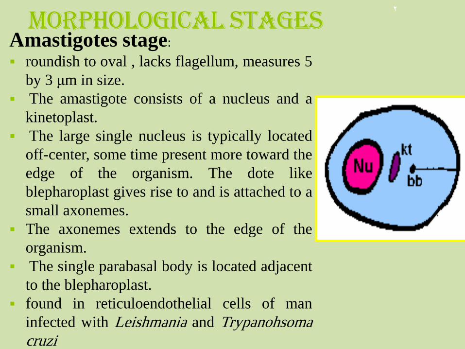
Morphological stages Amastigotes stage:
roundish to oval , lacks flagellum, measures 5
by 3 μm in size.
The amastigote consists of a nucleus and a
kinetoplast.
The large single nucleus is typically located
off-center, some time present more toward the
edge of the organism. The dote like
blepharoplast gives rise to and is attached to a
small axonemes.
The axonemes extends to the edge of the
organism.
The single parabasal body is located adjacent
to the blepharoplast.
found in reticuloendothelial cells of man
infected with Leishmania and Trypanohsoma
cruzi
2

Promastigotes: Lanceolate
shaped, measure 9 to 15μm
in length.
The large single nucleus is
located in or near the center
of the long and slender body.
The kinetoplast is located in
the anterior end of the
organism. A single free
flagellum extends anteriorly
from the axonemes.
This is the infective stage of
Leishmania to man.
3

• Epimastigotes: measures 9 to 15μm in length.
• The body is slightly wider than that of the promastigote.
• The large single nucleus is located in the posterior end of the organism.
• The kinetoplast is located anterior to the nucleus.
• An undulating membrane measuring half the body length forms into a free flagellum at the anterior end of the epimastigote.
4

Trypomastigotes: measures 12-35μm long by 2-4 μm wide.
assume the shape of the letters C or U in stained blood film.
The long, slender organism is characterized by a posteriorly located kinetoploast from which emerges a full body length undulating membrane. The single, large nucleus is located anterior to the kinetoplast.
An anterior free flagellum may or may not be present.
It is the infective stage of Trypanosoma
5

Clinical classification of Leishmaniasis
• Visceral leishmaniasis: (VL, kala azar) is the most severe form and if untreated has a mortality rate approaching 100%. This form is characterized by fever, weight loss, enlargement of the spleen and liver and anaemia. Caused exclusively by species of the L. donovani complex (L. donovani, L. infantum , L. chagasi)
• Mucocutaneous leishmaniasis:(MCL) produces lesions which can lead to extensive and disfiguring destruction of mucous membranes of the nose, mouth and throat cavities.
• Cutaneous leishmaniasis (CL): manifests with sores or
ulcers on: exposed parts of the body such as arms, legs
and face which may heal spontaneously, but the diffuse
form of CL does not heal and may relapse after treatment.
6

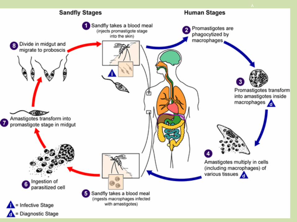
Life cycle • Host: Leishmania completes its life cycle in two hosts:
1. Vertebrate host (man)
2. Insect vector (female sand fly): Phlebotomus.
3.Infective form: Promastigote forms present in the mid gut(of insect) (majority)
• The sand fly vector becomes infected when feeding on the blood of an infected individual or an animal reservoir host.
• The leishmania parasites live in the macrophages as round, non-motile amastigotes (3-7 micrometers in diameter).
• The macrophages are ingested by the fly during the blood- meal and the amastigotes are released into the stomach of insect.
• immediately the amastigotes transform in to the motile, elongated (10-20 micrometers), flagellate promastigote form.
7
8

The promastigotes then migrate to the alimentary tract of the fly, where they live extracellularly and multiply by binary fission.
Four to five days after feeding the promastigotes move forward to the oesophagus and the salivary glands of the insect.
When the sandlfy next feeds on a mammalian host, it's proboscis pierces the skin and saliva containing anti-coagulant is injected into the wound to prevent the blood from clotting,
the leishmania promastigotes are transferred to the host along with the saliva.
Once in the host the promastigotes are taken up by the macrophages where they rapidly revert to the amastigote form
9
• multiplication inside the macrophages, leading to the lysis of the macrophages.
• The released amastigotes are taken up by additional macrophages and so the cycle continues.
• Ultimately all the organs containing macrophages and phagocytes are infected, especially the spleen, liver and bone marrow and less often in other locations such as the skin, intestinal mucosa and mesenteric lymph nodes.
• Small number of LD bodies can be found in blood
10

Insect vector
• The vector of the leishmania parasite is the blood-sucking female of the genus Phlebotomus in the old world and Lutzomyia in the new world.
11

Visceral Leishmaniasis • visceral leishmaniasis including Dum-dum fever,
Sikari disease, Burdwan fever, Shahib's disease
and tropical splenomegaly.
• The most commonly used term is Kala azar, which in
Hindi means black sickness or black fever.
• Visceral leishmaniasis is caused by the parasites
Leishmania donovani donovani, Leishmania donovani
infantum in the old world and by Leishmania donovani
chagasi in the new world.
12
Real image showing clinical features: splenomegaly seen in visceral leishmaniasis

Post kala-azar dermal leishmaniasis
• (PKDL) is a recurrence of Kala-azar that may appear
on the skin of affected individuals up to 20 years after
being partially treated, untreated or even in those
considered adequately treated..
13
Hypo-pigmented skin changes in early PKDL extensive facial nodular lesions in
late PKDL

Profile view of a teenage
boy suffering from visceral
leishmaniasis.
The boy exhibits
splenomegaly, distended
abdomen and severe
muscle wasting.
14

Diagnosis • Clinical diagnosis: such as splenomagaly,
hepatomegally and high undulating fever.
• Parasitological diagnosis: The various samples include:
• Spleen and liver biopsy; Part of the splenic aspirate can be used to make smears for direct microscopic examination and cultured.
• The sensitivity of splenic smear examination is excellent (>95%) but splenic puncture is less likely.??
• Liver biopsy material is less likely to demonstrate parasites on direct examination or on culture.??
15

Marrow and lymph gland puncture : Marrow obtained
from sternal or iliac crest puncture is a much safer but a painful method. It is less likely to demonstrate parasites in direct stained
films.
• Peripheral blood smear: (in HIV infected
• people).
16
Laboratory Diagnosis Parasitological diagnosis:
17
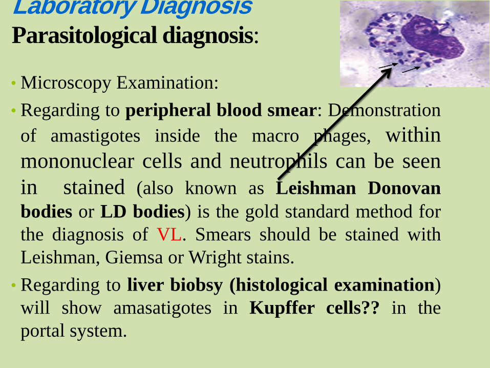
• Microscopy Examination:
• Regarding to peripheral blood smear: Demonstration
of amastigotes inside the macro phages, within
mononuclear cells and neutrophils can be seen
in stained (also known as Leishman Donovan
bodies or LD bodies) is the gold standard method for
the diagnosis of VL. Smears should be stained with
Leishman, Giemsa or Wright stains.
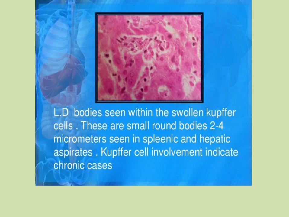
• Regarding to liver biobsy (histological examination)
will show amasatigotes in Kupffer cells?? in the
portal system.
18
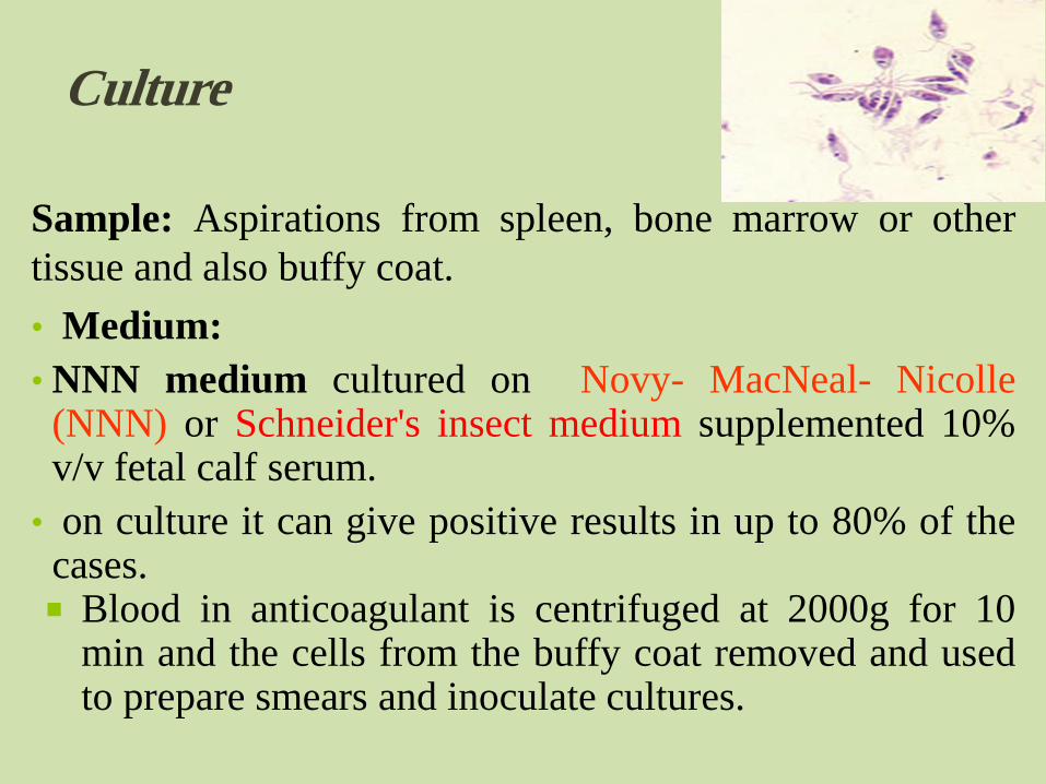
Culture
Sample: Aspirations from spleen, bone marrow or other
tissue and also buffy coat.
• Medium:
• NNN medium cultured on Novy- MacNeal- Nicolle (NNN) or Schneider's insect medium supplemented 10% v/v fetal calf serum.
• on culture it can give positive results in up to 80% of the cases. Blood in anticoagulant is centrifuged at 2000g for 10
min and the cells from the buffy coat removed and used to prepare smears and inoculate cultures.
19

Serological methods o Indirect fluorescent antibody test (IFAT) o Direct agglutination test o Enzyme linked immunosorbent assay (ELISA) o Complement fixation test PCR technique, this method based on the amilification of the
leishmania DNA by using specific primer which depend on the sequence of nitrogen bases in the leishmania genome.
20

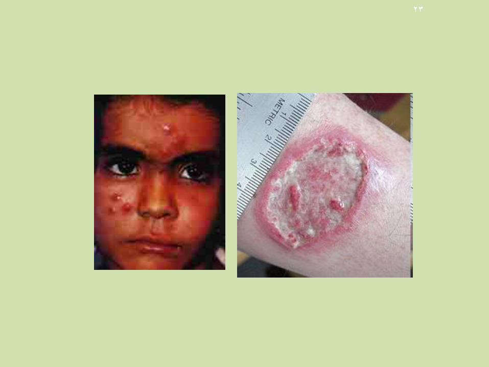
Cutaneous leishmaniasis a - L. tropica minor:
cause urban cutaneous leishmaniasis, is found in more
densely populated areas. reservoir host is dog Its lesion is dry, persists for months before ulcerating , and
has numerates amastigotes within it. Occurs primarily on the face.
21

b- L. tropica major
• cause rural cutaneous leishmaniasis, is found in thinly inhabited
regions.,
• reservoir host is rodent
• wet type, produces an acute infection with a duration of 3 to 6
months.
• Its papule ulcerates quickly, short duration, and contain few
amastigotes.
• The lesions occur primarily on the lower limbs, they are moist
and tend to ulcerate very early; there may be secondary or
satellite lesions.
22
23

: Dry lesion of cutaneous leishmaniasis, oriental sore ,or Baghdad boil (leishmania tropica)
94 24

wet lesion of cutaneous leishmaniasis (leishmania tropica)
95 25
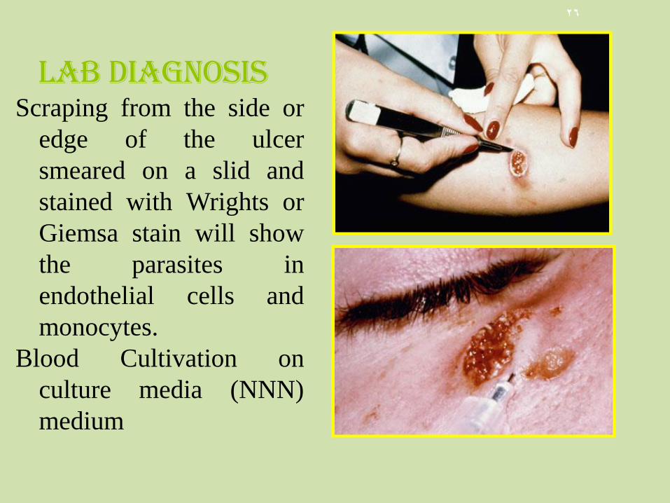
Lab diagnosis Scraping from the side or
edge of the ulcer
smeared on a slid and
stained with Wrights or
Giemsa stain will show
the parasites in
endothelial cells and
monocytes.
Blood Cultivation on
culture media (NNN)
medium
26
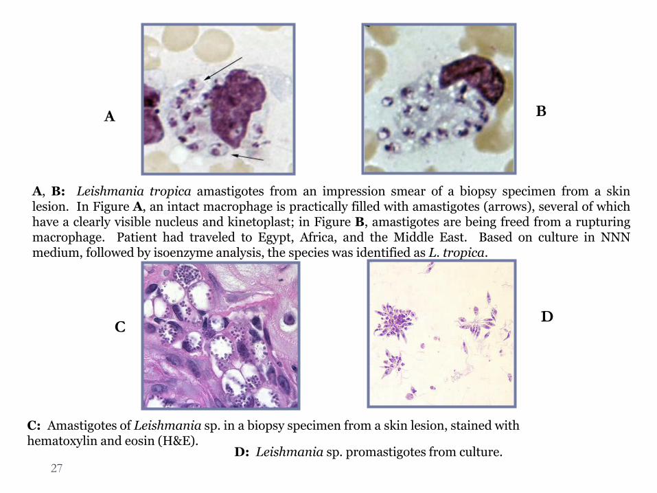
A, B: Leishmania tropica amastigotes from an impression smear of a biopsy specimen from a skin lesion. In Figure A, an intact macrophage is practically filled with amastigotes (arrows), several of which have a clearly visible nucleus and kinetoplast; in Figure B, amastigotes are being freed from a rupturing macrophage. Patient had traveled to Egypt, Africa, and the Middle East. Based on culture in NNN medium, followed by isoenzyme analysis, the species was identified as L. tropica.
C: Amastigotes of Leishmania sp. in a biopsy specimen from a skin lesion, stained with hematoxylin and eosin (H&E).
D: Leishmania sp. promastigotes from culture.
A B
C D
27
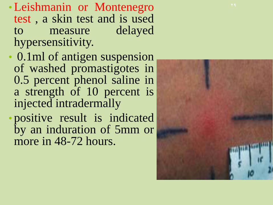
• Leishmanin or Montenegro test , a skin test and is used to measure delayed hypersensitivity.
• 0.1ml of antigen suspension of washed promastigotes in 0.5 percent phenol saline in a strength of 10 percent is injected intradermally
• positive result is indicated by an induration of 5mm or more in 48-72 hours.
29

Mucocutaneous leishmaniasis • L. brazilinensis produces a disease in humans known as
espundia, uta, or mucocutaneous leishmaniasis.
Morphologically, L. braziliensis cannot be differentiated from
L. tropica, L. mexicana, or L. donovani.
• Life cycle is similar to other type, except, the vector is
Lutzomyia.
• In some times the lesions appear as flat, ulcerated plaques that
remain open and oozing.
30

31

Fig : infected patients with leishmania brasiliensis ( mucocutaneous leishmaniasis
96 32

L. braziliensis, the parasites have a tendency to metastasize , or spread directly from the primery lesion to mucocutaneous zones.
The secondary lesion often involves the nasal system and buccal mucosa,causing degeneration of the cartilages and soft tissues
Invasion of the infection into the larynx and trachea destroys the voice.
The condition may last for many years, and death may result from secondary infection or respiratory complications.
33

Diagnosis
• Diagnosis is established by finding L-D bodies in affected tissues
34

35
Facebook: zainab alalwani Phone no.tel. : 07809054757