Bovine gd T Cells Are a Major Regulatory T Cell · PDF fileBovine gd T Cells Are a Major...
16
of May 19, 2018. This information is current as Cell Subset T Cells Are a Major Regulatory T δ γ Bovine Smith, Carolina Cubillos-Zapata and Bryan Charleston Efrain Guzman, Jayne Hope, Geraldine Taylor, Adrian L. ol.1303398 http://www.jimmunol.org/content/early/2014/05/31/jimmun published online 2 June 2014 J Immunol Material Supplementary 8.DCSupplemental http://www.jimmunol.org/content/suppl/2014/05/31/jimmunol.130339 average * 4 weeks from acceptance to publication Fast Publication! • Every submission reviewed by practicing scientists No Triage! • from submission to initial decision Rapid Reviews! 30 days* • Submit online. ? The JI Why Subscription http://jimmunol.org/subscription is online at: The Journal of Immunology Information about subscribing to Permissions http://www.aai.org/About/Publications/JI/copyright.html Submit copyright permission requests at: Email Alerts http://jimmunol.org/alerts Receive free email-alerts when new articles cite this article. Sign up at: Print ISSN: 0022-1767 Online ISSN: 1550-6606. Copyright © 2014 The Authors All rights reserved. 1451 Rockville Pike, Suite 650, Rockville, MD 20852 The American Association of Immunologists, Inc., is published twice each month by The Journal of Immunology by guest on May 19, 2018 http://www.jimmunol.org/ Downloaded from by guest on May 19, 2018 http://www.jimmunol.org/ Downloaded from
Transcript of Bovine gd T Cells Are a Major Regulatory T Cell · PDF fileBovine gd T Cells Are a Major...
of May 19, 2018.This information is current as
Cell Subset T Cells Are a Major Regulatory TδγBovine
Smith, Carolina Cubillos-Zapata and Bryan CharlestonEfrain Guzman, Jayne Hope, Geraldine Taylor, Adrian L.
ol.1303398http://www.jimmunol.org/content/early/2014/05/31/jimmun
published online 2 June 2014J Immunol
MaterialSupplementary
8.DCSupplementalhttp://www.jimmunol.org/content/suppl/2014/05/31/jimmunol.130339
average*
4 weeks from acceptance to publicationFast Publication! •
Every submission reviewed by practicing scientistsNo Triage! •
from submission to initial decisionRapid Reviews! 30 days* •
Submit online. ?The JIWhy
Subscriptionhttp://jimmunol.org/subscription
is online at: The Journal of ImmunologyInformation about subscribing to
Permissionshttp://www.aai.org/About/Publications/JI/copyright.htmlSubmit copyright permission requests at:
Email Alertshttp://jimmunol.org/alertsReceive free email-alerts when new articles cite this article. Sign up at:
Print ISSN: 0022-1767 Online ISSN: 1550-6606. Copyright © 2014 The Authors All rights reserved.1451 Rockville Pike, Suite 650, Rockville, MD 20852The American Association of Immunologists, Inc.,
is published twice each month byThe Journal of Immunology
by guest on May 19, 2018
http://ww
w.jim
munol.org/
Dow
nloaded from
by guest on May 19, 2018
http://ww
w.jim
munol.org/
Dow
nloaded from
The Journal of Immunology
Bovine gd T Cells Are a Major Regulatory T Cell Subset
Efrain Guzman,* Jayne Hope,† Geraldine Taylor,* Adrian L. Smith,‡
Carolina Cubillos-Zapata,*,1 and Bryan Charleston*
In humans and mice, gd T cells represent <5% of the total circulating lymphocytes. In contrast, the gd T cell compartment in
ruminants accounts for 15–60% of the total circulating mononuclear lymphocytes. Despite the existence of CD4+CD25high Foxp3+
T cells in the bovine system, these are neither anergic nor suppressive. We present evidence showing that bovine gd T cells are the
major regulatory T cell subset in peripheral blood. These gd T cells spontaneously secrete IL-10 and proliferate in response to
IL-10, TGF-b, and contact with APCs. IL-10–expressing gd T cells inhibit Ag-specific and nonspecific proliferation of CD4+ and
CD8+ T cells in vitro. APC subsets expressing IL-10 and TFG-b regulate proliferation of gd T cells producing IL-10. We propose that
gd T cells are a major regulatory T cell population in the bovine system. The Journal of Immunology, 2014, 193: 000–000.
Tcells expressing the gd TCR have been described as non-classical T cells, because unlike most TCR ab T cells,activation can be independent of MHC–peptide complexes.
In mice and humans, gd T cells represent between 1 and 5% of thecirculating lymphocytes, but are present at higher frequencies inepithelial sites (1). Many functions have been described for gdT cells including cytokine production, Ag presentation, and immuneregulation (2, 3). However, these various functions have been iden-tified mostly for mice and humans, species with “low” numbers ofcirculating gd T cells. In contrast, many other species such as cattle,sheep, pigs, and chickens are considered to have “high” numbers ofcirculating gd T cells, and the function of these is yet to be deter-mined. In the bovine system, gd T cells represent between 15 and 60%of the circulating lymphocytes (4), and a large proportion of bovine gdT cells express workshop cluster 1 (WC1), a transmembrane glyco-protein and member of the scavenger receptor cysteine-rich family,which is closely related to CD163. Although functional WC1 mole-cules have so far been identified only in ruminants, pigs, and camelids,WC1 orthologs have been identified in many other species (5).Regulation of the immune system is important to prevent au-
toimmunity and immunopathology. Regulatory T cells (Tregs) arenow recognized as a critical component of a balanced immunesystem (6, 7). The predominant Treg types are CD4+ and expresseither or both CD25 and the forkhead box transcription factor,Foxp3 (8). Despite the existence of bovine CD4+CD25high Foxp3+
T cells, these cells have been shown to be neither anergic nor
suppressive in vitro (9). Instead, mounting evidence supports thenotion that gd T cells are involved in immune suppression in rumi-nants. For example, depletion of gd T cells from PBMC culturesresulted in increased Ag-specific proliferation and cytokine produc-tion in ex vivo cultures of T cells (10–12).Tregs need to be licensed or activated to initiate and maintain
their regulatory role. Dendritic cells (DCs) can prevent, inhibit, ormodulate T cell–mediated responses through a variety of mecha-nisms ranging from the production of anti-inflammatory factors tothe induction of T cell responses, which result in deletion, anergy,or instruction of regulatory cells. Immature DCs have been pro-posed to be tolerogenic (13), and this function is thought to bea consequence of the presentation of Ag in the absence of co-stimulation or cytokines. In addition, tolerogenicity of DC subsetsmay be dependent on the secretion of anti-inflammatory signalssuch as IL-10, TGF-b, and retinoic acid, among others (14).In this report, we present evidence for the role of circulating
gd TCR+ cells as potent inhibitory T cells in the bovine system.Subsets of gd T cells secreted IL-10 ex vivo and proliferatedin response to IL-10, IL-4, and TGF-b, which, in turn, initiateda positive-feedback mechanism producing more IL-10 in prolif-erating gd T cells. IL-10–expressing gd T cells suppressed Ag-specific and nonspecific proliferation of CD4+ and CD8+ T cells.Suppressive gd T cells were present in both WC1+ and WC12 gd
TCR+ T cell populations, and were not stained with anti-Foxp3.We also identified specific subsets of APCs from various ana-tomical sites responsible for the expansion of gd T cells with sup-pressive function and show that in vitro infection of APCs withmodified vaccinia Ankara (MVA) increased the frequency ofIL-10–expressing gd T cells. These results suggest that a subset ofcirculating T cells expressing the gd TCR are a major regulatoryand suppressive T cell population in ruminants.
Materials and MethodsAnimals
Conventionally reared Holstein cattle (Bos taurus) from The Pirbright Instituteherd were used to obtain peripheral blood and tissues. All animals were atleast 6 mo of age and from a bovine virus diarrhea virus (BVDV)–free herd.All animal experiments were approved by The Pirbright Institute ethicscommittee according to the U.K. Animal (Scientific Procedures) Act 1986.
Vaccination and Ag-specific T cell assays
To obtain Ag-specific T cells, we vaccinated cattle (n = 10) with inactivatedFMDV (foot-and-mouth disease virus) vaccine (O1 Manisa/A22 Iraq;
*The Pirbright Institute, Surrey GU24 0NF, United Kingdom; †The Roslin InstituteUniversity of Edinburgh, Midlothian EH259RG, United Kingdom; and ‡Departmentof Zoology, University of Oxford, Oxford OX1 3PS, United Kingdom
1Current address: Tumour Immunology Department, Hospital La Paz, Madrid, Spain.
Received for publication December 20, 2013. Accepted for publication May 1, 2014.
This work was supported by the Biology and Biological Sciences Research Council,U.K. G.T., A.L.S., and B.C. are Jenner investigators.
Address correspondence and reprint requests to Dr. Efrain Guzman, The PirbrightInstitute, Ash Road, Woking, Surrey GU24 0NF, U.K. E-mail address: [email protected]
The online version of this article contains supplemental material.
Abbreviations used in this article: AdV5, adenovirus 5; ALDC, afferent-lymph DC;BVDV, bovine virus diarrhea virus; DC, dendritic cell; GM-CSF, granulocyte-macrophage CSF; MoDC, monocyte-derived DC; MVA, modified vaccinia Ankara;Treg, regulatory T cell; WC1, workshop cluster 1.
This is an open-access article distributed under the terms of the CC-BY 3.0 Unportedlicense.
Copyright � 2014 The Authors 0022-1767/14
www.jimmunol.org/cgi/doi/10.4049/jimmunol.1303398
Published June 2, 2014, doi:10.4049/jimmunol.1303398 by guest on M
ay 19, 2018http://w
ww
.jimm
unol.org/D
ownloaded from
Intervet, Milton Keynes, U.K.) as described previously (15). FMDV-specific proliferation, IFN-g ELISPOT, and intracellular cytokine stain-ing have all been described previously (15–17) using the FMDV vaccineAg for Ag-specific stimulation. In some experiments, UV-inactivated BVDVwas used as control Ag as described previously (18). In some assays, gdT cells were removed by MACS as described later, and autologous gd T cellsubsets were added back to the starting cultures at a ratio of 1 gd T cell to 1PBMC.
Separation and preparation of lymphocyte subsets
Heparinized venous blood was centrifuged at 300 3 g over Histopaque1083 (Sigma, Poole, U.K.), and the mononuclear cells were washed threetimes in PBS. Cells were either used immediately or frozen in FCS con-taining 10% DMSO (Sigma). CD14+ cells were purified by MACS usinganti-human CD14+ microbeads (Miltenyi Biotec, Surrey, U.K.) (19).Monocyte-derived DCs (MoDCs) were prepared by culturing CD14+ cellsfor 5 d at 37˚C in tissue culture media (RPMI 1640 [Invitrogen, Paisley,U.K.] containing 10% heat-inactivated FCS [Autogen Bioclear, U.K.], 2 mML-glutamine, 55 mM 2-ME, penicillin [100 U/ml], and streptomycin [100mg/ml]) and supplemented with recombinant bovine granulocyte-macro-phage CSF (GM-CSF) and IL-4 (The Pirbright Institute) (20). T cell subsetswere positively selected from peripheral blood using MACS (Miltenyi Bio-tech) with the following Abs: CC58 (anti-CD8b) (21), CC8 (anti-CD4) (22),CC15 (anti-WC1) (23), and GB21A (anti-gd TCR) (4) as described previ-ously (17) and following the manufacturer’s instructions. In some assays, gdT cells were negatively selected: PBMCs were depleted of MHC II+ cells(CC108, anti-MHC II) (24); CD8+ (CC58); CD4+ (CC8), B cells (CC21, anti-CD21; Serotec), and NK+ cells (AKS1, anti-NKp46) (25) using a MACS LDcolumn (Miltenyi Biotec) following the manufacturer’s instructions. In allcases, the purities of the selected populations were analyzed by flow cytometryand were confirmed to be .98% pure.
Lung-derived and afferent-lymph DCs
Lung-derived DCs were obtained as described previously (26, 27). Aportion of lung tissue obtained postmortem was excised and weighed.Three grams of lung tissue was placed in a petri dish and chopped veryfinely with a pair of scissors and a scalpel blade. The sliced tissue was thenplaced in a 250-ml polycarbonate conical flask together with l00 ml RPMI1640, 10% FCS containing 200 U/ml collagenase 4196 (Worthington) orhyaluronidase (BDH), and 50 U/ml DNase (Sigma). The tissue fragmentswere incubated for 2 h at 37˚C in an orbital shaker (200 rpm). After di-gestion, the tissue was sheared through a 20-ml syringe, filtered throughsterile gauze and muslin, and centrifuged for 10 min at 300 3 g at 20˚C.Mononuclear cells were separated on a Histopaque 1083 (Sigma) gradientas described earlier.
Afferent-lymph DCs (ALDCs) were obtained by surgical cannulation ofthe lymphatic vessels draining the suprascapular lymph node of cattle asdescribed previously (28). Anti-bovine mAbs used to distinguish DCs fromother cells were specific for MHC II (IL-A21), CD11c (IL-A16), DEC-205(CC98), CD8a (CC63), and CD172a (CC149) (29). All Abs were obtainedfrom The Pirbright Institute. DCs and subsets were flow sorted using aFACSAria II (Becton Dickson) as described previously (15).
In vitro proliferation assays
Lymphocyte proliferation was measured by CFDA-SE (referred to asCFSE) dilution and analyzed by flow cytometry (16). To assess proliferationof gd T cells in mixed cultures, we labeled PBMCs with CFSE (Invitrogen)following the manufacturer’s instructions and cultured them in tissueculture media for 5 d in 96-well U-bottom plates (Costar) at a concentra-tion of 1 3 106 cells/well in a total volume of 200 ml. In some cases, gdT cells were positively or negatively selected, labeled with CFSE, and 5 3105 cells were cultured in 96-well U-bottom plates (Costar) with the samenumber of autologous irradiated (3000 rad) CD14+ cells in the presence ofthe following blocking Abs: CC320 (anti–IL-10) (30), CC313 (anti–IL-4)(31), CC328 (anti–TNF-a) (32), and anti–TGF-b (clone 1D11; R&D Sys-tems), which have been previously shown to neutralize their target cytokine.In some cases, positively or negatively selected gd T cells were cultured inplates coated with 10 mg/ml anti-bovine CD3 (clone MM1A; VMRD) and inthe presence of 5 mg/ml anti-bovine CD28 (clone F849CD10; The PirbrightInstitute) (33), 5 ng/ml rTGF-b1, and 50 U/ml human rIL-2 (Roche) asdescribed previously (34). Polyclonal activation of T cells was performedusing 20 ng/ml phorbol 12-myristate 13-acetate and 1 mg/ml ionomycin. Forcontact-dependent proliferation assays, 5 3 105 irradiated CD14+ cells wereadded to the lower chamber of a 48-well Transwell plate (Costar); the 3-mminsert was carefully slotted in and autologous purified gd T cells added to theupper chamber in the presence or absence of blocking Abs described earlier.
In assays where gd T cells were cultured with APC subsets, gd T cells werepositively or negatively selected, labeled with CFSE, and 53 105 cells werecultured in 96-well U-bottom plates (Costar) with an equal number of au-tologous irradiated (3000 rad) FACS or MACS-sorted APCs (as describedearlier) in a final volume of 250 ml. Proliferation and Ag presentation assayswere performed as described earlier.
Flow cytometry
Fluorochrome-labeled mouse anti-bovine mAbs used in this study havebeen described in detail previously (35–39). For cell-surface molecules,these were: CC149-PerCP/Cy5.5 (anti-SIRPa), ILA-16-AlexaFluor 680/PE(anti-CD11c), ILA-21-PE (anti-MHCII), ILA-156-PE/Texas Red (anti-CD40), CC30-allophycocyanin/Cy5.5 (anti-CD4), CC63-allophycocyanin/Cy7 (anti-CD8), IL-A29-allophycocyanin (anti-WC1), BAQ159A (anti-WC1.1; VMRD), CACTB32A (anti-WC1.2; VMRD), GB21a-AlexaFluor405 (anti-gd TCR; VMRD), and ILA-111-AlexaFluor 610/PE (anti-CD25). IL7Ra (CD127) was detected using an anti-mouse CD127 Abraised in rats (clone RTK2758; Biolegend) and found to cross-react withbovine CD127 (this report). For intracellular staining, 10 mg/ml brefeldinA (Sigma) was added to cultured cells for 4 h and cells were harvested.Surface staining was performed in 96-well U-bottom plates (Costar) fol-lowed by fixation and permeabilization using BD Cytoperm/Cytofix kitfollowing the manufacturer’s instructions. Intracellular cytokine stainingwas performed with CC302-PE or -AF488 (anti–IFN-g), CC319-PE-Cy5.5(anti–IL-10), CC312-PE/Texas Red (anti–IL-4), sG9-FITC (anti-perforin;BD Pharmingen), TW4-2F8 (anti–TGF-b1–PE/Cy7; Biolegend), FJK16s-PE (anti-mouse/rat Foxp3 that cross-reacts with cattle (40); eBioscience), andAb86-allophycocyanin (anti–IL-2-AF657, a gift from A. Whelan, AnimalHealth and Veterinary Laboratories Agency, Weybridge, U.K.) (41). ControlmAbs were isotype and concentration matched anti-turkey rhinotracheitis virusmAbs (28, 42). All Abs were either raised against bovine or confirmed to becross-reactive with bovine cells and obtained from The Pirbright Institute ex-cept where noted. Dead cells were excluded using the 405-nm excitable dyeLive/Dead Aqua or propidium iodide (Invitrogen) following the manufacturer’sinstructions, and doublets were excluded using SSC-A/SSC-H. The cells wereacquired using an LSRFortessa (Becton Dickinson), and staining was analyzedusing FlowJo (TreeStar). Only live, single events were used for analysis, andat least 50,000 were used for postacquisition analysis. Proliferation analyseswere performed using the Cellular Proliferation module on FCS Expressv4 (DeNovo).
ELISA
Bovine cytokine ELISAs were performed on culture supernatants as de-scribed previously for IL-10 (30), IL-12p40/70 (43), IL-4 (31), and IFN-g(44). IL-2 was detected using the DuoSet Bovine IL-2 ELISA kit (R&DSystems) following the manufacturer’s instructions.
Viruses
E1- and E3-deleted recombinant human adenovirus 5 (AdV5) and MVAexpressing GFP were generated by the Jenner Institute Viral Vector CoreFacility, University of Oxford, Oxford, U.K., and have been describedpreviously (15). In some experiments, noncytopathic BVDV isolate PEC515was used as described previously (18).
Statistical analysis
Calculation of descriptive statistics (geometric statistics and SDs), non-parametric statistical analyses, and graphs were generated using GraphPadPrism for Windows v5.01 (GraphPad, San Diego, CA).
Resultsgd TCR+ T cells express IL-10 and have inhibitory function
Despite gd T cells representing a large component of the bovinelymphocyte system, the function of this cell population remainspoorly understood. To explore a potential role in immune regula-tion, expression of Foxp3 and IL-10 by bovine PBMCs were ana-lyzed ex vivo. Fig. 1A shows the typical frequency of CD4+ andCD8+ T cells in bovine peripheral blood. Of the CD4+ population,expression of IL-10 was not significantly higher compared with theisotype control staining, and ,1% expressed Foxp3 as previouslyreported (40) (Fig. 1B). Fig. 1D shows the typical frequencies of gdT cells and expression of WC1 in bovine peripheral blood. Between7 and 15% (mean 11.43%) of all gd TCR+ cells expressed IL-10,
2 BOVINE gd T CELLS ARE REGULATORY
by guest on May 19, 2018
http://ww
w.jim
munol.org/
Dow
nloaded from
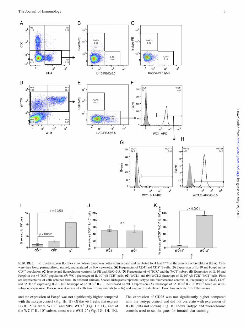
and the expression of Foxp3 was not significantly higher comparedwith the isotype control (Fig. 1E, 1I). Of the gd T cells that expressIL-10, 50% were WC12 and 50% WC1+ (Fig. 1F, 1J), and ofthe WC1+ IL-10+ subset, most were WC1.2+ (Fig. 1G, 1H, 1K).
The expression of CD25 was not significantly higher comparedwith the isotype control and did not correlate with expression ofIL-10 (data not shown). Fig. 1C shows isotype and fluorochromecontrols used to set the gates for intracellular staining.
FIGURE 1. gd T cells express IL-10 ex vivo. Whole blood was collected in heparin and incubated for 4 h at 37˚C in the presence of brefeldin A (BFA). Cells
were then fixed, permeabilized, stained, and analyzed by flow cytometry. (A) Frequencies of CD4+ and CD8+ T cells. (B) Expression of IL-10 and Foxp3 in the
CD4+ population. (C) Isotype and fluorochrome controls for PE and PE/Cy5.5. (D) Frequencies of gd TCR+ and the WC1+ subset. (E) Expression of IL-10 and
Foxp3 in the gd TCR+ population. (F) WC1 phenotype of IL-10+ gd TCR+ cells. (G) WC1.1 and (H) WC1.2 phenotype of IL-10+ gd TCR+ WC1+ cells. Plots
are representative of cells obtained from 16 different animals. Shaded histograms represent isotype and fluorochrome controls. (I) Frequency of CD4+, CD8+
and gd TCR+ expressing IL-10. (J) Phenotype of gd TCR+ IL-10+ cells based on WC1 expression. (K) Phenotype of gd TCR+ IL-10+ WC1+ based on WC1-
subgroup expression. Bars represent means of cells taken from animals (n = 16) and analyzed in duplicate. Error bars indicate SE of the means.
The Journal of Immunology 3
by guest on May 19, 2018
http://ww
w.jim
munol.org/
Dow
nloaded from
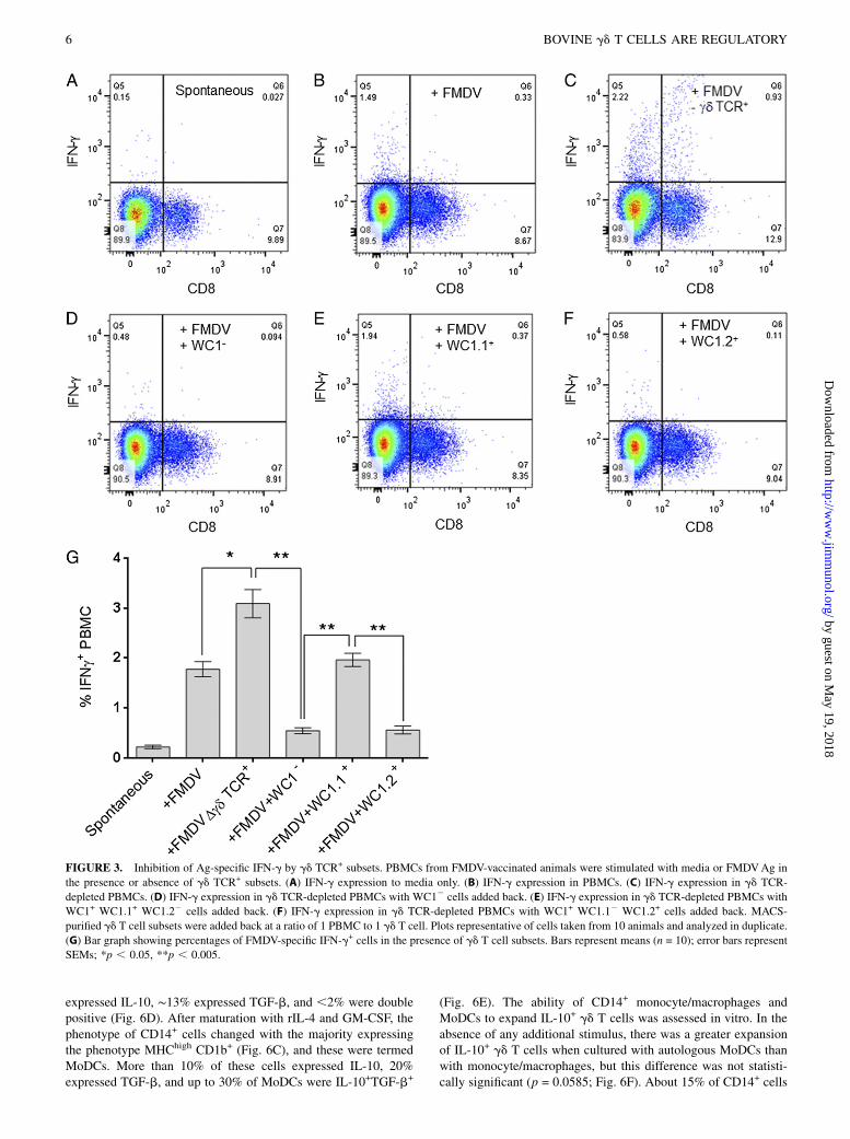
To confirm that gd T cells were involved in Ag-specific T cellregulation, we used cells from FMDV-vaccinated animals, becausevaccination with inactivated virus has been shown to induce Ag-specific CD4+ and CD8+ T cells (16, 45). PBLs from FMDV-vaccinated cattle were depleted of gd TCR+ cells, and FMDV-specific proliferation was measured in vitro. Fig. 2A, 2B showhigher FMDV-specific proliferation of CD4+ and CD8+ T cells insamples depleted of gd T cells. The depletion of gd T cells hada statistically significant effect not only on proliferation, but alsoon the number of cells producing IFN-g, IL-2, and IL-4. In thecase of CD8+ T cells, depletion of gd T cells resulted in a 5-foldincrease in IL-2 and a 2-fold increase in IFN-g expression (Fig.2C, 2E). For CD4+ T cells, depletion of gd T cells also resulted inincreased IFN-g and IL-4 expression (Fig. 2D, 2F). The quantityof cytokines released in these cultures was measured by ELISA,and there were statistically significant increases in the level ofIFN-g (p = 0.0057), IL-2 (p = 0.0143), and IL-4 (p , 0.001) inthose samples that had been depleted of gd T cells (Fig. 2G–I).To confirm that these effects were Ag specific, an irrelevant Ag(BVDV) was used in addition to media only. Responses to BVDVwere similar to responses to media (Fig. 2G–I). To identify the gdT cell subsets involved in this immune suppression, we depletedPBMCs from FMDV-vaccinated animals from all gd T cells byMACS and stimulated them with FMDV Ag in the presence ofautologous MACS-purified gd T cell subsets described earlierusing a ratio of 1 PBMC to 1 gd T cell. Cultures containing WC12
and WC1.2+ but not WC1.1+ gd T cells showed decreased IFN-gresponses to FMDV (Fig. 3A–G). Although there was a reductionin the percentage of IFNg+ cells in the +FMDV+WC1.1 groupcompared with the gd-depleted group, this was not statisticallysignificant (p = 0.1713). These results indicate that bovine gdT cells have potent inhibitory functions.
Expansion of IL-10 – expressing gd T cells in vitro
For expansion in vitro, human and murine Tregs (CD4+ CD25+
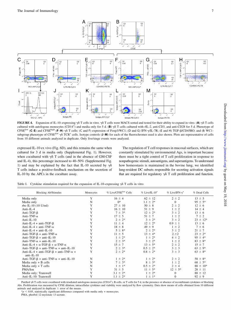
Foxp3+) require IL-2, engagement of the TCR via cross-linkingwith anti-CD3, and induction of Foxp3 by TGF-b (46, 47). Toidentify whether bovine IL-10–expressing gd T cells could beexpanded the same way, we cultured purified gd TCR+ cells withautologous peripheral blood monocytes (CD32sIg2MHCII+CD14+)alone or with rIL-2, anti-CD3, and anti-CD28 as described previ-ously (34). Between 7 and 15% of gd T cells cultured with mono-cytes and media only proliferated (Fig. 4A) and ∼50% of CFSElow
gd TCR+ cells were IL-10+; cells cultured with rIL-2, anti-CD3, andanti-CD28 did not proliferate, and most cells died within 36 h ofculture (Fig. 4B). Positively sorted gd T cells cultured in the absenceof monocytes did not survive .24 h in culture (data not shown).Both CFSElow and CFSEhigh gd T cells were WC12 and WC1+ andFoxp32 (Fig. 4C, 4F); within the WC1+ cells, only the WC1.2+
subset proliferated and expressed IL-10 (Fig. 4I), mirroring the resultsobtained ex vivo.Under the conditions tested, both proliferating and nonprolife-
rating IL-10+ gd TCR+ cells were found to be IFN-g2, eitherIL7R2 or IL7R+, TGF-b2, and most were CD45RO+ (Fig. 4D–H).To confirm that selection by MACS did not have a nonspecificeffect on the expansion of IL-10–producing gd T cells, we neg-atively selected peripheral blood gd TCR+ cells by MACS or flowsorted and cultured them with autologous monocytes as describedearlier with similar results (data not shown).To define the monocyte-derived signals required for the ex-
pansion of IL-10+ gd T cells, we cultured MACS-sorted gd T cellswith gamma-irradiated autologous monocytes and in the presenceof blocking Abs to various cytokines, individually and in combi-nation. Consistent with previous observations, gd T cells prolif-
erated in culture with autologous monocytes but did not survivein the absence of monocytes, and ,10% survival was observed inthe presence of autologous B cells (Table I). Anti–IL-4 or TNF-ablocking Abs did not have a significant effect on expression of IL-10 or proliferation of gd cells. Anti–TGF-b and anti–IL-10 indi-vidually and in combination reduced proliferation and expressionof IL-10 in gd T cells, and anti–IL-10 had the most significanteffect (Table I). These effects are partly explained by the increasein gd T cell death after incubation with anti–IL-10 and anti–TGF-b.Significant increases in IFN-g were only observed when gd T cellswere stimulated with mitogen. To investigate whether contact withmonocytes was required to expand IL-10–producing gd T cells, weused a Transwell system where irradiated CD14+ were placed in thelower chamber and autologous gd T cells in the upper chamber. Bypreventing the physical contact between gd T cells and monocytes,expansion of IL-10+ gd T cells did not occur and most cells diedwithin the first 2 days of the assay (Table I). These results show thatin vitro expansion of bovine IL-10+ gd T cells requires IL-10, TGF-b,and contact with APCs, in this case, peripheral blood CD14+
monocytes.In light of these results, and for all subsequent experiments,
positively sorted gd T cells were expanded in vitro for 5 d bycoculture with autologous monocytes and without any additionalcytokines.
In vitro–expanded gd T cells modulate proliferation andfunction of CD4+ and CD8+ T cells after specific andnonspecific stimulation
To investigate whether in vitro–expanded IL-10–expressing gd
T cells were capable of regulating proliferation and function ofT cells, we cultured purified CD4+ T cells with an equal number ofautologous CD14+ monocytes in the presence of rIL-2, anti-CD3,and anti-CD28. After the addition of in vitro–expanded, autolo-gous gd T cells at a ratio of 10:1 CD4+ T cells, polyclonal pro-liferation of CD4+ T cells was reduced (p = 0.0278; Fig. 5A, 5C)compared with CD4+ T cells expanded in the absence of gd
T cells. To confirm this effect in Ag-specific T cell proliferation,we cultured purified CD4+ T cells from FMDV-vaccinated animalswith autologous CD14+ cells loaded with FMDV Ag. In the ab-sence of gd T cells, CD4+ T cells proliferated in response to FMDVAg. However, after the addition of in vitro–expanded autologous gdT cells, FMDV-specific CD4+ proliferation was reduced (p = 0.0380;Fig. 5B, 5D).The effect of in vitro–expanded gd T cells on CTL phenotype was
also investigated. Purified CD8+ T cells were cultured with an equalnumber of autologous CD14+ cells that had been loaded with FMDVAg, in the presence or absence of in vitro–expanded autologous gdT cells. There was a reduction in IFN-g+ (p = 0.0048), perforin-positive (p = 0.0312), and double perforin/IFN-g+ (p = 0.0223)FMDV-specific CD8+ T cell frequencies in the presence of in vitro–expanded gd T cells (Fig. 5E–G). These results confirm the biologicalfunction of in vitro–expanded gd T cells, reducing Ag-specific andnonspecific proliferation and function of CD4+ and CD8+ T cells.
Suppressive gd T cells are induced by APCs of variousphenotypes
Efficient mechanisms for the induction of tolerance or maintenanceof homeostasis are necessary at sites of infection or vaccination,where APCs process and present both self- and non–self-Ags.In vitro–matured MoDCs have been used as a model to studyDC biology and have been shown to be required for the expansionof Tregs in mice and humans. Bovine peripheral blood CD14+
cells (Fig. 6A) expressed low to high levels of MHC class II,and most were CD1b2 (Fig. 6B). Less than 5% of these cells
4 BOVINE gd T CELLS ARE REGULATORY
by guest on May 19, 2018
http://ww
w.jim
munol.org/
Dow
nloaded from
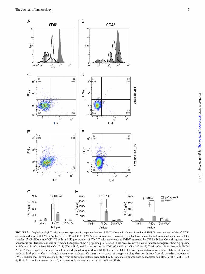
FIGURE 2. Depletion of gd T cells increases Ag-specific responses in vitro. PBMCs from animals vaccinated with FMDV were depleted of the gd TCR+
cells and cultured with FMDVAg for 5 d. CD4+ and CD8+ FMDV-specific responses were analyzed by flow cytometry and compared with nondepleted
samples. (A) Proliferation of CD8+ T cells and (B) proliferation of CD4+ T cells in response to FMDV measured by CFSE dilution. Gray histograms show
nonspecific proliferation to media only; white histograms show Ag-specific proliferation in the presence of gd T cells; hatched histograms show Ag-specific
proliferation in gd-depleted PBMCs. (C–F) IFN-g, IL-2, and IL-4 expression in CD8+ (C and E) and CD4+ (D and F) T cells after stimulation with FMDV
Ag in gd T cell–depleted samples (E and F) or nondepleted samples (C and D). Histograms and dot plots are representative of cells from 10 different animals
analyzed in duplicate. Only live/single events were analyzed. Quadrants were based on isotype staining (data not shown). Specific cytokine responses to
FMDVand nonspecific responses to BVDV from culture supernatants were tested by ELISA and compared with nondepleted samples. (G) IFN-g. (H) IL-2.
(I) IL-4. Bars indicate means (n = 10, analyzed in duplicate), and error bars indicate SEMs.
The Journal of Immunology 5
by guest on May 19, 2018
http://ww
w.jim
munol.org/
Dow
nloaded from
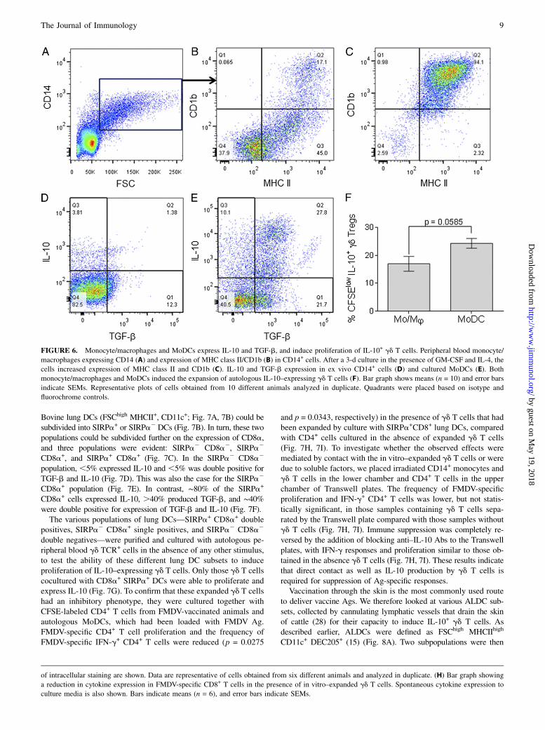
expressed IL-10, ∼13% expressed TGF-b, and ,2% were doublepositive (Fig. 6D). After maturation with rIL-4 and GM-CSF, thephenotype of CD14+ cells changed with the majority expressingthe phenotype MHChigh CD1b+ (Fig. 6C), and these were termedMoDCs. More than 10% of these cells expressed IL-10, 20%expressed TGF-b, and up to 30% of MoDCs were IL-10+TGF-b+
(Fig. 6E). The ability of CD14+ monocyte/macrophages andMoDCs to expand IL-10+ gd T cells was assessed in vitro. In theabsence of any additional stimulus, there was a greater expansionof IL-10+ gd T cells when cultured with autologous MoDCs thanwith monocyte/macrophages, but this difference was not statisti-cally significant (p = 0.0585; Fig. 6F). About 15% of CD14+ cells
FIGURE 3. Inhibition of Ag-specific IFN-g by gd TCR+ subsets. PBMCs from FMDV-vaccinated animals were stimulated with media or FMDVAg in
the presence or absence of gd TCR+ subsets. (A) IFN-g expression to media only. (B) IFN-g expression in PBMCs. (C) IFN-g expression in gd TCR-
depleted PBMCs. (D) IFN-g expression in gd TCR-depleted PBMCs with WC12 cells added back. (E) IFN-g expression in gd TCR-depleted PBMCs with
WC1+ WC1.1+ WC1.22 cells added back. (F) IFN-g expression in gd TCR-depleted PBMCs with WC1+ WC1.12 WC1.2+ cells added back. MACS-
purified gd T cell subsets were added back at a ratio of 1 PBMC to 1 gd T cell. Plots representative of cells taken from 10 animals and analyzed in duplicate.
(G) Bar graph showing percentages of FMDV-specific IFN-g+ cells in the presence of gd T cell subsets. Bars represent means (n = 10); error bars represent
SEMs; *p , 0.05, **p , 0.005.
6 BOVINE gd T CELLS ARE REGULATORY
by guest on May 19, 2018
http://ww
w.jim
munol.org/
Dow
nloaded from
expressed IL-10 ex vivo (Fig. 6D), and this remains the same whencultured for 3 d in media only (Supplemental Fig. 1). However,when cocultured with gd T cells (and in the absence of GM-CSFand IL-4), this percentage increased to 40–50% (Supplemental Fig.1) and may be explained by the fact that IL-10 secreted by gdT cells induce a positive-feedback mechanism on the secretion ofIL-10 by the APCs in the coculture assay.
The regulation of T cell responses in mucosal surfaces, which areconstantly stimulated by environmental Ags, is important becausethere must be a tight control of T cell proliferation in response tononpathogenic stimuli, autoantigens, and superantigens. To understandhow homeostasis is maintained in the bovine lung, we identifiedlung-resident DC subsets responsible for secreting activation signalsthat are required for regulatory gd T cell proliferation and function.
FIGURE 4. Expansion of IL-10–expressing gd T cells in vitro. gd T cells were MACS sorted and tested for their ability to expand in vitro. (A) gd T cells
cultured with autologous monocytes (CD14+) and media only for 5 d. (B) gd T cells cultured with rIL-2, anti-CD3, and anti-CD28 for 5 d. Phenotype of
CFSElow (C–E) and CFSEhigh (F–H) gd T cells: (C and F) expression of Foxp3/WC1; (D and G) IFN-g/IL-7R; (E and H) TGF-b/CD45RO; and (I) WC1-
subgroup phenotype of CFSElow gd TCR+ cells. Isotype controls (J–M) for each of the fluorochromes used is also shown. Plots are representative of cells
from 10 different animals analyzed in duplicate. Only live/singe events were analyzed.
Table I. Cytokine stimulation required for the expansion of IL-10–expressing gd T cells in vitro
Blocking Ab/Stimulus Monocytes % Live/CFSElow Cells % Live/IL-10+ % Live/IFN-g+ % Dead Cells
Media only Y 16 6 4 62 6 12 2 6 2 13 6 5Media only N 0* 1.1 6 2* 0 95 6 5*rbo IL-10 (10 U/ml) Y 27 6 10 50 6 8 2 6 2 12 6 6Anti–IL-4 Y 16 6 10 51 6 9 1 6 2 14 6 4Anti–TGF-b Y 7 6 3* 12 6 2* 3 6 2 13 6 6Anti–TNF-a Y 17 6 5 51 6 7 1 6 2 7 6 2Anti–IL-10 Y 2 6 2* 3 6 2* 3 6 2 23 6 12*Anti–IL-4 + anti–TGF-b Y 11 6 4 12 6 2* 0.9 6 1 13 6 6Anti–IL-4 + anti–TNF-a Y 18 6 8 49 6 9 1 6 2 7 6 6Anti–IL-4 + anti–IL-10 Y 5 6 8* 2 6 2* 3 6 2 21 6 7Anti–TGF-b + anti–TNF-a Y 11 6 5 13 6 4* 2.62 23 6 2*Anti–TGF-b + anti–IL-10 Y 1 6 2* 1 6 2* 4 6 2 95 6 4*Anti–TNF-a + anti–IL-10 Y 2 6 3* 3 6 2* 1 6 2 83 6 8*Anti–IL-4 + a-TGF-b + a-TNF-a Y 15 6 7 13 6 5* 2 6 2 15 6 7Anti–TGF-b + anti–TNF-a + anti–IL-10 Y 2 6 3* 0.5 6 2* 3 6 3 63 6 9*Anti–IL-4 + anti–TGF-b + anti–TNF-a +
anti–IL-10Y 2 6 2* 0.8 6 2* 3 6 3 63 6 8*
Anti–TGF-b + anti–TNF-a + anti–IL-10 N 1 6 2* 1 6 2* 2 6 2 58 6 8*Media only + B cells N 7 6 3* 8 6 3* 1 6 2 68 6 5*Media only + T cells N 1 6 1* 0.5 6 2* 2 6 4 88 6 10*PMA/Ion Y 31 6 5 11 6 5* 12 6 5* 28 6 11Media only: Transwell Y 3.1 6 2* 1 6 2* 0 88 6 12Anti–IL-10: Transwell Y 1.1 6 2* 1 6 1* 0 92 6 9
Purified gd T cells were cocultured with irradiated autologous monocytes (CD14+), B cells, or T cells for 5 d, in the presence or absence of recombinant cytokines or blockingAbs. Proliferation was measured by CFSE dilution; intracellular cytokines and viability were analyzed by flow cytometry. Data show means of cells obtained from 10 differentanimals and analyzed in duplicate 6 error of the means.
*p , 0.05, statistically significant difference compared with media only + monocytes.PMA, phorbol 12-myristate 13-acetate.
The Journal of Immunology 7
by guest on May 19, 2018
http://ww
w.jim
munol.org/
Dow
nloaded from
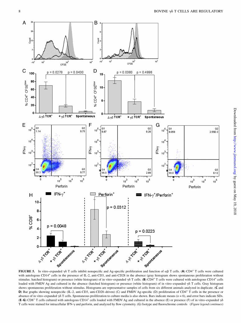
FIGURE 5. In vitro–expanded gd T cells inhibit nonspecific and Ag-specific proliferation and function of ab T cells. (A) CD4+ T cells were cultured
with autologous CD14+ cells in the presence of IL-2, anti-CD3, and anti-CD28 in the absence (gray histogram shows spontaneous proliferation without
stimulus. hatched histogram) or presence (white histogram) of in vitro–expanded gd T cells. (B) CD4+ T cells were cultured with autologous CD14+ cells
loaded with FMDVAg and cultured in the absence (hatched histogram) or presence (white histogram) of in vitro–expanded gd T cells. Gray histogram
shows spontaneous proliferation without stimulus. Histograms are representative samples of cells from six different animals analyzed in duplicate. (C and
D) Bar graphs showing nonspecific (IL-2, anti-CD3, anti-CD28–driven) (C) and FMDVAg-specific (D) proliferation of CD4+ T cells in the presence or
absence of in vitro–expanded gd T cells. Spontaneous proliferation to culture media is also shown. Bars indicate means (n = 6), and error bars indicate SDs.
(E–G) CD8+ T cells cultured with autologous CD14+ cells loaded with FMDVAg and cultured in the absence (E) or presence (F) of in vitro–expanded gd
T cells were stained for intracellular IFN-g and perforin, and analyzed by flow cytometry. (G) Isotype and fluorochrome controls (Figure legend continues)
8 BOVINE gd T CELLS ARE REGULATORY
by guest on May 19, 2018
http://ww
w.jim
munol.org/
Dow
nloaded from
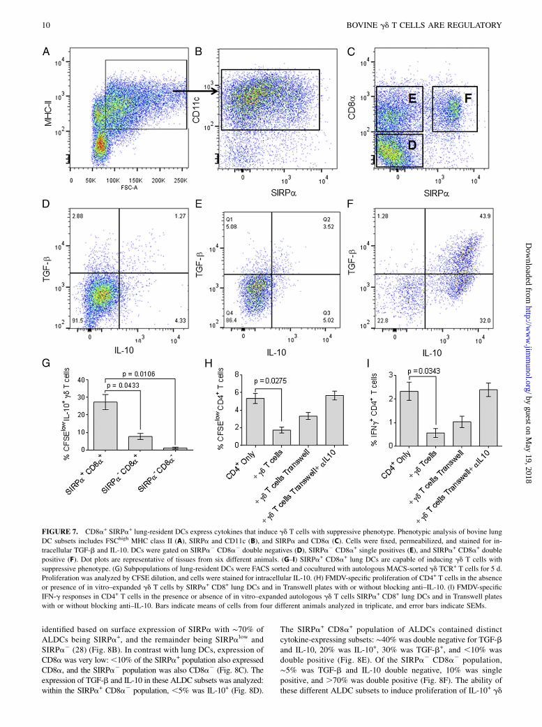
Bovine lung DCs (FSChigh MHCII+, CD11c+; Fig. 7A, 7B) could besubdivided into SIRPa+ or SIRPa2 DCs (Fig. 7B). In turn, these twopopulations could be subdivided further on the expression of CD8a,and three populations were evident: SIRPa2 CD8a2, SIRPa2
CD8a+, and SIRPa+ CD8a+ (Fig. 7C). In the SIRPa2 CD8a2
population, ,5% expressed IL-10 and ,5% was double positive forTGF-b and IL-10 (Fig. 7D). This was also the case for the SIRPa2
CD8a+ population (Fig. 7E). In contrast, ∼80% of the SIRPa+
CD8a+ cells expressed IL-10, .40% produced TGF-b, and ∼40%were double positive for expression of TGF-b and IL-10 (Fig. 7F).The various populations of lung DCs—SIRPa+ CD8a+ double
positives, SIRPa2 CD8a+ single positives, and SIRPa2 CD8a2
double negatives—were purified and cultured with autologous pe-ripheral blood gd TCR+ cells in the absence of any other stimulus,to test the ability of these different lung DC subsets to induceproliferation of IL-10–expressing gd T cells. Only those gd T cellscocultured with CD8a+ SIRPa+ DCs were able to proliferate andexpress IL-10 (Fig. 7G). To confirm that these expanded gd T cellshad an inhibitory phenotype, they were cultured together withCFSE-labeled CD4+ T cells from FMDV-vaccinated animals andautologous MoDCs, which had been loaded with FMDV Ag.FMDV-specific CD4+ T cell proliferation and the frequency ofFMDV-specific IFN-g+ CD4+ T cells were reduced (p = 0.0275
and p = 0.0343, respectively) in the presence of gd T cells that hadbeen expanded by culture with SIRPa+CD8+ lung DCs, comparedwith CD4+ cells cultured in the absence of expanded gd T cells(Fig. 7H, 7I). To investigate whether the observed effects weremediated by contact with the in vitro–expanded gd T cells or weredue to soluble factors, we placed irradiated CD14+ monocytes andgd T cells in the lower chamber and CD4+ T cells in the upperchamber of Transwell plates. The frequency of FMDV-specificproliferation and IFN-g+ CD4+ T cells was lower, but not statis-tically significant, in those samples containing gd T cells sepa-rated by the Transwell plate compared with those samples withoutgd T cells (Fig. 7H, 7I). Immune suppression was completely re-versed by the addition of blocking anti–IL-10 Abs to the Transwellplates, with IFN-g responses and proliferation similar to those ob-tained in the absence gd T cells (Fig. 7H, 7I). These results indicatethat direct contact as well as IL-10 production by gd T cells isrequired for suppression of Ag-specific responses.Vaccination through the skin is the most commonly used route
to deliver vaccine Ags. We therefore looked at various ALDC sub-sets, collected by cannulating lymphatic vessels that drain the skinof cattle (28) for their capacity to induce IL-10+ gd T cells. Asdescribed earlier, ALDCs were defined as FSChigh MHCIIhigh
CD11c+ DEC205+ (15) (Fig. 8A). Two subpopulations were then
of intracellular staining are shown. Data are representative of cells obtained from six different animals and analyzed in duplicate. (H) Bar graph showing
a reduction in cytokine expression in FMDV-specific CD8+ T cells in the presence of in vitro–expanded gd T cells. Spontaneous cytokine expression to
culture media is also shown. Bars indicate means (n = 6), and error bars indicate SEMs.
FIGURE 6. Monocyte/macrophages and MoDCs express IL-10 and TGF-b, and induce proliferation of IL-10+ gd T cells. Peripheral blood monocyte/
macrophages expressing CD14 (A) and expression of MHC class II/CD1b (B) in CD14+ cells. After a 3-d culture in the presence of GM-CSF and IL-4, the
cells increased expression of MHC class II and CD1b (C). IL-10 and TGF-b expression in ex vivo CD14+ cells (D) and cultured MoDCs (E). Both
monocyte/macrophages and MoDCs induced the expansion of autologous IL-10–expressing gd T cells (F). Bar graph shows means (n = 10) and error bars
indicate SEMs. Representative plots of cells obtained from 10 different animals analyzed in duplicate. Quadrants were placed based on isotype and
fluorochrome controls.
The Journal of Immunology 9
by guest on May 19, 2018
http://ww
w.jim
munol.org/
Dow
nloaded from
identified based on surface expression of SIRPa with ∼70% ofALDCs being SIRPa+, and the remainder being SIRPalow andSIRPa2 (28) (Fig. 8B). In contrast with lung DCs, expression ofCD8a was very low:,10% of the SIRPa+ population also expressedCD8a, and the SIRPa2 population was also CD8a2 (Fig. 8C). Theexpression of TGF-b and IL-10 in these ALDC subsets was analyzed:within the SIRPa+ CD8a2 population, ,5% was IL-10+ (Fig. 8D).
The SIRPa+ CD8a+ population of ALDCs contained distinctcytokine-expressing subsets: ∼40% was double negative for TGF-band IL-10, 20% was IL-10+, 30% was TGF-b+, and ,10% wasdouble positive (Fig. 8E). Of the SIRPa2 CD8a2 population,∼5% was TGF-b and IL-10 double negative, 10% was singlepositive, and .70% was double positive (Fig. 8F). The ability ofthese different ALDC subsets to induce proliferation of IL-10+ gd
FIGURE 7. CD8a+ SIRPa+ lung-resident DCs express cytokines that induce gd T cells with suppressive phenotype. Phenotypic analysis of bovine lung
DC subsets includes FSChigh MHC class II (A), SIRPa and CD11c (B), and SIRPa and CD8a (C). Cells were fixed, permeabilized, and stained for in-
tracellular TGF-b and IL-10. DCs were gated on SIRPa2 CD8a2 double negatives (D), SIRPa2 CD8a+ single positives (E), and SIRPa+ CD8a+ double
positive (F). Dot plots are representative of tissues from six different animals. (G–I) SIRPa+ CD8a+ lung DCs are capable of inducing gd T cells with
suppressive phenotype. (G) Subpopulations of lung-resident DCs were FACS sorted and cocultured with autologous MACS-sorted gd TCR+ T cells for 5 d.
Proliferation was analyzed by CFSE dilution, and cells were stained for intracellular IL-10. (H) FMDV-specific proliferation of CD4+ T cells in the absence
or presence of in vitro–expanded gd T cells by SIRPa+ CD8+ lung DCs and in Transwell plates with or without blocking anti–IL-10. (I) FMDV-specific
IFN-g responses in CD4+ T cells in the presence or absence of in vitro–expanded autologous gd T cells SIRPa+ CD8+ lung DCs and in Transwell plates
with or without blocking anti–IL-10. Bars indicate means of cells from four different animals analyzed in triplicate, and error bars indicate SEMs.
10 BOVINE gd T CELLS ARE REGULATORY
by guest on May 19, 2018
http://ww
w.jim
munol.org/
Dow
nloaded from
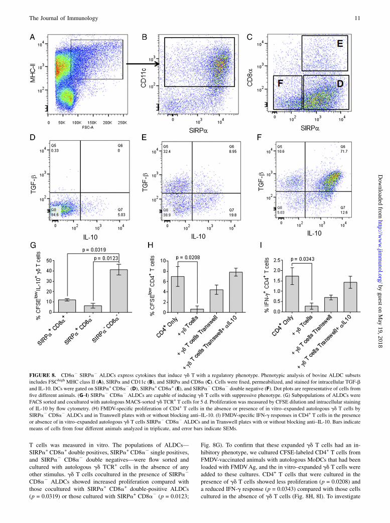
T cells was measured in vitro. The populations of ALDCs—SIRPa+ CD8a+ double positives, SIRPa+ CD8a2 single positives,and SIRPa2 CD8a2 double negatives—were flow sorted andcultured with autologous gd TCR+ cells in the absence of anyother stimulus. gd T cells cocultured in the presence of SIRPa2
CD8a2 ALDCs showed increased proliferation compared withthose cocultured with SIRPa+ CD8a+ double-positive ALDCs(p = 0.0319) or those cultured with SIRPa+ CD8a2 (p = 0.0123;
Fig. 8G). To confirm that these expanded gd T cells had an in-hibitory phenotype, we cultured CFSE-labeled CD4+ T cells fromFMDV-vaccinated animals with autologous MoDCs that had beenloaded with FMDVAg, and the in vitro–expanded gd T cells wereadded to these cultures. CD4+ T cells that were cultured in thepresence of gd T cells showed less proliferation (p = 0.0208) anda reduced IFN-g response (p = 0.0343) compared with those cellscultured in the absence of gd T cells (Fig. 8H, 8I). To investigate
FIGURE 8. CD8a2 SIRPa2 ALDCs express cytokines that induce gd T with a regulatory phenotype. Phenotypic analysis of bovine ALDC subsets
includes FSChigh MHC class II (A), SIRPa and CD11c (B), and SIRPa and CD8a (C). Cells were fixed, permeabilized, and stained for intracellular TGF-b
and IL-10. DCs were gated on SIRPa+ CD8a2 (D), SIRPa+ CD8a+ (E), and SIRPa2 CD8a2 double negative (F). Dot plots are representative of cells from
five different animals. (G–I) SIRPa2 CD8a2 ALDCs are capable of inducing gd T cells with suppressive phenotype. (G) Subpopulations of ALDCs were
FACS sorted and cocultured with autologous MACS-sorted gd TCR+ T cells for 5 d. Proliferation was measured by CFSE dilution and intracellular staining
of IL-10 by flow cytometry. (H) FMDV-specific proliferation of CD4+ T cells in the absence or presence of in vitro–expanded autologous gd T cells by
SIRPa2 CD8a2 ALDCs and in Transwell plates with or without blocking anti–IL-10. (I) FMDV-specific IFN-g responses in CD4+ T cells in the presence
or absence of in vitro–expanded autologous gd T cells SIRPa2 CD8a2 ALDCs and in Transwell plates with or without blocking anti–IL-10. Bars indicate
means of cells from four different animals analyzed in triplicate, and error bars indicate SEMs.
The Journal of Immunology 11
by guest on May 19, 2018
http://ww
w.jim
munol.org/
Dow
nloaded from
whether the observed effects were contact specific or due to sol-uble factors, we placed gd T cells in the lower chamber and CD4+
T cells in the upper chamber of Transwell plates as describedearlier. The frequency of FMDV-specific proliferation and IFN-g+
CD4+ T cells was lower, but not statistically significant, in thosesamples containing gd T cells separated by the Transwell platecompared with those samples without gd T cells (Fig. 8H, 8I).Immune suppression by the gd T cells was completely reversed bythe addition of blocking anti–IL-10 Abs to the Transwell plates(Fig. 8H, 8I). These results indicate that direct contact as well asIL-10 production by gd T cells is required for suppression of Ag-specific responses, and the ability to induce proliferation of gdT cells with a regulatory phenotype depends on the origin andphenotype of the APC.
Recombinant MVA induces the generation of IL-10–expressinggd T cells in vitro
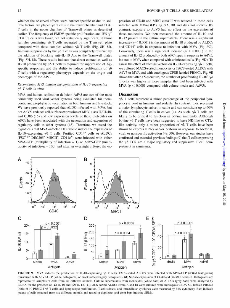
MVA and human replication-deficient AdV5 are two of the mostcommonly used viral vector systems being evaluated for thera-peutic and prophylactic vaccination in both humans and livestock.We have previously reported that ALDC infected with MVA, butnot AdV5, reduces cell-surface expression of MHC class II, CD40,and CD86 (15) and low expression levels of these molecules onAPCs have been associated with the generation and expansion ofregulatory cells in other systems (48). Therefore, we tested thehypothesis that MVA-infected DCs would induce the expansion ofIL-10–expressing gd T cells. Purified CD14+ cells or ALDCs(FSChigh DEC205+ MHCII+, CD11c+) were infected with eitherMVA-GFP (multiplicity of infection = 1) or AdV5-GFP (multi-plicity of infection = 100) and after an overnight culture, the ex-
pression of CD40 and MHC class II was reduced in those cellsinfected with MVA-GFP (Fig. 9A, 9B and data not shown). Bycontrast, exposure to AdV5 had no effect on the expression ofthese molecules. We then measured the amount of IL-10 andIL-12 present in the culture supernatants. There was a significantincrease (p, 0.0001) in the amount of IL-10 produced by ALDCsand CD14+ cells in response to infection with MVA (Fig. 9C).Conversely, there was a significant increase (p , 0.0001) in theamount of IL-12 produced by both APC types in response to AdV5,but not to MVAwhen compared with uninfected cells (Fig. 9D). Toassess the effect of vaccine vectors on IL-10–expressing gd T cells,we cultured MACS-sorted monocytes or FACS-sorted ALDCs withAdV5 or MVA and with autologous CFSE-labeled PBMCs. Fig. 9Eshows that after a 5-d culture, the number of proliferating IL-10+ gdT cells was higher in those samples that had been infected withMVA (p , 0.0001 compared with culture media and AdV5).
Discussiongd T cells represent a minor percentage of the peripheral lym-phocyte pool in humans and rodents. In contrast, they representa major lymphocyte subset in cattle and can constitute up to 60%of the circulating T cells in calves (4). As such, gd T cells arelikely to be critical to function in bovine immunity. Althoughbovine gd T cells have been suggested to have NK-like or CTL-like activity, only a minor proportion of gd T cells have beenshown to express IFN-g and/or perforin in response to bacterial,viral, or nonspecific activation (49, 50). However, our studies haveconfirmed and extended previous findings (9) that T cells expressingthe gd TCR are a major regulatory and suppressive T cell com-partment in ruminants.
FIGURE 9. MVA induces the production of IL-10–expressing gd T cells. FACS-sorted ALDCs were infected with MVA-GFP (dotted histograms)
transduced with AdV5-GFP (white histograms) or mock infected (gray histograms). (A) Surface expression of CD40 and (B) MHC class II. Histograms are
representative samples of cells from six different animals. Culture supernatants from monocytes (white bars) or ALDCs (gray bars) were analyzed by
ELISA for the presence of (C) IL-10 and (D) IL-12. (E) FACS-sorted ALDCs (from A and B) were cultured with autologous CFDA-SE–labeled PBMCs
(ratio of 10 PBMC:1 gd T cell), and lymphocyte proliferation, T cell subsets, and intracellular cytokines were measured by flow cytometry. Bars indicate
means of cells obtained from six different animals and tested in duplicate, and error bars indicate SEMs.
12 BOVINE gd T CELLS ARE REGULATORY
by guest on May 19, 2018
http://ww
w.jim
munol.org/
Dow
nloaded from
A large proportion of bovine gd T cells express WC1, a trans-membrane glycoprotein and member of the scavenger receptorcysteine-rich family, which is closely related to CD163. Func-tional WC1 molecules have so far been identified only in rumi-nants, pigs, and camelids (51, 52), although there is molecularevidence of the existence of WC1 orthologs in mice and humans(5). Human and murine gd T cell subsets are tissue specific asillustrated by the distribution of G and D gene segment usagein lymphocytes from various tissues (53, 54). The TCR d-chainrepertoire in cattle has been shown to be highly diverse and great-ly expanded to include 56 TRD variable (V) genes, 5 diversity (D)genes, 3 junctional (J) genes, and 1 constant (C) gene (55, 56).This expansion of TRD genes in cattle compared with humans andmice suggests a distinct role for gd T cells in ruminants (57).Until now, the induction, phenotype, and function of Tregs in
ruminants has not been fully described. However, there is indirectevidence of a regulatory role for gd T cells in cattle. For example, invivo depletion of WC1-expressing gd T cells in cattle after FMDVinfection resulted in a shorter period of viremia (5 d) compared withdepletion of CD4+ (∼6 d) or CD8+ T cells (∼7 d) (45). Another studyshowed that depletion of WC1+ gd T cells resulted in an enhancedAb response to the model Ag OVA (58). Similarly, enhanced localand systemic bovine respiratory syncytial virus–specific Ab responseswere seen post respiratory syncytial virus infection in WC1+-depletedcalves (59). One major drawback of these studies is that only cellsexpressing WC1 were depleted. The expression of WC1 in gd
T cells is variable in that 15–60% of all peripheral blood bovinegd T cells may express WC1, and thus in the aforementionedexperiments, not all gd T cells were depleted from the system.Even though there was likely to be incomplete depletion of all gdT cells in these in vivo studies, there was evidence that removingthese cells could enhance immune responses.Our data show for the first time, to our knowledge, that up to
15% of bovine gd T cells express IL-10 ex vivo (Fig. 1). These cellsdo not express either Foxp3 or IFN-g, and IL-10 production isnot related to WC1 expression. Further inspection of the WC1+
population shows that most WC1+ IL-10+ cells are of the WC1.2subset (Fig. 1). We and others (9, 40, 60) have unsuccessfully triedto identify bovine CD4+ CD25+ Foxp3+ IL-10+ cells with regu-latory function, and although the presence of bovine CD4+ Foxp3+
T cells has been reported (40) (Fig. 1), these cells do not appear tohave suppressive capacity. Because gd T cells represent a majorproportion of the lymphocytes in the blood of ruminants, we de-pleted these cells before stimulation of the remaining cells. Bydoing this, we were able to increase both polyclonal and Ag-specific proliferation and IFN-g, IL-2, and IL-4 released byCD4+ and CD8+ T cells (Fig. 2).It has been reported that bovine gd T cells proliferate in culture
in the absence of other stimuli (61–63). We now show that up to15% of these proliferating gd T cells secrete IL-10 and haveregulatory functions. Of the IL-10+–expressing gd T cells, twodistinct populations were observed: WC1+ and WC12 (Fig. 1J).IL7R and CD45RO have been suggested as alternative markers toidentify T cells with regulatory potential (64), and our data showthat ∼50% of the IL-10+ gd TCR+ cells express IL-7R and mostare CD45RO+ (Fig. 4D, 4E). In addition, we could not detectexpression of Foxp3 on expanding IL-10+ gd T cells. This indi-cates that the markers suggested so far for identifying Treg pop-ulations in humans and mice do not apply to cattle.Further analysis of the IL-10+WC1+ population shows only
those cells expressing the WC1.2 subgroup were able to prolif-erate and express IL-10 (Fig. 4F). In contrast, only those cellsexpressing the WC1.1 subgroup were able to express IFN-g inresponse to cytokine or mitogen stimulation. These data suggest
that differential expression of specific WC1 genes correlates withbovine gd T cell function and in the context of Ag as previouslysuggested (65, 66), and that the serological definition of WC1subsets does not correlate with function, because all serologicallydefined WC1 subgroups are able to express proinflammatory andanti-inflammatory cytokines (49, 57, 67).To determine the signals required to expand IL-10+ gd T cells,
we used Abs against several cytokines to try to inhibit this effect(Table I). The presence of IL-10 and TGF-b blocking Abs indi-vidually reduced the capacity of gd T cells to express IL-10 andproliferate, and in combination these two Abs almost completelyblocked this effect. The addition of IL-4 and TNF-a blockingAbs did not have a significant effect. Monocytes (CD14+) wererequired for gd T cell survival, and gd T cells needed to be inphysical contact with CD14+ cells. All these data suggest that forgd T cells to survive, express IL-10, and proliferate, soluble IL-10and, to a lesser extent, TGF-b are required along with other sig-nals provided by physical contact with CD14+ cells.We were unable to expand IL-10+ gd T cells by culturing with
anti-CD3 and anti-CD28 mAbs, indicating that these signals arenot required or are insufficient for maintenance of the major Tregcompartment in cattle. Murine and human Tregs require engage-ment of TCR for expansion and function (68); this may also be thecase in the bovine system, and more work is required to identifythese signals.Most assays to investigate Treg function rely on the expansion
in vitro of the Treg subset in question; therefore, we tested thesuppressive activity of in vitro–expanded gd T cells. Both poly-clonal and Ag-specific proliferation of CD4+ and CD8+ T cellswere reduced in the presence of in vitro–expanded gd T cells(Fig. 5A–D). Similarly, the frequency of IFN-g+ and perforin+
CD8+ T cells was reduced in the presence of gd T cells (Fig.5E–H). Hoek and colleagues (9) reported the inhibitory effect ofgd T cell WC1+ subpopulations on polyclonal activation of au-tologous T cells and observed that with WC1.1+ and WC1.2+ hadsuppressive function. However, the authors did not investigate theeffect of WC12 gd T cells or their regulatory function on Ag-specific responses. We did not identify a differential regulatoryrole between WC12 and WC1+ gd T cell subsets and within theWC1+, we observed that only the WC1.2+ had suppressive func-tions; therefore, more work is required to reconcile the observeddifferences.For T cells to have regulatory function, these need to obtain
the necessary signals from the surrounding environment, includingcytokines and DCs. These cells are sometimes called tolerogenicDCs and are a heterogeneous mix of APCs that differ not only withregard to phenotype, differentiation, and maturation status, but alsowith regard to tolerance-inducing capacity. Although immaturityappears to be a good indicator of DC tolerogenicity, mature DCsmay also contribute to the generation and maintenance of Tregs.Various subpopulations of DCs have been shown to maintain Tregs,and these include CD8a+, CD4+, CD103+, CD1032, CD11b+,CD205+, plasmacytoid DCs, and Langerhans cells (extensivelyreviewed in Ref. 48). The secretion of IL-10, TGF-b, and retinoicacid by DCs has been shown to be necessary for the induction ofTregs (69, 70). We investigated the ability of bovine DCs fromthree sources, peripheral blood, lung, and afferent lymph drainingthe skin, to induce IL-10+ gd T cells. Our data show that the majorAPC populations expressing IL-10 and TGF-b were in peripheralblood, monocytes (MHCII+ CD14+); in the lung MHCII+CD11c+
SIRPa+CD8a+ DCs; and in the afferent-lymph MHCII+ CD11c+
SIRPa2CD8a2 DCs (Figs. 6–8). All of these populations werecapable of inducing proliferation of IL-10+ gd T cells with sup-pressive function. Interestingly, the capacity of CD14+ to express
The Journal of Immunology 13
by guest on May 19, 2018
http://ww
w.jim
munol.org/
Dow
nloaded from
large amounts of IL-10 in culture is dependent on the presence ofIL-10–expressing gd T cells (Supplemental Fig. 1), and this maybe explained by a positive-feedback mechanism for the produc-tion of IL-10 as described in other systems (69, 70). It is possiblethat CD4+ and CD8+ T cells may become licensed regulatory cellswhen in contact with the DCs described earlier; however, we havenot yet investigated the role of bovine ab T cells subsets in im-mune regulation.The use of viral vectors to vaccinate against infectious diseases
has been investigated for many years. MVA and AdV5 are two ofthe most commonly used vaccine vectors being tested to date.MVA has been shown to be safe in laboratory animals and targetspecies. However, its efficacy as a vaccine delivery vector has notbeen consistent. We have previously shown that MVA reduces cell-surface expression of MHC class II, CD40, and CD86 (15), andinduces apoptosis in DCs, preventing optimal Ag expression andpresentation (71). We hypothesized the consequence of usingMVAwas that T cell responses were suboptimal not only becauseof poor Ag presentation and DC death, but also because of thegeneration of suppressive gd T cells. Our data show this is, in fact,the case: DCs infected with MVA upregulate the production of IL-10, inducing the proliferation of IL-10+ gd T cells. We confirmedthese results by inhibiting IL-10 using blocking Abs and remov-ing gd T cells from the system (data not shown). There are reportsof similar observations in other systems; for example, Kastenmullerand colleagues (72) showed in mice that MVA-induced Tregs se-lectively limit the number of effector T cells generated whereaspreserving the memory response by changing the amount of CD80and CD86 displayed on the MVA-infected DCs and the availabilityof IL-2.Although the role of bovine gd T cell in immune regulation is
unequivocal, it has also been observed that both bovine CD4+
and CD8+ T cells have suppressive functions in response to staphy-lococcal enterotoxin (73). Clearly, there are multiple sources ofimmune-regulating cells in cattle, as there are in humans and mice,and more work is required to identify their role in disease andimmune responses and to reconcile apparently opposing data.Increasing evidence supports the notion that gd T cells may
play an important role in immune regulation in humans and mice(74–76), producing large amounts of IL-10 and suppressing Ag-specific T cell proliferation and activation. Our data demonstratethat the ruminant immune system uses gd cells as a principalimmune-regulatory subset.
AcknowledgmentsWe thank the staff at The Pirbright Institute for care of cattle.
DisclosuresThe authors have no financial conflicts of interest.
References1. Komori, H. K., T. F. Meehan, and W. L. Havran. 2006. Epithelial and mucosal
gamma delta T cells. Curr. Opin. Immunol. 18: 534–538.2. Kabelitz, D., C. Peters, D. Wesch, and H. H. Oberg. 2013. Regulatory functions
of gd T cells. Int. Immunopharmacol. 16: 382–387.3. Pang, D. J., J. F. Neves, N. Sumaria, and D. J. Pennington. 2012. Understanding the
complexity of gd T-cell subsets in mouse and human. Immunology 136: 283–290.4. Davis, W. C., W. C. Brown, M. J. Hamilton, C. R. Wyatt, J. A. Orden,
A. M. Khalid, and J. Naessens. 1996. Analysis of monoclonal antibodies specificfor the gamma delta TcR. Vet. Immunol. Immunopathol. 52: 275–283.
5. Holm, D., D. R. Fink, J. Grønlund, S. Hansen, and U. Holmskov. 2009. Cloningand characterization of SCART1, a novel scavenger receptor cysteine-rich type Itransmembrane molecule. Mol. Immunol. 46: 1663–1672.
6. Bluestone, J. A., Q. Tang, and C. E. Sedwick. 2008. T regulatory cells in autoimmunediabetes: past challenges, future prospects. J. Clin. Immunol. 28: 677–684.
7. Brusko, T. M., A. L. Putnam, and J. A. Bluestone. 2008. Human regulatory T cells: rolein autoimmune disease and therapeutic opportunities. Immunol. Rev. 223: 371–390.
8. Hori, S., T. Nomura, and S. Sakaguchi. 2003. Control of regulatory T cell de-velopment by the transcription factor Foxp3. Science 299: 1057–1061.
9. Hoek, A., V. P. Rutten, J. Kool, G. J. Arkesteijn, R. J. Bouwstra, I. Van Rhijn, andA. P. Koets. 2009. Subpopulations of bovine WC1(+) gammadelta T cells ratherthan CD4(+)CD25(high) Foxp3(+) T cells act as immune regulatory cells exvivo. Vet. Res. 40: 6.
10. Rhodes, S. G., R. G. Hewinson, and H. M. Vordermeier. 2001. Antigen recog-nition and immunomodulation by gamma delta T cells in bovine tuberculosis. J.Immunol. 166: 5604–5610.
11. de Graaf, D. C., K. Walravens, J. Godfroid, and J. E. Peeters. 1998. A Crypto-sporidium parvum oocyst low molecular mass fraction evokes a CD4+ T-cell-dependent IFN-gamma response in bovine peripheral blood mononuclear cellcultures. Int. J. Parasitol. 28: 1875–1880.
12. Brown, W. C., W. C. Davis, S. H. Choi, D. A. Dobbelaere, and G. A. Splitter.1994. Functional and phenotypic characterization of WC1+ gamma/delta T cellsisolated from Babesia bovis-stimulated T cell lines. Cell. Immunol. 153: 9–27.
13. Xia, W., E. W. Wong, D. D. Mruk, and C. Y. Cheng. 2009. TGF-beta3 andTNFalpha perturb blood-testis barrier (BTB) dynamics by accelerating theclathrin-mediated endocytosis of integral membrane proteins: a new concept ofBTB regulation during spermatogenesis. Dev. Biol. 327: 48–61.
14. Smith, J. G., M. Silvestry, S. Lindert, W. Lu, G. R. Nemerow, and P. L. Stewart.2010. Insight into the mechanisms of adenovirus capsid disassembly fromstudies of defensin neutralization. PLoS Pathog. 6: e1000959.
15. Cubillos-Zapata, C., E. Guzman, A. Turner, S. C. Gilbert, H. Prentice,J. C. Hope, and B. Charleston. 2011. Differential effects of viral vectors onmigratory afferent lymph dendritic cells in vitro predict enhanced immunoge-nicity in vivo. J. Virol. 85: 9385–9394.
16. Guzman, E., G. Taylor, B. Charleston, and S. A. Ellis. 2010. Induction of a cross-reactive CD8(+) T cell response following foot-and-mouth disease virus vacci-nation. J. Virol. 84: 12375–12384.
17. Guzman, E., G. Taylor, B. Charleston, M. A. Skinner, and S. A. Ellis. 2008. AnMHC-restricted CD8+ T-cell response is induced in cattle by foot-and-mouthdisease virus (FMDV) infection and also following vaccination with inactivatedFMDV. J. Gen. Virol. 89: 667–675.
18. Glew, E. J., B. V. Carr, L. S. Brackenbury, J. C. Hope, B. Charleston, andC. J. Howard. 2003. Differential effects of bovine viral diarrhoea virus onmonocytes and dendritic cells. J. Gen. Virol. 84: 1771–1780.
19. Sopp, P., and C. J. Howard. 1997. Cross-reactivity of monoclonal antibodies todefined human leucocyte differentiation antigens with bovine cells. Vet. Immu-nol. Immunopathol. 56: 11–25.
20. Hope, J. C., D. Werling, R. A. Collins, B. Mertens, and C. J. Howard. 2000. Flt-3ligand, in combination with bovine granulocyte-macrophage colony-stimulatingfactor and interleukin-4, promotes the growth of bovine bone marrow deriveddendritic cells. Scand. J. Immunol. 51: 60–66.
21. MacHugh, N. D., A. Bensaid, C. J. Howard, W. C. Davis, and W. I. Morrison.1991. Analysis of the reactivity of anti-bovine CD8 monoclonal antibodies withcloned T cell lines and mouse L-cells transfected with bovine CD8. Vet.Immunol. Immunopathol. 27: 169–172.
22. Howard, C. J., P. Sopp, K. R. Parsons, D. J. McKeever, E. L. Taracha,B. V. Jones, N. D. MacHugh, and W. I. Morrison. 1991. Distinction of naive andmemory BoCD4 lymphocytes in calves with a monoclonal antibody, CC76, toa restricted determinant of the bovine leukocyte-common antigen, CD45. Eur. J.Immunol. 21: 2219–2226.
23. MacHugh, N. D., P. L. Wijngaard, H. C. Clevers, and W. C. Davis. 1993.Clustering of monoclonal antibodies recognizing different members of the WC1gene family. Vet. Immunol. Immunopathol. 39: 155–160.
24. Emery, D. L., N. K. Puri, J. H. Dufty, M. D. Gorrell, and M. R. Brandon. 1987. Afunctional and biochemical analysis of bovine class II MHC antigens usingmonoclonal antibodies. Vet. Immunol. Immunopathol. 16: 215–234.
25. Storset, A. K., S. Kulberg, I. Berg, P. Boysen, J. C. Hope, and E. Dissen. 2004.NKp46 defines a subset of bovine leukocytes with natural killer cell character-istics. Eur. J. Immunol. 34: 669–676.
26. Holt, P. G., A. Degebrodt, C. O’Leary, K. Krska, and T. Plozza. 1985. T cellactivation by antigen-presenting cells from lung tissue digests: suppression byendogenous macrophages. Clin. Exp. Immunol. 62: 586–593.
27. Holt, P. G., U. R. Kees, M. A. Shon-Hegrad, A. Rose, J. Ford, N. Bilyk,R. Bowman, and B. W. Robinson. 1988. Limiting-dilution analysis of T cellsextracted from solid human lung tissue: comparison of precursor frequencies forproliferative responses and lymphokine production between lung and bloodT cells from individual donors. Immunology 64: 649–654.
28. Hope, J. C., C. J. Howard, H. Prentice, and B. Charleston. 2006. Isolation andpurification of afferent lymph dendritic cells that drain the skin of cattle. Nat.Protoc. 1: 982–987.
29. Gliddon, D. R., J. C. Hope, G. P. Brooke, and C. J. Howard. 2004. DEC-205 ex-pression on migrating dendritic cells in afferent lymph. Immunology 111: 262–272.
30. Kwong, L. S., J. C. Hope, M. L. Thom, P. Sopp, S. Duggan, G. P. Bembridge,and C. J. Howard. 2002. Development of an ELISA for bovine IL-10. Vet.Immunol. Immunopathol. 85: 213–223.
31. Hope, J. C., L. S. Kwong, M. Thom, P. Sopp, W. Mwangi, W. C. Brown,G. H. Palmer, S. Wattegedera, G. Entrican, and C. J. Howard. 2005. Develop-ment of detection methods for ruminant interleukin (IL)-4. J. Immunol. Methods301: 114–123.
32. Kwong, L. S., M. Thom, P. Sopp, M. Rocchi, S. Wattegedera, G. Entrican, andJ. C. Hope. 2010. Production and characterization of two monoclonal antibodiesto bovine tumour necrosis factor alpha (TNF-alpha) and their cross-reactivitywith ovine TNF-alpha. Vet. Immunol. Immunopathol. 135: 320–324.
14 BOVINE gd T CELLS ARE REGULATORY
by guest on May 19, 2018
http://ww
w.jim
munol.org/
Dow
nloaded from
33. Hogg, A. E., K. Parsons, G. Taylor, A. Worth, P. Beverley, C. J. Howard, andB. Villarreal-Ramos. 2011. Characterization of age-related changes in bovineCD8+ T-cells. Vet. Immunol. Immunopathol. 140: 47–54.
34. Fantini, M. C., S. Dominitzki, A. Rizzo, M. F. Neurath, and C. Becker. 2007.In vitro generation of CD4+ CD25+ regulatory cells from murine naive T cells.Nat. Protoc. 2: 1789–1794.
35. Brooke, G. P., K. R. Parsons, and C. J. Howard. 1998. Cloning of two membersof the SIRP alpha family of protein tyrosine phosphatase binding proteins incattle that are expressed on monocytes and a subpopulation of dendritic cells andwhich mediate binding to CD4 T cells. Eur. J. Immunol. 28: 1–11.
36. Howard, C. J., W. I. Morrison, A. Bensaid, W. Davis, L. Eskra, J. Gerdes,M. Hadam, D. Hurley, W. Leibold, J. J. Letesson, et al. 1991. Summary ofworkshop findings for leukocyte antigens of cattle. Vet. Immunol. Immunopathol.27: 21–27.
37. Howard, C. J., and J. Naessens. 1993. Summary of workshop findings for cattle(tables 1 and 2). Vet. Immunol. Immunopathol. 39: 25–47.
38. Howard, C. J., P. Sopp, J. Brownlie, L. S. Kwong, K. R. Parsons, and G. Taylor.1997. Identification of two distinct populations of dendritic cells in afferentlymph that vary in their ability to stimulate T cells. J. Immunol. 159: 5372–5382.
39. Sallusto, F., M. Cella, C. Danieli, and A. Lanzavecchia. 1995. Dendritic cells usemacropinocytosis and the mannose receptor to concentrate macromolecules inthe major histocompatibility complex class II compartment: downregulation bycytokines and bacterial products. J. Exp. Med. 182: 389–400.
40. Gerner, W., M. Stadler, S. E. Hammer, D. Klein, and A. Saalm€uller. 2010.Sensitive detection of Foxp3 expression in bovine lymphocytes by flowcytometry. Vet. Immunol. Immunopathol. 138: 154–158.
41. Whelan, A. O., B. Villarreal-Ramos, H. M. Vordermeier, and P. J. Hogarth. 2011.Development of an antibody to bovine IL-2 reveals multifunctional CD4 T(EM)cells in cattle naturally infected with bovine tuberculosis. PLoS ONE 6: e29194.
42. Whelan, A. O., J. C. Hope, C. J. Howard, D. Clifford, R. G. Hewinson, andH. M. Vordermeier. 2003. Modulation of the bovine delayed-type hypersensi-tivity responses to defined mycobacterial antigens by a synthetic bacterial lip-opeptide. Infect. Immun. 71: 6420–6425.
43. Hope, J. C., L. S. Kwong, G. Entrican, S. Wattegedera, H. M. Vordermeier,P. Sopp, and C. J. Howard. 2002. Development of detection methods for rumi-nant interleukin (IL)-12. J. Immunol. Methods 266: 117–126.
44. Wattegedera, S. R., D. M. Watson, J. C. Hope, P. Kaiser, J. Sales, C. J. McInnes,and G. Entrican. 2010. Relative quantitative kinetics of interferon-gamma andinterleukin-10 mRNA and protein production by activated ovine peripheral bloodmononuclear cells. Vet. Immunol. Immunopathol. 136: 34–42.
45. Juleff, N., M. Windsor, E. A. Lefevre, S. Gubbins, P. Hamblin, E. Reid,K. McLaughlin, P. C. Beverley, I. W. Morrison, and B. Charleston. 2009. Foot-and-mouth disease virus can induce a specific and rapid CD4+ T-cell-independent neutralizing and isotype class-switched antibody response in na-ı̈ve cattle. J. Virol. 83: 3626–3636.
46. Amarnath, S., C. M. Costanzo, J. Mariotti, J. L. Ullman, W. G. Telford,V. Kapoor, J. L. Riley, B. L. Levine, C. H. June, T. Fong, et al. 2010. RegulatoryT cells and human myeloid dendritic cells promote tolerance via programmeddeath ligand-1. PLoS Biol. 8: e1000302.
47. Fantini, M. C., C. Becker, G. Monteleone, F. Pallone, P. R. Galle, andM. F. Neurath. 2004. Cutting edge: TGF-beta induces a regulatory phenotype inCD4+CD25- T cells through Foxp3 induction and down-regulation of Smad7. J.Immunol. 172: 5149–5153.
48. Maldonado, R. A., and U. H. von Andrian. 2010. How tolerogenic dendritic cellsinduce regulatory T cells. Adv. Immunol. 108: 111–165.
49. Price, S. J., and J. C. Hope. 2009. Enhanced secretion of interferon-gamma bybovine gammadelta T cells induced by coculture with Mycobacterium bovis-infected dendritic cells: evidence for reciprocal activating signals. Immunology126: 201–208.
50. Sopp, P., C. J. Howard, and J. C. Hope. 2006. Flow cytometric detection ofgamma interferon can effectively discriminate Mycobacterium bovis BCG-vaccinated cattle from M. bovis-infected cattle. Clin. Vaccine Immunol. 13:1343–1348.
51. O’Keeffe, M. A., S. A. Metcalfe, C. P. Cunningham, and I. D. Walker. 1999.Sheep CD4(+) alphabeta T cells express novel members of the T19 multigenefamily. Immunogenetics 49: 45–55.
52. Carr, M. M., C. J. Howard, P. Sopp, J. M. Manser, and K. R. Parsons. 1994.Expression on porcine gamma delta lymphocytes of a phylogenetically con-served surface antigen previously restricted in expression to ruminant gammadelta T lymphocytes. Immunology 81: 36–40.
53. Holtmeier, W., Y. Chowers, A. Lumeng, E. Morzycka-Wroblewska, andM. F. Kagnoff. 1995. The delta T cell receptor repertoire in human colon andperipheral blood is oligoclonal irrespective of V region usage. J. Clin. Invest. 96:1108–1117.
54. Asarnow, D. M., W. A. Kuziel, M. Bonyhadi, R. E. Tigelaar, P. W. Tucker, andJ. P. Allison. 1988. Limited diversity of gamma delta antigen receptor genes ofThy-1+ dendritic epidermal cells. Cell 55: 837–847.
55. Herzig, C. T., M. P. Lefranc, and C. L. Baldwin. 2010. Annotation and classi-fication of the bovine T cell receptor delta genes. BMC Genomics 11: 100.
56. Herzig, C. T., and C. L. Baldwin. 2009. Genomic organization and classificationof the bovine WC1 genes and expression by peripheral blood gamma deltaT cells. BMC Genomics 10: 191.
57. Guzman, E., S. Price, H. Poulsom, and J. Hope. 2012. Bovine gd T cells: cellswith multiple functions and important roles in immunity. Vet. Immunol. Immu-nopathol. 148: 161–167.
58. Howard, C. J., P. Sopp, K. R. Parsons, and J. Finch. 1989. In vivo depletion ofBoT4 (CD4) and of non-T4/T8 lymphocyte subsets in cattle with monoclonalantibodies. Eur. J. Immunol. 19: 757–764.
59. Taylor, G., L. H. Thomas, S. G. Wyld, J. Furze, P. Sopp, and C. J. Howard. 1995.Role of T-lymphocyte subsets in recovery from respiratory syncytial virus in-fection in calves. J. Virol. 69: 6658–6664.
60. Seo, K. S., W. C. Davis, M. J. Hamilton, Y. H. Park, and G. A. Bohach. 2009.Development of monoclonal antibodies to detect bovine FOXP3 in PBMCsexposed to a staphylococcal superantigen. Vet. Immunol. Immunopathol. 128:30–36.
61. Waters, W. R., M. V. Palmer, B. A. Pesch, S. C. Olsen, M. J. Wannemuehler, andD. L. Whipple. 2000. Lymphocyte subset proliferative responses of Mycobac-terium bovis-infected cattle to purified protein derivative. Vet. Immunol. Immu-nopathol. 77: 257–273.
62. Hanby-Flarida, M. D., O. J. Trask, T. J. Yang, and C. L. Baldwin. 1996. Mod-ulation of WC1, a lineage-specific cell surface molecule of gamma/delta T cellsaugments cellular proliferation. Immunology 88: 116–123.
63. Clevers, H., N. D. MacHugh, A. Bensaid, S. Dunlap, C. L. Baldwin, A. Kaushal,K. Iams, C. J. Howard, and W. I. Morrison. 1990. Identification of a bovinesurface antigen uniquely expressed on CD4-CD8- T cell receptor gamma/delta+T lymphocytes. Eur. J. Immunol. 20: 809–817.
64. Venken, K., N. Hellings, T. Broekmans, K. Hensen, J. L. Rummens, andP. Stinissen. 2008. Natural naive CD4+CD25+CD127low regulatory T cell(Treg) development and function are disturbed in multiple sclerosis patients:recovery of memory Treg homeostasis during disease progression. J. Immunol.180: 6411–6420.
65. Rogers, A. N., D. G. Vanburen, E. E. Hedblom, M. E. Tilahun, J. C. Telfer, andC. L. Baldwin. 2005. Gammadelta T cell function varies with the expressed WC1coreceptor. J. Immunol. 174: 3386–3393.
66. Blumerman, S. L., C. T. Herzig, A. N. Rogers, J. C. Telfer, and C. L. Baldwin.2006. Differential TCR gene usage between WC1- and WC1+ ruminant gam-madelta T cell subpopulations including those responding to bacterial antigen.Immunogenetics 58: 680–692.
67. Lahmers, K. K., J. F. Hedges, M. A. Jutila, M. Deng, M. S. Abrahamsen, andW. C. Brown. 2006. Comparative gene expression by WC1+ gammadelta andCD4+ alphabeta T lymphocytes, which respond to Anaplasma marginale,demonstrates higher expression of chemokines and other myeloid cell-associatedgenes by WC1+ gammadelta T cells. J. Leukoc. Biol. 80: 939–952.
68. Pacholczyk, R., and J. Kern. 2008. The T-cell receptor repertoire of regulatoryT cells. Immunology 125: 450–458.
69. Wakkach, A., N. Fournier, V. Brun, J. P. Breittmayer, F. Cottrez, and H. Groux.2003. Characterization of dendritic cells that induce tolerance and T regulatory 1cell differentiation in vivo. Immunity 18: 605–617.
70. Yamazaki, S., D. Dudziak, G. F. Heidkamp, C. Fiorese, A. J. Bonito, K. Inaba,M. C. Nussenzweig, and R. M. Steinman. 2008. CD8+ CD205+ splenic dendriticcells are specialized to induce Foxp3+ regulatory T cells. J. Immunol. 181:6923–6933.
71. Guzman, E., C. Cubillos-Zapata, M. G. Cottingham, S. C. Gilbert, H. Prentice,B. Charleston, and J. C. Hope. 2012. Modified vaccinia virus Ankara-basedvaccine vectors induce apoptosis in dendritic cells draining from the skin viaboth the extrinsic and intrinsic caspase pathways, preventing efficient antigenpresentation. J. Virol. 86: 5452–5466.
72. Kastenmuller, W., G. Gasteiger, N. Subramanian, T. Sparwasser, D. H. Busch,Y. Belkaid, I. Drexler, and R. N. Germain. 2011. Regulatory T cells selectivelycontrol CD8+ T cell effector pool size via IL-2 restriction. J. Immunol. 187:3186–3197.
73. Seo, K. S., S. U. Lee, Y. H. Park, W. C. Davis, L. K. Fox, and G. A. Bohach.2007. Long-term staphylococcal enterotoxin C1 exposure induces soluble factor-mediated immunosuppression by bovine CD4+ and CD8+ T cells. Infect. Immun.75: 260–269.
74. K€uhl, A. A., N. N. Pawlowski, K. Grollich, M. Blessenohl, J. Westermann,M. Zeitz, C. Loddenkemper, and J. C. Hoffmann. 2009. Human peripheralgammadelta T cells possess regulatory potential. Immunology 128: 580–588.
75. Casetti, R., C. Agrati, M. Wallace, A. Sacchi, F. Martini, A. Martino, A. Rinaldi,and M. Malkovsky. 2009. Cutting edge: TGF-beta1 and IL-15 Induce FOXP3+gammadelta regulatory T cells in the presence of antigen stimulation. J.Immunol. 183: 3574–3577.
76. Li, X., N. Kang, X. Zhang, X. Dong, W. Wei, L. Cui, D. Ba, and W. He. 2011.Generation of human regulatory gammadelta T cells by TCRgammadelta stim-ulation in the presence of TGF-beta and their involvement in the pathogenesis ofsystemic lupus erythematosus. J. Immunol. 186: 6693–6700.
The Journal of Immunology 15
by guest on May 19, 2018
http://ww
w.jim
munol.org/
Dow
nloaded from