BMC Immunology BioMed Central - COnnecting REpositories · BioMed Central Page 1 of 13 (page number...
13
BioMed Central Page 1 of 13 (page number not for citation purposes) BMC Immunology BMC Immunology 2002, 3 x Research article Increased expression of ICAM-1, VCAM-1, MCP-1, and MIP-1α by spinal perivascular macrophages during experimental allergic encephalomyelitis in rats Nils Hofmann 1 , Nina Lachnit 1 , Michael Streppel 2 , Brigitte Witter 1 , Wolfram F Neiss 1 , Orlando Guntinas-Lichius 2 and Doychin N Angelov* 1 Address: 1 Institut für Anatomie der Universität zu Köln, Germany and 2 Klinik für Hals-, Nasen- und Ohrenheilkunde der Universität zu Köln, Germany E-mail: Nils Hofmann - [email protected]; Nina Lachnit - [email protected]; Michael Streppel - [email protected]; Brigitte Witter - [email protected]; Wolfram F Neiss - [email protected]; Orlando Guntinas- Lichius - [email protected]; Doychin N Angelov* - [email protected] *Corresponding author Abstract Background: T-cells extravasation and CNS parenchyma infiltration during autoimmune neurodegenerative disease can be evoked by local antigen presenting cells. Studying the chemoattracting potential of spinal perivascular macrophages (SPM) during experimental allergic encephalomyelitis (EAE), we observed numerous infiltrates of densely-packed mononuclear cells. Apart from the poor spatial and optical resolution, no differentiation between the resident SPM (mabs ED1 + , ED2 + ) and the just recruited monocytes/macrophages (mab ED1 + ) was possible. Results: This is why we labeled SPM by injections of different fluoresecent dyes into the lateral cerebral ventricle before induction of active EAE. Within an additional experimental set EAE was induced by an intraperitoneal injection of T-cells specifically sensitized to myelin basic protein (MBP) and engineered to express the green fluorescent protein (GFP). In both experiments we observed a strong activation of SPM (mabs OX6 + , SILK6 + , CD40 + , CD80 + , CD86 + ) which was accompanied by a consistently increased expression of ICAM-1, VCAM-1, and the chemokines MCP-1 and MIP-1α. Conclusion: These observations indicate that SPM play a role in promoting lymphocyte extravasation. Background Antigen specificity during an autoimmune attack is affect- ed by antigen presentation and recognition, antigen ex- pression and the response of target organs [1]. Accordingly, accumulating evidence shows that the ex- travasation of lymphocytes into the CNS perivascular (Vir- chow-Robin) space in the course of progressing autoimmune disease may be initiated by resident antigen presenting cells [2–4]. It is generally acknowledged that, under conditions of a structurally intact blood-brain barrier, the cerebral/spinal perivascular macrophages (CPM/SPM) are the antigen presenting cells of the brain [5–9]. CPM/SPM exhibit mor- Published: 26 August 2002 BMC Immunology 2002, 3:11 Received: 19 April 2002 Accepted: 26 August 2002 This article is available from: http://www.biomedcentral.com/1471-2172/3/11 © 2002 Hofmann et al; licensee BioMed Central Ltd. This article is published in Open Access: verbatim copying and redistribution of this article are per- mitted in all media for any non-commercial purpose, provided this notice is preserved along with the article's original URL.
Transcript of BMC Immunology BioMed Central - COnnecting REpositories · BioMed Central Page 1 of 13 (page number...
BioMed CentralBMC Immunology
BMC Immunology 2002, 3 xResearch articleIncreased expression of ICAM-1, VCAM-1, MCP-1, and MIP-1α by spinal perivascular macrophages during experimental allergic encephalomyelitis in ratsNils Hofmann1, Nina Lachnit1, Michael Streppel2, Brigitte Witter1, Wolfram F Neiss1, Orlando Guntinas-Lichius2 and Doychin N Angelov*1
Address: 1Institut für Anatomie der Universität zu Köln, Germany and 2Klinik für Hals-, Nasen- und Ohrenheilkunde der Universität zu Köln, Germany
E-mail: Nils Hofmann - [email protected]; Nina Lachnit - [email protected]; Michael Streppel - [email protected]; Brigitte Witter - [email protected]; Wolfram F Neiss - [email protected]; Orlando Guntinas-Lichius - [email protected]; Doychin N Angelov* - [email protected]
*Corresponding author
AbstractBackground: T-cells extravasation and CNS parenchyma infiltration during autoimmuneneurodegenerative disease can be evoked by local antigen presenting cells. Studying thechemoattracting potential of spinal perivascular macrophages (SPM) during experimental allergicencephalomyelitis (EAE), we observed numerous infiltrates of densely-packed mononuclear cells.Apart from the poor spatial and optical resolution, no differentiation between the resident SPM(mabs ED1+, ED2+) and the just recruited monocytes/macrophages (mab ED1+) was possible.
Results: This is why we labeled SPM by injections of different fluoresecent dyes into the lateralcerebral ventricle before induction of active EAE. Within an additional experimental set EAE wasinduced by an intraperitoneal injection of T-cells specifically sensitized to myelin basic protein(MBP) and engineered to express the green fluorescent protein (GFP). In both experiments weobserved a strong activation of SPM (mabs OX6+, SILK6+, CD40+, CD80+, CD86+) which wasaccompanied by a consistently increased expression of ICAM-1, VCAM-1, and the chemokinesMCP-1 and MIP-1α.
Conclusion: These observations indicate that SPM play a role in promoting lymphocyteextravasation.
BackgroundAntigen specificity during an autoimmune attack is affect-ed by antigen presentation and recognition, antigen ex-pression and the response of target organs [1].Accordingly, accumulating evidence shows that the ex-travasation of lymphocytes into the CNS perivascular (Vir-chow-Robin) space in the course of progressing
autoimmune disease may be initiated by resident antigenpresenting cells [2–4].
It is generally acknowledged that, under conditions of astructurally intact blood-brain barrier, the cerebral/spinalperivascular macrophages (CPM/SPM) are the antigenpresenting cells of the brain [5–9]. CPM/SPM exhibit mor-
Published: 26 August 2002
BMC Immunology 2002, 3:11
Received: 19 April 2002Accepted: 26 August 2002
This article is available from: http://www.biomedcentral.com/1471-2172/3/11
© 2002 Hofmann et al; licensee BioMed Central Ltd. This article is published in Open Access: verbatim copying and redistribution of this article are per-mitted in all media for any non-commercial purpose, provided this notice is preserved along with the article's original URL.
Page 1 of 13(page number not for citation purposes)
BMC Immunology 2002, 3 http://www.biomedcentral.com/1471-2172/3/11
phological features consistent with macrophages [10], ex-press the scavenger receptor [8], the MajorHistocompatibility Complex (MHC) class II glycoproteinson their surface [6], and act as scavengers in the cerebralblood-brain interface zone [11]. CPM/SPM retain thephagocytosed material and remain within the perivascu-lar space for up to 2 years [11], with a rather slow turnoverrate of about 6% per month [12]. Due to presence of Fcand complement receptors on their surface and expres-sion of macrophage specific antigens [5], CPM/SPM areconsidered to be "the only macrophages found in the tis-sues of the CNS" [13].
CPM/SPM differ from pericytes with respect to their mor-phology and anatomic localization in the perivascularspace [14]. Pericytes are – like elsewhere in the body –completely ensheathed by split layers of the vascular basallamina and separated from the CNS tissue by the mem-brana limitans gliae perivascularis. CPM/SPM, on the con-trary, are never enclosed within the basal lamina of theblood vessels. In this way, placed along the lymphaticdrainage pathways of the brain, CPM/SPM are in a primeposition not only for antigen presentation, but also for in-fluencing endothelial cells and leukocyte migration fromthe blood [15].
This is why we decided to study their role in experimentalallergic encephalomyelitis (EAE), which is considered torepresent an animal model for multiple sclerosis [16].Our special interest was focused on the immunocyto-chemically detectable expression of cell adhesion mole-cules and chemokines by CPM/SPM.
Observation of spinal-cord sections from rats with EAE,however, revealed numerous densely-packed mononu-clear cells in the Virchow-Robin space, which seriouslyjeopardized the imunocytochemical differentiation be-tween the resident SPM (mab ED2+, mab ED1+) and therecently extravasated monocytes/macrophages (mabED1+, mab ED2-) [6,14,17].
To circumvent this poor spatial and optical resolution welabeled SPM by intracerebroventricular (icv) injections ofdifferent fluorescent dyes before induction of active EAE(injection with myelin basic protein in complete Freund'sadjuvant). In additional experiments EAE was induced bythe intraperitoneal injection of T-cells specifically primedto myelin basic protein (MBP) and engineered to expressthe green fluorescent protein (GFP) [18,19].
In both sets of experiments we observed a strong activa-tion of SPM (mabs OX6+, SILK6+, CD40+, CD80+,CD86+) which was accompanied by a consistently in-creased expression of ICAM-1, VCAM-1, and the chemok-ines MCP-1 and MIP-1α. We conclude that, indicating the
sites for lymphocytic extravasation, SPM play an impor-tant role in the initiation of local inflammatory processes.
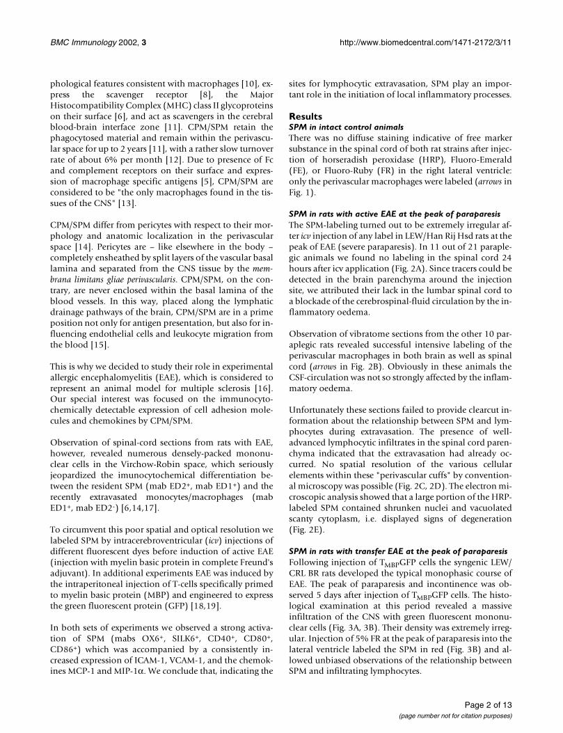
ResultsSPM in intact control animalsThere was no diffuse staining indicative of free markersubstance in the spinal cord of both rat strains after injec-tion of horseradish peroxidase (HRP), Fluoro-Emerald(FE), or Fluoro-Ruby (FR) in the right lateral ventricle:only the perivascular macrophages were labeled (arrows inFig. 1).
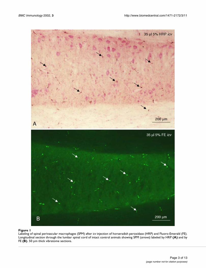
SPM in rats with active EAE at the peak of paraparesisThe SPM-labeling turned out to be extremely irregular af-ter icv injection of any label in LEW/Han Rij Hsd rats at thepeak of EAE (severe paraparesis). In 11 out of 21 paraple-gic animals we found no labeling in the spinal cord 24hours after icv application (Fig. 2A). Since tracers could bedetected in the brain parenchyma around the injectionsite, we attributed their lack in the lumbar spinal cord toa blockade of the cerebrospinal-fluid circulation by the in-flammatory oedema.
Observation of vibratome sections from the other 10 par-aplegic rats revealed successful intensive labeling of theperivascular macrophages in both brain as well as spinalcord (arrows in Fig. 2B). Obviously in these animals theCSF-circulation was not so strongly affected by the inflam-matory oedema.
Unfortunately these sections failed to provide clearcut in-formation about the relationship between SPM and lym-phocytes during extravasation. The presence of well-advanced lymphocytic infiltrates in the spinal cord paren-chyma indicated that the extravasation had already oc-curred. No spatial resolution of the various cellularelements within these "perivascular cuffs" by convention-al microscopy was possible (Fig. 2C, 2D). The electron mi-croscopic analysis showed that a large portion of the HRP-labeled SPM contained shrunken nuclei and vacuolatedscanty cytoplasm, i.e. displayed signs of degeneration(Fig. 2E).
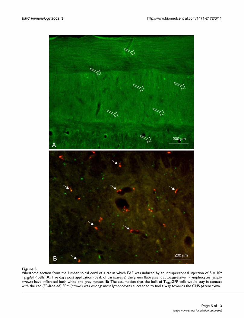
SPM in rats with transfer EAE at the peak of paraparesisFollowing injection of TMBPGFP cells the syngenic LEW/CRL BR rats developed the typical monophasic course ofEAE. The peak of paraparesis and incontinence was ob-served 5 days after injection of TMBPGFP cells. The histo-logical examination at this period revealed a massiveinfiltration of the CNS with green fluorescent mononu-clear cells (Fig. 3A, 3B). Their density was extremely irreg-ular. Injection of 5% FR at the peak of paraparesis into thelateral ventricle labeled the SPM in red (Fig. 3B) and al-lowed unbiased observations of the relationship betweenSPM and infiltrating lymphocytes.
Page 2 of 13(page number not for citation purposes)
BMC Immunology 2002, 3 http://www.biomedcentral.com/1471-2172/3/11
Figure 1Labeling of spinal perivascular macrophages (SPM) after icv injection of horseradish peroxidase (HRP) and Fluoro-Emerald (FE).Longitudinal section through the lumbar spinal cord of intact control animals showing SPM (arrows) labeled by HRP (A) and byFE (B). 50 µm thick vibratome sections.
Page 3 of 13(page number not for citation purposes)
BMC Immunology 2002, 3 http://www.biomedcentral.com/1471-2172/3/11
Figure 2Labeling of spinal perivascular macrophages (SPM) after icv injection of HRP in rats with EAE. A: Spinal cord of a rat with asevere paraparesis showing large erythrocytic infiltrates but no labeling of SPM; 50 µm vibratome section. B: The successfulHRP-DAB labeling of SPM (arrows) in rats with EAE reveals more labeled SPM than in normal control animals; 50 µm vibratomesection. C: The dense perivascular leukocytic infiltrates in the spinal cord parenchyma of rats with severe paraparesis allowonly poor spatial resolution between the endothelial and extravasated cells in 50 µm thick vibratome sections. D: No differen-tiation among the various types of extravasated cells filling the periavscular space was possible also in plastic semithin sections(0.5 µm thick) counterstained with toluidine blue. E: Rat with severe paraparesis. Electron micrograph showing a HRP-labeledSPM with vacuolated cytoplasm. Counterstaining with uranyl acetate and lead citrate.
Page 4 of 13(page number not for citation purposes)
BMC Immunology 2002, 3 http://www.biomedcentral.com/1471-2172/3/11
Figure 3Vibratome section from the lumbar spinal cord of a rat in which EAE was induced by an intraperitoneal injection of 5 × 10⁶
TMBPGFP cells. A: Five days post application (peak of paraparesis) the green fluorescent autoaggressive T-lymphocytes (emptyarrows) have infiltrated both white and grey matter. B: The assumption that the bulk of TMBPGFP cells would stay in contactwith the red (FR-labeled) SPM (arrows) was wrong: most lymphocytes succeeded to find a way towards the CNS parenchyma.
Page 5 of 13(page number not for citation purposes)
BMC Immunology 2002, 3 http://www.biomedcentral.com/1471-2172/3/11
These new data falsified one expectation that had beenbased on earlier work. Our hypothesis, that most (if notall) of the red-fluorescent SPM would be approached andcontacted by the extravasated autoagressive lymphocytes,turned out to be wrong. Despite a general impression thatsome TMBPGFP cells adhered to SPM, most lymphocytesfound a way into the CNS parenchyma through the wallsof capillaries and venules which do not possess a SPM lin-ing.
Thus, the invasion of leukocytes (non-labeled as well asTMBPGFP cells) occurred despite the barrier role of SPM toCNS infiltration which we had previously shown in a dif-ferent kind of experiment [4]. Being a component of aneuroprotective mechanism of extraparenchymal antigenpresentation and prevention of infiltrates, activated SPMcan impede blood cells from crossing the lamina superfi-cialis gliae limitans and infiltrate CNS parenchyma underconditions of a non-inflammatory immune response to exoge-nous proteins[4]. During EAE, however, SPM fail to stop theinfiltration of CNS parenchyma by highly activated lym-phocytes (primed against MBP). Obviously the degree ofT cell activation, the number of activated T cells, and the
localization of the target antigen in the CNS are all impor-tant factors in determining the extent of intraparenchymalT cell infiltration.
Immunlogical pertinence of the non-phagocytic cellsMany non-phagocytic cells in the spinal perivascularspace, which were not labeled by the icv applied fluores-cent dyes, could be stained by the lymphocyte-recogniz-ing antibodies R73, W3/25, and OX-50 (data not shown;see however Fig. 4 in Walther et al., 2001) [4].
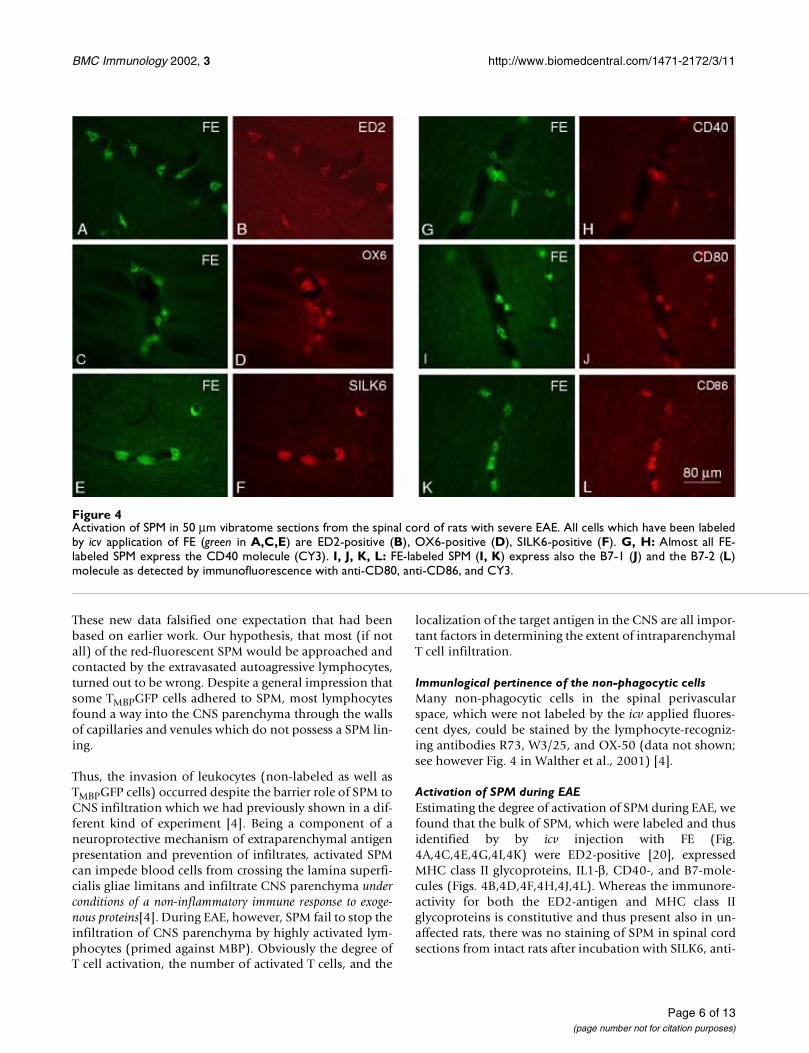
Activation of SPM during EAEEstimating the degree of activation of SPM during EAE, wefound that the bulk of SPM, which were labeled and thusidentified by by icv injection with FE (Fig.4A,4C,4E,4G,4I,4K) were ED2-positive [20], expressedMHC class II glycoproteins, IL1-β, CD40-, and B7-mole-cules (Figs. 4B,4D,4F,4H,4J,4L). Whereas the immunore-activity for both the ED2-antigen and MHC class IIglycoproteins is constitutive and thus present also in un-affected rats, there was no staining of SPM in spinal cordsections from intact rats after incubation with SILK6, anti-
Figure 4Activation of SPM in 50 µm vibratome sections from the spinal cord of rats with severe EAE. All cells which have been labeledby icv application of FE (green in A,C,E) are ED2-positive (B), OX6-positive (D), SILK6-positive (F). G, H: Almost all FE-labeled SPM express the CD40 molecule (CY3). I, J, K, L: FE-labeled SPM (I, K) express also the B7-1 (J) and the B7-2 (L)molecule as detected by immunofluorescence with anti-CD80, anti-CD86, and CY3.
Page 6 of 13(page number not for citation purposes)
BMC Immunology 2002, 3 http://www.biomedcentral.com/1471-2172/3/11
CD40, anti-CD80, anti-CD86 (data not shown; see how-ever the right column of Fig. 3 in Walther et al., 2001) [4].
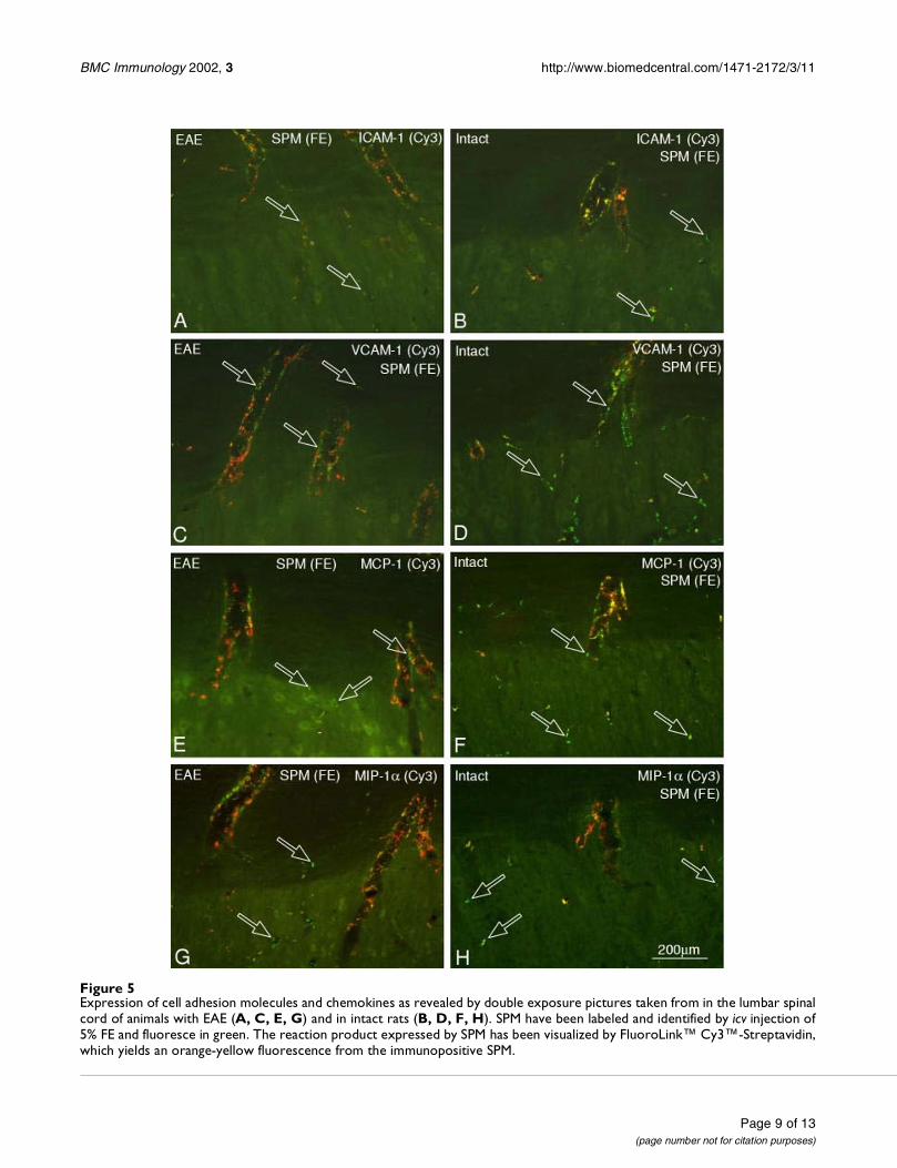
Expression of cell adhesion molecules by SPM during EAETo elucidate the putative chemoattracting potential ofSPM, which were labeled green by an icv injection of FE,we investigated whether they expressed molecules pro-moting extravasation of leukocytes. Despite the commonview that during ongoing inflammation the expression ofadhesion molecules by the cerebral endothelium is notsufficient to support leukocyte recruitment [21–23], wecompared the expression of cell adhesion molecules be-tween unaffected rats and animals with EAE. Followingincubation with polyclonal anti-ICAM-1 and anti-VCAM-1 and visualization of the immunoreaction product by thered fluorescence of Cy3, we observed much more immu-nopositive SPM (orange-yellow fluorescence) in the para-plegic rats (Fig. 5A,5C) than in the intact controls (Fig.5B,5D). Although the numerous and densely-packed cellsin and around the Virchow-Robin space rendered difficultthe discrimination between endothelial cells, recruitedmacrophages and activated SPM, these results are in linewith the earlier findings demonstrating expression ofVCAM-1 mRNA in cerebral perivascular macrophages inthe acute phase of immune-mediated injury [24].
Production of chemokines by SPM during EAEChemokines are proposed to play a role in CNS inflam-matory disease at the stage of leukocyte recruitment in theperivascular space in response to activated antigen-specif-ic T-lymphocytes [25,26]. Together with their receptorsthe chemokines build a complex and sophisticated bio-chemical signaling system involved in the regulation ofleukocyte and lymphocyte trafficking [27,28]. Both, adhe-sion molecule expression as well as production of chem-okines within the CNS have been reported (reviewed inPrat et al., 2001) [29]. However, sound morphological ev-idence elucidating mechanism(s) by which precisely iden-tified SPM (but not perivascular glial cells or perivascularastrocytes) could promote extravasation of leukocytesinto the Virchow-Robin space and thus initiate inflamma-tory relapses, is still missing.
The chemokines MCP-1 (monocyte chemoattractant pro-tein) and MIP-1α (macrophage inflammatory protein 1α)are known to play crucial roles in recruiting macrophagesand monocytes [30–32]. Using monoclonal mouse anti-bodies we observed more MCP-1 and MIP-1α positiveSPM (Fig. 5E,5G) in animals with EAE than in intact con-trol rats (Fig. 5F,5H).
DiscussionClinical importance of T-cell extravasationA major issue in the pathogenesis of any autoimmuneneurological disease is how autologous proteins, that had
been pathologically altered "behind" an intact blood-brain barrier (BBB) can mediate T-lymphocyte entry intothe central nervous system (CNS) and trigger inflammato-ry relapses [33]. These relapses consist of (i) early proin-flammatory response, (ii) recruitment of antigen specificT-lymphocytes, and (iii) invasion of leukocytes (mono-cytes/macrophages, T- and B-lymphocytes) into the brainparenchyma [34–38].
The last process listed, i.e. the accumulation of immuneeffectors around the cerebral microvasculature ("perivas-cular lymphocytic cuffs") is considered to initiate or exac-erbate an inflammatory process, which is accompanied bya localized breakdown of the blood-brain barrier to serumproteins, leading to focal oedema [39,40]. The "bystanderdemyelination" [41,42], together with the subsequent in-terruption of axons [43,44], provide the pathologic corre-late of irreversible neurologic impairment [45].
In contrast, the molecular mechanisms which rule the firsttwo processes, i.e. the proinflammatory response and thesubsequent CNS-recruitment of T-lymphocytes, remainunclear. This lack of sound knowledge is very disappoint-ing because in the course of many immune-mediated dis-eases such as multiple sclerosis, postinfectiousencephalomyelitis, viral encephalitis, the entry of T-lym-phocytes into the CNS is one of the earliest events in theirpathogenesis [46–48]. Morphologically, T-lymphocyteshave been shown to cross the BBB via a transendothelialroute, preferentially in parajunctional areas of endothelialcells [49]. Interestingly, there appears to be a particularlyhigh density of endothelial adhesion molecule expressionin these areas [50]. Although the large number of T-lym-phocytes seen in specific regions or plaques in the acutestage of multiple sclerosis suggests that there is specific tar-geting in the immune response [15], the basic mecha-nisms ruling the extravasation of lymphocytes into theperivascular (Virchow-Robin) space and the subsequentinfiltration of the brain-parenchyma have not been entire-ly elucidated yet.
Possible role of T-cell receptors (TCR)The generally accepted current theory is based on the factthat CNS is continuously patrolled by activated T-lym-phocytes [49–51]. Upon antigenic stimulation to a blastphase [52] these lymphocytes which carry T cell receptors(TCR) for CNS autoantigens initiate an "encephalitogenicimmune response", i.e. if these "antigen seeking" T-cellsencounter their antigen in the CNS [46,52–54] they couldpromote an inflammatory reaction through the secretionof pro-inflammatory cytokines [55–58]. This leads to theupregulation of endothelial adhesion molecules and thelocal production of chemokines, which in concert facili-tate the entry of circulating B- and T-lymphocytes [59] andinflammatory effector cells into the local lesion sites [60].
Page 7 of 13(page number not for citation purposes)
BMC Immunology 2002, 3 http://www.biomedcentral.com/1471-2172/3/11
This "direct" or "chemotaxis-based" theory of extravasa-tion and infiltration acknowledges the "site-specific lym-phocyte entry" as a key event for the progression of diseaseand outcome [21,36,61–63].
Possible role(s) of the resident antigen presenting cellsSince, however, the inflammation-triggering antigen rec-ognition by T-lymphocytes can take place only after pres-entation of antigen in association with the MHC class IImolecule, the role of the resident antigen presenting cells(APC) during lymphocyte extravasation is a matter of con-tinuously expanding interest [53,64–67]. Earlier work hasshown that the most probable candidate for antigen pres-entation within the rat CNS are the ED2-positive CPM/SPM [reviewed in [68]].
Due to their strategic location distal to the blood-en-dothelial interface and proximal to the glia limitans,CPM/SPM are able to phagocytize neuronal debris in thecerebral interstitium and to contact extravasated T-lym-phocytes [9,69]. CPM/SPM synthesize MHC class II glyco-proteins constitutively and, following neuronal death,transform into IL-1β secreting neuronophages [70,71].Recent evidence also implicates SPM as primary targets ofhuman immunodeficiency virus (HIV) and simian immu-nodeficiency virus (SIV) infection in the CNS of humansand macaques [72]. Anyway, these cells must not bemixed up with other "sentinels" of the immune systemwithin the brain, e.g. the ED2-negative dendritic cells inthe meninges and choroid plexus [73].
Accordingly, current knowledge shows that since the syn-thesis of T-cell receptors (TCR) is directly dependent onthe concentration of MHC class II peptides expressed byAPC [74], the latter are able to mediate the trafficking ofspecific antigen-primed T-lymphocytes [75] or B-lym-phocytes [76,77] into the cerebral area. Thus, the capacityto activate lymphocytes for transfer of severe experimentalautoimmune encephalomyelitis has been attributed tocerebral APC [2,75,78]. Furthermore, activating memory/blast CD4+ T-lymphocytes through secretion of cytokines[79,80] and co-stimulatory molecules, APC could play akey role in the repetitive lymphocytic invasions and leu-kocyte recruitment [81–83]. Finally, selective depletion ofblood-borne or resident perivascular macrophages duringEAE causes suppression of clinical symptoms [84–89].
ConclusionsIn the present study we used a combination of a techniquefor invasive, but very reliable labeling of SPM plus an im-munocytochemcal analysis of their activity status. Study-ing the reactivity of identified SPM in very small details,this report provides evidence for increased expression ofcell adhesion molecules (ICAM-1, VCAM-1) and intensi-fied production of chemokines (MCP-1, MIP-1α) by SPM
during EAE. Since these findings, which could enhance T-cell extravasation, were observed in both "antigen-in-duced or active" and "T cell transfer-induced" forms ofEAE, we suppose that they reflect a common neurobio-logical response of antigen-presenting SPM upon interac-tion with primed T-lymphocytes.
Materials and MethodsAnimalsYoung adult (3 months of age) male Lewis rats were used:Twenty-six animals were from the strain LEW/Han Rij Hsd(purchased from Harlan Winkelmann, D-33176 Borchen,Germany) and 16 rats from the strain LEW/CRL BR (pur-chased from Charles River Deutschland, D-97633Sulzfeld, Germany). Before and after experiments all ratswere kept on standard laboratory food and tap water ad li-bitum with an artificial light-dark cycle of 12 hours lighton, 12 hours light off. All experiments were conducted inaccordance with the German law on Animal Protectionand all procedures were approved by the Local Animal-Protection Committe (Bezirksregierung Köln, Az.23.203.2-K 35, 33/98).
Overview of experimentsSixteen LEW/Han Rij Hsd animals comprised the intactcontrol group and were used to optimize the labeling ofspinal perivascular macrophages (SPM) by intracerebrov-entricular (icv) injections of horseradish peroxidase (HRP;4 rats) or fluorochrome-conjugated dextrans (4 rats). Oth-er 8 rats were used to immunocytochemically demon-strate the basic expression of molecules promoting SPMactivation and lymphocyte extravasation.
The antigen-induced EAE (active EAE) group consistedof 10 rats which were used to immunocytochemicallydemonstrate the reactive changes in SPM during EAE.
The T cell transfer-induced EAE (transfer EAE) groupconsisted of 16 LEW/CRL BR rats. Eight animals were usedas intact controls. The other 8 rats received an intraperito-neal injection of 5 × 10⁶ autoaggressive MBP-specific T-lymphocytes in 3 ml 10% FCS-containing medium. Sincethese cells had been genetically engineered to express thegreen fluorescent protein (GFP), they were readily detect-able within the heterogenous perivascular infiltrates("cuffs") in the spinal cord of rats with EAE [18]. A pre-ceeding labeling of the SPM in red fluorescence (Fluoro-Ruby) revealed the complex relationships between the res-ident SPM and the extravasated GFP-transduced, MBP-specific (TMBPGFP) lymphocytes.
Labeling of SPM employing intracerebroventricular (icv) injectionsAfter an intraperitoneal injection of Ketamin plus Xylazin(100 mg Ketanest® plus 5 mg Rompun® per kg body
Page 8 of 13(page number not for citation purposes)
BMC Immunology 2002, 3 http://www.biomedcentral.com/1471-2172/3/11
Figure 5Expression of cell adhesion molecules and chemokines as revealed by double exposure pictures taken from in the lumbar spinalcord of animals with EAE (A, C, E, G) and in intact rats (B, D, F, H). SPM have been labeled and identified by icv injection of5% FE and fluoresce in green. The reaction product expressed by SPM has been visualized by FluoroLink™ Cy3™-Streptavidin,which yields an orange-yellow fluorescence from the immunopositive SPM.
Page 9 of 13(page number not for citation purposes)
BMC Immunology 2002, 3 http://www.biomedcentral.com/1471-2172/3/11
weight) the rats were fixed in a stereotactic apparatus andreceived 35 µl of a 5% solution of horseradish peroxidase(HRP, Type VIA, Sigma, No. P 6782), fluorescein-dextran(Fluoro-Emerald, FE; Molecular Probes, Cat. No. D-1820)or rhodamine-dextran (Fluoro-Ruby, FR; MolecularProbes D-1817) in 0.9% NaCl saline into the right lateralcerebral ventricle. All solutions were injected over a periodof 5 min [90] using a very thin (10 µm thick) glass pipetteto minimize tissue damage [91].
Induction of experimental allergic encephalomyelitis (EAE)EAE was induced in 37 LEW/Han Rij Hsd rats by a singleintradermal (hind footpad) injection containing 25 µgmyelin basic protein from guinea pig (MBP; SigmaM2295), 100 µg Mycobacterium tuberculosis H37 RA(Difco Laboratories, Cat. Nr. 3114-33-8) in 50 µl Com-plete Freund's Adjuvant (CFA; Difco Laboratories, Cat. Nr.0638-60-7).
Tissue processingFixationAll rats were anesthetized with ether, their vascular systemrinsed for 60 sec with 0.9% NaCl saline and fixed for atleast 40 min by transcardial perfusion with 1.0 litre ofperiodate-lysine-paraformaldehyde (PLP) fixative [92].The PLP fixative was separately mixed from premade stemstock solutions immediately prior to the perfusion of eachanimal. The CNS (cerebrum, cerebellum, brainstem, spi-nal cord) was removed and stored in a 4% (w/v) parafor-maldehyde in 0.1 M phosphate buffer, pH 7,4.
Visualizing of HRP-labeled SPMIn contrast to our previous work [4] we focused our obser-vations on the lumbar spinal cord only: Since the majorvisible symptom during EAE is the severe paraparesis ofthe hind limbs, most of the pathological alterationsshould be expected, localized and observed in this region.Longitudinal vibratome sections (50 µm thick) of the spi-nal cord were incubated for 20 min in 0.75% (w/v) 3,3'-diaminobenzidine tetrahydrochloride (150 mg in 200 mlbuffer; DAB, Sigma, D 5637) plus 0.01% (v/v) H2O2 plus0.075% (w/v) nickel chloride in 0.05 M Tris-HCl buffer,pH 7.6. This procedure yielded a very intensive dark-pur-ple to black reaction product exclusively in the cytoplasmof the SPM. Control tissue (sections from brains whichhad not received HRP injections) contained no reactionproduct. For electron microscopy some of the sectionswere postfixed in 1% OsO4 + 1.5% K3
IIIFe(CN)6[93], de-hydrated in graded acetones, and embedded flat in Arald-ite CY212 (Fluka, No. 44610).
Visualizing of FE- and FR-labeled SPMTo detect the green fluorescence of FE or the red fluores-cence of FR vibratome sections (50 µm thick) of the spinal
cord were illuminated through filter set 10 (excitation BP450-490, beamsplitter FT 510, emission BP 515-565) orfilter set 15 (Excitation BP 546/12, Emission LP 590) of aCarl Zeiss microscope.
ImmunocytochemistryPrimary antibodies(1) Mouse monoclonal OX-6 (Serotec, MCA 46, 1:1000)recognizing rat MHC class II (Ia) antigen. (2) Mouse mon-oclonal SILK 6 (Serotec, MCA-1397, 1:500) recognizingrecombinant rat IL-1β. (3) Armenian hamster mono-clonal anti-mouse CD40 (BD PharMingen, 09401A,1:100) to test for presence of the costimulatory moleculeCD40 on SPM. (4) Mouse monoclonal CD80 (BDPharMingen, 22661D, 1:100) to test for presence of theco-stimulatory molecule B7-1 on rat SPM. (5) Mousemonoclonal CD86 (BD PharMingen, 22671D, 1:100) totest for presence of the co-stimulatory molecule B7-2 onrat SPM. (6) Monoclonal mouse anti-rat TCR alpha/beta(clone R73, Serotec MCA 453G, 1:20) recognizing a con-stant determinant of the α/β T cell receptor. (7) Mono-clonal mouse anti-rat CD4 (clone number W3/25, SerotecMCA55G, 1:50) recognizing the CD4 cell surface glyco-protein expressed by helper T cells and thymocytes. (8)Monoclonal mouse anti-rat CD44 (clone OX-50, SerotecMCA643XZ, 1:50) recognizing the CD44 cell surface anti-gen expressed by T cells, B cells, macrophages, and thymo-cytes. (9) Polyclonal goat anti-rat ICAM-1(M-19; SantaCruz Biotechnology Inc., sc-1511, 1:50) recognizing ratintercellular adhesion molecule-1. (10) Polyclonal goatanti-rat VCAM-1(C-19; Santa Cruz Biotechnology Inc., sc-1504, 1:50) recognizing rat vascular cell adhesion mole-cule-1. (11) Polyclonal rabbit anti-rat MCP-1 (Cedarlane,No. CL9576AP, 1:500). recognizing the CC-chemokine"monocyte chemoattractant protein-1" which promotesthe directed migration of inflammatory cells. (12) Poly-clonal rabbit anti-rat MIP-1α (Cedarlane, No. CL9577AP,1:2000) recognizing the chemokine "macrophage inflam-matory protein-1α ".
Secondary antibodies(1) Biotinylated goat anti-rabbit IgG (DAKO, No. E0432).(2) Biotinylated goat anti-mouse IgG (Fab-specific, Sigma,B-0529). (3) Biotinylated goat anti-mouse IgG (Fc specif-ic, pre-absorbed with human IgG and rat serum proteins,Sigma, No. B-9904). (4) Biotinylated rabbit anti-goat IgG(DAKO, No. E0466). In order to minimize the back-ground staining of neurons the biotinylated IgGs were al-ways pre-absorbed with the individual rat's spleen protein[70]. (5) Biotin-conjugated mouse anti-hamster IgG(cocktail; PharMingen, 12102D).
Standard incubation protocolFree floating vibratome sections were immunostained ona shaker at room temperature through the following steps:
Page 10 of 13(page number not for citation purposes)
BMC Immunology 2002, 3 http://www.biomedcentral.com/1471-2172/3/11
(1) 0.6% (v/v) H2O2 in buffer (to block the endogenousperoxidase activity) for 30 min; (2) 5.0% (w/v) bovine se-rum albumin (BSA, Sigma No. A-9647) in 0.1 M Tris-buff-ered saline (TBS) pH 7.6 for 60 min; (3) primary antibody(see above) diluted in TBS plus 0.8% (w/v) BSA for 2 h;(4) 5.0% (v/v) normal goat serum (NGS, Vector No. S-1000) or normal rabbit serum (NRS, DAKO, No. X0902)plus 0.8% BSA in TBS for 15 min. (5) 1:400 dilution of thecorresponding biotinylated secondary antibody in TBS for1 h; (6) 1:100 diluted FluoroLink™ Cy2™-Streptavidin(Amersham Life Science, PA 42001) or FluoroLink™Cy3™-Streptavidin (Amersham Life Science, PA 43001)for 1 h. Steps (1) and (5) were followed by two 10 minwashes in TBS. Steps (3) and (6) were followed by four 10min washes in TBS. Finally, sections were dehydrated withethanol and Histoclear (a nontoxic xylene-substitute) andcoverslipped.
ControlsOmission of the primary or secondary antibody yieldedblank sections in which only the green SPM fluoresced.Incubation of sections with non-relevant biotinylated sec-ondary antibodies (e.g. goat anti-rabbit IgG for recogni-tion of mouse primary antibodies) yielded also blanksections.
Fluorescence microscopyFE-labeled SPM and Cy2-immunopositive cells were visu-alized through filter set 10 of a Carl Zeiss fluorescence mi-croscope. FR-labeled SPM and Cy3-immunopositive cellswere observed through Zeiss filter set 15 (excitationBP546/12, beamsplitter FT580, emission LP590). BothFE-labeled SPM and CY3-immunopositive cells could besimultaneously observed employing Zeiss filter set 25 (ex-citation TBP400/495/570, beamsplitter FT410/505/585,emission TBP460/530/610).
Authors' contributionsN.H. participated in the icv labelings and counts of theSPP. N.L. performed the immunocytochemistry. M.S. par-ticipated in the icv labelings. B.W. performed the intra-peritoneal injections of TMBPGFP lymphocytes. W.F.N.participated in the coordination of the study. O.G.L. par-ticipated in the icv labelings. D.N.A. designed the study,induced EAE, participated in the icv labelings and per-fused the animals.
All authors read and approved the final manuscript.
AcknowledgementsThis work has been financially supported by the Imhoff-Foundation (D.N.A.), the Jean Uhrmacher-Foundation (O.G.-L.), and the Megapharm GmbH, Germany (M.S.). The authors are thankful to Dr. Alexander Flügel (Max-Planck-Institut für Neurobiologie, Martinsried, Germany) for the gen-erously provided TMBPGFP cells. The skillful technical assistance of Mrs. Il-ona Rohrmann and Mrs. Dirkje Felder and the fine photographical work of Mrs. Inge Koch are highly appreciated.
References1. Marrack P, Kappler J, Kotzin B: Autoimmune disease:why and
where it occurs. Nature Medicine 2001, 7:899-9052. Perry VH: A revised view of the central nervous system mi-
croenvironment and major histocompatibility complex classII antigen presentation. J Neuroimmunol 1998, 90:113-121
3. Archelos JJ, Hartung H-P: Pathogenetic role of autoantibodies inneurological disease. TINS 2000, 23:317-327
4. Walther M, Popratiloff AS, Lachnit N, Hofmann N, Streppel M, Gun-tinas-Lichius O, Neiss WF, Angelov DN: Exogenous antigen con-taining perivascular phagocytes induce a non-encephalitogenic extravasation of primed lymphocytes. JNeuroimmunol 2001, 117:30-42
5. Hickey WF, Kimura H: Perivascular microglial cells of the CNSare bone marrow derived and present antigen in vivo. Science1988, 239:290-292
6. Graeber MB, Streit WJ, Kreutzberg GW: Identity of ED2-positiveperivascular cells in rat brain. J Neurosci Res 1989, 22:103-106
7. Graeber MB, Streit WJ, Büringer D, Larry Sparks D, Kreutzberg GW:Ultrastructural location of major histocompatibility com-plex (MHC) class II positive perivascular J Neuropathol 1992,cells in histologically normal human brain.:303-311
8. Mato N, Ookara S, Sakamoto A, Aikawa E, Ogawa T, Mitsuhashi U,Masuzawa T, Suzuki H, Honda M, Yazaki Y, Watanabe E, Luoma J, Yla-Herttuala S, Fraser I, Gordon S, Kodama T: Involvement of specificmacrophage-lineage cell surrounding arterioles in barrierand scavenger function in brain cortex. Proc Natl Acad Sci USA1996, 93:3269-3274
9. Angelov DN, Walther M, Streppel M, Guntinas-Lichius O, Neiss WF:The cerebral perivascular cells. Advances in Anatomy, Embryologyand Cell Biology Springer Verlag Berlin 1998, 147:
10. Mori S, Leblond CP: Identification of microglia in light and elec-tron microscopy. J Comp Neuol 1969, 135:57-80
11. Kida S, Steart PV, Zhang ET, Weller RO: Perivascular cells act asscavengers in the cerebral perivascular spaces and remaindistinct from pericytes, microglia and macrophages. Acta Neu-ropathol 1993, 85:646-652
12. Bechmann I, Kwidzinski E, Kovac AD, Simbürger E, Horvath T, GimsaU, Dirnag U, Priller J, Nitsch R: Turnover of rat brain perivascu-lar cells. Exp Neurol 2001, 168:242-249
13. Dijkstra CD, Damoiseaux JGMC: Macrophage heterogeneity es-tablished by immunocytochemistry. Progr Histochem Cytochem1993, 27:1-65
14. Graeber MM, Streit WJ, Kiefer R, Schoen S, Kreutzberg GW: Newexpression of myelomonocytic antigens by microglia andperivascular cells following lethal motor neuron injury. J Neu-roimmunol 1990, 27:121-132
15. Weller RO, Engelhardt B, Phillips MJ: Lymphocyte targeting ofthe central nervous system: A review of afferent and efferentCNS-immune pathways. Brain Pathol 1996, 6:275-288
16. Raine CS: The Dale E. McFarlin memorial lecture: The immu-nology of the multiple sclerosis lesion. Ann Neurol 1994, 36:S61-S72
17. Dijkstra CD, Döpp EA, van den Berg TK, Damoiseaux JGMC: Mon-oclonal antibodies against rat macrophages. J Immunol Methods1994, 174:21-23
18. Flügel A, Willem M, Berkowicz T, Wekerle H: Gene transfer intoCD4+ T lymphocytes: green fluorescent protein-engineered,encephalitogenic T cells illuminate brain autoimmune re-sponses. Nature Medicine 1999, 5:843-847
19. Flügel A, Bradl M: New tools to trace populations of inflamma-tory cells in the CNS. Glia 2001, 36:125-136
20. Dijkstra CD, Döpp EA, Joling P, Kraal G: The heterogeneity ofmononuclear phagocytes in lymphoid organs: distinct mac-rophage subpopulations in the rat recognized by monoclonalantibodies ED1, ED2 and ED3. Immunology 1985, 54:589-599
21. Engelhardt B, Conley FK, Butcher EC: Cell-adhesion molecules onvessels during J Neuroimmunol 1994, inflammation in the mousecentral nervous system:199-208
22. Bell MD, Perry VH: Adhesion molecule expression on murinecerebral endothelium following the injection of a proinflam-mogen or during acute neuronal J Neurocytol 1995, degenera-tion.:695-710
23. Perry VH, Bell MD, Brown HC, Matyszak MK: Inflamation in thenervous system. Curr Opin Neurobiol 1995, 5:636-641
Page 11 of 13(page number not for citation purposes)
http://www.ncbi.nlm.nih.gov/entrez/query.fcgi?cmd=Retrieve&db=PubMed&dopt=Abstract&list_uids=9817438
http://www.ncbi.nlm.nih.gov/entrez/query.fcgi?cmd=Retrieve&db=PubMed&dopt=Abstract&list_uids=9817438
http://www.ncbi.nlm.nih.gov/entrez/query.fcgi?cmd=Retrieve&db=PubMed&dopt=Abstract&list_uids=9817438
http://www.ncbi.nlm.nih.gov/entrez/query.fcgi?cmd=Retrieve&db=PubMed&dopt=Abstract&list_uids=9817438
http://www.ncbi.nlm.nih.gov/entrez/query.fcgi?cmd=Retrieve&db=PubMed&dopt=Abstract&list_uids=3276004
http://www.ncbi.nlm.nih.gov/entrez/query.fcgi?cmd=Retrieve&db=PubMed&dopt=Abstract&list_uids=3276004
http://www.ncbi.nlm.nih.gov/entrez/query.fcgi?cmd=Retrieve&db=PubMed&dopt=Abstract&list_uids=3276004
http://www.ncbi.nlm.nih.gov/entrez/query.fcgi?cmd=Retrieve&db=PubMed&dopt=Abstract&list_uids=2926837
http://www.ncbi.nlm.nih.gov/entrez/query.fcgi?cmd=Retrieve&db=PubMed&dopt=Abstract&list_uids=2926837
http://www.ncbi.nlm.nih.gov/entrez/query.fcgi?cmd=Retrieve&db=PubMed&dopt=Abstract&list_uids=2926837
http://www.ncbi.nlm.nih.gov/entrez/query.fcgi?cmd=Retrieve&db=PubMed&dopt=Abstract&list_uids=8622926
http://www.ncbi.nlm.nih.gov/entrez/query.fcgi?cmd=Retrieve&db=PubMed&dopt=Abstract&list_uids=8622926
http://www.ncbi.nlm.nih.gov/entrez/query.fcgi?cmd=Retrieve&db=PubMed&dopt=Abstract&list_uids=8622926
http://www.ncbi.nlm.nih.gov/entrez/query.fcgi?cmd=Retrieve&db=PubMed&dopt=Abstract&list_uids=8337943
http://www.ncbi.nlm.nih.gov/entrez/query.fcgi?cmd=Retrieve&db=PubMed&dopt=Abstract&list_uids=8337943
http://www.ncbi.nlm.nih.gov/entrez/query.fcgi?cmd=Retrieve&db=PubMed&dopt=Abstract&list_uids=8337943
http://www.ncbi.nlm.nih.gov/entrez/query.fcgi?cmd=Retrieve&db=PubMed&dopt=Abstract&list_uids=8337943
http://www.ncbi.nlm.nih.gov/entrez/query.fcgi?cmd=Retrieve&db=PubMed&dopt=Abstract&list_uids=2332482
http://www.ncbi.nlm.nih.gov/entrez/query.fcgi?cmd=Retrieve&db=PubMed&dopt=Abstract&list_uids=2332482
http://www.ncbi.nlm.nih.gov/entrez/query.fcgi?cmd=Retrieve&db=PubMed&dopt=Abstract&list_uids=2332482
http://www.ncbi.nlm.nih.gov/entrez/query.fcgi?cmd=Retrieve&db=PubMed&dopt=Abstract&list_uids=2332482
http://www.ncbi.nlm.nih.gov/entrez/query.fcgi?cmd=Retrieve&db=PubMed&dopt=Abstract&list_uids=8864284
http://www.ncbi.nlm.nih.gov/entrez/query.fcgi?cmd=Retrieve&db=PubMed&dopt=Abstract&list_uids=8864284
http://www.ncbi.nlm.nih.gov/entrez/query.fcgi?cmd=Retrieve&db=PubMed&dopt=Abstract&list_uids=8864284
http://www.ncbi.nlm.nih.gov/entrez/query.fcgi?cmd=Retrieve&db=PubMed&dopt=Abstract&list_uids=8864284
http://www.ncbi.nlm.nih.gov/entrez/query.fcgi?cmd=Retrieve&db=PubMed&dopt=Abstract&list_uids=8017891
http://www.ncbi.nlm.nih.gov/entrez/query.fcgi?cmd=Retrieve&db=PubMed&dopt=Abstract&list_uids=8017891
http://www.ncbi.nlm.nih.gov/entrez/query.fcgi?cmd=Retrieve&db=PubMed&dopt=Abstract&list_uids=8017891
http://www.ncbi.nlm.nih.gov/entrez/query.fcgi?cmd=Retrieve&db=PubMed&dopt=Abstract&list_uids=8083524
http://www.ncbi.nlm.nih.gov/entrez/query.fcgi?cmd=Retrieve&db=PubMed&dopt=Abstract&list_uids=8083524
http://www.ncbi.nlm.nih.gov/entrez/query.fcgi?cmd=Retrieve&db=PubMed&dopt=Abstract&list_uids=8083524
http://www.ncbi.nlm.nih.gov/entrez/query.fcgi?cmd=Retrieve&db=PubMed&dopt=Abstract&list_uids=3882559
http://www.ncbi.nlm.nih.gov/entrez/query.fcgi?cmd=Retrieve&db=PubMed&dopt=Abstract&list_uids=3882559
http://www.ncbi.nlm.nih.gov/entrez/query.fcgi?cmd=Retrieve&db=PubMed&dopt=Abstract&list_uids=3882559
http://www.ncbi.nlm.nih.gov/entrez/query.fcgi?cmd=Retrieve&db=PubMed&dopt=Abstract&list_uids=3882559
http://www.ncbi.nlm.nih.gov/entrez/query.fcgi?cmd=Retrieve&db=PubMed&dopt=Abstract&list_uids=8580715
BMC Immunology 2002, 3 http://www.biomedcentral.com/1471-2172/3/11
24. Jander S, Pohl J, Gillen C, Schroeter M, Stoll G: Vascular cell adhe-sion molecule-1 mRNA is expressed in immune-mediatedand ischemic injury of the rat nervous system. J Neuroimmunol1996, 70:75-80
25. Ransohoff RM, Glabinski AR, Tani M: Chemokines in immune-mediated inflammation of the central nervous system. Cy-tokine and Growth Factor Rev 1996, 7:35-46
26. Ransohoff RM, Tani M, Glabinski AR, Chernosky A, Krivacic K, Peter-son JW, Chien H-F, Trapp BD: Chemokines and chemokine re-ceptors in model neurological pathologies: Molecular andimmunohistochemical approaches. Methods Enzymol 1997,287:319-348
27. Aloisi F: Immune function of microglia. Glia 2001, 36:165-17928. Baggiolini M: Chemokines in pathology and medicine. J Intern
Med 2001, 250:91-10429. Prat A, Biernacki K, Wosik K, Antel JP: Glial cell influence on the
human blood-brain-barrier. Glia 2001, 36:145-15530. Calvo CF, Yoshimura T, Gelman M, Mallat M: Production of Mono-
cyte Chemotactic Protein-1 by rat brain macrophages. Eur JNeurosci 1996, 8:1725-1734
31. Glabinski AR, Tuohy VK, Ransohoff RM: Expression of chemok-ines RANTES, MIP-1alpha and GRO-alpha correlates withinflammation in acute experimental autoimmune encepha-lomyelitis. Neuroimmunomodulation 1998, 5:166-171
32. Rezaie P, Trillo-Pazos G, Everall IP, Male DK: Expression of β-chemokines and chemokine receptors in human fetal astro-cyte and microglial co-cultures: potential role of chemokinesin the developing CNS. Glia 2002, 37:64-75
33. Wekerle H, Linington C, Lassmann H, Meyermann R: Cellular im-mune reactivity within the CNS. TINS 1986, 9:271-277
34. Hickey WF, Kimura H: Graft-vs-host disease elicits expressionof class I and class II hostocompatibility antigens and thepresence of scattered T lymphocytes in rat central nervoussystem. Proc Natl Acad Sci USA 1987, 84:2082-2086
35. Paterson PY, Day ED, Whitacre CC: Neuroimmunologic diseas-es: Effector cell responses and immunoregulatory mecha-nisms. Immunol Rev 1981, 55:89-120
36. Raivich G, Jones LL, Kloss CUA, Werner A, Neumann H, KreutzbergGW: Immune surveillance in the injured nervous system: T-lymphocytes invade the axotomized mouse facial motor nu-cleus and aggregate around sites of neuronal degeneration. JNeurosc 1998, 18:5804-5816
37. Stoll G, Jander S: The role of microglia and macrophages in thepathophysiology of the CNS. Progr Neurobiol 1999, 58:233-247
38. Flügel A, Schwaiger F-W, Neumann H, Medana I, Willem M, WekerleH, Kreutzberg GW, Graeber MB: Neuronal FasL induces celldeath of encephalithogenic T lymphocytes. Brain Pathol 2000,10:353-364
39. Brosnan CF, Claudio L, Tansey FA, Martiney J: Mechanism of au-toimmune neuropathies. Ann Neurol 1990, 27(suppl):75-79
40. Ptak W, Herzog WR, Askenase PW: Delayed-type hypersensitiv-ity initiation by early-acting cells that are antigen mis-matched or MHC incompatible with late-acting, delayed-type hypersensitivity effector T cells. J Immunol 1991, 146:469-75
41. Bauer J, Sminia T, Wouterlood FG, Dijkstra CD: Phagocytic activ-ity of macrophages and microglial cells during the course ofacute and chronic relapsing experimental autoimmune en-cephalomyelitis. J Neurosci Res 1994, 38:365-375
42. Wisniewski HM, Bloom BR: Primary demyelination as a non-specific consequence of a cell-mediated immune reaction. JExp Med 1975, 141:346-359
43. Ferguson B, Matyszak MK, Esiri MM, Perry VH: Axonal damage inacute multiple sclerosis lesions. Brain 1997, 120:393-399
44. Trapp BD, Peterson J, Ransohoff RM, Rudick R, Mörk S, Bö L: Axonaltransection in the lesions of multiple sclerosis. N Engl J Med1998, 338:278-285
45. Kiefer R, Kieseier BC, Stoll G, Hartung H-P: The role of macro-phages in the immune-mediated damage to the peripheralnervous system. Progr Neurobiol 2001, 64:109-127
46. Hickey WF, Hsu BL, Kimura H: T-lymphocyte entry into the cen-tral nervous system. J Neurosci Res 1991, 28:254-260
47. Lassmann H, Zimprich F, Vass K, Hickey WF: Microglial cells are acomponent of the perivascular glia limitans. J Neurosci Res1991, 28:236-243
48. Muhallab S, Lidman O, Weissert R, Olsson T, Svenningsson A: Intra-CNS activation by antigen-specific T lymphocytes in experi-mental autoimmune encephalomyelitis. J Neuroimmunol 2001,113:201-211
49. Raine CS, Cannella B, Duijvestijn AM, Cross AH: Homing to cen-tral nervous system vasculature by antigen-specific lym-phocytes. II. Lymphocyte/endothelial cell adhesion duringthe initial stages of autoimmune demyelination. Lab Invest1990, 63:476-489
50. Lassmann H, Zimprich F, Rössler K, Vass K: Inflammation in thenervous system. Rev Neurol (Paris) 1991, 147:763-781
51. Lassmann H, Vass K, Brunner C, Seutelberger F: Characterizationof inflammatory infiltrates in experimental allergic encepha-lomyelitis. Progr Neuropathol 1986, 6:33-62
52. Hickey WF: Basic principles of immunological surveillance ofthe normal central nervous system. Glia 2001, 36:118-124
53. Cross AH, Cannella B, Brosnan CF, Raine CS: Homing to centralnervous system vasculature by antigen specific lymphocytes.I. Localization of 14C-labeled cells during acute, chronic andrelapsing experimental allergic encephalomyelitis. Lab Invest1990, 63:162-170
54. Krakowsky ML, Owens T: Naive T lymphocytes traffic to in-flamed central nervous system, but require antigen recogni-tion for activation. Eur J Immunol 2000, 30:1002-1009
55. Male D, Pryce G, Rahman J: Comparison of the immunologicalproperties of rat cerebral and aortic endothelium. J Neuroim-munol 1990, 30:167-172
56. Berger T, Weerth S, Kojima K, Linington C, Wekerle H, Lassmann H:Experimental autoimmune encephalomyelitis: the antigenspecificity of T-lymphocytes determines the topography oflesions in the central and peripheral nervous system. Lab In-vest 1997, 76:355-364
57. Serpe CJ, Kohm AP, Huppenbauer CB, Sanders VM, Jones KJ: Exac-erbation of facial motoneuron loss after facial nerve transec-tion in severe combined immunodeficient (scid) mice. JNeurosci 1999, 19RCN:1-5
58. Delves PJ, Roitt IM: The immune system. N Engl J Med 2000,343:37-49
59. Knopf PM, Harling-Berg CJ, Cserr HF, Basu D, Sirulnick EJ, Nolan SC,Park JT, Keir G, Thompson EJ, Hickey WF: Antigen-dependent in-trathecal synthesis in the normal rat brain: tissue entry andlocal retention of antigen specific B cells. J Immunol 1998,161:692-701
60. Lassmann H: Basic mechanisms of brain inflammation. J NeuralTransm Suppl 1997, 50:183-190
61. Engelhardt JI, Tajti J, Appel SH: Lymphocytic infiltrates in the spi-nal cord in amyotrophic lateral sclerosis. Arch Neurol 1993,50:30-36
62. Kawamata T, Akiyama A, Yamada T, McGeer PL: Immunologic re-actions in amyotrophic lateral sclerosis brain and spinalcord. Am J Pathol 1992, 140:691-707
63. Schluesener HJ, Seid K, Kretzschmar J, Meyermann R: Leukocytechemotactic factor, a natural ligand to CD4, is expressed bylymphocytes and microglial cells of the MS plaque. J NeurosciRes 1996, 44:606-611
64. Cross AH, O'Mara T, Raine CS: Chronologic localization of my-elin-reactive cells in the lesions of relapsing EAE: implica-tions for the study of multiple sclerosis. Neurology 1993,43:1028-1033
65. Morimoto K, Craig Hooper D, Bornhorst A, Corisdeo S, Bette M, FuZF, Schäfer MK-H, Koprowsky H, Weihe E, Dietzschold B: Intrinsicresponses to Borna disease virus infection of the centralnervous system. Proc Natl Acad Sci USA 1996, 93:13345-13350
66. Hu P, Pollard J, Hunt N, Chan-Ling T: Microvascular and cellularresponses in the retina of rats with acute experimental aller-gic encephalomyelitis (EAE). Brain Pathol 1998, 8:487-498
67. Deiniger MH, Zhao Y, Schluesener HJ: CP-10, a chemotactic pep-tide, is expressed in lesions of experimental autoimmune en-cephalomyelitis, neuritis, uveitis and in C6 gliomas. JNeuroimmunol 1999, 93:156-163
68. Raivich G, Bohatscheck M, Kloss CUA, Werner A, Neumann H, JonesLL, Kreutzberg GW: Neuroglial activation repertoire in the in-jured brain: graded response, molecular mechanisms andcues to physiological function. Brain Res Rev 1999, 30:77-105
69. Lawrence JM, Morris RJ, Wilson DJ, Raisman G: Mechanisms of al-lograft rejection in the rat brain. Neuroscience 1990, 37:431-462
Page 12 of 13(page number not for citation purposes)
http://www.ncbi.nlm.nih.gov/entrez/query.fcgi?cmd=Retrieve&db=PubMed&dopt=Abstract&list_uids=8862137
http://www.ncbi.nlm.nih.gov/entrez/query.fcgi?cmd=Retrieve&db=PubMed&dopt=Abstract&list_uids=8862137
http://www.ncbi.nlm.nih.gov/entrez/query.fcgi?cmd=Retrieve&db=PubMed&dopt=Abstract&list_uids=8862137
http://www.ncbi.nlm.nih.gov/entrez/query.fcgi?cmd=Retrieve&db=PubMed&dopt=Abstract&list_uids=8862137
http://www.ncbi.nlm.nih.gov/entrez/query.fcgi?cmd=Retrieve&db=PubMed&dopt=Abstract&list_uids=9330331
http://www.ncbi.nlm.nih.gov/entrez/query.fcgi?cmd=Retrieve&db=PubMed&dopt=Abstract&list_uids=9330331
http://www.ncbi.nlm.nih.gov/entrez/query.fcgi?cmd=Retrieve&db=PubMed&dopt=Abstract&list_uids=9330331
http://www.ncbi.nlm.nih.gov/entrez/query.fcgi?cmd=Retrieve&db=PubMed&dopt=Abstract&list_uids=9330331
http://www.ncbi.nlm.nih.gov/entrez/query.fcgi?cmd=Retrieve&db=PubMed&dopt=Abstract&list_uids=9330331
http://www.ncbi.nlm.nih.gov/entrez/query.fcgi?cmd=Retrieve&db=PubMed&dopt=Abstract&list_uids=8921263
http://www.ncbi.nlm.nih.gov/entrez/query.fcgi?cmd=Retrieve&db=PubMed&dopt=Abstract&list_uids=8921263
http://www.ncbi.nlm.nih.gov/entrez/query.fcgi?cmd=Retrieve&db=PubMed&dopt=Abstract&list_uids=8921263
http://www.ncbi.nlm.nih.gov/entrez/query.fcgi?cmd=Retrieve&db=PubMed&dopt=Abstract&list_uids=9730682
http://www.ncbi.nlm.nih.gov/entrez/query.fcgi?cmd=Retrieve&db=PubMed&dopt=Abstract&list_uids=9730682
http://www.ncbi.nlm.nih.gov/entrez/query.fcgi?cmd=Retrieve&db=PubMed&dopt=Abstract&list_uids=9730682
http://www.ncbi.nlm.nih.gov/entrez/query.fcgi?cmd=Retrieve&db=PubMed&dopt=Abstract&list_uids=9730682
http://www.ncbi.nlm.nih.gov/entrez/query.fcgi?cmd=Retrieve&db=PubMed&dopt=Abstract&list_uids=3550805
http://www.ncbi.nlm.nih.gov/entrez/query.fcgi?cmd=Retrieve&db=PubMed&dopt=Abstract&list_uids=3550805
http://www.ncbi.nlm.nih.gov/entrez/query.fcgi?cmd=Retrieve&db=PubMed&dopt=Abstract&list_uids=3550805
http://www.ncbi.nlm.nih.gov/entrez/query.fcgi?cmd=Retrieve&db=PubMed&dopt=Abstract&list_uids=3550805
http://www.ncbi.nlm.nih.gov/entrez/query.fcgi?cmd=Retrieve&db=PubMed&dopt=Abstract&list_uids=6165674
http://www.ncbi.nlm.nih.gov/entrez/query.fcgi?cmd=Retrieve&db=PubMed&dopt=Abstract&list_uids=6165674
http://www.ncbi.nlm.nih.gov/entrez/query.fcgi?cmd=Retrieve&db=PubMed&dopt=Abstract&list_uids=6165674
http://www.ncbi.nlm.nih.gov/entrez/query.fcgi?cmd=Retrieve&db=PubMed&dopt=Abstract&list_uids=6165674
http://www.ncbi.nlm.nih.gov/entrez/query.fcgi?cmd=Retrieve&db=PubMed&dopt=Abstract&list_uids=2137320
http://www.ncbi.nlm.nih.gov/entrez/query.fcgi?cmd=Retrieve&db=PubMed&dopt=Abstract&list_uids=2137320
http://www.ncbi.nlm.nih.gov/entrez/query.fcgi?cmd=Retrieve&db=PubMed&dopt=Abstract&list_uids=2137320
http://www.ncbi.nlm.nih.gov/entrez/query.fcgi?cmd=Retrieve&db=PubMed&dopt=Abstract&list_uids=1987274
http://www.ncbi.nlm.nih.gov/entrez/query.fcgi?cmd=Retrieve&db=PubMed&dopt=Abstract&list_uids=1987274
http://www.ncbi.nlm.nih.gov/entrez/query.fcgi?cmd=Retrieve&db=PubMed&dopt=Abstract&list_uids=1987274
http://www.ncbi.nlm.nih.gov/entrez/query.fcgi?cmd=Retrieve&db=PubMed&dopt=Abstract&list_uids=1987274
http://www.ncbi.nlm.nih.gov/entrez/query.fcgi?cmd=Retrieve&db=PubMed&dopt=Abstract&list_uids=7932870
http://www.ncbi.nlm.nih.gov/entrez/query.fcgi?cmd=Retrieve&db=PubMed&dopt=Abstract&list_uids=7932870
http://www.ncbi.nlm.nih.gov/entrez/query.fcgi?cmd=Retrieve&db=PubMed&dopt=Abstract&list_uids=7932870
http://www.ncbi.nlm.nih.gov/entrez/query.fcgi?cmd=Retrieve&db=PubMed&dopt=Abstract&list_uids=7932870
http://www.ncbi.nlm.nih.gov/entrez/query.fcgi?cmd=Retrieve&db=PubMed&dopt=Abstract&list_uids=9126051
http://www.ncbi.nlm.nih.gov/entrez/query.fcgi?cmd=Retrieve&db=PubMed&dopt=Abstract&list_uids=9126051
http://www.ncbi.nlm.nih.gov/entrez/query.fcgi?cmd=Retrieve&db=PubMed&dopt=Abstract&list_uids=9126051
http://www.ncbi.nlm.nih.gov/entrez/query.fcgi?cmd=Retrieve&db=PubMed&dopt=Abstract&list_uids=9445407
http://www.ncbi.nlm.nih.gov/entrez/query.fcgi?cmd=Retrieve&db=PubMed&dopt=Abstract&list_uids=9445407
http://www.ncbi.nlm.nih.gov/entrez/query.fcgi?cmd=Retrieve&db=PubMed&dopt=Abstract&list_uids=9445407
http://www.ncbi.nlm.nih.gov/entrez/query.fcgi?cmd=Retrieve&db=PubMed&dopt=Abstract&list_uids=2033653
http://www.ncbi.nlm.nih.gov/entrez/query.fcgi?cmd=Retrieve&db=PubMed&dopt=Abstract&list_uids=2033653
http://www.ncbi.nlm.nih.gov/entrez/query.fcgi?cmd=Retrieve&db=PubMed&dopt=Abstract&list_uids=2033653
http://www.ncbi.nlm.nih.gov/entrez/query.fcgi?cmd=Retrieve&db=PubMed&dopt=Abstract&list_uids=2033652
http://www.ncbi.nlm.nih.gov/entrez/query.fcgi?cmd=Retrieve&db=PubMed&dopt=Abstract&list_uids=2033652
http://www.ncbi.nlm.nih.gov/entrez/query.fcgi?cmd=Retrieve&db=PubMed&dopt=Abstract&list_uids=2033652
http://www.ncbi.nlm.nih.gov/entrez/query.fcgi?cmd=Retrieve&db=PubMed&dopt=Abstract&list_uids=1700193
http://www.ncbi.nlm.nih.gov/entrez/query.fcgi?cmd=Retrieve&db=PubMed&dopt=Abstract&list_uids=1700193
http://www.ncbi.nlm.nih.gov/entrez/query.fcgi?cmd=Retrieve&db=PubMed&dopt=Abstract&list_uids=1700193
http://www.ncbi.nlm.nih.gov/entrez/query.fcgi?cmd=Retrieve&db=PubMed&dopt=Abstract&list_uids=1700193
http://www.ncbi.nlm.nih.gov/entrez/query.fcgi?cmd=Retrieve&db=PubMed&dopt=Abstract&list_uids=1780606
http://www.ncbi.nlm.nih.gov/entrez/query.fcgi?cmd=Retrieve&db=PubMed&dopt=Abstract&list_uids=1780606
http://www.ncbi.nlm.nih.gov/entrez/query.fcgi?cmd=Retrieve&db=PubMed&dopt=Abstract&list_uids=1780606
http://www.ncbi.nlm.nih.gov/entrez/query.fcgi?cmd=Retrieve&db=PubMed&dopt=Abstract&list_uids=1696331
http://www.ncbi.nlm.nih.gov/entrez/query.fcgi?cmd=Retrieve&db=PubMed&dopt=Abstract&list_uids=1696331
http://www.ncbi.nlm.nih.gov/entrez/query.fcgi?cmd=Retrieve&db=PubMed&dopt=Abstract&list_uids=1696331
http://www.ncbi.nlm.nih.gov/entrez/query.fcgi?cmd=Retrieve&db=PubMed&dopt=Abstract&list_uids=1696331
http://www.ncbi.nlm.nih.gov/entrez/query.fcgi?cmd=Retrieve&db=PubMed&dopt=Abstract&list_uids=9121118
http://www.ncbi.nlm.nih.gov/entrez/query.fcgi?cmd=Retrieve&db=PubMed&dopt=Abstract&list_uids=9121118
http://www.ncbi.nlm.nih.gov/entrez/query.fcgi?cmd=Retrieve&db=PubMed&dopt=Abstract&list_uids=9121118
http://www.ncbi.nlm.nih.gov/entrez/query.fcgi?cmd=Retrieve&db=PubMed&dopt=Abstract&list_uids=9121118
http://www.ncbi.nlm.nih.gov/entrez/query.fcgi?cmd=Retrieve&db=PubMed&dopt=Abstract&list_uids=9121118
http://www.ncbi.nlm.nih.gov/entrez/query.fcgi?cmd=Retrieve&db=PubMed&dopt=Abstract&list_uids=9670944
http://www.ncbi.nlm.nih.gov/entrez/query.fcgi?cmd=Retrieve&db=PubMed&dopt=Abstract&list_uids=9670944
http://www.ncbi.nlm.nih.gov/entrez/query.fcgi?cmd=Retrieve&db=PubMed&dopt=Abstract&list_uids=9670944
http://www.ncbi.nlm.nih.gov/entrez/query.fcgi?cmd=Retrieve&db=PubMed&dopt=Abstract&list_uids=9670944
http://www.ncbi.nlm.nih.gov/entrez/query.fcgi?cmd=Retrieve&db=PubMed&dopt=Abstract&list_uids=9670944
http://www.ncbi.nlm.nih.gov/entrez/query.fcgi?cmd=Retrieve&db=PubMed&dopt=Abstract&list_uids=9120418
http://www.ncbi.nlm.nih.gov/entrez/query.fcgi?cmd=Retrieve&db=PubMed&dopt=Abstract&list_uids=9120418
http://www.ncbi.nlm.nih.gov/entrez/query.fcgi?cmd=Retrieve&db=PubMed&dopt=Abstract&list_uids=8093428
http://www.ncbi.nlm.nih.gov/entrez/query.fcgi?cmd=Retrieve&db=PubMed&dopt=Abstract&list_uids=8093428
http://www.ncbi.nlm.nih.gov/entrez/query.fcgi?cmd=Retrieve&db=PubMed&dopt=Abstract&list_uids=8093428
http://www.ncbi.nlm.nih.gov/entrez/query.fcgi?cmd=Retrieve&db=PubMed&dopt=Abstract&list_uids=1347673
http://www.ncbi.nlm.nih.gov/entrez/query.fcgi?cmd=Retrieve&db=PubMed&dopt=Abstract&list_uids=1347673
http://www.ncbi.nlm.nih.gov/entrez/query.fcgi?cmd=Retrieve&db=PubMed&dopt=Abstract&list_uids=1347673
http://www.ncbi.nlm.nih.gov/entrez/query.fcgi?cmd=Retrieve&db=PubMed&dopt=Abstract&list_uids=1347673
http://www.ncbi.nlm.nih.gov/entrez/query.fcgi?cmd=Retrieve&db=PubMed&dopt=Abstract&list_uids=8794952
http://www.ncbi.nlm.nih.gov/entrez/query.fcgi?cmd=Retrieve&db=PubMed&dopt=Abstract&list_uids=8794952
http://www.ncbi.nlm.nih.gov/entrez/query.fcgi?cmd=Retrieve&db=PubMed&dopt=Abstract&list_uids=8794952
http://www.ncbi.nlm.nih.gov/entrez/query.fcgi?cmd=Retrieve&db=PubMed&dopt=Abstract&list_uids=8794952
http://www.ncbi.nlm.nih.gov/entrez/query.fcgi?cmd=Retrieve&db=PubMed&dopt=Abstract&list_uids=7684116
http://www.ncbi.nlm.nih.gov/entrez/query.fcgi?cmd=Retrieve&db=PubMed&dopt=Abstract&list_uids=7684116
http://www.ncbi.nlm.nih.gov/entrez/query.fcgi?cmd=Retrieve&db=PubMed&dopt=Abstract&list_uids=7684116
http://www.ncbi.nlm.nih.gov/entrez/query.fcgi?cmd=Retrieve&db=PubMed&dopt=Abstract&list_uids=7684116
http://www.ncbi.nlm.nih.gov/entrez/query.fcgi?cmd=Retrieve&db=PubMed&dopt=Abstract&list_uids=8917593
http://www.ncbi.nlm.nih.gov/entrez/query.fcgi?cmd=Retrieve&db=PubMed&dopt=Abstract&list_uids=8917593
http://www.ncbi.nlm.nih.gov/entrez/query.fcgi?cmd=Retrieve&db=PubMed&dopt=Abstract&list_uids=9669699
http://www.ncbi.nlm.nih.gov/entrez/query.fcgi?cmd=Retrieve&db=PubMed&dopt=Abstract&list_uids=9669699
http://www.ncbi.nlm.nih.gov/entrez/query.fcgi?cmd=Retrieve&db=PubMed&dopt=Abstract&list_uids=9669699
http://www.ncbi.nlm.nih.gov/entrez/query.fcgi?cmd=Retrieve&db=PubMed&dopt=Abstract&list_uids=9669699
http://www.ncbi.nlm.nih.gov/entrez/query.fcgi?cmd=Retrieve&db=PubMed&dopt=Abstract&list_uids=2133352
BMC Immunology 2002, 3 http://www.biomedcentral.com/1471-2172/3/11
70. Angelov DN, Neiss WF, Streppel M, Walther M, Guntinas-Lichius O,Stennert E: ED2-positive perivascular cells act as neu-ronophages during delayed neuronal loss in the facial nucleusof the rat. Glia 1996, 16:129-139
71. Angelov DN, Streppel M, Walther M, Guntinas-Lichius O, van DamAM, Stennert E, Neiss WF: ED2-positive perivascular neu-ronophages produce Interleukin-1β during delayed neuronalloss in the facial nucleus of the rat. J Neurosci Res 1998, 54:820-827
72. Williams KC, Corey S, Westmoreland SV, Pauley D, Knight H, de-Bakker C, Alvarez X, Lackner AA: Perivascular macrophages arethe primary cell type productively infected by simian immu-nodeficiency virus in the brains of macaques: implications forthe neuropathogenesis of AIDS. J Exp Med 2001, 193:905-915
73. McMenamin PG: Distribution and phenotype of dendritic cellsand resident tissue macrophages in the dura mater, lep-tomeninges, and choroid plexus of the rat brain as demon-strated in wholemount preparations. J Comp Neurol 1999,405:553-562
74. Grakoui A, Bromley SK, Sumen C, Davis MM, Shaw AS, Allen PM,Dustin ML: The immunological synapse: a molecular machinecontrolling T cell activation. Science 1999, 285:207-208
75. Matsumoto Y, Hara N, Tanaka R, Fujihara M: Immunohistochemi-cal analysis of the rat central nervous system during experi-mental allergic encephalomyelitis, with special reference toIa-positive cells with dendritic morphology. J Neuroimmunol1986, 136:3668-3676
76. Litzenburger T, Fässler R, Bauer J, Lassmann H, Linington C, WekerleH, Iglesias A: B lymphocytes producing demyelinating autoan-tibodies: development and function in gene-targeted trans-genic mice. J Exp Med 1998, 188:169-180
77. Cross AH, Trotter JL, Lyons J-A: B cells and antibodies in CNSdemyelinating disease. J Neuroimmunol 2001, 112:1-14
78. Gautam AM, Glynn P: Lewis rat lymphoid dendritic cells can ef-ficiently present homologous myelin basic protein to en-cephalitogenic lymphocytes. J Neuroimmunol 1989, 22:113-121
79. Martin M, Resch K: Interleukin 1: more than a mediator be-tween leukocytes. TINS 1988, 9:171-177
80. Croft M, Dubey C: Accessory molecule and costimulation re-quirements for CD4 T cell response. Crit Rev Immunol 1997,17:89-118
81. Sun D, Wekerle H: Ia restricted encephalitogenic T lym-phocytes mediating EAE lyse autoantigen-presenting astro-cytes. Nature 1986, 320:70-72
82. Matsumoto Y, Ohmori J, Fujihara M: Immune regulation by braincells in the central nervous system: microglia but not astro-cytes present myelin basic protein to encephalitogenic T-cells under in vivo mimicking conditions. Immunology 1992,76:209-216
83. Brosnan CF, Raine CS: Mechanisms of immune injury in multi-ple sclerosis. Brain Pathol 1996, 6:243-257
84. Brosnan C, Bornstein MB, Bloom BR: The effect of macrophagedepletion on clinical and pathological expression of experi-mental allergic encephalomyelitis. J Immunol 1981, 126:614-620
85. Bauer J, Huitinga I, Zhao W, Lassmann H, Hickey W, Dijkstra CD:The role of macrophages, perivascular cells, and microglialcells in the pathogenesis of experimental autoimmune en-cephalomyelitis. Glia 1995, 15:437-446
86. Huitinga I, van Rooijen N, de Groot CJA, Uitdehaag BMJ, Dikstra CD:Suppression of experimental allergic encephalomyelitis inLewis rats after elimination of macrophages. J Exp Med 1990,172:1025-1033
87. Huitinga I, Ruuls SR, Jung S, van Rooijen N, Hartung H-P, Dikstra CD:Macrophages in T cell line-mediated, demyelinating, andchronic relapsing experimental autoimmune encephalomy-elitis in Lewin rats. Clin Exp Immunol 1995, 344-351
88. Tran EH, Hoekstra K, van Rooijen N, Dijkstra CD, Owens T: Im-mune invasion of the central nervous system parenchymaand experimental allergic encephalomyelitis, but not leuko-cyte extravasation from blood, are prevented in macro-phage depleted mice. J Immunol 1998, 161:3767-3775
89. Polfliet MMJ, van de Veerdonk F, Döpp E, van Kesteren-Hendrikx EM,van Rooijen N, Dijkstra CD, van der Berg T: The role of perivas-cular and meningeal macrophages in experimental allergicencephalomyelitis. J Neuroimmunol 2002, 122:1-8
90. Wagner HJ, Pilgrim C, Brandl J: Penetration and removal ofhorseradish peroxidase injected into the cerebrospinal fluid:role of cerebral perivascular spaces, endothelium and micro-glia. Acta Neuropathol 1974, 27:299-315
91. Mingheti L, Walsh DT, Levi G, Perry VH: In vivo expression of cy-clooxygenase-2 in rat brain following intraparenchymal in-jection of bacterial endotoxin and inflammatory cytokines. JNeuropathol Exp Neurol 1999, 58:1184-1191
92. McLean IW, Nakane PK: Periodate-lysine-paraformaldehydefixative a new fixative for immunoelectron microscopy. J His-tochem Cytochem 1974, 22:1077-1087
93. Langford LA, Coggeshall RE: The use of potassium ferricyanidein neural fixation. Anat Rec 1980, 197:297-303
Publish with BioMed Central and every scientist can read your work free of charge
"BioMedcentral will be the most significant development for disseminating the results of biomedical research in our lifetime."
Paul Nurse, Director-General, Imperial Cancer Research Fund
Publish with BMC and your research papers will be:
available free of charge to the entire biomedical community
peer reviewed and published immediately upon acceptance
cited in PubMed and archived on PubMed Central
yours - you keep the copyright
[email protected] your manuscript here:http://www.biomedcentral.com/manuscript/
BioMedcentral.com
Page 13 of 13(page number not for citation purposes)
http://www.ncbi.nlm.nih.gov/entrez/query.fcgi?cmd=Retrieve&db=PubMed&dopt=Abstract&list_uids=8929900
http://www.ncbi.nlm.nih.gov/entrez/query.fcgi?cmd=Retrieve&db=PubMed&dopt=Abstract&list_uids=8929900
http://www.ncbi.nlm.nih.gov/entrez/query.fcgi?cmd=Retrieve&db=PubMed&dopt=Abstract&list_uids=8929900
http://www.ncbi.nlm.nih.gov/entrez/query.fcgi?cmd=Retrieve&db=PubMed&dopt=Abstract&list_uids=8929900
http://www.ncbi.nlm.nih.gov/entrez/query.fcgi?cmd=Retrieve&db=PubMed&dopt=Abstract&list_uids=8929900
http://www.ncbi.nlm.nih.gov/entrez/query.fcgi?cmd=Retrieve&db=PubMed&dopt=Abstract&list_uids=9856866
http://www.ncbi.nlm.nih.gov/entrez/query.fcgi?cmd=Retrieve&db=PubMed&dopt=Abstract&list_uids=9856866
http://www.ncbi.nlm.nih.gov/entrez/query.fcgi?cmd=Retrieve&db=PubMed&dopt=Abstract&list_uids=9856866
http://www.ncbi.nlm.nih.gov/entrez/query.fcgi?cmd=Retrieve&db=PubMed&dopt=Abstract&list_uids=9856866
http://www.ncbi.nlm.nih.gov/entrez/query.fcgi?cmd=Retrieve&db=PubMed&dopt=Abstract&list_uids=9653093
http://www.ncbi.nlm.nih.gov/entrez/query.fcgi?cmd=Retrieve&db=PubMed&dopt=Abstract&list_uids=9653093
http://www.ncbi.nlm.nih.gov/entrez/query.fcgi?cmd=Retrieve&db=PubMed&dopt=Abstract&list_uids=9653093
http://www.ncbi.nlm.nih.gov/entrez/query.fcgi?cmd=Retrieve&db=PubMed&dopt=Abstract&list_uids=9653093
http://www.ncbi.nlm.nih.gov/entrez/query.fcgi?cmd=Retrieve&db=PubMed&dopt=Abstract&list_uids=9653093
http://www.ncbi.nlm.nih.gov/entrez/query.fcgi?cmd=Retrieve&db=PubMed&dopt=Abstract&list_uids=2466870
http://www.ncbi.nlm.nih.gov/entrez/query.fcgi?cmd=Retrieve&db=PubMed&dopt=Abstract&list_uids=2466870
http://www.ncbi.nlm.nih.gov/entrez/query.fcgi?cmd=Retrieve&db=PubMed&dopt=Abstract&list_uids=2466870
http://www.ncbi.nlm.nih.gov/entrez/query.fcgi?cmd=Retrieve&db=PubMed&dopt=Abstract&list_uids=2466870
http://www.ncbi.nlm.nih.gov/entrez/query.fcgi?cmd=Retrieve&db=PubMed&dopt=Abstract&list_uids=9034725
http://www.ncbi.nlm.nih.gov/entrez/query.fcgi?cmd=Retrieve&db=PubMed&dopt=Abstract&list_uids=9034725
http://www.ncbi.nlm.nih.gov/entrez/query.fcgi?cmd=Retrieve&db=PubMed&dopt=Abstract&list_uids=9034725
http://www.ncbi.nlm.nih.gov/entrez/query.fcgi?cmd=Retrieve&db=PubMed&dopt=Abstract&list_uids=2419764
http://www.ncbi.nlm.nih.gov/entrez/query.fcgi?cmd=Retrieve&db=PubMed&dopt=Abstract&list_uids=2419764
http://www.ncbi.nlm.nih.gov/entrez/query.fcgi?cmd=Retrieve&db=PubMed&dopt=Abstract&list_uids=2419764
http://www.ncbi.nlm.nih.gov/entrez/query.fcgi?cmd=Retrieve&db=PubMed&dopt=Abstract&list_uids=2419764
http://www.ncbi.nlm.nih.gov/entrez/query.fcgi?cmd=Retrieve&db=PubMed&dopt=Abstract&list_uids=1378815
http://www.ncbi.nlm.nih.gov/entrez/query.fcgi?cmd=Retrieve&db=PubMed&dopt=Abstract&list_uids=1378815
http://www.ncbi.nlm.nih.gov/entrez/query.fcgi?cmd=Retrieve&db=PubMed&dopt=Abstract&list_uids=1378815
http://www.ncbi.nlm.nih.gov/entrez/query.fcgi?cmd=Retrieve&db=PubMed&dopt=Abstract&list_uids=1378815
http://www.ncbi.nlm.nih.gov/entrez/query.fcgi?cmd=Retrieve&db=PubMed&dopt=Abstract&list_uids=8864282
http://www.ncbi.nlm.nih.gov/entrez/query.fcgi?cmd=Retrieve&db=PubMed&dopt=Abstract&list_uids=8864282
http://www.ncbi.nlm.nih.gov/entrez/query.fcgi?cmd=Retrieve&db=PubMed&dopt=Abstract&list_uids=8864282
http://www.ncbi.nlm.nih.gov/entrez/query.fcgi?cmd=Retrieve&db=PubMed&dopt=Abstract&list_uids=6256443
http://www.ncbi.nlm.nih.gov/entrez/query.fcgi?cmd=Retrieve&db=PubMed&dopt=Abstract&list_uids=6256443
http://www.ncbi.nlm.nih.gov/entrez/query.fcgi?cmd=Retrieve&db=PubMed&dopt=Abstract&list_uids=6256443
http://www.ncbi.nlm.nih.gov/entrez/query.fcgi?cmd=Retrieve&db=PubMed&dopt=Abstract&list_uids=6256443
http://www.ncbi.nlm.nih.gov/entrez/query.fcgi?cmd=Retrieve&db=PubMed&dopt=Abstract&list_uids=8926037
http://www.ncbi.nlm.nih.gov/entrez/query.fcgi?cmd=Retrieve&db=PubMed&dopt=Abstract&list_uids=8926037
http://www.ncbi.nlm.nih.gov/entrez/query.fcgi?cmd=Retrieve&db=PubMed&dopt=Abstract&list_uids=8926037
http://www.ncbi.nlm.nih.gov/entrez/query.fcgi?cmd=Retrieve&db=PubMed&dopt=Abstract&list_uids=8926037
http://www.ncbi.nlm.nih.gov/entrez/query.fcgi?cmd=Retrieve&db=PubMed&dopt=Abstract&list_uids=8926037
http://www.ncbi.nlm.nih.gov/entrez/query.fcgi?cmd=Retrieve&db=PubMed&dopt=Abstract&list_uids=2145387
http://www.ncbi.nlm.nih.gov/entrez/query.fcgi?cmd=Retrieve&db=PubMed&dopt=Abstract&list_uids=2145387
http://www.ncbi.nlm.nih.gov/entrez/query.fcgi?cmd=Retrieve&db=PubMed&dopt=Abstract&list_uids=2145387
http://www.ncbi.nlm.nih.gov/entrez/query.fcgi?cmd=Retrieve&db=PubMed&dopt=Abstract&list_uids=2145387
http://www.ncbi.nlm.nih.gov/entrez/query.fcgi?cmd=Retrieve&db=PubMed&dopt=Abstract&list_uids=7743675
http://www.ncbi.nlm.nih.gov/entrez/query.fcgi?cmd=Retrieve&db=PubMed&dopt=Abstract&list_uids=7743675
http://www.ncbi.nlm.nih.gov/entrez/query.fcgi?cmd=Retrieve&db=PubMed&dopt=Abstract&list_uids=7743675
http://www.ncbi.nlm.nih.gov/entrez/query.fcgi?cmd=Retrieve&db=PubMed&dopt=Abstract&list_uids=7743675
http://www.ncbi.nlm.nih.gov/entrez/query.fcgi?cmd=Retrieve&db=PubMed&dopt=Abstract&list_uids=7743675
http://www.ncbi.nlm.nih.gov/entrez/query.fcgi?cmd=Retrieve&db=PubMed&dopt=Abstract&list_uids=9759903
http://www.ncbi.nlm.nih.gov/entrez/query.fcgi?cmd=Retrieve&db=PubMed&dopt=Abstract&list_uids=9759903
http://www.ncbi.nlm.nih.gov/entrez/query.fcgi?cmd=Retrieve&db=PubMed&dopt=Abstract&list_uids=9759903
http://www.ncbi.nlm.nih.gov/entrez/query.fcgi?cmd=Retrieve&db=PubMed&dopt=Abstract&list_uids=9759903
http://www.ncbi.nlm.nih.gov/entrez/query.fcgi?cmd=Retrieve&db=PubMed&dopt=Abstract&list_uids=4366438
http://www.ncbi.nlm.nih.gov/entrez/query.fcgi?cmd=Retrieve&db=PubMed&dopt=Abstract&list_uids=4366438
http://www.ncbi.nlm.nih.gov/entrez/query.fcgi?cmd=Retrieve&db=PubMed&dopt=Abstract&list_uids=4366438
http://www.ncbi.nlm.nih.gov/entrez/query.fcgi?cmd=Retrieve&db=PubMed&dopt=Abstract&list_uids=4366438
http://www.ncbi.nlm.nih.gov/entrez/query.fcgi?cmd=Retrieve&db=PubMed&dopt=Abstract&list_uids=4374474
http://www.ncbi.nlm.nih.gov/entrez/query.fcgi?cmd=Retrieve&db=PubMed&dopt=Abstract&list_uids=4374474
http://www.ncbi.nlm.nih.gov/entrez/query.fcgi?cmd=Retrieve&db=PubMed&dopt=Abstract&list_uids=4374474
http://www.ncbi.nlm.nih.gov/entrez/query.fcgi?cmd=Retrieve&db=PubMed&dopt=Abstract&list_uids=6776846
http://www.ncbi.nlm.nih.gov/entrez/query.fcgi?cmd=Retrieve&db=PubMed&dopt=Abstract&list_uids=6776846


![BMC Gastroenterology BioMed Central · 2017. 8. 28. · BMC Gastroenterology Research article ... MAP kinase [33], and AMP-activated protein kinase [34]. Further-more, several different](https://static.fdocument.org/doc/165x107/609f415b38f68d540772e0a3/bmc-gastroenterology-biomed-central-2017-8-28-bmc-gastroenterology-research.jpg)
![BMC Biochemistry BioMed Centralepimer of testosterone (T). Its concentration in the urine is used as reference substance in the control of T abuse [1]. EpiT was identified for the](https://static.fdocument.org/doc/165x107/61149e2ae73d631b836b794e/bmc-biochemistry-biomed-central-epimer-of-testosterone-t-its-concentration-in.jpg)