Blood First Edition Paper, prepublished online March 12, 2013; DOI
38
CD141 + dendritic cells produce prominent amounts of IFN-α after dsRNA recognition and can be targeted via DEC-205 in humanized mice α Blood First Edition Paper, prepublished online March 12, 2013; DOI 10.1182/blood-2012-12-473413 Copyright © 2013 American Society of Hematology For personal use only. on November 29, 2018. by guest www.bloodjournal.org From
Transcript of Blood First Edition Paper, prepublished online March 12, 2013; DOI
1
CD141+ dendritic cells produce prominent amounts of IFN-α after dsRNA
recognition and can be targeted via DEC-205 in humanized mice
Sonja Meixlsperger1,*
, Carol S. Leung1,*
, Patrick C. Rämer1, Maggi Pack
2, Liliana D.
Vanoaica1, Gaëlle Breton
2, Steve Pascolo
3, Andres M. Salazar
4, Andrzej Dzionek
5,
Jürgen Schmitz5, Ralph M. Steinman
2,† and Christian Münz
1,‡
1Viral Immunobiology, Institute of Experimental Immunology, University of Zürich,
Switzerland
2Laboratory of Cellular Physiology and Immunology, The Rockefeller University, New
York, USA
3Department of Oncology, University Hospital of Zürich, Switzerland
4Oncovir, Inc., Washington, DC 20008
5Department of Research and Development, Miltenyi Biotec GmbH, Bergisch-
Gladbach, Germany
*Authors contributed equally to this work
†deceased
‡Address correspondence to: Christian Münz, Viral Immunobiology, Institute of
Experimental Immunology, University of Zürich, Winterthurerstrasse 190, CH-8057
Zürich, Switzerland, Tel.: +41-44-635-3716, Fax.: +42-44-635-6883, e-mail:
Short title: IFN-α production by human conventional DCs
Scientific category: immunology
Blood First Edition Paper, prepublished online March 12, 2013; DOI 10.1182/blood-2012-12-473413
Copyright © 2013 American Society of Hematology
For personal use only.on November 29, 2018. by guest www.bloodjournal.orgFrom
2
KEY POINTS
• Human CD141+ cDCs not only produce IL-12, but also yield large amounts of
IFN-α after TLR3 stimulation with synthetic dsRNA.
• Targeting of antigen to DEC-205 and synthetic dsRNA as adjuvant for CD141+
DC maturation induces CD4+ T cell responses in humanized mice.
ABSTRACT
Functional differences between human dendritic cell (DC) subsets and potential
benefits to target them with vaccines remain poorly defined. Here we describe that
mice with reconstituted human immune system components (huNSG mice) develop
all human conventional and plasmacytoid DC compartments in lymphoid organs.
Testing different TLR agonists for DC maturation in vivo, we found that IL-12p70 and
IFN-α production correlated with maturation of CD141+ (BDCA3
+) conventional DCs
in huNSG mice. Furthermore, depletion of CD141+ DCs before stimulation
significantly reduced IFN-α levels in vivo. This DC subset produced similar total
amounts, but different subtypes of IFN-α in response to synthetic double-stranded
(ds) RNA, compared to plasmacytoid DCs in response to a single-stranded RNA
equivalent. Moreover, synthetic dsRNA as adjuvant and antigen targeting to the
endocytic receptor DEC-205, a combination that focusses antigen presentation for T
cell priming on CD141+ DCs, stimulated antigen specific human CD4
+ T cell responses.
Thus, the human CD141+
DC subset is a prominent source of IFN-α and IL-12
production, and should be further evaluated for vaccine development.
For personal use only.on November 29, 2018. by guest www.bloodjournal.orgFrom
3
INTRODUCTION
Dendritic cells (DC) are professional antigen presenting cells that get activated upon
sensing of pathogens and induce robust innate and adaptive immune responses. This
so-called maturation leads to MHC and co-stimulatory molecule up-regulation,
enhanced migration to secondary lymphoid tissues and cytokine production by DCs
1,2. In humans, the major DC subsets in the steady state are conventional DCs (cDC)
and plasmacytoid DCs (pDC) 3. Those differ in their expression of surface markers,
toll-like receptors (TLRs) and in the cytokines produced after activation. cDCs are
positive for CD11c and carry either CD1c (BDCA1) or CD141 (BDCA3) 4,5
. CD1c+ cDCs
express TLR1 through TLR8 and TLR10, which enable them to respond to pathogen-
associated molecular patterns (PAMPs) like double- (ds) and single-stranded (ss) RNA
6-8. The small subset of CD1c
-CD141
+ cDCs expresses TLR1/2/3/6/8 and 10, and
efficiently cross-presents antigens to CD8+ T cells
8-12. Upon activation, cDCs are
primarily known to secrete IL-12, TNF-α and IL-6. pDCs on the other hand are CD11c
negative and CD123, CD303 (BDCA2) and CD304 (BDCA4) positive 5. In contrast to
cDCs, they sense ssRNA and unmethylated DNA by TLR7 and TLR9, respectively 6. In
response, they secrete type I interferons (IFN) and play an important role in immune
reactions to viral infection. pDCs also express TLR1/6 and 10 7. While the TLR
expression of human DC subsets has been characterized in vitro, human pDC and
cDC responses to TLR agonists have just started to be investigated in vivo. Of note,
efficient maturation of human DCs by adjuvants that induce signaling of TLRs or
PAMP-receptors seems necessary for successful vaccination.
Mice reconstituted with human immune system components are a valuable
tool to study human immune cells in vivo, as well as to characterize the infection and
For personal use only.on November 29, 2018. by guest www.bloodjournal.orgFrom
4
immune response to pathogens with a strictly human tropism in a small animal
model 13,14
. The two major strains that are used for these studies are either mice
deficient in recombination activating gene 2 (Rag2) and the cytokine receptor
common gamma chain on BALB/c background (BALB/c Rag2-/-γc
-/-) or γc-deficient,
non-obese diabetic (NOD) mice with the Prkdcscid
mutation (NOD-scid γc-/-
, NSG). On
both genetic backgrounds, injection of CD34+ human hematopoietic stem or
progenitor cells reconstitutes all major leukocyte subsets including CD11c+ cDCs as
well as CD123+ pDCs
15-18. Although in vivo presence of reconstituted human DC
subsets in NSG mice reconstituted with human immune system components
(huNSG) has been described 19
, the functions of these human DC subsets in these
models have not been explored in detail.
Here, we describe the composition and organ distribution of the DC
compartment in huNSG mice. Furthermore, we study their maturation by different
TLR agonists in vivo by monitoring maturation marker expression and cytokine
secretion. Surprisingly, we found that maturation of CD141+ cDCs correlates with the
highest production of both IL-12p70 and IFN-α in vivo, and that enriched CD141+ DC
populations produce similar amounts of IFN-α upon synthetic dsRNA exposure as
pDCs after recognition of a ssRNA surrogate. Depletion of CD141+
cDCs in huNSG
mice prior to stimulation significantly reduced IFN-α production in vivo. Accordingly,
human donor-derived CD141+
cDC produced IFN-α after stimulation with synthetic
dsRNA. Furthermore, synthetic dsRNA as adjuvant and targeting antigen to the DEC-
205 receptor, a combination that matures antigen loaded CD141+ DCs, led to priming
of antigen-specific human CD4+ T cell responses after vaccination. Our study,
For personal use only.on November 29, 2018. by guest www.bloodjournal.orgFrom

5
therefore, identifies the CD141+
cDC subset as a prominent source of IFN-α after
dsRNA recognition and as competent to prime T cell responses.
MATERIALS AND METHODS
Generation of HuNSG mice and cell isolation.
NOD. Cg-Prkdcscid
Il2rgtm1Wjl
/SzJ (NSG) mice were obtained from The Jackson
Laboratory. HuNSG mice were generated as described by reconstitution with human
fetal liver derived CD34+ hematopoietic progenitor cells (Advanced Bioscience
Resources) 20
. Animal protocols were approved by the Cantonal Veterinary Office
Zürich.
Cells from Heparin-blood were obtained after centrifugation for 10 min at
400g and erythrocyte lysis. Serum was processed on BD Microtainers (BD
Biosciences). Spleens were digested in HBSS with CaCl2/MgCl2 containing 0.4 mg/mL
collagenase D (Roche Diagnostics) and 20 μg/mL DNAse (Roche Diagnostics) before
mashing and erythrocytes lysis. BM cells were flushed from excised femurs.
TLR agonists
CpG ODN 2216 was purchased from InvivoGen, Glycopyranosyl Lipid Adjuvant (GLA)
IDC 1001 from IDRI (Infectious Disease Research Institute) and R848 from Enzo Life
Sciences. PolyICLC was obtained from Oncovir Inc. and protamine/RNA from S.
Pascolo (RNA: AGUGUUAUUCUUGUAUGG, 1:4 protamine).
For personal use only.on November 29, 2018. by guest www.bloodjournal.orgFrom
6
Flow cytometry
Per sample, 4 x 10⁶ cells were stained. Antibodies used are described in
supplemental information. For intracellular cytokine staining, we used the BD
Biosciences Cytofix/Cytoperm TM
Plus kit. Samples were acquired on a BD
LSRFortessa™ using FACS Diva software (BD Biosciences). Analysis was performed
with FlowJo Software (Tristar).
Histology
Spleens were frozen in OCT medium, sectioned at 6-8 μm and immunostained for
1h. Sections were washed in PBS and mounted in Aqua Poly/Mount (Polysciences).
Cytokine ELISAs
IFN-α pan, IFN-α2, IL-12p70 and IFN-γ ELISA kits were purchased from Mabtech.
Preparation of human PBMCs and DC isolation
Preparation of human PBMCs and DC isolation are described in the supplemental
methods.
Q-PCR
RNA was isolated with the RNeasy® Micro Kit (QIAGEN). Reverse transcription and Q-
PCR reactions were performed as described 21
and run on a BIO-RAD iCycler IQ.
For personal use only.on November 29, 2018. by guest www.bloodjournal.orgFrom
7
Vaccination experiments
αDEC-205-Alexa647 or isotype-Alexa647 monoclonal antibodies (mAbs) were a kind
gift of Dr. C. Cheong (Montreal, Canada). αDEC-205-EBNA1 or isotype-EBNA1, IFN-
γ enzyme ELISPOT and peptide libraries have been described 22
.
Generation of EBNA1 specific T cell clones
IFN-γ secreting cells were enriched from splenocytes of vaccinated huNSG mice by
an IFN-γ-secretion assay-cell enrichment and detection kit (Miltenyi Biotech) and
cloned by limiting dilution as described 23
. Functional T cell assays are described in
the supplemental methods.
Statistical analysis
Mann-Whitney tests were performed for analysis in Fig. 2, 3, 4 and 7. Data in Fig. 5
and 6 were analyzed by paired T-test using GraphPad Prism Software (Version 5.0a).
For personal use only.on November 29, 2018. by guest www.bloodjournal.orgFrom
8
RESULTS
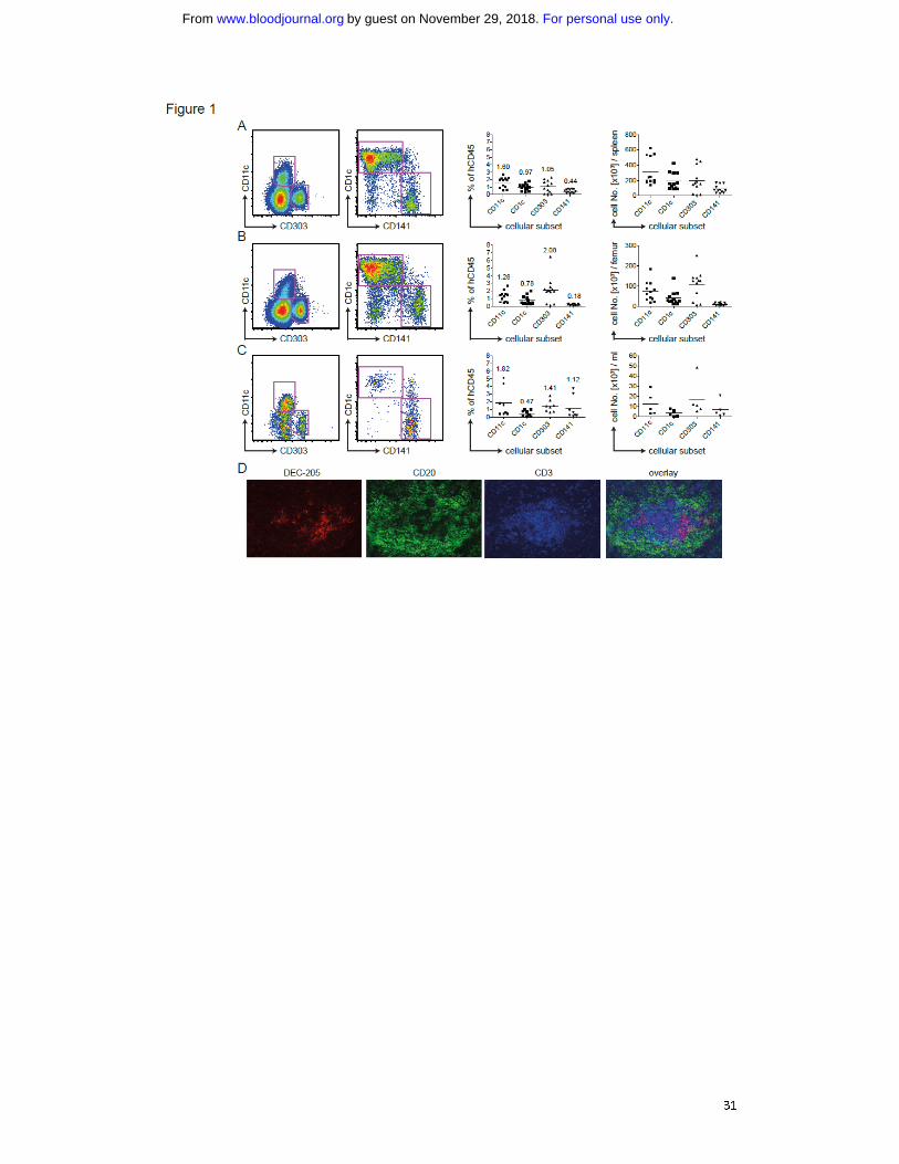
Robust reconstitution of all human DC subsets in huNSG mice
We reconstituted newborn NSG mice with CD34+ human fetal liver-derived
hematopoietic progenitor cells and analyzed the DC compartment 4 months later. By
flow cytometry staining, we defined human DCs as being positive for human CD45
and HLA-DR, but negative for lineage markers CD3 (T cells), CD19 (B cells), CD56 (NK
cells), CD14 (mainly monocytes) and CD16 (mainly NK cells) (Fig. S1A). We subdivided
the CD11c+ cDC population into CD1c or CD141 positive cells. The pDC subset was
defined as CD11c-CD303
+ (Fig. 1A). We detected all three human DC subsets in
spleen (Fig. 1A), bone marrow (BM; Fig. 1B) and blood (Fig. 1C). On average, DCs
made up 3% of reconstituted cells. In spleen and BM, the majority of cDCs was
CD1c+, while in blood CD141
+ cDCs predominated. The highest percentage of pDCs
was located in the BM, exceeding 2% of human CD45+ cells in most mice. Those
trends reflected the absolute numbers of DC subsets per spleen (Fig. 1A), femur (Fig.
1B) or milliliter of blood (Fig. 1C). Per spleen, we detected on average 300,000 cDCs
and 200,000 pDCs.
We analyzed the distribution of human DCs in spleens of huNSG mice by
immunofluorescence microscopy on histological sections. As in flow cytometric
staining (Fig. S1B/C), we detected both DEC-205 high and low cells. They were of DC
morphology and in T cell zones of white pulp areas (Fig. 1D). Those, we could not
subdivide by CD1c and CD141, as staining was unspecific.
In conclusion, we identified the main constitutive human DC subsets in
primary and secondary lymphoid organs of huNSG mice. Interestingly, the frequency
of the CD141+ DCs in blood and bone marrow of huNSG mice is higher than in human
For personal use only.on November 29, 2018. by guest www.bloodjournal.orgFrom
9
blood and bone marrow 8. Furthermore, the reconstituted cDCs in huNSG mice home
to similar areas as their counterparts in human spleen 24
.
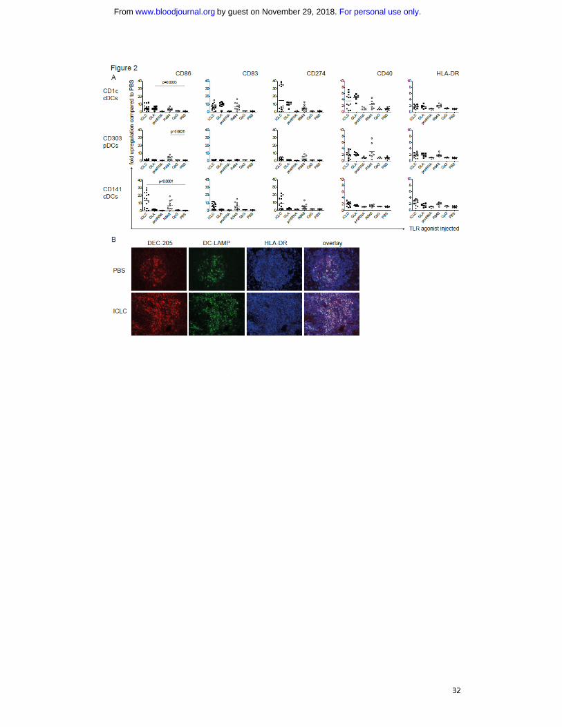
Ligation of RNA receptors matures human conventional and plasmacytoid DCs in
vivo
To test the functional competence of reconstituted DCs in huNSG mice, we analyzed
whether intraperitoneal (i.p.) injection of TLR agonists matured human DCs in vivo.
We used the TLR3 ligand polyICLC 25,26
, the TLR4 ligand GLA 27
, the TLR7/8 ligands
R848 28
and protamine/RNA 29
, as well as CpG as a TLR9 agonist 30
. After 14h (Fig.
S2A), we sacrificed the mice and assessed surface levels of maturation markers
(CD86, CD83, CD274, CD40 and HLA-DR) on the different splenic DC subsets (Fig.
S1D). Both cDC subsets significantly up-regulated maturation markers when injected
with polyICLC, in line with TLR3 expression on CD1c+ and CD141
+ cDCs in human (Fig.
2A/S2B). In contrast, only CD1c+ cDCs should have TLR4. Accordingly, only this cDC
subset matured significantly in response to GLA. R848 not only matured cDCs, but
also up-regulated CD40, CD86, CD274 and HLA-DR expression on pDCs significantly,
yet to a lesser extent (Fig. 2A). Neither protamine/RNA nor CpG ODN2216
phenotypically matured human DCs (Fig. 2A), even though those compounds
induced, albeit weak, murine cytokine production by DCs in non-reconstituted NSG
mice (Fig. S3A). With both being particulate formulations, their bioactivity might
however improve with intraveneous compared to intraperitoneal injection. Changes
in CD83, CD86 and CD274 expression were highest after polyICLC and R848 injection,
while CD40 and HLA-DR expression was less affected by the TLR agonists (Fig. 2A).
Furthermore, CD86 was most dramatically up-regulated on CD141+ DCs after
For personal use only.on November 29, 2018. by guest www.bloodjournal.orgFrom
10
polyICLC stimulation (more than 10 fold). In summary, the effect of a given TLR
agonist depends on the DC subset as well as on the maturation marker analyzed.
In addition, we chose the stimulus most efficacious for reconstituted cDCs in
this setting, polyICLC, to study DC maturation in tissue sections. We observed an
increase of DEC-205+ cells in spleens of polyICLC treated mice (Fig. 2B). Whereas
unstimulated spleens only displayed small areas of DEC-205+
DC-LAMP+ cDCs, DC-
LAMP+ mature cDC numbers increased dramatically after polyICLC injection (Fig. 2B).
Of note, mature cDCs were preferentially found in T cell zones of splenic white pulp.
In general, all reconstituted constitutive DC compartments of huNSG mice
matured in response to TLR agonists in vivo. However, only those targeting RNA
receptors (TLR3/7/ 8) induced robust maturation of cDCs and pDCs.
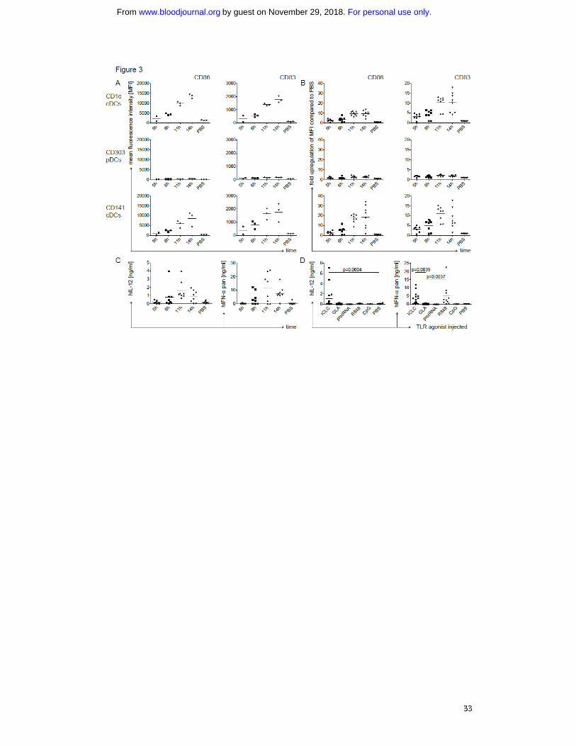
Human conventional DC populations mature slowly in response to TLR agonists in
vivo
Next, we characterized the kinetics of DC maturation in huNSG mice after polyICLC
injection. Maturation markers were maximally up-regulated at 11h to 14h (Fig.
3A/B). Again, we detected the highest up-regulation of CD86 on CD141+ cDCs and
pDCs were only minimally activated (Fig 2A/3A). In general, the kinetics of DC
maturation in huNSG mice resemble activation of human DCs in vitro, while TLR
agonists activate mouse DCs considerably faster 25
.
Only stimulation of RNA receptors elicits cytokine production by human DCs in vivo
As a functional readout for DC activation, we measured human cytokines in the
serum of huNSG mice at different time points after polyICLC injection. We analyzed
For personal use only.on November 29, 2018. by guest www.bloodjournal.orgFrom
11
IL-12p70 as a cDC signature cytokine and IFN-α, which is secreted at high levels by
activated pDCs 6. Concentrations of those cytokines peaked 11h after injection (Fig.
3C). Remarkably, they consistently reached around 1 ng/ml for IL-12p70 and up to 25
ng/ml for IFN-α.
In addition, we monitored cytokine production after injection of different TLR
agonists. In line with DC maturation, injection of polyICLC or R848 induced strong
cytokine production, while protamine/RNA and CpG did not (Fig 3D). Serum IL-12
and IFN-α concentrations varied after polyICLC injection between experiments,
possibly due to HPC donor variation or differences in the used TLR agonist batches.
These cytokines, however, were co-regulated in individual huNSG mice and
reconstitution levels of DC subsets was more homogeneous than these variations in
cytokine production. Surprisingly, GLA did not elicit even low human cytokine levels,
despite its capacity to mature CD1c+ cDCs (Fig. 2A).
This suggests that RNA receptor (TLR3/7/8) ligation fully matures human
cDCs and pDCs with up-regulation of co-stimulatory molecules and cytokine
production, while TLR4 and 9 agonists only partially mature DCs in this experimental
setting. Particularly, TLR4 agonists seem to be poor activators of cytokine production
by constitutive human DC subsets, while potently activating mouse DCs to produce
IL-6 and IL-12 (Fig. S3A).
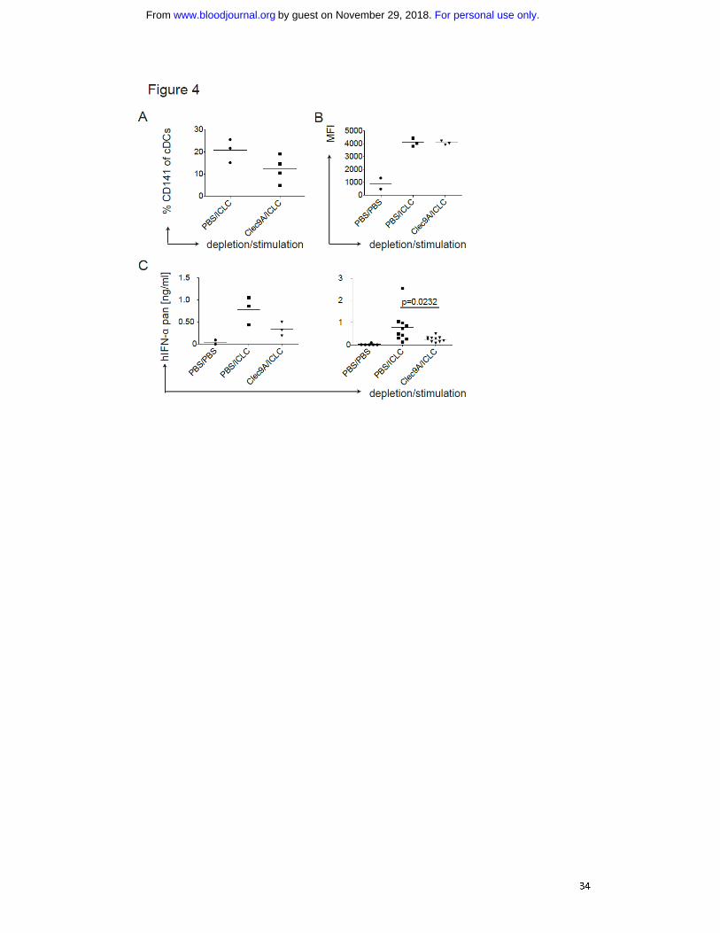
Depletion of CD141+
cDCs reduces IFN-α levels in the serum of huNSG mice
As polyICLC, which mainly matures cDCs (Fig. 2A), induced similar serum levels of
IFN-α as detected after injection of the pDC activating TLR ligand R848 (Fig. 3D), we
hypothesized that human cDCs could be a non-recognized source for considerable
For personal use only.on November 29, 2018. by guest www.bloodjournal.orgFrom
12
amounts of IFN-α. Furthermore, polyICLC matured both human cDC subsets and led
to IFN-α production in vivo, while GLA matured only CD1c+ cDCs without detectable
cytokine secretion (Fig. 2A and 3D), suggesting that the CD141+ cDC subset could be
producing the IFN-α. To test this, we depleted CD141+ cDCs in huNSG mice by
injecting an antibody recognizing the CD141+ cDC-specific molecule Clec9A
31,32.
Depletion of CD141+
cDCs was transient and partial (Fig. 4A) and left the other DC
populations mostly unaltered (Fig. S3B). Subsequently, we injected polyICLC and
analyzed serum IFN-α levels, only considering mice showing equal CD86 expression
on splenic CD1c+ cDCs (Fig. 4B). A decrease in CD141
+ cDCs resulted in significantly
diminished IFN-α levels (Fig. 4C).
Taken together, data generated in huNSG mice strongly suggests that the
CD141+ cDC subset is a main source of IFN-α production after stimulation with
synthetic dsRNA.
Human donor-derived CD141+
cDCs are potent producers of IFN-α
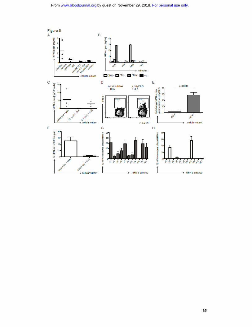
To confirm this hypothesis in a strictly human setting, we isolated cDCs from PBMCs
of healthy volunteers. We stimulated the cDC and the non-cDC fraction with GLA,
polyICLC or R848 and analyzed IFN-α in the supernatants. Indeed, cDCs produced
considerable amounts of IFN-α in response to polyICLC (Fig. 5A/B). They secreted
much less IFN-α in response to R848 and, in line with huNSG data, none after GLA
stimulation. The non-cDC fraction produced only low IFN-α levels in response to
polyICLC (Fig. 5A/B), probably originating from residual cDCs. For R848 stimulation of
non-cDCs, we attributed at least part of the IFN-α to pDCs by intracellular cytokine
staining using flow cytometry (not shown).
For personal use only.on November 29, 2018. by guest www.bloodjournal.orgFrom
13
We next tested which cDC subset in the human samples produces the IFN-α.
Therefore, we enriched CD141+ cDCs, CD304
+ pDCs and CD1c
+ cDCs sequentially
from PBMCs (Fig. S4A-D), stimulated them with TLR agonists and measured IFN-α in
the supernatants. Due to low CD141+ cDC recovery, we only tested polyICLC
stimulation for this subset, while the other DC populations were incubated with all
TLR stimuli. CD141+ cDC supernatants contained by far the highest concentration of
IFN-α after polyICLC stimulation (Fig. 5C). Despite donor variation, CD141+
cDCs of all
donors tested produced high amounts of IFN-α in response to polyICLC. Only with
R848, pDCs produced IFN-α copiously (Fig. 5C/D). In contrast, CD1c+ cDCs did not
secrete IFN-α after GLA stimulation (Fig. 5C/D), which is the TLR agonist inducing
strong up-regulation of maturation markers on CD1c+ cDCs in huNSG mice (Fig. 2A).
Additionally, we established an intracellular cytokine staining for IFN-α in CD141+
cDCs. In enriched, polyICLC-stimulated cDCs, we detected an IFN-α-specific signal in
CD141+
cDCs (Fig. 5E), which was significantly higher than the fold-increase in CD1c+
cDCs (Fig. 5F). Moreover, we compared the IFN-α subtypes produced by pDCs after
R848 stimulation in contrast to polyICLC-stimulated CD141+ cDCs. While pDCs
produced 50% of IFN-α2, this subtype only accounts for 5% of the IFN-α produced by
CD141+
cDCs (Fig. 5G). This was reflected in the transcriptional profiles of CD141+
cDCs showing a much broader IFN-α repertoire than pDCs, with very low IFN-α2
transcription (Fig. 5H/I). Thus, CD141+ cDCs from human blood produce significant
IFN-α amounts in response to TLR3 stimulation, consisting of other IFN-α subtypes
than those produced by pDCs in response to TLR7 ligation.
For personal use only.on November 29, 2018. by guest www.bloodjournal.orgFrom
14
Priming of human CD4+
T cell responses after antigen targeting to DEC-205
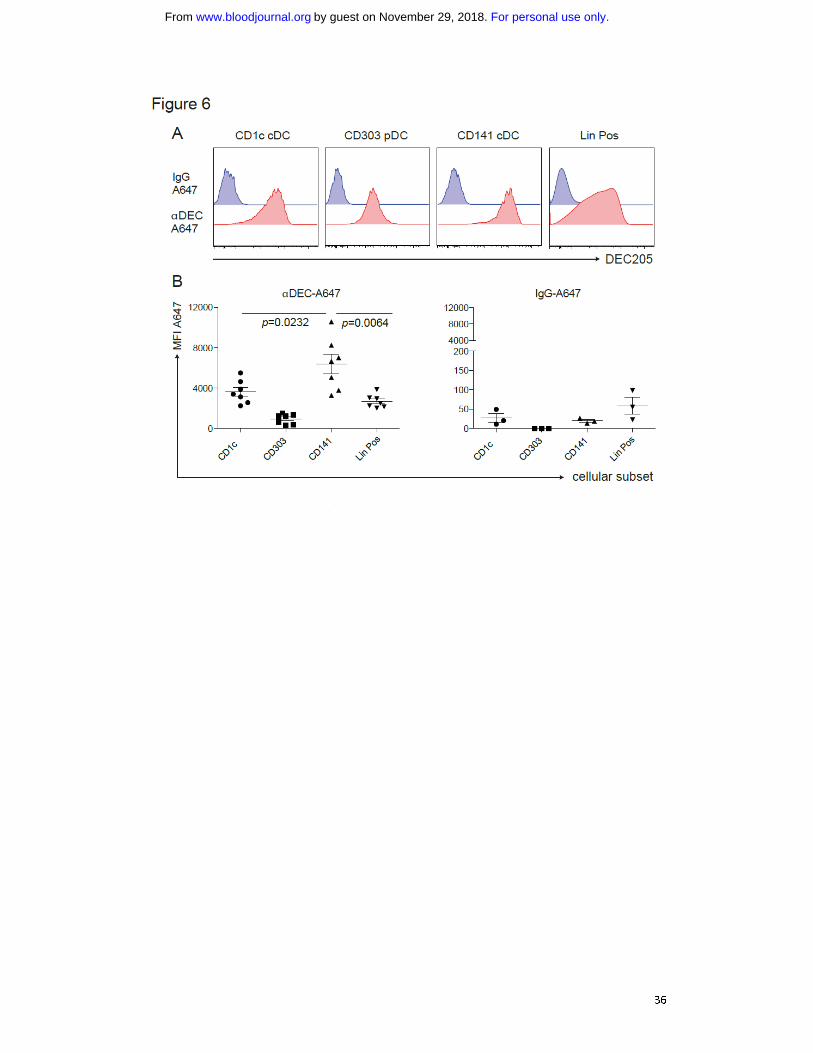
Finally, we tested the ability of this unique CD141+ cDC subset to prime immune
responses in vivo. As they highly express DEC-205, we aimed to target them by an
αDEC-205 mAb with polyICLC as an adjuvant that mainly elicits immunostimulatory
cytokine production by this DC subset. To validate specific targeting, we injected 5
μg of Alexa-647-labeled αDEC-205 or the labeled isotype control mAb into huNSG
mice. After 3 to 5h, we examined antibody uptake by different splenic DC subsets
(Fig. 6A/B). Unlike the isotype mAb, the αDEC-205 mAb labeled most of the CD141+
cDCs. αDEC-205 also bound other DC subsets and leukocyte populations, as shown in
mice 33
and in line with DEC-205 expression on other human cell populations 34,35
.
However, labeling by αDEC-205 mAb reached the highest levels in CD141+ cDCs
among different DC subsets and lineage positive cells. This infers that antigens
targeted to DEC-205 are most efficiently taken up by CD141+ cDCs, and combining it
with polyICLC that triggers immunostimulatory cytokine production by this DC
subset, should only render CD141+ DCs capable to prime T cell responses.
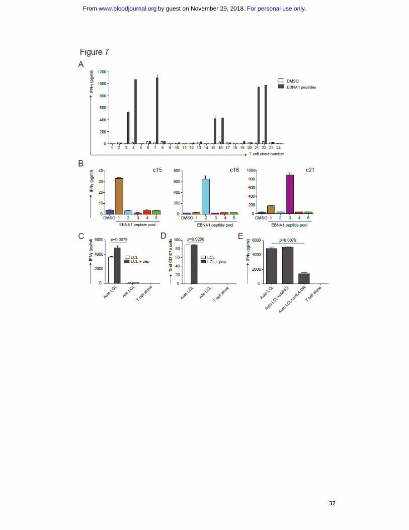
We have previously shown that the combination of poly(I:C) and DEC-205-
targeted antigen elicits peptide specific T cell responses against the Epstein-Barr
virus (EBV) nuclear antigen 1 (EBNA1) in huNSG mice 22
. To further characterize this T
cell response, we repeated the immunization experiment with αDEC-205-EBNA1
mAb but used the more stable polyICLC instead to preferentially mature CD141+ DCs
(Fig. S5A). From the splenocytes of the responding huNSG mice, we cloned the
EBNA1 specific T cells. Several clones responded to EBNA1 peptides (Fig. 7A) and all
of those were positive for human CD45, CD3 and CD4. Some reacted to only one out
of five EBNA1 peptide subpools (Fig. 7B). Notably, these clones were specific for
For personal use only.on November 29, 2018. by guest www.bloodjournal.orgFrom
15
different epitopes as they reacted to different subpools (Fig. 7B). This indicates that
targeting CD141+ cDCs by linking EBNA1 to DEC-205 with polyICLC as adjuvant
induces T cell responses with broad epitope specificities in huNSG mice. To evaluate
the effector functions of these clones, we generated autologous lymphoblastoid cell
lines (LCLs) as they serve as an in vitro model for EBV positive tumors. This was
achieved by infecting huNSG mice from the same reconstitution as the immunized
mice with EBV and subsequent isolation of LCLs from their splenocytes. The T cell
clones were incubated with those autologous or allogeneic LCLs loaded with or
without EBNA1 peptides, and we monitored IFN-γ production. Autologous LCLs
induced strong responses of the EBNA1 specific T cell clones while allogeneic LCLs
did not (Fig. 7C/S5B). In addition, we evaluated the cytotoxic potential of these
clones towards LCLs. In line with IFN-γ secretion, they expressed the degranulation
marker CD107a in autologous LCL co-cultures (Fig. 7D/S5B). Furthermore, we could
block responses of the CD4+ T cell clone c16 by αHLA-DR mAb but not αHLA-class I
mAb indicating that the T cell response towards LCLs is strictly HLA class II restricted
(Fig 7E). Taken together, this suggests that targeting antigen to DEC-205 in
combination with a strong stimulus for CD141+
cDC activation as adjuvant elicits
CD4+ T cell responses with protective function against autologous EBV transformed B
cells. In addition, it reports for the first time at the clonal level antigen specific CD4+
T cell responses in mice with reconstituted human immune system components.
For personal use only.on November 29, 2018. by guest www.bloodjournal.orgFrom
16
DISCUSSION
This study shows that DCs reconstituted in huNSG mice resemble their counterparts
in humans. Human CD1c+ cDCs respond to the TLR3 agonist polyICLC, the TLR4
agonist GLA and the TLR7/8 agonist R848 like in huNSG mice. In contrast, CD141+
cDCs lack TLR4 and are therefore unresponsive to GLA. Since high IL-12p70
production is a hallmark of CD141+
conventional DCs 8,9
, we mainly observed it in
response to dsRNA that matures this particular subset of human cDCs. As
anticipated, pDCs up-regulate maturation markers following TLR7 stimulation with
R848. Weaker TLR7/8 agonists, like protamine/RNA, and TLR9 agonists, like CpG,
however, did not mature pDCs in our hands. Therefore, RNA receptor engagement
seems to be most efficient for human DC stimulation in vivo.
Interestingly, we observed not only robust human IL-12p70 production in
response to TLR3 ligation, but also efficient IFN-α production, which could not be
enhanced upon R848 mediated maturation of pDCs (data now shown). While murine
IFN-α in response to poly(I:C) stimulation is mainly produced by non-hematopoietic
cells 25,36
, huNSG mice lack human non-hematopoietic tissues and, therefore,
produce significant human IFN-α amounts with their hematopoietic cells. Among
human hematopoietic cells, CD141+
cDCs have been proposed as type I and III IFN
sources, producing IFN-β and -λ, respectively 8,37
. In addition, we report here that
this subset is also a prominent source of IFN-α after dsRNA stimulation in vitro and in
vivo. In vivo, polyICLC stimulation leads to similar serum IFN-α levels in huNSG mice
as observed in healthy volunteers, namely 5ng/ml 38
. This correlates with phenotypic
maturation of CD141+
cDC maturation and reaches similar levels as R848 stimulation.
Of note, extrapolating from all analyzed organs, the total cell number of CD141+ cDCs
For personal use only.on November 29, 2018. by guest www.bloodjournal.orgFrom
17
should be 4x lower than for pDCs in huNSG mice, yet these cells raise serum IFN-α
concentrations to similar levels. Furthermore, in peripheral blood of healthy human
donors, IFN-α production upon polyICLC stimulation resides mainly in cDCs,
especially following CD141 enrichment. Of note, CD141
+ cell-enriched populations
produce similar IFN-α levels as pDCs after R848 stimulation. Therefore, CD141+
cDCs
have the dual capacity to produce IL-12p70 and type I IFN, which makes them an
attractive adjuvant target. RNA receptor ligands could serve as agonists to mature
this cDC subset during vaccination.
Apart from harnessing CD141+
cDCs for vaccination, it would be interesting to
assess their contribution to IFN-α production in an infection model. However, this is
hampered by the transient nature of CD141+
cDC depletion in huNSG mice. Of note,
this is in line with the fast repopulation of DCs after diphtheria toxin injection in
CD11c-DTR transgenic mice 39
. Furthermore, most infectious agents activate multiple
cell types by various TLRs, which would mask the effect of CD141+
cDC after
stimulation by dsRNA molecules. However, one could also envision a role of CD141+
cDC in resolving viral infection independent of absolute IFN-α production. As shown
(Fig. 5G/I), pDCs mainly produce IFN-α2 and IFN-α14. CD141+
cDCs, on the other
hand, secrete several IFN-α subtypes, which could be needed to orchestrate a
protective immune response against certain viral infections.
Although it remains so far unclear whether functional CD141+
cDCs also
reconstitute in other immune compromised mouse strains, reconstituted NOD-scid
mice have cDCs (lin–CD11c
+HLA-DR
+) and pDCs (lin
–IL-3R
+HLA-DR
+)
40-42. Although
they develop less DCs outside the BM than huNSG mice (this study and 43
), human
DCs from reconstituted NOD-scid mice can stimulate alloreactive human T cell
For personal use only.on November 29, 2018. by guest www.bloodjournal.orgFrom
18
responses in vitro 40-43
. Furthermore, they up-regulate maturation markers and
secrete IFN-α upon influenza virus infection 40 and produce IL-12 after
administration of LPS 41
or poly(I:C) 42
. Since functional human DCs reconstitute in
immune compromised mice, but NSG mice reconstitute especially high numbers of
all human constitutive DC populations in spleen and blood, huNSG mice should be
further explored for vaccine development.
We had previously observed that antigen targeted to DEC-205 on DCs with
poly(I:C) as adjuvant induced IFN-γ producing T cells in huNSG mice 22
. Similarly,
polyICLC plus DEC-205-targeted antigen elicited T cell responses in non-human
primates 44
and in this study CD4+ T cell responses in huNSG mice. While antigen
specific CD8+ T cell responses have been documented in mice with reconstituted
human immune system components 20,45-47
, our study shows for the first time
antigen specific CD4+ T cell responses, which could be verified at the clonal level, in
one of these models. Interestingly, both DEC-205 and TLR3 expression are highest on
human CD141+
cDCs (10
and this study), and, therefore, this antigen/adjuvant
formulation might preferentially harnesses this small, but potent DC population for
vaccination. Indeed, DEC-205 targeting of antigens to the mouse counterpart of
CD141+
cDCs, CD8α+ cDCs, enhances priming of T cell responses 100-fold, and
targeting to other receptors like Langerin and CLEC9A on CD8α+ cDCs similarly
augments vaccine-induced immune responses 33
. With the additional IFN-α
production by this DC subset in humans, it becomes even more attractive for vaccine
development. Furthermore, the role of CD141+ cDCs in innate and adaptive
immunity to important human pathogens as well as in inducing tolerance in
autoimmune settings could be explored in huNSG mice. In addition, they should be
For personal use only.on November 29, 2018. by guest www.bloodjournal.orgFrom
19
important tools to test vaccine regiments in a pre-clinical setting with hopefully
improved predictive value for immunogenicity in humans.
ACKNOWLEDGEMENTS
This work was supported by grants from the National Cancer Institute
(R01CA108609), the Sassella Foundation (10/02, 11/02 and 12/02), Cancer Research
Switzerland (KFS-02652-08-2010), the Association for International Cancer Research
(11-0516), KFSPMS
and KFSPHLD
of the University of Zurich, the Vontobel Foundation,
the Baugarten Foundation, the EMDO Foundation, the Sobek Foundation, Fondation
Acteria, Novartis and the Swiss National Science Foundation (310030_143979 and
CRSII3_136241) to C.M.. S.M., C.S.L. and P.C.R. were supported by junior research
fellowships from the University of Zürich. C.S.L. was also supported by the Croucher
Foundation Hong Kong.
AUTHORSHIP CONTRIBUTIONS
S.M. and C.S.L. designed and performed research. M.P., P.C.R. and L.D.V. performed
experiments. R.M.S. and C.M. designed research. Remaining authors contributed
essential information or vital reagents. S.M., C.S.L. and C.M. wrote the manuscript.
CONFLICT OF INTEREST
A.M.S. heads Oncovir Inc., which provided polyICLC. A.D. and J.S. work for Miltenyi
Biotec, which provided the anti-Clec9A antibody. The authors have no other financial
interests.
For personal use only.on November 29, 2018. by guest www.bloodjournal.orgFrom
20
REFERENCES
1. Steinman RM, Banchereau J. Taking dendritic cells into medicine. Nature.
2007;449(7161):419-426.
2. Steinman RM. Decisions about dendritic cells: past, present, and future. Annu
Rev Immunol. 2012;30:1-22.
3. Palucka K, Banchereau J, Mellman I. Designing vaccines based on biology of
human dendritic cell subsets. Immunity. 2010;33(4):464-478.
4. MacDonald KP, Munster DJ, Clark GJ, Dzionek A, Schmitz J, Hart DN.
Characterization of human blood dendritic cell subsets. Blood.
2002;100(13):4512-4520.
5. Dzionek A, Fuchs A, Schmidt P, et al. BDCA-2, BDCA-3, and BDCA-4: three
markers for distinct subsets of dendritic cells in human peripheral blood. J
Immunol. 2000;165(11):6037-6046.
6. Coffman RL, Sher A, Seder RA. Vaccine adjuvants: putting innate immunity to
work. Immunity. 2010;33(4):492-503.
7. Iwasaki A, Medzhitov R. Toll-like receptor control of the adaptive immune
responses. Nat Immunol. 2004;5(10):987-995.
8. Jongbloed SL, Kassianos AJ, McDonald KJ, et al. Human CD141+ (BDCA-3)
+
dendritic cells (DCs) represent a unique myeloid DC subset that cross-presents
necrotic cell antigens. J Exp Med. 2010;207(6):1247-1260.
9. Poulin LF, Salio M, Griessinger E, et al. Characterization of human DNGR-1+
BDCA3+ leukocytes as putative equivalents of mouse CD8alpha
+ dendritic cells. J
Exp Med. 2010;207(6):1261-1271.
For personal use only.on November 29, 2018. by guest www.bloodjournal.orgFrom

21
10. Robbins SH, Walzer T, Dembele D, et al. Novel insights into the relationships
between dendritic cell subsets in human and mouse revealed by genome-wide
expression profiling. Genome Biol. 2008;9(1):R17.
11. Bachem A, Guttler S, Hartung E, et al. Superior antigen cross-presentation and
XCR1 expression define human CD11c+CD141
+ cells as homologues of mouse
CD8+ dendritic cells. J Exp Med. 2010;207(6):1273-1281.
12. Crozat K, Guiton R, Contreras V, et al. The XC chemokine receptor 1 is a
conserved selective marker of mammalian cells homologous to mouse
CD8alpha+ dendritic cells. J Exp Med. 2010;207(6):1283-1292.
13. Rämer PC, Chijioke O, Meixlsperger S, Leung CS, Münz C. Mice with human
immune system components as in vivo models for infections with human
pathogens. Immunol Cell Biol. 2011;89(3):408-416.
14. Legrand N, Ploss A, Balling R, et al. Humanized mice for modeling human
infectious disease: challenges, progress, and outlook. Cell Host Microbe.
2009;6(1):5-9.
15. Ishikawa F, Yasukawa M, Lyons B, et al. Development of functional human blood
and immune systems in NOD/SCID/IL2 receptor gamma chainnull
mice. Blood.
2005;106(5):1565-1573.
16. Traggiai E, Chicha L, Mazzucchelli L, et al. Development of a human adaptive
immune system in cord blood cell-transplanted mice. Science.
2004;304(5667):104-107.
17. Shultz LD, Lyons BL, Burzenski LM, et al. Human lymphoid and myeloid cell
development in NOD/LtSz-scid IL2R gamma null mice engrafted with mobilized
human hemopoietic stem cells. J Immunol. 2005;174(10):6477-6489.
For personal use only.on November 29, 2018. by guest www.bloodjournal.orgFrom
22
18. Melkus MW, Estes JD, Padgett-Thomas A, et al. Humanized mice mount specific
adaptive and innate immune responses to EBV and TSST-1. Nat Med.
2006;12(11):1316-1322.
19. Tanaka S, Saito Y, Kunisawa J, et al. Development of mature and functional
human myeloid subsets in hematopoietic stem cell-engrafted
NOD/SCID/IL2rgammaKO mice. J Immunol. 2012;188(12):6145-6155.
20. Strowig T, Gurer C, Ploss A, et al. Priming of protective T cell responses against
virus-induced tumors in mice with human immune system components. J Exp
Med. 2009;206(6):1423-1434.
21. Puig M, Tosh KW, Schramm LM, et al. TLR9 and TLR7 agonists mediate distinct
type I IFN responses in humans and nonhuman primates in vitro and in vivo. J
Leukoc Biol. 2012;91(1):147-158.
22. Gurer C, Strowig T, Brilot F, et al. Targeting the nuclear antigen 1 of Epstein Barr
virus to the human endocytic receptor DEC-205 stimulates protective T-cell
responses. Blood. 2008;112:1231-1239.
23. Leung CS, Haigh TA, Mackay LK, Rickinson AB, Taylor GS. Nuclear location of an
endogenously expressed antigen, EBNA1, restricts access to macroautophagy
and the range of CD4 epitope display. Proc Natl Acad Sci U S A.
24. Pack M, Trumpfheller C, Thomas D, et al. DEC-205/CD205+ dendritic cells are
abundant in the white pulp of the human spleen, including the border region
between the red and white pulp. Immunology. 2008;123(3):438-446.
25. Longhi MP, Trumpfheller C, Idoyaga J, et al. Dendritic cells require a systemic
type I interferon response to mature and induce CD4+ Th1 immunity with poly
IC as adjuvant. J Exp Med. 2009;206(7):1589-1602.
For personal use only.on November 29, 2018. by guest www.bloodjournal.orgFrom
23
26. Stahl-Hennig C, Eisenblatter M, Jasny E, et al. Synthetic double-stranded RNAs
are adjuvants for the induction of T helper 1 and humoral immune responses to
human papillomavirus in rhesus macaques. PLoS Pathog. 2009;5(4):e1000373.
27. Coler RN, Bertholet S, Moutaftsi M, et al. Development and characterization of
synthetic glucopyranosyl lipid adjuvant system as a vaccine adjuvant. PLoS ONE.
2011;6(1):e16333.
28. Jurk M, Heil F, Vollmer J, et al. Human TLR7 or TLR8 independently confer
responsiveness to the antiviral compound R-848. Nat Immunol. 2002;3(6):499.
29. Scheel B, Teufel R, Probst J, et al. Toll-like receptor-dependent activation of
several human blood cell types by protamine-condensed mRNA. Eur J Immunol.
2005;35(5):1557-1566.
30. Hemmi H, Takeuchi O, Kawai T, et al. A Toll-like receptor recognizes bacterial
DNA. Nature. 2000;408(6813):740-745.
31. Sancho D, Mourao-Sa D, Joffre OP, et al. Tumor therapy in mice via antigen
targeting to a novel, DC-restricted C-type lectin. J Clin Invest. 2008;118(6):2098-
2110.
32. Huysamen C, Willment JA, Dennehy KM, Brown GD. CLEC9A is a novel activation
C-type lectin-like receptor expressed on BDCA3+ dendritic cells and a subset of
monocytes. J Biol Chem. 2008;283(24):16693-16701.
33. Idoyaga J, Lubkin A, Fiorese C, et al. Comparable T helper 1 (Th1) and CD8 T-cell
immunity by targeting HIV gag p24 to CD8 dendritic cells within antibodies to
Langerin, DEC205, and Clec9A. Proc Natl Acad Sci U S A. 2011;108(6):2384-2389.
34. Kato M, McDonald KJ, Khan S, et al. Expression of human DEC-205 (CD205)
multilectin receptor on leukocytes. Int Immunol. 2006;18(6):857-869.
For personal use only.on November 29, 2018. by guest www.bloodjournal.orgFrom
24
35. Leung CS, Maurer MA, Meixlsperger S, et al. Robust T cell stimulation by Epstein-
Barr virus-transformed B cells after antigen targeting to DEC-205. Blood. 2013.
Prepublished on 2013/01/09 as DOI blood-2012-08-450775.
36. Gitlin L, Barchet W, Gilfillan S, et al. Essential role of mda-5 in type I IFN
responses to polyriboinosinic:polyribocytidylic acid and encephalomyocarditis
picornavirus. Proc Natl Acad Sci U S A. 2006;103:8459-8464.
37. Lauterbach H, Bathke B, Gilles S, et al. Mouse CD8alpha+ DCs and human BDCA3
+
DCs are major producers of IFN-lambda in response to poly IC. J Exp Med.
2010;207(12):2703-2717.
38. Caskey M, Lefebvre F, Filali-Mouhim A, et al. Synthetic double-stranded RNA
induces innate immune responses similar to a live viral vaccine in humans. J Exp
Med. 2011;208(12):2357-2366.
39. Jung S, Unutmaz D, Wong P, et al. In vivo depletion of CD11c+ dendritic cells
abrogates priming of CD8+ T cells by exogenous cell-associated antigens.
Immunity. 2002;17(2):211-220.
40. Palucka AK, Gatlin J, Blanck JP, et al. Human dendritic cell subsets in NOD/SCID
mice engrafted with CD34+ hematopoietic progenitors. Blood.
2003;102(9):3302-3310.
41. Cravens PD, Melkus MW, Padgett-Thomas A, Islas-Ohlmayer M, Del PMM,
Garcia JV. Development and activation of human dendritic cells in vivo in a
xenograft model of human hematopoiesis. Stem Cells. 2005;23(2):264-278.
42. Vuckovic S, Abdul Wahid FS, Rice A, et al. Compartmentalization of allogeneic T-
cell responses in the bone marrow and spleen of humanized NOD/SCID mice
For personal use only.on November 29, 2018. by guest www.bloodjournal.orgFrom
25
containing activated human resident myeloid dendritic cells. Exp Hematol.
2008;36(11):1496-1506.
43. Ishikawa F, Niiro H, Iino T, et al. The developmental program of human dendritic
cells is operated independently of conventional myeloid and lymphoid
pathways. Blood. 2007;110(10):3591-3660.
44. Flynn BJ, Kastenmuller K, Wille-Reece U, et al. Immunization with HIV Gag
targeted to dendritic cells followed by recombinant New York vaccinia virus
induces robust T-cell immunity in nonhuman primates. Proc Natl Acad Sci U S A.
2011;108(17):7131-7136.
45. Shultz LD, Saito Y, Najima Y, et al. Generation of functional human T-cell subsets
with HLA-restricted immune responses in HLA class I expressing NOD/SCID/IL2r
gamma(null) humanized mice. Proc Natl Acad Sci U S A. 2010;107(29):13022-
13027.
46. Jaiswal S, Pazoles P, Woda M, et al. Enhanced humoral and HLA-A2-restricted
dengue virus-specific T-cell responses in humanized BLT NSG mice. Immunology.
2012;136(3):334-343.
47. Brainard DM, Seung E, Frahm N, et al. Induction of robust cellular and humoral
virus-specific adaptive immune responses in human immunodeficiency virus-
infected humanized BLT mice. J Virol. 2009;83(14):7305-7321.
For personal use only.on November 29, 2018. by guest www.bloodjournal.orgFrom
26
FIGURE LEGENDS
Fig. 1: Human DC subsets in huNSG mice. A) Flow cytometric staining of CD11c-,
CD303+ pDCs or CD1c
+ and CD141
+ on CD11c
+ cDCs of the spleen (left panels).
Samples were pre-gated as singlet, live cells positive for hCD45 and HLA-DR and
negative for lineage, CD14 and CD16. Percentages of those subsets in relation to live,
singlet, human CD45+ cells are shown in the middle. The numbers on top of the data
points indicate the average percentage of human CD45 positive cells for each
analyzed DC subset. Absolute numbers of DC populations are shown on the right.
Graphs represent data from mice from four independent reconstitutions. Each data
point represents one individually analyzed mouse. B) as in A) for DC subsets of the
BM. C) as in A) for DC subsets of the blood. D) Immunofluoresence microscopy for
DC markers on spleen sections. DEC-205 in red as DC-marker, CD20 in green as B cell
marker and CD3 in blue as T cell marker.
Fig. 2: Maturation of human DC subsets upon TLR ligand injection in vivo. A) Fold-
up-regulation of CD86, CD83, CD274, CD40 and HLA-DR on CD1c+ cDCs (top), CD303
+
pDCs (middle) and CD141+ cDCs (bottom). HuNSG mice were injected i.p. with 50
μg/mouse polyICLC, 20 μg/mouse GLA IDC 1001, 25 μg/mouse protamine/RNA, 20
μg/mouse R848, 50 μg/mouse CpG ODN 2216 or PBS. 14h after injection,
splenocytes were isolated and stained for flow cytometry. Fold up-regulation was
calculated from the MFI in relation to the mean of the corresponding PBS samples.
The graph represents composite data from five independent experiments. Each data
point represents one individually analyzed mouse. Statistical analysis was performed
with the Mann-Whitney test. B) Immunohistochemistry for DC markers on spleen
sections. HuNSG mice were injected with PBS (top) or with polyICLC (bottom) and
For personal use only.on November 29, 2018. by guest www.bloodjournal.orgFrom
27
sacrificed after 14h. DEC-205 serves as a DC marker, DC-LAMP and HLA-DR as DC
maturation markers.
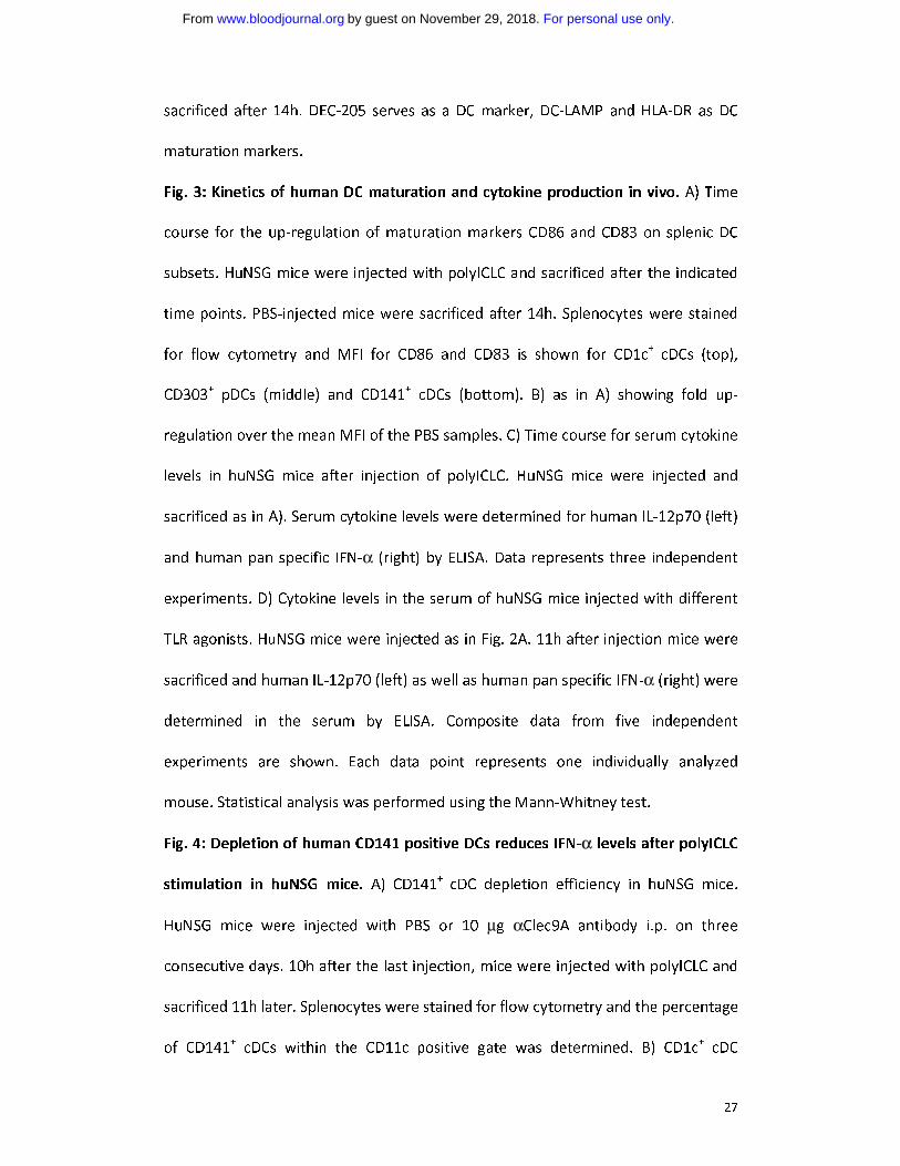
Fig. 3: Kinetics of human DC maturation and cytokine production in vivo. A) Time
course for the up-regulation of maturation markers CD86 and CD83 on splenic DC
subsets. HuNSG mice were injected with polyICLC and sacrificed after the indicated
time points. PBS-injected mice were sacrificed after 14h. Splenocytes were stained
for flow cytometry and MFI for CD86 and CD83 is shown for CD1c+ cDCs (top),
CD303+ pDCs (middle) and CD141
+ cDCs (bottom). B) as in A) showing fold up-
regulation over the mean MFI of the PBS samples. C) Time course for serum cytokine
levels in huNSG mice after injection of polyICLC. HuNSG mice were injected and
sacrificed as in A). Serum cytokine levels were determined for human IL-12p70 (left)
and human pan specific IFN-α (right) by ELISA. Data represents three independent
experiments. D) Cytokine levels in the serum of huNSG mice injected with different
TLR agonists. HuNSG mice were injected as in Fig. 2A. 11h after injection mice were
sacrificed and human IL-12p70 (left) as well as human pan specific IFN-α (right) were
determined in the serum by ELISA. Composite data from five independent
experiments are shown. Each data point represents one individually analyzed
mouse. Statistical analysis was performed using the Mann-Whitney test.
Fig. 4: Depletion of human CD141 positive DCs reduces IFN-α levels after polyICLC
stimulation in huNSG mice. A) CD141+ cDC depletion efficiency in huNSG mice.
HuNSG mice were injected with PBS or 10 μg αClec9A antibody i.p. on three
consecutive days. 10h after the last injection, mice were injected with polyICLC and
sacrificed 11h later. Splenocytes were stained for flow cytometry and the percentage
of CD141+ cDCs within the CD11c positive gate was determined. B) CD1c
+ cDC
For personal use only.on November 29, 2018. by guest www.bloodjournal.orgFrom
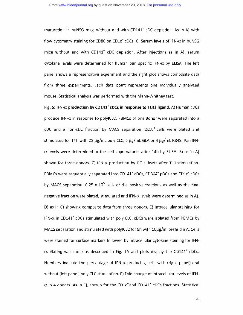
28
maturation in huNSG mice without and with CD141+ cDC depletion. As in A) with
flow cytometry staining for CD86 on CD1c+ cDCs. C) Serum levels of IFN-α in huNSG
mice without and with CD141+ cDC depletion. After injections as in A), serum
cytokine levels were determined for human pan specific IFN-α by ELISA. The left
panel shows a representative experiment and the right plot shows composite data
from three experiments. Each data point represents one individually analyzed
mouse. Statistical analysis was performed with the Mann-Whitney test.
Fig. 5: IFN-α production by CD141+
cDCs in response to TLR3 ligand. A) Human cDCs
produce IFN-α in response to polyICLC. PBMCs of one donor were separated into a
cDC and a non-cDC fraction by MACS separation. 2x10⁵
cells were plated and
stimulated for 14h with 25 μg/mL polyICLC, 5 μg/mL GLA or 4 μg/mL R848. Pan IFN-
α levels were determined in the cell supernatants after 14h by ELISA. B) as in A)
shown for three donors. C) IFN-α production by DC subsets after TLR stimulation.
PBMCs were sequentially separated into CD141+ cDCs, CD304
+ pDCs and CD1c
+ cDCs
by MACS separation. 0.25 x 10⁵ cells of the positive fractions as well as the final
negative fraction were plated, stimulated and IFN-α levels were determined as in A).
D) as in C) showing composite data from three donors. E) Intracellular staining for
IFN-α in CD141+ cDCs stimulated with polyICLC. cDCs were isolated from PBMCs by
MACS separation and stimulated with polyICLC for 9h with 10μg/ml brefeldin A. Cells
were stained for surface markers followed by intracellular cytokine staining for IFN-
α. Gating was done as described in Fig. 1A and plots display the CD141+ cDCs.
Numbers indicate the percentage of IFN-α producing cells with (right panel) and
without (left panel) polyICLC stimulation. F) Fold change of intracellular levels of IFN-
α in 4 donors. As in E), shown for the CD1c+
and CD141+ cDCs fractions. Statistical
For personal use only.on November 29, 2018. by guest www.bloodjournal.orgFrom
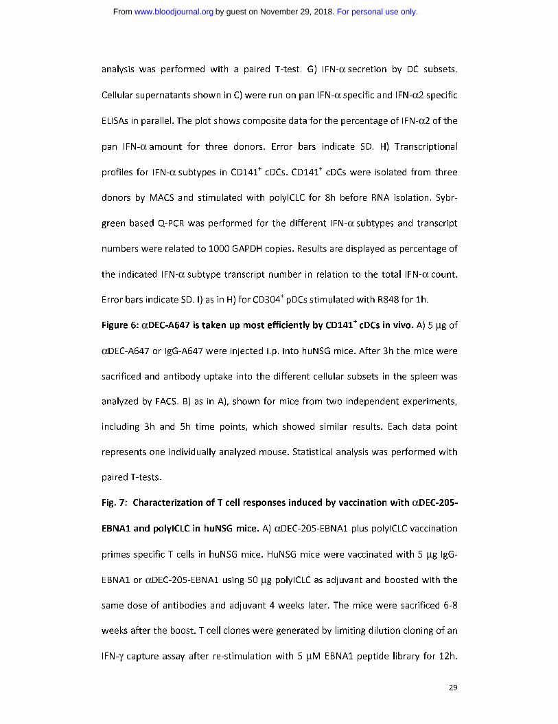
29
analysis was performed with a paired T-test. G) IFN-α secretion by DC subsets.
Cellular supernatants shown in C) were run on pan IFN-α specific and IFN-α2 specific
ELISAs in parallel. The plot shows composite data for the percentage of IFN-α2 of the
pan IFN-α amount for three donors. Error bars indicate SD. H) Transcriptional
profiles for IFN-α subtypes in CD141+ cDCs. CD141
+ cDCs were isolated from three
donors by MACS and stimulated with polyICLC for 8h before RNA isolation. Sybr-
green based Q-PCR was performed for the different IFN-α subtypes and transcript
numbers were related to 1000 GAPDH copies. Results are displayed as percentage of
the indicated IFN-α subtype transcript number in relation to the total IFN-α count.
Error bars indicate SD. I) as in H) for CD304+ pDCs stimulated with R848 for 1h.
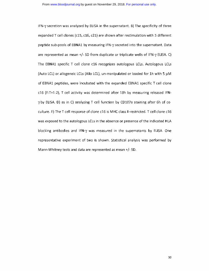
Figure 6: αDEC-A647 is taken up most efficiently by CD141+
cDCs in vivo. A) 5 μg of
αDEC-A647 or IgG-A647 were injected i.p. into huNSG mice. After 3h the mice were
sacrificed and antibody uptake into the different cellular subsets in the spleen was
analyzed by FACS. B) as in A), shown for mice from two independent experiments,
including 3h and 5h time points, which showed similar results. Each data point
represents one individually analyzed mouse. Statistical analysis was performed with
paired T-tests.
Fig. 7: Characterization of T cell responses induced by vaccination with αDEC-205-
EBNA1 and polyICLC in huNSG mice. A) αDEC-205-EBNA1 plus polyICLC vaccination
primes specific T cells in huNSG mice. HuNSG mice were vaccinated with 5 μg IgG-
EBNA1 or αDEC-205-EBNA1 using 50 μg polyICLC as adjuvant and boosted with the
same dose of antibodies and adjuvant 4 weeks later. The mice were sacrificed 6-8
weeks after the boost. T cell clones were generated by limiting dilution cloning of an
IFN-γ capture assay after re-stimulation with 5 μM EBNA1 peptide library for 12h.
For personal use only.on November 29, 2018. by guest www.bloodjournal.orgFrom
30
IFN-γ secretion was analyzed by ELISA in the supernatant. B) The specificity of three
expanded T cell clones (c15, c16, c21) are shown after restimulation with 5 different
peptide sub-pools of EBNA1 by measuring IFN-γ secreted into the supernatant. Data
are represented as mean +/- SD from duplicate or triplicate wells of IFN-γ ELISA. C)
The EBNA1 specific T cell clone c16 recognizes autologous LCLs. Autologous LCLs
(Auto LCL) or allogeneic LCLs (Allo LCL), un-manipulated or loaded for 1h with 5 μM
of EBNA1 peptides, were incubated with the expanded EBNA1 specific T cell clone
c16 (E:T=1:2), T cell activity was determined after 18h by measuring released IFN-
γ by ELISA. D) as in C) analyzing T cell function by CD107a staining after 6h of co-
culture. E) The T cell response of clone c16 is MHC class II-restricted. T cell clone c16
was exposed to the autologous LCLs in the absence or presence of the indicated HLA
blocking antibodies and IFN-γ was measured in the supernatants by ELISA. One
representative experiment of two is shown. Statistical analysis was performed by
Mann-Whitney tests and data are represented as mean +/- SD.
For personal use only.on November 29, 2018. by guest www.bloodjournal.orgFrom
31
For personal use only.on November 29, 2018. by guest www.bloodjournal.orgFrom
32
For personal use only.on November 29, 2018. by guest www.bloodjournal.orgFrom
33
For personal use only.on November 29, 2018. by guest www.bloodjournal.orgFrom
34
For personal use only.on November 29, 2018. by guest www.bloodjournal.orgFrom
35
For personal use only.on November 29, 2018. by guest www.bloodjournal.orgFrom
36
For personal use only.on November 29, 2018. by guest www.bloodjournal.orgFrom
37
For personal use only.on November 29, 2018. by guest www.bloodjournal.orgFrom
doi:10.1182/blood-2012-12-473413Prepublished online March 12, 2013;
MünzSteve Pascolo, Andres M. Salazar, Andrzej Dzionek, Jürgen Schmitz, Ralph M. Steinman and Christian Sonja Meixlsperger, Carol S. Leung, Patrick C. Rämer, Maggi Pack, Liliana D. Vanoaica, Gaëlle Breton, recognition and can be targeted via DEC-205 in humanized mice
after dsRNAα dendritic cells produce prominent amounts of IFN-+CD141
http://www.bloodjournal.org/site/misc/rights.xhtml#repub_requestsInformation about reproducing this article in parts or in its entirety may be found online at:
http://www.bloodjournal.org/site/misc/rights.xhtml#reprintsInformation about ordering reprints may be found online at:
http://www.bloodjournal.org/site/subscriptions/index.xhtmlInformation about subscriptions and ASH membership may be found online at:
digital object identifier (DOIs) and date of initial publication. indexed by PubMed from initial publication. Citations to Advance online articles must include final publication). Advance online articles are citable and establish publication priority; they areappeared in the paper journal (edited, typeset versions may be posted when available prior to Advance online articles have been peer reviewed and accepted for publication but have not yet
Copyright 2011 by The American Society of Hematology; all rights reserved.Hematology, 2021 L St, NW, Suite 900, Washington DC 20036.Blood (print ISSN 0006-4971, online ISSN 1528-0020), is published weekly by the American Society of
For personal use only.on November 29, 2018. by guest www.bloodjournal.orgFrom