BJP RESEARCH PAPER - Actinogen
13
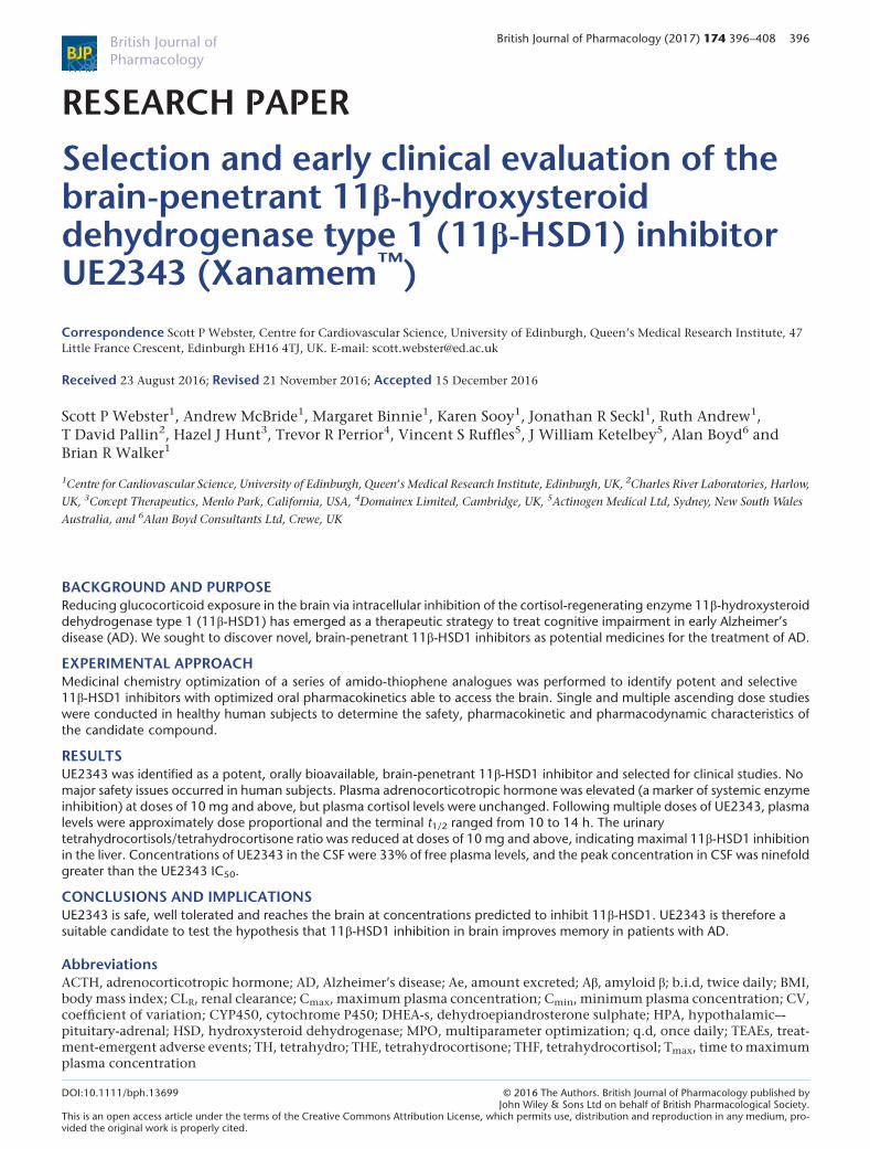
RESEARCH PAPER Selection and early clinical evaluation of the brain-penetrant 11β-hydroxysteroid dehydrogenase type 1 (11β-HSD1) inhibitor UE2343 (Xanamem ™ ) Correspondence Scott P Webster, Centre for Cardiovascular Science, University of Edinburgh, Queen’s Medical Research Institute, 47 Little France Crescent, Edinburgh EH16 4TJ, UK. E-mail: [email protected] Received 23 August 2016; Revised 21 November 2016; Accepted 15 December 2016 Scott P Webster 1 , Andrew McBride 1 , Margaret Binnie 1 , Karen Sooy 1 , Jonathan R Seckl 1 , Ruth Andrew 1 , T David Pallin 2 , Hazel J Hunt 3 , Trevor R Perrior 4 , Vincent S Ruffles 5 , J William Ketelbey 5 , Alan Boyd 6 and Brian R Walker 1 1 Centre for Cardiovascular Science, University of Edinburgh, Queen’s Medical Research Institute, Edinburgh, UK, 2 Charles River Laboratories, Harlow, UK, 3 Corcept Therapeutics, Menlo Park, California, USA, 4 Domainex Limited, Cambridge, UK, 5 Actinogen Medical Ltd, Sydney, New South Wales Australia, and 6 Alan Boyd Consultants Ltd, Crewe, UK BACKGROUND AND PURPOSE Reducing glucocorticoid exposure in the brain via intracellular inhibition of the cortisol-regenerating enzyme 11β-hydroxysteroid dehydrogenase type 1 (11β-HSD1) has emerged as a therapeutic strategy to treat cognitive impairment in early Alzheimer’s disease (AD). We sought to discover novel, brain-penetrant 11β-HSD1 inhibitors as potential medicines for the treatment of AD. EXPERIMENTAL APPROACH Medicinal chemistry optimization of a series of amido-thiophene analogues was performed to identify potent and selective 11β-HSD1 inhibitors with optimized oral pharmacokinetics able to access the brain. Single and multiple ascending dose studies were conducted in healthy human subjects to determine the safety, pharmacokinetic and pharmacodynamic characteristics of the candidate compound. RESULTS UE2343 was identified as a potent, orally bioavailable, brain-penetrant 11β-HSD1 inhibitor and selected for clinical studies. No major safety issues occurred in human subjects. Plasma adrenocorticotropic hormone was elevated (a marker of systemic enzyme inhibition) at doses of 10 mg and above, but plasma cortisol levels were unchanged. Following multiple doses of UE2343, plasma levels were approximately dose proportional and the terminal t 1/2 ranged from 10 to 14 h. The urinary tetrahydrocortisols/tetrahydrocortisone ratio was reduced at doses of 10 mg and above, indicating maximal 11β-HSD1 inhibition in the liver. Concentrations of UE2343 in the CSF were 33% of free plasma levels, and the peak concentration in CSF was ninefold greater than the UE2343 IC 50 . CONCLUSIONS AND IMPLICATIONS UE2343 is safe, well tolerated and reaches the brain at concentrations predicted to inhibit 11β-HSD1. UE2343 is therefore a suitable candidate to test the hypothesis that 11β-HSD1 inhibition in brain improves memory in patients with AD. Abbreviations ACTH, adrenocorticotropic hormone; AD, Alzheimer’s disease; Ae, amount excreted; Aβ, amyloid β; b.i.d, twice daily; BMI, body mass index; CL R , renal clearance; C max , maximum plasma concentration; C min , minimum plasma concentration; CV, coefficient of variation; CYP450, cytochrome P450; DHEA-s, dehydroepiandrosterone sulphate; HPA, hypothalamic–- pituitary-adrenal; HSD, hydroxysteroid dehydrogenase; MPO, multiparameter optimization; q.d, once daily; TEAEs, treat- ment-emergent adverse events; TH, tetrahydro; THE, tetrahydrocortisone; THF, tetrahydrocortisol; T max , time to maximum plasma concentration BJP British Journal of Pharmacology British Journal of Pharmacology (2017) 174 396–408 396 DOI:10.1111/bph.13699 © 2016 The Authors. British Journal of Pharmacology published by John Wiley & Sons Ltd on behalf of British Pharmacological Society. This is an open access article under the terms of the Creative Commons Attribution License, which permits use, distribution and reproduction in any medium, pro- vided the original work is properly cited.
Transcript of BJP RESEARCH PAPER - Actinogen
RESEARCH PAPER
Selection and early clinical evaluation of thebrain-penetrant 11β-hydroxysteroiddehydrogenase type 1 (11β-HSD1) inhibitorUE2343 (Xanamem™)Correspondence Scott P Webster, Centre for Cardiovascular Science, University of Edinburgh, Queen’s Medical Research Institute, 47Little France Crescent, Edinburgh EH16 4TJ, UK. E-mail: [email protected]
Received 23 August 2016; Revised 21 November 2016; Accepted 15 December 2016
Scott P Webster1, Andrew McBride1, Margaret Binnie1, Karen Sooy1, Jonathan R Seckl1, Ruth Andrew1,T David Pallin2, Hazel J Hunt3, Trevor R Perrior4, Vincent S Ruffles5, J William Ketelbey5, Alan Boyd6 andBrian R Walker1
1Centre for Cardiovascular Science, University of Edinburgh, Queen’s Medical Research Institute, Edinburgh, UK, 2Charles River Laboratories, Harlow,
UK, 3Corcept Therapeutics, Menlo Park, California, USA, 4Domainex Limited, Cambridge, UK, 5Actinogen Medical Ltd, Sydney, New South Wales
Australia, and 6Alan Boyd Consultants Ltd, Crewe, UK
BACKGROUND AND PURPOSEReducing glucocorticoid exposure in the brain via intracellular inhibition of the cortisol-regenerating enzyme 11β-hydroxysteroiddehydrogenase type 1 (11β-HSD1) has emerged as a therapeutic strategy to treat cognitive impairment in early Alzheimer’sdisease (AD). We sought to discover novel, brain-penetrant 11β-HSD1 inhibitors as potential medicines for the treatment of AD.
EXPERIMENTAL APPROACHMedicinal chemistry optimization of a series of amido-thiophene analogues was performed to identify potent and selective11β-HSD1 inhibitors with optimized oral pharmacokinetics able to access the brain. Single and multiple ascending dose studieswere conducted in healthy human subjects to determine the safety, pharmacokinetic and pharmacodynamic characteristics ofthe candidate compound.
RESULTSUE2343 was identified as a potent, orally bioavailable, brain-penetrant 11β-HSD1 inhibitor and selected for clinical studies. Nomajor safety issues occurred in human subjects. Plasma adrenocorticotropic hormone was elevated (a marker of systemic enzymeinhibition) at doses of 10 mg and above, but plasma cortisol levels were unchanged. Following multiple doses of UE2343, plasmalevels were approximately dose proportional and the terminal t1/2 ranged from 10 to 14 h. The urinarytetrahydrocortisols/tetrahydrocortisone ratio was reduced at doses of 10 mg and above, indicating maximal 11β-HSD1 inhibitionin the liver. Concentrations of UE2343 in the CSF were 33% of free plasma levels, and the peak concentration in CSF was ninefoldgreater than the UE2343 IC50.
CONCLUSIONS AND IMPLICATIONSUE2343 is safe, well tolerated and reaches the brain at concentrations predicted to inhibit 11β-HSD1. UE2343 is therefore asuitable candidate to test the hypothesis that 11β-HSD1 inhibition in brain improves memory in patients with AD.
AbbreviationsACTH, adrenocorticotropic hormone; AD, Alzheimer’s disease; Ae, amount excreted; Aβ, amyloid β; b.i.d, twice daily; BMI,body mass index; CLR, renal clearance; Cmax, maximum plasma concentration; Cmin, minimum plasma concentration; CV,coefficient of variation; CYP450, cytochrome P450; DHEA-s, dehydroepiandrosterone sulphate; HPA, hypothalamic–-pituitary-adrenal; HSD, hydroxysteroid dehydrogenase; MPO, multiparameter optimization; q.d, once daily; TEAEs, treat-ment-emergent adverse events; TH, tetrahydro; THE, tetrahydrocortisone; THF, tetrahydrocortisol; Tmax, time tomaximumplasma concentration
BJP British Journal ofPharmacology
British Journal of Pharmacology (2017) 174 396–408 396
DOI:10.1111/bph.13699 © 2016 The Authors. British Journal of Pharmacology published byJohn Wiley & Sons Ltd on behalf of British Pharmacological Society.
This is an open access article under the terms of the Creative Commons Attribution License, which permits use, distribution and reproduction in any medium, pro-vided the original work is properly cited.

IntroductionChronically elevated levels of the glucocorticoid stress hor-mone cortisol are associated with a variety of conditions in-cluding Cushing’s syndrome, which is characterized by themetabolic complications of central obesity, dyslipidaemia,hypertension and glucose dysregulation (Anagnostis et al.,2009). In Cushing’s syndrome, neuropsychiatric problemsalso occur, including cognitive impairment, which is relatedto excess cortisol exposure in key areas of the brain responsi-ble for memory (Starkman et al., 1999). Increased glucocorti-coid exposure in the brain is linked to age-related cognitivedecline where cortisol levels align with reductions in hippo-campal volumes and consequent memory impairment(Lupien et al., 1998; MacLullich et al., 2005; Lupien et al.,2009). More subtle alterations in glucocorticoid levels,exemplified by higher morning cortisol levels, have also beenshown to be associated with impaired cognitive performance(Reynolds et al., 2010).
In Alzheimer’s disease (AD), elevated plasma cortisollevels are associated with accelerated disease progression,while higher cortisol levels in CSF are associated with morerapid clinical worsening and cognitive decline in mild cogni-tive impairment of the AD type (Cernansky et al., 2006;Kornhuber and Jessen, 2015; Popp et al., 2015). Individualswith the APOE-ε4 allele, a key risk factor for AD, have higherCSF cortisol (Peskind et al., 2001). Studies in rodents haveprovided further evidence that excess glucocorticoids link todisease progression with synthetic glucocorticoid treatmentleading to increased amyloid β (Aβ) formation, reduced Aβdegradation and increased τ expression in the brain (Greenet al., 2006). Reducing glucocorticoid action may thereforebe beneficial in the treatment of AD.
Directly manipulating the hypothalamic–pituitary-adrenal (HPA) axis in AD is not attractive, since a reductionin circulating cortisol risks impairing the stress response.However, tissue-specific modulation of intracellular cortisollevels without concomitant reductions in plasma cortisolcan be achieved by inhibiting the cortisol-generating enzyme
11β-hydroxysteroid dehydrogenase type 1 (11β-HSD1) in rele-vant brain areas including the hippocampus. Pharmacologi-cal inhibition of 11β-HSD1 leads to reduction in Aβ plaquesin the brains of Tg2576-AD mice and to improvements inmemory (Sooy et al., 2015). In aged rodents, modulation of11β-HSD1 by genetic knockdown or pharmacological inhibi-tion improves memory (Yau et al., 2001; Sooy et al., 2010;Mohler et al., 2011; Yau et al., 2014), while treatment of cogni-tively impaired healthy elderly men and patients with type 2diabetes, with the non-selective 11β-HSD1 inhibitorcarbenoxolone, has been shown to improve memory(Sandeep et al., 2004). Lowering cortisol levels in the brainvia inhibition of 11β-HSD1 has thus emerged as a therapeuticstrategy for the treatment of cognitive impairment in early-stage AD (Reynolds and Webster, 2015).
Here, we report the discovery of the brain-penetrant11β-HSD1 inhibitor UE2343 and the results of Phase 1 clin-ical studies to establish its safety, pharmacokinetics, distri-bution to the brain and tissue-specific pharmacodynamics.
Methods
Chemical synthesisThe synthetic methods for the preparation of compounds 1to 9 are reported in patent applications WO2011033255 andWO2011135276 (Webster et al., 2011a,b).
11β-HSD1 inhibitionInhibition of human 11β-HSD1 was determined in HEK293cells stably transfected with a construct containing the full-length gene coding for the human 11β-HSD1 enzyme accord-ing to the protocol described by Sooy et al., 2010.
Microsomal stabilityMicrosomal stability was determined using pooled human,rat or dog liver microsomes according to the method de-scribed in WO2011135276 (Webster et al., 2011a).
Tables of Links
TARGETS
Voltage-gated ion channelsa Enzymesc
hERG (Kv11.1) channels 3α-reductase (AKR1C3)
Nuclear hormone receptorsb 5α-reductase
Glucocorticoid receptor (GR) 11β-HSD1
Mineralocorticoid receptor (MR) CYP1A2
CYP2C9
CYP2C19
CYP2D6
CYP3A4
LIGANDS
4-androstenedione
ACTH
Amyloid β
Cortisol
Cortisone
DHEA-s
Testosterone
These Tables list key protein targets and ligands in this article which are hyperlinked to corresponding entries in http://www.guidetopharmacology.org,the common portal for data from the IUPHAR/BPS Guide to PHARMACOLOGY (Southan et al., 2016), and are permanently archived in the Concise Guide toPHARMACOLOGY 2015/16 (a,b,cAlexander et al., 2015a,b,c).
Discovery/clinical evaluation of 11β-HSD1 inhibitor UE2343 BJP
British Journal of Pharmacology (2017) 174 396–408 397
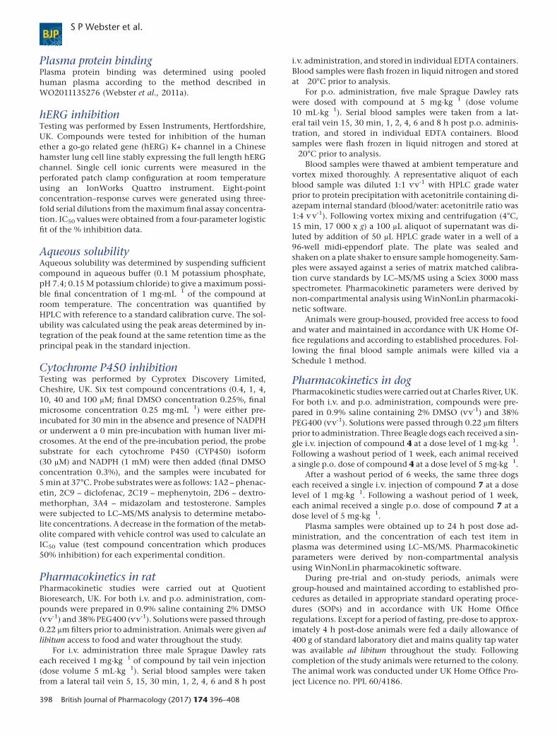
Plasma protein bindingPlasma protein binding was determined using pooledhuman plasma according to the method described inWO2011135276 (Webster et al., 2011a).
hERG inhibitionTesting was performed by Essen Instruments, Hertfordshire,UK. Compounds were tested for inhibition of the humanether a go-go related gene (hERG) K+ channel in a Chinesehamster lung cell line stably expressing the full length hERGchannel. Single cell ionic currents were measured in theperforated patch clamp configuration at room temperatureusing an IonWorks Quattro instrument. Eight-pointconcentration–response curves were generated using three-fold serial dilutions from themaximum final assay concentra-tion. IC50 values were obtained from a four-parameter logisticfit of the % inhibition data.
Aqueous solubilityAqueous solubility was determined by suspending sufficientcompound in aqueous buffer (0.1 M potassium phosphate,pH 7.4; 0.15 M potassium chloride) to give a maximum possi-ble final concentration of 1 mg·mL�1 of the compound atroom temperature. The concentration was quantified byHPLC with reference to a standard calibration curve. The sol-ubility was calculated using the peak areas determined by in-tegration of the peak found at the same retention time as theprincipal peak in the standard injection.
Cytochrome P450 inhibitionTesting was performed by Cyprotex Discovery Limited,Cheshire, UK. Six test compound concentrations (0.4, 1, 4,10, 40 and 100 μM; final DMSO concentration 0.25%, finalmicrosome concentration 0.25 mg·mL�1) were either pre-incubated for 30 min in the absence and presence of NADPHor underwent a 0 min pre-incubation with human liver mi-crosomes. At the end of the pre-incubation period, the probesubstrate for each cytochrome P450 (CYP450) isoform(30 μM) and NADPH (1 mM) were then added (final DMSOconcentration 0.3%), and the samples were incubated for5min at 37°C. Probe substrates were as follows: 1A2 – phenac-etin, 2C9 – diclofenac, 2C19 – mephenytoin, 2D6 – dextro-methorphan, 3A4 – midazolam and testosterone. Sampleswere subjected to LC–MS/MS analysis to determine metabo-lite concentrations. A decrease in the formation of the metab-olite compared with vehicle control was used to calculate anIC50 value (test compound concentration which produces50% inhibition) for each experimental condition.
Pharmacokinetics in ratPharmacokinetic studies were carried out at QuotientBioresearch, UK. For both i.v. and p.o. administration, com-pounds were prepared in 0.9% saline containing 2% DMSO(v.v-1) and 38% PEG400 (v.v-1). Solutions were passed through0.22 μm filters prior to administration. Animals were given adlibitum access to food and water throughout the study.
For i.v. administration three male Sprague Dawley ratseach received 1 mg·kg�1 of compound by tail vein injection(dose volume 5 mL·kg�1). Serial blood samples were takenfrom a lateral tail vein 5, 15, 30 min, 1, 2, 4, 6 and 8 h post
i.v. administration, and stored in individual EDTA containers.Blood samples were flash frozen in liquid nitrogen and storedat �20°C prior to analysis.
For p.o. administration, five male Sprague Dawley ratswere dosed with compound at 5 mg·kg�1 (dose volume10 mL·kg�1). Serial blood samples were taken from a lat-eral tail vein 15, 30 min, 1, 2, 4, 6 and 8 h post p.o. adminis-tration, and stored in individual EDTA containers. Bloodsamples were flash frozen in liquid nitrogen and stored at�20°C prior to analysis.
Blood samples were thawed at ambient temperature andvortex mixed thoroughly. A representative aliquot of eachblood sample was diluted 1:1 v.v-1 with HPLC grade waterprior to protein precipitation with acetonitrile containing di-azepam internal standard (blood/water: acetonitrile ratio was1:4 v v-1). Following vortex mixing and centrifugation (4°C,15 min, 17 000 x g) a 100 μL aliquot of supernatant was di-luted by addition of 50 μL HPLC grade water in a well of a96-well midi-eppendorf plate. The plate was sealed andshaken on a plate shaker to ensure sample homogeneity. Sam-ples were assayed against a series of matrix matched calibra-tion curve standards by LC–MS/MS using a Sciex 3000 massspectrometer. Pharmacokinetic parameters were derived bynon-compartmental analysis using WinNonLin pharmacoki-netic software.
Animals were group-housed, provided free access to foodand water and maintained in accordance with UK Home Of-fice regulations and according to established procedures. Fol-lowing the final blood sample animals were killed via aSchedule 1 method.
Pharmacokinetics in dogPharmacokinetic studies were carried out at Charles River, UK.For both i.v. and p.o. administration, compounds were pre-pared in 0.9% saline containing 2% DMSO (v.v-1) and 38%PEG400 (v.v-1). Solutions were passed through 0.22 μm filtersprior to administration. Three Beagle dogs each received a sin-gle i.v. injection of compound 4 at a dose level of 1 mg·kg�1.Following a washout period of 1 week, each animal receiveda single p.o. dose of compound 4 at a dose level of 5 mg·kg�1.
After a washout period of 6 weeks, the same three dogseach received a single i.v. injection of compound 7 at a doselevel of 1 mg·kg�1. Following a washout period of 1 week,each animal received a single p.o. dose of compound 7 at adose level of 5 mg·kg�1.
Plasma samples were obtained up to 24 h post dose ad-ministration, and the concentration of each test item inplasma was determined using LC–MS/MS. Pharmacokineticparameters were derived by non-compartmental analysisusing WinNonLin pharmacokinetic software.
During pre-trial and on-study periods, animals weregroup-housed and maintained according to established pro-cedures as detailed in appropriate standard operating proce-dures (SOPs) and in accordance with UK Home Officeregulations. Except for a period of fasting, pre-dose to approx-imately 4 h post-dose animals were fed a daily allowance of400 g of standard laboratory diet and mains quality tap waterwas available ad libitum throughout the study. Followingcompletion of the study animals were returned to the colony.The animal work was conducted under UK Home Office Pro-ject Licence no. PPL 60/4186.
BJP S P Webster et al.
398 British Journal of Pharmacology (2017) 174 396–408
Tissue exposure measurements in ratsThe circulating plasma levels and tissue distribution of com-pounds were determined according to the method describedin WO2011135276 (Webster et al., 2011a). Animals weregroup-housed, had free access to food and water and weremaintained in accordance with UK Home Office regulations.The animal work was conducted under UK Home Office Pro-ject Licence no. PPL 60/3915.
Compounds were prepared in 0.9% saline containing2% DMSO (v.v-1) and 38% PEG400 (v.v-1) and administeredto male Sprague Dawley rats by oral gavage such that thefinal concentration of compound was 10 mg·kg�1. Rats(n = 3 per time point) were killed by decapitation at 1, 4and 6 h post dosing, and trunk blood and tissues (liver,adipose and brain) were excised. Blood samples at 0.5 and2 h post dosing were taken by a tail nick from the 4 and6 h rats respectively.
Compounds were triple extracted from plasma (preparedfrom blood by a high speed centrifugation step) and spikedwith 1 μg of a standard compound using ethyl acetate. Ex-tracts were dried under nitrogen and re-suspended in 60%methanol/40% ammonium acetate (5 mM).
A known weight of tissue was homogenized in 3 volumesof Krebs buffer. The compoundwas triple extracted with ethylacetate from the supernatant of a low speed spin spiked with1 mg of a standard compound. Extracts were dried under ni-trogen and re-suspended in 60% methanol/40% ammoniumacetate (5 mM).
Samples from plasma and tissue were analysed by TSQQuantum Discovery Tandem Mass Spectrometer and Sur-veyor Liquid Chromatogram (Thermo, Hemel-Hempstead,UK). Then 10 μL of each sample was injected in a mobilephase consisting of 60% methanol/40% ammonium acetate(5 mM) at a flow rate of 0.5 mL·min�1. The column usedwas a BDS hypersil C18, 50 × 2.1mmwith a 5 μmparticle size.
Each compound was tuned with a spray voltage of 3000 Vand a capillary temperature of 300°C, and values for tubelens, CID and product ions were determined. The peak areafor each compound and for the internal standard was deter-mined, and the concentration of compound . g-1 of tissue ormL-1 of plasma was calculated by comparison of the peak arearatio to a standard curve.
Compliance with requirements for studies usinganimalsStudies using animals were carried out in accordance with UKHome Office regulations under appropriate project licences.Pharmacokinetic and tissue exposure measurements werecarried out using standard study designs, and the numbersof animals used per study were in line with those requiredto support regulatory submissions to the UK Medicines andHealthcare products Regulatory Agency. Animal studies arereported in compliance with the ARRIVE guidelines (Kil-kenny et al., 2010; McGrath & Lilley, 2015).
Clinical study design and subjectcharacteristicsSingle ascending dose study. A double-blind, randomized,placebo-controlled single ascending dose study was carriedout at Simbec Research Ltd, Merthyr Tydfil, UK. Each cohort
consisted of six healthy male subjects and two females ofnon-child bearing potential. Two male ‘dose leader’ subjects(1 active : 1 placebo) were dosed first in each cohort. Theremainder of the cohort (5 active : 1 placebo) were dosed24 h later. Dose cohorts were 2, 5, 10, 18, 25 and 35 mg.Safety, pharmacokinetic and pharmacodynamic assessmentswere made at pre-determined time-points during each partof the study. Dose escalation was dependent uponsatisfactory review of the blinded safety (up to Day 6) andpharmacokinetic (up to 24 h post-dose) data from thepreceding cohort.
Multiple ascending dose study. A double-blind, randomized,placebo-controlled multiple ascending dose study wascarried out at Linear Clinical Research Ltd, Nedlands,Australia. Each cohort consisted of eight healthy malesubjects. Each subject received UE2343 or placebo twicedaily (b.i.d ) (12 hourly) in the fed state for 9 days (Day 1 toDay 9) and once on the morning of Day 10 (19 doses intotal). Dose cohorts were 10, 20 and 35 mg. Dose escalationand selection was dependent upon satisfactory review of theblinded safety (up to Day 17 ± 2) and plasmapharmacokinetics (up to Day 12) data from the precedingcohort. Safety, pharmacokinetic and pharmacodynamicassessments were made at pre-determined time-pointsduring each part of the study.
CSF study. A non-randomized, open label, multiple dosephase I study to determine the pharmacokinetic parametersof UE2343 in CSF was carried out at Linear Clinical ResearchLtd, Nedlands, Australia. Four healthy male subjects eachreceived 35 mg UE2343 b.i.d (12 hourly) fed (30 min afterthe start of a standard meal) for 3 days (Day 1 to Day 3) andonce on the morning of Day 4 (seven doses in total). Plasmapharmacokinetic samples were taken from each subject onDay 1 and Day 4. A single CSF sample was taken from eachsubject at 5 h post-final dose on Day 4, corresponding to theplasma Tmax (time to maximum plasma concentration).
Approval for each clinical study was obtained followingreview of protocols by local research ethics committees.
Analytical methodsLaboratory evaluation of clinical safety parameters was car-ried out using standard validated methods. Measurement ofplasma pharmacodynamic markers [adrenocorticotropic hor-mone (ACTH), cortisol, 4-androstenedione, testosterone anddehydroepiandrosterone sulphate (DHEA-s)] was carried outusing commercially available kits. Each method was fully val-idated prior to analysis. Urinary steroid metabolites weremeasured according to established methods (Boonen et al.,2013). UE2343 levels in plasma, urine and CSF was deter-mined by LC–MS/MS using fully validated methods.
Statistical analysisThe data and statistical analysis comply with the recom-mendations on experimental design and analysis in phar-macology (Curtis et al., 2015). Levels of probability,P < 0.05 were deemed to constitute the threshold for statis-tical significance. Pharmacokinetic parameters, except Tmax
(time of maximum observed drug concentration), are givenas mean ± coefficient of variation (CV%). Tmax is given as
Discovery/clinical evaluation of 11β-HSD1 inhibitor UE2343 BJP
British Journal of Pharmacology (2017) 174 396–408 399
median ± range. For each of the pharmacodynamic parame-ters [ACTH, cortisol, adrenal androgens, tetrahydrocortisols(THFs)/tetrahydrocortisone (THE) ratio], the maximum %change from baseline for each subject was subjected toANOVA. For selected data, an additional covariate factorfor the baseline was also included in the ANOVA/analysisof covariance (ANCOVA) model. Associated P-values <0.05for each comparison have been presented.
The ACTH levels for each individual given a single dose ofUE2343 on Day 1 (0 and 23 h post-dose) were also subjectedto a fixed effects ANOVA, with a fixed effect for dose level.The associated P-values <0.05 between 0 and 23 h post-dosehave been presented.
Unless stated otherwise, data has been plotted asmean ± SEM.
Results
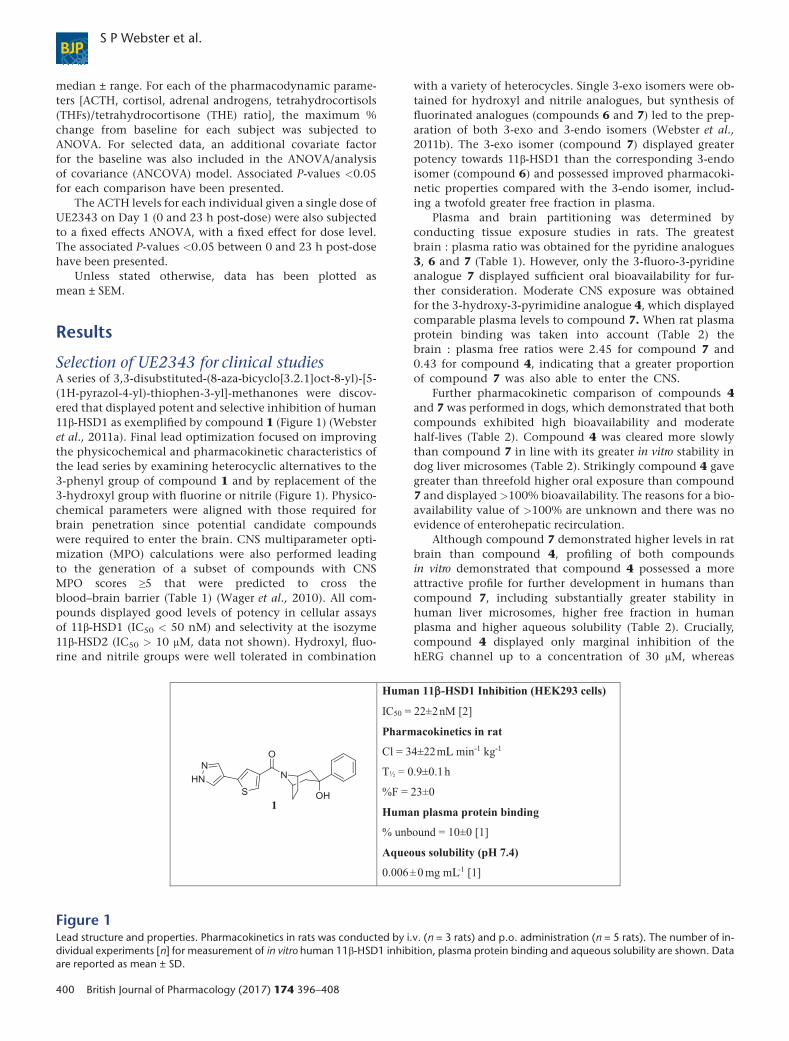
Selection of UE2343 for clinical studiesA series of 3,3-disubstituted-(8-aza-bicyclo[3.2.1]oct-8-yl)-[5-(1H-pyrazol-4-yl)-thiophen-3-yl]-methanones were discov-ered that displayed potent and selective inhibition of human11β-HSD1 as exemplified by compound 1 (Figure 1) (Websteret al., 2011a). Final lead optimization focused on improvingthe physicochemical and pharmacokinetic characteristics ofthe lead series by examining heterocyclic alternatives to the3-phenyl group of compound 1 and by replacement of the3-hydroxyl group with fluorine or nitrile (Figure 1). Physico-chemical parameters were aligned with those required forbrain penetration since potential candidate compoundswere required to enter the brain. CNS multiparameter opti-mization (MPO) calculations were also performed leadingto the generation of a subset of compounds with CNSMPO scores ≥5 that were predicted to cross theblood–brain barrier (Table 1) (Wager et al., 2010). All com-pounds displayed good levels of potency in cellular assaysof 11β-HSD1 (IC50 < 50 nM) and selectivity at the isozyme11β-HSD2 (IC50 > 10 μM, data not shown). Hydroxyl, fluo-rine and nitrile groups were well tolerated in combination
with a variety of heterocycles. Single 3-exo isomers were ob-tained for hydroxyl and nitrile analogues, but synthesis offluorinated analogues (compounds 6 and 7) led to the prep-aration of both 3-exo and 3-endo isomers (Webster et al.,2011b). The 3-exo isomer (compound 7) displayed greaterpotency towards 11β-HSD1 than the corresponding 3-endoisomer (compound 6) and possessed improved pharmacoki-netic properties compared with the 3-endo isomer, includ-ing a twofold greater free fraction in plasma.
Plasma and brain partitioning was determined byconducting tissue exposure studies in rats. The greatestbrain : plasma ratio was obtained for the pyridine analogues3, 6 and 7 (Table 1). However, only the 3-fluoro-3-pyridineanalogue 7 displayed sufficient oral bioavailability for fur-ther consideration. Moderate CNS exposure was obtainedfor the 3-hydroxy-3-pyrimidine analogue 4, which displayedcomparable plasma levels to compound 7. When rat plasmaprotein binding was taken into account (Table 2) thebrain : plasma free ratios were 2.45 for compound 7 and0.43 for compound 4, indicating that a greater proportionof compound 7 was also able to enter the CNS.
Further pharmacokinetic comparison of compounds 4and 7 was performed in dogs, which demonstrated that bothcompounds exhibited high bioavailability and moderatehalf-lives (Table 2). Compound 4 was cleared more slowlythan compound 7 in line with its greater in vitro stability indog liver microsomes (Table 2). Strikingly compound 4 gavegreater than threefold higher oral exposure than compound7 and displayed >100% bioavailability. The reasons for a bio-availability value of >100% are unknown and there was noevidence of enterohepatic recirculation.
Although compound 7 demonstrated higher levels in ratbrain than compound 4, profiling of both compoundsin vitro demonstrated that compound 4 possessed a moreattractive profile for further development in humans thancompound 7, including substantially greater stability inhuman liver microsomes, higher free fraction in humanplasma and higher aqueous solubility (Table 2). Crucially,compound 4 displayed only marginal inhibition of thehERG channel up to a concentration of 30 μM, whereas
Figure 1Lead structure and properties. Pharmacokinetics in rats was conducted by i.v. (n = 3 rats) and p.o. administration (n = 5 rats). The number of in-dividual experiments [n] for measurement of in vitro human 11β-HSD1 inhibition, plasma protein binding and aqueous solubility are shown. Dataare reported as mean ± SD.
BJP S P Webster et al.
400 British Journal of Pharmacology (2017) 174 396–408
compound 7 was a moderate hERG inhibitor (IC50 = 3.1 μM,Table 2). Subsequent in vitro profiling of compound 4revealed a clean off-target profile in a diversity screen of 29enzymes and 72 receptors, including the glucocorticoidand mineralocorticoid receptors (data not shown). For com-pound 4, no significant CYP450 inhibition was observed atisoforms 1A2, 2D6, 2C9 or 3A4 ((IC50 > 50 μM). However,moderate inhibition of isoform 2C19 (IC50 = 1.7 μM) wasobserved. No time-dependent inhibition was observed atany of the CYP450 enzymes tested. Compound 4 was thuschosen for further development in humans and designatedwith the code UE2343.
Subject demographics and safetyA single ascending dose study was conducted in 36 healthymale subjects (35 Caucasians and 1 Afro-Caribbean) and 12healthy Caucasian females of non-child bearing potential.For males, the mean age was 34.1 ± 9.7 years and the meanbody mass index (BMI) was 26.0 ± 2.4 kg·m�2. For females,the mean age was 49.3 ± 9.1 years and the mean BMIwas 25.1 ± 2.1 kg·m�2. Each cohort consisted of six malesand two females. The incidence of treatment-emergent
adverse events (TEAEs) was considered to be low (numberof subjects with ≥1 TEAEs: placebo = 6, UE2343 = 9), andnone was associated with any clinically significant changesin vital signs, ECG, biochemistry, haematology or urinaly-sis data.
A multiple ascending dose study was conducted in 24healthy male subjects (4 Caucasians and 20 Asians) with amean age of 26.7 ± 5.4 years and a mean BMI of 23.9 ± 1.9-kg·m�2. No serious TEAEs or TEAEs that led to subject with-drawal from the study were reported, and all TEAEs weremild or moderate in intensity. The most common TEAE washeadache reported in 7/24 subjects; diarrhoea was reportedin 2/24 subjects, and thrombophlebitis was reported in 3/24subjects.
A study to determine the amount of UE2343 in CSF wasconducted in four healthy Asian male subjects with meanage of 29 ± 11 years and mean BMI of 24.6 ± 1.4 kg·m�2. AllTEAEs were mild to moderate in intensity. Vital signsremained stable during the study, and no out of the rangevalues were recorded for any of the subjects during thestudy. Increased alanine aminotransferase was reported ina single subject.
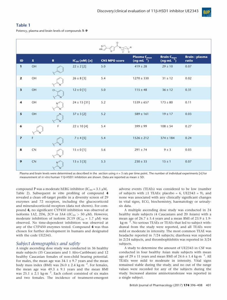
Table 1Potency, plasma and brain levels of compounds 1–9
ID X R IC50 (nM) [n] CNS MPO scorePlasma Cmax
(ng·mL�1)Brain Cmax
(ng·mL�1)Brain : plasmaratio
1 OH 22 ± 2 [2] 5.0 419 ± 28 29 ± 10 0.07
2 OH 26 ± 8 [3] 5.4 1270 ± 330 31 ± 12 0.02
3 OH 12 ± 0 [1] 5.0 115 ± 48 36 ± 12 0.31
4 OH 24 ± 15 [31] 5.2 1539 ± 657 173 ± 80 0.11
5 OH 37 ± 3 [2] 5.2 589 ± 161 19 ± 17 0.03
6 F 22 ± 10 [4] 5.4 399 ± 99 108 ± 54 0.27
7 F 7 ± 4 [5] 5.4 1526 ± 212 374 ± 184 0.24
8 CN 15 ± 0 [1] 5.6 291 ± 74 9 ± 3 0.03
9 CN 13 ± 3 [3] 5.3 230 ± 33 15 ± 1 0.07
Plasma and brain levels were determined as described in the section using n = 3 rats per time point. The number of individual experiments [n] formeasurement of in vitro human 11β-HSD1 inhibition are shown. Data are reported as mean ± SD.
Discovery/clinical evaluation of 11β-HSD1 inhibitor UE2343 BJP
British Journal of Pharmacology (2017) 174 396–408 401
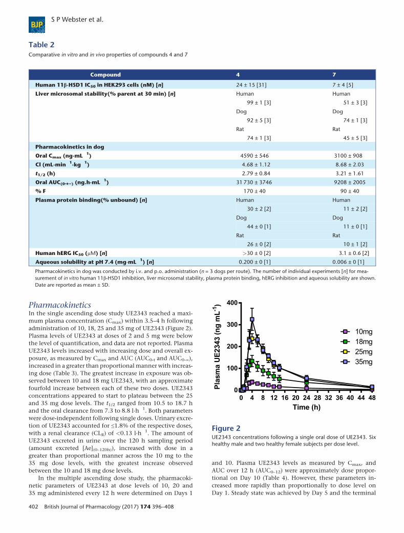
PharmacokineticsIn the single ascending dose study UE2343 reached a maxi-mum plasma concentration (Cmax) within 3.5–4 h followingadministration of 10, 18, 25 and 35 mg of UE2343 (Figure 2).Plasma levels of UE2343 at doses of 2 and 5 mg were belowthe level of quantification, and data are not reported. PlasmaUE2343 levels increased with increasing dose and overall ex-posure, as measured by Cmax and AUC (AUC0-t and AUC0-∞),increased in a greater than proportional manner with increas-ing dose (Table 3). The greatest increase in exposure was ob-served between 10 and 18 mg UE2343, with an approximatefourfold increase between each of these two doses. UE2343concentrations appeared to start to plateau between the 25and 35 mg dose levels. The t1/2 ranged from 10.5 to 18.7 hand the oral clearance from 7.3 to 8.8 l·h�1. Both parameterswere dose-independent following single doses. Urinary excre-tion of UE2343 accounted for ≤1.8% of the respective doses,with a renal clearance (CLR) of <0.13 l·h�1. The amount ofUE2343 excreted in urine over the 120 h sampling period(amount excreted [Ae](0–120h)), increased with dose in agreater than proportional manner across the 10 mg to the35 mg dose levels, with the greatest increase observedbetween the 10 and 18 mg dose levels.
In the multiple ascending dose study, the pharmacoki-netic parameters of UE2343 at dose levels of 10, 20 and35 mg administered every 12 h were determined on Days 1
and 10. Plasma UE2343 levels as measured by Cmax, andAUC over 12 h (AUC0–12) were approximately dose propor-tional on Day 10 (Table 4). However, these parameters in-creased more rapidly than proportionally to dose level onDay 1. Steady state was achieved by Day 5 and the terminal
Table 2Comparative in vitro and in vivo properties of compounds 4 and 7
Compound 4 7
Human 11β-HSD1 IC50 in HEK293 cells (nM) [n] 24 ± 15 [31] 7 ± 4 [5]
Liver microsomal stability(% parent at 30 min) [n] Human Human
99 ± 1 [3] 51 ± 3 [3]
Dog Dog
92 ± 5 [3] 74 ± 1 [3]
Rat Rat
74 ± 1 [3] 45 ± 5 [3]
Pharmacokinetics in dog
Oral Cmax (ng·mL�1) 4590 ± 546 3100 ± 908
Cl (mL·min�1·kg�1) 4.68 ± 1.12 8.68 ± 2.03
t1/2 (h) 2.79 ± 0.84 3.21 ± 1.61
Oral AUC(0➔∞) (ng.h·mL�1) 31 730 ± 3746 9208 ± 2005
% F 170 ± 40 90 ± 40
Plasma protein binding(% unbound) [n] Human Human
30 ± 2 [2] 11 ± 2 [2]
Dog Dog
44 ± 0 [1] 11 ± 0 [1]
Rat Rat
26 ± 0 [2] 10 ± 1 [2]
Human hERG IC50 (μM) [n] >30 ± 0 [2] 3.1 ± 0.6 [2]
Aqueous solubility at pH 7.4 (mg·mL�1) [n] 0.200 ± 0 [1] 0.006 ± 0 [1]
Pharmacokinetics in dog was conducted by i.v. and p.o. administration (n = 3 dogs per route). The number of individual experiments [n] for mea-surement of in vitro human 11β-HSD1 inhibition, liver microsomal stability, plasma protein binding, hERG inhibition and aqueous solubility are shown.Date are reported as mean ± SD.
Figure 2UE2343 concentrations following a single oral dose of UE2343. Sixhealthy male and two healthy female subjects per dose level.
BJP S P Webster et al.
402 British Journal of Pharmacology (2017) 174 396–408
t1/2 ranged from 10 to 14 h. The median time to peak concen-tration following each dose ranged from 4 to 6 h. There wasconsiderable accumulation with 12 hourly dosing, with in-crease in AUC0–12 fromfirst dose to steady state of greater thanfourfold at the highest dose level. The Ae in urine was low,with the fraction excreted in the dosing interval approxi-mately 4% at steady state.
The concentrations of UE2343 in plasma and CSF weredetermined during administration of 35 mg b.i.d for 4 days.Mean Cmax on Days 1 and 4 was reached at 5 h post-dose(Table 5). The extent of accumulation of UE2343 in plasmaafter repeated daily dosing on Day 4 was 3.3-fold, consistentwith an elimination t1/2 in the order of 1 day. Between-subjectvariability in the extent of systemic exposure to UE2343 washigh with geometric CVs for Cmax and AUC0–12 of 59 to81%. A single CSF sample was taken from each subject at5 h post-dose on Day 4. The mean concentration ofUE2343 in the CSF was 69.8 ng·mL�1, ranging from 41.2to 99.9 ng·mL�1 and 7.46 to 11.9% of simultaneous totalplasma levels (Table 5).
HPA axis hormones and steroidsFollowing single dose administration of UE2343, there wasno effect on serum cortisol, 4-androstenedione or testoster-one levels at any of the doses tested (Supporting InformationFig. S1A–C). DHEA-s was higher following UE2343 adminis-tration when compared to placebo with statistically signifi-cant increases observed at 2 (P < 0.05, data not shown), 5(P < 0.05, data not shown) and 35 mg (P < 0.05) UE2343.The increases in DHEA-s were of similar magnitude indicat-ing that there was no apparent dose–response relationship(Supporting Information Fig. S1D). There was a dose-dependent increase in plasma ACTH across the 18–35 mgdose range (Figure 3A). When compared with placebo, a sta-tistically significant increase was observed for the 35 mg doselevel (P < 0.05). The time to maximum % change was shownto occur at a median of 23–27 h post-dose at all dose levels ex-cept 2 mg UE2343, and values had returned to baseline by ap-proximately 47 h post-dose. Within-group comparisons alsorevealed a statistically significant increase in ACTH 23 hpost-dose when compared with pre-dose levels following 10
Table 3Pharmacokinetic parameters following a single oral dose of UE2343 in humans
Pharmacokineticparameter
UE2343
10 mg Mean(CV%)
18 mg Mean(CV%)
25 mg Mean(CV%)
35 mg Mean(CV%)
Cmax (ng·mL�1) 40.4 (62%) 136 (46%) 278 (30%) 329 (37%)
Tmax (h)1 3.5 (3–6) 4.0 (3–4) 4.0 (3–6) 4.0 (4–6)
t1/2 (h) 18.7 (59%) 13.6 (61%) 10.5 (35%) 11.4 (24%)
AUC0-t (ng.h·mL�1) 550 (90%) 2291 (87%) 3486 (39%) 4016 (30%)
AUC0-∞ (ng.h·mL�1) 856 (70%) 2581 (84%) 3825 (35%) 4261 (29%)
Oral clearance (L·h�1) 18.5 (75%) 14.5 (90%) 7.32 (38%) 8.78 (28%)
Ae(0–120h) (μg) 52.5 (85%) 300 (66%) 438 (37%) 501 (15%)
Ae(0–120h) (%) 0.53 (85%) 1.67 (66%) 1.75 (37%) 1.43 (15%)
CLR (L·h�1) 0.07 (86%) 0.16 (50%) 0.13 (46%) 0.12 (16%)
Single ascending dose studies were conducted in six healthy male and two healthy female subjects per dose level.1Median ± range
Table 4Pharmacokinetic parameters following multiple oral doses of UE2343 in humans
Pharmacokineticparameter
Day 1 Day 10
10 mg UE2343Mean(CV%)
20 mg UE2343Mean(CV%)
35 mg UE2343Mean(CV%)
10 mg UE2343Mean(CV%)
20 mg UE2343Mean(CV%)
35 mg UE2343Mean(CV%)
Cmax (ng·mL�1) 29.1 (61%) 89.2 (71%) 268 (34%) 207 (58%) 365 (29%) 892 (30%)
Tmax (h)1 6.0 (4–8) 4.0 (3–10) 4.0 (3–6) 4.0 (3–6) 3.0 (2–6) 4.0 (3–6)
t1/2 (h) nd nd nd 13.8 (21%) 11.2 (25%) 9.99 (14%)
AUC0–12h(ng.h·mL�1) 207 (79%) 623 (69%) 1843 (33%) 1917 (66%) 3175 (30%) 7909 (36%)
Oral clearance (L·h�1) nd nd nd 7.24 (60%) 6.85 (33%) 5.02 (43%)
Ae(0–12h) (μg) 34.6 (50%) 107 (92%) 362 (46%) 408 (46%) 644 (38%) 1345 (29%)
Ae(0–12h) (%) 0.35 (50%) 0.54 (92%) 1.03 (46%) 4.08 (46%) 3.22 (38%) 3.84 (29%)
Multiple ascending dose studies were conducted in eight healthy male subjects per dose level.1Median ± range
Discovery/clinical evaluation of 11β-HSD1 inhibitor UE2343 BJP
British Journal of Pharmacology (2017) 174 396–408 403
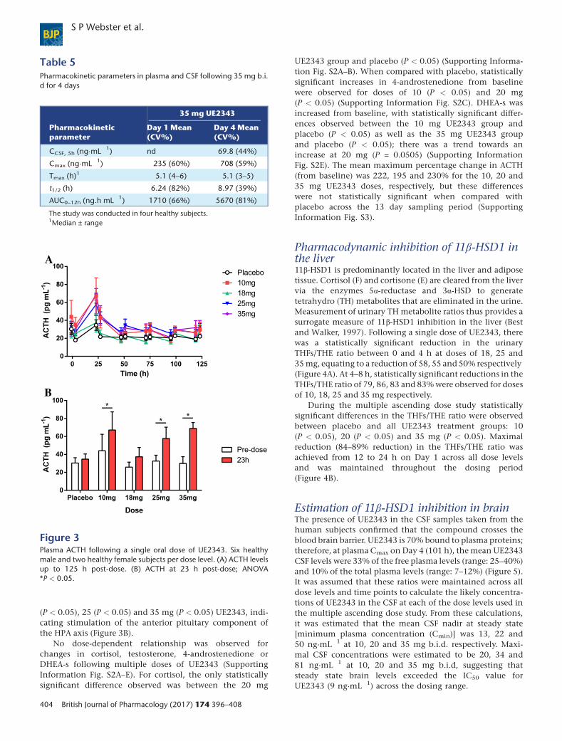
(P < 0.05), 25 (P < 0.05) and 35 mg (P < 0.05) UE2343, indi-cating stimulation of the anterior pituitary component ofthe HPA axis (Figure 3B).
No dose-dependent relationship was observed forchanges in cortisol, testosterone, 4-androstenedione orDHEA-s following multiple doses of UE2343 (SupportingInformation Fig. S2A–E). For cortisol, the only statisticallysignificant difference observed was between the 20 mg
UE2343 group and placebo (P < 0.05) (Supporting Informa-tion Fig. S2A–B). When compared with placebo, statisticallysignificant increases in 4-androstenedione from baselinewere observed for doses of 10 (P < 0.05) and 20 mg(P < 0.05) (Supporting Information Fig. S2C). DHEA-s wasincreased from baseline, with statistically significant differ-ences observed between the 10 mg UE2343 group andplacebo (P < 0.05) as well as the 35 mg UE2343 groupand placebo (P < 0.05); there was a trend towards anincrease at 20 mg (P = 0.0505) (Supporting InformationFig. S2E). The mean maximum percentage change in ACTH(from baseline) was 222, 195 and 230% for the 10, 20 and35 mg UE2343 doses, respectively, but these differenceswere not statistically significant when compared withplacebo across the 13 day sampling period (SupportingInformation Fig. S3).
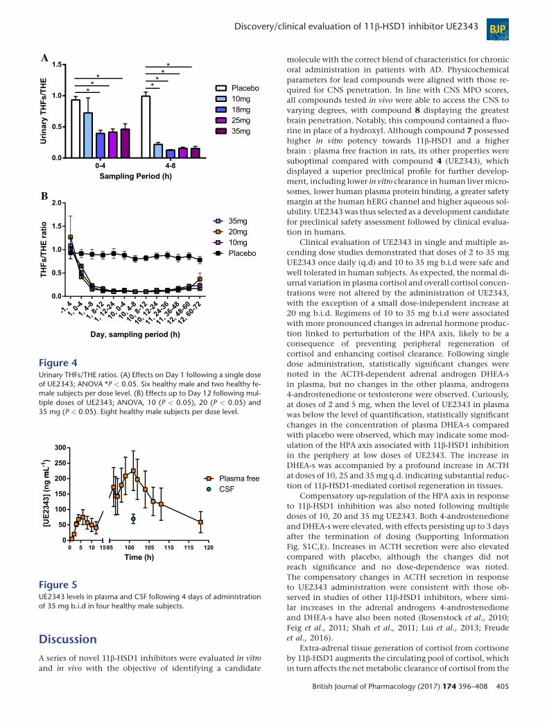
Pharmacodynamic inhibition of 11β-HSD1 inthe liver11β-HSD1 is predominantly located in the liver and adiposetissue. Cortisol (F) and cortisone (E) are cleared from the livervia the enzymes 5α-reductase and 3α-HSD to generatetetrahydro (TH) metabolites that are eliminated in the urine.Measurement of urinary TH metabolite ratios thus provides asurrogate measure of 11β-HSD1 inhibition in the liver (Bestand Walker, 1997). Following a single dose of UE2343, therewas a statistically significant reduction in the urinaryTHFs/THE ratio between 0 and 4 h at doses of 18, 25 and35mg, equating to a reduction of 58, 55 and 50% respectively(Figure 4A). At 4–8 h, statistically significant reductions in theTHFs/THE ratio of 79, 86, 83 and 83%were observed for dosesof 10, 18, 25 and 35 mg respectively.
During the multiple ascending dose study statisticallysignificant differences in the THFs/THE ratio were observedbetween placebo and all UE2343 treatment groups: 10(P < 0.05), 20 (P < 0.05) and 35 mg (P < 0.05). Maximalreduction (84–89% reduction) in the THFs/THE ratio wasachieved from 12 to 24 h on Day 1 across all dose levelsand was maintained throughout the dosing period(Figure 4B).
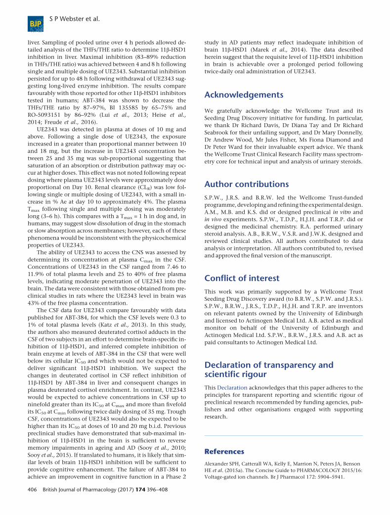
Estimation of 11β-HSD1 inhibition in brainThe presence of UE2343 in the CSF samples taken from thehuman subjects confirmed that the compound crosses theblood brain barrier. UE2343 is 70% bound to plasma proteins;therefore, at plasma Cmax on Day 4 (101 h), the mean UE2343CSF levels were 33% of the free plasma levels (range: 25–40%)and 10% of the total plasma levels (range: 7–12%) (Figure 5).It was assumed that these ratios were maintained across alldose levels and time points to calculate the likely concentra-tions of UE2343 in the CSF at each of the dose levels used inthe multiple ascending dose study. From these calculations,it was estimated that the mean CSF nadir at steady state[minimum plasma concentration (Cmin)] was 13, 22 and50 ng·mL�1 at 10, 20 and 35 mg b.i.d. respectively. Maxi-mal CSF concentrations were estimated to be 20, 34 and81 ng·mL�1 at 10, 20 and 35 mg b.i.d, suggesting thatsteady state brain levels exceeded the IC50 value forUE2343 (9 ng·mL�1) across the dosing range.
Table 5Pharmacokinetic parameters in plasma and CSF following 35 mg b.i.d for 4 days
Pharmacokineticparameter
35 mg UE2343
Day 1 Mean(CV%)
Day 4 Mean(CV%)
CCSF, 5h (ng·mL�1) nd 69.8 (44%)
Cmax (ng·mL�1) 235 (60%) 708 (59%)
Tmax (h)1 5.1 (4–6) 5.1 (3–5)
t1/2 (h) 6.24 (82%) 8.97 (39%)
AUC0–12h (ng.h mL�1) 1710 (66%) 5670 (81%)
The study was conducted in four healthy subjects.1Median ± range
A
B
Figure 3Plasma ACTH following a single oral dose of UE2343. Six healthymale and two healthy female subjects per dose level. (A) ACTH levelsup to 125 h post-dose. (B) ACTH at 23 h post-dose; ANOVA*P < 0.05.
BJP S P Webster et al.
404 British Journal of Pharmacology (2017) 174 396–408
DiscussionA series of novel 11β-HSD1 inhibitors were evaluated in vitroand in vivo with the objective of identifying a candidate
molecule with the correct blend of characteristics for chronicoral administration in patients with AD. Physicochemicalparameters for lead compounds were aligned with those re-quired for CNS penetration. In line with CNS MPO scores,all compounds tested in vivo were able to access the CNS tovarying degrees, with compound 8 displaying the greatestbrain penetration. Notably, this compound contained a fluo-rine in place of a hydroxyl. Although compound 7 possessedhigher in vitro potency towards 11β-HSD1 and a higherbrain : plasma free fraction in rats, its other properties weresuboptimal compared with compound 4 (UE2343), whichdisplayed a superior preclinical profile for further develop-ment, including lower in vitro clearance in human liver micro-somes, lower human plasma protein binding, a greater safetymargin at the human hERG channel and higher aqueous sol-ubility. UE2343 was thus selected as a development candidatefor preclinical safety assessment followed by clinical evalua-tion in humans.
Clinical evaluation of UE2343 in single and multiple as-cending dose studies demonstrated that doses of 2 to 35 mgUE2343 once daily (q.d) and 10 to 35 mg b.i.d were safe andwell tolerated in human subjects. As expected, the normal di-urnal variation in plasma cortisol and overall cortisol concen-trations were not altered by the administration of UE2343,with the exception of a small dose-independent increase at20 mg b.i.d. Regimens of 10 to 35 mg b.i.d were associatedwith more pronounced changes in adrenal hormone produc-tion linked to perturbation of the HPA axis, likely to be aconsequence of preventing peripheral regeneration ofcortisol and enhancing cortisol clearance. Following singledose administration, statistically significant changes werenoted in the ACTH-dependent adrenal androgen DHEA-sin plasma, but no changes in the other plasma, androgens4-androstenedione or testosterone were observed. Curiously,at doses of 2 and 5 mg, when the level of UE2343 in plasmawas below the level of quantification, statistically significantchanges in the concentration of plasma DHEA-s comparedwith placebo were observed, which may indicate some mod-ulation of the HPA axis associated with 11β-HSD1 inhibitionin the periphery at low doses of UE2343. The increase inDHEA-s was accompanied by a profound increase in ACTHat doses of 10, 25 and 35mg q.d. indicating substantial reduc-tion of 11β-HSD1-mediated cortisol regeneration in tissues.
Compensatory up-regulation of the HPA axis in responseto 11β-HSD1 inhibition was also noted following multipledoses of 10, 20 and 35 mg UE2343. Both 4-androstenedioneand DHEA-s were elevated, with effects persisting up to 3 daysafter the termination of dosing (Supporting InformationFig. S1C,E). Increases in ACTH secretion were also elevatedcompared with placebo, although the changes did notreach significance and no dose-dependence was noted.The compensatory changes in ACTH secretion in responseto UE2343 administration were consistent with those ob-served in studies of other 11β-HSD1 inhibitors, where simi-lar increases in the adrenal androgens 4-androstenedioneand DHEA-s have also been noted (Rosenstock et al., 2010;Feig et al., 2011; Shah et al., 2011; Lui et al., 2013; Freudeet al., 2016).
Extra-adrenal tissue generation of cortisol from cortisoneby 11β-HSD1 augments the circulating pool of cortisol, whichin turn affects the net metabolic clearance of cortisol from the
A
B
Figure 4Urinary THFs/THE ratios. (A) Effects on Day 1 following a single doseof UE2343; ANOVA *P < 0.05. Six healthy male and two healthy fe-male subjects per dose level. (B) Effects up to Day 12 following mul-tiple doses of UE2343; ANOVA, 10 (P < 0.05), 20 (P < 0.05) and35 mg (P < 0.05). Eight healthy male subjects per dose level.
Figure 5UE2343 levels in plasma and CSF following 4 days of administrationof 35 mg b.i.d in four healthy male subjects.
Discovery/clinical evaluation of 11β-HSD1 inhibitor UE2343 BJP
British Journal of Pharmacology (2017) 174 396–408 405
liver. Sampling of pooled urine over 4 h periods allowed de-tailed analysis of the THFs/THE ratio to determine 11β-HSD1inhibition in liver. Maximal inhibition (83–89% reductionin THFs/THE ratio) was achieved between 4 and 8 h followingsingle and multiple dosing of UE2343. Substantial inhibitionpersisted for up to 48 h following withdrawal of UE2343 sug-gesting long-lived enzyme inhibition. The results comparefavourably with those reported for other 11β-HSD1 inhibitorstested in humans; ABT-384 was shown to decrease theTHFs/THE ratio by 87–97%, BI 135585 by 65–75% andRO-5093151 by 86–92% (Lui et al., 2013; Heise et al.,2014; Freude et al., 2016).
UE2343 was detected in plasma at doses of 10 mg andabove. Following a single dose of UE2343, the exposureincreased in a greater than proportional manner between 10and 18 mg, but the increase in UE2343 concentration be-tween 25 and 35 mg was sub-proportional suggesting thatsaturation of an absorption or distribution pathway may oc-cur at higher doses. This effect was not noted following repeatdosing where plasma UE2343 levels were approximately doseproportional on Day 10. Renal clearance (CLR) was low fol-lowing single or multiple dosing of UE2343, with a small in-crease in % Ae at day 10 to approximately 4%. The plasmaTmax following single and multiple dosing was moderatelylong (3–6 h). This compares with a Tmax = 1 h in dog and, inhumans, may suggest slow dissolution of drug in the stomachor slow absorption across membranes; however, each of thesephenomena would be inconsistent with the physicochemicalproperties of UE2343.
The ability of UE2343 to access the CNS was assessed bydetermining its concentration at plasma Cmax in the CSF.Concentrations of UE2343 in the CSF ranged from 7.46 to11.9% of total plasma levels and 25 to 40% of free plasmalevels, indicating moderate penetration of UE2343 into thebrain. The data were consistent with those obtained from pre-clinical studies in rats where the UE2343 level in brain was43% of the free plasma concentration.
The CSF data for UE2343 compare favourably with datapublished for ABT-384, for which the CSF levels were 0.3 to1% of total plasma levels (Katz et al., 2013). In this study,the authors also measured deuterated cortisol adducts in theCSF of two subjects in an effort to determine brain-specific in-hibition of 11β-HSD1, and inferred complete inhibition ofbrain enzyme at levels of ABT-384 in the CSF that were wellbelow its cellular IC50 and which would not be expected todeliver significant 11β-HSD1 inhibition. We suspect thechanges in deuterated cortisol in CSF reflect inhibition of11β-HSD1 by ABT-384 in liver and consequent changes inplasma deuterated cortisol enrichment. In contrast, UE2343would be expected to achieve concentrations in CSF up toninefold greater than its IC50 at Cmax and more than fivefoldits IC50 at Cmin following twice daily dosing of 35 mg. TroughCSF, concentrations of UE2343 would also be expected to behigher than its IC50 at doses of 10 and 20 mg b.i.d. Previouspreclinical studies have demonstrated that sub-maximal in-hibition of 11β-HSD1 in the brain is sufficient to reversememory impairments in ageing and AD (Sooy et al., 2010;Sooy et al., 2015). If translated to humans, it is likely that sim-ilar levels of brain 11β-HSD1 inhibition will be sufficient toprovide cognitive enhancement. The failure of ABT-384 toachieve an improvement in cognitive function in a Phase 2
study in AD patients may reflect inadequate inhibition ofbrain 11β-HSD1 (Marek et al., 2014). The data describedherein suggest that the requisite level of 11β-HSD1 inhibitionin brain is achievable over a prolonged period followingtwice-daily oral administration of UE2343.
Acknowledgements
We gratefully acknowledge the Wellcome Trust and itsSeeding Drug Discovery initiative for funding. In particular,we thank Dr Richard Davis, Dr Diana Tay and Dr RichardSeabrook for their unfailing support, and Dr Mary Donnelly,Dr Andrew Wood, Mr Jules Fisher, Ms Fiona Diamond andDr Peter Ward for their invaluable expert advice. We thanktheWellcome Trust Clinical Research Facility mass spectrom-etry core for technical input and analysis of urinary steroids.
Author contributionsS.P.W., J.R.S. and B.R.W. led the Wellcome Trust-fundedprogramme,developingand refining theexperimentaldesign.A.M., M.B. and K.S. did or designed preclinical in vitro andin vivo experiments. S.P.W., T.D.P., H.J.H. and T.R.P. did ordesigned the medicinal chemistry. R.A. performed urinarysteroid analysis. A.B., B.R.W., V.S.R. and J.W.K. designed andreviewed clinical studies. All authors contributed to dataanalysis or interpretation. All authors contributed to, revisedand approved the final version of themanuscript.
Conflict of interestThis work was primarily supported by a Wellcome TrustSeeding Drug Discovery award (to B.R.W., S.P.W. and J.R.S.).S.P.W., B.R.W., J.R.S., T.D.P., H.J.H. and T.R.P. are inventorson relevant patents owned by the University of Edinburghand licensed to Actinogen Medical Ltd. A.B. acted as medicalmonitor on behalf of the University of Edinburgh andActinogen Medical Ltd. S.P.W., B.R.W., J.R.S. and A.B. act aspaid consultants to Actinogen Medical Ltd.
Declaration of transparency andscientific rigourThis Declaration acknowledges that this paper adheres to theprinciples for transparent reporting and scientific rigour ofpreclinical research recommended by funding agencies, pub-lishers and other organisations engaged with supportingresearch.
References
Alexander SPH, Catterall WA, Kelly E, Marrion N, Peters JA, BensonHE et al. (2015a). The Concise Guide to PHARMACOLOGY 2015/16:Voltage-gated ion channels. Br J Pharmacol 172: 5904–5941.
BJP S P Webster et al.
406 British Journal of Pharmacology (2017) 174 396–408
Alexander SPH, Cidlowski JA, Kelly E, Marrion N, Peters JA, BensonHE et al. (2015b). The Concise Guide to PHARMACOLOGY 2015/16:Nuclear hormone receptors. Br J Pharmacol 172: 5956–5978.
Alexander SPH, Fabbro D, Kelly E, Marrion N, Peters JA, Benson HEet al. (2015c). The Concise Guide to PHARMACOLOGY 2015/16:Enzymes. Br J Pharmacol 172: 6024–6109.
Anagnostis P, Athyros VG, Tziomalos K, Karagiannis A, MikhailidisDP (2009). Clinical review: the pathogenetic role of cortisol in themetabolic syndrome: a hypothesis. J Clin Endocrinol Metab 94:2692–2701.
Best R, Walker BR (1997). Additional value of measurement of urinarycortisone and unconjugated cortisol metabolites in assessing theactivity of 11β-hydroxysteroid dehydrogenase in vivo. ClinEndocrinol (Oxf) 47: 231–236.
Boonen E, Vervenne H, Meersseman P, Andrew R, Mortier L, DeclercqPE et al. (2013). Reduced cortisol metabolism during critical illness. NEngl J Med 368: 1477–1488.
Cernansky JG, Dong H, Fagan AM, Wang L, Xiong C, HoltzmanDM et al. (2006). Plasma cortisol and progression of dementia insubjects with Alzheimer-type dementia. Am J Psychiatry 163:2164–2169.
Curtis MJ, Bond RA, Spina D, hluwalia A A, Alexander SPA, GiembyczMA et al. (2015). Experimental design and analysis and theirreporting: new guidance for publication in BJP. Br J Pharmacol 172:3461–3471.
Feig PU, Shah S, Hermanowski-Vosatka A, Plotkin D, Springer MS,Donahue S et al. (2011). Effects of an 11β-hydroxysteroiddehydrogenase type 1 inhibitor, MK-0916, in patients with type 2diabetes mellitus and metabolic syndrome. Diabetes Obes Metab 13:498–504.
Freude S, Heise T, Woerle HJ, Jungnik A, Rauch T, Hamilton B et al.(2016). Safety, pharmacokinetics and pharmacodynamics of BI135585, a selective 11β-hydroxysteroid dehydrogenase-1 (HSD1)inhibitor in humans: liver and adipose tissue 11β-HSD1 inhibitionafter acute and multiple administrations over 2 weeks. Diabetes ObesMetab 18: 486–490.
Green KN, Billings LM, Roozendaal B, McGaugh JL, LaFerla FM(2006). Glucocorticoids increase amyloid-β and tau pathology in amouse model of Alzheimer’s disease. J Neurosci 26: 9047–9056.
Heise T, Morrow L, Hompesch M, Häring HU, Kapitza C, Abt M et al.(2014). Safety, efficacy and weight effect of two 11β-HSD1 inhibitorsin metformin-treated patients with type 2 diabetes. Diabetes ObesMetab 16: 1070–1077.
Katz DA, Liu W, Locke C, Jacobson P, Barnes DM, Basu R et al. (2013).Peripheral and central nervous system inhibition of 11β-hydroxysteroid dehydrogenase type 1 in man by the novel inhibitorABT-384. Transl Psychiatry 3: e295.
Kilkenny C, Browne W, Cuthill IC, Emerson M, Altman DG (2010).Animal research: reporting in vivo experiments: the ARRIVEguidelines. Br J Pharmacol 160: 1577–1579.
Kornhuber J, Jessen F (2015). Cerebrospinal fluid cortisol and clinicaldisease progression in MCI and dementia of Alzheimer’s type.Neurobiol Aging 36: 601–607.
Lui W, Katz DA, Locke C, Daszkowski D, Wang Y, Rieser MJ et al.(2013). Clinical safety, pharmacokinetics, and pharmacodynamics ofthe 11β-hydroxysteroid dehydrogenase type 1 Inhibitor ABT-384 inhealthy volunteers and elderly adults. Clin Pharmacol Drug Dev 2:133–151.
Lupien SJ, LeonM, Santi S, Convit A, Tarshish C, Nair NP et al. (1998).Cortisol levels during human aging predict hippocampal atrophy andmemory deficits. Nat Neurosci 1: 69–73.
Lupien SJ, McEwen BS, Gunnar MR, Heim C (2009). Effects of stressthroughout the lifespan on the brain, behavior and cognition. NatRev Neurosci 10: 434–445.
MacLullich AM, Deary IJ, Starr JM, Ferguson KJ, Wardlaw JM, Seckl JR(2005). Plasma cortisol levels, brain volumes and cognition inhealthy elderly men. Psychoneuroendocrinology 30: 505–515.
Marek GJ, Katz DA, Meier A, Greco N, Zhang W, Liu W et al. (2014).Efficacy and safety evaluation of HSD-1 inhibitor ABT-384 inAlzheimer’s disease. Alzheimers Dement 10: S364–S373.
McGrath JC, Lilley E (2015). Implementing guidelines on reportingresearch using animals (ARRIVE etc.): new requirements forpublication in BJP. Br J Pharmacol 172: 3189–3193.
Mohler EG, Browman KE, Roderwald VA, Cronin EA, Markosyn S,Bitner RS et al. (2011). Acute inhibition of 11β-hydroxysteroiddehydrogenase type-1 improves memory in rodent models ofcognition. J Neurosci 31: 5406–5413.
Peskind ER, Wilkinson CW, Petrie EC, Schellenberg GD, Raskind MA(2001). Increased CSF cortisol in AD is a function of APOE genotype.Neurology 56: 1094–1098.
Popp J, Wolfsgruber S, Heuser I, Peters O, Hüll M, Schröder J et al.(2015). Cerebrospinal fluid cortisol and clinical disease progression inMCI and dementia of Alzheimer’s type. Neurobiol Aging 36:601–607.
Reynolds RM, Strachan MW, Labad J, Lee AJ, Frier BM, Fowkes FGet al. (2010). Morning cortisol levels and cognitive abilities in peoplewith type 2 diabetes: the Edinburgh type 2 diabetes study. DiabetesCare 33: 714–720.
Reynolds RM, Webster SP (2015). Translational research in stressneuroendocrinology. In: Russell JA, Shipston MJ (eds).Neuroendocrinology of Stress, First edn. John Wiley & Sons:Singapore.
Rosenstock J, Banarer S, Fonseca VA, Inzucchi SE, SunW, YaoW et al.(2010). The 11β-hydroxysteroid dehydrogenase type 1 inhibitorINCB13739 improves hyperglycemia in patients with type 2 diabetesinadequately controlled by metformin monotherapy. Diabetes Care33: 1516–1522.
Sandeep TC, Yau JLW, MacLullich AM, Noble J, Deary IJ, Walker BRet al. (2004). 11β-hydroxysteroid dehydrogenase inhibition improvescognitive function in healthy elderly men and type 2 diabetics. ProcNatl Acad Sci U S A 101: 6734–6739.
Shah S, Hermanowski-Vosatka A, Gibson K, Ruck RA, Jia G, Zhang Jet al. (2011). Efficacy and safety of the selective 11β-HSD-1 inhibitorsMK-0736 and MK-0916 in overweight and obese patients withhypertension. J Am Soc Hypertens 5: 166–176.
Sooy K, Webster SP, Noble J, Binnie M, Walker BR, Seckl JR et al.(2010). Partial deficiency or short term inhibition of 11β-hydroxysteroid dehydrogenase type 1 improves cognitive function inaging mice. J Neurosci 30: 13867–13872.
Sooy K, Noble J, McBride A, Binnie M, Yau JL, Seckl JR et al. (2015).Cognitive and disease-modifying effects of 11ß-hydroxysteroiddehydrogenase type 1 inhibition in male Tg2576 mice, a model ofAlzheimer’s disease. Endocrinology 156: 4592–4603.
Southan C, Sharman JL, Benson HE, Faccenda E, Pawson AJ,Alexander SP et al. (2016). The IUPHAR/BPS Guide toPHARMACOLOGY in 2016: towards curated quantitative
Discovery/clinical evaluation of 11β-HSD1 inhibitor UE2343 BJP
British Journal of Pharmacology (2017) 174 396–408 407
interactions between 1300 protein targets and 6000 ligands. Nucl.Acids Res. 44: D1054–D1068.
Starkman MN, Giordani B, Gebarski SS, Berent S, Schork MA,Schteingart DE (1999). Decrease in cortisol reverses humanhippocampal atrophy following treatment of Cushing’s disease. BiolPsychiatry 46: 1595–1602.
Wager TT, Hou X, Verhoest PR, Villalobos A (2010). Moving beyondrules: the development of a central nervous system multiparameteroptimization (CNS MPO) approach to enable alignment of druglikeproperties. ACS Chem Nerosci 1: 435–449.
Webster SP, Seckl JR, Walker BR, Ward P, Pallin TD, Dyke HJ et al.(2011a). (4-Phenyl-piperidin-1-yl)-[5-(1H-pyrazol-4-yl)-thiophen-3-yl]-methanone Compounds and Their Use. Int PCTApplicationWO2011033255.
Webster SP, Seckl JR, Walker BR, Ward P, Pallin TD, Dyke HJ et al.(2011b). 3,3-Disubstituted-(8-aza-bicyclo[3.2.1]oct-8-yl)-[5-(1 h-pyrazol-4-yl)-thiophen-3-yl]-methanone and related compounds andtheir use. Int PCTApplication WO2011135276.
Yau JL, Noble J, Kenyon CJ, Hibberd C, Kotelevtsev Y, Mullins JJ et al.(2001). Lack of tissue glucocorticoid reactivation in 11beta-hydroxysteroid dehydrogenase type 1 knockout mice amelioratesage-related learning impairments. Proc Natl Acad Sci U S A 98:4716–4721.
Yau JLW, Wheelan N, Noble J, Walker BR, Webster SP, Kenyon CJet al. (2014). Intrahippocampal glucocorticoids generated by 11β-HSD1 affect memory in aged mice. Neurobiol Aging 36: 334–343.
Supporting Information
Additional Supporting Information may be found in the on-line version of this article at the publisher’s web-site:
http://doi.org/10.1111/bph.13699
Figure S1 Adrenal steroid levels following single dose ad-ministration. 6 healthy male and 2 healthy female subjectsper dose level. A. Cortisol, B. 4-androstenedione, C. testoster-one and D. DHEA-s.Figure S2 Adrenal steroid levels following multiple dose ad-ministration. 8 healthy male subjects per dose level. A. Day 1cortisol, B. Day 10 cortisol, C. 4-androstenedione, D. testos-terone, E. DHEA-s.Figure S3 ACTH levels following multiple dosing. 8 healthymale subjects per dose level. A. Day 1 and B. Day 10.
BJP S P Webster et al.
408 British Journal of Pharmacology (2017) 174 396–408
![Research Paper links between cellular senescence ... · Research Paper macromolecules (RNA, protein, lipid) and organelles [12, 13], increased secretion of pro-inflammatory substances](https://static.fdocument.org/doc/165x107/5f60b7919daa4954fe45d092/research-paper-links-between-cellular-senescence-research-paper-macromolecules.jpg)