
Biosynthesis of α-pyrones - Journals · Beilstein J. Org. Chem. 2016, 12, 571–588. 573 Figure 4:...
18
571 Biosynthesis of α-pyrones Till F. Schäberle Review Open Access Address: Institute for Pharmaceutical Biology, University of Bonn, Nußallee 6, 53115 Bonn, Germany Email: Till F. Schäberle - [email protected] Keywords: α-pyrones; biological activity; interconnecting ketosynthases; natural product; polyketides Beilstein J. Org. Chem. 2016, 12, 571–588. doi:10.3762/bjoc.12.56 Received: 28 December 2015 Accepted: 02 March 2016 Published: 24 March 2016 This article is part of the Thematic Series "Natural products in synthesis and biosynthesis II". Guest Editor: J. S. Dickschat © 2016 Schäberle; licensee Beilstein-Institut. License and terms: see end of document. Abstract The α-pyrone moiety is a structural feature found in a huge variety of biologically active metabolites. In recent times new insights into additional biosynthetic mechanisms, yielding in such six-membered unsaturated ester ring residues have been obtained. The purpose of this mini-review is to give a brief overview of α-pyrones and the mechanisms forming the basis of their natural synthe- sis. Especially the chain interconnecting enzymes, showing homology to ketosynthases which catalyze Claisen-like condensation reactions, will be presented. 571 Introduction α-Pyrones (1, also 2-pyrones) represent a moiety widespread in nature (Figure 1). The motif of a six-membered cyclic unsatu- rated ester is present in a large number of natural products, and molecules containing α-pyrones can be found in all three king- doms of life. Additionally α-pyrones, especially the structurally simple ones, i.e., triacetic acid lactone (2) and tetraacetic acid lactone (3) (Figure 1), represent widely exploited building blocks in synthetic chemistry. Examples are the syntheses of compounds like α-chymotrypsin, coumarins, pheromones, and solanopyrones [1]. Known biological functions reach from intermediates and end products in primary metabolism to signaling molecules and molecules which are applied for defense against competitors and predators. The biological activ- ities these compounds exhibit is immense, including antimicro- bial [2], antitumor [3,4], and cytotoxic activities [5]. Aflatoxins, produced by several Aspergillus species, are known to cause food poisoning due to their cytotoxic activity. They can regu- larly be found in improperly stored food, hence, entering the food supply chain [6]. Further coumarin derivatives, e.g., umbelliferone (4), esculetin (5), and scopoletin (6), are subject of investigation due to their pharmacological properties, i.e., anticancer effects (Figure 1) [7]. α-Pyrones have also been shown to be HIV protease [8-10] and selective COX-2 inhibi- tors [11,12], and further, signaling functions were attributed to them. Already in the 1990s an unusual dialkyl-substituted α-pyrone (supellapyrone, 7) was detected to be the cockroach sex pheromone [13], and recently it was reported that so called photopyrones (8–15) act as signaling molecules in the cell–cell communication system of the bacterium Photorhabdus lumi- nescens (Figure 1) [14].
Transcript of Biosynthesis of α-pyrones - Journals · Beilstein J. Org. Chem. 2016, 12, 571–588. 573 Figure 4:...
571
Biosynthesis of α-pyronesTill F. Schäberle
Review Open Access
Address:Institute for Pharmaceutical Biology, University of Bonn, Nußallee 6,53115 Bonn, Germany
Email:Till F. Schäberle - [email protected]
Keywords:α-pyrones; biological activity; interconnecting ketosynthases; naturalproduct; polyketides
Beilstein J. Org. Chem. 2016, 12, 571–588.doi:10.3762/bjoc.12.56
Received: 28 December 2015Accepted: 02 March 2016Published: 24 March 2016
This article is part of the Thematic Series "Natural products in synthesisand biosynthesis II".
Guest Editor: J. S. Dickschat
© 2016 Schäberle; licensee Beilstein-Institut.License and terms: see end of document.
AbstractThe α-pyrone moiety is a structural feature found in a huge variety of biologically active metabolites. In recent times new insights
into additional biosynthetic mechanisms, yielding in such six-membered unsaturated ester ring residues have been obtained. The
purpose of this mini-review is to give a brief overview of α-pyrones and the mechanisms forming the basis of their natural synthe-
sis. Especially the chain interconnecting enzymes, showing homology to ketosynthases which catalyze Claisen-like condensation
reactions, will be presented.
571
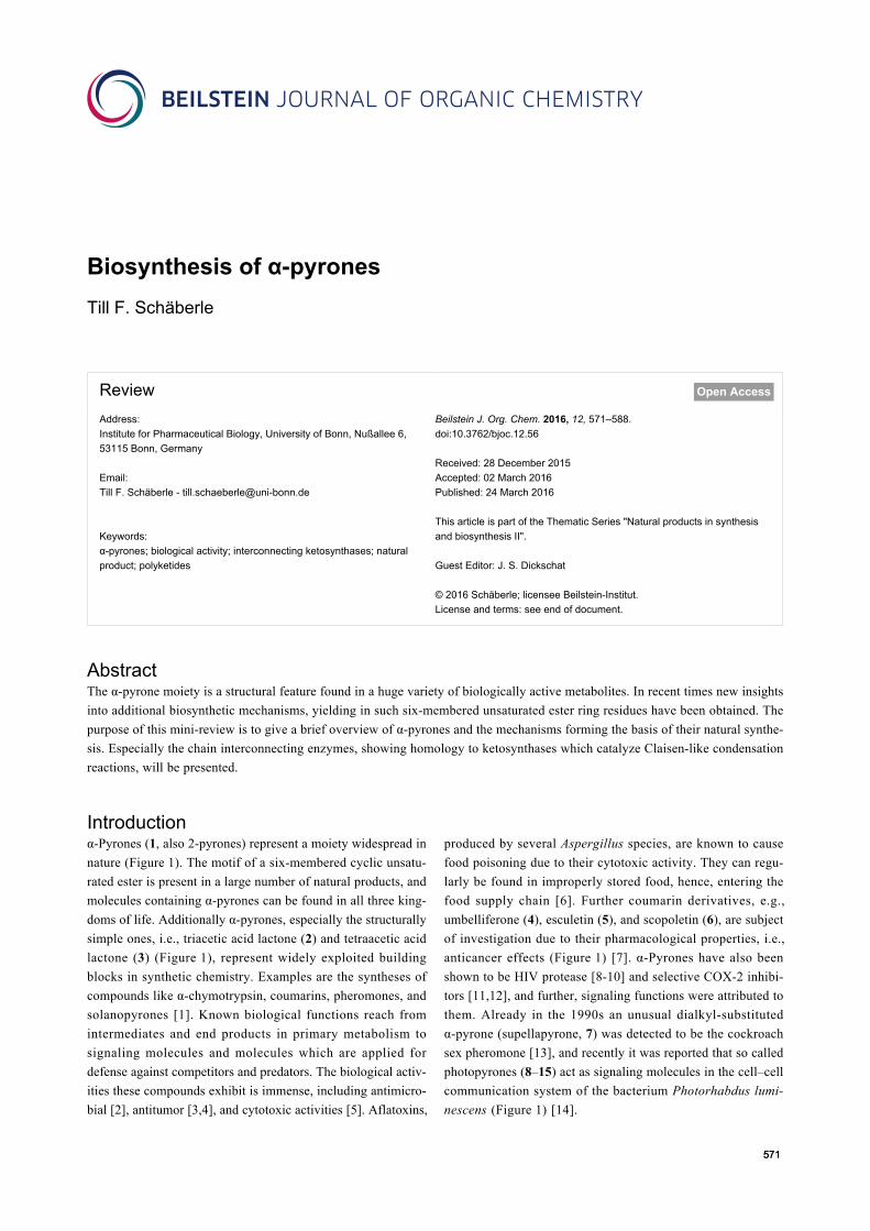
Introductionα-Pyrones (1, also 2-pyrones) represent a moiety widespread in
nature (Figure 1). The motif of a six-membered cyclic unsatu-
rated ester is present in a large number of natural products, and
molecules containing α-pyrones can be found in all three king-
doms of life. Additionally α-pyrones, especially the structurally
simple ones, i.e., triacetic acid lactone (2) and tetraacetic acid
lactone (3) (Figure 1), represent widely exploited building
blocks in synthetic chemistry. Examples are the syntheses of
compounds like α-chymotrypsin, coumarins, pheromones, and
solanopyrones [1]. Known biological functions reach from
intermediates and end products in primary metabolism to
signaling molecules and molecules which are applied for
defense against competitors and predators. The biological activ-
ities these compounds exhibit is immense, including antimicro-
bial [2], antitumor [3,4], and cytotoxic activities [5]. Aflatoxins,
produced by several Aspergillus species, are known to cause
food poisoning due to their cytotoxic activity. They can regu-
larly be found in improperly stored food, hence, entering the
food supply chain [6]. Further coumarin derivatives, e.g.,
umbelliferone (4), esculetin (5), and scopoletin (6), are subject
of investigation due to their pharmacological properties, i.e.,
anticancer effects (Figure 1) [7]. α-Pyrones have also been
shown to be HIV protease [8-10] and selective COX-2 inhibi-
tors [11,12], and further, signaling functions were attributed to
them. Already in the 1990s an unusual dialkyl-substituted
α-pyrone (supellapyrone, 7) was detected to be the cockroach
sex pheromone [13], and recently it was reported that so called
photopyrones (8–15) act as signaling molecules in the cell–cell
communication system of the bacterium Photorhabdus lumi-
nescens (Figure 1) [14].
Beilstein J. Org. Chem. 2016, 12, 571–588.
572
Figure 1: Selected monocyclic and monobenzo α-pyrone structures.
Since the biological activities of α-pyrones are very diverse,
these compounds are in the focus of synthetic chemists [15].
Hence, the phenomenal abundance of natural products and of
chemically synthesized derivatives therefrom justifies several
reviews, and comprehensive articles exist [1,16]. However, in
the present review the diverse biosynthesis of α-pyrones will be
the focus. Different mechanisms for the biosynthesis of these
mostly polyketide-derived structures exist, thus it is assumed
that the route towards α-pyrones has been developed several
times in evolution. They can be built up by the catalytic activi-
ties of the different types of polyketide synthase (PKS) systems,
and especially the final ring formation yielding in the α-pyrone
moiety can be accomplished in different ways. The different
biosynthetic routes towards an α-pyrone ring will be presented.
The biosynthetic mechanisms to yield saturated lactones, like
the statin drug lovastatin, which is in application for lowering
cholesterol, will not be discussed.
Review1 Occurrence and activitiesIn this chapter special sub-types of α-pyrones will be described.
The compounds are grouped into three categories depending on
their structural features: (i) dibenzo-α-pyrones, (ii) monocyclic
α-pyrones, and (iii) monobenzo-α-pyrones.
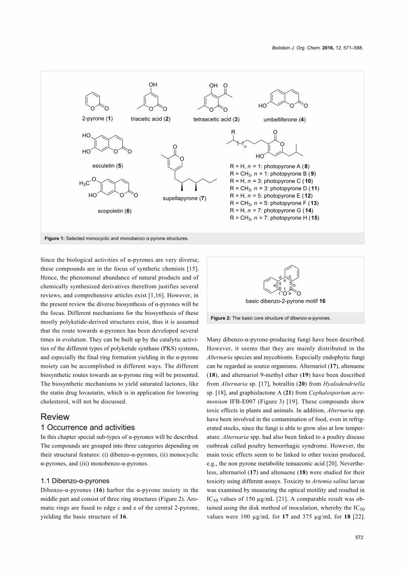
1.1 Dibenzo-α-pyronesDibenzo-α-pyrones (16) harbor the α-pyrone moiety in the
middle part and consist of three ring structures (Figure 2). Aro-
matic rings are fused to edge c and e of the central 2-pyrone,
yielding the basic structure of 16.
Figure 2: The basic core structure of dibenzo-α-pyrones.
Many dibenzo-α-pyrone-producing fungi have been described.
However, it seems that they are mainly distributed in the
Alternaria species and mycobionts. Especially endophytic fungi
can be regarded as source organisms. Alternariol (17), altenuene
(18), and alternariol 9-methyl ether (19) have been described
from Alternaria sp. [17], botrallin (20) from Hyalodendriella
sp. [18], and graphislactone A (21) from Cephalosporium acre-
monium IFB-E007 (Figure 3) [19]. These compounds show
toxic effects in plants and animals. In addition, Alternaria spp.
have been involved in the contamination of food, even in refrig-
erated stocks, since the fungi is able to grow also at low temper-
ature. Alternaria spp. had also been linked to a poultry disease
outbreak called poultry hemorrhagic syndrome. However, the
main toxic effects seem to be linked to other toxins produced,
e.g., the non pyrone metabolite tenuazonic acid [20]. Neverthe-
less, alternariol (17) and altenuene (18) were studied for their
toxicity using different assays. Toxicity to Artemia salina larvae
was examined by measuring the optical motility and resulted in
IC50 values of 150 µg/mL [21]. A comparable result was ob-
tained using the disk method of inoculation, whereby the IC50
values were 100 µg/mL for 17 and 375 µg/mL for 18 [22].
Beilstein J. Org. Chem. 2016, 12, 571–588.
573
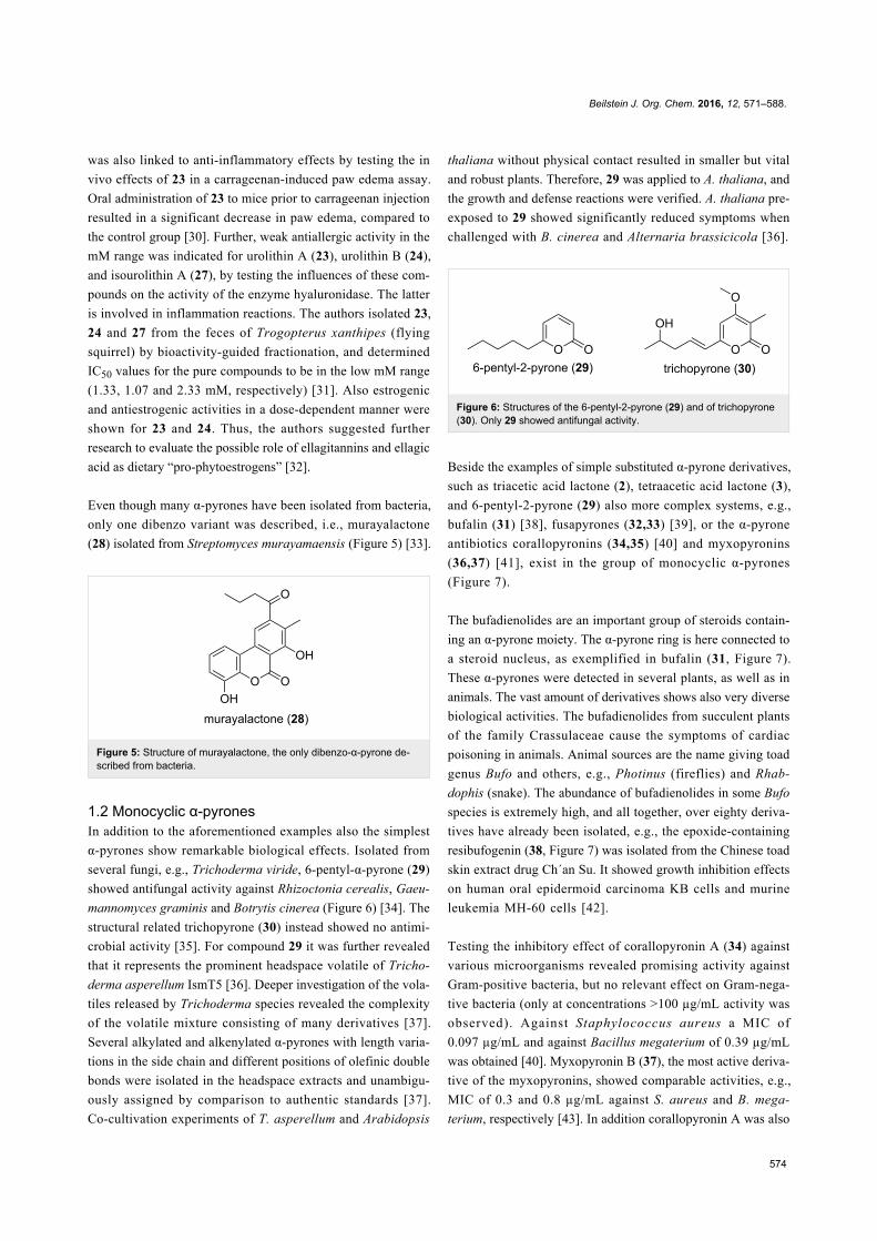
Figure 4: Structure of ellagic acid and of the urolithins, the latter metabolized from ellagic acid by intestinal bacteria.
Further, alternariol (17) and derivatives were tested against
L5178Y mouse lymphoma cells. Here 17 was the most active
compound with an EC50 value of 1.7 μg/mL [23]. In another in
vitro assay, this time a biochemical assay using protein kinase,
the IC50 values were determined, and 17 inhibited 10 out of the
24 kinases tested. The results of the MTT and the kinase assay
showed a similar pattern, and hence it was concluded that pro-
tein kinase inhibition should be one mechanism leading to the
cytotoxicity of 17. In a study using human colon carcinoma
cells to elucidate the cell death mode and the pathways trig-
gered by 17, the induction of an apoptotic process was revealed.
Further investigations showed that cell death was mediated
through a mitochondria-dependent pathway [24]. In murine
hepatoma cells it was shown that 17 and its methyl ether 19
interfere with the transcription factor and by inducing the
so-called aryl hydrocarbon receptor, apoptosis is mediated by
inducing cytochrome P450 1A1 [25]. For alternariol 9-methyl
ether (19) and the graphislactone A (21) cytotoxic effects
against the human cancer cell line SW1116 with IC50 values be-
tween 8.5 and 21 μg/mL were reported [26].
Figure 3: Selected dibenzo-α-pyrones.
These toxic fungi-derived metabolites are often pathogenic to
plants, and are therefore called phytotoxins. Phytotoxins are
divided into host-specific and host non-specific toxins, whereby
the here named Alternaria-derived dibenzo-α-pyrones 17, 18,
and 19 represent host-specific phytotoxins [26].
Several dibenzo-α-pyrones have been isolated from plant parts.
Purified from roots, bulbi, heartwood, or whole plant material,
the origin of some plant-derived pyrones is not finally clarified,
since the production by endophytic fungi cannot be excluded.
Djalonensone was isolated from Anthocleista djalonensis
(Loganiaceae) roots, but is identical to alternariol 9-methyl
ether (the corresponding bioactivities are described above.) The
latter was isolated from a series of fungi including endophytic
species. Thus, the possibility that a fungus is the real producer
cannot be ruled out. In addition, production by a fungus and
modification of the metabolites by plant enzymes is also
possible. Further α-pyrone plant secondary metabolites are
ellagitannins and ellagic acid (22) [27] (Figure 4). These
metabolites are important constituents of different foods, e.g.,
berries, nuts, medicinal plants and tisanes, as well as of grapes
and oak-aged wines. These natural products are not absorbed in
the intestinal tract; rather they are metabolized by intestinal
bacteria, yielding so called urolithins (23–27, Figure 4). There-
fore, it can be assumed that the urolithins are responsible for the
biological activities related to the intake of ellagitannins by
higher organisms. Such urolithins show different phenolic
hydroxylation patterns and have been isolated from animal
feces.
Concerning the activity urolithin A (23), urolithin B (24), and
isourolithin A (27), all isolated from fruits of Trapa natans
(water chestnut) showed antioxidant activity [28]. Testing
urolithins A, B, C, D (23–26) in an assay using myelomono-
cytic HL-60 cells showed antioxidant activities for 23, 25 and
26. These three derivatives inhibited the reactive oxygen
species (ROS)-dependent oxygenation of the non-fluorescent
2’,7’-dichlorodihydrofluorescein (DCFH) to the fluorescent
2’,7’-dichlorofluorescein (DCF) [29]. This antioxidant activity
Beilstein J. Org. Chem. 2016, 12, 571–588.
574
was also linked to anti-inflammatory effects by testing the in
vivo effects of 23 in a carrageenan-induced paw edema assay.
Oral administration of 23 to mice prior to carrageenan injection
resulted in a significant decrease in paw edema, compared to
the control group [30]. Further, weak antiallergic activity in the
mM range was indicated for urolithin A (23), urolithin B (24),
and isourolithin A (27), by testing the influences of these com-
pounds on the activity of the enzyme hyaluronidase. The latter
is involved in inflammation reactions. The authors isolated 23,
24 and 27 from the feces of Trogopterus xanthipes (flying
squirrel) by bioactivity-guided fractionation, and determined
IC50 values for the pure compounds to be in the low mM range
(1.33, 1.07 and 2.33 mM, respectively) [31]. Also estrogenic
and antiestrogenic activities in a dose-dependent manner were
shown for 23 and 24. Thus, the authors suggested further
research to evaluate the possible role of ellagitannins and ellagic
acid as dietary “pro-phytoestrogens” [32].
Even though many α-pyrones have been isolated from bacteria,
only one dibenzo variant was described, i.e., murayalactone
(28) isolated from Streptomyces murayamaensis (Figure 5) [33].
Figure 5: Structure of murayalactone, the only dibenzo-α-pyrone de-scribed from bacteria.
1.2 Monocyclic α-pyronesIn addition to the aforementioned examples also the simplest
α-pyrones show remarkable biological effects. Isolated from
several fungi, e.g., Trichoderma viride, 6-pentyl-α-pyrone (29)
showed antifungal activity against Rhizoctonia cerealis, Gaeu-
mannomyces graminis and Botrytis cinerea (Figure 6) [34]. The
structural related trichopyrone (30) instead showed no antimi-
crobial activity [35]. For compound 29 it was further revealed
that it represents the prominent headspace volatile of Tricho-
derma asperellum IsmT5 [36]. Deeper investigation of the vola-
tiles released by Trichoderma species revealed the complexity
of the volatile mixture consisting of many derivatives [37].
Several alkylated and alkenylated α-pyrones with length varia-
tions in the side chain and different positions of olefinic double
bonds were isolated in the headspace extracts and unambigu-
ously assigned by comparison to authentic standards [37].
Co-cultivation experiments of T. asperellum and Arabidopsis
thaliana without physical contact resulted in smaller but vital
and robust plants. Therefore, 29 was applied to A. thaliana, and
the growth and defense reactions were verified. A. thaliana pre-
exposed to 29 showed significantly reduced symptoms when
challenged with B. cinerea and Alternaria brassicicola [36].
Figure 6: Structures of the 6-pentyl-2-pyrone (29) and of trichopyrone(30). Only 29 showed antifungal activity.
Beside the examples of simple substituted α-pyrone derivatives,
such as triacetic acid lactone (2), tetraacetic acid lactone (3),
and 6-pentyl-2-pyrone (29) also more complex systems, e.g.,
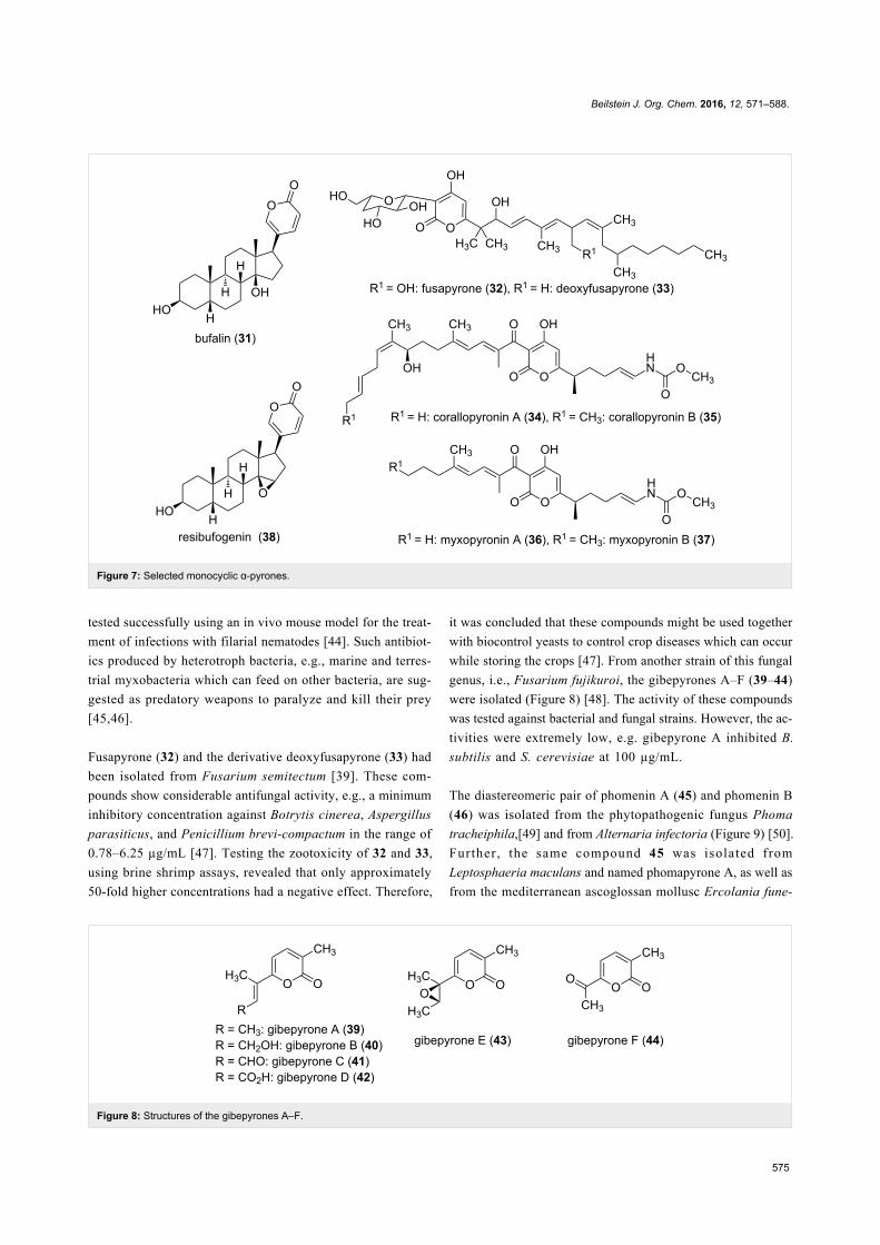
bufalin (31) [38], fusapyrones (32,33) [39], or the α-pyrone
antibiotics corallopyronins (34,35) [40] and myxopyronins
(36,37) [41], exist in the group of monocyclic α-pyrones
(Figure 7).
The bufadienolides are an important group of steroids contain-
ing an α-pyrone moiety. The α-pyrone ring is here connected to
a steroid nucleus, as exemplified in bufalin (31, Figure 7).
These α-pyrones were detected in several plants, as well as in
animals. The vast amount of derivatives shows also very diverse
biological activities. The bufadienolides from succulent plants
of the family Crassulaceae cause the symptoms of cardiac
poisoning in animals. Animal sources are the name giving toad
genus Bufo and others, e.g., Photinus (fireflies) and Rhab-
dophis (snake). The abundance of bufadienolides in some Bufo
species is extremely high, and all together, over eighty deriva-
tives have already been isolated, e.g., the epoxide-containing
resibufogenin (38, Figure 7) was isolated from the Chinese toad
skin extract drug Ch´an Su. It showed growth inhibition effects
on human oral epidermoid carcinoma KB cells and murine
leukemia MH-60 cells [42].
Testing the inhibitory effect of corallopyronin A (34) against
various microorganisms revealed promising activity against
Gram-positive bacteria, but no relevant effect on Gram-nega-
tive bacteria (only at concentrations >100 µg/mL activity was
observed). Against Staphylococcus aureus a MIC of
0.097 µg/mL and against Bacillus megaterium of 0.39 µg/mL
was obtained [40]. Myxopyronin B (37), the most active deriva-
tive of the myxopyronins, showed comparable activities, e.g.,
MIC of 0.3 and 0.8 µg/mL against S. aureus and B. mega-
terium, respectively [43]. In addition corallopyronin A was also
Beilstein J. Org. Chem. 2016, 12, 571–588.
575
Figure 7: Selected monocyclic α-pyrones.
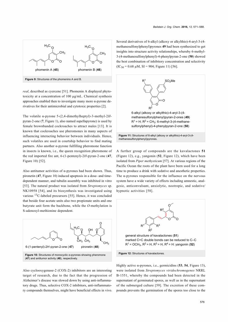
Figure 8: Structures of the gibepyrones A–F.
tested successfully using an in vivo mouse model for the treat-
ment of infections with filarial nematodes [44]. Such antibiot-
ics produced by heterotroph bacteria, e.g., marine and terres-
trial myxobacteria which can feed on other bacteria, are sug-
gested as predatory weapons to paralyze and kill their prey
[45,46].
Fusapyrone (32) and the derivative deoxyfusapyrone (33) had
been isolated from Fusarium semitectum [39]. These com-
pounds show considerable antifungal activity, e.g., a minimum
inhibitory concentration against Botrytis cinerea, Aspergillus
parasiticus, and Penicillium brevi-compactum in the range of
0.78–6.25 µg/mL [47]. Testing the zootoxicity of 32 and 33,
using brine shrimp assays, revealed that only approximately
50-fold higher concentrations had a negative effect. Therefore,
it was concluded that these compounds might be used together
with biocontrol yeasts to control crop diseases which can occur
while storing the crops [47]. From another strain of this fungal
genus, i.e., Fusarium fujikuroi, the gibepyrones A–F (39–44)
were isolated (Figure 8) [48]. The activity of these compounds
was tested against bacterial and fungal strains. However, the ac-
tivities were extremely low, e.g. gibepyrone A inhibited B.
subtilis and S. cerevisiae at 100 µg/mL.
The diastereomeric pair of phomenin A (45) and phomenin B
(46) was isolated from the phytopathogenic fungus Phoma
tracheiphila,[49] and from Alternaria infectoria (Figure 9) [50].
Further, the same compound 45 was isolated from
Leptosphaeria maculans and named phomapyrone A, as well as
from the mediterranean ascoglossan mollusc Ercolania fune-
Beilstein J. Org. Chem. 2016, 12, 571–588.
576
Figure 9: Structures of the phomenins A and B.
real, described as cyercene [51]. Phomenin A displayed phyto-
toxicity at a concentration of 100 µg/mL. Chemical synthesis
approaches enabled then to investigate many more α-pyrone de-
rivatives for their antimicrobial and cytotoxic properties [2].
The volatile α-pyrone 5-(2,4-dimethylheptyl)-3-methyl-2H-
pyran-2-one (7, Figure 1), also named supellapyrone) is used by
female brownbanded cockroaches to attract males [13]. It is
known that cockroaches use pheromones in many aspects of
influencing interacting behavior between individuals. Hence,
such volatiles are used in courtship behavior to find mating
partners. Also another α-pyrone fulfilling pheromone function
in insects is known, i.e., the queen recognition pheromone of
the red imported fire ant, 6-(1-pentenyl)-2H-pyran-2-one (47,
Figure 10) [52].
Also antitumor activities of α-pyrones had been shown. Thus,
pironetin (47, Figure 10) induced apoptosis in a dose- and time-
dependent manner, and tubulin assembly was inhibited in vitro
[53]. The natural product was isolated from Streptomyces sp.
NK10958 [54], and its biosynthesis was investigated using
various 13C-labeled precursors [55]. Hence, it was concluded
that beside four acetate units also two propionate units and one
butyrate unit form the backbone, while the O-methylation is
S-adenosyl-methionine dependent.
Figure 10: Structures of monocyclic α-pyrones showing pheromone(47) and antitumor activity (48), respectively.
Also cyclooxygenase-2 (COX-2) inhibitors are an interesting
target of research, due to the fact that the progression of
Alzheimer’s disease was slowed down by using anti-inflamma-
tory drugs. Thus, selective COX-2 inhibitors, anti-inflammato-
ry compounds themselves, might have beneficial effects in vivo.
Several derivatives of 6-alkyl (alkoxy or alkylthio)-4-aryl-3-(4-
methanesulfonylphenyl)pyrones 49 had been synthesized to get
insights into structure activity relationships, whereby 6-methyl-
3-(4-methanesulfonylphenyl)-4-phenylpyran-2-one (50) showed
the best combination of inhibitory concentration and selectivity
(IC50 = 0.68 µM, SI = 904; Figure 11) [56].
Figure 11: Structures of 6-alkyl (alkoxy or alkylthio)-4-aryl-3-(4-methanesulfonylphenyl)pyrones.
A further group of compounds are the kavalactones 51
(Figure 12), e.g., yangonin (52, Figure 12), which have been
isolated from Piper methysticum [57]. At various regions of the
Pacific Ocean the roots of the plant have been used for a long
time to produce a drink with sedative and anesthetic properties.
The α-pyrones responsible for the influence on the nervous
system have a wide variety of effects including amnestic, anal-
gesic, anticonvulsant, anxiolytic, nootropic, and sedative/
hypnotic activities [58].
Figure 12: Structures of kavalactones.
Highly active α-pyrones, i.e., germicidins (53, 54, Figure 13),
were isolated from Streptomyces viridochromogenes NRRL
B-1551, whereby the compounds had been detected in the
supernatant of germinated spores, as well as in the supernatant
of the submerged culture [59]. The excretion of these com-
pounds prevents the germination of the spores too close to the
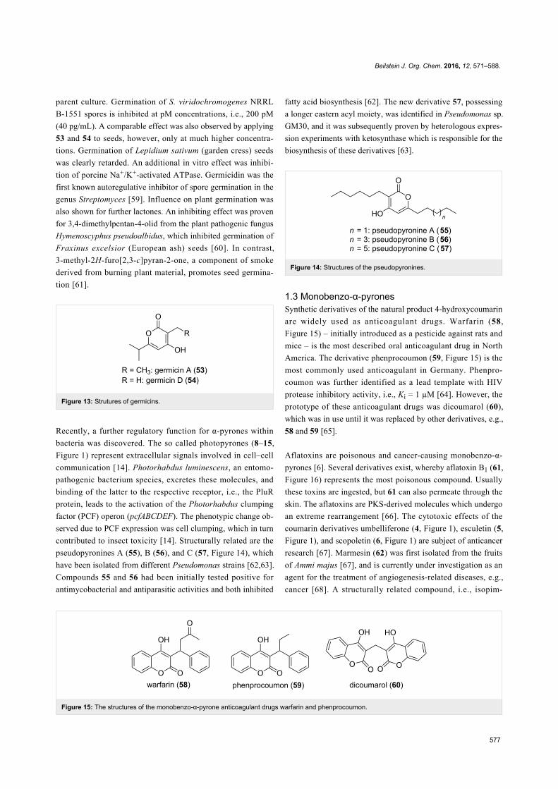
Beilstein J. Org. Chem. 2016, 12, 571–588.
577
Figure 15: The structures of the monobenzo-α-pyrone anticoagulant drugs warfarin and phenprocoumon.
parent culture. Germination of S. viridochromogenes NRRL
B-1551 spores is inhibited at pM concentrations, i.e., 200 pM
(40 pg/mL). A comparable effect was also observed by applying
53 and 54 to seeds, however, only at much higher concentra-
tions. Germination of Lepidium sativum (garden cress) seeds
was clearly retarded. An additional in vitro effect was inhibi-
tion of porcine Na+/K+-activated ATPase. Germicidin was the
first known autoregulative inhibitor of spore germination in the
genus Streptomyces [59]. Influence on plant germination was
also shown for further lactones. An inhibiting effect was proven
for 3,4-dimethylpentan-4-olid from the plant pathogenic fungus
Hymenoscyphus pseudoalbidus, which inhibited germination of
Fraxinus excelsior (European ash) seeds [60]. In contrast,
3-methyl-2H-furo[2,3-c]pyran-2-one, a component of smoke
derived from burning plant material, promotes seed germina-
tion [61].
Figure 13: Strutures of germicins.
Recently, a further regulatory function for α-pyrones within
bacteria was discovered. The so called photopyrones (8–15,
Figure 1) represent extracellular signals involved in cell–cell
communication [14]. Photorhabdus luminescens, an entomo-
pathogenic bacterium species, excretes these molecules, and
binding of the latter to the respective receptor, i.e., the PluR
protein, leads to the activation of the Photorhabdus clumping
factor (PCF) operon (pcfABCDEF). The phenotypic change ob-
served due to PCF expression was cell clumping, which in turn
contributed to insect toxicity [14]. Structurally related are the
pseudopyronines A (55), B (56), and C (57, Figure 14), which
have been isolated from different Pseudomonas strains [62,63].
Compounds 55 and 56 had been initially tested positive for
antimycobacterial and antiparasitic activities and both inhibited
fatty acid biosynthesis [62]. The new derivative 57, possessing
a longer eastern acyl moiety, was identified in Pseudomonas sp.
GM30, and it was subsequently proven by heterologous expres-
sion experiments with ketosynthase which is responsible for the
biosynthesis of these derivatives [63].
Figure 14: Structures of the pseudopyronines.
1.3 Monobenzo-α-pyronesSynthetic derivatives of the natural product 4-hydroxycoumarin
are widely used as anticoagulant drugs. Warfarin (58,
Figure 15) – initially introduced as a pesticide against rats and
mice – is the most described oral anticoagulant drug in North
America. The derivative phenprocoumon (59, Figure 15) is the
most commonly used anticoagulant in Germany. Phenpro-
coumon was further identified as a lead template with HIV
protease inhibitory activity, i.e., Ki = 1 µM [64]. However, the
prototype of these anticoagulant drugs was dicoumarol (60),
which was in use until it was replaced by other derivatives, e.g.,
58 and 59 [65].
Aflatoxins are poisonous and cancer-causing monobenzo-α-
pyrones [6]. Several derivatives exist, whereby aflatoxin B1 (61,
Figure 16) represents the most poisonous compound. Usually
these toxins are ingested, but 61 can also permeate through the
skin. The aflatoxins are PKS-derived molecules which undergo
an extreme rearrangement [66]. The cytotoxic effects of the
coumarin derivatives umbelliferone (4, Figure 1), esculetin (5,
Figure 1), and scopoletin (6, Figure 1) are subject of anticancer
research [67]. Marmesin (62) was first isolated from the fruits
of Ammi majus [67], and is currently under investigation as an
agent for the treatment of angiogenesis-related diseases, e.g.,
cancer [68]. A structurally related compound, i.e., isopim-
Beilstein J. Org. Chem. 2016, 12, 571–588.
578
Figure 16: Structures of selected monobenzo-α-pyrones.
pinellin (63), was also first isolated from fruits of Ammi majus
[69]. It was shown that 63 blocks DNA adduct formation and
skin tumor initiation in mice [70]. Psoralen (64), isolated from
plants, e.g., Ficus carica, had been used against skin diseases
due to its mutagenic effect [71].
Bacterial monobenzo-α-pyrones were isolated from the
myxobacterium Stigmatella aurantiaca MYX-030. Myxo-
coumarins A (65) and B (66) were identified, and 65 was tested
for antifungal activity [72]. It showed a promising activity
against agronomically important pathogens, e.g., complete inhi-
bition of Magnaporthe grisea and Phaeosphaeria nodorum at
67 µg/mL, and Botrytis cinerea was inhibited at 200 µg/mL.
2 BiosynthesisEven though the α-pyrones possessing interesting activities
were in the focus of chemical synthesis approaches for a long
time, for most of them the clarification of the biosynthesis
remained unknown for many years.
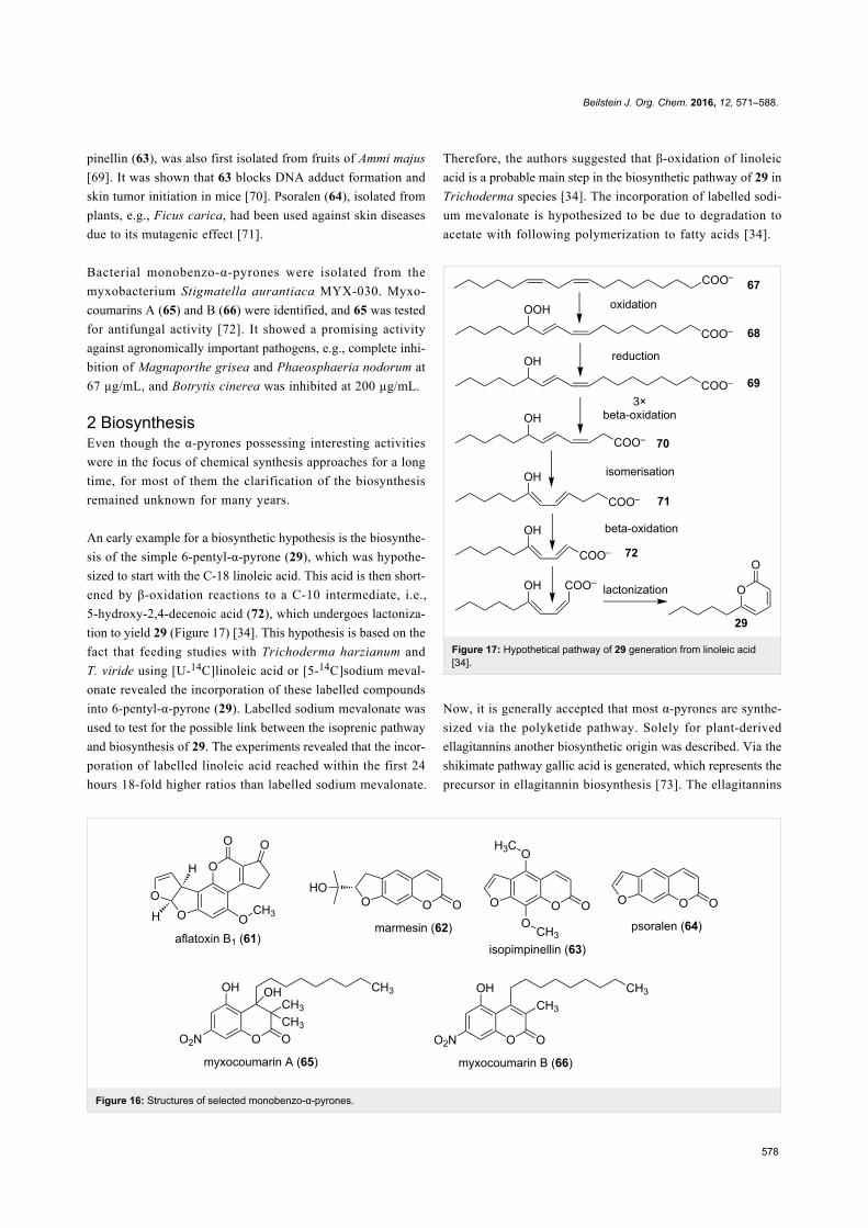
An early example for a biosynthetic hypothesis is the biosynthe-
sis of the simple 6-pentyl-α-pyrone (29), which was hypothe-
sized to start with the C-18 linoleic acid. This acid is then short-
ened by β-oxidation reactions to a C-10 intermediate, i.e.,
5-hydroxy-2,4-decenoic acid (72), which undergoes lactoniza-
tion to yield 29 (Figure 17) [34]. This hypothesis is based on the
fact that feeding studies with Trichoderma harzianum and
T. viride using [U-14C]linoleic acid or [5-14C]sodium meval-
onate revealed the incorporation of these labelled compounds
into 6-pentyl-α-pyrone (29). Labelled sodium mevalonate was
used to test for the possible link between the isoprenic pathway
and biosynthesis of 29. The experiments revealed that the incor-
poration of labelled linoleic acid reached within the first 24
hours 18-fold higher ratios than labelled sodium mevalonate.
Therefore, the authors suggested that β-oxidation of linoleic
acid is a probable main step in the biosynthetic pathway of 29 in
Trichoderma species [34]. The incorporation of labelled sodi-
um mevalonate is hypothesized to be due to degradation to
acetate with following polymerization to fatty acids [34].
Figure 17: Hypothetical pathway of 29 generation from linoleic acid[34].
Now, it is generally accepted that most α-pyrones are synthe-
sized via the polyketide pathway. Solely for plant-derived
ellagitannins another biosynthetic origin was described. Via the
shikimate pathway gallic acid is generated, which represents the
precursor in ellagitannin biosynthesis [73]. The ellagitannins
Beilstein J. Org. Chem. 2016, 12, 571–588.
579
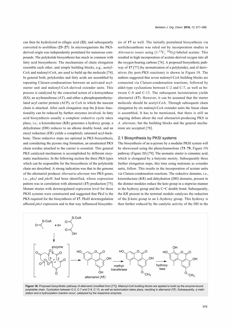
Figure 18: Proposed biosynthetic pathway of alternariol (modified from [77]). Malonyl-CoA building blocks are applied to build up the enzyme-boundpolyketide chain. Cyclization between C-2, C-7 and C-8, C-13, as well as lactonization takes place, resulting in alternariol (17). Subsequently, a meth-ylation and a hydroxylation reaction occur, catalyzed by the respective enzymes.
can then be hydrolyzed to ellagic acid (22), and subsequently
converted to urolithins (23–27). In microorganisms the PKS-
derived origin was independently postulated for numerous com-
pounds. The polyketide biosynthesis has much in common with
fatty acid biosynthesis: The mechanisms of chain elongation
resemble each other, and simple building blocks, e.g., acetyl-
CoA and malonyl-CoA, are used to build up the molecule [74].
In general both, polyketides and fatty acids are assembled by
repeating Claisen-condensations between an activated acyl-
starter unit and malonyl-CoA-derived extender units. This
process is catalyzed by the concerted action of a ketosynthase
(KS), an acyltransferase (AT), and either a phosphopantetheiny-
lated acyl carrier protein (ACP), or CoA to which the nascent
chain is attached. After each elongation step the β-keto func-
tionality can be reduced by further enzymes involved. In fatty
acid biosynthesis usually a complete reductive cycle takes
place, i.e., a ketoreductase (KR) generates a hydroxy group, a
dehydratase (DH) reduces to an alkene double bond, and an
enoyl reductase (ER) yields a completely saturated acyl-back-
bone. These reductive steps are optional in PKS biosynthesis,
and considering the pyrone ring formation, an unsaturated PKS
chain residue attached to the carrier is essential. This general
PKS catalyzed mechanism is accomplished by different enzy-
matic machineries. In the following section the three PKS types
which can be responsible for the biosynthesis of the polyketide
chain are described. A strong indication was that in the genome
of the alternariol producer Alternaria alternate two PKS genes,
i.e., pksJ and pksH, had been identified, whose expression
pattern was in correlation with alternariol (17) production [75].
Mutant strains with downregulated expression level for these
PKSI systems were constructed and suggested that PksJ is the
PKS required for the biosynthesis of 17. PksH downregulation
affected pksJ expression and in that way influenced biosynthe-
sis of 17 as well. The initially postulated biosynthesis via
norlichexanthone was ruled out by incorporation studies in
Alternaria tenuis using [1-13C, 18O2]-labeled acetate. This
resulted in high incorporation of acetate-derived oxygen into all
the oxygen-bearing carbons [76]. A proposed biosynthetic path-
way of 17 [77] (by aromatization of a polyketide), and of deriv-
atives (by post-PKS reactions) is shown in Figure 18. The
authors suggested that seven malonyl-CoA building blocks are
connected via Claisen-condensation reactions, followed by
aldol-type cyclizations between C-2 and C-7, as well as be-
tween C-8 and C-13. The subsequent lactonization yields
alternariol (17). However, it can be assumed that the starter
molecule should be acetyl-CoA. Through subsequent chain
elongation by six malonyl-CoA extender units the linear chain
is assembled. It has to be mentioned, that there is still an
ongoing debate about the real alternariol-producing PKS in
A. alternate, but the building blocks and the general mecha-
nism are accepted [78].
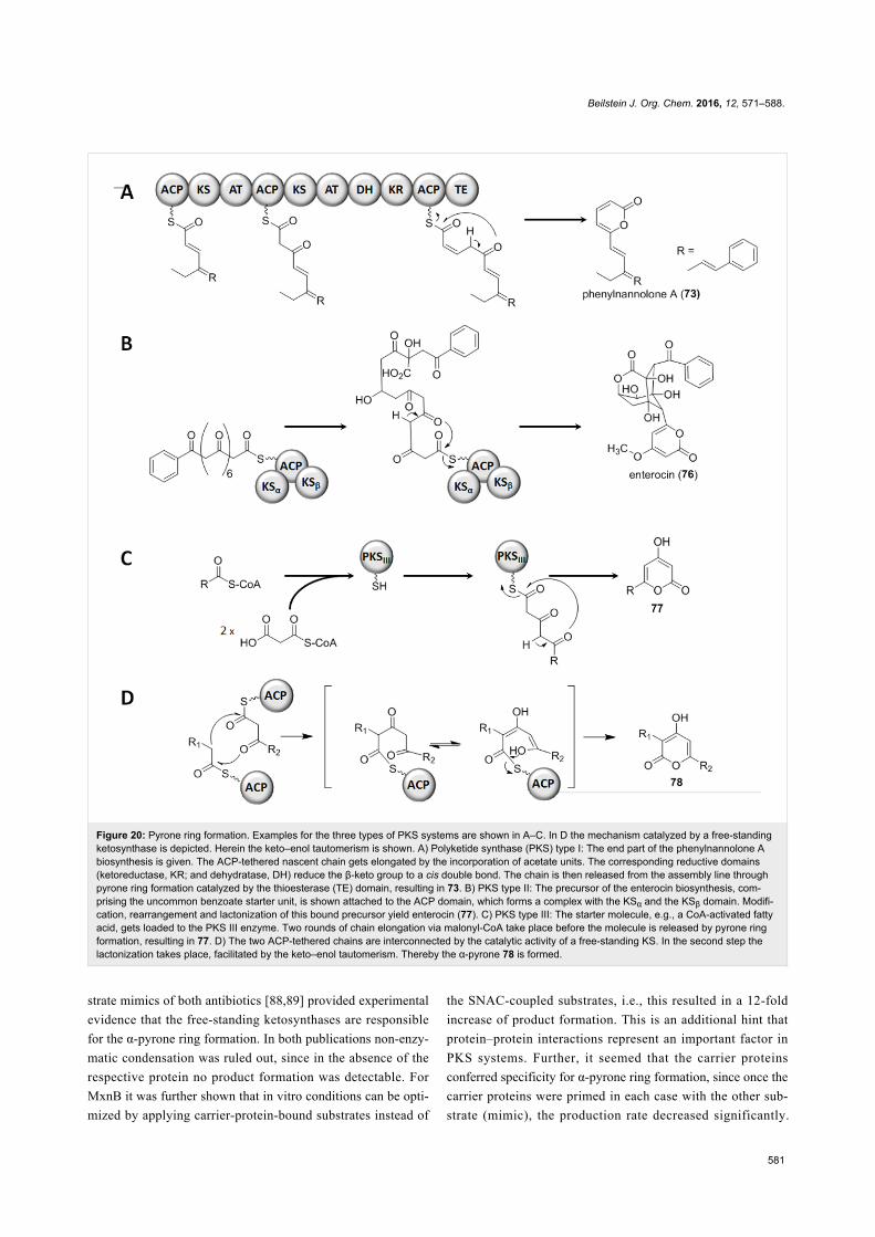
2.1 Biosynthesis by PKSI systemsThe biosynthesis of an α-pyrone by a modular PKSI system will
be showcased using the phenylnannolone (73–75, Figure 19)
pathway (Figure 20) [79]. The aromatic starter is cinnamic acid,
which is elongated by a butyrate moiety. Subsequently three
further elongation steps, this time using malonate as extender
units, follow. This results in the incorporation of acetate units
via Claisen-condensation reactions. The reductive domains, i.e.,
ketoreductase (KR) and dehydration (DH) domains, present in
the distinct modules reduce the keto group in a stepwise manner
to the hydroxy group and the C=C double bond. Subsequently,
the KR present in the terminal module catalyzes the reduction
of the β-keto group to an L-hydroxy group. This hydroxy is
then further reduced by the catalytic activity of the DH in the
Beilstein J. Org. Chem. 2016, 12, 571–588.
580
Figure 19: Structures of phenylnannolones and of enterocin, bothbiosynthesized via polyketide synthase systems.
terminal module, which results in a cis-configured double bond.
Through the formation of the cis double bond the sterical
arrangement of the nascent chain favors the lactone ring closure
which results in the α-pyrone moiety. Hence, the polyketide is
released from the assembly line, whereby the thioesterase (TE)
domain catalyzes the ring-closure and therewith also the off-
loading from the PKSI system [79]. A comparable mechanism,
in which a TE is involved in off-loading the nascent chain from
the PKS assembly line by lactonization, was described for other
natural products, e.g., the isochromanone ring formation for the
ajudazols A and B in Chondromyces crocatus Cm c5 [80].
2.2 Biosynthesis by PKSII systemsIn the type II PKS-catalyzed biosynthesis, the subunit type of
such megaenzyme systems, the starter molecule and the
extender units, mostly malonate molecules, are assembled at the
same ACP. A lactonization at the ACP-bound terminus yields
the pyrone ring. As an example the enterocin (76, Figure 19)
biosynthesis will be regarded (Figure 20). In the marine
bacterium Streptomyces maritimus a gene cluster correspond-
ing to enterocin (enc) biosynthesis was identified [81]. The
minimal enc PKS, EncABC, is encoded by a set of genes archi-
tecturally similar to most other type II PKS clusters. EncA
represents the KSα, EncB the KSβ, and EncC the ACP domain.
First, an uncommon benzoate starter unit gets elongated by
seven malonate molecules. This nascent carbon chain under-
goes a rare Favorskii-like rearrangement and lactonization to
yield the polyketide 76.
2.3 Biosynthesis by PKSIII systemsType III PKSs are relatively small molecules, since in contrast
to the PKSs of type I and II they solely consist of a single
ketosynthase. A single KS connects the CoA-bound starter and
extender units; and also in this system the final lactonization of
the peptide-bound polyketide chain results in the pyrone ring.
Type III systems synthesize a variety of aromatic polyketides.
First discovered in plants, later PKS III systems have also been
described in fungi and bacteria. BpsA (for Bacillus pyrone
synthase) was analyzed in vivo and in vitro [82]. These experi-
ments revealed BpsA to be indeed the enzyme responsible for
the synthesis of triketide pyrones. The substrates used by BpsA
are long-chain fatty acyl-CoAs and malonyl-CoAs – either as
starter or as elongation building blocks, respectively
(Figure 20). Generating B. subtilis mutant strains, overex-
pressing the bpsA gene, yielded in triketide pyrenes. Once the
adjacent gene bpsB, the latter coding for a methyltransferase,
was co-overexpressed, the methylated variants, i.e., triketide
pyrone methyl ethers, were synthesized. The pyrone-forming
activity of BpsA was also proven in vitro, using heterologously
expressed protein. Thereby, the chain length of the acyl residue
had only minor influence on the pyrone formation, since many
substrates had been accepted. This could be expected, since the
α-pyrone formation takes place at the enzyme-tethered end of
the nascent chain, resulting in off-loading.
2.4 Biosynthesis by free-standing ketosynthasesIn contrast to the α-pyrone formation by intramolecular cycliza-
tion reactions, also the condensation of two polyketide chains
can result in a pyrone ring. Such a mechanism was indicated by
feeding experiments for the antibiotically active compounds 36
[83] and 34 [84]. The resulting labeling pattern clearly showed
that the central α-pyrone ring of the molecule was not the result
of a usual intramolecular reaction. Rather, an interconnection of
two independent chains should form the central ring structure.
In addition further molecules, e.g., photopyrones (8–15) from
Photorhabdus luminescens are synthesized by such a head-to-
head condensation of two acyl moieties [60]. Also the csypy-
rones (79–81, Figure 21), first reported from Aspergillus
oryzae, are composed of two independent chains which are
interconnected thereafter [85]. Recently, the biosynthetic origin
of the pseudopyronines A (55) and B (56) in Pseudomonas
putida BW11M1 was clarified – and again two chains are fused
to yield the final products [86]. Thus, it can be assumed that this
mechanism is exemplified quite often in natural products.
Therefore, in the next paragraph the chain interconnecting
mechanism will be described.
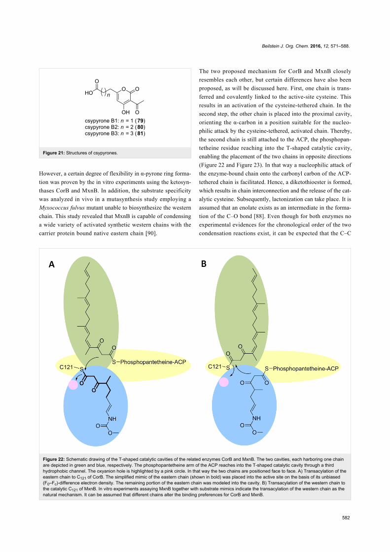
For α-pyrone antibiotics, the corallopyronin and myxopyronin
derivatives, free-standing KSs encoded in the respective cluster,
i.e., CorB and MxnB, were suggested as the chain-intercon-
necting enzymes [84,87]. These enzymes have now been inves-
tigated in detail.
In vitro assays using NAC thioesters of the western and eastern
chains in the biosynthesis of 36 [88], as well as simplified sub-
Beilstein J. Org. Chem. 2016, 12, 571–588.
581
Figure 20: Pyrone ring formation. Examples for the three types of PKS systems are shown in A–C. In D the mechanism catalyzed by a free-standingketosynthase is depicted. Herein the keto–enol tautomerism is shown. A) Polyketide synthase (PKS) type I: The end part of the phenylnannolone Abiosynthesis is given. The ACP-tethered nascent chain gets elongated by the incorporation of acetate units. The corresponding reductive domains(ketoreductase, KR; and dehydratase, DH) reduce the β-keto group to a cis double bond. The chain is then released from the assembly line throughpyrone ring formation catalyzed by the thioesterase (TE) domain, resulting in 73. B) PKS type II: The precursor of the enterocin biosynthesis, com-prising the uncommon benzoate starter unit, is shown attached to the ACP domain, which forms a complex with the KSα and the KSβ domain. Modifi-cation, rearrangement and lactonization of this bound precursor yield enterocin (77). C) PKS type III: The starter molecule, e.g., a CoA-activated fattyacid, gets loaded to the PKS III enzyme. Two rounds of chain elongation via malonyl-CoA take place before the molecule is released by pyrone ringformation, resulting in 77. D) The two ACP-tethered chains are interconnected by the catalytic activity of a free-standing KS. In the second step thelactonization takes place, facilitated by the keto–enol tautomerism. Thereby the α-pyrone 78 is formed.
strate mimics of both antibiotics [88,89] provided experimental
evidence that the free-standing ketosynthases are responsible
for the α-pyrone ring formation. In both publications non-enzy-
matic condensation was ruled out, since in the absence of the
respective protein no product formation was detectable. For
MxnB it was further shown that in vitro conditions can be opti-
mized by applying carrier-protein-bound substrates instead of
the SNAC-coupled substrates, i.e., this resulted in a 12-fold
increase of product formation. This is an additional hint that
protein–protein interactions represent an important factor in
PKS systems. Further, it seemed that the carrier proteins
conferred specificity for α-pyrone ring formation, since once the
carrier proteins were primed in each case with the other sub-
strate (mimic), the production rate decreased significantly.
Beilstein J. Org. Chem. 2016, 12, 571–588.
582
Figure 22: Schematic drawing of the T-shaped catalytic cavities of the related enzymes CorB and MxnB. The two cavities, each harboring one chainare depicted in green and blue, respectively. The phosphopantetheine arm of the ACP reaches into the T-shaped catalytic cavity through a thirdhydrophobic channel. The oxyanion hole is highlighted by a pink circle. In that way the two chains are positioned face to face. A) Transacylation of theeastern chain to C121 of CorB. The simplified mimic of the eastern chain (shown in bold) was placed into the active site on the basis of its unbiased(F0–Fc)-difference electron density. The remaining portion of the eastern chain was modeled into the cavity. B) Transacylation of the western chain tothe catalytic C121 of MxnB. In vitro experiments assaying MxnB together with substrate mimics indicate the transacylation of the western chain as thenatural mechanism. It can be assumed that different chains alter the binding preferences for CorB and MxnB.
Figure 21: Structures of csypyrones.
However, a certain degree of flexibility in α-pyrone ring forma-
tion was proven by the in vitro experiments using the ketosyn-
thases CorB and MxnB. In addition, the substrate specificity
was analyzed in vivo in a mutasynthesis study employing a
Myxococcus fulvus mutant unable to biosynthesize the western
chain. This study revealed that MxnB is capable of condensing
a wide variety of activated synthetic western chains with the
carrier protein bound native eastern chain [90].
The two proposed mechanism for CorB and MxnB closely
resembles each other, but certain differences have also been
proposed, as will be discussed here. First, one chain is trans-
ferred and covalently linked to the active-site cysteine. This
results in an activation of the cysteine-tethered chain. In the
second step, the other chain is placed into the proximal cavity,
orienting the α-carbon in a position suitable for the nucleo-
philic attack by the cysteine-tethered, activated chain. Thereby,
the second chain is still attached to the ACP, the phosphopan-
tetheine residue reaching into the T-shaped catalytic cavity,
enabling the placement of the two chains in opposite directions
(Figure 22 and Figure 23). In that way a nucleophilic attack of
the enzyme-bound chain onto the carbonyl carbon of the ACP-
tethered chain is facilitated. Hence, a diketothioester is formed,
which results in chain interconnection and the release of the cat-
alytic cysteine. Subsequently, lactonization can take place. It is
assumed that an enolate exists as an intermediate in the forma-
tion of the C–O bond [88]. Even though for both enzymes no
experimental evidences for the chronological order of the two
condensation reactions exist, it can be expected that the C–C
Beilstein J. Org. Chem. 2016, 12, 571–588.
583
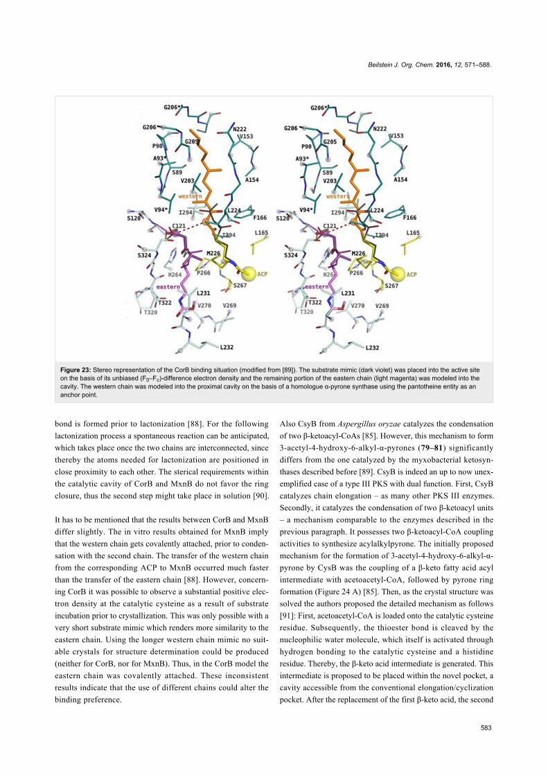
Figure 23: Stereo representation of the CorB binding situation (modified from [89]). The substrate mimic (dark violet) was placed into the active siteon the basis of its unbiased (F0–Fc)-difference electron density and the remaining portion of the eastern chain (light magenta) was modeled into thecavity. The western chain was modeled into the proximal cavity on the basis of a homologue α-pyrone synthase using the pantotheine entity as ananchor point.
bond is formed prior to lactonization [88]. For the following
lactonization process a spontaneous reaction can be anticipated,
which takes place once the two chains are interconnected, since
thereby the atoms needed for lactonization are positioned in
close proximity to each other. The sterical requirements within
the catalytic cavity of CorB and MxnB do not favor the ring
closure, thus the second step might take place in solution [90].
It has to be mentioned that the results between CorB and MxnB
differ slightly. The in vitro results obtained for MxnB imply
that the western chain gets covalently attached, prior to conden-
sation with the second chain. The transfer of the western chain
from the corresponding ACP to MxnB occurred much faster
than the transfer of the eastern chain [88]. However, concern-
ing CorB it was possible to observe a substantial positive elec-
tron density at the catalytic cysteine as a result of substrate
incubation prior to crystallization. This was only possible with a
very short substrate mimic which renders more similarity to the
eastern chain. Using the longer western chain mimic no suit-
able crystals for structure determination could be produced
(neither for CorB, nor for MxnB). Thus, in the CorB model the
eastern chain was covalently attached. These inconsistent
results indicate that the use of different chains could alter the
binding preference.
Also CsyB from Aspergillus oryzae catalyzes the condensation
of two β-ketoacyl-CoAs [85]. However, this mechanism to form
3-acetyl-4-hydroxy-6-alkyl-α-pyrones (79–81) significantly
differs from the one catalyzed by the myxobacterial ketosyn-
thases described before [89]. CsyB is indeed an up to now unex-
emplified case of a type III PKS with dual function. First, CsyB
catalyzes chain elongation – as many other PKS III enzymes.
Secondly, it catalyzes the condensation of two β-ketoacyl units
– a mechanism comparable to the enzymes described in the
previous paragraph. It possesses two β-ketoacyl-CoA coupling
activities to synthesize acylalkylpyrone. The initially proposed
mechanism for the formation of 3-acetyl-4-hydroxy-6-alkyl-α-
pyrone by CysB was the coupling of a β-keto fatty acid acyl
intermediate with acetoacetyl-CoA, followed by pyrone ring
formation (Figure 24 A) [85]. Then, as the crystal structure was
solved the authors proposed the detailed mechanism as follows
[91]: First, acetoacetyl-CoA is loaded onto the catalytic cysteine
residue. Subsequently, the thioester bond is cleaved by the
nucleophilic water molecule, which itself is activated through
hydrogen bonding to the catalytic cysteine and a histidine
residue. Thereby, the β-keto acid intermediate is generated. This
intermediate is proposed to be placed within the novel pocket, a
cavity accessible from the conventional elongation/cyclization
pocket. After the replacement of the first β-keto acid, the second
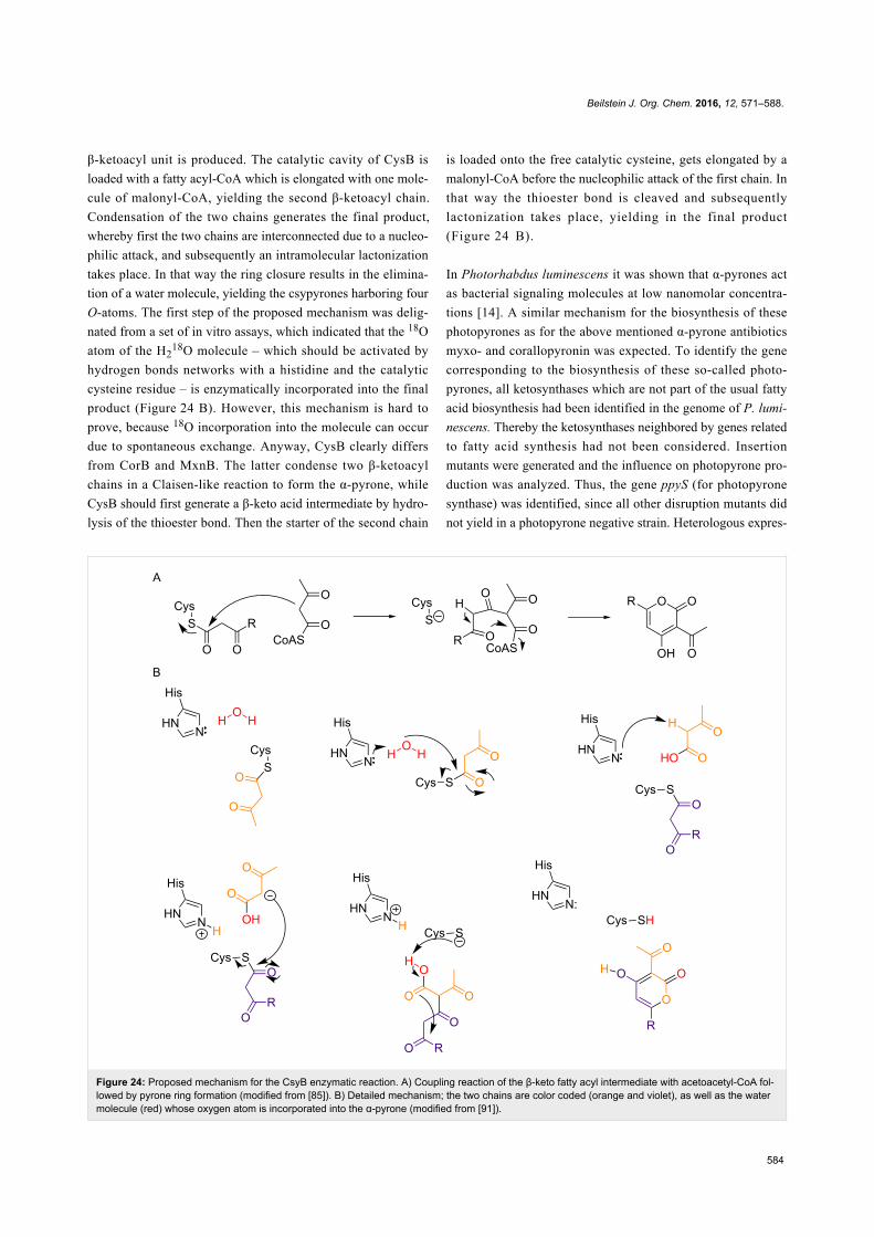
Beilstein J. Org. Chem. 2016, 12, 571–588.
584
Figure 24: Proposed mechanism for the CsyB enzymatic reaction. A) Coupling reaction of the β-keto fatty acyl intermediate with acetoacetyl-CoA fol-lowed by pyrone ring formation (modified from [85]). B) Detailed mechanism; the two chains are color coded (orange and violet), as well as the watermolecule (red) whose oxygen atom is incorporated into the α-pyrone (modified from [91]).
β-ketoacyl unit is produced. The catalytic cavity of CysB is
loaded with a fatty acyl-CoA which is elongated with one mole-
cule of malonyl-CoA, yielding the second β-ketoacyl chain.
Condensation of the two chains generates the final product,
whereby first the two chains are interconnected due to a nucleo-
philic attack, and subsequently an intramolecular lactonization
takes place. In that way the ring closure results in the elimina-
tion of a water molecule, yielding the csypyrones harboring four
O-atoms. The first step of the proposed mechanism was delig-
nated from a set of in vitro assays, which indicated that the 18O
atom of the H218O molecule – which should be activated by
hydrogen bonds networks with a histidine and the catalytic
cysteine residue – is enzymatically incorporated into the final
product (Figure 24 B). However, this mechanism is hard to
prove, because 18O incorporation into the molecule can occur
due to spontaneous exchange. Anyway, CysB clearly differs
from CorB and MxnB. The latter condense two β-ketoacyl
chains in a Claisen-like reaction to form the α-pyrone, while
CysB should first generate a β-keto acid intermediate by hydro-
lysis of the thioester bond. Then the starter of the second chain
is loaded onto the free catalytic cysteine, gets elongated by a
malonyl-CoA before the nucleophilic attack of the first chain. In
that way the thioester bond is cleaved and subsequently
lactonization takes place, yielding in the final product
(Figure 24 B).
In Photorhabdus luminescens it was shown that α-pyrones act
as bacterial signaling molecules at low nanomolar concentra-
tions [14]. A similar mechanism for the biosynthesis of these
photopyrones as for the above mentioned α-pyrone antibiotics
myxo- and corallopyronin was expected. To identify the gene
corresponding to the biosynthesis of these so-called photo-
pyrones, all ketosynthases which are not part of the usual fatty
acid biosynthesis had been identified in the genome of P. lumi-
nescens. Thereby the ketosynthases neighbored by genes related
to fatty acid synthesis had not been considered. Insertion
mutants were generated and the influence on photopyrone pro-
duction was analyzed. Thus, the gene ppyS (for photopyrone
synthase) was identified, since all other disruption mutants did
not yield in a photopyrone negative strain. Heterologous expres-
Beilstein J. Org. Chem. 2016, 12, 571–588.
585
sion of ppyS in E. coli, together with the bkdABC operon
(encoding the branched chain α-ketoacid dehydrogenase (Bkd)
complex) and ngrA (encoding a phosphopantetheinyl-trans-
ferase which is essential to generate the holo-acyl carrier pro-
tein BkdB) for the biosynthesis of branched-chain iso-fatty acid,
resulted in the production of photopyrone derivatives. This was
a functional proof that PpyS catalyzes the formation of
α-pyrones, as indicated before by feeding experiments with
stable isotope-labeled precursors. PpyS should connect
5-methyl-3-oxohexanoyl thioester and different thioesters of
straight-chain and iso-branched chain fatty acids [14]. The
mechanism proposal also includes the catalytic cysteine. The
first chain, i.e., thioester-activated 9-methyldecanoic acid, gets
covalently tethered to that important residue within the active
site. This reflects the same mechanism as for the other KS-like
enzymes described. Also for PpyS the proposal postulates that
the α-carbon of the enzyme-bound chain acts as a nucleophile.
Thus, this activated carbon executes a nucleophilic attack on the
carbonyl carbon of chain two, i.e., 5-methyl-3-oxohexanoyl
thioester, which is itself synthesized by the Bkd complex. In
that way a C–C bond is formed, and both chains are still at-
tached to the catalytic cysteine residue. This bound intermedi-
ate undergoes a further deprotonation, which enables the forma-
tion of the α-pyrone ring. Through the ring closure the α-pyrone
is released from PpyS. This second deprotonation can occur
spontaneously, or enzyme catalyzed. In contrast to the cases of
myxopyronins 36 and 37 and corallopyronins 34 and 35, no
PKSI system provides the ACP-bound chains. Therefore, the
substrates for the chain interconnection might be either ACP or
CoA bound. This would be depending on their origin in the cell,
either fatty acid biosynthesis or degradation. The flexibility of
the system in regard to the first chain to be bound to PpyS was
already shown by the photopyrones A–H, which differ in the
chain length and in the either branched or unbranched starting
unit.
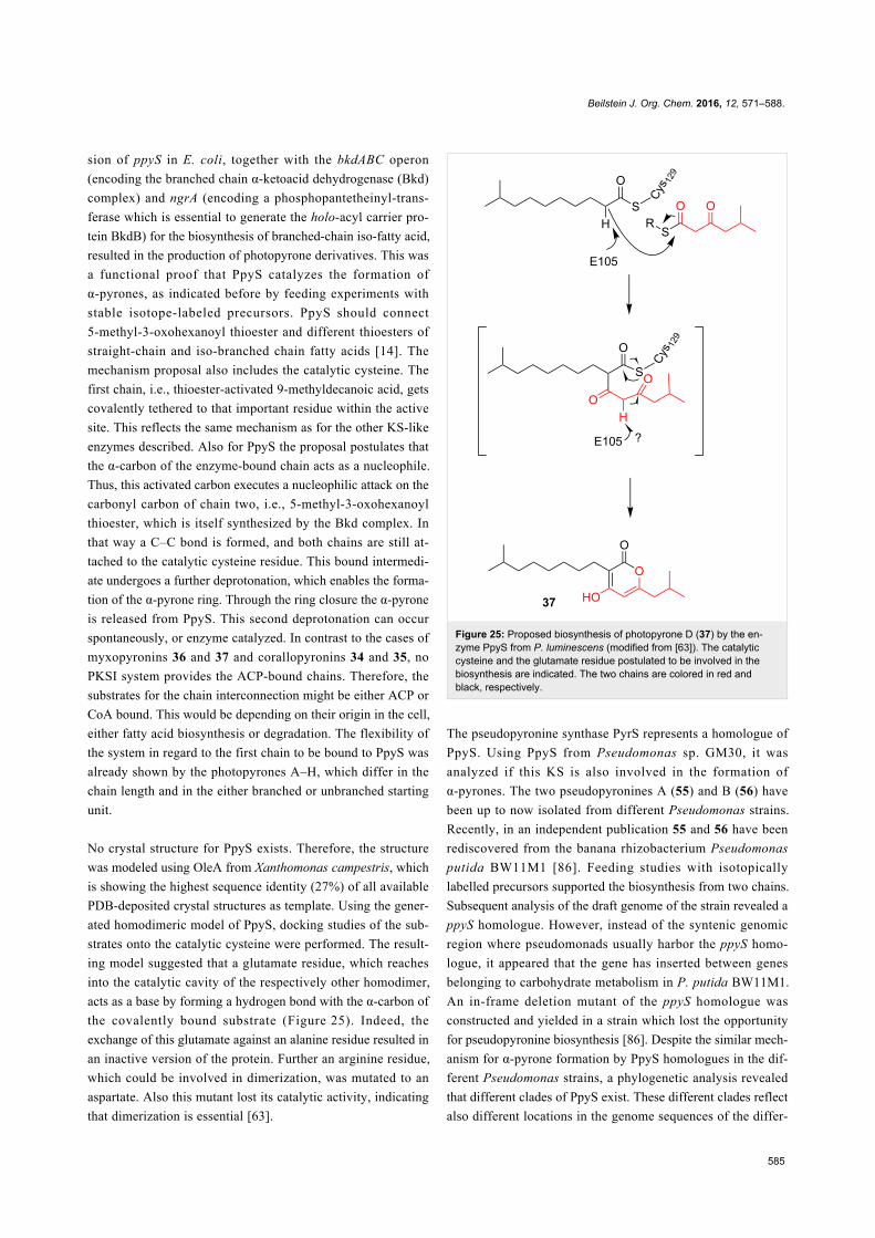
No crystal structure for PpyS exists. Therefore, the structure
was modeled using OleA from Xanthomonas campestris, which
is showing the highest sequence identity (27%) of all available
PDB-deposited crystal structures as template. Using the gener-
ated homodimeric model of PpyS, docking studies of the sub-
strates onto the catalytic cysteine were performed. The result-
ing model suggested that a glutamate residue, which reaches
into the catalytic cavity of the respectively other homodimer,
acts as a base by forming a hydrogen bond with the α-carbon of
the covalently bound substrate (Figure 25). Indeed, the
exchange of this glutamate against an alanine residue resulted in
an inactive version of the protein. Further an arginine residue,
which could be involved in dimerization, was mutated to an
aspartate. Also this mutant lost its catalytic activity, indicating
that dimerization is essential [63].
Figure 25: Proposed biosynthesis of photopyrone D (37) by the en-zyme PpyS from P. luminescens (modified from [63]). The catalyticcysteine and the glutamate residue postulated to be involved in thebiosynthesis are indicated. The two chains are colored in red andblack, respectively.
The pseudopyronine synthase PyrS represents a homologue of
PpyS. Using PpyS from Pseudomonas sp. GM30, it was
analyzed if this KS is also involved in the formation of
α-pyrones. The two pseudopyronines A (55) and B (56) have
been up to now isolated from different Pseudomonas strains.
Recently, in an independent publication 55 and 56 have been
rediscovered from the banana rhizobacterium Pseudomonas
putida BW11M1 [86]. Feeding studies with isotopically
labelled precursors supported the biosynthesis from two chains.
Subsequent analysis of the draft genome of the strain revealed a
ppyS homologue. However, instead of the syntenic genomic
region where pseudomonads usually harbor the ppyS homo-
logue, it appeared that the gene has inserted between genes
belonging to carbohydrate metabolism in P. putida BW11M1.
An in-frame deletion mutant of the ppyS homologue was
constructed and yielded in a strain which lost the opportunity
for pseudopyronine biosynthesis [86]. Despite the similar mech-
anism for α-pyrone formation by PpyS homologues in the dif-
ferent Pseudomonas strains, a phylogenetic analysis revealed
that different clades of PpyS exist. These different clades reflect
also different locations in the genome sequences of the differ-
Beilstein J. Org. Chem. 2016, 12, 571–588.
586
ent Pseudomonas species: On a taxonomic level closely related
strains harbor the ppyS homologue in the same region of their
genome. Therefore, it can be assumed that the genetic informa-
tion coding for the enzyme needed to synthesize pseudopy-
ronines was acquired several times. Hence, Pseudomonas
species from different habitats, e.g., rhizosphere, soil, water,
acquired the gene set independently [86].
In summary different types of chain-interconnecting KSs which
catalyze α-pyrone ring formation were identified in the last
years. One mechanism is to fuse two ketoacyl moieties, as
exemplified by CorB and MxnB. Another mechanism is the
fusion of one ketoacyl moiety with one acyl moiety, as shown
for PpyS-like KSs. All evolved from FabH-type KSs, but form
different clades in phylogenetic analyses. PpyS-like enzymes
show the conserved glutamate residue – indicating a mecha-
nism distinct from the ketoacyl–ketoacyl-connecting KSs – and
were identified in different bacterial genera, i.e., Burkholderia,
Legionella, Nocardia, Microcystis and Streptomyces, therewith
also in clinically relevant pathogens [63]. Future work will
reveal which natural products are biosynthesized by such KSs,
and which relevance these products have.
ConclusionThe α-pyrones show an extraordinary wide variation in biologi-
cal activities, independently if structurally simple or complex,
naturally or non-naturally synthesized. Therefore, α-pyrones
represent a rich source for isolation studies and lead discovery.
Now, new insights into the biosynthesis of these molecules
through chain interconnecting ketosynthases were obtained.
This opens up the possibility to use these enzymes as tools;
both, in bio- as well as in semi-synthetic approaches. The poten-
tial of these enzymes in combinatorial biosynthesis has to be
further evaluated in the future.
AcknowledgementsThe German Federal Ministry of Education and Research is
thanked for funding.
References1. McGlacken, G. P.; Fairlamb, I. J. S. Nat. Prod. Rep. 2005, 22,
369–385. doi:10.1039/b416651p2. Fairlamb, I. J. S.; Marrison, L. R.; Dickinson, J. M.; Lu, F.-J.;
Schmidt, J. P. Bioorg. Med. Chem. 2004, 12, 4285–4299.doi:10.1016/j.bmc.2004.01.051
3. Suzuki, K.; Kuwahara, A.; Yoshida, H.; Fujita, S.; Nishikiori, T.;Nakagawa, T. J. Antibiot. 1997, 50, 314–317.doi:10.7164/antibiotics.50.314
4. Kondoh, M.; Usui, T.; Kobayashi, S.; Tsuchiya, K.; Nishikawa, K.;Nishikiori, T.; Mayumi, T.; Osada, H. Cancer Lett. 1998, 126, 29–32.doi:10.1016/S0304-3835(97)00528-4
5. Calderón-Montaño, J. M.; Burgos-Morón, E.; Orta, M. L.; Pastor, N.;Austin, C. A.; Mateos, S.; López-Lázaro, M. Toxicol. Lett. 2013, 222,64–71. doi:10.1016/j.toxlet.2013.07.007
6. Villers, P. Front. Microbiol. 2014, 5, No. 158.doi:10.3389/fmicb.2014.00158
7. Musa, M. A.; Cooperwood, J. S.; Khan, M. O. F. Curr. Med. Chem.2008, 15, 2664–2679. doi:10.2174/092986708786242877
8. Thaisrivongs, S.; Romero, D. L.; Tommasi, R. A.; Janakiraman, M. N.;Strohbach, J. W.; Turner, S. R.; Biles, C.; Morge, R. R.; Johnson, P. D.;Aristoff, P. A.; Tomich, P. K.; Lynn, J. C.; Horng, M.-M.; Chong, K.-T.;Hinshaw, R. R.; Howe, W. J.; Finzel, B. C.; Watenpaugh, K. D.J. Med. Chem. 1996, 39, 4630–4642. doi:10.1021/jm960228q
9. Poppe, S. M.; Slade, D. E.; Chong, K. T.; Hinshaw, R. R.;Pagano, P. J.; Markowitz, M.; Ho, D. D.; Mo, H.; Gorman, R. R., III;Dueweke, T. J.; Thaisrivongs, S.; Tarpley, W. G.Antimicrob. Agents Chemother. 1997, 41, 1058–1063.
10. Turner, S. R.; Strohbach, J. W.; Tommasi, R. A.; Aristoff, P. A.;Johnson, P. D.; Skulnick, H. I.; Dolak, L. A.; Seest, E. P.; Tomich, P. K.;Bohanon, M. J.; Horng, M.-M.; Lynn, J. C.; Chong, K.-T.;Hinshaw, R. R.; Watenpaugh, K. D.; Janakiraman, M. N.;Thaisrivongs, S. J. Med. Chem. 1998, 41, 3467–3476.doi:10.1021/jm9802158
11. Yeh, P.-P.; Daniels, D. S. B.; Cordes, D. B.; Slawin, A. M. Z.;Smith, A. D. Org. Lett. 2014, 16, 964–967. doi:10.1021/ol403697h
12. Liaw, C.-C.; Yang, Y.-L.; Lin, C.-K.; Lee, J.-C.; Liao, W.-Y.;Shen, C.-N.; Sheu, J.-H.; Wu, S.-H. Org. Lett. 2015, 17, 2330–2333.doi:10.1021/acs.orglett.5b00739
13. Charlton, R. E.; Webster, F. X.; Zhang, A.; Schal, C.; Liang, D.;Sreng, I.; Roelofs, W. L. Proc. Natl. Acad. Sci. U. S. A. 1993, 90,10202–10205. doi:10.1073/pnas.90.21.10202
14. Brachmann, A. O.; Brameyer, S.; Kresovic, D.; Hitkova, I.; Kopp, Y.;Manske, C.; Schubert, K.; Bode, H. B.; Heermann, R. Nat. Chem. Biol.2013, 9, 573–578. doi:10.1038/nchembio.1295
15. Lee, J. S. Mar. Drugs 2015, 13, 1581–1620. doi:10.3390/md1303158116. Dickinson, J. M. Nat. Prod. Rep. 1993, 10, 71–98.
doi:10.1039/np993100007117. Harvan, D. J.; Pero, R. W. Adv. Chem. Ser. 1976, 149, 344–355.
doi:10.1021/ba-1976-0149.ch01518. Luo, H.; Liu, H.; Cao, Y.; Xu, D.; Mao, Z.; Mou, Y.; Meng, J.; Lai, D.;
Liu, Y.; Zhou, L. Molecules 2014, 19, 14221–14234.doi:10.3390/molecules190914221
19. Song, Y. C.; Huang, W. Y.; Sun, C.; Wang, F. W.; Tan, R. X.Biol. Pharm. Bull. 2005, 28, 506–509. doi:10.1248/bpb.28.506
20. Griffin, G. F.; Chu, F. S. Appl. Environ. Microbiol. 1983, 46, 1420–1422.21. Zajkowski, P.; Grabarkiewicz-Szcesna, J.; Schmidt, R. Mycotoxin Res.
1991, 7, 11–15. doi:10.1007/BF0319215822. Panigrahi, S.; Dallin, S. J. Sci. Food Agric. 1994, 66, 493–496.
doi:10.1002/jsfa.274066041123. Aly, A. H.; Edrada-Ebel, R.; Indriani, I. D.; Wray, V.; Müller, W. E. G.;
Totzke, F.; Zirrgiebel, U.; Schächtele, C.; Kubbutat, M. H. G.;Lin, W. H.; Proksch, P.; Ebel, R. J. Nat. Prod. 2008, 71, 972–980.doi:10.1021/np070447m
24. Bensassi, F.; Gallerne, C.; Sharaf El Dein, O.; Hajlaoui, M. R.;Bacha, H.; Lemaire, C. Toxicol. In Vitro 2012, 26, 915–923.doi:10.1016/j.tiv.2012.04.014
25. Schreck, I.; Deigendesch, U.; Burkhardt, B.; Marko, D.; Weiss, C.Arch. Toxicol. 2012, 86, 625–632. doi:10.1007/s00204-011-0781-3
26. Mao, Z.; Sun, W.; Fu, L.; Luo, H.; Lai, D.; Zhou, L. Molecules 2014, 19,5088–5108. doi:10.3390/molecules19045088
Beilstein J. Org. Chem. 2016, 12, 571–588.
587
27. Grasser, G. Synthetic Tannins; Crosby Lockwood & Son: London,1922.
28. Yoshiaki, S.; Shizuo, T. Nat. Med. 2001, 55, 247–250.29. Bialonska, D.; Kasimsetty, S. G.; Khan, S. I.; Ferreira, D.
J. Agric. Food Chem. 2009, 57, 10181–10186. doi:10.1021/jf902579430. Ishimoto, H.; Shibata, M.; Myojin, Y.; Ito, H.; Sugimoto, Y.; Tai, A.;
Hatano, T. Bioorg. Med. Chem. Lett. 2011, 21, 5901–5904.doi:10.1016/j.bmcl.2011.07.086
31. Jeong, S.-J.; Kim, N.-Y.; Kim, D.-H.; Kang, T.-H.; Ahn, N.-H.;Miyamoto, T.; Higuchi, R.; Kim, Y.-C. Planta Med. 2000, 66, 76–77.doi:10.1055/s-0029-1243114
32. Larrosa, M.; González-Sarrías, A.; García-Conesa, M. T.;Tomás-Barberán, F. A.; Espín, J. C. J. Agric. Food Chem. 2006, 54,1611–1620. doi:10.1021/jf0527403
33. Melville, C. R.; Gould, S. J. J. Nat. Prod. 1994, 57, 597–601.doi:10.1021/np50107a005
34. Serrano-Carreon, L.; Hathout, Y.; Bensoussan, M.; Belin, J. M.Appl. Environ. Microbiol. 1993, 59, 2945–2950.
35. Abdel-Lateff, A.; Fisch, K.; Wright, A. D. Z. Naturforsch., C 2009, 64,186–192.
36. Kottb, M.; Gigolashvili, T.; Großkinsky, D. K.; Piechulla, B.Front. Microbiol. 2015, 6, No. 995. doi:10.3389/fmicb.2015.00995
37. Wickel, S. M.; Citron, C. A.; Dickschat, J. S. Eur. J. Org. Chem. 2013,2906–2913. doi:10.1002/ejoc.201300049
38. Pettit, G. R.; Houghton, L. E.; Knight, J. C.; Bruschweiler, F.J. Org. Chem. 1970, 35, 2895–2898. doi:10.1021/jo00834a008
39. Evidente, A.; Conti, L.; Altomare, C.; Bottalico, A.; Sindona, G.;Segre, A. L.; Logrieco, A. Nat. Toxins 1994, 2, 4–13.doi:10.1002/nt.2620020103
40. Irschik, H.; Jansen, R.; Höfle, G.; Gerth, K.; Reichenbach, H.J. Antibiot. 1985, 38, 145–152. doi:10.7164/antibiotics.38.145
41. Irschik, H.; Gerth, K.; Höfle, G.; Kohl, W.; Reichenbach, H. J. Antibiot.1983, 36, 1651–1658. doi:10.7164/antibiotics.36.1651
42. Kamano, Y.; Nogawa, T.; Yamashita, A.; Hayashi, M.; Inoue, M.;Drašar, P.; Pettit, G. R. J. Nat. Prod. 2002, 65, 1001–1005.doi:10.1021/np0200360
43. Schäberle, T. F.; Lohr, F.; Schmitz, A.; König, G. M. Nat. Prod. Rep.2014, 31, 953–972. doi:10.1039/c4np00011k
44. Schiefer, A.; Schmitz, A.; Schäberle, T. F.; Specht, S.; Lämmer, C.;Johnston, K. L.; Vassylyev, D. G.; König, G. M.; Hoerauf, A.; Pfarr, K.J. Infect. Dis. 2012, 206, 249–257. doi:10.1093/infdis/jis341
45. Dávila-Céspedes, A.; Hufendiek, P.; Crüsemann, M.; Schäberle, T. F.;König, G. M. Beilstein J. Org. Chem. submitted.
46. Korp, J.; Vela Gurovic, M. S.; Nett, M. Beilstein J. Org. Chem. 2016,12, in press.
47. Altomare, C.; Pengue, R.; Favilla, M.; Evidente, A.; Visconti, A.J. Agric. Food Chem. 2004, 52, 2997–3001. doi:10.1021/jf035233z
48. Barrero, A. F.; Oltra, J. E.; Herrador, M. M.; Cabrera, E.;Sanchez, J. F.; Quílez, J. F.; Rojas, F. J.; Reyes, J. F. Tetrahedron1993, 49, 141–150. doi:10.1016/S0040-4020(01)80514-7
49. Tringali, C.; Parisi, A.; Piatelli, M.; di San Lio, G. M. Nat. Prod. Lett.1993, 3, 101–106. doi:10.1080/10575639308043845
50. Ivanova, L.; Petersen, D.; Uhlig, S. Toxicon 2010, 55, 1107–1114.doi:10.1016/j.toxicon.2009.12.017
51. Vardaro, R. R.; Di Marzo, V.; Marin, A.; Cimino, G. Tetrahedron 1992,48, 9561–9566. doi:10.1016/S0040-4020(01)88324-1
52. Rocca, J. R.; Tumlinson, J. H.; Glancey, B. M.; Lofgren, C. S.Tetrahedron Lett. 1983, 24, 1889–1892.doi:10.1016/S0040-4039(00)81798-0
53. Kondoh, M.; Usui, T.; Nishikiori, T.; Mayumi, T.; Osada, H. Biochem. J.1999, 340, 411–416. doi:10.1042/bj3400411
54. Kobayashi, S.; Tsuchiya, K.; Harada, T.; Nishide, M.; Kurokawa, T.;Nakagawa, T.; Shimada, N.; Kobayashi, K. J. Antibiot. 1994, 47,697–702. doi:10.7164/antibiotics.47.697
55. Kobayashi, S.; Tsuchiya, K.; Nishide, M.; Nishikiori, T.; Nakagawa, T.;Shimada, N. J. Antibiot. 1995, 48, 893–895.doi:10.7164/antibiotics.48.893
56. Rao, P. N. P.; Amini, M.; Li, H.; Habeeb, A. G.; Knaus, E. E.J. Med. Chem. 2003, 46, 4872–4882. doi:10.1021/jm0302391
57. Bilia, A. R.; Scalise, L.; Bergonzi, M. C.; Vincieri, F. F.J. Chromatogr. B: Anal. Technol. Biomed. Life Sci. 2004, 812,203–214. doi:10.1016/S1570-0232(04)00644-0
58. Sarris, J.; Kavanagh, D. J. J. Altern. Complementary Med. 2009, 15,827–836. doi:10.1089/acm.2009.0066
59. Petersen, F.; Zähner, H.; Metzger, J. W.; Freund, S.; Hummel, R.-P.J. Antibiot. 1993, 46, 1126–1138. doi:10.7164/antibiotics.46.1126
60. Citron, C. A.; Junker, C.; Schulz, B.; Dickschat, J. S.Angew. Chem., Int. Ed. 2014, 53, 4346–4349.doi:10.1002/anie.201402290
61. Flematti, G. R.; Ghisalberti, E. L.; Dixon, K. W.; Trengove, R. D.Science 2004, 305, 977. doi:10.1126/science.1099944
62. Giddens, A. C.; Nielsen, L.; Boshoff, H. I.; Tasdemir, D.; Perozzo, R.;Kaiser, M.; Wang, F.; Sacchettini, J. C.; Copp, B. R. Tetrahedron 2008,64, 1242–1249. doi:10.1016/j.tet.2007.11.075
63. Kresovic, D.; Schempp, F.; Cheikh-Ali, Z.; Bode, H. B.Beilstein J. Org. Chem. 2015, 11, 1412–1417. doi:10.3762/bjoc.11.152
64. Thaisrivongs, S.; Tomich, P. K.; Watenpaugh, K. D.; Chong, K.-T.;Howe, W. J.; Yang, C.-P.; Strohbach, J. W.; Turner, S. R.;McGrath, J. P.; Bohanon, M. J.; Lynn, J. C.; Mulichak, A. M.;Spinelli, P. A.; Hinshaw, R. A.; Pagano, P. J.; Moon, J. B.;Ruwart, M. J.; Wilkinson, K. F.; Rush, B. D.; Zipp, G. L.; Dalga, R. J.;Schwende, F. J.; Howard, G. M.; Padbury, G. E.; Toth, L. N.; Zhao, Z.;Koeplinger, K. A.; Kakuk, T. J.; Cole, S. L.; Zaya, R. M.; Piper, R. C.;Jeffrey, P. J. Med. Chem. 1994, 37, 3200–3204.doi:10.1021/jm00046a002
65. Richards, R. K. Science 1943, 97, 313.doi:10.1126/science.97.2518.313
66. Townsend, C. A. Nat. Prod. Rep. 2014, 31, 1260–1265.doi:10.1039/C4NP00092G
67. Balbaa, S. I.; Hilal, S. H.; Haggag, M. Y. Planta Med. 1973, 23,191–195. doi:10.1055/s-0028-1099432
68. Kim, J. H.; Kim, J.-K.; Ahn, E.-K.; Ko, H.-J.; Cho, Y.-R.; Lee, C. H.;Kim, Y. K.; Bae, G.-U.; Oh, J. S.; Seo, D.-W. Cancer Lett. 2015, 369,323–330. doi:10.1016/j.canlet.2015.09.021
69. Abdel-Hay, F. M.; Abu-Mustafa, E. A.; Fayez, M. B. E.Naturwissenschaften 1966, 53, 406. doi:10.1007/BF00625773
70. Kleiner, H. E.; Vulimiri, S. V.; Starost, M. F.; Reed, M. J.; DiGiovanni, J.Carcinogenesis 2002, 23, 1667–1675. doi:10.1093/carcin/23.10.1667
71. George, W. M.; Burks, J. W., Jr. AMA Arch. Dermatol. 1955, 71, 14–18.doi:10.1001/archderm.1955.01540250016004
72. Gulder, T. A. M.; Neff, S.; Schüz, T.; Winkler, T.; Gees, R.;Böhlendorf, B. Beilstein J. Org. Chem. 2013, 9, 2579–2585.doi:10.3762/bjoc.9.293
73. Werner, R. A.; Rossmann, A.; Schwarz, C.; Bacher, A.; Schmidt, H.-L.;Eisenreich, W. Phytochemistry 2004, 65, 2809–2813.doi:10.1016/j.phytochem.2004.08.020
74. Hertweck, C. Angew. Chem., Int. Ed. 2009, 48, 4688–4716.doi:10.1002/anie.200806121
Beilstein J. Org. Chem. 2016, 12, 571–588.
588
75. Saha, D.; Fetzner, R.; Burkhardt, B.; Podlech, J.; Metzler, M.; Dang, H.;Lawrence, C.; Fischer, R. PLoS One 2012, 7, e40564.doi:10.1371/journal.pone.0040564
76. Dasenbrock, J.; Simpson, T. J. J. Chem. Soc., Chem. Commun. 1987,1235–1236. doi:10.1039/C39870001235
77. Sun, J.; Awakawa, T.; Noguchi, H.; Abe, I. Bioorg. Med. Chem. Lett.2012, 22, 6397–6400. doi:10.1016/j.bmcl.2012.08.063
78. Throckmorton, K.; Wiemann, P.; Keller, N. P. Toxins 2015, 7,3572–3607. doi:10.3390/toxins7093572
79. Bouhired, S. M.; Crüsemann, M.; Almeida, C.; Weber, T.; Piel, J.;Schäberle, T. F.; König, G. M. ChemBioChem 2014, 15, 757–765.doi:10.1002/cbic.201300676
80. Buntin, K.; Weissman, K. J.; Müller, R. ChemBioChem 2010, 11,1137–1146. doi:10.1002/cbic.200900712
81. Piel, J.; Hertweck, C.; Shipley, P. R.; Hunt, D. M.; Newman, M. S.;Moore, B. S. Chem. Biol. 2000, 7, 943–955.doi:10.1016/S1074-5521(00)00044-2
82. Nakano, C.; Ozawa, H.; Akanuma, G.; Funa, N.; Horinouchi, S.J. Bacteriol. 2009, 191, 4916–4923. doi:10.1128/JB.00407-09
83. Kohl, W.; Irschik, H.; Reichenbach, H.; Höfle, G. Liebigs Ann. Chem.1984, 1088–1093. doi:10.1002/jlac.198419840605
84. Erol, Ö.; Schäberle, T. F.; Schmitz, A.; Rachid, S.; Gurgui, C.;El Omari, M.; Lohr, F.; Kehraus, S.; Piel, J.; Müller, R.; König, G. M.ChemBioChem 2010, 11, 1253–1265. doi:10.1002/cbic.201000085
85. Hashimoto, M.; Koen, T.; Takahashi, H.; Suda, C.; Kitamoto, K.; Fujii, I.J. Biol. Chem. 2014, 289, 19976–19984. doi:10.1074/jbc.M114.569095
86. Bauer, J. S.; Ghequire, M. G. K.; Nett, M.; Josten, M.; Sahl, H.-G.;De Mot, R.; Gross, H. ChemBioChem 2015, 16, 2491–2497.doi:10.1002/cbic.201500413
87. Sucipto, H.; Wenzel, S. C.; Müller, R. ChemBioChem 2013, 14,1581–1589. doi:10.1002/cbic.201300289
88. Sucipto, H.; Sahner, J. H.; Prusov, E.; Wenzel, S. C.; Hartmann, R. W.;Koehnke, J.; Müller, R. Chem. Sci. 2015, 6, 5076–5085.doi:10.1039/C5SC01013F
89. Zocher, G.; Vilstrup, J.; Heine, D.; Hallab, A.; Goralski, E.;Hertweck, C.; Stahl, M.; Schäberle, T. F.; Stehle, T. Chem. Sci. 2015,6, 6525–6536. doi:10.1039/C5SC02488A
90. Sahner, J. H.; Sucipto, H.; Wenzel, S. C.; Groh, M.; Hartmann, R. W.;Müller, R. ChemBioChem 2015, 16, 946–953.doi:10.1002/cbic.201402666
91. Mori, T.; Yang, D.; Matsui, T.; Hashimoto, M.; Morita, H.; Fujii, I.; Abe, I.J. Biol. Chem. 2015, 290, 5214–5225. doi:10.1074/jbc.M114.626416
License and TermsThis is an Open Access article under the terms of the
Creative Commons Attribution License
(http://creativecommons.org/licenses/by/2.0), which
permits unrestricted use, distribution, and reproduction in
any medium, provided the original work is properly cited.
The license is subject to the Beilstein Journal of Organic
Chemistry terms and conditions:
(http://www.beilstein-journals.org/bjoc)
The definitive version of this article is the electronic one
which can be found at:
doi:10.3762/bjoc.12.56


![Rh(II)-mediated domino [4 + 1]-annulation of α ... · Beilstein J. Org. Chem. 2017, 13, 2569–2576. 2572 Figure 2: The structures of compounds 4a and 3b according to the data of](https://static.fdocument.org/doc/165x107/5f68622bf4baa60e6d317822/rhii-mediated-domino-4-1-annulation-of-beilstein-j-org-chem-2017.jpg)