Biomedical Research 2017; 28 (4): 1693-1700 Effect of ... · group 1 every day through...
8
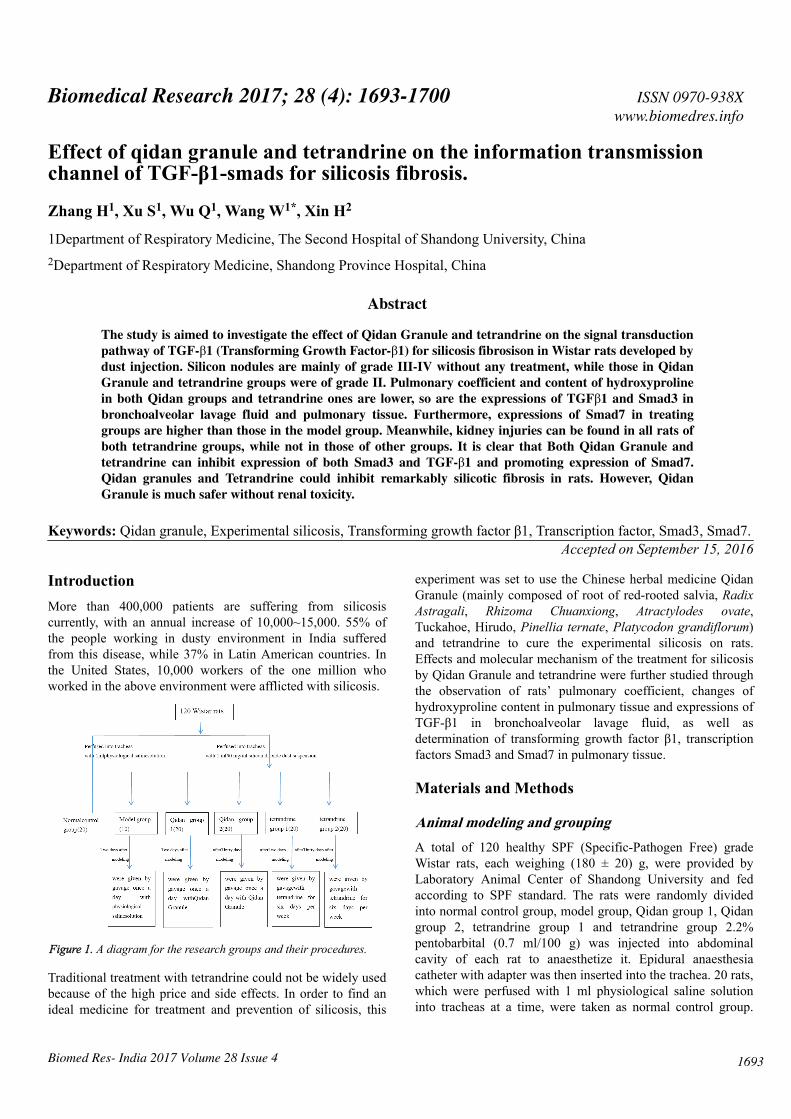
Effect of qidan granule and tetrandrine on the information transmission channel of TGF-β1-smads for silicosis fibrosis. Zhang H 1 , Xu S 1 , Wu Q 1 , Wang W 1* , Xin H 2 1Department of Respiratory Medicine, The Second Hospital of Shandong University, China 2 Department of Respiratory Medicine, Shandong Province Hospital, China Abstract The study is aimed to investigate the effect of Qidan Granule and tetrandrine on the signal transduction pathway of TGF-β1 (Transforming Growth Factor-β1) for silicosis fibrosison in Wistar rats developed by dust injection. Silicon nodules are mainly of grade III-IV without any treatment, while those in Qidan Granule and tetrandrine groups were of grade II. Pulmonary coefficient and content of hydroxyproline in both Qidan groups and tetrandrine ones are lower, so are the expressions of TGFβ1 and Smad3 in bronchoalveolar lavage fluid and pulmonary tissue. Furthermore, expressions of Smad7 in treating groups are higher than those in the model group. Meanwhile, kidney injuries can be found in all rats of both tetrandrine groups, while not in those of other groups. It is clear that Both Qidan Granule and tetrandrine can inhibit expression of both Smad3 and TGF-β1 and promoting expression of Smad7. Qidan granules and Tetrandrine could inhibit remarkably silicotic fibrosis in rats. However, Qidan Granule is much safer without renal toxicity. Keywords: Qidan granule, Experimental silicosis, Transforming growth factor β1, Transcription factor, Smad3, Smad7. Accepted on September 15, 2016 Introduction More than 400,000 patients are suffering from silicosis currently, with an annual increase of 10,000~15,000. 55% of the people working in dusty environment in India suffered from this disease, while 37% in Latin American countries. In the United States, 10,000 workers of the one million who worked in the above environment were afflicted with silicosis. Figure 1. A diagram for the research groups and their procedures. Traditional treatment with tetrandrine could not be widely used because of the high price and side effects. In order to find an ideal medicine for treatment and prevention of silicosis, this experiment was set to use the Chinese herbal medicine Qidan Granule (mainly composed of root of red-rooted salvia, Radix Astragali, Rhizoma Chuanxiong, Atractylodes ovate, Tuckahoe, Hirudo, Pinellia ternate, Platycodon grandiflorum) and tetrandrine to cure the experimental silicosis on rats. Effects and molecular mechanism of the treatment for silicosis by Qidan Granule and tetrandrine were further studied through the observation of rats’ pulmonary coefficient, changes of hydroxyproline content in pulmonary tissue and expressions of TGF-β1 in bronchoalveolar lavage fluid, as well as determination of transforming growth factor β1, transcription factors Smad3 and Smad7 in pulmonary tissue. Materials and Methods Animal modeling and grouping A total of 120 healthy SPF (Specific-Pathogen Free) grade Wistar rats, each weighing (180 ± 20) g, were provided by Laboratory Animal Center of Shandong University and fed according to SPF standard. The rats were randomly divided into normal control group, model group, Qidan group 1, Qidan group 2, tetrandrine group 1 and tetrandrine group 2.2% pentobarbital (0.7 ml/100 g) was injected into abdominal cavity of each rat to anaesthetize it. Epidural anaesthesia catheter with adapter was then inserted into the trachea. 20 rats, which were perfused with 1 ml physiological saline solution into tracheas at a time, were taken as normal control group. ISSN 0970-938X www.biomedres.info Biomed Res- India 2017 Volume 28 Issue 4 1693 Biomedical Research 2017; 28 (4): 1693-1700
Transcript of Biomedical Research 2017; 28 (4): 1693-1700 Effect of ... · group 1 every day through...
Effect of qidan granule and tetrandrine on the information transmissionchannel of TGF-β1-smads for silicosis fibrosis.
Zhang H1, Xu S1, Wu Q1, Wang W1*, Xin H2
1Department of Respiratory Medicine, The Second Hospital of Shandong University, China2Department of Respiratory Medicine, Shandong Province Hospital, China
Abstract
The study is aimed to investigate the effect of Qidan Granule and tetrandrine on the signal transductionpathway of TGF-β1 (Transforming Growth Factor-β1) for silicosis fibrosison in Wistar rats developed bydust injection. Silicon nodules are mainly of grade III-IV without any treatment, while those in QidanGranule and tetrandrine groups were of grade II. Pulmonary coefficient and content of hydroxyprolinein both Qidan groups and tetrandrine ones are lower, so are the expressions of TGFβ1 and Smad3 inbronchoalveolar lavage fluid and pulmonary tissue. Furthermore, expressions of Smad7 in treatinggroups are higher than those in the model group. Meanwhile, kidney injuries can be found in all rats ofboth tetrandrine groups, while not in those of other groups. It is clear that Both Qidan Granule andtetrandrine can inhibit expression of both Smad3 and TGF-β1 and promoting expression of Smad7.Qidan granules and Tetrandrine could inhibit remarkably silicotic fibrosis in rats. However, QidanGranule is much safer without renal toxicity.
Keywords: Qidan granule, Experimental silicosis, Transforming growth factor β1, Transcription factor, Smad3, Smad7.Accepted on September 15, 2016
IntroductionMore than 400,000 patients are suffering from silicosiscurrently, with an annual increase of 10,000~15,000. 55% ofthe people working in dusty environment in India sufferedfrom this disease, while 37% in Latin American countries. Inthe United States, 10,000 workers of the one million whoworked in the above environment were afflicted with silicosis.
Figure 1. A diagram for the research groups and their procedures.
Traditional treatment with tetrandrine could not be widely usedbecause of the high price and side effects. In order to find anideal medicine for treatment and prevention of silicosis, this
experiment was set to use the Chinese herbal medicine QidanGranule (mainly composed of root of red-rooted salvia, RadixAstragali, Rhizoma Chuanxiong, Atractylodes ovate,Tuckahoe, Hirudo, Pinellia ternate, Platycodon grandiflorum)and tetrandrine to cure the experimental silicosis on rats.Effects and molecular mechanism of the treatment for silicosisby Qidan Granule and tetrandrine were further studied throughthe observation of rats’ pulmonary coefficient, changes ofhydroxyproline content in pulmonary tissue and expressions ofTGF-β1 in bronchoalveolar lavage fluid, as well asdetermination of transforming growth factor β1, transcriptionfactors Smad3 and Smad7 in pulmonary tissue.
Materials and Methods
Animal modeling and groupingA total of 120 healthy SPF (Specific-Pathogen Free) gradeWistar rats, each weighing (180 ± 20) g, were provided byLaboratory Animal Center of Shandong University and fedaccording to SPF standard. The rats were randomly dividedinto normal control group, model group, Qidan group 1, Qidangroup 2, tetrandrine group 1 and tetrandrine group 2.2%pentobarbital (0.7 ml/100 g) was injected into abdominalcavity of each rat to anaesthetize it. Epidural anaesthesiacatheter with adapter was then inserted into the trachea. 20 rats,which were perfused with 1 ml physiological saline solutioninto tracheas at a time, were taken as normal control group.
ISSN 0970-938Xwww.biomedres.info
Biomed Res- India 2017 Volume 28 Issue 4 1693
Biomedical Research 2017; 28 (4): 1693-1700
The other 100 rats were perfused into tracheas with 1 ml 50mg/ml silicon dioxide dust suspension (produced by SigmaCompany, was diluted into 50 mg/ml suspension byphysiological saline solution, content of free silica ≥ 99%, <5µm particles ≥ 99%, 8000 U/ml penicillin was added beforeuse) and 0.25 ml air, then were rotated immediately afterwardsto distribute the injection evenly in the lungs. 10 of these ratswill be randomly chosen to form the model group. Two daysafter modeling, Qidan Granule (3,125 mg/kg, which wascomposed of crude extract of Chinese herbal medicine such asroot of red-rooted salvia, Radix Astragali, RhizomaChuanxiong, Atractylodes ovate, Tuckahoe, Hirudo, Pinelliaternate, Platycodon grandiflorum, produced by preparation labin Shandong Provincial Hospital) was given to rats of Qidangroup 1 every day through administration by gavage, andtetrandrine (22 mg/kg) to those of tetrandrine group 1 six daysper week. Thirty days after modeling, Qidan 2 group rats weregiven by gavage once a day with Qidan Granule (3,125 mg/kg). Tetrandrine (22 mg/kg, purchased from Hangzhou SiworIndustrial & Trading Co., Ltd., was diluted by 0.2%hydrochloric acid solution) were given to rats of tetrandrine 2for six days per week through administration by gavage.Physiological saline solution was given to rats of the normalcontrol group every day (2 ml/rat) through administration bygavage. Three months later, drugs were stopped in tetrandrinegroup for one month, because of its strong renal toxicity. Thisstudy was carried out in strict accordance with therecommendations in the Guide for the Care and Use ofLaboratory Animals of the National Institutes of Health. Theanimal use protocol has been reviewed and approved by theInstitutional Animal Care and Use Committee (IACUC) of TheSecond Hospital of Shandong University (The research groupsand their procedures were shown in Figure 1).
Observation items and determination methodsFive months after modeling, the rats were weighed beforebeing executed. 2% pentobarbital (0.7 ml/100 g) was injectedto abdominal cavity of the rats to make them anaesthetized.Ten rats were randomly taken from each group, and epiduralanaesthesia catheter with adapter was inserted into tracheas ofthese rats. A dose of 4 ml 37°C physiological saline solutionwas perfused slowly into each rat’s lungs, and then drawn out30 seconds later. The procedure was repeated for three times. 2ml bronchoalveolar lavage fluid, which was centrifuged in1,500 rpm, 4°C, was collected. After extracting thesupernatant, TGF-β1 expressions were examined according tooperation procedures of ELISA (Enzyme-Linked ImmunoSorbent Assay) reagent kit (TGF-β1 ELISA reagent kit waspurchased from American Alpha Diagnostic Intl). Pulmonarytissue of the other 60 rats was picked out. Connective tissuesuch as trachea and bronchus was removed. The pulmonarytissue was rinsed in physiological saline solution and weighedafter the moisture was absorbed by filter paper. Pulmonarycoefficient was calculated as: pulmonary coefficient=wetweight of the lung (mg)/body weight of the rat (g). The rightlung was picked out to examine the content of hydroxyprolineby using alkali hydrolysis (according to the instructions of
hydroxyproline reagent kit, which purchased from NanjingJiancheng Bioengineering Institute.). Apex and base of the leftlung were cut off, and the middle part of the left lung wasantisepticised in 10% buffered formalin, paraffin embedded,then cut into 5 μm thick sections, which were processed withHematoxylin-Eosin (HE) staining and observed under ordinaryoptical microscope to examine the cellular construction ofsilicon nodules. The silicosis was classified into 4 gradesaccording to the 4-grade classification methods ofexperimental silicosis: Grade 0 with no pathological changes,Grade I cellular nodules, Grade II cellular fibrosis nodules,Grade III fibrocyte nodules and Grade IV with fibrosis siliconnodules and nodule fusion in some parts. Expressions of TGF-β1, Smad3 and Smad7 in pulmonary tissue were detected byusing immunohistochemical method (according to theoperation procedures of immunohistochemical reagent kit,which purchased from Wuhan Boster Biological TechnologyCo., Ltd.). The remaining pulmonary tissue was preserved inliquid nitrogen. The left kidney was antisepticised in 10%buffered formalin, paraffin embedded, then cut into 5 μm thick,processed by Hematoxylin-Eosin (HE) staining, and observedunder ordinary optical microscope to examine the pathologicalchanges of the renal tissue (All chemicals and their origins'were explained in Table 1).
Table 1. Chemicals and their origins.
Chemicals Origins
Silicon dioxide dust Sigma Company
Qidan Granule Preparation lab in Shandong ProvincialHospital
ELISA reagent kit American Alpha Diagnostic Intl
Hydroxyproline reagent kit Nanjing Jiancheng BioengineeringInstitute
Immunohistochemical reagent kit Wuhan Boster Biological Technology Co.,Ltd.
Statistical analysisAll numerical values were expressed in ± SD and calculated bySPSS11.5 statistical software. Comparison among groups wasconducted by one-way analysis of variance, and the varianceswere analyzed by Pearson correlation analysis.
Results
Pathological examination of pulmonary diseasesRats’ lungs in the normal control group were pink and smoothwithout any abnormal changes. Volume and weight of thelungs in the model group increased. The lungs became hard.On the surface and slices, there were pale millet-sized nodules,which tended to inosculate. The nodules were hard and stuckout. Bleeding spots can be observed on the lungs of both Qidangroups and tetrandrine groups. Occasionally there were palemillet nodules on the surface and slices of the lung, which,however, did not stick out, and there was no inosculation.
Zhang/Xu/Wu/Wang/Xin
1694 Biomed Res- India 2017 Volume 28 Issue 4
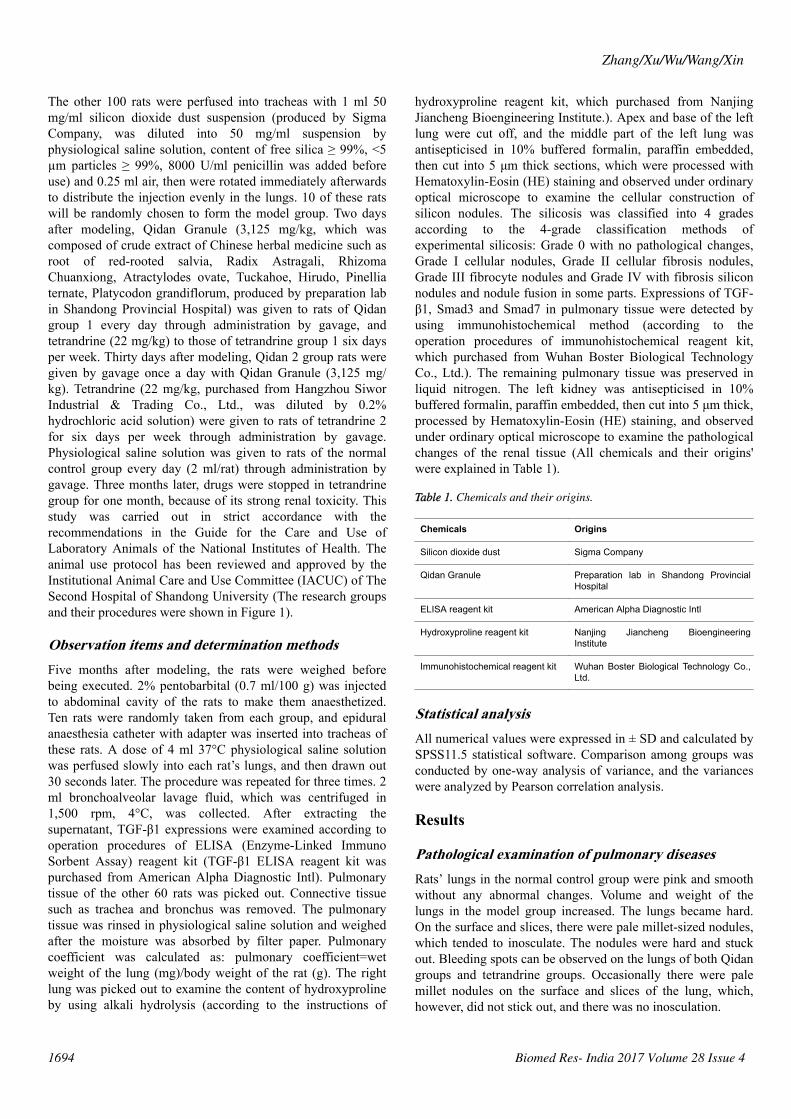
Observations under optical microscopeResults of HE staining: It was shown in Figure 2. that noobvious morphological changes occurred under microscopeobservation in the normal control group. In the model group,pulmonary tissue structures were damaged and alveoluscollapsed. And there was obvious fiber hyperplasia, especiallyaround the small blood vessels and bronchioles. A largenumber of silicon nodules of different sizes, mainly of GradeIII~IV could be seen in the pulmonary tissue, some of whichwere inosculated into pieces. There was abundant collagenfiber hyperplasia in the center of the nodules, and hyalinizationcould be seen. Only a small number of fibroblasts andmacrophages were distributed around the nodules. Siliconnodules of rats in Qidan groups and tetrandrine groups aremainly of Grade II. Scattered silicon nodules could be seenafter HE staining. There was no fusion. The nodules werecomposed of fibroblasts, macrophages and collagen fiber. Theareas of Silicon nodules were measured. It can be seen fromTable 2 that both the total and Grade III~IV areas of Siliconnodules in the Qidan group 1 and 2, and tetrandrine group 1and 2 were much lower than those in the model group. And thedifferences were quite significant (P<0.05). While both thetotal and Grade II~IV areas of Silicon nodules in Qidan 1 and
2, and tetrandrine 1 and 2 were higher than those in the normalcontrol group, with significant differences (P<0.05).
Figure 2. HE staining of pulmonary tissue of the rats in each group.A. HE staining of pulmonary tissue of the rats in the normal controlgroup (X100); B. HE staining of pulmonary tissue of the rats in themodel group (X100); C. HE staining of pulmonary tissue of the ratsin Qidan group 1 (X100); D. HE staining of pulmonary tissue of therats in Qidan group 2 (X100); E. HE staining of the pulmonary tissueof the rats in tetrandrine group 1 (X100); F. HE staining ofpulmonary tissue of the rats in tetrandrine group 2 (X100).
Table 2. The areas of Silicon nodules of rats in each group (um2, x ± S).
Group Quantityof rats
Total aeras Grade I Grade II Grade III Grade IV
Control group 10 0.00 ± 0.00 0.00 ± 0.00 0.00 ± 0.00 0.00 ± 0.00 0.00 ± 0.00
Model group 10 3482.25 ± 307.98* 0.00 ± 0.00 0.00 ± 0.00 1860.88 ± 329.51 * 1621.36 ± 195.56*
Qidan group 1 10 1704.65 ± 134.71* 796.84 ± 210.17* 807.07 ± 166.59* 100.73 ± 40.21* 0.00 ± 0.00
Qidan group 2 10 1693.88 ± 148.43* 775.39 ± 169.47* 811.55 ± 187.37* 106.94 ± 38.59* 0.00 ± 0.00
Terandrine group 1 10 1720.12 ± 123.38* 818.97 ± 173.41* 791.93 ± 154.88* 109.21 ± 28.93* 0.00 ± 0.00
Terandrine group 2 10 1739.804 ± 102.68* 839.45 ± 169.03* 786.60 ± 161.39* 113.74 ± 28.40* 0.00 ± 0.00
Note: Compared with the model group, P<0.05; and compared with the normal control group, *P<0.05.
Pulmonary coefficient and content of hydroxyprolineIt can be seen from Table 3 the pulmonary coefficient andcontent of hydroxyproline in the normal control group, Qidangroup 1 and 2, and tetrandrine group 1 and 2 were much lowerthan those in the model group. And the differences were quitesignificant (P<0.05) while the pulmonary coefficient in Qidan1 and 2, and tetrandrine 1 and 2 were higher than those in thenormal control group, with significant differences (P<0.05).However, the content of hydroxyproline in Qidan group 1 and2, and tetrandrine 1 and 2 were not significantly different fromnormal control group.
Expressions of TGF-β1 in bronchial lavage fluid ofthe ratsIn Table 4 expressions of TGF-β1 in bronchial lavage fluid inthe normal control group, Qidan 1 and 2, and tetrandrine 1 and2 were lower than those in the model group, with significant
differences (P<0.05). Expressions of TGF-β1 in bronchiallavage fluid in the model group, Qidan group and tetrandrinegroup were higher than those in the normal control group, withsignificant differences (P<0.05).
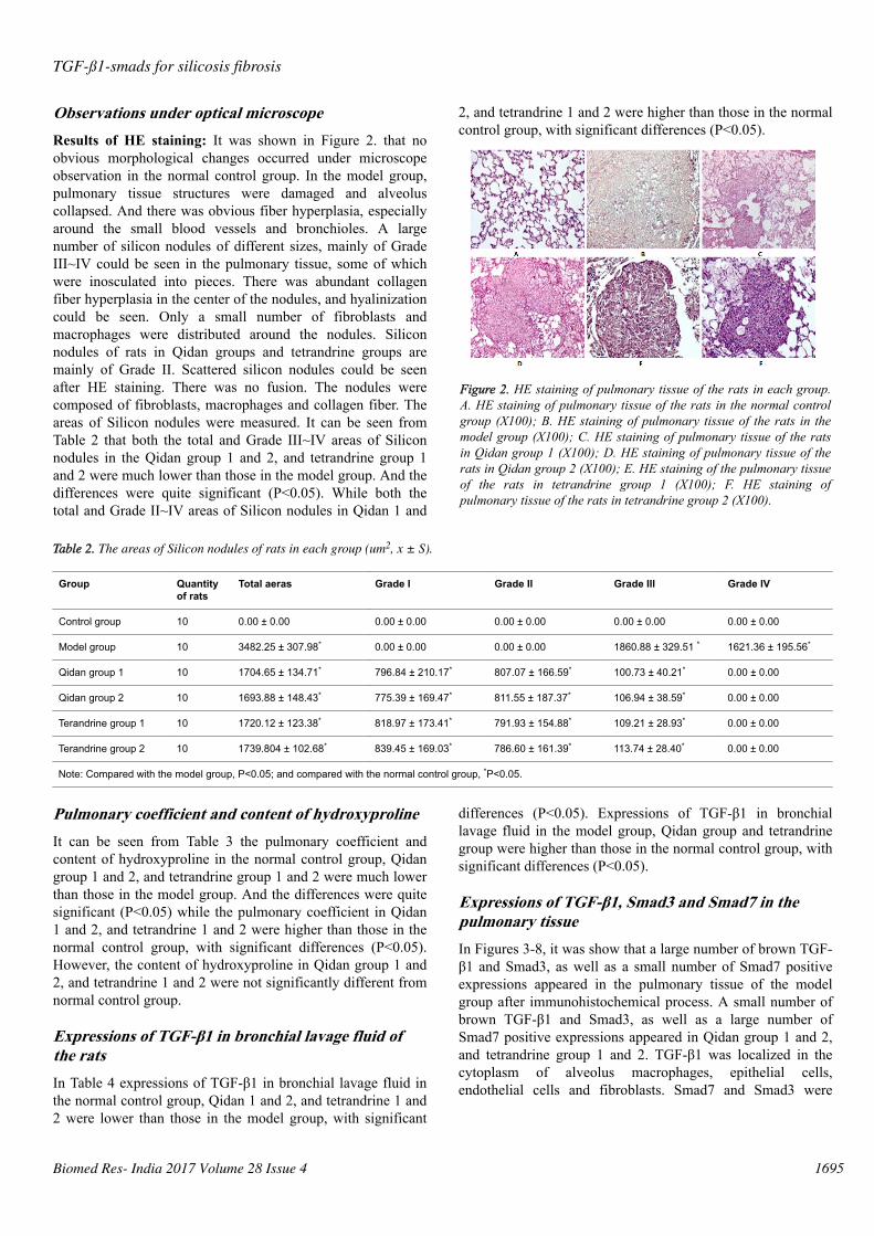
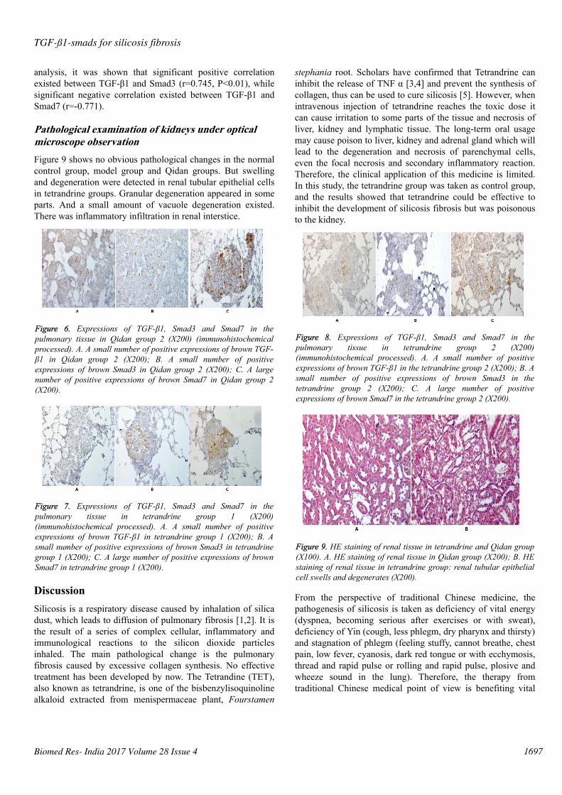
Expressions of TGF-β1, Smad3 and Smad7 in thepulmonary tissueIn Figures 3-8, it was show that a large number of brown TGF-β1 and Smad3, as well as a small number of Smad7 positiveexpressions appeared in the pulmonary tissue of the modelgroup after immunohistochemical process. A small number ofbrown TGF-β1 and Smad3, as well as a large number ofSmad7 positive expressions appeared in Qidan group 1 and 2,and tetrandrine group 1 and 2. TGF-β1 was localized in thecytoplasm of alveolus macrophages, epithelial cells,endothelial cells and fibroblasts. Smad7 and Smad3 were
TGF-ß1-smads for silicosis fibrosis
Biomed Res- India 2017 Volume 28 Issue 4 1695
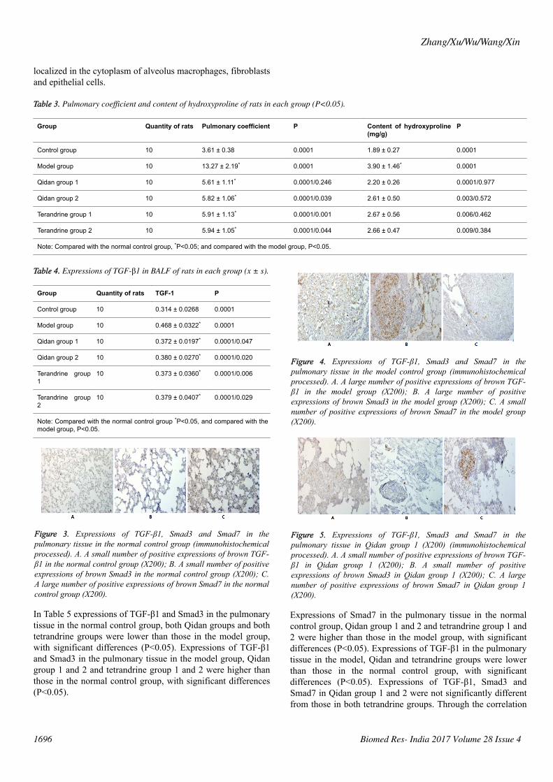
localized in the cytoplasm of alveolus macrophages, fibroblastsand epithelial cells.
Table 3. Pulmonary coefficient and content of hydroxyproline of rats in each group (P<0.05).
Group Quantity of rats Pulmonary coefficient P Content of hydroxyproline(mg/g)
P
Control group 10 3.61 ± 0.38 0.0001 1.89 ± 0.27 0.0001
Model group 10 13.27 ± 2.19* 0.0001 3.90 ± 1.46* 0.0001
Qidan group 1 10 5.61 ± 1.11* 0.0001/0.246 2.20 ± 0.26 0.0001/0.977
Qidan group 2 10 5.82 ± 1.06* 0.0001/0.039 2.61 ± 0.50 0.003/0.572
Terandrine group 1 10 5.91 ± 1.13* 0.0001/0.001 2.67 ± 0.56 0.006/0.462
Terandrine group 2 10 5.94 ± 1.05* 0.0001/0.044 2.66 ± 0.47 0.009/0.384
Note: Compared with the normal control group, *P<0.05; and compared with the model group, P<0.05.
Table 4. Expressions of TGF-β1 in BALF of rats in each group (x ± s).
Group Quantity of rats TGF-1 P
Control group 10 0.314 ± 0.0268 0.0001
Model group 10 0.468 ± 0.0322* 0.0001
Qidan group 1 10 0.372 ± 0.0197* 0.0001/0.047
Qidan group 2 10 0.380 ± 0.0270* 0.0001/0.020
Terandrine group1
10 0.373 ± 0.0360* 0.0001/0.006
Terandrine group2
10 0.379 ± 0.0407* 0.0001/0.029
Note: Compared with the normal control group *P<0.05, and compared with themodel group, P<0.05.
Figure 3. Expressions of TGF-β1, Smad3 and Smad7 in thepulmonary tissue in the normal control group (immunohistochemicalprocessed). A. A small number of positive expressions of brown TGF-β1 in the normal control group (X200); B. A small number of positiveexpressions of brown Smad3 in the normal control group (X200); C.A large number of positive expressions of brown Smad7 in the normalcontrol group (X200).
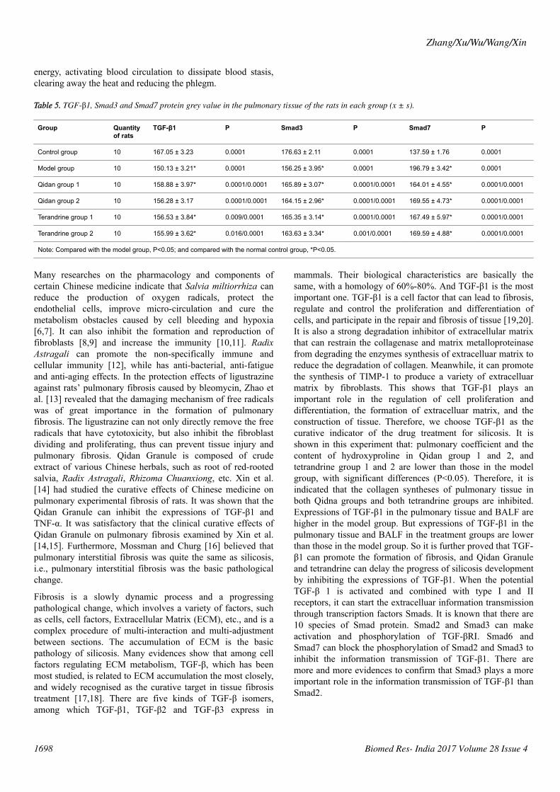
In Table 5 expressions of TGF-β1 and Smad3 in the pulmonarytissue in the normal control group, both Qidan groups and bothtetrandrine groups were lower than those in the model group,with significant differences (P<0.05). Expressions of TGF-β1and Smad3 in the pulmonary tissue in the model group, Qidangroup 1 and 2 and tetrandrine group 1 and 2 were higher thanthose in the normal control group, with significant differences(P<0.05).
Figure 4. Expressions of TGF-β1, Smad3 and Smad7 in thepulmonary tissue in the model control group (immunohistochemicalprocessed). A. A large number of positive expressions of brown TGF-β1 in the model group (X200); B. A large number of positiveexpressions of brown Smad3 in the model group (X200); C. A smallnumber of positive expressions of brown Smad7 in the model group(X200).
Figure 5. Expressions of TGF-β1, Smad3 and Smad7 in thepulmonary tissue in Qidan group 1 (X200) (immunohistochemicalprocessed). A. A small number of positive expressions of brown TGF-β1 in Qidan group 1 (X200); B. A small number of positiveexpressions of brown Smad3 in Qidan group 1 (X200); C. A largenumber of positive expressions of brown Smad7 in Qidan group 1(X200).
Expressions of Smad7 in the pulmonary tissue in the normalcontrol group, Qidan group 1 and 2 and tetrandrine group 1 and2 were higher than those in the model group, with significantdifferences (P<0.05). Expressions of TGF-β1 in the pulmonarytissue in the model, Qidan and tetrandrine groups were lowerthan those in the normal control group, with significantdifferences (P<0.05). Expressions of TGF-β1, Smad3 andSmad7 in Qidan group 1 and 2 were not significantly differentfrom those in both tetrandrine groups. Through the correlation
Zhang/Xu/Wu/Wang/Xin
1696 Biomed Res- India 2017 Volume 28 Issue 4
analysis, it was shown that significant positive correlationexisted between TGF-β1 and Smad3 (r=0.745, P<0.01), whilesignificant negative correlation existed between TGF-β1 andSmad7 (r=-0.771).
Pathological examination of kidneys under opticalmicroscope observationFigure 9 shows no obvious pathological changes in the normalcontrol group, model group and Qidan groups. But swellingand degeneration were detected in renal tubular epithelial cellsin tetrandrine groups. Granular degeneration appeared in someparts. And a small amount of vacuole degeneration existed.There was inflammatory infiltration in renal interstice.
Figure 6. Expressions of TGF-β1, Smad3 and Smad7 in thepulmonary tissue in Qidan group 2 (X200) (immunohistochemicalprocessed). A. A small number of positive expressions of brown TGF-β1 in Qidan group 2 (X200); B. A small number of positiveexpressions of brown Smad3 in Qidan group 2 (X200); C. A largenumber of positive expressions of brown Smad7 in Qidan group 2(X200).
Figure 7. Expressions of TGF-β1, Smad3 and Smad7 in thepulmonary tissue in tetrandrine group 1 (X200)(immunohistochemical processed). A. A small number of positiveexpressions of brown TGF-β1 in tetrandrine group 1 (X200); B. Asmall number of positive expressions of brown Smad3 in tetrandrinegroup 1 (X200); C. A large number of positive expressions of brownSmad7 in tetrandrine group 1 (X200).
DiscussionSilicosis is a respiratory disease caused by inhalation of silicadust, which leads to diffusion of pulmonary fibrosis [1,2]. It isthe result of a series of complex cellular, inflammatory andimmunological reactions to the silicon dioxide particlesinhaled. The main pathological change is the pulmonaryfibrosis caused by excessive collagen synthesis. No effectivetreatment has been developed by now. The Tetrandine (TET),also known as tetrandrine, is one of the bisbenzylisoquinolinealkaloid extracted from menispermaceae plant, Fourstamen
stephania root. Scholars have confirmed that Tetrandrine caninhibit the release of TNF α [3,4] and prevent the synthesis ofcollagen, thus can be used to cure silicosis [5]. However, whenintravenous injection of tetrandrine reaches the toxic dose itcan cause irritation to some parts of the tissue and necrosis ofliver, kidney and lymphatic tissue. The long-term oral usagemay cause poison to liver, kidney and adrenal gland which willlead to the degeneration and necrosis of parenchymal cells,even the focal necrosis and secondary inflammatory reaction.Therefore, the clinical application of this medicine is limited.In this study, the tetrandrine group was taken as control group,and the results showed that tetrandrine could be effective toinhibit the development of silicosis fibrosis but was poisonousto the kidney.
Figure 8. Expressions of TGF-β1, Smad3 and Smad7 in thepulmonary tissue in tetrandrine group 2 (X200)(immunohistochemical processed). A. A small number of positiveexpressions of brown TGF-β1 in the tetrandrine group 2 (X200); B. Asmall number of positive expressions of brown Smad3 in thetetrandrine group 2 (X200); C. A large number of positiveexpressions of brown Smad7 in the tetrandrine group 2 (X200).
Figure 9. HE staining of renal tissue in tetrandrine and Qidan group(X100). A. HE staining of renal tissue in Qidan group (X200); B. HEstaining of renal tissue in tetrandrine group: renal tubular epithelialcell swells and degenerates (X200).
From the perspective of traditional Chinese medicine, thepathogenesis of silicosis is taken as deficiency of vital energy(dyspnea, becoming serious after exercises or with sweat),deficiency of Yin (cough, less phlegm, dry pharynx and thirsty)and stagnation of phlegm (feeling stuffy, cannot breathe, chestpain, low fever, cyanosis, dark red tongue or with ecchymosis,thread and rapid pulse or rolling and rapid pulse, plosive andwheeze sound in the lung). Therefore, the therapy fromtraditional Chinese medical point of view is benefiting vital
TGF-ß1-smads for silicosis fibrosis
Biomed Res- India 2017 Volume 28 Issue 4 1697
energy, activating blood circulation to dissipate blood stasis,clearing away the heat and reducing the phlegm.
Table 5. TGF-β1, Smad3 and Smad7 protein grey value in the pulmonary tissue of the rats in each group (x ± s).
Group Quantityof rats
TGF-β1 P Smad3 P Smad7 P
Control group 10 167.05 ± 3.23 0.0001 176.63 ± 2.11 0.0001 137.59 ± 1.76 0.0001
Model group 10 150.13 ± 3.21* 0.0001 156.25 ± 3.95* 0.0001 196.79 ± 3.42* 0.0001
Qidan group 1 10 158.88 ± 3.97* 0.0001/0.0001 165.89 ± 3.07* 0.0001/0.0001 164.01 ± 4.55* 0.0001/0.0001
Qidan group 2 10 156.28 ± 3.17 0.0001/0.0001 164.15 ± 2.96* 0.0001/0.0001 169.55 ± 4.73* 0.0001/0.0001
Terandrine group 1 10 156.53 ± 3.84* 0.009/0.0001 165.35 ± 3.14* 0.0001/0.0001 167.49 ± 5.97* 0.0001/0.0001
Terandrine group 2 10 155.99 ± 3.62* 0.016/0.0001 163.63 ± 3.34* 0.001/0.0001 169.59 ± 4.88* 0.0001/0.0001
Note: Compared with the model group, P<0.05; and compared with the normal control group, *P<0.05.
Many researches on the pharmacology and components ofcertain Chinese medicine indicate that Salvia miltiorrhiza canreduce the production of oxygen radicals, protect theendothelial cells, improve micro-circulation and cure themetabolism obstacles caused by cell bleeding and hypoxia[6,7]. It can also inhibit the formation and reproduction offibroblasts [8,9] and increase the immunity [10,11]. RadixAstragali can promote the non-specifically immune andcellular immunity [12], while has anti-bacterial, anti-fatigueand anti-aging effects. In the protection effects of ligustrazineagainst rats’ pulmonary fibrosis caused by bleomycin, Zhao etal. [13] revealed that the damaging mechanism of free radicalswas of great importance in the formation of pulmonaryfibrosis. The ligustrazine can not only directly remove the freeradicals that have cytotoxicity, but also inhibit the fibroblastdividing and proliferating, thus can prevent tissue injury andpulmonary fibrosis. Qidan Granule is composed of crudeextract of various Chinese herbals, such as root of red-rootedsalvia, Radix Astragali, Rhizoma Chuanxiong, etc. Xin et al.[14] had studied the curative effects of Chinese medicine onpulmonary experimental fibrosis of rats. It was shown that theQidan Granule can inhibit the expressions of TGF-β1 andTNF-α. It was satisfactory that the clinical curative effects ofQidan Granule on pulmonary fibrosis examined by Xin et al.[14,15]. Furthermore, Mossman and Churg [16] believed thatpulmonary interstitial fibrosis was quite the same as silicosis,i.e., pulmonary interstitial fibrosis was the basic pathologicalchange.
Fibrosis is a slowly dynamic process and a progressingpathological change, which involves a variety of factors, suchas cells, cell factors, Extracellular Matrix (ECM), etc., and is acomplex procedure of multi-interaction and multi-adjustmentbetween sections. The accumulation of ECM is the basicpathology of silicosis. Many evidences show that among cellfactors regulating ECM metabolism, TGF-β, which has beenmost studied, is related to ECM accumulation the most closely,and widely recognised as the curative target in tissue fibrosistreatment [17,18]. There are five kinds of TGF-β isomers,among which TGF-β1, TGF-β2 and TGF-β3 express in
mammals. Their biological characteristics are basically thesame, with a homology of 60%-80%. And TGF-β1 is the mostimportant one. TGF-β1 is a cell factor that can lead to fibrosis,regulate and control the proliferation and differentiation ofcells, and participate in the repair and fibrosis of tissue [19,20].It is also a strong degradation inhibitor of extracellular matrixthat can restrain the collagenase and matrix metalloproteinasefrom degrading the enzymes synthesis of extracelluar matrix toreduce the degradation of collagen. Meanwhile, it can promotethe synthesis of TIMP-1 to produce a variety of extracelluarmatrix by fibroblasts. This shows that TGF-β1 plays animportant role in the regulation of cell proliferation anddifferentiation, the formation of extracelluar matrix, and theconstruction of tissue. Therefore, we choose TGF-β1 as thecurative indicator of the drug treatment for silicosis. It isshown in this experiment that: pulmonary coefficient and thecontent of hydroxyproline in Qidan group 1 and 2, andtetrandrine group 1 and 2 are lower than those in the modelgroup, with significant differences (P<0.05). Therefore, it isindicated that the collagen syntheses of pulmonary tissue inboth Qidna groups and both tetrandrine groups are inhibited.Expressions of TGF-β1 in the pulmonary tissue and BALF arehigher in the model group. But expressions of TGF-β1 in thepulmonary tissue and BALF in the treatment groups are lowerthan those in the model group. So it is further proved that TGF-β1 can promote the formation of fibrosis, and Qidan Granuleand tetrandrine can delay the progress of silicosis developmentby inhibiting the expressions of TGF-β1. When the potentialTGF-β 1 is activated and combined with type I and IIreceptors, it can start the extracelluar information transmissionthrough transcription factors Smads. It is known that there are10 species of Smad protein. Smad2 and Smad3 can makeactivation and phosphorylation of TGF-βRI. Smad6 andSmad7 can block the phosphorylation of Smad2 and Smad3 toinhibit the information transmission of TGF-β1. There aremore and more evidences to confirm that Smad3 plays a moreimportant role in the information transmission of TGF-β1 thanSmad2.
Zhang/Xu/Wu/Wang/Xin
1698 Biomed Res- India 2017 Volume 28 Issue 4

Kobayashi et al. [21] used TGF-β1 to stimulate the pulmonaryfibroblasts that were infected by Smad2-siRNA and Smad3-siRNA, and found that collagen synthesis of pulmonaryfibroblasts cured by Smad3-siRNA, expressions of α-smoothmuscle action (α-SMA) and the contraction of α-SMA did notdiffer from those in the control group. On the contrary, TGF-β1can increase the collagen synthesis of pulmonary fibroblastscured by Smad2-siRNA, expressions of α-SMA andcontraction of α-SMA. Therefore, it is indicated that TGF-β1transfers information through Smad3 to cause the activationand collagen synthesis of fibroblasts. Smad7 is of some valuein the treatment for pulmonary fibrosis. It can prevent TGF-β1from inducing the pulmonary fibroblasts to differentiate intomyofibroblasts. So we choose Smad3 and Smad7 asobservation indicators to make further study on the molecularmechanism of the inhibition of fibrosis by Qidan Granule andtetrandrine through TGF-β1. This study shows that,expressions of Smad3 in the model group are higher than thosein the normal control group, Qidan 1, 2, and tetrandrine 1, 2,while expressions of Smad7 are lower than those in the normalcontrol group, Qidan 1, 2, and tetrandrine 1, 2. Correlationanalysis shows that significant positive correlation existsbetween TGF-β1 and Smad3 (r=0.745, P<0.01), whilesignificant negative correlation exists between TGF-β1 andSmad7 (r=0.745, P<0.01). Therefore, Qidan Granule andtetrandrine can block the information transmission channel ofTGF-β1 through the inhibition of Smad3 expressions and thepromotion of Smad7 expressions to inhibit the formation offibrosis and delay the progress of silicosis development.
Meanwhile, the results show that pulmonary coefficient,content of hydroxyproline and expressions of TGF-β1, Smad3and Smad7 in Qidan group 1 and 2 do not differ significantlyfrom those in tetrandrine groups. This may be related to thespecial pathogenesis of silicosis, which as respiratory diseasecaused by inhalation of dissociated silica dust, is different fromother pulmonary interstitial fibrosis, and leads to diffusion ofpulmonary fibrosis. It is the result of a series of complexcellular, inflammatory and immunological reactions wheninhale silicon dioxide particles, which continuously stimulatesthe fibroblasts to accelerate the formation of collagen fiber.Qidan Granule and terandrine can inhibit the formation offibrosis and delay the progress of silicosis development byblocking the information transmission channel of TGF-β1,even grade II-III silicon nodules have already formed. There isno significant difference compared with earlier medicaltreatments.
AcknowledgmentsThis study was supported by the National Natural ScienceFoundation of China (No. 81473485).
References1. Jalloul AS, Banks DE. The health effects of silica exposure.
In: Rom WN, ed. Environmental and occupationalmedicine. Philadelphia, PA: Lippincott Williams & Wilkins2007; 4: 365-387.
2. Greenberg MI, Waksman J, Curtis J. Silicosis: a review. DisMon 2007; 53: 394-416.
3. Chen L, Chen L, Lv Y, Cui Z, Bei G, Qin G, Zhou J, Ge T.Tetrandrine ameliorates cognitive ampairment viainhibiting astrocyte-derived S100B activation in a rat modelof chronic cerebral hypoperfusion. Neurol Res 2013; 35:614-621.
4. Lin TY, Tseng SH, Li SJ, Chen JC, Shieh JS, Chen Y.Tetrandrine increased the survival rate of mice withlipopolysaccharide-induced endotoxemia. J Trauma 2009;66: 411-417.
5. He Y, Liu B, Miao Q. Inhibition of mRNA expression ofsilicotic collagen gene by tetrandrine. Zhonghua Yu FangYi Xue Za Zhi 1995; 29: 18-20.
6. Hung YC, Wang PW, Pan TL. Functional proteomics revealthe effect of Salvia miltiorrhiza aqueous extract againstvascular atherosclerotic lesions. Biochim Biophys Acta2010; 1804: 1310-1321.
7. Han JY, Horie Y, Miura S, Akiba Y, Guo J, Li D, Fan JY,Liu YY, Hu BH, An LH, Chang X, Xu M, Guo DA, Sun K,Yang JY, Fang SP, Xian MJ, Kizaki M, Nagata H, Hibi T.Compound Danshen injection improves endotoxin-inducedmicrocirculatory disturbance in rat mesentery. World JGastroenterol 2007; 13: 3581-3591.
8. He S, Yang Y, Liu X, Huang W, Zhang X, Yang S, ZhangX. Compound Astragalus and Salvia miltiorrhiza extractinhibits cell proliferation, invasion and collagen synthesisin keloid fibroblasts by mediating transforming growthfactor-beta/Smad pathway. Br J Dermatol 2012; 166:564-754.
9. Zhang M, Cao SR, Zhang R, Jin JL, Zhu YF. The inhibitoryeffect of salvianolic acid B on TGF-beta1-inducedproliferation and differentiation in lung fibroblasts. ExpLung Res 2014; 40: 172-185.
10. Liang XY, Li HN, Yang XY, Zhou WY, Niu JG, Chen BD.Effect of Danshen aqueous extract on serum hs-CRP, IL-8,IL-10, TNF-alpha levels, and IL-10 mRNA, TNF-alphamRNA expression levels, cerebral TGF-beta1 positiveexpression level and its neuroprotective mechanisms in CIRrats. Mol Biol Rep 2013; 40: 3419-3427.
11. Wong CK, Tse PS, Wong EL, Leung PC, Fung KP, LamCW. Immunomodulatory effects of yun zhi and danshencapsules in health subjects--a randomized, double-blind,placebo-controlled, crossover study. Int Immunopharmacol2004; 4: 201-211.
12. Jian J, Wu Z. Influences of traditional Chinese medicine onnon-specific immunity of Jian Carp (Cyprinus carpio var.Jian). Fish Shellfish Immunol 2004; 16: 185-191.
13. Zhao J, Dai H, Zhang YJ. Research of ligustrazineprotection over rats’ pulmonary interstitial fibrosis causedby bleomycin. Med J Chinese People's Liberation Army2005; 30: 455.
14. Xin HT, Song XJ, Xue SH, Mu XY, Wang J, Jin CJ, HangC. The Infection of Qidan Granule to TGF-β and TGF-αexpressions in rats’ pulmonary interstitial fibrosis. Journalof Capital Medical University 2004; 25: 49-52.
TGF-ß1-smads for silicosis fibrosis
Biomed Res- India 2017 Volume 28 Issue 4 1699
15. Xin HT, Liu QH, Zhang X, Hang C, Jin CJ, Zhang JP.Clinic study of Qidan Granule therapy of pulmonaryinterstitial fibrosis. Shandong Medicine 2002; 42: 20-21.
16. Mossman BT, Churg A. Mechanisms in pathogenesis ofasbestosis and silicosis. Am J Respir Crit Care Med 1998;157: 1666-1680.
17. Coker PK, Laurent GJ, Shahzeidi S, Lympany PA, du BoisRM, Jeffery PK, Mc Anulty RJ. Transforming GrowthFactors-β1, -β2, and -β3 stimulate fibroblast procollagenproduction in vitro but are differentially expressed duringbleomycin-induced lung fibrosis. Am J Pathol 1997; 150:981-991.
18. Zhao Y, Shah DU. Expression of transforming growthfactor- beta type I and type II receptors is altered in ratlungs undergoing bleomycin-induced pulmonary fibrosis.Exp Mol Pathol 2000; 69: 67-78.
19. Fernandez IE, Eickelberg O. The impact of TGF-b on lungfibrosis. From targeting to biomarkers. Proc Am ThoracSoc 2012; 9: 111-116.
20. Letterio JJ, Roberts AB. Regulation of immune responsesby TGF-beta. Annu Rev Immunol 1998; 16: 137-161.
21. Kobayashi T, Liu X, Wen FQ. Smad3 Mediates TGF-Beta1-Induced Collagen Gel Contraction by HumanPulmonary Fibroblasts. Biochem Biophys Res Commun2006; 339: 290-295.
*Correspondence toWang W
Department of Respiratory Medicine
The Second Hospital of Shandong University
China
Zhang/Xu/Wu/Wang/Xin
1700 Biomed Res- India 2017 Volume 28 Issue 4