Biologia seminario pp
38
STIMULATION O F A LFA 1- ADREN O CE PTOR OR ANG O TENSIN TYPE 1 REC E PT O R ENH A NCES DNA SYNTH ESIS I N H U M AN- I ND U C E D PLURI P OTENT S T E M CELLS V I A G Q-COUPL ED RECE P TO R- D E PE NDENT SIGNA L ING P ATHWAYS ANG E L IC A MAR I A SE R NA CAMPUZANO AN D AN A ISABEL VA LLEJ O
-
Upload
angelica-maria -
Category
Documents
-
view
92 -
download
1
Transcript of Biologia seminario pp

STIMULA
TION O
F ALF
A1-
ADRENOCEPTOR O
R ANGOTE
NSIN
TYPE
1 R
ECEPTOR E
NHANCES DNA
SYNTH
ESIS IN
HUMAN-IN
DUCED
PLURIP
OTENT
STEM C
ELLS V
IA G
Q-
COUPLED R
ECEPTOR-D
EPENDENT
SIGNALIN
G PATH
WAY
S
AN
GE
LI C
A M
AR
I A S
ER
NA
CA
MP
UZ
AN
O A
ND
AN
A I S
AB
EL
VA
LL
EJ O

INTRODUCTION
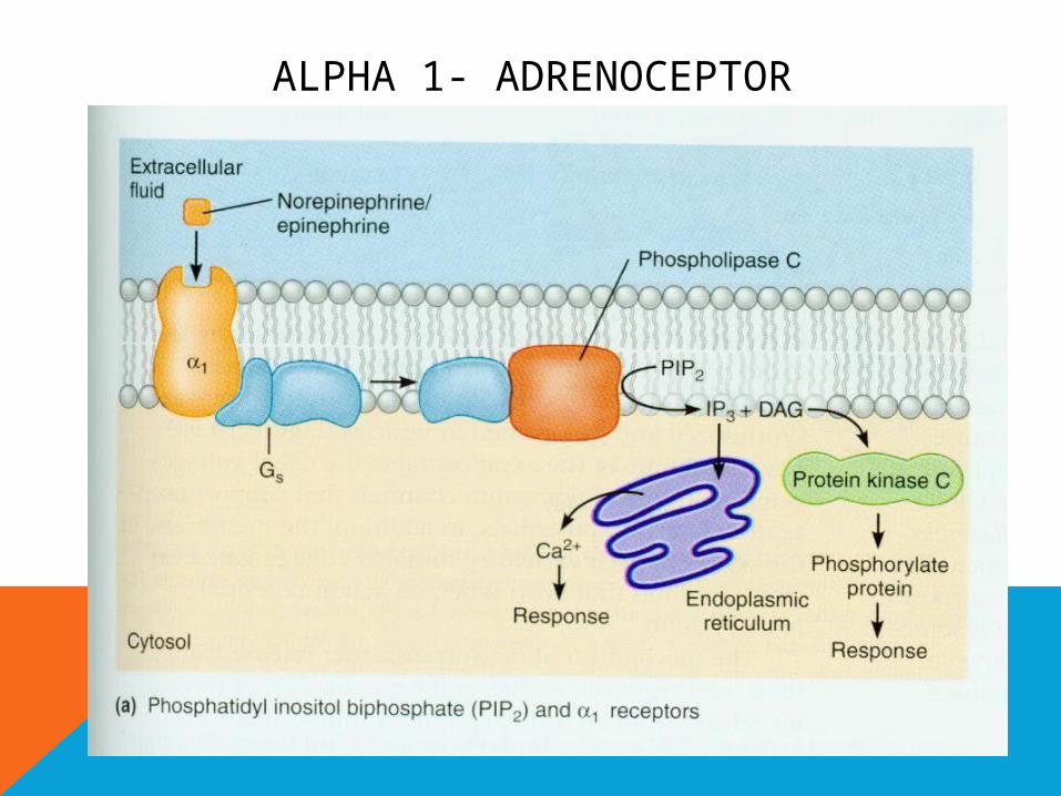
• Previous studies suggested that stimulation of α1-adrenoceptors enhances proliferation not only in somatic cells such as vascular muscle cells or adventitial but also in undifferentiated stem cells such as mouse induced pluripotent stem cells. α1-Adrenoceptors are Gq coupled receptors. Other Gq-coupled receptors such as angiotensin type 1 receptor are known to enhance cell growth in undifferentiated stem cells
INTRODUCTION
• Theres a lot of studies that have shown the mechanism proliferation of iPS, However, it remains unclear whether a signaling pathway downstream of Gq-coupled receptors is involved in proliferation of human iPS cells. In addition, no studies have shown whether human iPS cells express α1-adrenoceptors or AT1 receptor.

ANGIOTENSINE T1 RECEPTOR
AT1

ANGIOTENSINE T1 RECEPTOR
AT1
• Vasoconstriction• Aldosterone and
vasopressin secretion
• Vascular smooth muscle cells proliferation
• Cardiac contractility
• Human cultured breast epithelial cells


ALPHA 1- ADRENOCEPTOR
ALPHA 1- ADRENOCEPTOR
STEM CELL
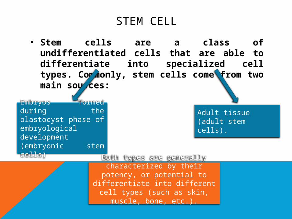
• Stem cells are a class of undifferentiated cells that are able to differentiate into specialized cell types. Commonly, stem cells come from two main sources:
Embryos formed during the blastocyst phase of embryological development (embryonic stem cells)
Adult tissue (adult stem cells).
Both types are generally characterized by their potency, or
potential to differentiate into different cell types (such as skin,
muscle, bone, etc.).
OBJECTIVE
• To reveal the expression of alpha 1- adrenoceptor and AT1 receptor in human IPS cells and their involvement in DNA synthesis. To future regenerative therapy.
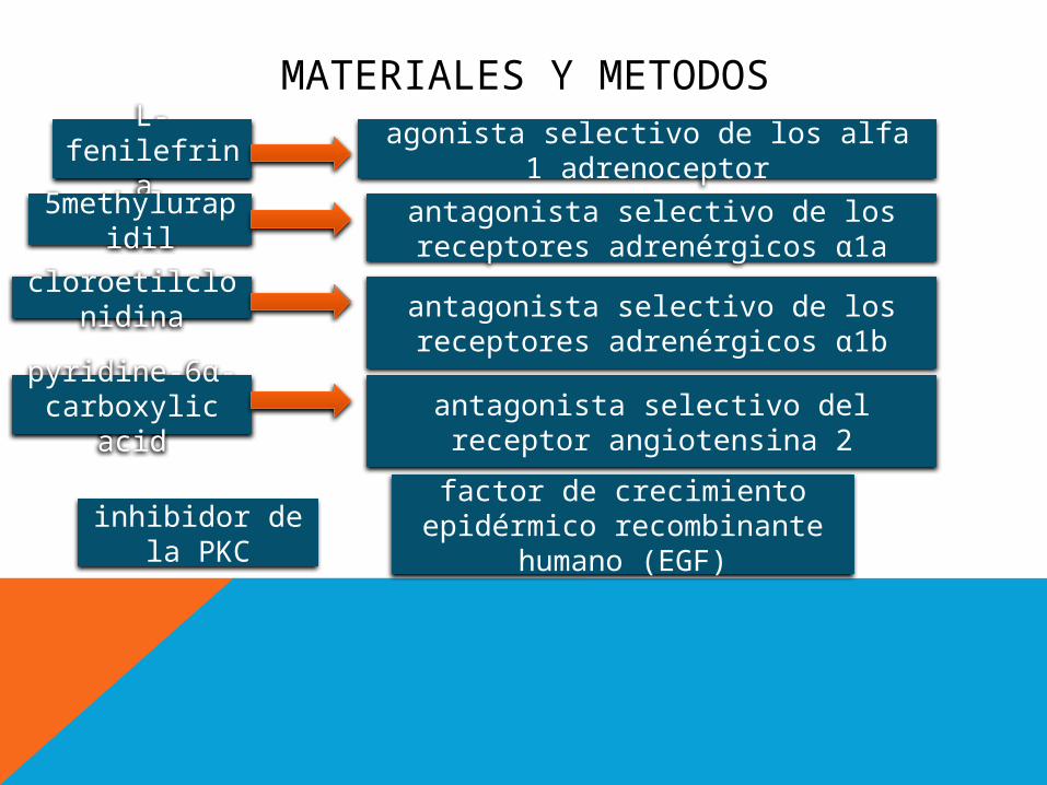
MATERIALES Y METODOS
L-fenilefrina agonista selectivo de los alfa 1
adrenoceptor5methylurapi
dilantagonista selectivo de los receptores adrenérgicos α1a
cloroetilclonidina antagonista selectivo de los
receptores adrenérgicos α1b
pyridine-6α-carboxylic acid
antagonista selectivo del receptor angiotensina 2
inhibidor de la PKC
factor de crecimiento epidérmico recombinante
humano (EGF)
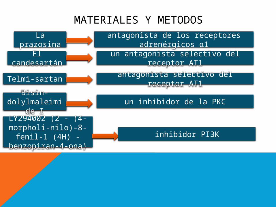
MATERIALES Y METODOSLa
prazosinaantagonista de los receptores
adrenérgicos α1El
candesartánun antagonista selectivo del receptor
AT1
Telmi-sartan antagonista selectivo del receptor AT1
Bisin-dolylmaleimid
e Iun inhibidor de la PKC
LY294002 (2 - (4-morpholi-nilo)-8-fenil-1 (4H) - benzopiran-
4-ona)
inhibidor PI3K

CULTIVOS DE CÉLULAS IPS HUMANAS
Las Células iPS humanas se expandieron en MEFs tratadas en placas de cultivo recubiertas con gelatina al 0,1% a 37 C en un entorno de 5% de CO2.
El medio de cultivo contiene 20% de suero “knock out” , 0,1 mM de aminoácidos no esenciales (Invitrogen) penicilina y estreptomicina (Invitrogen), 500ng/ml de factor de crecimiento de fibroblastos recombinante humano BFGF.

• Para eliminar MEFs contaminadas, las colonias de células iPS se pasaron hasta 4 veces sin células alimentadoras en placas de cultivo de 60 mm recubiertas con Matrigel.
• las colonias de células se despegaron utilizando tripsina 0,25% de EDTA y luego se re-sembraron en placas a una densidad de 1,5 x 10(5) células/cm2. Después de la incubación durante 24 h, el medio de cultivo fue reemplazado por un medio de cultivo sin bFGF.

INMUNOFLUORESCENCIA
luego de 24 h, las células fueron re-sembradas en portaobjetos revestidos con Matrigel y se fijaron con 4% de paraformaldehído a 41C durante 30 min. Luego se les añadio 10% de suero bovino fetal en PBS, los portaobjetos se incubaron con anticuerpo monoclonal anti-α1-adrenoceptores en PBS a 4C durante la noche. Los portaobjetos se incubaron con anticuerpo anti-IgG conjugado con Alexa Fluor 594 (1:200; Invitrogen) en PBS durante 1 h a temperatura ambiente. (5 PBS)
• BrdU fue el enfoque utilizado para controlar la síntesis de ADNc. el ensayo inmunoenzimático ELISA se realizó usando marcaje BrdU y kit de detección III para detectar la incorporación de BrdU en el ADN celular. luego de 24 h de re-recubrir en Matrigel , las células se cultivaron en el medio de cultivo sin bFGF por 24 h y se trataron con o sin L-fenilefrina o Ang II. luego de 24 h de realizado el tratamiento, se añadió BrdU al medio de cultivo y se incorpora en el ADN recién sintetizado durante 2 h
INCORPORACIÓN DE BrdU

• Después de la fijación de células, se añadió un ac marcado con peroxidasa para BrdU. En el paso final, se añadió el sustrato de peroxidasa y la enzima catalizo la escisión de este, produciendo un producto coloreado.

WESTERN BLOT
• luego del tratamiento, las células fueron lisadas y luego centrifugadas, Los extractos de proteínas se sometieron a electroforesis en un gel de poliacrilamida y se transfirieron a una membrana de difluoruro de polivinilideno con un equipo de transferencia semiseca. se aplicaron anticuerpos en diluciones en PBS que contenía suero de albúmina bovina: a1a, b, d -adrenoceptor anti-humano,tecnologia de señalizacioncelular, Gq de conejo anti-humano.
• Se añadió el respectivo anticuerpo secundario conjugado con peroxidasa, y las proteínas inmunorreactivas se visualizaron utilizando ECL Western Blotting Detection Kit. La densidad de las bandas se normalizó utilizando β-actina como estándar interno.

ARN CORTO DE INTERFERENCIA
Células iPS fueron transfectadas con los ARN interferentes cortos (siRNAs) mediante el uso de un agente de transfección. Un controlador siRNA, fue desarrollado para minimizar los efectos de off- target.
Luego de la transfeccion las células crecieron por 24 hr y luego estimuladas con o sin l-phenylephrina, Ang 2.
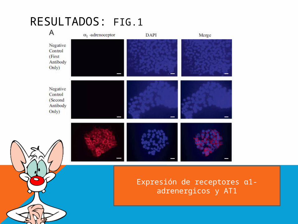
RESULTADOS: FIG.1
Expresión de receptores α1-adrenergicos y AT1

RESULTADOS: FIG.1
Expresión de receptores α1-adrenergicos y AT1

RESULTADOS
TRATAMIENTO CONBrdU+I-fenilefrina+
Prazosina Cloro-
etilclonidina BMY7378
yohimbina5-
metilurapidil
Efectos de los agonistas y antagonistas de los receptores
α1-adrenergicos en la síntesis de DNA en células humanas iPS

RESULTADOS
TRATAMIENTO CONBrdU+AngII+
PD123319
Candesartan o telmisartan
Efectos de los antagonistas de los receptores de angiotensina (AngII) en la síntesis de DNA en
células humanas iPS

RESULTADOS
Bisindolylmaleimide I
Go 6976

RESULTADOS
LY294002
PD98059
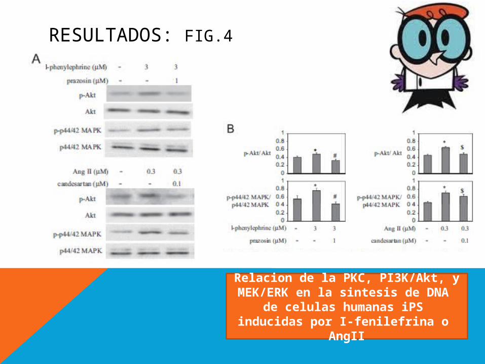
RESULTADOS: FIG.4
Relacion de la PKC, PI3K/Akt, y MEK/ERK en la sintesis de DNA
de celulas humanas iPS inducidas por I-fenilefrina o
AngII
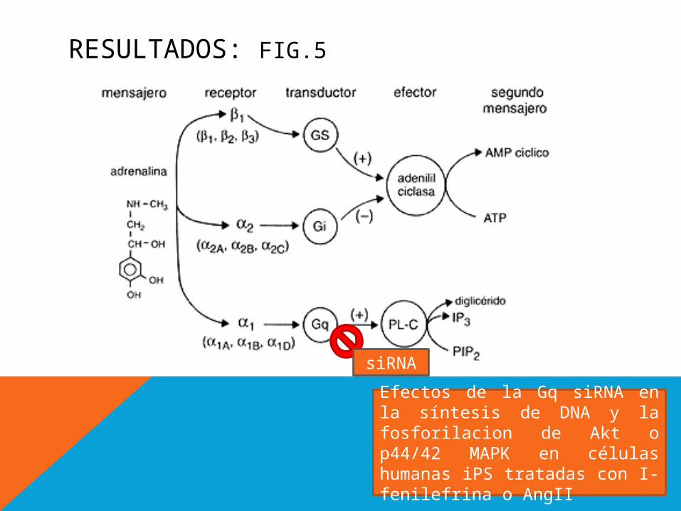
RESULTADOS: FIG.5
siRNA
Efectos de la Gq siRNA en la síntesis de DNA y la fosforilacion de Akt o p44/42 MAPK en células humanas iPS tratadas con I-fenilefrina o AngII

RESULTADOS: FIG.5
Efectos de la Gq siRNA en la síntesis de DNA y la fosforilacion de Akt o p44/42 MAPK en células humanas iPS tratadas con I-fenilefrina o AngII
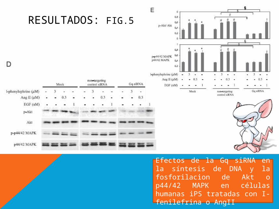
RESULTADOS: FIG.5
Efectos de la Gq siRNA en la síntesis de DNA y la fosforilacion de Akt o p44/42 MAPK en células humanas iPS tratadas con I-fenilefrina o AngII
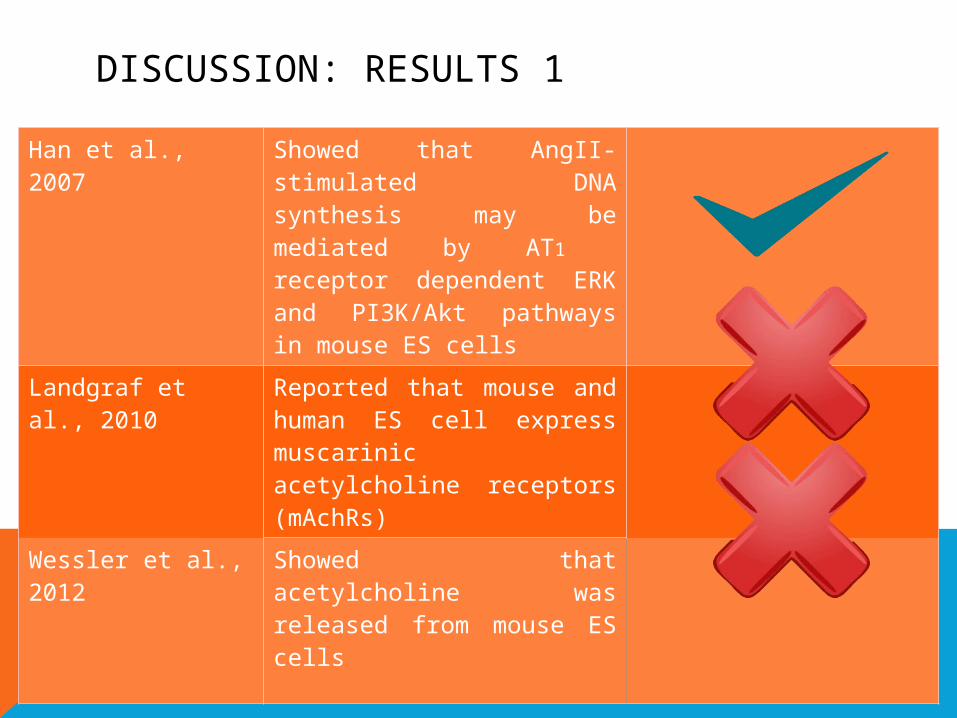
DISCUSSION: RESULTS 1
Han et al., 2007 Showed that AngII-stimulated DNA synthesis may be mediated by AT1
receptor dependent ERK and PI3K/Akt pathways in mouse ES cells
Landgraf et al., 2010
Reported that mouse and human ES cell express muscarinic acetylcholine receptors (mAchRs)
Wessler et al., 2012
Showed that acetylcholine was released from mouse ES cells

DISCUSSION: RESULTS 2
Han et al., 2006 Reported that PKC activation may be involved in mouse ES cell proliferation
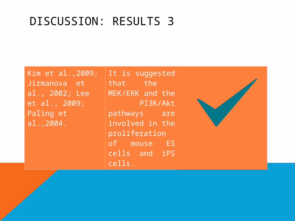
DISCUSSION: RESULTS 3
Kim et al.,2009; Jirmanova et al., 2002; Lee et al., 2009; Paling et al.,2004.
It is suggested that the MEK/ERK and the PI3K/Akt pathways are involved in the proliferation of mouse ES cells and iPS cells.

DISCUSSION: RESULTS 4Heo and Han, 2006
The activation of Gq-couple receptors promotes PKC activation
Zhao et al., 2005; Leon et al., 2011; Tang et al., 2012
Reported that PKC may be a key regulator of ERK activation induced by various stimuli

CONCLUSION
1. The results of this study validate that stimulation of α1-adrenoceptors or AT1 receptor may lead to a significant increase in human iPS cell proliferation via Gq-coupled receptor signaling pathways.
2. It is important to establish the methodology by which iPS cells can be produced in large quantities, because iPS cells may be used as a future source for regenerative therapy.
3. Understanding the physiological role of Gq-coupled receptors, such as α1-adrenoceptor or AT1 receptor in human iPS cells may be useful in the development of better culture conditions.
4. This improvment can be a step forward the new technics of treatment with regenerative therapy

MAPA CONCEPTUAL: ANGELICA MARIA SERNA

MAPA CONCEPTUAL: ANA ISABEL VALLEJO

GRACIAS