Bioengineering a Single-Protein...
10
Bioengineering a Single-Protein Junction Marta P. Ruiz, †,‡,§ Albert C. Aragone ̀ s, †,‡,§ Nuria Camarero, ‡,§ J. G. Vilhena, #,○ Maria Ortega, # Linda A. Zotti, # Rube ́ n Pe ́ rez,* ,#,Δ Juan Carlos Cuevas, #,Δ Pau Gorostiza,* ,‡,§,∥ and Ismael Díez-Pe ́ rez* ,†,‡,§ † Departament of Materials Science and Physical Chemistry & Institute of Theoretical and Computational Chemistry (IQTCUB), University of Barcelona, Martí i Franque ̀ s, 1, Barcelona 08028, Spain ‡ Institute for Bioengineering of Catalonia (IBEC), The Barcelona Institute of Science and Technology (BIST), Baldiri Reixac 15-21, Barcelona 08028, Spain § Centro Investigació n Biome ́ dica en Red (CIBER-BBN), Campus Río Ebro-Edificio I+D, Poeta Mariano Esquillor s/n, 50018 Zaragoza, Spain ∥ Catalan Institution for Research and Advanced Studies (ICREA) # Departamento de Física Teó rica de la Materia Condensada, Universidad Autó noma de Madrid, E-28049 Madrid, Spain ○ Department of Macromolecular Structures, Centro Nacional de Biotecnología, Consejo Superior de Investigaciones Científicas, 28049 Cantoblanco, Madrid, Spain Δ Condensed Matter Physics Center (IFIMAC), Universidad Autó noma de Madrid, E-28049 Madrid, Spain * S Supporting Information ABSTRACT: Bioelectronics moves toward designing nano- scale electronic platforms that allow in vivo determinations. Such devices require interfacing complex biomolecular moieties as the sensing units to an electronic platform for signal transduction. Inevitably, a systematic design goes through a bottom-up understanding of the structurally related electrical signatures of the biomolecular circuit, which will ultimately lead us to tailor its electrical properties. Toward this aim, we show here the first example of bioengineered charge transport in a single-protein electrical contact. The results reveal that a single point-site mutation at the docking hydrophobic patch of a Cu-azurin causes minor structural distortion of the protein blue Cu site and a dramatic change in the charge transport regime of the single-protein contact, which goes from the classical Cu-mediated two-step transport in this system to a direct coherent tunneling. Our extensive spectroscopic studies and molecular-dynamics simulations show that the proteins’ folding structures are preserved in the single-protein junction. The DFT-computed frontier orbital of the relevant protein segments suggests that the Cu center participation in each protein variant accounts for the different observed charge transport behavior. This work is a direct evidence of charge transport control in a protein backbone through external mutagenesis and a unique nanoscale platform to study structurally related biological electron transfer. ■ INTRODUCTION Biological electron transfer (ET) is the key step in many basic cellular processes such as respiration and photosynthesis. 1 Nature has developed highly specialized molecular building blocks capable of transporting charge with unprecedented efficiency, i.e., fast and at long distances. 2,3 Understanding the mechanisms behind biological ET is key to elucidate the changes in the charge transport regime caused by specific structural variations of the associated molecular machinery, which ultimately lead to, for instance, malfunctioning of the mitochondria. Such subtle ET changes are typically caused by specific mutations of one or more residues in the involved ET proteins, and they have been directly linked to well-described severe pathologies related to the overproduction of reactive oxygen species. 4−6 Fundamental knowledge is not the only gain from studying biological ET. Such knowledge can also be exploited to design bioelectronic devices. Such studies would ultimately unveil what are the key parameters to be controlled in the transduction of electrical signals from active biomolecules, and direct us, for instance, to the design of the next generation of highly specific optoelectronic sensors. 7,8 In order to comply with the increasingly demanding downsizing of the micro- electronics industry, the latest bioelectronic advances focus Received: June 13, 2017 Published: October 5, 2017 Article pubs.acs.org/JACS © 2017 American Chemical Society 15337 DOI: 10.1021/jacs.7b06130 J. Am. Chem. Soc. 2017, 139, 15337−15346 Cite This: J. Am. Chem. Soc. 2017, 139, 15337-15346
Transcript of Bioengineering a Single-Protein...

Bioengineering a Single-Protein JunctionMarta P. Ruiz,†,‡,§ Albert C. Aragones,†,‡,§ Nuria Camarero,‡,§ J. G. Vilhena,#,○ Maria Ortega,#
Linda A. Zotti,# Ruben Perez,*,#,Δ Juan Carlos Cuevas,#,Δ Pau Gorostiza,*,‡,§,∥
and Ismael Díez-Perez*,†,‡,§
†Departament of Materials Science and Physical Chemistry & Institute of Theoretical and Computational Chemistry (IQTCUB),University of Barcelona, Martí i Franques, 1, Barcelona 08028, Spain‡Institute for Bioengineering of Catalonia (IBEC), The Barcelona Institute of Science and Technology (BIST), Baldiri Reixac 15-21,Barcelona 08028, Spain§Centro Investigacion Biomedica en Red (CIBER-BBN), Campus Río Ebro-Edificio I+D, Poeta Mariano Esquillor s/n, 50018Zaragoza, Spain∥Catalan Institution for Research and Advanced Studies (ICREA)#Departamento de Física Teorica de la Materia Condensada, Universidad Autonoma de Madrid, E-28049 Madrid, Spain○Department of Macromolecular Structures, Centro Nacional de Biotecnología, Consejo Superior de Investigaciones Científicas,28049 Cantoblanco, Madrid, SpainΔCondensed Matter Physics Center (IFIMAC), Universidad Autonoma de Madrid, E-28049 Madrid, Spain
*S Supporting Information
ABSTRACT: Bioelectronics moves toward designing nano-scale electronic platforms that allow in vivo determinations.Such devices require interfacing complex biomolecularmoieties as the sensing units to an electronic platform forsignal transduction. Inevitably, a systematic design goesthrough a bottom-up understanding of the structurally relatedelectrical signatures of the biomolecular circuit, which willultimately lead us to tailor its electrical properties. Toward thisaim, we show here the first example of bioengineered chargetransport in a single-protein electrical contact. The resultsreveal that a single point-site mutation at the dockinghydrophobic patch of a Cu-azurin causes minor structuraldistortion of the protein blue Cu site and a dramatic change inthe charge transport regime of the single-protein contact, which goes from the classical Cu-mediated two-step transport in thissystem to a direct coherent tunneling. Our extensive spectroscopic studies and molecular-dynamics simulations show that theproteins’ folding structures are preserved in the single-protein junction. The DFT-computed frontier orbital of the relevantprotein segments suggests that the Cu center participation in each protein variant accounts for the different observed chargetransport behavior. This work is a direct evidence of charge transport control in a protein backbone through external mutagenesisand a unique nanoscale platform to study structurally related biological electron transfer.
■ INTRODUCTION
Biological electron transfer (ET) is the key step in many basiccellular processes such as respiration and photosynthesis.1
Nature has developed highly specialized molecular buildingblocks capable of transporting charge with unprecedentedefficiency, i.e., fast and at long distances.2,3 Understanding themechanisms behind biological ET is key to elucidate thechanges in the charge transport regime caused by specificstructural variations of the associated molecular machinery,which ultimately lead to, for instance, malfunctioning of themitochondria. Such subtle ET changes are typically caused byspecific mutations of one or more residues in the involved ETproteins, and they have been directly linked to well-described
severe pathologies related to the overproduction of reactiveoxygen species.4−6
Fundamental knowledge is not the only gain from studyingbiological ET. Such knowledge can also be exploited to designbioelectronic devices. Such studies would ultimately unveilwhat are the key parameters to be controlled in thetransduction of electrical signals from active biomolecules,and direct us, for instance, to the design of the next generationof highly specific optoelectronic sensors.7,8 In order to complywith the increasingly demanding downsizing of the micro-electronics industry, the latest bioelectronic advances focus
Received: June 13, 2017Published: October 5, 2017
Article
pubs.acs.org/JACS
© 2017 American Chemical Society 15337 DOI: 10.1021/jacs.7b06130J. Am. Chem. Soc. 2017, 139, 15337−15346
Cite This: J. Am. Chem. Soc. 2017, 139, 15337-15346

mainly on bottom-up perspectives,8 aiming for maximumsensitivities, high signal-to-noise ratios, and enhanced efficiencyin order to reduce energy consumption.Several ubiquitous redox proteins in biological ET have
become model biomolecular systems to study, owing to theirstructural robustness against mutagenesis.9 These systems havealso been suggested as versatile building blocks in molecularelectronic devices such as logic gates and multistate memorydevices.10−12 The structural similarity between the two redoxstates of the metal center in most model redox proteinsprovides a functional advantage, as the characteristic reorgan-ization free energy (λ) for the redox ET is kept within lowvalues, 0.6−0.8 eV,13,14 allowing high ET rates.9 The exactatomistic origin of such structural invariability that leads to lowλ values has been a longstanding debate.15,16 Redox proteinsalso present a large tunability of their redox potentials at minorstructural costs. Many studies have dug into the redox potentialcontrol of metalloproteins by means of point-site mutations inorder to correlate it with the molecular structure.17,18 Theredox midpoint potential exhibited by the band c-typecytochromes varies between −400 and 400 mV vs a normalhydrogen electrode (NHE) upon specific chemical modifica-tion of the Fe-heme surrounding.19 The reactivity andassociated function of ET proteins depend critically on itsredox potential, whose value is greatly affected by differences inthe first coordination sphere of the metal center.20,21 Forexample, it has been shown in blue-copper proteins that ashorter (stronger) axial Cu(II)−S(thioether) bond (as opposedto a longer (weaker) Cu(II)−S(thiolate) bond) results in agreen site (as opposed to the blue site). This difference resultsin a tetragonally distorted structure, thus substantiallymodifying the redox potential of the protein.20,22 Not onlymutations at the residues closer to the protein metal site areresponsible for such redox tunability. It has been demonstratedthat modifications of the secondary coordination sphere in Cuproteins can widely affect the protein redox potential throughcontrolling the hydrophobicity and hydrogen-bonding inter-actions.17,23 Such large potential tunability through pointmutations of the outer protein sphere highlights the profoundimportance of the redox potential control of biologicalmolecules in carrying out a wide variety of bioenergeticprocesses.24
Model redox proteins have been integrated in nano/microscale devices as the charge transport material. Cu-azurinconductance signatures have been recently observed inmicroscale solid-state devices,25−27 which demonstrates theircompatibility when hybridized to an electronic platform. Redoxprotein models such as Cu-azurin and cytochrome b562 havealso been extensively analyzed at the single-protein level byseveral groups.28−34 These pioneering studies established ametal-mediated electron transport through the metalloprotein,which has been usually pictured as to the electron jumpingfrom one electrode terminal to the Cu(II) metal center and,sequentially, from the Cu(I) center to the second electrode.28
The exact details of this mechanisms are, however, underdebate.34,35 With independence of the exact mechanistic details,these works demonstrated the feasibility of such hybridbiointerfaces to work as active components in nanoscalecircuits. We have recently exploited these capabilities to buildsingle-protein junctions displaying unique electrical signaturesby profiting from their exceptional redox properties, namely, alow operational voltage field-effect transistor36 and a redoxconductance-switching device.37
Here we present an example of bioengineering chargetransport in a single-protein junction. The copper-bindingprotein azurin from Pseudomonas aeruginosa has been exploitedto compare charge transport of single-protein electrical contactsmade of a wild-type (Wt) structure and a mutant (K41C),where, in the latter, the natural lysine (Lys) 41 residue has beenreplaced by a cysteine (Cys) (Figure 1a). This single pointmutation has a twofold effect; first, the new solvent-exposedthiol (−SH) group will serve as a new chemical connection toone of the external electrode terminals, and second, themodification is in the secondary coordination sphere of the Cu
Figure 1. Structural assessment of the proteins and scheme of thesingle-protein junction setup. (a) Structural models of the studiedwild-type azurin (left structure extracted from the Protein Data Bank(PDB)40) and its K41C variant (right structure). Natural cysteines(Cys3 and Cys26) are colored in orange. The wild-type amino acidLys41 (blue) was substituted by a Cys41 (green) through a point-sitemutation. (b) Superimposed structures of WT (cyan) and K41C(magenta) after a 500 ns molecular dynamics simulation (left panel).Representation of the same orientation of the surface hydrophobic/philic residues of the wild-type azurin (right panel, color legend: blue-cyan-orange-red ranges from very hydrophobic to very hydrophilic).(c) Stable adsorption configurations of the wild-type over a Ausubstrate after 150 ns molecular dynamics simulation. The hydro-phobic pocket is represented with a transparent green Connollysurface. (d) Absorption spectra of the wild-type (blue trace) and theK41C (green trace) in ammonium acetate buffer (pH 4.55). (e)Electrochemical cyclic voltammetry response of the two proteinvariants adsorbed on a functionalized Au(111) substrate in 50 mMammonium acetate buffer (pH 4.55). The electrochemical potentialscan rate was set to 50 mV/s (see further details in the SupportingInformation, section 4). (f) Schematic representation of the single-protein junction using an EC-STM setup.
Journal of the American Chemical Society Article
DOI: 10.1021/jacs.7b06130J. Am. Chem. Soc. 2017, 139, 15337−15346
15338

center, which is expected to influence the metal redoxbehavior17,23 and, hence, the transport regime through theprotein matrix. Individual proteins of both variants weretrapped between two metal electrodes in a physiologicalenvironment using an electrochemical scanning tunnelingmicroscope (EC-STM) configuration, and the charge transportwas characterized as a function of an applied electrochemicalgate voltage and temperature. We have successfully imple-mented a static blinking modality in the past38,39 that has beenexploited here to preserve the folding structure of the proteinduring the single-molecule transport measurements. All-atommolecular-dynamics (MD) simulations suggest that theelectrode−protein−electrode junction occurs via two well-localized sites on the protein, i.e., the hydrophobic patch andthe natural Cys residues. Despite that comparable orientationsof both Wt and K41C proteins bridges are expected, the resultsshow acute differences in the charge transport mechanism ofthe single-protein junction between the Wt and the mutantvariant, observing in the latter a complete shutdown of the two-step sequential tunneling character typically described in theWt.28 Ab initio calculations of the relevant ET pathwayfragment including the modified residue 41 show the poorparticipation of the Cu center in the transport-relevantmolecular frontier orbital of the K41C mutant. These resultsfully account for the observed conduction changes within theframework of the coherent tunneling mechanism for the single-protein junction of the bioengineered protein.
■ RESULTS AND DISCUSSIONExperimental Characterization and Molecular Dy-
namics Simulations for the Assessment of the Mutant’sStructure and Activity. Details on the protein expression andpurification including the Wt and the K41C mutant (Figure 1a)can be found in the Experimental and Computational Methodssection and in the Supporting Information, section 2. Theproteins’ structures in Figure 1a have been both represented onthe basis of the Wt crystalline structure. Despite the lack ofcrystalline structure of the K41C mutant, this section showsfirm evidence of both the preservation of its folding structureand electrical activity. We have first run exceedingly long (500ns) molecular dynamics (MD) simulations for the entireprotein structure immersed in an aqueous medium and seen nomajor structural changes after introducing the mutation at the41 position (see Figure 1b and the Supporting Information,section 7b), which assesses the robustness of the Cu-azurinstructure against modifications of its outer sphere. This result isconsistent with the findings obtained in other single pointmutants of azurin for which the crystal structure exists.17 Ontop of the MD results, Figure 1d shows the UV−visibleabsorption spectra for the Wt Cu-azurin and its K41C variant inthe working ammonium acetate buffer, both displaying strongligand-to-metal charge transfer (LMCT) absorption bandswithin the range of the typical Cu proteins blue site, 634 and615 nm, respectively, for Wt and K41C. The exact position ofthese bands has been extensively used to extract details on theCu ligand-field structure in blue Cu proteins upon specificmutations. The observed slight blue shift for the K41C suggestsa small perturbation of the Cu binding site upon mutation,17,21
which is ascribed to a decrease in the ligand-field energy as aresult of the axial coordination weakening of the methionine(Met) 121 residue binding the Cu metal center.20,22 Thisdifferent “energetics” of the chemical surrounding for the Cucenter has direct impact on the protein redox properties. A
decrease in the ligand-field energy typically worsens the stabilityof the Cu(II) oxidation form because the −S coordination tothe Met elongates, thus destabilizing the oxidized Cu form andraising the protein redox potential.17,22,41 Figure 1e shows thecyclic voltammograms of both Wt and K41C variants of theCu-azurin adsorbed on a Au(111) surface (see theExperimental and Computational Methods section and theSupporting Information, section 4, for more details). Theelectrode surface functionalization conducted to record thevoltammogram of the proteins (see Figure S4a) assures theconformation of both proteins with the hydrophobic patchfacing the Au electrode. In agreement with the spectroscopicsignal, the measured mid-redox potential displays an anodicshift of ∼50 mV for the K41C mutant with respect to the Wt,which points to a destabilization of the Cu(II) oxidation state.Moreover, the larger cathodic (Ured)-to-anodic (Uox) peaksseparation (Uox−Ured of 0.038 and 0.096 V for the Wt andK41C, respectively) evidences a reversibility loss in the K41Cmutant versus the Wt,42 owing to the stabilization of one of theCu oxidation states and implying a markedly slower electrontransfer rate in the electrochemical channel. Finally, the foldingstate of the K41C mutant structure has been tested out bymeasuring the extension of the fluorescence quenching of theinner tryptophan (Trp) 48 residue.43 The fluorescenceemission around 310 nm for the K41C variant evidences acorrect folding of its structure (see details in the SupportingInformation, section 3).
Electrochemical Gate-Dependent Single-ProteinTransport. We have formed and electrically characterizedsingle-protein junctions in a physiological environment(ammonium acetate buffer pH 4.5) for both target systems,i.e., the Wt and the K41C variant. To this aim, the targetproteins are absorbed onto a clean Au(111) electrode surfaceand an electrochemical scanning tunneling microscope (EC-STM) break-junction approach is used to bridge individualproteins between both the Au(111) substrate and the Au STMtip electrodes under electrochemical control (Figure 1f). The150 ns MD simulations under the same working conditions(see the Supporting Information, section 7b) show that Cu-azurin adsorption to gold occurs either through the naturallypresent Cys residues or through the hydrophobic patch (Figure1c), which reinforces the pre-established idea that the contactbetween the tip-protein-surface occurs via these two sites,36,44
i.e., STM tip-hydrophobic-patch/Cys-surface or vice versa. Inaddition, our simulations confirm that the process of adsorptionoccurs with a minor loss of the secondary structure of theprotein (Supporting Information, section 7b), in agreementwith the measured electrochemical activity for both systems(Figure 1e). Both simulations and experiments indicate that theprotein attachment to a Au substrate is stable and allowsimaging individual redox-active proteins on the electrodesurface for long time periods of up to several hours (see theSupporting Information, section 5). A similar anchoringgeometry will be expected for the K41C mutant given thatthis residue is located in the hydrophobic patch, one of the twopreferential adsorption sites observed for the Wt. In order tovalidate this assumption, we have performed two additionalMD simulations concerning the adsorption of the K41C andApo-azurin over gold along the orientation O1. The results arepresented in Figure S23 and show that, in the three cases (i.e.,Wt-Holo, K41C, and Wt-Apo), the final adsorption config-uration is very similar, thus strengthening our hypothesis thatthis point mutation will not produce significant modifications in
Journal of the American Chemical Society Article
DOI: 10.1021/jacs.7b06130J. Am. Chem. Soc. 2017, 139, 15337−15346
15339

the final adsorption configuration and, therefore, it shall notalter the contact surface-protein-tip obtained for the Wt-Holoazurin.The break-junction approach is operated in two different
modalities, namely, dynamic tapping and static blinking. In theformer, the STM tip is continuously approached and retractedto/from the Au electrode substrate where the target protein isanchored. When an individual protein binds the STM tipelectrode and closes the gap, a single-protein junction isformed, displaying a typical quantum conductance plateaufeature in the current versus distance retracting curve.45
Hundreds of such retracting curves (Figure 2b−d) are thenaccumulated into a single conductance histogram for eachparticular applied electrochemical potential (Figure 2a). Wehave previously demonstrated the feasibility of this method tostudy the charge transport in a single-azurin (Wt) junction as afunction of the applied electrochemical (EC gate) potential36
(EC gate = −Usample, where Usample is the substrate electro-chemical potential). Figure 2 shows the dynamic measurementsof the single-protein transport conductance values of the K41Cmutant as a function of the applied EC gate. In stark contrastwith the Wt behavior, the single-protein junction of the K41Cmutant shows no transistor behavior; i.e., the conductance isinvariable versus the EC gate within the relevant redoxpotential window. The transport behavior in the Wt ischaracterized by a two-step sequential tunneling that resultsin a maximum in the conductance versus EC gate curve,29,36
and suggests a direct tunneling transport in the K41C mutant.25
Single-biomolecular transport results by means of thedynamic STM break-junction approach has been previouslyshown in many instances.32,36,46−49 However, the force exertedover the folding structure of the biomolecule in every pullingcycle (Figure 2b−d) might disrupt its structure and lead to amisinterpretation of the single-biomolecule transport data.
Figure 2. Dynamic single-protein transport of the K41C junction. (a) Conductance histograms at three extreme EC gate potentials (−USample in ourEC-STM configuration) covering the redox potential window for the K41C protein. The histograms were built out of hundreds of retracting curves(conductance (G) vs retraction distance (nm)) from the break junction experiments displaying quantum conductance plateau features. (b−d) Threerepresentative retracting curves containing plateau features at the three applied EC gate potentials (dark gray 0.1 V, black −0.1 V, light gray −0.3 V).An offset was applied in the X-axis in all plots for better visualization. A constant 300 mV voltage bias (Vbias = Usample − Utip, where Usample and Utip arethe Au substrate and STM tip electrochemical potentials, respectively) was applied.
Figure 3. Gate-dependent single-protein transport. (a) Representative “blinks” (blue traces) identified in the transients of the current flowingbetween the two electrodes at a constant distance (2−3.5 nm) and Vbias (300 mV). Such blinks are observed when a protein spans the gap betweenthe EC-STM tip and the Au substrate electrodes. When the protein disconnects from one of the electrodes, the current drops down to the initial setpoint level. G = Istep/Vbias is used to obtain the conductance values. (b) Semilog 2D-blinking maps for both proteins (Wt top and K41C bottom) atdifferent EC gate potentials. Several tens (up to a hundred) of individual blinking traces like that shown in part a are accumulated to build each 2Dmap without any selection. The counts have been normalized for each map versus the maximum value so that each 2D map has its maximum countset to 1. The far right graphs summarize the average single-protein conductance (G) vs the EC gate (V) for both studied proteins. The averageconductance values were obtained from the Gaussian fits of the maxima in the vertical 1D histogram for each 2D map (see Figure S8 in theSupporting Information, section 5). The error bars in these plots are extracted from the full width at half-maximum (fwhm) of the Gaussian fits.
Journal of the American Chemical Society Article
DOI: 10.1021/jacs.7b06130J. Am. Chem. Soc. 2017, 139, 15337−15346
15340

Here we rule out this uncertainty by using a static version of theEC-STM break-junction approach, namely, the blinkingmode39,50 (see more details in the Supporting Information,section 5). Briefly, the STM tip electrode is initially approachedto the Au(111) substrate where the proteins have beenpreadsorbed to a distance of 1.5−2 nm, which is achieved byimposing a small (∼100 pA) tunneling current set point at anapplied bias voltage difference between the two electrodes (biasvoltage (Vbias) = Usample − Utip). Once the STM tip-to-substrategap is mechanically stable, the current feedback loop is turnedoff and the tunneling current is monitored. Spontaneoustrapping of proteins between both Au electrodes results in atelegraphic noise in the measured tunneling current flowingbetween the electrodes, which appears in the form of sudden“jumps” (blinks) (Figure 3a).39,51 In order to corroborate thatthe observed blinks correspond to protein trapping events,larger electrode−electrode separations (up to 3.5 nm) wereimposed by retracting the STM tip electrode further away fromthe surface. We observe that the large protein backbone (∼4nm) is still capable of closing large electrode−electrode gapseparations, resulting in a similar telegraphic signal (see theSupporting Information, section 5), yet at lower success rates.In support of this picture, we have run MD simulations of anSTM tip approaching a Wt azurin stably absorbed on theAu(111) substrate (see the Supporting Information, section 7)and observed that the protein folding structure remains stableeven after the STM tip establishes physical contact to it atdistances close to 2.5 nm (see the Supporting Informationmovie and Supporting Information, section 7b).Figure 3b panels show the accumulation of tens of such
blinking traces into single 2D maps at different applied EC gatepotentials for the Wt (top panel) and the K41C mutant(bottom panel). Such 2D blinking maps39,52 are built by settingall blinking features from individual current transients (Figure3a) to a common time origin and subtracted tunnelingbackground, and represent solely the net conductance flowingthrough the protein junction. The horizontal fringe observed inthe 2D blinking maps represents the conductance dispersion ofthe single-protein junction, whose average values arerepresented as a function of the applied EC gate in the farright graph of Figure 3b. Comparable conductance values areobtained from the static 2D maps (Figure 3b) and the dynamicbreak-junction histograms (Figure 2) for the K41C variant, 5 ×10⁻⁶G0 and 3 × 10⁻⁶G0 respectively (G0 = 77.5 μS), being on
average slightly larger in the former, which might evidence thedetrimental effect of the protein “stretching” in the lattermethod. The single-protein conductance in Figure 3b for theK41C follows the same nondependent EC gate behaviorobserved in the dynamic break-junction experiments (Figure2), with a slightly lower off-resonance current (at an EC gatevoltage of −0.3 V) when compared to the single-Wt junction(Figure 3b, upper panel). As expected in the latter case, amaximum in the conductance versus the EC gate potential nearthe mid-redox value is observed for the Wt (Figure 3b, far rightpanel)36 as opposed to the invariability for the single-K41Cmutant junction conductance behavior within the same relevantredox potential range. These results suggest a sharp change inthe transport behavior of the single-protein electrical contact,going from a classic sequential two-step tunneling for the Wt,observed in a number of other redox molecular junctions,36,53,54
to a fully coherent tunneling for the K41C mutant. A numericalversion of the two-step tunneling model is given in eq 1 andexpresses the enhanced current (Ienh in nanoamps) flowingthrough the molecular junction53
=
+
λ λ ξη γ
λ λ ξη γ
+ +
+ − − −
I V1820 [e
e ]
T V
T V V
enh bias((2898/ )( ) )
((2898/ )( ) ) 1bias bias
bias2
2
(1)
where ξ and γ are model parameters ranging between 0 and 1describing the fraction of the overpotential η in volts (η =Usample − Uredox, where Uredox corresponds to the protein redoxmidpoint extracted from Figure 1e) and the Vbias (in volts) feltby the reactive redox center in the molecular junctions,respectively. T (in °C) is the temperature, and λ (in eV) is thereorganization energy (the energy difference between theoxidized and reduced states of the protein). All other physicalconstants have been numerically processed for simplicity takingreasonable assumptions of our experimental setup such as fullyadiabatic limit (strong molecule/electrode coupling), fixedjunction geometry for every measured blink, Au as electrodematerial, and aqueous solution as the environment. A fit of theWt experimental results (blue trace in Figure 3b, far left graph)yields reasonable values for λ, ξ, and γ, 0.27 eV, 0.95, and 0.79,respectively,28,36 evidencing low reorganization energies, largeEC gating efficiencies, and a slight asymmetric Vbias distributiondue to the usually asymmetric location of the Cu center in themolecular junction. It is important to note that the two-stepsequential tunneling model fits the current versus the EC gate
Figure 4. Temperature-dependent single-protein transport. Semilog 2D-blinking maps for both proteins (Wt top and K41C bottom) at differenttemperatures (from 5 to 35 °C), constant distance (2 to 3.5 nm) and Vbias (300 mV). The applied EC gate value was set to 0 and −100 mV for Wtand K41C mutant, respectively. The counts have been normalized for each map versus the maximum value, so each 2D map has its maximum countset to 1. The far right graph summarizes the single-protein conductance (G) vs temperature (°C) for both studied proteins. The averageconductance values were obtained from the maxima Gaussian fits in the vertical 1D histogram for each 2D map (see Figure S9 in the SupportingInformation, section 5). The error bars in these plots are extracted from the full fwhm of the Gaussian fits.
Journal of the American Chemical Society Article
DOI: 10.1021/jacs.7b06130J. Am. Chem. Soc. 2017, 139, 15337−15346
15341
voltage behavior, but it is not used to evaluate the absolutevalues of the current flowing through the single-proteinjunction. Accurate calculated values of the current flowingthrough the protein junction would require the introduction ofcoupling terms to both junction electrodes, which are difficultto model given the lack of information on the protein/electrodecontact geometry.Another relevant piece of information that can be extracted
from the 2D blinking map is related to the characterization ofthe single-protein junction lifetime (X-axis in Figure 3b). Ingeneral, larger lifetime values for the K41C mutant are observed(see full lifetime statistics in the Supporting Information,section 5), which points toward a more stable protein/STM tipthiol bond thanks to the newly introduced Cys41 residue. Theslight lifetime differences among the applied EC gate voltagesmight be due to different electrostatic stabilization of the thiolbond at the Au/protein interface and need to be furtherstudied. In support of the general single-protein junctionstabilization for the K41C, we have functionalized Aunanoparticles (NPs) with both Wt and K41C variants anddetected a significantly larger percentage of multimeric Au NPstructures in the latter (see the Supporting Information, section6), evidencing the enhanced K41C bridging capability. Such aconfiguration leads to a similar metal/protein/metal orientationin the K41C junction as compared to the Wt protein (see theSupporting Information, section 7).Temperature-Dependent Single-Protein Transport. In
order to search for possible sources of fully incoherenttransport in any of the studied single-protein junctions (i.e.,hopping transport regime55,56), we have conducted single-protein transport measurements as a function of temperature.Figure 4 shows the temperature-dependent single-proteinconductance results for both proteins at EC gate potentials of0 and −100 mV for the Wt and the K41C mutant, respectively,near their respective redox midpoint potentials. The temper-ature range covers the room temperature conditions andapproaches physiologically relevant values (∼37 °C).57 Themaximum temperature values were kept below 40 °C toprevent any denaturalization of the protein structures (84.4 °Cfor the Wt58 and >70 °C for the K41C (see the SupportingInformation, section 3b). Temperature-dependent single-protein conductance experiments are subjected to severemechanical instabilities originated by thermal drift, whichprevents reliable evaluation of the single-protein junctionlifetime, usually resulting in similar values for both protein
variants. The invariance of the single-protein junctionconductance versus temperature in both proteins rules outany fully incoherence source of transport within the relevanttemperature range and underscores the important fact that theprotein is able to maintain a high level of coherence even forvery different transport regimes, namely, two-step sequentialand direct tunneling. Similar behavior has also been observed inmicroscale solid-state devices sandwiching a Wt azurinprotein.25 The temperature-dependent conductance withinthe two-step sequential scenario (see eq 1) shows a veryshallow trend as we approach the η = 0 point (Usample=Uredox),at which the measurements were performed (see theSupporting Information, section 7c, where the temperaturedependence of eq 1 is represented at different η values).Building upon this mechanistic analysis, the conductanceinvariance of the K41C single-protein junction against bothEC gate potential and temperature suggests a complete loss ofthe Wt sequential tunneling character, turning into a fullycoherent tunneling regime.59
Computational Studies of the Electronic Properties.To understand the observed transition in the charge transportregimes between the two studied single-protein junctions, wehave performed ab initio computational calculations in thestructurally relevant proteins segment for charge transport. Wehave used DFT computational methods (see the SupportingInformation, section 7, and the Experimental and Computa-tional Methods section for more details) using the long-rangecorrected B3LYP functional (CAM-B3LYP) to visualize thedistribution of the frontier orbitals nearby the redox activeCu(II) metal center for the Wt azurin and the K41C variants.Due to the intrinsic computational limitations, we haveconsidered only the sequence fraction involving the firstcoordination sphere of the Cu(II) center and the segment ofthe second coordination including the mutated residue at the41 position (Figure 5),60,61 and for that reason, thesecomputational results are qualitative in nature. Such a proteinfragment constitutes the ET pathway connecting the solvent-exposed hydrophobic patch of the protein, that will be moreaccessible to the STM tip electrode,14,29,62 and the active metalcenter. The protein folding structure of the fragment used forthe electronic structure calculations (Figure 5) has been takenfrom the crystalline Wt structure40 for both protein variants(Figure 1a). This is a good approximation given the minordisruption of the Cu coordination field after the outer-spheremutation is performed, as deduced from our spectroscopic
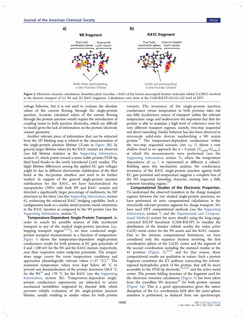
Figure 5. Electronic structure calculations. Isosurface plots (isovalue = 0.02) of the lowest unoccupied frontier molecular orbital (LUMO) involvedin the electron transport of (a) Wt and (b) K41C fragments. Calculations were done at the CAM-B3LYP/6311G+(d) level of DFT.
Journal of the American Chemical Society Article
DOI: 10.1021/jacs.7b06130J. Am. Chem. Soc. 2017, 139, 15337−15346
15342

characterization and MD simulations (Figure 1b−d). Finestructural analyses of the mutation effect will have to beconsidered for cases involving major structural changes in themutant protein folding.63
In our single-protein junction experiments, the electrons areinjected from the STM tip electrode into the Cu(II)-protein.Figure 5a shows the representation of the DFT-calculatedLUMO frontier orbital for the ET-relevant fragment of the Wtazurin, showing significant LUMO contribution close to themetal center and its first coordination sphere in the oxidizedstate (Cu(II)) of the protein. The orbital energy valuesresulting from the DFT calculation (see the SupportingInformation, section 7a) indicate that the energetic positionof the LUMO fragment for this system (around −6 eV) iscloser to the gold Fermi level (∼−5 eV) than the highestoccupied molecular orbital (HOMO) fragment (positioned atlower energies than −7 eV). Assuming their uncertainty due tothe self-interaction errors in DFT,64,65 this suggests the LUMOfragment as the domination transport orbital. Such a statementis based on the assumption that the position of the energylevels in the considered protein fragment is not affected eitherby the presence of the rest of the protein or by the couplingwith the gold electrodes. Although a transport calculation onthe whole coupled system should be performed, this iscomputationally highly demanding and beyond the reach ofstate-of-the-art computational facilities. We believe that ourassumption is based on a plausible scenario: in our MDsimulations, the Cu ion is located at a distance of approximately14 Å from the gold surface. At such a distance, molecularorbitals are expected to be decoupled from the electrodes andto be unaffected by both Coulomb screening effects66 and bythe electrostatic balance established at the interface.67
It is important to distinguish the orbital picture representedin Figure 5 from previously calculated orbitals of the redox Cusite of a metalloprotein. The latter corresponds to the well-established biological electron transfer field that describes theelectron exchange with the metal center in a metalloprotein,which directly relates to the electrochemical response (Figure1e). Such an orbital has been previously ascribed to a Cu(II)-SOMO.60 Figure 5 instead is an attempt of describing electrontransport across the entire protein when it bridges two metalleads in a nanoscale molecular device. Such a picture isintended to be captured by visualizing the low lying (closer tothe electrodes’ Fermi energy) molecular orbitals of the entiretransport-relevant fragment of the protein in Figure 5.As opposed to what is observed in the single-protein Wt
junction, Figure 5b shows that the LUMO fragment in theK41C is mainly localized at the mutated Cys41 residue, withessentially no contribution near the Cu coordination sphereregion. The LUMO distribution around the metal siteevidences the implication of the protein redox state in theobserved charge transport through the Wt protein, inagreement with a two-step sequential tunneling mechanismmediated by the metal redox center. We then hypothesize thatthe disappearance of the two-step sequential character in theK41C variant is caused by the lack of LUMO fragmentdistribution at the first Cu coordination sphere. The closestfragment orbital around the Cu center in the K41C variantappears now at the LUMO+2, further energetically separated,∼1.5 eV (Supporting Information, section 7a), from theLUMO-fragment transport channel. This conclusion issupported by the similar transport behavior observed for asingle-protein junction with an Apo (lack of Cu) variant (see
the Supporting Information, section 5b). Within an ETpathway picture,68 while both conduction channels, namely,sequential two-step (involving the redox metal center) andtunneling (nonredox), are present in the Wt single-proteinelectrical contact, the former is shut down in the K41C mutantcase.59,69 This conclusion is supported by the observed slowerkinetics and anodically shifted electrochemistry for the K41Cmutant (Figure 1e). The observed slower Cu(II)/Au electrontransfer kinetics in the mutant foresees a larger energy penaltyfor the redox conduction channel (Cu redox signal) whenelectrons are transported through the entire protein junction,which translates into the non-Cu-mediated coherent tunnelingbeing the dominant pathway in the K41C case. In an effort torationalize the observed orbital transport picture with the redoxproperties of the metalloprotein, we suggest that theparticipation of the first Cu coordination sphere in the orbitaltransport channel, which can be tailored through proteinmutagenesis, is a requirement to observe electrochemicalconductance modulation in a single-protein junction.
■ CONCLUSIONS
We present a mechanistic analysis of bioengineered chargetransport in a single-protein junction. We have exploited ourstatic STM-based blinking approach39 to transiently trapindividual Cu-azurin metalloproteins between two metal leadsand characterize their electrical properties as a function of thetwo key experimental parameters for charge transport for suchredox molecular systems, namely, the electrochemical gatevoltage and the temperature. The results prove that theconduction channels in the single-protein electrical contact canbe finely tuned by performing point-site mutations in the outerprotein structure involving mild structural changes in theprotein backbone. In short, we have modified a wild-typePseudomonas aeruginosa blue Cu-azurin at its secondary Cucoordination sphere (Lys41 residue) by introducing a Cys41residue that caused minor structural modifications, as seen byboth spectroscopy and MD simulations. Such minor structuralchanges have been ascribed to variations in the hydrophobicityand/or H-bonding network in the protein peptidic structure.17
The slight reduction of charge density at the Cu centeroriginated by the newly introduced Cys41 residue dramaticallyalters the charge transport behavior of the single-proteinjunction. Our MD simulations of the Au substrate/protein/STM tip junction suggest that the protein structure is preservedduring our measurements, ruling out the possibility that thesechanges arise from any major structural rearrangement. Theoriginal two-step sequential charge transport in the Wt is shutdown to give rise to a single-protein electrical contactdisplaying an almost fully coherent transport mechanism inthe mutant, i.e., invariant to electrochemical gate voltage andtemperature. Our DFT orbital calculations for the relevantsegment to the protein transport (extracted from thecrystallographic structure) suggest a simple yet intuitiveexplanation for the observed sharp transport transition, whichis based on the lack of contribution of the first Cu coordinationsphere in the transport-dominant frontier orbital for themodified protein. This interpretation should however be testedby a complete transport calculation on the metal−protein−metal system. Furthermore, the role of all the possibleconformational changes due to thermal fluctuations, aspredicted by the molecular dynamics simulations, in thetransport mechanism should be investigated.
Journal of the American Chemical Society Article
DOI: 10.1021/jacs.7b06130J. Am. Chem. Soc. 2017, 139, 15337−15346
15343

These results demonstrate the feasibility of tailoring thecharge transport in a nanoscale biomolecular electrical contactand bring new horizons toward real bottom-up approaches toengineer the next generation of biosensors, biotransistors, orany platform requiring the optimization of the biomolecule/electrode electrical communication. The outcomes of this workgo beyond a detailed interfacial study of the protein/electrodeelectrical contact. The observed abrupt transition in the ETbehavior upon a single external mutation in the protein−protein “docking” patch of a functional redox protein pointstoward a plausible biological mechanism to control coherencein biological ET through minor structural/chemical changes.Similar mechanisms have been recently suggested in relevantbimolecular structures such as DNA.70
■ EXPERIMENTAL AND COMPUTATIONALMETHODS
A complete description of the mutagenesis procedures and character-ization methodologies employed in this work has been included in theSupporting Information.Spectroscopic Methods. The UV−vis absorption spectra were
obtained at ambient temperature using an Infinite M200 PROMultimode Microplate Reader from Tecan. Protein samples wereapproximately 7 μM protein in 50 mM ammonium acetate pH 4.5buffer. Fluorescence spectra were recorded using a Horibaspectrofluorometer. The Trp48 fluorescence band, which has intrinsicfluorescence, was monitored after exciting at 290 nm. Both Wt andK41C variants were measured as well as their denaturized homologues.Electrochemical Measurements. Cyclic voltammetry (CV) was
undertaken using an Autolab PGSTAT-12 Galvanostat-Potentiostat(Metrohm Autolab). The three-electrode cell is composed by aplatinum wire as a counter electrode, a miniaturized ultralow leakagemembrane Ag/AgCl (SSC) as a reference electrode, and aCH3(CH2)5S-functionalized Au(111) as a working electrode. All ofthe electrolyte solutions were deoxygenated with purified argon. Tocapture the protein CVs, the gold is immersed overnight in a 50 mMhexanethiol solution. Then, it is rinsed with abundant ethanol andwater to remove the noncovalently bound hexanethiols. Thefunctionalized Au is covered with a droplet of 50 mM ammoniumacetate pH 4.5 containing the protein for at least 2 h. The azurinattaches to the hexanethiols by its hydrophobic patch near the copperion (see the Supporting Information, section 4).Electrochemical Scanning Tunnelling Microscopy (EC-STM).
All experiments were performed with a PicoSPM microscope head anda PicoStat bipotentiostat (Agilent, USA) controlled by Dulcineaelectronics (Nanotec Electronica, Spain) using the WSxM 4.13software. Two different cells were used: a liquid cell with a standardsample plate for the room temperature measurements and a Peltier(Cold MAC) sample plate for temperature-controlled experiments. Afour-electrode cell is required for the bipotentiostatic control: a Pt:Ir(80:20) wire as a counter electrode, a miniaturized ultralow leakagemembrane Ag/AgCl (SSC) as a reference electrode, and the twoworking electrodes, a Au(111) substrate and a STM tip, whosepotentials US and UP, respectively, are expressed against the same Ag/AgCl reference electrode. A 50 mM ammonium acetate pH 4.55 bufferwas used for the measurements, previously filtered and deoxygenatedwith an Ar stream. The EC-STM probes were mechanically cut from a0.25 mm diameter Au wire (99.99%), and they are made of 1 cm inlength, annealed with a butane flame, and coated with Apiezon wax tominimize leakage current when immersed in the working aqueousbuffer. The leakage current of our tips was typically of <10 pA.71
Molecular Dynamics Simulations. All of the simulations wereperformed using the AMBER14 software suite72 with NVIDIA GPUacceleration.73−75 The parmbsc0 modification76 of the Cornell ff99force field77 was used to describe all standard amino acids present inthe azurin. The interatomic potentials of the copper atom and itscorresponding five ligands were described using a force field derivedfrom quantum mechanical simulations.78 This force field has been
widely used to model the blue-copper azurin protein.18,79−81 Inparticular, recent experiments80 have shown how early stages ofmechanical unfolding of this protein are well described by this forcefield. In all of our simulations, the system is fully embedded in a watermedium. The water is described using the explicit TIP3P model,82
while Joung/Cheatham parameters were used to describe the sodiumcounterions.83,84 For the gold atoms, we have resorted to theCHARMM-METAL force field,85,86 since it is thermodynamicallyconsistent with the AMBERFF used to describe the protein and it hasbeen successfully employed to study inorganic−bioorganic inter-faces.86 We have used periodic boundary conditions and the particlemesh Ewald method (with standard defaults and a real-space cutoff of10 Å) to account for long-range electrostatic interactions. van derWaals contacts were truncated at a real space cutoff of 10 Å for all ofthe simulations also. The SHAKE algorithm was used to constrainbonds containing hydrogen, thus allowing us to use an integration stepof 2 fs. Coordinates were saved every 1000 steps.
Electronic Structure Calculations. The geometry of the Wtazurin structure was obtained from the PDB (1AZU code). The K41Cgeometry was based on 1AZU replacing the Lys41 for a Cys41. Thisstructure is supported by spectroscopic measurements and moleculardynamic simulation (see main discussion). The calculated frontierorbitals for both protein fragments were obtained with DensityFunctional methods (DFT) using the software package Gaussian 0987
and employing a long-range-corrected variant CAM-B3LYP method88
and 6-311G+(d) as basis set. The ab initio calculations were conductedin the protein fragments relevant to the ET involving the residues 41−45, 46, 112, 117, 121, and 129.
■ ASSOCIATED CONTENT*S Supporting InformationThe Supporting Information is available free of charge on theACS Publications website at DOI: 10.1021/jacs.7b06130.
Chemicals and materials, K41C bioengineering methods,spectroscopic characterization, electrochemical tests,ECSTM technical details, TEM characterization ofprotein-conjugated NP, and computational details(PDF)Molecular dynamics movie (MPG)
■ AUTHOR INFORMATIONCorresponding Authors*[email protected]*[email protected]*[email protected] A. Zotti: 0000-0002-5292-6759Ruben Perez: 0000-0001-5896-541XIsmael Díez-Perez: 0000-0003-0513-8888NotesThe authors declare no competing financial interest.
■ ACKNOWLEDGMENTSThe plasmid pet3a carrying the azurin structural gene (TableS1) from E. coli was kindly provided from Dr. Jeff Warren(California Institute of Technology, Pasadena). We thank Prof.Eliseo Ruiz of the University of Barcelona for his support in thefluorescence characterization. We thank the financial supportfrom the Spanish MINECO (projects MDM-2014-0377,CTQ2015-71406-ERC, CTQ2015-64579-C3-3-P, CTQ2016-80066R, RYC-2011-07951, CSD2010-00024, FIS2014-53488-P, MAT2014-54484-P, and MAT2014-58982-JIN), the COSTAction MP1303, and the European Research Council (ERC)under the European Union’s Horizon 2020 research and
Journal of the American Chemical Society Article
DOI: 10.1021/jacs.7b06130J. Am. Chem. Soc. 2017, 139, 15337−15346
15344

innovation grant Agreement No. 681299. P.G. thanks EUCommission (ERANET SynBio MODULIGHTOR Project),Catalan Government (CERCA Programme and 2014SGR-1251grant) and the Ramon Areces foundation. J.C.C. thanks theDFG and SFB 767 for sponsoring his stay at the University ofKonstanz as Mercator Fellow. The authors thankfully acknowl-edge the computer resources, technical expertise, and assistanceprovided by the Red Espanola de Supercomputacion (RES) atthe Minotauro and Marenostrum supercomputers (BSC,Barcelona) and Altamira (IFCA, Santander) Supercomputers.We also acknowledge the support provided by the computingfacilities of the Extremadura Research Centre for AdvancedTechnologies (CETA-CIEMAT), funded by the EuropeanRegional Development Fund (ERDF). CETA-CIEMATbelongs to CIEMAT and the Government of Spain.
■ REFERENCES(1) Broda, E. The Evolution of the Bioenergetic Processes; Elsevier:Amsterdam, The Netherlands, 1975.(2) Progress in Inorganic Chemistry; Lippard, S. J., Ed.; John Wiley &Sons, Inc.: Hoboken, NJ, 1984; Vol. 32.(3) Gray, H. B.; Winkler, J. R. Proc. Natl. Acad. Sci. U. S. A. 2005, 102(10), 3534.(4) Sue, C. M.; Schon, E. A. Brain Pathol. 2000, 10 (3), 442.(5) Stadtman, E. R.; Berlett, B. S. Drug Metab. Rev. 1998, 30 (2), 225.(6) Gray, H. B.; Winkler, J. R. Biochim. Biophys. Acta, Bioenerg. 2010,1797 (9), 1563.(7) Willner, I.; Katz, E. Bioelectronics: from theory to applications;Wiley-VCH: Weinheim, Germany, 2005.(8) Zhang, A.; Lieber, C. M. Chem. Rev. 2016, 116 (1), 215.(9) Gray, H. B.; Winkler, J. R. Annu. Rev. Biochem. 1996, 65, 537.(10) D’Amico, S.; Maruccio, G.; Visconti, P.; D’Amone, E.; Bramanti,A.; Cingolani, R.; Rinaldi, R. IEE Proceedings - Nanobiotechnology. IETDigital Library October 1, 2004; pp 173−175.(11) Lee, T.; Kim, S.-U.; Min, J.; Choi, J.-W. Adv. Mater. 2010, 22(4), 510.(12) Amdursky, N.; Marchak, D.; Sepunaru, L.; Pecht, I.; Sheves, M.;Cahen, D. Adv. Mater. 2014, 26 (42), 7142.(13) Gray, H. B.; Malmstrom, B. G.; Williams, R. J. P. JBIC, J. Biol.Inorg. Chem. 2000, 5 (5), 551.(14) Cascella, M.; Magistrato, A.; Tavernelli, I.; Carloni, P.;Rothlisberger, U. Proc. Natl. Acad. Sci. U. S. A. 2006, 103 (52), 19641.(15) Olsson, M. H. M.; Ryde, U.; Roos, B. O. Protein Sci. 1998, 7,2659.(16) Farver, O.; Marshall, N. M.; Wherland, S.; Lu, Y.; Pecht, I. Proc.Natl. Acad. Sci. U. S. A. 2013, 110 (26), 10536.(17) Marshall, N. M.; Garner, D. K.; Wilson, T. D.; Gao, Y.-G.;Robinson, H.; Nilges, M. J.; Lu, Y. Nature 2009, 462 (7269), 113.(18) Zanetti-Polzi, L.; Bortolotti, C. A.; Daidone, I.; Aschi, M.;Amadei, A.; Corni, S. Org. Biomol. Chem. 2015, 13 (45), 11003.(19) Springs, S. L.; Bass, S. E.; McLendon, G. L. Biochemistry 2000,39 (20), 6075.(20) Clark, K. M.; Yu, Y.; Marshall, N. M.; Sieracki, N. A.; Nilges, M.J.; Blackburn, N. J.; van der Donk, W. A.; Lu, Y. J. Am. Chem. Soc.2010, 132 (29), 10093.(21) Lancaster, K. M.; DeBeer George, S.; Yokoyama, K.; Richards, J.H.; Gray, H. B. Nat. Chem. 2009, 1 (9), 711.(22) Warren, J. J.; Lancaster, K. M.; Richards, J. H.; Gray, H. B. J.Inorg. Biochem. 2012, 115, 119.(23) Lancaster, K. M.; Sproules, S.; Palmer, J. H.; Richards, J. H.;Gray, H. B. J. Am. Chem. Soc. 2010, 132 (41), 14590.(24) Mauk, A. G.; Moore, G. R. JBIC, J. Biol. Inorg. Chem. 1997, 2(1), 119.(25) Sepunaru, L.; Pecht, I.; Sheves, M.; Cahen, D. J. Am. Chem. Soc.2011, 133 (8), 2421.(26) Li, W.; Sepunaru, L.; Amdursky, N.; Cohen, S. R.; Pecht, I.;Sheves, M.; Cahen, D. ACS Nano 2012, 6 (12), 10816.
(27) Yu, X.; Lovrincic, R.; Sepunaru, L.; Li, W.; Vilan, A.; Pecht, I.;Sheves, M.; Cahen, D. ACS Nano 2015, 9 (10), 9955.(28) Chi, Q.; Farver, O.; Ulstrup, J. Proc. Natl. Acad. Sci. U. S. A.2005, 102 (45), 16203.(29) Chi, Q.; Zhang, J.; Andersen, J. E. T.; Ulstrup, J. J. Phys. Chem. B2001, 105 (20), 4669.(30) Baldacchini, C.; Bizzarri, A. R.; Cannistraro, S. Eur. Polym. J.2016, 83, 407.(31) Baldacchini, C.; Kumar, V.; Bizzarri, A. R.; Cannistraro, S. Appl.Phys. Lett. 2015, 106 (18), 183701.(32) Della Pia, E. A.; Chi, Q.; Jones, D. D.; Macdonald, J. E.; Ulstrup,J.; Elliott, M. Nano Lett. 2011, 11 (1), 176.(33) Alessandrini, A.; Salerno, M.; Frabboni, S.; Facci, P. Appl. Phys.Lett. 2005, 86 (13), 133902.(34) Alessandrini, A.; Corni, S.; Facci, P.; Facci, P.; Sola, M.; Facci,P.; Facci, P.; Harris, G.; Lindsay, S. M. Phys. Chem. Chem. Phys. 2006, 8(38), 4383.(35) Baldea, I. J. Phys. Chem. C 2013, 117 (48), 25798.(36) Artes, J. M.; Díez-Perez, I.; Gorostiza, P. Nano Lett. 2012, 12(6), 2679.(37) Artes, J. M.; Lopez-Martínez, M.; Díez-Perez, I.; Sanz, F.;Gorostiza, P. Small 2014, 10 (13), 2537.(38) Díez-Perez, I.; Hihath, J.; Lee, Y.; Yu, L.; Adamska, L.;Kozhushner, M. a; Oleynik, I. I.; Tao, N. Nat. Chem. 2009, 1 (8), 635.(39) Aragones, A. C.; Haworth, N. L.; Darwish, N.; Ciampi, S.;Bloomfield, N. J.; Wallace, G. G.; Diez-Perez, I.; Coote, M. L. Nature2016, 531 (7592), 88.(40) Adman, E. T.; Jensen, L. H. Isr. J. Chem. 1981, 21, 8.(41) Garner, D. K.; Vaughan, M. D.; Hwang, H. J.; Savelieff, M. G.;Berry, S. M.; Honek, J. F.; Lu, Y. J. Am. Chem. Soc. 2006, 128 (49),15608.(42) Bard, A. J.; Faulkner, L. R. Electrochemical methods: fundamentalsand applications; Wiley: Weinheim, Germany, 2001.(43) Huang, Q.; Quinones, E. Arch. Biochem. Biophys. 2008, 477 (1),175.(44) Artes, J. M.; Lopez-Martínez, M.; Giraudet, A.; Díez-Perez, I.;Sanz, F.; Gorostiza, P. J. Am. Chem. Soc. 2012, 134 (50), 20218.(45) Xu, B.; Tao, N. J. Science 2003, 301 (5637), 1221.(46) Hihath, J.; Xu, B.; Zhang, P.; Tao, N. Proc. Natl. Acad. Sci. U. S.A. 2005, 102 (47), 16979.(47) Artes, J. M.; Li, Y.; Qi, J.; Anantram, M. P.; Hihath, J. Nat.Commun. 2015, 6, 8870.(48) Sek, S.; Misicka, A.; Swiatek, K.; Maicka, E. J. Phys. Chem. B2006, 110 (39), 19671.(49) Scullion, L.; Doneux, T.; Bouffier, L.; Fernig, D. G.; Higgins, S.J.; Bethell, D.; Nichols, R. J. J. Phys. Chem. C 2011, 115 (16), 8361.(50) Haiss, W.; Wang, C.; Grace, I.; Batsanov, A. S.; Schiffrin, D. J.;Higgins, S. J.; Bryce, M. R.; Lambert, C. J.; Nichols, R. J. Nat. Mater.2006, 5 (12), 995.(51) Chang, S.; He, J.; Kibel, A.; Lee, M.; Sankey, O.; Zhang, P.;Lindsay, S. Nat. Nanotechnol. 2009, 4 (5), 297.(52) Aragones, A. C.; Darwish, N.; Saletra, W. J.; Perez-García, L.;Sanz, F.; Puigmartí-Luis, J.; Amabilino, D. B.; Díez-Perez, I. Nano Lett.2014, 14 (8), 4751.(53) Pobelov, I. V.; Li, Z.; Wandlowski, T. J. Am. Chem. Soc. 2008,130 (47), 16045.(54) Díez-Perez, I.; Li, Z.; Guo, S.; Madden, C.; Huang, H.; Che, Y.;Yang, X.; Zang, L.; Tao, N. ACS Nano 2012, 6 (8), 7044.(55) Engelkes, V. B.; Beebe, J. M.; Frisbie, C. D. J. Am. Chem. Soc.2004, 126 (43), 14287.(56) Hines, T.; Diez-Perez, I.; Hihath, J.; Liu, H.; Wang, Z. S.; Zhao,J.; Zhou, G.; Mullen, K.; Tao, N. J. Am. Chem. Soc. 2010, 132 (33),11658.(57) Tsuji, A.; Kaneko, Y.; Takahashi, K.; Ogawa, M.; Goto, S.Microbiol. Immunol. 1982, 26 (1), 15.(58) Guzzi, R.; La Rosa, C.; Grasso, D.; Milardi, D.; Sportelli, L.Biophys. Chem. 1996, 60 (1), 29.(59) Migliore, A.; Nitzan, A. J. Am. Chem. Soc. 2013, 135 (25), 9420.
Journal of the American Chemical Society Article
DOI: 10.1021/jacs.7b06130J. Am. Chem. Soc. 2017, 139, 15337−15346
15345
(60) Solomon, E. I.; Szilagyi, R. K.; DeBeer George, S.; Basumallick,L. Chem. Rev. 2004, 104 (2), 419.(61) Blanco-Rodríguez, A. M.; Di Bilio, A. J.; Shih, C.; Museth, A. K.;Clark, I. P.; Towrie, M.; Cannizzo, A.; Sudhamsu, J.; Crane, B. R.;Sykora, J.; Winkler, J. R.; Gray, H. B.; Zalis, S.; Vlcek, A. Chem. - Eur. J.2011, 17 (19), 5350.(62) Venkat, A. S.; Corni, S.; Di Felice, R. Small 2007, 3 (8), 1431.(63) Ghosh, S.; Xie, X.; Dey, A.; Sun, Y.; Scholes, C. P.; Solomon, E.I. Proc. Natl. Acad. Sci. U. S. A. 2009, 106 (13), 4969.(64) Rincon-García, L.; Ismael, A. K.; Evangeli, C.; Grace, I.; Rubio-Bollinger, G.; Porfyrakis, K.; Agraït, N.; Lambert, C. J. Nat. Mater.2015, 15 (3), 289.(65) Kim, M.-C.; Sim, E.; Burke, K. Phys. Rev. Lett. 2013, 111 (7),73003.(66) Stadler, R.; Geskin, V.; Cornil, J. Phys. Rev. B: Condens. MatterMater. Phys. 2009, 79 (11), 113408.(67) Brooke, C.; Vezzoli, A.; Higgins, S. J.; Zotti, L. A.; Palacios, J. J.;Nichols, R. J. Phys. Rev. B: Condens. Matter Mater. Phys. 2015, 91,195438.(68) Beratan, D.; Onuchic, J.; Winkler; Gray, H. Science 1992, 258(5089), 1740.(69) Garrigues, A. R.; Yuan, L.; Wang, L.; Mucciolo, E. R.; Thompon,D.; Del Barco, E.; Nijhuis, C. A. Sci. Rep. 2016, 6, 26517.(70) Liu, C.; Xiang, L.; Zhang, Y.; Zhang, P.; Beratan, D. N.; Li, Y.;Tao, N. Nat. Chem. 2016, 8 (10), 941.(71) Guell, A. G.; Díez-Perez, I.; Gorostiza, P.; Sanz, F. Anal. Chem.2004, 76 (17), 5218.(72) http://ambermd.org/, 2014.(73) Salomon-Ferrer, R.; Gotz, A. W.; Poole, D.; Le Grand, S.;Walker, R. C. J. Chem. Theory Comput. 2013, 9 (9), 3878.(74) Gotz, A. W.; Williamson, M. J.; Xu, D.; Poole, D.; Le Grand, S.;Walker, R. C. J. Chem. Theory Comput. 2012, 8 (5), 1542.(75) Le Grand, S.; Gotz, A. W.; Walker, R. C. Comput. Phys.Commun. 2013, 184 (2), 374.(76) Perez, A.; Marchan, I.; Svozil, D.; Sponer, J.; Cheatham, T. E.;Laughton, C. A.; Orozco, M. Biophys. J. 2007, 92 (11), 3817.(77) Cornell, W. D.; Cieplak, P.; Bayly, C. I.; Gould, I. R.; Merz, K.M.; Ferguson, D. M.; Spellmeyer, D. C.; Fox, T.; Caldwell, J. W.;Kollman, P. A. J. Am. Chem. Soc. 1995, 117 (19), 5179.(78) van den Bosch, M.; Swart, M.; Snijders, J. G.; Berendsen, H. J.C.; Mark, A. E.; Oostenbrink, C.; van Gunsteren, W. F.; Canters, G.W. ChemBioChem 2005, 6 (4), 738.(79) Paltrinieri, L.; Borsari, M.; Ranieri, A.; Battistuzzi, G.; Corni, S.;Bortolotti, C. A. J. Phys. Chem. Lett. 2013, 4 (5), 710.(80) Beedle, A. E. M.; Lezamiz, A.; Stirnemann, G.; Garcia-Manyes,S. Nat. Commun. 2015, 6, 7894.(81) Zanetti-Polzi, L.; Corni, S.; Daidone, I.; Amadei, A. Phys. Chem.Chem. Phys. 2016, 18 (27), 18450.(82) Jorgensen, W. L.; Chandrasekhar, J.; Madura, J. D.; Impey, R.W.; Klein, M. L. J. Chem. Phys. 1983, 79 (2), 926.(83) Joung, I. S.; Cheatham, T. E. J. Phys. Chem. B 2009, 113 (40),13279.(84) Li, P.; Roberts, B. P.; Chakravorty, D. K.; Merz, K. M. J. Chem.Theory Comput. 2013, 9 (6), 2733.(85) Heinz, H.; Lin, T.-J.; Kishore Mishra, R.; Emami, F. S. Langmuir2013, 29 (6), 1754.(86) Heinz, H.; Ramezani-Dakhel, H. Chem. Soc. Rev. 2016, 45 (2),412.(87) Frisch, M. J.; Trucks, G. W.; Schlegel, H. B.; Scuseria, G. E.;Robb, M. A.; Cheeseman, J. R.; Scalmani, G.; Barone, V.; Mennucci,B.; Petersson, G. A.; Nakatsuji, H.; Caricato, M.; Li, X.; Hratchian, H.P.; Izmaylov, A. F.; Bloino, J.; Zheng, G.; Sonnenberg, J. L.; Hada, M.;Ehara, M.; Toyota, K.; Fukuda, R.; Hasegawa, J.; Ishida, M.; Nakajima,T.; Honda, Y.; Kitao, O.; Nakai, H.; Vreven, T.; Montgomery, J. A., Jr.;Peralta, J. E.; Ogliaro, F.; Bearpark, M.; Heyd, J. J.; Brothers, E.; Kudin,K. N.; Staroverov, V. N.; Kobayashi, R.; Normand, J.; Raghavachari, K.;Rendell, A.; Burant, J. C.; Iyengar, S. S.; Tomasi, J.; Cossi, M.; Rega,N.; Millam, J. M.; Klene, M.; Knox, J. E.; Cross, J. B.; Bakken, V.;Adamo, C.; Jaramillo, J.; Gomperts, R.; Stratmann, R. E.; Yazyev, O.;
Austin, A. J.; Cammi, R.; Pomelli, C.; Ochterski, J. W.; Martin, R. L.;Morokuma, K.; Zakrzewski, V. G.; Voth, G. A.; Salvador, P.;Dannenberg, J. J.; Dapprich, S.; Daniels, A. D.; Farkas, O.;Foresman, J. B.; Ortiz, J. V; Cioslowski, J.; Fox, D. J. Gaussian 09;Gaussian, Inc.: Wallingford, CT, 2009.(88) Yanai, T.; Tew, D. P.; Handy, N. C. Chem. Phys. Lett. 2004, 393(1−3), 51.
Journal of the American Chemical Society Article
DOI: 10.1021/jacs.7b06130J. Am. Chem. Soc. 2017, 139, 15337−15346
15346