BIOCHIMICA ET BIOPHYSICÄ ACTA - Open Access LMU · PDF fileBIOCHIMICA ET BIOPHYSICÄ...
13
BIOCHIMICA ET BIOPHYSICÄ ACTA MANAGING EDITORS L.L.M. van Deenen (Utrecht) P. Borst (Amsterdam) G.K. Radda (Oxford) (Chairman) P. Cohen (Dundee) E.C. Slater (Amsterdam) K. van Dam (Amsterdam) EDITORS J. Amesz (Leiden) M.D. Houslay (Glasgow) P.G.W. Plagemann (Minneapolis) H.R.V. Arnstein (London) R.C. Hughes(London) 1. Ragan (Southampton) J. Barber (London) J.B. Jackson (Birmingham) P. Randle (Oxford) 1. Björkhem (Huddinge) P. Jolles (Paris) A. Razin (Jerusalem) S.L. Bonting (Nijmegen) P.L. Jargensen (Ärhus) A.M. Scanu (Chicago) B. Borgström (Lund) Y. Kagawa (Tochigi-ken) H.J. Schatzmann (Bern) J. Brächet (Brussels) K.-A. Karlsson (Gothenburg) J. Seelig (Basel) Α.Ε. Braunstein (Moscow) M. Kates (Ottawa) N. Sharon (Rehovot) J. Bremer (Oslo) Y. Kaziro (Tokyo) D. Shugar (Warsaw) R. Burgess (Madison, Wl) R. Kinne (New York) W.D. Stein (Jerusalem) E. Carafoli (Zürich) C. Klee (Bethesda) J.M. Tager (Amsterdam) J.E. Cleaver (San Francisco) A. Kleinzeller (Philadelphia) T. Taniguchi (Tokyo) C. Crane-Robinson (Portsmouth) B. de Kruijff (Utrecht) J.P. Thornber (Los Angeles) MP. Czech (Worcester, MA) P. Läuger (Constance) 1. Tinoco (Berkeley) J.W. Daly (Bethesda) A.G. Lee (Southampton) K.F. Tipton (Dublin) J.G. Demaille (Montpellier) D.M. Lee (Oklahoma City) IP. Trayer (Birmingham) KT. Douglas (Colchester) F. Maley (Albany, NY) 1. Trowbridge (San Diego) S. Ebashi (Tokyo) D. Marsh (Göttingen) C.L. Tsou (Beijing) L. Ernster (Stockholm) V. Massey (Ann Arbor) D.E. Vance (Vancouver) S. Ferguson (Birmingham) D.J. Morre (Lafayette) T. Vänngärd (Gothenburg) J. Gergely (Boston, MA) J.F. Morrison (Canberra) A. Watts (Oxford) F. Gibson (Canberra) L.E. Mortenson (Athens, GA) C. Weissmann (Zürich) W.B. Gratzer (London) G. Nelsestuen (St. Paul, MN) Μ.Κ.F. Wikström (Helsinki) M. Hamberg (Stockholm) D.G. Nicholls (Dundee) R.J.P. Williams (Oxford) T.E. Hardingham (London) C. Nicolau (Orleans) R.G. Williamson (London) J.L. Harwood (Cardiff) Y. Nishizuka (Kobe) C. Wraight (Urbana) E. Heinz (New York) R.C. Nordlie (Grand Forks) K. Wüthrich (Zürich) E.J.M. Helmreich (Würzburg) S. Numa (Kyoto) R.F.A. Zwaal (Maastricht) Τ. Honjo (Kyoto) C. Pace-Asciak (Toronto) ELSEVIER SCIENCE PUBLISHERS B.V. AMSTERDAM
Transcript of BIOCHIMICA ET BIOPHYSICÄ ACTA - Open Access LMU · PDF fileBIOCHIMICA ET BIOPHYSICÄ...
BIOCHIMICA ET BIOPHYSICÄ ACTA
M A N A G I N G EDITORS
L.L.M. van Deenen (Utrecht) P. Borst (Amsterdam) G.K. Radda (Oxford)
(Chairman) P. Cohen (Dundee) E.C. Slater (Amsterdam) K. van Dam (Amsterdam)
EDITORS
J. Amesz (Leiden) M.D. Houslay (Glasgow) P.G.W. Plagemann (Minneapolis)
H.R.V. Arnstein (London) R.C. Hughes(London) 1. Ragan (Southampton)
J. Barber (London) J.B. Jackson (Birmingham) P. Randle (Oxford)
1. Björkhem (Huddinge) P. Jolles (Paris) A. Razin (Jerusalem)
S.L. Bonting (Nijmegen) P.L. Jargensen (Ärhus) A.M. Scanu (Chicago)
B. Borgström (Lund) Y. Kagawa (Tochigi-ken) H.J. Schatzmann (Bern)
J. Brächet (Brussels) K.-A. Karlsson (Gothenburg) J. Seelig (Basel)
Α.Ε. Braunstein (Moscow) M. Kates (Ottawa) N. Sharon (Rehovot)
J. Bremer (Oslo) Y. Kaziro (Tokyo) D. Shugar (Warsaw)
R. Burgess (Madison, Wl) R. Kinne (New York) W.D. Stein (Jerusalem)
E. Carafoli (Zürich) C. Klee (Bethesda) J.M. Tager (Amsterdam)
J.E. Cleaver (San Francisco) A. Kleinzeller (Philadelphia) T. Taniguchi (Tokyo)
C. Crane-Robinson (Portsmouth) B. de Kruijff (Utrecht) J.P. Thornber (Los Angeles)
MP. Czech (Worcester, MA) P. Läuger (Constance) 1. Tinoco (Berkeley)
J.W. Daly (Bethesda) A.G. Lee (Southampton) K.F. Tipton (Dublin)
J.G. Demaille (Montpellier) D.M. Lee (Oklahoma City) IP. Trayer (Birmingham)
KT. Douglas (Colchester) F. Maley (Albany, NY) 1. Trowbridge (San Diego)
S. Ebashi (Tokyo) D. Marsh (Göttingen) C.L. Tsou (Beijing)
L. Ernster (Stockholm) V. Massey (Ann Arbor) D.E. Vance (Vancouver)
S. Ferguson (Birmingham) D.J. Morre (Lafayette) T. Vänngärd (Gothenburg)
J. Gergely (Boston, MA) J.F. Morrison (Canberra) A. Watts (Oxford)
F. Gibson (Canberra) L.E. Mortenson (Athens, GA) C. Weissmann (Zürich)
W.B. Gratzer (London) G. Nelsestuen (St. Paul, MN) Μ.Κ.F. Wikström (Helsinki)
M. Hamberg (Stockholm) D.G. Nicholls (Dundee) R.J.P. Williams (Oxford)
T.E. Hardingham (London) C. Nicolau (Orleans) R.G. Williamson (London)
J.L. Harwood (Cardiff) Y. Nishizuka (Kobe) C. Wraight (Urbana)
E. Heinz (New York) R.C. Nordlie (Grand Forks) K. Wüthrich (Zürich)
E.J.M. Helmreich (Würzburg) S. Numa (Kyoto) R.F.A. Zwaal (Maastricht)
Τ. Honjo (Kyoto) C. Pace-Asciak (Toronto)
ELSEVIER SCIENCE PUBLISHERS B.V. AMSTERDAM
BIOCHIMICA E T BIOPHYSICA ACTA
INTERNATIONAL JOURNAL OF BIOCHEMISTRY AND BIOPHYSICS
bioetjergetics
810 (1985) (B74)
ELSEVIER SCIENCE PUBLISHERS B.V. AMSTERDAM
Biochimica et Biophyska Acta 810 (1985) 73-83 Elsevier
73
BBA 41867
Protein /lipid interactions in phospholipid monolayers containing the bacterial antenna protein B800-850
Wolfgang M. Heckl a , Mathias Lösche a , Hugo Scheer b
and Helmuth Möhwald a
" Physics Department (E22), Technical University Munich, James Franckstrasse, 8046 Garching and h Botanical Institute, LMU Munich, Μ enzinger Strasse 67, 8000 Munich (F.R.G.)
(Received April 10th, 1985)
Key words: Protein-lipid interaction; Fluorescence microscopy; Reaction center; Light-harvesting complex; Phospholipid monolayer; (Rps. sphaeroides)
Studies on monomolecular layers of phospholipids containing the antenna protein B800-850 (LHCP) and in some cases additionally the reaction center of the photosynthetic bacterium Rhodopseudomonas sphaeroides are reported. Information on monolayer preparation as well as on protein /l ipid and protein/protein interaction is obtained by means of fluorescence spectroscopy and microscopy at the air/water interface in combination with film balance experiments. It is shown that a homogeneous distribution of functional proteins can be achieved. This can be transformed into a regular pattern-like distribution by inducing a phospholipid phase transition. Although the L H C P preferentially partitions into the fluid lipid phase, it decreases the lateral pressure necessary to crystallize the lipid. This is probably due to an increase in order of the fluid phase. A pressure-induced conformation change of the L H C P is detected via a drastic change in fluorescence yield. A highly efficient energy transfer from L H C P to the reaction center is observed. This proves the quantitative reconstitution of both types of proteins and indicates protein aggregation also in the monolayer.
Introduction
P r o t e i n / l i p i d interactions are of fundamental importance for the function o f biological membranes, and their study therefore is one pr ime objective o f many research efforts. F r o m a b iophysical po in t o f view these studies have gained interest because techniques to isolate, characterize
Abbreviations: L H C P , bacterial antenna protein B800-850 (light-harvesting chlorophyll-protein); D L P A , DL-a-dilauryl-phosphatidic acid; D M PA, DL-a-dimyristoylphosphatidic acid; D M P E , DL-a-dimyristoylphosphatidylethanolamine; D L P C , DL-a-dilaurylphosphatidylcholine; D P - N B D - P E , DL -a-d i -palmitoylnitrobenzoaxadiazolephosphatidylethanolamine; BChl, bacteriochlorophyll a\ L D A O , N,/V-dimethyldodecyl-amine-yV-oxide (lauryldimethylamine-N-oxide).
and reconstri tute membrane proteins have been developed to a h igh degree. Thus systems w i t h a rather well-defined structure can be designed. One biologica l system to wh ich the above statements apply especially wel l is the bacterial photosynthetic apparatus [ 1 - 3 ] . I ts function depends on membrane organisat ion and can be studied by opt ica l techniques i n a detailed way. The investigat ion of intermolecular interactions in monolayers at the a i r / water interface provides the advantage that this model system and its environmental parameters can be varied to a large extent. This concerns chemical compos i t ion , temperature, lateral pressure, phosphol ip id phase state and the electrostatics o f the interfacial area.
O f special impor tance for the studies reported
0005-2728/85/$03.30 © 1985 Elsevier Science Publishers B.V. (Biomedical Division)
74
here are achievements o f fluorescence microscopy applied to monolayers developed recently by our group [4,14]. This technique enables us to control the growh of large crystalline domains of phospholipids as has also been reported by two other groups [5,6]. I n this work fluorescence microscopy is applied to moni tor the functional reconsti tution of proteins into monolayers, to study p r o t e i n / l i p i d and p r o t e i n / p r o t e i n interactions. O n doing this we specialize in the antenna protein B800-850 ( L H C P ) and the reaction center of the photosyn-thetic bacterium Rhodopseudomonas sphaeroides R26 reconstituted into monolayers of charged and uncharged phospholipids. The function is characterized by fluorescence properties of the antenna and fluorescence quenching due to energy transfer to the reaction center. Us ing these model proteins we demonstrate not only the potential of the technique, but addi t ional ly provide surprising results on the interdependence between protein function and membrane environment.
Experimental
Materials Rps. sphaeroides 2.4.1. was grown anaerobically
on Hutner 's medium [7]. Reaction centers were prepared by a modif icat ion of the procedure of Jolchine and Reiss-Housson [8] which involved two subsequent extractions of the chromatophores w i t h 0.25% L D A O (Cogdell , R.J., personal communicat ion, L D A O from Fluka, Buchs, Switzerland). The B800-850 light-harvesting complex was isolated from the reaction-center-depleted pellet according to Ref. 9.
A l l bacteriochlorophyll protein complexes were dialyzed against T r i s - H C l buffer (20 m M ; p H 8.0) containing L D A O (5 · 10 ~ 4 M ) . The l ipids used in this investigation, D L P A , D M P A , D M P E and D L P C , were purchased f rom Fluka (Buchs, Switzerland) and checked for pur i ty by thin-layer chromatography, and used wi thout further pu r i f i cat ion. The dye labeled l i p id D P - N B D - P E was purchased from A v a n t i Polar Lipids , Inc. (Bi r mingham, A L , U.S.A.) .
The water used was destilled and filtered ( M i l l i es Mi l l i po re Corp. , El Paso, T X , U.S.A.) . The solvents used were reagent grade.
Protein reconstitution into phospholipid monolayers T o prepare monolayers the phospholipid or
p h o s p h o l i p i d / d y e mix ture , respectively, was spread from a c h l o r o f o r m / m e t h a n o l solution (3 : 1 , both Merck, Darmstadt , F .R.G.) . Af ter solvent evaporation and recording the pressure-area isotherm the l ip id monomolecular f i lm was reex-panded in to the f lu id state to yield a pressure of 1-5 n i N / m .
The antenna protein was kept in an 0 .1% L D A O solution at p H 7 as described in the Materials subsection. Just before spreading an aliquot this solution was di luted 1 :20 in to Mil l ipore-f i l te red water. 50 μ\ of this solution were then spread in approx. 30 droplets onto the f lu id phosphol ipid monolayer in order to establish a homogenoeus d is t r ibut ion . Each droplet spreads over an area of some 10 c m 2 thus d is t r ibut ing the protein solution on the surface. This rather simple procedure yields a homogeneous L H C P / p h o s p h o l i p i d layer at least over dimensions larger than 2 μπ\ over the whole surface as can be judged by fluorescence m i croscopy.
I t is well k n o w n that trace amounts ( > 1 mol%) of amphiphatic impurit ies in the phosphol ipid monolayer seriously affect the pressure-area characteristics in that the transit ion pressure is in creased and, at higher impur i t y concentrations, the phase transi t ion disappears [10]. Therefore, to check the detergent influence, up to 20-fold the amount of L D A O applied in the protein experiments was added to the phosphol ipid surface layer. This yielded an area increase of about 7% explaining why i n the experiments cited below the detergent does not affect the pressure-area diagram [11]. This addi t ional ly proves that the concentrat ion of detergent solubilized in the phosphol ip id layer is well below 1 mol%, but does not exclude the possibili ty of boundary detergent at the protein molecules, especially at those protein regions st icking in to the gaseous phase. A quanti tat ive study concerning the exchange of detergent between p r o t e i n / l i p i d monolayers and subphase is in progress i n our laboratory.
Subsequent reconsti tution of the reaction center in to the an tenna /phosphol ip id monolayer was then achieved by the same procedure described above. The functional integrity of the reaction center was demonstrated after transfer o f the
75
monolayer onto solid support measuring flash-induced absorption changes at 870 nm. This yielded a surface reaction center concentration of 1 0 1 0
c m " 2 and a decay t ime of 85 ms, which is characteristic for reaction centers containing one qu inone [1].
Fluorescence microscopy and spectroscopy T h e fluorescence microscopic device was the
one described elsewhere [12]. The excitation l ight wavelengths were between 440 and 490 n m (Zeiss K P 490 and Schott B G 18), corresponding to the carotenoid absorption. For moni to r ing fluorescence intensity or recording fluorescence spectra the emission f rom the field of view was focused on to a l ight pipe connected to the entrance slit of a 20 c m grat ing monochromator ( Jobin-Yvon Η 20 V ) . The emission was detected using a peltier cooled red sensitive photomul t ip l ie r tube ( R C A C 31034) and a photon-count ing device. The spectral resolution was 8 nm.
Results
Functional reconstitution of Β800-850 into phospholipid monolayers
Reconsti tut ion of membrane proteins in to monolayers at the a i r / w a t e r interface generally leads to the question to which extent the incorpora t ion is (1) homogeneous, (2) quantitative, and (3) funct ional .
T o contr ibute to these problems w i t h the technique introduced above we concentrate on the wel l -known pattern format ion of monomolecular layers of pure phospholipids exhibi t ing crystalline growth i n the flat region of the ττ-Α isotherm. W i t h the pro te in spreading method developed that is described in the second subsection under Experiments we aimed to test (a) whether or not the model peptides show aff ini ty for incorporat ion into one of two l i p i d phases coexisting, and (b) how the fluorescence quantum yield reflects different qualities of the protein environment in the phosphol ipid monolayer.
F rom bo th area increase on spreading the protein solution and fluorescence microscopic observation we deduce that a quanti tat ive and homogeneous reconsti tut ion in to the monolayer was successful as w i l l be shown i n the for thcoming
480 S60 640 720 800 880 wave l eng th (nm]
480 560 6 4 0 720 800 880 w a v e l e n g t h [nm]
480 560 640 720 800 880 w a v e l e n g t h [nm]
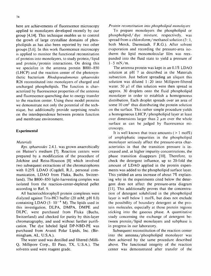
Fig. 1. Fluorescence spectrum of L H C P in D L P A monolayers at the air/water interface. Lipid molecular density is 10/7 n m - 2 , protein/lipid ratio is 1:100. Spectral resolution, 8 nm. (a) Freshly prepared monolayer; (b) same monolayer after 1 h without inert gas atmosphere; (c) spectrum of L H C P in 0.005% L D A O solution.
subsection D L P A . The functional integr i ty of the proteins was assessed by means of the fluorescence spectra. These are shown for freshly prepared in Fig . l a and for aged i n Fig. l b antenna proteins reconstituted in to D L P A . For comparison the f lu -
76
orescence spectrum of L H C P in detergent solution (F ig . l c ) is added.
Fig. l a and c show the pronounced emission band at λ = 858 n m due to the two BChl chromo-phores absorbing at 850 n m which are probably associated w i t h the heavier (9 k D a ) subunit [13]. The lack of fluorescence from the BChl chromo-phores absorbing at 800 n m in the lighter subunit demonstrates an efficient energy transfer to the 'd imer ic ' BChl . We therefore conclude on the functional integr i ty of the relevant part of the prote in containing the chromophores.
Wi thou t the protective inert gas atmosphere we observe a second fluorescence band rising at 800 n m in the t ime course of hours ( l b ) . This indicates disrupt ion of energy transfer to the 'd imeric ' energy trap.
By keeping the monolayer under an argon atmosphere and i n d i m light we are able to preserve the L H C P funct ion over hours as judged by the above cr i ter ion (data not shown).
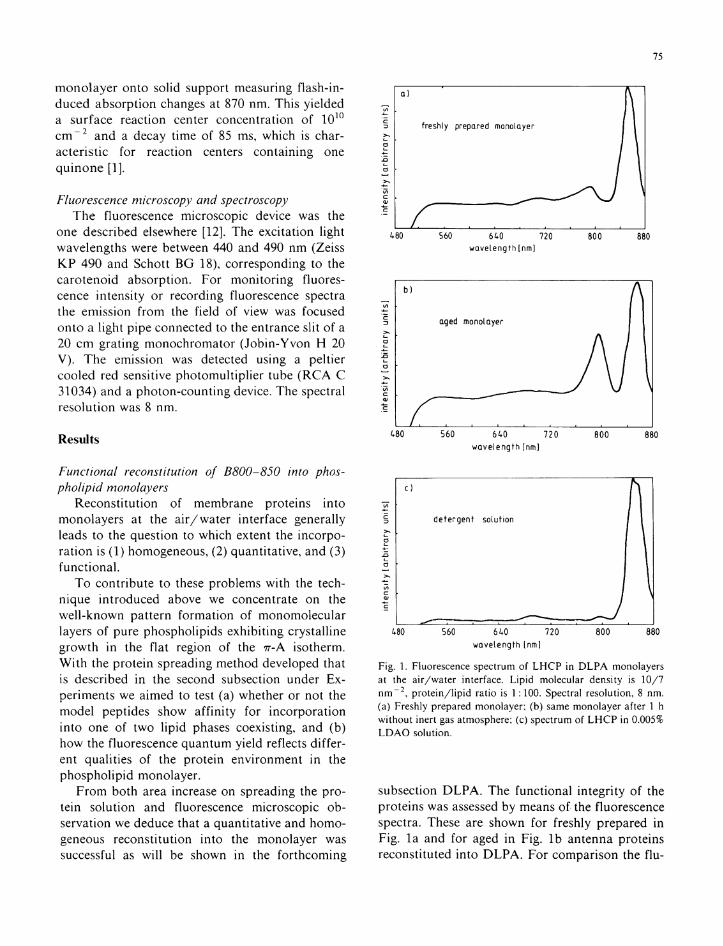
Protein / lipid interaction DLPA. Fig. 2 presents the ττ-Α isotherms of
D L P A at T = 2 1 ° C ( p H 5.5) wi thout (leftmost trace) and after deposition of 3 · 1 0 1 3 molecules and 6 · 1 0 1 3 molecules o f the antenna protein, respectively. For the subphase p H between 4 and 8, the D L P A head group is known to be singly deprotonated. The concentration cp o f the prote in solution added was determined by absorption
molecular area A [ n r n 2 /molecule DLPAj
Fig. 2. Pressure-area diagrams of D L P A monolayers containing L H C P in different protein/lipid ratios. The ratios given in the figure are calculated assuming complete incorporation. T, 21 °C; pH 5.5. In some diagrams there are indications for two break points in the slope between region I and II.
spectroscopy using the value o f 3.19 · Ι Ο 5 Μ - 1 · c m " 1 for the ext inct ion coefficient at λ = 850 n m (Scheer, H . and Cogdell , R.J., unpubl ished results). The most interesting features o f the isotherms after protein deposition are ( i ) the increase i n area and ( i i ) the decrease of the pressure 7 r m indicative of the onset of the l i p i d phase t rans i t ion .
Calculat ing the average area Ap per L H C P protomer ( α , ß - B C h l 3 ) in the monolayer at a lateral pressure π o f 30 m N / m we ob ta in Λ ρ = Ap/Np~ 20 n m 2 , where Ap is the area increase due to incorporat ion of Np p ro te in molecules or a radius of rp ~ 2.5 n m per L H C P molecule. The decrease of 7 7 m on antenna incorpora t ion is of the order of 10 m N / m per 1 mol% L H C P added.
Con t ro l experiments were performed by measuring the isotherms of the p r o t e i n / l i p i d -monolayer at different temperatures (data not shown). Hence we deduce the temperature dependence of the pressure 7 r m o f 1.4 m N · m " 1 · K - 1 . For the pure phosphol ip id monolayer values of 1 m N • m " 1 · K " 1 are reported [15].
Observation of the fluorescence microscopic images leads to the same conclusion. A s reported earlier [4 -6 ,10 ,14] p h o s p h o l i p i d monolayers labeled w i t h fluorescent dyes exhibi t a characteristic pattern format ion on compression f rom the f lu id state to the state of coexisting crystal l ine and f lu id phases. This effect is due to different so lubi l i ties of the dye molecules in different phosphol ip id phases. I n analogy w i t h this we may assume that L H C P is squeezed in to the f l u i d l i p i d phase. A n alternative explanation that L H C P remains i n sol i d domains, but is quenched there, can be ruled out from electron microscopic observations [16]. The p r o t e i n / l i p i d monolayer shows homogeneous fluorescence in the f lu id state (region I i n F ig . 2).
I t is impor tan t to note that fluorescence emission leading to an image is collected f rom the focal plane as can be demonstrated by imaging the entrance diaphragm of the exci ta t ion l ight . On compression in to the phase coexistence region (region I I in Fig. 2) pattern format ion is observed as shown in F ig . 3. The fact that these patterns can be imaged shows that the fluorescence is emit ted f rom molecules w i t h i n the focal plane. This again proves that L H C P is incorporated in to the monolayer, or at least adsorbed to i t .
O n further compressing the p r o t e i n / l i p i d
77
50
UQ
ω 3 30 OU
^•20 ο
Ii ίο
DMPE
lipid protein ratio
\ 1 : 500
U---0
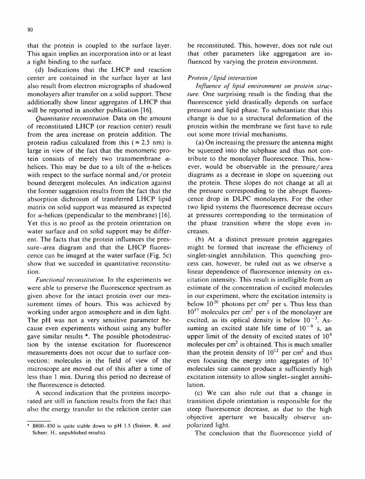
0 3 0 4 0 5 0 6 0 7 0 8 0 9 1 0 11 molecular area A [Tim 2 /molecule DMPE) ]
Fig. 4. Pressure-area diagrams of D M P E monolayers without and with L H C P incorporated. 7\ 22°C (pH 8), using 10 mM Tris buffer.
b
c
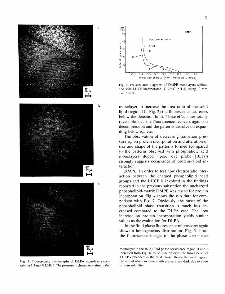
Fig. 3. Fluorescence micrographs of D L P A monolayers containing 1.5 mol% L H C P . The pressure is chosen to maintain the
monolayer to increase the area ratio of the solid l i p i d (region I I I , Fig. 2) the fluorescence decreases below the detection l i m i t . These effects are total ly reversible, i.e., the fluorescence recovers again on decompression and the patterns dissolve on expand ing below 7 7 n v etc.
The observation of decreasing transi t ion pressure 7Tm on protein incorporat ion and alteration of size and shape of the patterns formed (compared to the patterns observed w i t h phospha t ide acid monolayers doped l i po id dye probe [10,17]) strongly suggests occurrance of p r o t e i n / l i p i d i n teraction.
DMPE. I n order to test how electrostatic interaction between the charged phosphol ipid head groups and the L H C P is involved in the findings reported i n the previous subsection the uncharged phosphol ipid-matr ix D M P E was tested for protein incorporat ion. F ig . 4 shows the π-Α data for comparison w i t h Fig . 2. Obviously, the onset of the phosphol ip id phase transi t ion is much less decreased compared to the D L P A case. The area increase on protein incorporat ion yields similar values as the evaluation for D L P A .
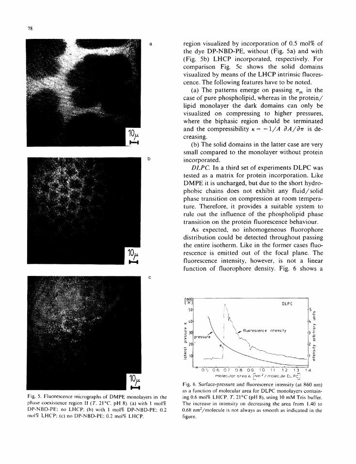
I n the f lu id phase fluorescence microscopy again shows a homogeneous d is t r ibu t ion . F ig . 5 shows the fluorescence images in the phase coexistence
monolayer in the solid/fluid phase coexistence region II and is increased from Fig. 3a to 3c. One observes the fluorescence of L H C P embedded in the fluid phase. Hence the solid regions, the size of which increases with pressure, are dark due to a low protein solubility.
78
a
b
c
Fig. 5. Fluorescence micrographs of D M P E monolayers in the phase coexistence region II (T. 21 ° C pH 8). (a) with 1 mol% DP-NBD-PE: no L H C P ; (b) with 1 mol% DP-NBD-PE; 0.2 mo\% L H C P ; (c) no DP-NBD-PE; 0.2 mol% L H C P .
region visualized by incorpora t ion o f 0.5 mo l% of the dye D P - N B D - P E , wi thou t (F ig . 5a) and w i t h (F ig . 5b) L H C P incorporated, respectively. For comparison Fig . 5c shows the sol id domains visualized by means of the L H C P intr insic fluorescence. The fo l lowing features have to be noted.
(a) The patterns emerge on passing nm i n the case o f pure phosphol ipid , whereas i n the p r o t e i n / l i p i d monolayer the dark domains can only be visualized on compressing to higher pressures, where the biphasic region should be terminated and the compressibil i ty κ = -I/A dA/dir is decreasing.
(b) The solid domains in the latter case are very small compared to the monolayer w i thou t protein incorporated.
DLPC. I n a th i rd set o f experiments D L P C was tested as a mat r ix for protein incorpora t ion . Like D M P E it is uncharged, but due to the short hydrophobic chains does not exhibi t any f l u i d / s o l i d phase transi t ion on compression at r o o m temperature. Therefore, i t provides a suitable system to rule out the influence of the phosphol ip id phase transi t ion on the protein fluorescence behaviour.
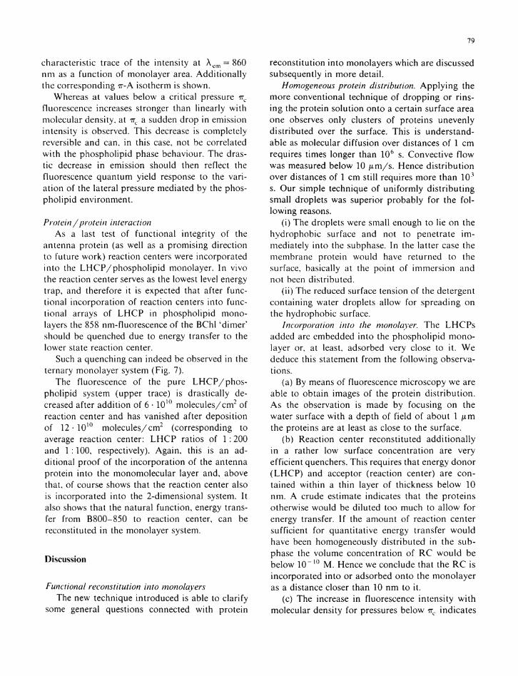
As expected, no inhomogeneous f luorophore d i s t r ibu t ion could be detected throughout passing the entire isotherm. L ike i n the former cases fluorescence is emitted out of the focal plane. The fluorescence intensity, however, is not a linear funct ion of f luorophore density. Fig . 6 shows a
0 5 0 6 0 7 0 .8 0 9 10 11 12 1.3 1 4 molecular area A | nm 2 / rnolecj le D l P C '
Fig. 6. Surface-pressure and fluorescence intensity (at 860 nm) as a function of molecular area for D L P C monolayers containing 0.6 mol% L H C P . 7\ 21 °C (pH 8), using 10 mM Tris buffer. The increase in intensity on decreasing the area from 1.40 to 0.68 nm2/molecule is not always as smooth as indicated in the figure.
79
characteristic trace of the intensity at λ 0 Π 1 = 860 n m as a function of monolayer area. Add i t i ona l l y the corresponding 7Γ-Α isotherm is shown.
Whereas at values below a cri t ical pressure TTC
fluorescence increases stronger than linearly w i t h molecular density, at 7rc a sudden drop in emission intensity is observed. This decrease is completely reversible and can, in this case, not be correlated w i t h the phosphol ipid phase behaviour. The drastic decrease in emission should then reflect the fluorescence quantum yield response to the var i a t ion of the lateral pressure mediated by the phospho l ip id environment.
Protei η/protein interaction As a last test of functional integrity of the
antenna prote in (as well as a promising direction to future work ) reaction centers were incorporated into the L H C P / p h o s p h o l i p i d monolayer. I n vivo the reaction center serves as the lowest level energy trap, and therefore i t is expected that after functional incorporat ion of reaction centers into funct ional arrays of L H C P in phosphol ipid monolayers the 858 nm-fluorescence of the BChl 'd imer ' should be quenched due to energy transfer to the lower state reaction center.
Such a quenching can indeed be observed in the ternary monolayer system (Fig. 7).
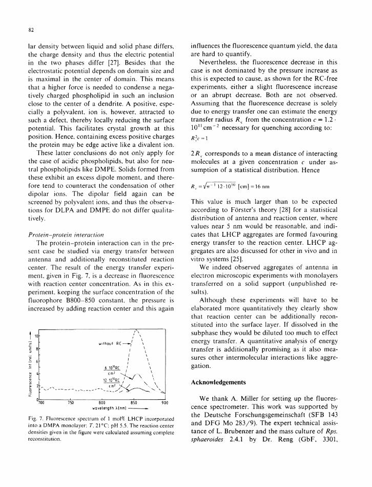
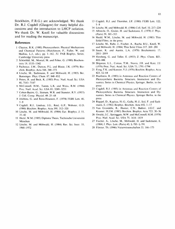
The fluorescence of the pure L H C P / p h o s phol ip id system (upper trace) is drastically decreased after addi t ion of 6 · 1 0 1 0 molecu les /cm 2 of reaction center and has vanished after deposition of 12 · 1 0 1 0 mo lecu le s / cm 2 (corresponding to average reaction center: L H C P ratios of 1 :200 and 1 :100, respectively). Again , this is an addit ional p roof of the incorporat ion of the antenna protein into the monomolecular layer and, above that, of course shows that the reaction center also is incorporated into the 2-dimensional system. I t also shows that the natural function, energy transfer from B800-850 to reaction center, can be reconstituted i n the monolayer system.
Discussion
Functional reconstitution into monolayers The new technique introduced is able to clarify
some general questions connected w i t h protein
reconsti tution into monolayers which are discussed subsequently in more detail.
Homogeneous protein distribution. A p p l y i n g the more conventional technique of d ropping or rinsing the protein solution onto a certain surface area one observes only clusters of proteins unevenly distr ibuted over the surface. This is understandable as molecular diffusion over distances o f 1 cm requires times longer than 10 6 s. Convective f low was measured below 10 /xm/s . Hence d i s t r ibu t ion over distances of 1 cm st i l l requires more than 1 0 3
s. Our simple technique of un i fo rmly d i s t r ibu t ing small droplets was superior probably for the fo l lowing reasons.
(i) The droplets were small enough to lie on the hydrophobic surface and not to penetrate i m mediately into the subphase. I n the latter case the membrane protein would have returned to the surface, basically at the point of immersion and not been distr ibuted.
( i i ) The reduced surface tension of the detergent containing water droplets al low for spreading on the hydrophobic surface.
Incorporation into the monolayer. The L H C P s added are embedded in to the phosphol ip id monolayer or, at least, adsorbed very close to i t . We deduce this statement f rom the fo l lowing observations.
(a) By means of fluorescence microscopy we are able to obtain images of the protein d i s t r ibu t ion . A s the observation is made by focusing on the water surface w i t h a depth of field of about 1 μητ the proteins are at least as close to the surface.
(b) Reaction center reconstituted addi t iona l ly i n a rather low surface concentration are very efficient quenchers. This requires that energy donor ( L H C P ) and acceptor (reaction center) are contained w i t h i n a th in layer of thickness below 10 n m . A crude estimate indicates that the proteins otherwise would be di luted too much to a l low for energy transfer. I f the amount of reaction center sufficient for quanti tat ive energy transfer w o u l d have been homogeneously dis tr ibuted in the sub-phase the volume concentration of R C w o u l d be below 1 0 " 1 0 M . Hence we conclude that the R C is incorporated into or adsorbed onto the monolayer as a distance closer than 10 n m to i t .
(c) The increase in fluorescence intensity w i t h molecular density for pressures below 7r c indicates
80
that the protein is coupled to the surface layer. This again implies an incorporat ion in to or at least a t ight b inding to the surface.
(d) Indications that the L H C P and reaction center are contained in the surface layer at last also result f rom electron micrographs of shadowed monolayers after transfer on a solid support. These addi t ional ly show linear aggregates of L H C P that w i l l be reported i n another publ icat ion [16].
Quantitative reconstitution. Data on the amount of reconstituted L H C P (or reaction center) result f rom the area increase on protein addi t ion. The protein radius calculated from this ( ~ 2.5 nm) is large in view of the fact that the monomeric protein consists of merely two transmembrane a-helices. This may be due to a t i l t o f the α-helices w i t h respect to the surface normal a n d / o r protein bound detergent molecules. A n indicat ion against the former suggestion results from the fact that the absorption dichroism of transferred L H C P l i p i d matr ix on solid support was measured as expected for α-helices (pependicular to the membrane) [16]. Ye t this is no proof as the protein orientat ion on water surface and on solid support may be different. The facts that the protein influences the pressure-area diagram and that the L H C P fluorescence can be imaged at the water surface (Fig . 5c) show that we succeded in quanti tat ive reconstitut ion .
Functional reconstitution. I n the experiments we were able to preserve the fluorescence spectrum as given above for the intact protein over our measurement times of hours. This was achieved by work ing under argon atmosphere and in d i m light. The p H was not a very sensitive parameter because even experiments wi thout using any buffer gave similar results *. The possible photodestruc-t ion by the intense excitation for fluorescence measurements does not occur due to surface convection: molecules in the field of view of the microscope are moved out of this after a t ime of less than 1 min . D u r i n g this period no decrease of the fluorescence is detected.
A second indicat ion that the proteins incorporated are sti l l in function results from the fact that also the energy transfer to the reaction center can
* B800-850 is quite stable down to pH 1.5 (Steiner, R. and Scheer, H., unpublished results).
be reconstituted. This, however, does not rule out that other parameters l ike aggregation are i n fluenced by varying the protein environment.
Protein / lipid interaction Influence of lipid environment on protein struc
ture. One surprising result is the f inding that the fluorescence yield drastically depends on surface pressure and l i p i d phase. T o substantiate that this change is due to a structural deformation o f the prote in w i t h i n the membrane we first have to rule out some more t r iv ia l mechanisms.
(a) O n increasing the pressure the antenna might be squeezed in to the subphase and thus not contr ibute to the monolayer fluorescence. This , however, wou ld be observable i n the pressure/area diagrams as a decrease in slope on squeezing out the protein. These slopes do not change at all at the pressure corresponding to the abrupt fluorescence drop in D L P C monolayers. For the other two l i p i d systems the fluorescence decrease occurs at pressures corresponding to the terminat ion of the phase transit ion where the slope even i n creases.
(b) A t a dist inct pressure protein aggregates might be formed that increase the efficiency o f singlet-singlet annihi la t ion. This quenching process can, however, be ruled out as we observe a linear dependence of fluorescence intensity on exci ta t ion intensity. This result is intel l igible f rom an estimate of the concentration of excited molecules in our experiment, where the excitation intensity is below 10 2 0 photons per c m 2 per s. Thus less than 1 0 1 7 molecules per c m 2 per s o f the monolayer are excited, as its opt ical density is below 10 ~ 3 . Assuming an excited state life t ime of 10 ~ 9 s, an upper l i m i t o f the density o f excited states o f 1 0 s
molecules per c m 2 is obtained. This is much smaller than the prote in density of 1 0 1 2 per c m 2 and thus even focusing the energy in to aggregates o f 1 0 3
molecules size cannot produce a sufficiently high excitat ion intensity to al low singlet-singlet annih i la t ion .
(c) We can also rule out that a change in transi t ion dipole or ientat ion is responsible for the steep fluorescence decrease, as due to the high objective aperture we basically observe un-polarized l ight .
The conclusion that the fluorescence y ie ld of
81
the B800-850 prote in depends on environment is suppor ted f rom the fact that its value also varies by more than a factor of 3 for the protein in different detergent micelles [18]. Hence, even in cases where the pressure inside different micelles is not expected to vary too much the fluorescence properties are affected.
T o be able to conclude on the pigment environment inside the prote in i t is impor tant to ask for the molecular mechanisms leading to a change in fluorescence yield. As the t ransi t ion is electronical ly a l lowed, its t ransi t ion dipole moment is not sensitively dependent on the environment of the f luorophore group [19]. This also holds for the d imer i f the transit ion originates from the lower of the two energy levels that result f rom the elect ronic interact ion of the monomer excited states [20,21]. As this argument is val id in the present si tuat ion the transit ion dipole moment quite probably remains unaffected by a pressure change. Hence we suppose either the energy transfer w i t h i n L H C P or the rate of radiationless transitions is varied. The latter can be caused by freezing in or a l lowing a v ibra t ion to serve as a decay channel. This possibi l i ty was discussed by Pearlstein [22].
Al ternat ive ly , a radiationless transit ion can be induced by a change in the energetic posi t ion of the lowest excited singlet state due to a variat ion of po rphy r in environment w i t h i n the protein. The ch lo rophyl l microenvironment has already been discussed as con t r ibu t ing significantly to the excited state energy [13], and could thus also open more efficient decay paths by creating a degeneracy between the excited state and the proper vibrational level o f the electronic ground state. Wi thou t detailed structural data, no dis t inct ion between the two mechanisms is possible.
The possibi l i ty of variat ion in energy transfer from carotenoid to the BChl 'd imer ic ' trap, proposed by Cogdell [23], w i l l be investigated after modif ica t ion of the apparatus. I f energy transfer is involved fluorescence should not be affected by pressure var ia t ion i f the excitation occured in to the BChl bands instead of the carotenoid band.
Besides this biophysical aspect that a change in the membrane environment causes a change in protein structure and thus in a functional p roperty, one may also speculate i f this is a biological ly important regulation mechanism. The pressure
near 20 m N / m is not entirely improbable for the inside of a membrane. A local pressure change, e.g., as a consequence of a change in the ionic environment of the membrane after electron transfer w i t h i n the reaction center could open a radiationless decay channel and thus p roh ib i t too frequent energy transfer to the reaction center. This idea is in t r iguing, but also highly speculative, and deserves further consideration in dedicated experiments w i th vesicle systems.
Influence of LHCP on the lipid matrix. Concerning the influence on the l i p i d matr ix we obtained as most interesting results (a) that although the prote in preferentially part i t ions in to the l iquid phase it reduces the pressure necessary for sol idif i cat ion, and (b) that it removes the l i qu id inclusions w i t h i n the dendrites. Result (a) is surprising as one usually expects that the incorporat ion of the ' i m p u r i t y ' protein in the monolayer creates disorder in the system which therefore is rendered more diff icul t to crystallize [24]. This, however, is not necessarily true in the present case. The protein may form a boundary of ordered l ipids, thus reducing the entropy of the l iqu id state. Crysta l l i zat ion therefore needs a smaller entropy change. This reduces the free-energy change of the transit ion and thus also the transit ion pressure. Similar ly a change in transit ion temperature of phospho l ip id vesicles due to protein incorporat ion is discussed by means of Landau-de Gennes theory ( H e l m , C , Lösche , Μ . and M ö h w a l d , Η . , unpublished results).
The fact that this reduction of irm is very pronounced for the charged l i p i d , but less for the uncharged one points to the importance of electrostatic forces: the protein tends to attract negatively charged lipids, but not uncharged ones. This f inding is understood regarding the amino acid sequence of B800-850 [12]. Positively charged lysins are posit ioned at the end of long hydrophobic regions capable of fo rming α-hel ices . As the negatively charged amino acids may protrude in to the water phase an attractive Coulomb interaction may occur i n the polar headgroup region.
The second effect, the removal o f the l i qu id inclusions in the dendrites has been observed after addi t ion of divalent ions and may thus be caused by the same mechanism [26]. These inclusions may be stabilized by electrostatic forces: as the molecu-
82
lar density between l i qu id and solid phase differs, the charge density and thus the electric potential in the two phases differ [27]. Besides that the electrostatic potential depends on domain size and is maximal in the center of domain. This means that a higher force is needed to condense a negatively charged phosphol ipid in such an inclusion close to the center of a dendrite. A positive, especially a polyvalent, ion is, however, attracted to such a defect, thereby locally reducing the surface potential . This facilitates crystal growth at this posit ion. Hence, containing excess positive charges the protein may be edge active like a divalent ion.
These latter conclusions do not only apply for the case of acidic phospholipids, but also for neutral phospholipids like D M P E . Solids formed from these exhibit an excess dipole moment, and therefore tend to counteract the condensation of other dipolar ions. The dipolar field again can be screened by polyvalent ions, and thus the observations for D L P A and D M P E do not differ qualitatively.
Protein-protein interaction The pro te in -pro te in interaction can in the pre
sent case be studied via energy transfer between antenna and addi t ional ly reconstituted reaction center. The result of the energy transfer experiment, given in Fig. 7, is a decrease in fluorescence w i t h reaction center concentration. As in this experiment, keeping the surface concentration of the fluorophore B800-850 constant, the pressure is increased by adding reaction center and this again
1 1 0
1 Β
r 6
a
w i t h o u t RC-
/ \ / \ ' \
6 1010RC ' \
12-101°RC / ^ \ — 1
\ \
700 750 800 850 wavelength X[nm]
Fig. 7. Fluorescence spectrum of 1 mol% L H C P incorporated into a DMPA monolayer; Γ, 21 °C; pH 5.5. The reaction center densities given in the figure were calculated assuming complete reconstitution.
influences the fluorescence quantum yield, the data are hard to quantify.
Nevertheless, the fluorescence decrease in this case is not dominated by the pressure increase as this is expected to cause, as shown for the RC-free experiments, either a slight fluorescence increase or an abrupt decrease. Both are not observed. Assuming that the fluorescence decrease is solely due to energy transfer one can estimate the energy transfer radius Ä c from the concentration c = 1.2 · 1 0 n c m - 2 necessary for quenching according to:
R]c = \
2Rc corresponds to a mean distance of interacting molecules at a given concentration c under assumption of a statistical d is t r ibut ion . Hence
tf( =\/7r 1 12-10 1 0 [cm]=16nm
This value is much larger than to be expected according to Fö r s t e r ' s theory [28] for a statistical d is t r ibut ion of antenna and reaction center, where values near 5 n m wou ld be reasonable, and i n d i cates that L H C P aggregates are formed favouring energy transfer to the reaction center. L H C P aggregates are also discussed for other in vivo and in v i t ro systems [25].
We indeed observed aggregates of antenna in electron microscopic experiments wi th monolayers transferred on a solid support (unpublished results).
Al though these experiments w i l l have to be elaborated more quanti tat ively they clearly show that reaction center can be addi t ional ly reconstituted in to the surface layer. I f dissolved in the subphase they wou ld be di luted too much to effect energy transfer. A quanti tat ive analysis o f energy transfer is addi t ional ly promising as it also measures other intermolecular interactions l ike aggregation.
Acknowledgements
We thank A . M i l l e r for setting up the fluorescence spectrometer. This work was supported by the Deutsche Forschungsgemeinschaft (SFB 143 and D F G M o 283 /9 ) . The expert technical assistance of L . Brubenzer and the mass culture o f Rps. sphaeroides 2.4.1 by D r . Reng ( G b F , 3301,
83
S t ö c k h e i m , F .R.G. ) are acknowledged. We thank D r . R.J. Cogdell (Glasgow) for many helpful discussions and the in t roduct ion to L H C P isolation. W e thank D r . W . K n o l l for valuable discussions and for reading the manuscript.
References 1 Clayton, R . K . (1980) Photosynthesis. Physical Mechanisms
and Chemical Patterns (Hutchinson, F. , Fuller, W. and Mullins. L . J . , eds.), pp. 1-162, JU PAB Biophys. Series, Cambridge University press
2 Schönfeld. Μ., Montal. Μ. and Feher, G . (1980) Biochemistry 19, 1535-1542
3 Pachence, J.M., Dutton, P.L. and Blasie, J .K. (1979) Bio-chim. Biophys. Acta 548, 348-373
4 Lösche, Μ.. Sackmann, Ε. and Möhwald, Η. (1983) Ber. Bunsenges. Phys. Chem. 87, 848-852
5 Peters, R. and Beck, K. (1983) Proc. Natl. Acad. Sei. USA 80, 7183-7187
6 McConnell, H.M., Tamm, L . K . and Weiss, R.M. (1984) Proc. Natl. Acad. Sei. USA 81, 3249-3253
7 Cohen-Bazire. G. , Sistrum, W.R. and Staneier, R .Y. (1957) J. Cell. Comp. Physiol. 49, 25-68
8 Jolchine, G . and Reiss-Housson, F. (1974) F E B S Lett. 40, 5-8
9 Cogdell, R.J . , Lindsay, J .G. , Reid, G.P., Webster, G.D. (1980) Biochim. Biophys. Acta 591. 312-320
10 Lösche. M. and Möhwald, H. (1984) Eur. Biophys. J. 11. 35-42
11 Heckl, W.M. (1985) Diploma Thesis, Technische Universität München
12 Lösche, M. and Möhwald. H. (1984) Rev. Sei. Instr. 55, 1968-1972
13 Cogdell, R.J. and Thornber, J.P. (1980) F E B S Lett. 122, 1-8
14 Lösche. M. and Möhwald, H. (1984) Coli. Surf. 10. 217-224 15 Albrecht, O.. Gruler, H. and Sackmann, E. (1978) J. Phys.
(Paris) 39, 301-313 16 Heckl, W.M.. Lösche. M. and Möhwald, H. (1985) Thin
Solid Films, in the press 17 Lösche, Μ., Rabe. J . . Fischer, Α., Rucha. B.U., Knoll, W.
and Möhwald, H. (1984) Thin Solid Films 117, 269-280 18 Sauer, K. and Austin, L.A. (1978) Biochemistry 17.
2011-2019 19 Herzberg, G . and Teller, E. (1933) Z. Phys. Chem. B21,
410-446 20 Shipman. L . L . , Cotton, T.M., Norris, J.R. and Katz, J.J.
(1976) Proc. Nati. Acad. Sei. USA 73, 1791-1794 21 Fong, F .K. and Koester, V.J. (1976) Biochim. Biophys. Acta
423, 52-64 22 Pearlstein, R. (1985) in Antennas and Reaction Centers of
Photosynthetic Bacteria. Structure, Interaction and Dynamics, Series in Chemical Physics. Springer, Berlin, in the press
23 Cogdell, R.J. (1985) in Antennas and Reaction Centers of Photosynthetic Bacteria. Structure. Interaction and Dynamics, Series in Chemical Physics, Springer Berlin, in the press
24 Rüppel, D., Kapitza, H.-G., Galla, H.-J., Sixl. F . and Sack-rnann, E. (1982) Biophys. Biochim. Acta 692, 1-17
25 Van Grondelle, R., Hunter, C.N., Bakker, J .G .C. and Kramer, H.J.M. (1983) Biochim. Biophys. Acta 723, 30-36
26 Owicki, J .C. , Springgate, M.W. and McConnell, H.M. (1978) Proc. Natl. Acad. Sei. USA 75, 1616-1619
27 Fischer. Α., Lösche, Μ., Möhwald, Η. and Sackmann, Ε. (1984) J. Phys. Lett. (Paris) 45, L-785-L-791
28 Förster, Th. (1946) Naturwissenschaften 33, 166-175