Bidirectional Regulation of Intravenous General...
15
Anesthesiology, V 118 • No 3 562 March 2013 ABSTRACT Background: γ-aminobutyric acid A (GABA A ) receptors mediate the actions of several intravenous general anesthet- ics. However, the contribution of α3-containing GABA A receptors to the action of these drugs is unknown. Methods: e authors compared anesthetic endpoints (hypnosis, immobility, hypothermia) in response to various intravenous anesthetics in mice lacking the α3 subunit of the GABA A receptor (α3 knockout) and in wild-type mice. Fur- thermore, the authors generated and analyzed conditional mutant mice expressing the GABA A receptor α3 subunit exclusively in noradrenergic neurons. Results: α3 knockout mice displayed decreased hypnotic and hypothermic responses to etomidate and midazolam, but an increased response to pentobarbital. e hypnotic response to ketamine was unaltered, whereas the hypo- thermic response was increased. In contrast, the hypnotic but not the hypothermic response to medetomidine was increased. e combination of ketamine/xylazine displayed increased hypnotic, immobilizing, and hypothermic effects in α3 knockout mice. Mice expressing the α3 subunit exclu- sively in noradrenergic neurons were generated to assess whether the lack of α3 subunits on noradrenergic neurons may be responsible for this effect. In these mice, the increases of the hypnotic and immobilizing actions induced by ket- amine/xylazine were largely absent, whereas the increase in the hypothermic action was still present. Conclusion: α3-containing GABA A receptors bidirec- tionally regulate essential anesthetic actions: they mediate anesthetic actions of etomidate and midazolam, known to selectively act at GABA A receptors, and they negatively constrain anesthetic actions of compounds with targets partly or exclusively distinct from GABA A receptors such as medetomidine, ketamine, and pentobarbital. Furthermore, our results indicate that α3-containing GABA A receptors on noradrenergic neurons may contribute to this constraint. γ -AMINOBUTYRIC acid A (GABA A ) receptors are chlo- ride channels, which mediate fast synaptic inhibition and tonic inhibition in the central nervous system. ey are composed of five subunits drawn from a repertoire of eight Bidirectional Regulation of Intravenous General Anesthetic Actions by α3-containing γ-aminobutyric Acid A Receptors Carolin J. Straub, Ph.D.,* Hew Mun Lau, B.Sc.,† Rosanna Parlato, Ph.D.,‡ Guenther Schuetz, M.D.,§ Jean-Marc Fritschy, Ph.D.,‖ Uwe Rudolph, M.D.# * Predoctoral Research Fellow, Laboratory of Genetic Neuro- pharmacology, McLean Hospital and Department of Psychiatry, Harvard Medical School, Belmont, Massachusetts, and Institute of Pharmacology and Toxicology, University of Zürich, Zürich, Swit- zerland. † Associate in Psychiatry, Laboratory of Genetic Neuro- pharmacology, McLean Hospital and Department of Psychiatry, Harvard Medical School, Belmont, Massachusetts. ‡ Postdoctoral Research Fellow, § Helmholtz Professor, Molecular Biology of the Cell I, German Cancer Research Center, Heidelberg, Germany. ‖ Professor, Institute of Pharmacology and Toxicology, University of Zürich, Zürich, Switzerland. # Associate Professor, Laboratory of Genetic Neuropharmacology, McLean Hospital and Department of Psychiatry, Harvard Medical School, Belmont, Massachusetts. Received from the Laboratory of Genetic Neuropharmacology, McLean Hospital and the Department of Psychiatry, Harvard Medical School. Submitted for publication January 5, 2012. Accepted for publi- cation October 8, 2012. The research described in this publication was supported by institutional funds from the University of Zurich, Zurich, Switzerland and from McLean Hospital, Belmont, Massachusetts. UR’s work on the manuscript was supported in part by award number R01-GM086448 from the National Institute of General Medical Sciences of the National Institutes of Health (Bethesda, Maryland). The views supported in this manuscript do not necessarily represent the views of the National Institute of General Medical Sciences or of the National Institutes of Health. In the last three years, Uwe Rudolph provided professional services to Sunovion Pharmaceuticals (Marlbourough, Massachusetts) and to Concert Pharmaceuticals (Lexington, Massachu- setts). The authors thank Drs. Alo C. Basu (College of the Holy Cross, Worcester, Massachusetts) and Elif Engin (McLean Hospital, Belmont, Massachusetts) for advice on statistical analysis and Dr. Elif Engin for critically reading the manuscript. Address correspondence to Dr. Rudolph: Laboratory of Genetic Neuropharmacology, McLean Hospital, Mail stop 145, Harvard Medical School, 115 Mill Street, Belmont, MA 02478. urudolph@mclean. harvard.edu. Information on purchasing reprints may be found at www.anesthesiology.org or on the masthead page at the beginning of this issue. ANESTHESIOLOGY’S articles are made freely accessible to all read- ers, for personal use only, 6 months from the cover date of the issue. Copyright © 2013, the American Society of Anesthesiologists, Inc. Lippincott Williams & Wilkins. Anesthesiology 2013; 118:562–76 What We Already Know about This Topic • The γ-aminobutyric acid A (GABA A ) receptor system mediates actions of many general anesthetics in the central nervous system. However, the contributions of the different GABA A re- ceptor subtypes and the role of noradrenergic neurons require further clarification What This Article Tells Us That Is New • GABA A receptors containing the α3 subunit mediate anes- thetic responses to etomidate and midazolam. Lack of the α3 subunit results in enhanced responses to the combination ketamine/xylazine, an effect that is rescued in part by expres- sion of the α3 subunit selectively in noradrenergic neurons, indicating an involvement of noradrenergic neurons in modu- lating hypnosis Downloaded From: http://anesthesiology.pubs.asahq.org/pdfaccess.ashx?url=/data/journals/jasa/931124/ on 07/09/2018
-
Upload
vuongkhanh -
Category
Documents
-
view
216 -
download
0
Transcript of Bidirectional Regulation of Intravenous General...
Anesthesiology, V 118 • No 3 562 March 2013
ABSTRACT
Background: γ-aminobutyric acidA (GABAA) receptors mediate the actions of several intravenous general anesthet-ics. However, the contribution of α3-containing GABAA receptors to the action of these drugs is unknown.Methods: The authors compared anesthetic endpoints (hypnosis, immobility, hypothermia) in response to various intravenous anesthetics in mice lacking the α3 subunit of the GABAA receptor (α3 knockout) and in wild-type mice. Fur-thermore, the authors generated and analyzed conditional mutant mice expressing the GABAA receptor α3 subunit exclusively in noradrenergic neurons.
Results: α3 knockout mice displayed decreased hypnotic and hypothermic responses to etomidate and midazolam, but an increased response to pentobarbital. The hypnotic response to ketamine was unaltered, whereas the hypo-thermic response was increased. In contrast, the hypnotic but not the hypothermic response to medetomidine was increased. The combination of ketamine/xylazine displayed increased hypnotic, immobilizing, and hypothermic effects in α3 knockout mice. Mice expressing the α3 subunit exclu-sively in noradrenergic neurons were generated to assess whether the lack of α3 subunits on noradrenergic neurons may be responsible for this effect. In these mice, the increases of the hypnotic and immobilizing actions induced by ket-amine/xylazine were largely absent, whereas the increase in the hypothermic action was still present.Conclusion: α3-containing GABAA receptors bidirec-tionally regulate essential anesthetic actions: they mediate anesthetic actions of etomidate and midazolam, known to selectively act at GABAA receptors, and they negatively constrain anesthetic actions of compounds with targets partly or exclusively distinct from GABAA receptors such as medetomidine, ketamine, and pentobarbital. Furthermore, our results indicate that α3-containing GABAA receptors on noradrenergic neurons may contribute to this constraint.
γ-AMInoButyrIc acidA (GABAA) receptors are chlo-ride channels, which mediate fast synaptic inhibition
and tonic inhibition in the central nervous system. They are composed of five subunits drawn from a repertoire of eight
Bidirectional Regulation of Intravenous General Anesthetic Actions by α3-containing γ-aminobutyric AcidA Receptors
Carolin J. Straub, Ph.D.,* Hew Mun Lau, B.Sc.,† Rosanna Parlato, Ph.D.,‡ Guenther Schuetz, M.D.,§ Jean-Marc Fritschy, Ph.D.,‖ Uwe Rudolph, M.D.#
* Predoctoral Research Fellow, Laboratory of Genetic Neuro-pharmacology, McLean Hospital and Department of Psychiatry, Harvard Medical School, Belmont, Massachusetts, and Institute of Pharmacology and Toxicology, University of Zürich, Zürich, Swit-zerland. † Associate in Psychiatry, Laboratory of Genetic Neuro-pharmacology, McLean Hospital and Department of Psychiatry, Harvard Medical School, Belmont, Massachusetts. ‡ Postdoctoral Research Fellow, § Helmholtz Professor, Molecular Biology of the Cell I, German Cancer Research Center, Heidelberg, Germany. ‖ Professor, Institute of Pharmacology and Toxicology, University of Zürich, Zürich, Switzerland. # Associate Professor, Laboratory of Genetic Neuropharmacology, McLean Hospital and Department of Psychiatry, Harvard Medical School, Belmont, Massachusetts.
Received from the Laboratory of Genetic Neuropharmacology, McLean Hospital and the Department of Psychiatry, Harvard Medical School. Submitted for publication January 5, 2012. Accepted for publi-cation October 8, 2012. The research described in this publication was supported by institutional funds from the University of Zurich, Zurich, Switzerland and from McLean Hospital, Belmont, Massachusetts. UR’s work on the manuscript was supported in part by award number R01-GM086448 from the National Institute of General Medical Sciences of the National Institutes of Health (Bethesda, Maryland). The views supported in this manuscript do not necessarily represent the views of the National Institute of General Medical Sciences or of the National Institutes of Health. In the last three years, Uwe Rudolph provided professional services to Sunovion Pharmaceuticals (Marlbourough, Massachusetts) and to Concert Pharmaceuticals (Lexington, Massachu-setts). The authors thank Drs. Alo C. Basu (College of the Holy Cross, Worcester, Massachusetts) and Elif Engin (McLean Hospital, Belmont, Massachusetts) for advice on statistical analysis and Dr. Elif Engin for critically reading the manuscript.
Address correspondence to Dr. Rudolph: Laboratory of Genetic Neuropharmacology, McLean Hospital, Mail stop 145, Harvard Medical School, 115 Mill Street, Belmont, MA 02478. [email protected]. Information on purchasing reprints may be found at www.anesthesiology.org or on the masthead page at the beginning of this issue. Anesthesiology’s articles are made freely accessible to all read-ers, for personal use only, 6 months from the cover date of the issue.
Copyright © 2013, the American Society of Anesthesiologists, Inc. Lippincott Williams & Wilkins. Anesthesiology 2013; 118:562–76
What We Already Know about This Topic
• The γ-aminobutyric acidA (GABAA) receptor system mediates actions of many general anesthetics in the central nervous system. However, the contributions of the different GABAA re-ceptor subtypes and the role of noradrenergic neurons require further clarification
What This Article Tells Us That Is New
• GABAA receptors containing the α3 subunit mediate anes-thetic responses to etomidate and midazolam. Lack of the α3 subunit results in enhanced responses to the combination ketamine/xylazine, an effect that is rescued in part by expres-sion of the α3 subunit selectively in noradrenergic neurons, indicating an involvement of noradrenergic neurons in modu-lating hypnosis
ALn
Bidirectional Modulation of General Anesthetic Action
Straub et al.
March
10.1097/ALn.0b013e3182800d76
3
Anjana
2013
Downloaded From: http://anesthesiology.pubs.asahq.org/pdfaccess.ashx?url=/data/journals/jasa/931124/ on 07/09/2018
Anesthesiology 2013; 118:562-76 563 Straub et al.
PERIOPERATIVE MEDICINE
different subunit families comprising at least 19 different genes (α1–6, β1–3, γ1–3, δ, ε, θ, π, ρ1–3).1–4 Depending on the exact subunit composition GABAA receptors possess different physiological and pharmacological properties.1,2 Many general anesthetics potentiate the activity of GABAA receptors, and the role of several GABAA receptors sub-types in mediating anesthetic effects has been described.3,4 We have previously shown that β3(n265M) knock-in mice display dramatically reduced hypnotic responses to etomi-date and pentobarbital and cannot be immobilized by these compounds, demonstrating that a subpopulation of GABAA receptors is essential for the anesthetic actions of these anes-thetic agents.5 Most frequently, the GABAA receptors are classified by the α subunit that is present. on the basis of studies in global knockout (Ko) mice, the α1 subunit,6 but not the α57 and α6 subunits,8 contribute to the hypnotic actions of intravenous general anesthetics. However, the sig-nificance of α3 subunit–containing GABAA receptors for anesthetic effects remained to be clarified.
Performing stereotaxic surgeries we made the unexpected and surprising observation that a combination of ketamine, an N-methyl-D-aspartate receptor antagonist and xylazine, an α2-adrenergic agonist, induces remarkably prolonged anes-thesia in α3Ko mice compared with wild-type (Wt) mice. The α3 subunit of the GABAA receptor is present in 10–15% of all GABAA receptors in the central nervous system and GABAA receptors containing the α3 subunit are expressed widely in the brain.1,2 However, in monoaminergic neurons the α3 subunit seems to be the major α subunit expressed. High expression levels in noradrenergic neurons9–11 suggest that α3 subunit–containing GABAA receptors might be important modulators of the noradrenergic system.
The noradrenergic system (cell groups A1–A10), which includes the locus coeruleus (A6) whose neurons project widely throughout the neuraxis and other noradrenergic nuclei (e.g., A1 and A2, which project mainly to the hypo-thalamus) plays an important role in the regulation of sleep and arousal as well as thermoregulation.12–14 Activity of the noradrenaline-containing locus coeruleus neurons depends on the behavioral state (e.g., sleeping or waking) of the organ-ism and this system is likely involved in switching between these different states.15 Moreover, α2-adrenoceptor agonists such as dexmedetomidine have been shown to exert their sed-ative/hypnotic effects through endogenous sleep pathways,16 at least in part through inhibition of the locus coeruleus.17
In order to obtain insights into the role of α3-containing GABAA receptors in general anesthetic actions, we investigated (1) in α3Ko mice whether α3-containing GABAA receptors mediate the action of intravenous general anesthetics known to act at least in part via GABAA receptors such as etomidate, midazolam, and pentobarbital; (2) whether α3-containing GABAA receptors play a role in the actions of the N-methyl-D-aspartate receptor antagonist ketamine and of the α2-adrenergic agonist medetomidine, either alone or in combination, using α3Ko mice; and (3)
whether α3-containing GABAA receptors on noradrenergic neurons are responsible for the increased sensitivity to ketamine/xylazine. to this end, using conditional cre-loxP-mediated recombination limited to noradrenergic neurons, we generated α3Ko mice in which the α3 subunit was reintroduced exclusively in noradrenergic neurons.
Materials and Methods
AnimalsMale mice on the c57BL/6J background between 8 and 18 weeks of age at beginning of experiments were used. Biochem-ical and morphological studies were performed in Zurich (Switzerland) with approval of the cantonal Veterinary office of Zurich and performed in accordance with international guidelines on animal use and care (European community council Directive 86⁄609⁄EEc). All behavioral experiments were conducted in Belmont in accordance with the national Institutes of Health guide for the care and use of Labora-tory Animals, and were approved by the McLean Hospital Institutional Animal care and use committee, Belmont, Massachusetts. Mice lacking the α3 subunit of the GABAA receptor (α3Ko mice) have been described previously.18 For behavioral testing mice were group-housed (3–4 per cage) and kept under a reversed 12/12 h light–dark cycle (lights on from 9:00 PM to 9:00 AM) for at least 2 weeks before beginning of experimental testing and throughout the study.
Generation of Global Rescue MiceThe generation of mice lacking the α3 subunit of the GABAA receptor (α3Ko mice) has been described in detail.18 The absence of α3 messenger rnA was demonstrated by reverse transcription polymerase chain reaction, and the absence of the α3 protein by Western blot and immunohistochemistry.18 As the nomenclature of exons in the Gabra3 gene has changed over time, exon 4 in the study by yee et al.18 is identical to exon 5 as described here. These mice carry an artificial exon flanked by loxP sites in the Gabra3 gene, which is located on the X-chromosome. In mice that carry both the mutant gene and express the cre recombinase this artificial exon is removed, thereby restoring α3 subunit expression. Mice in which the α3 subunit is rescued globally have been generated by crossing α3Ko mice with EIIa-cre mice, which express the cre recombinase in oocytes and preimplantation stages of the embryo.19 Mice carrying both the mutant gene and the cre recombinase gene have been selected by polymerase chain reaction genotyping. These mice were crossed with c57BL/6J Wt mice. Hemizygote male mice carrying only the mutant gene were then selected for experiments to ensure that the res-cue of the α3 subunit had been present in the germline.
Generation of Neuron-specific Rescue MiceMice expressing the α3 subunit of the GABAA recep-tor exclusively in noradrenergic (dopamine β hydroxy-lase [DBH]-rescue mice) and dopaminergic (dopamine
Downloaded From: http://anesthesiology.pubs.asahq.org/pdfaccess.ashx?url=/data/journals/jasa/931124/ on 07/09/2018
Anesthesiology 2013; 118:562-76 564 Straub et al.
Bidirectional Modulation of General Anesthetic Action
transporter [DAt]-rescue mice) neurons, respectively, have been generated by crossing female α3Ko mice with male mice expressing the improved cre (icre) recombinase spe-cifically in (nor)adrenergic (DBH-icre mice) or dopami-nergic neurons (DAt-icre mice). DBH-icre mice express the icre recombinase under the control of the promoter of the DBH gene and have been described previously.20,21 DAt-icre mice express the icre recombinase under the control of the promoter of the DAt gene and have been described pre-viously.22 Male mice carrying both the α3Ko gene and the respective icre transgene have been selected by polymerase chain reaction genotyping.
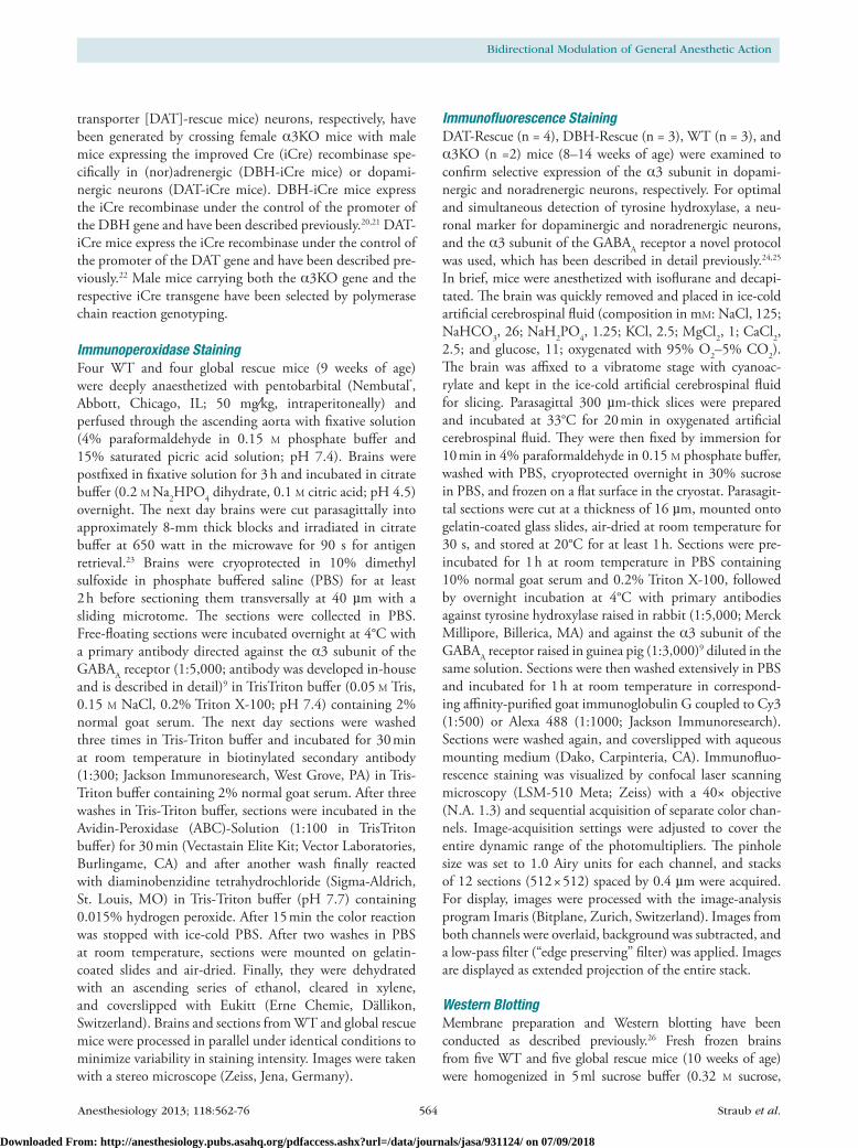
Immunoperoxidase StainingFour Wt and four global rescue mice (9 weeks of age) were deeply anaesthetized with pentobarbital (nembutal®, Abbott, chicago, IL; 50 mg⁄kg, intraperitoneally) and perfused through the ascending aorta with fixative solution (4% paraformaldehyde in 0.15 M phosphate buffer and 15% saturated picric acid solution; pH 7.4). Brains were postfixed in fixative solution for 3 h and incubated in citrate buffer (0.2 M na2HPo4 dihydrate, 0.1 M citric acid; pH 4.5) overnight. The next day brains were cut parasagittally into approximately 8-mm thick blocks and irradiated in citrate buffer at 650 watt in the microwave for 90 s for antigen retrieval.23 Brains were cryoprotected in 10% dimethyl sulfoxide in phosphate buffered saline (PBS) for at least 2 h before sectioning them transversally at 40 µm with a sliding microtome. The sections were collected in PBS. Free-floating sections were incubated overnight at 4°c with a primary antibody directed against the α3 subunit of the GABAA receptor (1:5,000; antibody was developed in-house and is described in detail)9 in tristriton buffer (0.05 M tris, 0.15 M nacl, 0.2% triton X-100; pH 7.4) containing 2% normal goat serum. The next day sections were washed three times in tris-triton buffer and incubated for 30 min at room temperature in biotinylated secondary antibody (1:300; Jackson Immunoresearch, West Grove, PA) in tris-triton buffer containing 2% normal goat serum. After three washes in tris-triton buffer, sections were incubated in the Avidin-Peroxidase (ABc)-Solution (1:100 in tristriton buffer) for 30 min (Vectastain Elite Kit; Vector Laboratories, Burlingame, cA) and after another wash finally reacted with diaminobenzidine tetrahydrochloride (Sigma-Aldrich, St. Louis, Mo) in tris-triton buffer (pH 7.7) containing 0.015% hydrogen peroxide. After 15 min the color reaction was stopped with ice-cold PBS. After two washes in PBS at room temperature, sections were mounted on gelatin-coated slides and air-dried. Finally, they were dehydrated with an ascending series of ethanol, cleared in xylene, and coverslipped with Eukitt (Erne chemie, Dällikon, Switzerland). Brains and sections from Wt and global rescue mice were processed in parallel under identical conditions to minimize variability in staining intensity. Images were taken with a stereo microscope (Zeiss, Jena, Germany).
Immunofluorescence StainingDAt-rescue (n = 4), DBH-rescue (n = 3), Wt (n = 3), and α3Ko (n =2) mice (8–14 weeks of age) were examined to confirm selective expression of the α3 subunit in dopami-nergic and noradrenergic neurons, respectively. For optimal and simultaneous detection of tyrosine hydroxylase, a neu-ronal marker for dopaminergic and noradrenergic neurons, and the α3 subunit of the GABAA receptor a novel protocol was used, which has been described in detail previously.24,25 In brief, mice were anesthetized with isoflurane and decapi-tated. The brain was quickly removed and placed in ice-cold artificial cerebrospinal fluid (composition in mM: nacl, 125; naHco3, 26; naH2Po4, 1.25; Kcl, 2.5; Mgcl2, 1; cacl2, 2.5; and glucose, 11; oxygenated with 95% o2–5% co2). The brain was affixed to a vibratome stage with cyanoac-rylate and kept in the ice-cold artificial cerebrospinal fluid for slicing. Parasagittal 300 µm-thick slices were prepared and incubated at 33°c for 20 min in oxygenated artificial cerebrospinal fluid. They were then fixed by immersion for 10 min in 4% paraformaldehyde in 0.15 M phosphate buffer, washed with PBS, cryoprotected overnight in 30% sucrose in PBS, and frozen on a flat surface in the cryostat. Parasagit-tal sections were cut at a thickness of 16 µm, mounted onto gelatin-coated glass slides, air-dried at room temperature for 30 s, and stored at 20°c for at least 1 h. Sections were pre-incubated for 1 h at room temperature in PBS containing 10% normal goat serum and 0.2% triton X-100, followed by overnight incubation at 4°c with primary antibodies against tyrosine hydroxylase raised in rabbit (1:5,000; Merck Millipore, Billerica, MA) and against the α3 subunit of the GABAA receptor raised in guinea pig (1:3,000)9 diluted in the same solution. Sections were then washed extensively in PBS and incubated for 1 h at room temperature in correspond-ing affinity-purified goat immunoglobulin G coupled to cy3 (1:500) or Alexa 488 (1:1000; Jackson Immunoresearch). Sections were washed again, and coverslipped with aqueous mounting medium (Dako, carpinteria, cA). Immunofluo-rescence staining was visualized by confocal laser scanning microscopy (LSM-510 Meta; Zeiss) with a 40× objective (n.A. 1.3) and sequential acquisition of separate color chan-nels. Image-acquisition settings were adjusted to cover the entire dynamic range of the photomultipliers. The pinhole size was set to 1.0 Airy units for each channel, and stacks of 12 sections (512 × 512) spaced by 0.4 µm were acquired. For display, images were processed with the image-analysis program Imaris (Bitplane, Zurich, Switzerland). Images from both channels were overlaid, background was subtracted, and a low-pass filter (“edge preserving” filter) was applied. Images are displayed as extended projection of the entire stack.
Western BlottingMembrane preparation and Western blotting have been conducted as described previously.26 Fresh frozen brains from five Wt and five global rescue mice (10 weeks of age) were homogenized in 5 ml sucrose buffer (0.32 M sucrose,
Downloaded From: http://anesthesiology.pubs.asahq.org/pdfaccess.ashx?url=/data/journals/jasa/931124/ on 07/09/2018
Anesthesiology 2013; 118:562-76 565 Straub et al.
PERIOPERATIVE MEDICINE
5 mM ethylenediaminetetraacetic acid, 10 mM tris pH 7.5, 0.02% nan3, phenylmethanesulfonyl fluoride 1:500, complete, mini protease inhibitor cocktail [roche Diagnostics, Mannheim, Germany]) and centrifuged for 10 min at 1,000g. The supernatant was collected and the step repeated. Both supernatants were combined and centrifuged for 20 min at 40,000g to obtain the crude membrane fraction. The pellet was resuspended in 3 ml sucrose buffer. Protein concentration was determined using the BcA protein assay kit (Pierce, rockford, IL). Samples were incubated for 5 min at 95°c with an equal volume of 125 mM tris-Hcl pH 6.8, 20% glycerol, 0.002% bromphenol blue, 10% β-mercaptoethanol, 4% sodium dodecyl sulfate and stored at −20°c. Before use samples were incubated for 10 min at 60°c and diluted to a concentration of 1 mg/ml. Aliquots with increasing protein content (2.5, 5, 7.5, and 10 μg) were subjected to sodium dodecyl sulfate polyacrylamide gel electrophoresis using 10% minigels (Mini protean II; Bio-rad, Hercules, cA). Proteins were transferred onto nitrocellulose membranes using a trans Blot Mini cell (Bio-rad). The blots were blocked for 1–2 h in 10 mM tris-Hcl pH 8, 0.15 M nacl, 0.05% tween 20 containing 5% nonfat dry milk at room temperature, followed by incubation with affinity-purified antiserum against the α3 subunit of the GABAA receptor (1:500)9 together with a monoclonal antibody directed against β-actin (1:20,000, Merck Millipore) overnight at 4°c in 10 mM tris-Hcl pH 8, 0.15 M nacl, 0.05% tween 20/5% nonfat dry milk. The blots were washed once with 20 mM tris pH 7.5, 60 mM nacl, 2 mM ethylenediaminetetraacetic acid, 0.4% SDS, 0.4% triton-X 100, and 0.4% deoxycholate and four times with 10 mM tris-Hcl pH 8, 0.15 M nacl, 0.05% tween 20. Incubation with the appropriate horseradish peroxidase-conjugated secondary antibodies was carried out for 1 h at room temperature. After extensive washing immunoreactivity was detected by the enhanced chemoluminescence method (Super Signal West Pico chemoluminescence, Pierce), images were captured using a Fujifilm (tokyo, Japan) LAS-1000 Plus Gel Documentation System, and immunoreactive bands were quantified with the AIDA software (Version 3.25, raytest, Pforzheim, Germany). Actin immunoreactivity was used to monitor equal sample loading. The specificity of the antibody against the α3 subunit has been confirmed previously using α3Ko mice.18
DrugsKetamine-Hcl/Xylazine-Hcl solution (Sigma-Aldrich) was administered at a volume of 20 ml/kg. Ketamine-Hcl (Sigma-Aldrich), medetomidine-Hcl solution (Domitor®, Pfizer Animal Health, new york, ny) and pentobarbital-na (Sigma-Aldrich) were administered at a volume of 10 ml/kg. Etomidate (Amidate®, Hospira, Lake Forest, IL) was admin-istered at a volume of 15 ml/kg. Midazolam-Hcl solution (Bedford Laboratories, Bedford, oH) was administered at a volume of 16.5 ml/kg. All drugs were dissolved or diluted in physiological (0.9%) saline where necessary and drug doses were calculated as mg/kg base. All drugs were administered
by intraperitoneal injection. The doses administered are reported in the results section.
Behavioral Testingtests were conducted during the dark phase (9:00 AM–9:00 PM) and each test was conducted at approximately the same time of day. The tester was blind to the genotype of the animals. Animals were tested in three cohorts. cohort 1 containing Wt, α3Ko, DBH-rescue, and DBH-icre mice received a single injection of ketamine/xylazine and anesthetic properties were observed. cohort 2 containing Wt and α3Ko mice were first tested for medetomidine-induced seda-tion in the locomotor activity test, 3 days later for medetomi-dine-induced hypothermia, and 4–6 days later for anesthetic properties of ketamine, medetomidine, or pentobarbital. Ani-mals received increasing doses of ketamine, medetomidine, or pentobarbital every 7 days. Mice were counterbalanced for drug experience. cohort 3 containing Wt and α3Ko mice were first tested for anesthetic properties of midazolam and 7 days later for anesthetic properties of etomidate.
Locomotor ActivityAnimals were moved into the testing room at least 1 h before testing. Animals were injected with medetomidine (30 µg/kg or 60 µg/kg) or vehicle immediately before testing. The test-ing apparatus consisted of four evenly illuminated (approxi-mately 130 lux) clear plexiglas (J. Freeman, Inc., Dorchester, MA) boxes (41 × 41 × 31 cm) separated by white cardboard. Boxes were cleaned with 70% ethanol, between animals. Locomotor activity was recorded for 60 min using the video-tracking system EthoVisionXt (noldus Information tech-nology, Wageningen, The netherlands).
Medetomidine-induced HypothermiaAnimals were moved into the testing room directly before testing. Body temperature was determined using a tH-5 digital thermometer (Physitemp Instruments, clifton, nJ) with a rEt3-Iso thermal probe for mice (inserted 2 cm deep) immediately before drug injection (vehicle, 60 µg/kg, 120 µg/kg medetomidine) and 30, 60, 90, and 120 min thereaf-ter. Mice were kept together in their home cage throughout the experiment.
Anesthetic EndpointsThe assessment of anesthetic end points was adapted from published protocols.27–29 Animals were moved into the test-ing room directly before testing. Each animal was placed in a clean holding cage without bedding material immediately after drug injection. Body temperature was determined using a tH-5 digital thermometer (Physitemp Instru-ments) with an rEt3-Iso thermal probe for mice (inserted 2 cm deep) immediately before drug injection and every 30 min thereafter. once the animal stopped moving it was turned on its back. Beginning of loss of righting reflex (Lorr) was defined as the time-point when the animal
Downloaded From: http://anesthesiology.pubs.asahq.org/pdfaccess.ashx?url=/data/journals/jasa/931124/ on 07/09/2018
Anesthesiology 2013; 118:562-76 566 Straub et al.
Bidirectional Modulation of General Anesthetic Action
was placed on its back. End of Lorr was defined as the time-point when the animal righted itself spontaneously from dorsal to sternal recumbency with all four extremities touching the ground. The time between beginning and end of Lorr was defined as duration of Lorr. After regain-ing the righting reflex animals were turned on their back every minute and time to righting was scored: less than 2 s (score = 1), between 2 and 10 s (score = 2), more than 10 s (score = 3). Animals were defined as having fully recovered when they were able to right themselves three times con-secutively within 2 s. time to recovery was defined as the time between beginning of Lorr and full recovery. A total Lorr score was calculated by adding the score values for every minute from beginning of Lorr until full recov-ery. The loss of hind limb withdrawal reflex (LHWr) was determined every 5 min by pinching the paw firmly with an atraumatic forceps alternating between the left and right hind paw until animals regained the righting reflex. LHWr was defined as present if no signs of reaction to the pinch were observed.
Anesthetic properties of medetomidine were assessed using a different protocol. Animals were moved into the testing room directly before testing and each animal was placed in a clean holding cage without bedding material immediately after drug injection. Animals were gently rolled on their side 30, 60, 90, and 120 min after injection and time to righting defined as completely returning to the prone position with all four extremities touching the ground was noted. Lorr was defined as present if animals were not able to roll back to the prone position within 10 s.
Statistical AnalysisData were analyzed using the appropriate statistical test (Student t test, AnoVA followed by post hoc Dunnett t test, AnoVA with repeated measures followed by post hoc t tests with Bonferroni correction) or Sheffé post hoc test. Hypothesis testing was two-tailed. Data for medetomidine-induced Lorr were analyzed by fitting standard least-squares mixed models with subject as a random effect. Drug dose and genotype were included as fixed effects. All effects were assessed by F tests based on the fitted regression models. This analysis was followed by post hoc Mann–Whitney u tests. Data are expressed as the mean ± SEM. P value less than 0.05 was considered to be statistically significant. Statistical analysis was performed using the SPSS (SPSS, Inc., chicago, IL) and GraphPad (GraphPad Software, Inc., La Jolla, cA) software packages.
ResultsWhen performing stereotaxic surgeries for implantation of electrodes into the medial forebrain bundle under ketamine/xylazine (139/21 mg/kg intraperitoneally) anesthesia, we unexpectedly observed that using the same doses of anes-thetics α3Ko mice were anesthetized longer than Wt mice
(approximately 130 min compared with 80 min), prompting us to investigate the role of α3-containing GABAA receptors in the action of intravenous general anesthetics.
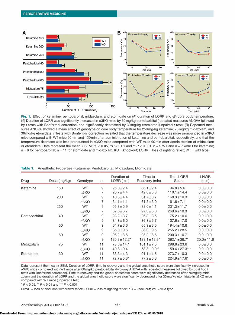
Anesthetic Endpoints with Etomidate, Midazolam, and PentobarbitalThe anesthetic effects of etomidate, midazolam and pento-barbital, three compounds known to act at least in part via GABAA receptors, were assessed by determining the follow-ing endpoints: duration of Lorr, time to recovery and total Lorr score as a measure of hypnosis, LHWr as a measure of immobility, and body temperature as a measure of hypo-thermic effects of the drug (fig. 1 and table 1).
EtomidateEtomidate (30 mg/kg) was less effective in α3Ko mice compared with Wt mice. Duration of Lorr (Wt: 88.3 ± 4.3 min, α3Ko: 72.7 ± 5.8 min) and total Lorr score (Wt: 273.7 ± 10.3, α3Ko: 224.9 ± 17.5) were significantly decreased in α3Ko mice (unpaired t test, P = 0.0439 and P = 0.0263, respectively), whereas time to recovery was not dif-ferent (Wt: 91.1 ± 4.5 min, α3Ko: 77.2 ± 5.8 min, unpaired t test, P = 0.0710). Anesthetic-induced decrease in body temperature was smaller in α3Ko mice compared with Wt controls (two-way AnoVA with repeated measures of time: significant effect of time F(2,40), P < 0.0001; significant effect of genotype F(1,20) = 4.90, P = 0.0386; significant time × genotype interaction F(2,40) = 8.12, P = 0.0011). Post hoc t tests with Bonferroni correction showed that the temperature differed significantly 90 min after injection (P < 0.01).
MidazolamThe hypnotic action of midazolam (75 mg/kg) was decreased in α3Ko mice compared with Wt mice. Although the difference in the duration of Lorr failed to reach significance (unpaired t test, P = 0.0691), the time to recovery (Wt: 101.1 ± 7.5 min, α3Ko: 53.8 ± 9.9 min) and the total Lorr score (Wt: 298.8 ± 23.6, α3Ko: 159.4 ± 27.3) were significantly lower in α3Ko mice (unpaired t test, P = 0.0011 and P = 0.001, respectively). Similarly, the reduction in body temperature was smaller in α3Ko mice (two-way AnoVA with repeated measures of time: significant effect of time F(2,40) = 17.65, P < 0.0001; significant effect of genotype F(1,20), P = 0.0395 and significant time × genotype interaction F(2,40), P = 0.0008). Post hoc t tests with Bonferroni correction showed that the temperature difference was significant 90 min after injection (P < 0.01).
PentobarbitalFor pentobarbital two-way AnoVAs using drug dose and genotype as factors revealed main effects of drug and geno-type on Lorr (drug F(2,32) = 160.92, P < 0.0001; geno-type F(1,16) = 5.64, P = 0.304), on time to recovery (drug F(2,32) = 163.15, P < 0.0001; genotype F(1,16) = 5.51, P = 0.0321) and on total Lorr score (drug F(2,32) = 158.06,
Downloaded From: http://anesthesiology.pubs.asahq.org/pdfaccess.ashx?url=/data/journals/jasa/931124/ on 07/09/2018
Anesthesiology 2013; 118:562-76 567 Straub et al.
PERIOPERATIVE MEDICINE
Table 1. Anesthetic Properties (Ketamine, Pentobarbital, Midazolam, Etomidate)
Drug Dose (mg/kg) Genotype nDuration of LORR (min)
Time to Recovery (min)
Total LORR Score
LHWR (min)
Ketamine 150 WT 9 25.0 ± 2.4 36.1 ± 2.4 94.8 ± 5.6 0.0 ± 0.0α3KO 7 26.7 ± 4.4 42.0 ± 5.3 110.1 ± 14.4 0.0 ± 0.0
200 WT 9 40.3 ± 4.4 61.7 ± 3.7 168.3 ± 10.3 0.0 ± 0.0α3KO 7 34.1 ± 1.1 61.3 ± 3.0 161.6 ± 7.1 0.0 ± 0.0
250 WT 9 56.8 ± 5.9 83.0 ± 4.1 231.3 ± 11.7 0.0 ± 0.0α3KO 7 62.6 ± 6.7 97.3 ± 5.8 269.6 ± 18.3 0.0 ± 0.0
Pentobarbital 40 WT 9 23.2 ± 3.7 26.3 ± 3.5 75.2 ± 10.6 0.0 ± 0.0α3KO 9 34.8 ± 6.0 36.8 ± 5.7 107.6 ± 17.0 0.0 ± 0.0
50 WT 9 64.7 ± 3.6 65.9 ± 3.5 194.4 ± 10.6 0.0 ± 0.0α3KO 9 85.0 ± 9.5 86.0 ± 9.5 255.2 ± 28.5 0.0 ± 0.0
60 WT 9 96.2 ± 3.6 98.2 ± 3.6 290.3 ± 10.7 0.0 ± 0.0α3KO 9 126.8 ± 12.2* 129.1 ± 12.3* 382.1 ± 36.7* 25.0 ± 11.6
Midazolam 75 WT 11 73.5 ± 14.1 101.1 ± 7.5 298.8 ± 23.6 0.0 ± 0.0α3KO 11 40.8 ± 9.4 53.8 ± 9.9** 159.4 ± 27.3*** 0.0 ± 0.0
Etomidate 30 WT 11 88.3 ± 4.3 91.1 ± 4.5 273.7 ± 10.3 0.0 ± 0.0α3KO 11 72.7 ± 5.8* 77.2 ± 5.8 224.9 ± 17.6* 0.0 ± 0.0
Data represent the mean ± SEM. Duration of LORR, time to recovery and the global anesthetic score were significantly increased in α3KO mice compared with WT mice after 60 mg/kg pentobarbital (two-way ANOVA with repeated measures followed by post hoc t tests with Bonferroni correction). Time to recovery and the global anesthetic score were significantly decreased after 75 mg/kg mida-zolam and the duration of LORR and the global anesthetic score were significantly decreased after 30 mg/kg etomidate in α3KO mice compared with WT mice (unpaired t test).* P < 0.05, ** P < 0.01 and *** P < 0.001.LHWR = loss of hind limb withdrawal reflex; LORR = loss of righting reflex; KO = knockout; WT = wild type.
Fig. 1. Effect of ketamine, pentobarbital, midazolam, and etomidate on (A) duration of LORR and (B) core body temperature. (A) Duration of LORR was significantly increased in α3KO mice by 60 mg/kg pentobarbital (repeated measures ANOVA followed by t tests with Bonferroni correction) and significantly decreased by 30 mg/kg etomidate (unpaired t test). (B) Repeated mea-sures ANOVA showed a mean effect of genotype on core body temperature for 250 mg/kg ketamine, 75 mg/kg midazolam, and 30 mg/kg etomidate. t Tests with Bonferroni correction revealed that the temperature decrease was more pronounced in α3KO mice compared with WT mice 60 min and 120 min after administration of ketamine and pentobarbital, respectively, and that the temperature decrease was less pronounced in α3KO mice compared with WT mice 90 min after administration of midazolam or etomidate. Data represent the mean ± SEM; *P < 0.05, **P < 0.01 and ***P < 0.001, n = 9 WT and n = 7 α3KO for ketamine; n = 9 for pentobarbital; n = 11 for etomidate and midazolam. KO = knockout; LORR = loss of righting reflex; WT = wild type.
Downloaded From: http://anesthesiology.pubs.asahq.org/pdfaccess.ashx?url=/data/journals/jasa/931124/ on 07/09/2018
Anesthesiology 2013; 118:562-76 568 Straub et al.
Bidirectional Modulation of General Anesthetic Action
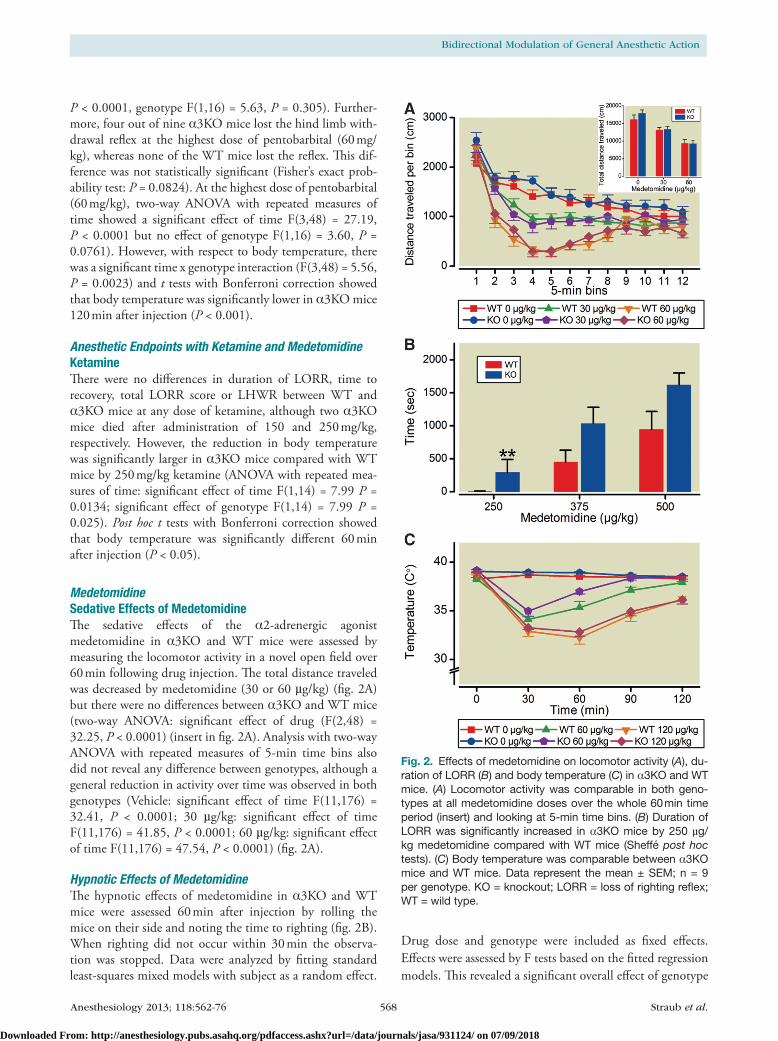
P < 0.0001, genotype F(1,16) = 5.63, P = 0.305). Further-more, four out of nine α3Ko mice lost the hind limb with-drawal reflex at the highest dose of pentobarbital (60 mg/kg), whereas none of the Wt mice lost the reflex. This dif-ference was not statistically significant (Fisher’s exact prob-ability test: P = 0.0824). At the highest dose of pentobarbital (60 mg/kg), two-way AnoVA with repeated measures of time showed a significant effect of time F(3,48) = 27.19, P < 0.0001 but no effect of genotype F(1,16) = 3.60, P = 0.0761). However, with respect to body temperature, there was a significant time x genotype interaction (F(3,48) = 5.56, P = 0.0023) and t tests with Bonferroni correction showed that body temperature was significantly lower in α3Ko mice 120 min after injection (P < 0.001).
Anesthetic Endpoints with Ketamine and MedetomidineKetamineThere were no differences in duration of Lorr, time to recovery, total Lorr score or LHWr between Wt and α3Ko mice at any dose of ketamine, although two α3Ko mice died after administration of 150 and 250 mg/kg, respectively. However, the reduction in body temperature was significantly larger in α3Ko mice compared with Wt mice by 250 mg/kg ketamine (AnoVA with repeated mea-sures of time: significant effect of time F(1,14) = 7.99 P = 0.0134; significant effect of genotype F(1,14) = 7.99 P = 0.025). Post hoc t tests with Bonferroni correction showed that body temperature was significantly different 60 min after injection (P < 0.05).
MedetomidineSedative Effects of MedetomidineThe sedative effects of the α2-adrenergic agonist medetomidine in α3Ko and Wt mice were assessed by measuring the locomotor activity in a novel open field over 60 min following drug injection. The total distance traveled was decreased by medetomidine (30 or 60 µg/kg) (fig. 2A) but there were no differences between α3Ko and Wt mice (two-way AnoVA: significant effect of drug (F(2,48) = 32.25, P < 0.0001) (insert in fig. 2A). Analysis with two-way AnoVA with repeated measures of 5-min time bins also did not reveal any difference between genotypes, although a general reduction in activity over time was observed in both genotypes (Vehicle: significant effect of time F(11,176) = 32.41, P < 0.0001; 30 µg/kg: significant effect of time F(11,176) = 41.85, P < 0.0001; 60 µg/kg: significant effect of time F(11,176) = 47.54, P < 0.0001) (fig. 2A).
Hypnotic Effects of MedetomidineThe hypnotic effects of medetomidine in α3Ko and Wt mice were assessed 60 min after injection by rolling the mice on their side and noting the time to righting (fig. 2B). When righting did not occur within 30 min the observa-tion was stopped. Data were analyzed by fitting standard least-squares mixed models with subject as a random effect.
Drug dose and genotype were included as fixed effects. Effects were assessed by F tests based on the fitted regression models. This revealed a significant overall effect of genotype
Fig. 2. Effects of medetomidine on locomotor activity (A), du-ration of LORR (B) and body temperature (C) in α3KO and WT mice. (A) Locomotor activity was comparable in both geno-types at all medetomidine doses over the whole 60 min time period (insert) and looking at 5-min time bins. (B) Duration of LORR was significantly increased in α3KO mice by 250 µg/kg medetomidine compared with WT mice (Sheffé post hoc tests). (C) Body temperature was comparable between α3KO mice and WT mice. Data represent the mean ± SEM; n = 9 per genotype. KO = knockout; LORR = loss of righting reflex; WT = wild type.
Downloaded From: http://anesthesiology.pubs.asahq.org/pdfaccess.ashx?url=/data/journals/jasa/931124/ on 07/09/2018
Anesthesiology 2013; 118:562-76 569 Straub et al.
PERIOPERATIVE MEDICINE
(F[1,16] = 8.90, P = 0.0088) and dose (F[1,34] = 36.96, P < 0.0001). Post hoc Sheffé tests showed that the Lorr was significantly increased in α3Ko mice (298 ± 192 s) com-pared with Wt mice (6 ± 4 s) by 250 µg/kg medetomidine (P = 0.0098). At this dose, two of nine Wt mice lost the righting reflex (loss of righting reflex defined as inability to right within 10 s) in contrast to seven of nine α3Ko mice.
Hypothermic Effects of MedetomidineThe hypothermic effects of medetomidine in α3Ko and Wt mice were assessed by determining the rectal body tem-perature immediately before drug or vehicle injection and every 30 min thereafter over a period of 120 min (fig. 2c).
The baseline body temperature before injection was increased in α3Ko mice compared with Wt mice in the groups that received vehicle (Wt = 38.29 ± 0.11°c, α3Ko = 39.01 ± 0.18°c, unpaired t test, P < 0.01) and 60 µg/kg medetomidine (Wt = 38.297 ± 0.15°c, α3Ko = 39.13 ± 0.17°c, unpaired t test, P < 0.01) in α3Ko mice (39.0 ± 0.2°c), but not in the group that received 120 µg/kg medetomidine (Wt = 38.82 ± 0.14°c, α3Ko = 38.97 ± 0.09°c, independent samples t test, P = 0.398). This precluded direct statistical analysis of the hypothermic effects of 60 µg/kg medetomidine. For the 120µg/kg medetomidine dose, a genotype × time two-way AnoVA did not reveal a significant genotype main
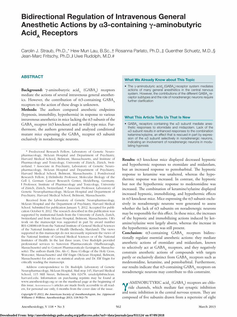
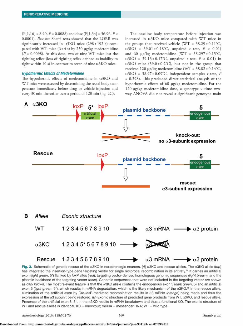
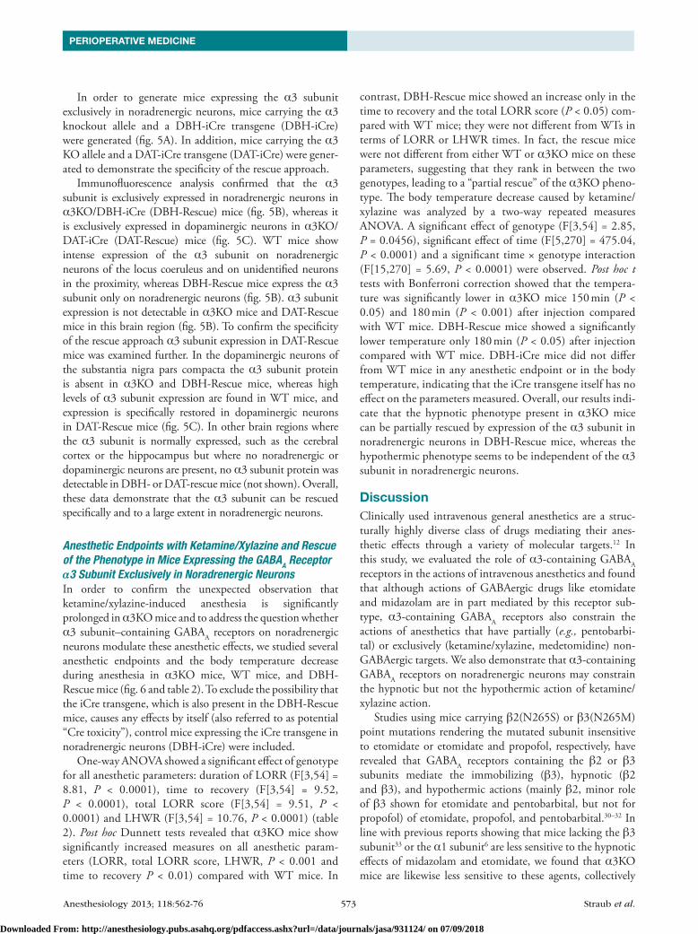
Fig. 3. Schematic of genetic rescue of the α3KO in noradrenergic neurons. (A) α3KO and rescue alleles. The α3KO allele (top) has integrated the insertion-type gene targeting vector for single reciprocal recombination in its entirety.18 It carries an artificial exon (light green, 5*) flanked by loxP sites (red), targeting vector-derived homologous genomic sequences (light brown), and the plasmid backbone of the targeting vector (blue). Genomic sequences that were not included in the targeting vector are shown as dark brown. The most relevant feature is that the α3KO allele contains the endogenous exon 5 (dark green, 5) and an artificial exon 5 (light green, 5*), which results in mRNA degradation, which is the likely mechanism of the α3KO.18 In the rescue allele, elimination of the artificial exon by Cre-loxP-mediated recombination results in α3 mRNA (orange) being made and thus the expression of the α3 subunit being restored. (B) Exonic structure of predicted gene products from WT, α3KO, and rescue allele. Presence of the artificial exon 5, 5*, in the α3KO results in mRNA breakdown and thus a functional KO. The exonic structure of WT and rescue alleles is identical. KO = knockout; mRNA = messenger RNA; WT = wild type.
Downloaded From: http://anesthesiology.pubs.asahq.org/pdfaccess.ashx?url=/data/journals/jasa/931124/ on 07/09/2018
Anesthesiology 2013; 118:562-76 570 Straub et al.
Bidirectional Modulation of General Anesthetic Action
effect or genotype × time interaction. In the entire study, a difference in baseline temperature was only present in 2 of the 11 experiments performed in total with midazolam, pentobarbital, and ketamine. We therefore assume that extraneous undefined factors specific to the experiments with medetomidine are responsible for this baseline
difference, and that overall Wt mice and α3Ko mice do not display a difference in baseline body temperature. When we analyzed the change in body temperature from baseline, we still did not obtain any significant differences between genotypes both for 60 µg/kg and for 120 µg/kg medetomidine (not shown).
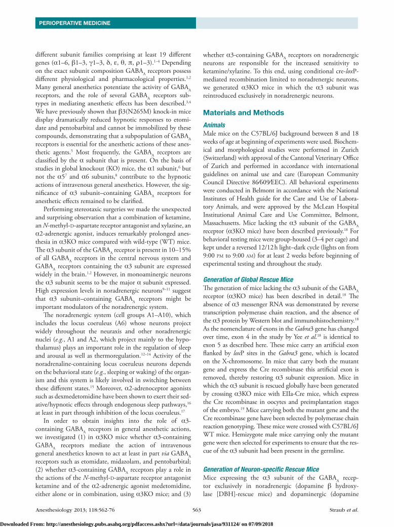
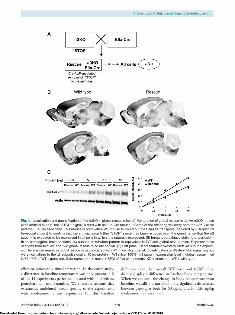
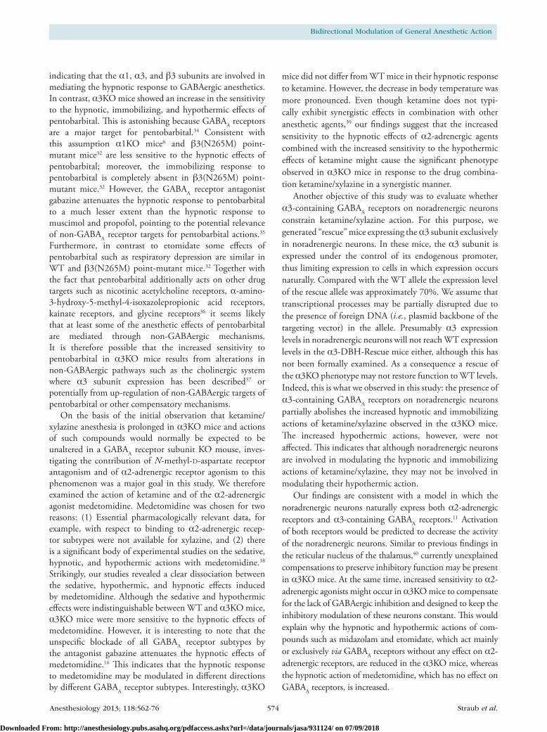
Fig. 4. Localization and quantification of the α3KO in global rescue mice. (A) Generation of global rescue mice. An α3KO mouse (with artificial exon 5, the “STOP” signal) is bred with an EIIa-Cre mouse.19 Some of the offspring will carry both the α3KO allele and the EIIa-Cre transgene. This mouse is bred with a WT mouse to breed out the EIIa-Cre transgene (depicted by 2 sequential horizontal arrows) to confirm that the artificial exon 5 (the “STOP” signal) has been removed from the germline, so that the α3 subunit is expected to be expressed in all cells in which it is naturally expressed. (B) Immunoperoxidase staining of perfusion-fixed parasagittal brain sections: α3 subunit distribution pattern is equivalent in WT and global rescue mice. Representative sections from two WT and two global rescue mice are shown. (C) Left panel: Representative Western Blot: α3 subunit expres-sion level is decreased in global rescue mice compared with WT mice. Right panel: Quantification of Western blot signal: signals were normalized to the α3 subunit signal at 10 μg protein in WT mice (100%). α3 subunit expression level in global rescue mice is 70 ± 7% of WT expression. Data represent the mean ± SEM of five experiments. KO = knockout; WT = wild type.
Downloaded From: http://anesthesiology.pubs.asahq.org/pdfaccess.ashx?url=/data/journals/jasa/931124/ on 07/09/2018
Anesthesiology 2013; 118:562-76 571 Straub et al.
PERIOPERATIVE MEDICINE
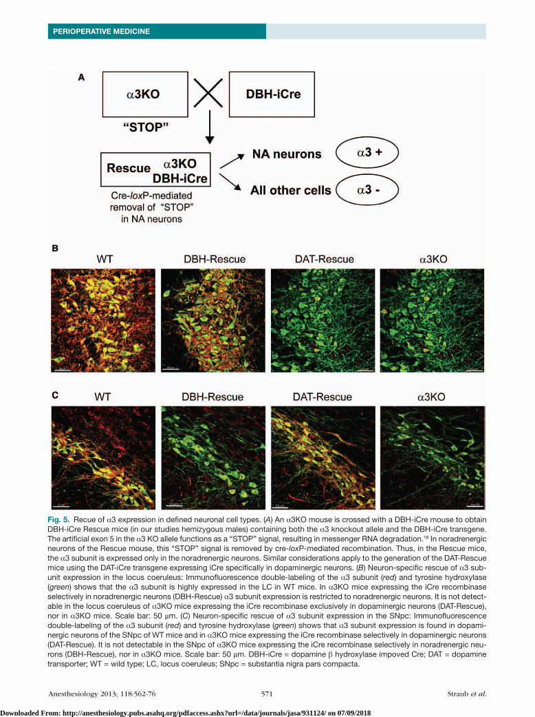
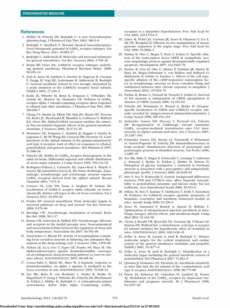
Fig. 5. Recue of α3 expression in defined neuronal cell types. (A) An α3KO mouse is crossed with a DBH-iCre mouse to obtain DBH-iCre Rescue mice (in our studies hemizygous males) containing both the α3 knockout allele and the DBH-iCre transgene. The artificial exon 5 in the α3 KO allele functions as a “STOP” signal, resulting in messenger RNA degradation.18 In noradrenergic neurons of the Rescue mouse, this “STOP” signal is removed by cre-loxP-mediated recombination. Thus, in the Rescue mice, the α3 subunit is expressed only in the noradrenergic neurons. Similar considerations apply to the generation of the DAT-Rescue mice using the DAT-iCre transgene expressing iCre specifically in dopaminergic neurons. (B) Neuron-specific rescue of α3 sub-unit expression in the locus coeruleus: Immunofluorescence double-labeling of the α3 subunit (red) and tyrosine hydroxylase (green) shows that the α3 subunit is highly expressed in the LC in WT mice. In α3KO mice expressing the iCre recombinase selectively in noradrenergic neurons (DBH-Rescue) α3 subunit expression is restricted to noradrenergic neurons. It is not detect-able in the locus coeruleus of α3KO mice expressing the iCre recombinase exclusively in dopaminergic neurons (DAT-Rescue), nor in α3KO mice. Scale bar: 50 µm. (C) Neuron-specific rescue of α3 subunit expression in the SNpc: Immunofluorescence double-labeling of the α3 subunit (red) and tyrosine hydroxylase (green) shows that α3 subunit expression is found in dopami-nergic neurons of the SNpc of WT mice and in α3KO mice expressing the iCre recombinase selectively in dopaminergic neurons (DAT-Rescue). It is not detectable in the SNpc of α3KO mice expressing the iCre recombinase selectively in noradrenergic neu-rons (DBH-Rescue), nor in α3KO mice. Scale bar: 50 µm. DBH-iCre = dopamine β hydroxylase impoved Cre; DAT = dopamine transporter; WT = wild type; LC, locus coeruleus; SNpc = substantia nigra pars compacta.
Downloaded From: http://anesthesiology.pubs.asahq.org/pdfaccess.ashx?url=/data/journals/jasa/931124/ on 07/09/2018
Anesthesiology 2013; 118:562-76 572 Straub et al.
Bidirectional Modulation of General Anesthetic Action
Generation of Mice Expressing the GABAA Receptor α3 Subunit Exclusively in Noradrenergic Neuronsα3Ko mice used in this study have been generated using an insertion targeting strategy (fig. 1 in reference 18) with an insertion-type targeting vector containing exon 5 of the Gabra3 gene flanked by two loxP sites. This vector was inserted into the target locus in its entirety by single reciprocal recombination, resulting in a duplication of exon 5 (i.e., exon 4 in fig. 1 in the study by yee et al.).18 The resulting α3Ko mice carry both an endogenous exon 5 and an artificial exon 5* (5* in fig. 3A). This artificial exon 5* provides a “StoP” signal functionally disrupting the Gabra3 gene (fig. 3, A and B). We predicted that a cre-loxP-mediated excision of this loxP-flanked artificial exon 5* would functionally restore the Gabra3 gene. In a proof-of-principle experiment, α3Ko mice were crossed with EIIa-cre mice, which express the cre
transgene at early embryonic stages19 (fig. 4A). As predicted, cre-loxP-mediated recombination resulted in the elimination of the loxP-flanked artificial exon 5*. Subsequently, the Ella-cre transgene was bred out to ensure that the rescue of the α3 subunit had been present in the germline. These global rescue mice were examined for α3 subunit expression. Immu-nohistochemical analysis revealed that α3 subunit expression is restored in these global rescue mice and the distribution pattern of α3 subunit expression is indistinguishable from Wt mice (fig. 4B). Western blot analysis showed that α3 subunit expression levels in global rescue mice are 70 ± 7% compared with Wt mice (fig. 4c). Thus, the genomic rear-rangement in the rescue allele, which includes the presence of a plasmid backbone, reduces the expression of the α3 subunit by approximately 30% without affecting its regional distribu-tion. The precise mechanisms of this reduction are unknown.
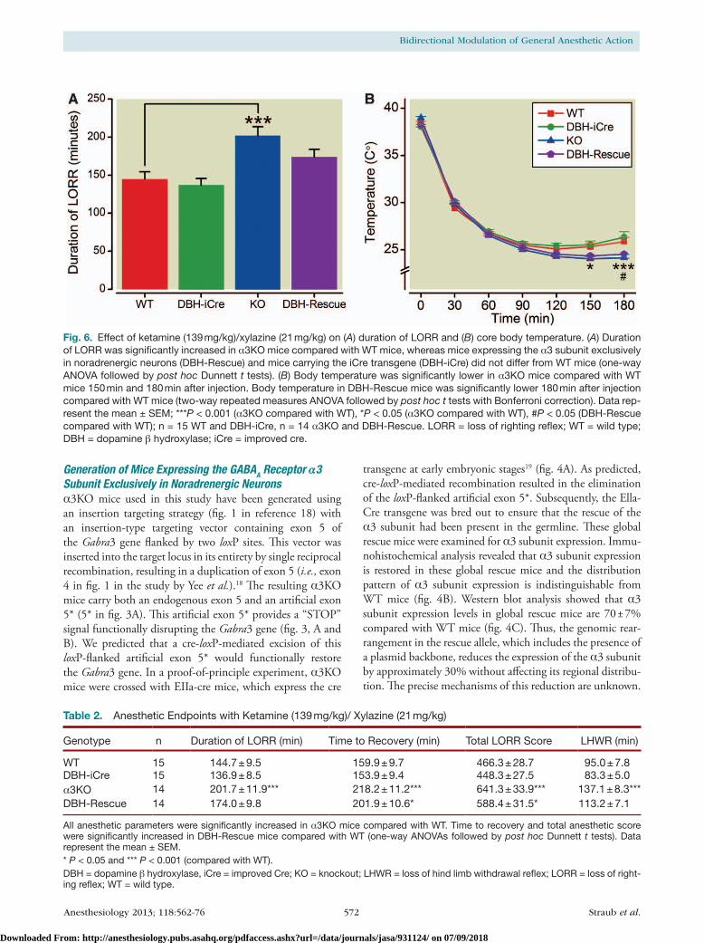
Fig. 6. Effect of ketamine (139 mg/kg)/xylazine (21 mg/kg) on (A) duration of LORR and (B) core body temperature. (A) Duration of LORR was significantly increased in α3KO mice compared with WT mice, whereas mice expressing the α3 subunit exclusively in noradrenergic neurons (DBH-Rescue) and mice carrying the iCre transgene (DBH-iCre) did not differ from WT mice (one-way ANOVA followed by post hoc Dunnett t tests). (B) Body temperature was significantly lower in α3KO mice compared with WT mice 150 min and 180 min after injection. Body temperature in DBH-Rescue mice was significantly lower 180 min after injection compared with WT mice (two-way repeated measures ANOVA followed by post hoc t tests with Bonferroni correction). Data rep-resent the mean ± SEM; ***P < 0.001 (α3KO compared with WT), *P < 0.05 (α3KO compared with WT), #P < 0.05 (DBH-Rescue compared with WT); n = 15 WT and DBH-iCre, n = 14 α3KO and DBH-Rescue. LORR = loss of righting reflex; WT = wild type; DBH = dopamine β hydroxylase; iCre = improved cre.
Table 2. Anesthetic Endpoints with Ketamine (139 mg/kg)/ Xylazine (21 mg/kg)
Genotype n Duration of LORR (min) Time to Recovery (min) Total LORR Score LHWR (min)
WT 15 144.7 ± 9.5 159.9 ± 9.7 466.3 ± 28.7 95.0 ± 7.8DBH-iCre 15 136.9 ± 8.5 153.9 ± 9.4 448.3 ± 27.5 83.3 ± 5.0α3KO 14 201.7 ± 11.9*** 218.2 ± 11.2*** 641.3 ± 33.9*** 137.1 ± 8.3***DBH-Rescue 14 174.0 ± 9.8 201.9 ± 10.6* 588.4 ± 31.5* 113.2 ± 7.1
All anesthetic parameters were significantly increased in α3KO mice compared with WT. Time to recovery and total anesthetic score were significantly increased in DBH-Rescue mice compared with WT (one-way ANOVAs followed by post hoc Dunnett t tests). Data represent the mean ± SEM.* P < 0.05 and *** P < 0.001 (compared with WT).DBH = dopamine β hydroxylase, iCre = improved Cre; KO = knockout; LHWR = loss of hind limb withdrawal reflex; LORR = loss of right-ing reflex; WT = wild type.
Downloaded From: http://anesthesiology.pubs.asahq.org/pdfaccess.ashx?url=/data/journals/jasa/931124/ on 07/09/2018
Anesthesiology 2013; 118:562-76 573 Straub et al.
PERIOPERATIVE MEDICINE
In order to generate mice expressing the α3 subunit exclusively in noradrenergic neurons, mice carrying the α3 knockout allele and a DBH-icre transgene (DBH-icre) were generated (fig. 5A). In addition, mice carrying the α3 Ko allele and a DAt-icre transgene (DAt-icre) were gener-ated to demonstrate the specificity of the rescue approach.
Immunofluorescence analysis confirmed that the α3 subunit is exclusively expressed in noradrenergic neurons in α3Ko/DBH-icre (DBH-rescue) mice (fig. 5B), whereas it is exclusively expressed in dopaminergic neurons in α3Ko/DAt-icre (DAt-rescue) mice (fig. 5c). Wt mice show intense expression of the α3 subunit on noradrenergic neurons of the locus coeruleus and on unidentified neurons in the proximity, whereas DBH-rescue mice express the α3 subunit only on noradrenergic neurons (fig. 5B). α3 subunit expression is not detectable in α3Ko mice and DAt-rescue mice in this brain region (fig. 5B). to confirm the specificity of the rescue approach α3 subunit expression in DAt-rescue mice was examined further. In the dopaminergic neurons of the substantia nigra pars compacta the α3 subunit protein is absent in α3Ko and DBH-rescue mice, whereas high levels of α3 subunit expression are found in Wt mice, and expression is specifically restored in dopaminergic neurons in DAt-rescue mice (fig. 5c). In other brain regions where the α3 subunit is normally expressed, such as the cerebral cortex or the hippocampus but where no noradrenergic or dopaminergic neurons are present, no α3 subunit protein was detectable in DBH- or DAt-rescue mice (not shown). overall, these data demonstrate that the α3 subunit can be rescued specifically and to a large extent in noradrenergic neurons.
Anesthetic Endpoints with Ketamine/Xylazine and Rescue of the Phenotype in Mice Expressing the GABAA Receptor α3 Subunit Exclusively in Noradrenergic NeuronsIn order to confirm the unexpected observation that ketamine/xylazine-induced anesthesia is significantly prolonged in α3Ko mice and to address the question whether α3 subunit–containing GABAA receptors on noradrenergic neurons modulate these anesthetic effects, we studied several anesthetic endpoints and the body temperature decrease during anesthesia in α3Ko mice, Wt mice, and DBH-rescue mice (fig. 6 and table 2). to exclude the possibility that the icre transgene, which is also present in the DBH-rescue mice, causes any effects by itself (also referred to as potential “cre toxicity”), control mice expressing the icre transgene in noradrenergic neurons (DBH-icre) were included.
one-way AnoVA showed a significant effect of genotype for all anesthetic parameters: duration of Lorr (F[3,54] = 8.81, P < 0.0001), time to recovery (F[3,54] = 9.52, P < 0.0001), total Lorr score (F[3,54] = 9.51, P < 0.0001) and LHWr (F[3,54] = 10.76, P < 0.0001) (table 2). Post hoc Dunnett tests revealed that α3Ko mice show significantly increased measures on all anesthetic param-eters (Lorr, total Lorr score, LHWr, P < 0.001 and time to recovery P < 0.01) compared with Wt mice. In
contrast, DBH-rescue mice showed an increase only in the time to recovery and the total Lorr score (P < 0.05) com-pared with Wt mice; they were not different from Wts in terms of Lorr or LHWr times. In fact, the rescue mice were not different from either Wt or α3Ko mice on these parameters, suggesting that they rank in between the two genotypes, leading to a “partial rescue” of the α3Ko pheno-type. The body temperature decrease caused by ketamine/xylazine was analyzed by a two-way repeated measures AnoVA. A significant effect of genotype (F[3,54] = 2.85, P = 0.0456), significant effect of time (F[5,270] = 475.04, P < 0.0001) and a significant time × genotype interaction (F[15,270] = 5.69, P < 0.0001) were observed. Post hoc t tests with Bonferroni correction showed that the tempera-ture was significantly lower in α3Ko mice 150 min (P < 0.05) and 180 min (P < 0.001) after injection compared with Wt mice. DBH-rescue mice showed a significantly lower temperature only 180 min (P < 0.05) after injection compared with Wt mice. DBH-icre mice did not differ from Wt mice in any anesthetic endpoint or in the body temperature, indicating that the icre transgene itself has no effect on the parameters measured. overall, our results indi-cate that the hypnotic phenotype present in α3Ko mice can be partially rescued by expression of the α3 subunit in noradrenergic neurons in DBH-rescue mice, whereas the hypothermic phenotype seems to be independent of the α3 subunit in noradrenergic neurons.
Discussionclinically used intravenous general anesthetics are a struc-turally highly diverse class of drugs mediating their anes-thetic effects through a variety of molecular targets.12 In this study, we evaluated the role of α3-containing GABAA receptors in the actions of intravenous anesthetics and found that although actions of GABAergic drugs like etomidate and midazolam are in part mediated by this receptor sub-type, α3-containing GABAA receptors also constrain the actions of anesthetics that have partially (e.g., pentobarbi-tal) or exclusively (ketamine/xylazine, medetomidine) non-GABAergic targets. We also demonstrate that α3-containing GABAA receptors on noradrenergic neurons may constrain the hypnotic but not the hypothermic action of ketamine/xylazine action.
Studies using mice carrying β2(n265S) or β3(n265M) point mutations rendering the mutated subunit insensitive to etomidate or etomidate and propofol, respectively, have revealed that GABAA receptors containing the β2 or β3 subunits mediate the immobilizing (β3), hypnotic (β2 and β3), and hypothermic actions (mainly β2, minor role of β3 shown for etomidate and pentobarbital, but not for propofol) of etomidate, propofol, and pentobarbital.30–32 In line with previous reports showing that mice lacking the β3 subunit33 or the α1 subunit6 are less sensitive to the hypnotic effects of midazolam and etomidate, we found that α3Ko mice are likewise less sensitive to these agents, collectively
Downloaded From: http://anesthesiology.pubs.asahq.org/pdfaccess.ashx?url=/data/journals/jasa/931124/ on 07/09/2018
Anesthesiology 2013; 118:562-76 574 Straub et al.
Bidirectional Modulation of General Anesthetic Action
indicating that the α1, α3, and β3 subunits are involved in mediating the hypnotic response to GABAergic anesthetics. In contrast, α3Ko mice showed an increase in the sensitivity to the hypnotic, immobilizing, and hypothermic effects of pentobarbital. This is astonishing because GABAA receptors are a major target for pentobarbital.34 consistent with this assumption α1Ko mice6 and β3(n265M) point-mutant mice32 are less sensitive to the hypnotic effects of pentobarbital; moreover, the immobilizing response to pentobarbital is completely absent in β3(n265M) point-mutant mice.32 However, the GABAA receptor antagonist gabazine attenuates the hypnotic response to pentobarbital to a much lesser extent than the hypnotic response to muscimol and propofol, pointing to the potential relevance of non-GABAA receptor targets for pentobarbital actions.35 Furthermore, in contrast to etomidate some effects of pentobarbital such as respiratory depression are similar in Wt and β3(n265M) point-mutant mice.32 together with the fact that pentobarbital additionally acts on other drug targets such as nicotinic acetylcholine receptors, α-amino-3-hydroxy-5-methyl-4-isoxazolepropionic acid receptors, kainate receptors, and glycine receptors36 it seems likely that at least some of the anesthetic effects of pentobarbital are mediated through non-GABAergic mechanisms. It is therefore possible that the increased sensitivity to pentobarbital in α3Ko mice results from alterations in non-GABAergic pathways such as the cholinergic system where α3 subunit expression has been described37 or potentially from up-regulation of non-GABAergic targets of pentobarbital or other compensatory mechanisms.
on the basis of the initial observation that ketamine/xylazine anesthesia is prolonged in α3Ko mice and actions of such compounds would normally be expected to be unaltered in a GABAA receptor subunit Ko mouse, inves-tigating the contribution of N-methyl-D-aspartate receptor antagonism and of α2-adrenergic receptor agonism to this phenomenon was a major goal in this study. We therefore examined the action of ketamine and of the α2-adrenergic agonist medetomidine. Medetomidine was chosen for two reasons: (1) Essential pharmacologically relevant data, for example, with respect to binding to α2-adrenergic recep-tor subtypes were not available for xylazine, and (2) there is a significant body of experimental studies on the sedative, hypnotic, and hypothermic actions with medetomidine.38 Strikingly, our studies revealed a clear dissociation between the sedative, hypothermic, and hypnotic effects induced by medetomidine. Although the sedative and hypothermic effects were indistinguishable between Wt and α3Ko mice, α3Ko mice were more sensitive to the hypnotic effects of medetomidine. However, it is interesting to note that the unspecific blockade of all GABAA receptor subtypes by the antagonist gabazine attenuates the hypnotic effects of medetomidine.16 This indicates that the hypnotic response to medetomidine may be modulated in different directions by different GABAA receptor subtypes. Interestingly, α3Ko
mice did not differ from Wt mice in their hypnotic response to ketamine. However, the decrease in body temperature was more pronounced. Even though ketamine does not typi-cally exhibit synergistic effects in combination with other anesthetic agents,39 our findings suggest that the increased sensitivity to the hypnotic effects of α2-adrenergic agents combined with the increased sensitivity to the hypothermic effects of ketamine might cause the significant phenotype observed in α3Ko mice in response to the drug combina-tion ketamine/xylazine in a synergistic manner.
Another objective of this study was to evaluate whether α3-containing GABAA receptors on noradrenergic neurons constrain ketamine/xylazine action. For this purpose, we generated “rescue” mice expressing the α3 subunit exclusively in noradrenergic neurons. In these mice, the α3 subunit is expressed under the control of its endogenous promoter, thus limiting expression to cells in which expression occurs naturally. compared with the Wt allele the expression level of the rescue allele was approximately 70%. We assume that transcriptional processes may be partially disrupted due to the presence of foreign DnA (i.e., plasmid backbone of the targeting vector) in the allele. Presumably α3 expression levels in noradrenergic neurons will not reach Wt expression levels in the α3-DBH-rescue mice either, although this has not been formally examined. As a consequence a rescue of the α3Ko phenotype may not restore function to Wt levels. Indeed, this is what we observed in this study: the presence of α3-containing GABAA receptors on noradrenergic neurons partially abolishes the increased hypnotic and immobilizing actions of ketamine/xylazine observed in the α3Ko mice. The increased hypothermic actions, however, were not affected. This indicates that although noradrenergic neurons are involved in modulating the hypnotic and immobilizing actions of ketamine/xylazine, they may not be involved in modulating their hypothermic action.
our findings are consistent with a model in which the noradrenergic neurons naturally express both α2-adrenergic receptors and α3-containing GABAA receptors.11 Activation of both receptors would be predicted to decrease the activity of the noradrenergic neurons. Similar to previous findings in the reticular nucleus of the thalamus,40 currently unexplained compensations to preserve inhibitory function may be present in α3Ko mice. At the same time, increased sensitivity to α2-adrenergic agonists might occur in α3Ko mice to compensate for the lack of GABAergic inhibition and designed to keep the inhibitory modulation of these neurons constant. This would explain why the hypnotic and hypothermic actions of com-pounds such as midazolam and etomidate, which act mainly or exclusively via GABAA receptors without any effect on α2-adrenergic receptors, are reduced in the α3Ko mice, whereas the hypnotic action of medetomidine, which has no effect on GABAA receptors, is increased.
Downloaded From: http://anesthesiology.pubs.asahq.org/pdfaccess.ashx?url=/data/journals/jasa/931124/ on 07/09/2018
Anesthesiology 2013; 118:562-76 575 Straub et al.
PERIOPERATIVE MEDICINE
References 1. Möhler H, Fritschy JM, Rudolph U: A new benzodiazepine
pharmacology. J Pharmacol Exp Ther 2002; 300:2–8
2. Rudolph U, Knoflach F: Beyond classical benzodiazepines: Novel therapeutic potential of GABA
A receptor subtypes. Nat
Rev Drug Discov 2011; 10:685–97
3. Rudolph U, Antkowiak B: Molecular and neuronal substrates for general anaesthetics. Nat Rev Neurosci 2004; 5:709–20
4. Bonin RP, Orser BA: GABA(A) receptor subtypes underly-ing general anesthesia. Pharmacol Biochem Behav 2008; 90:105–12
5. Jurd R, Arras M, Lambert S, Drexler B, Siegwart R, Crestani F, Zaugg M, Vogt KE, Ledermann B, Antkowiak B, Rudolph U: General anesthetic actions in vivo strongly attenuated by a point mutation in the GABA(A) receptor beta3 subunit. FASEB J 2003; 17:250–2
6. Kralic JE, Wheeler M, Renzi K, Ferguson C, O’Buckley TK, Grobin AC, Morrow AL, Homanics GE: Deletion of GABA
A receptor alpha 1 subunit-containing receptors alters responses to ethanol and other anesthetics. J Pharmacol Exp Ther 2003; 305:600–7
7. Cheng VY, Martin LJ, Elliott EM, Kim JH, Mount HT, Taverna FA, Roder JC, Macdonald JF, Bhambri A, Collinson N, Wafford KA, Orser BA: Alpha5GABAA receptors mediate the amnes-tic but not sedative-hypnotic effects of the general anesthetic etomidate. J Neurosci 2006; 26:3713–20
8. Homanics GE, Ferguson C, Quinlan JJ, Daggett J, Snyder K, Lagenaur C, Mi ZP, Wang XH, Grayson DR, Firestone LL: Gene knockout of the alpha6 subunit of the gamma-aminobutyric acid type A receptor: Lack of effect on responses to ethanol, pentobarbital, and general anesthetics. Mol Pharmacol 1997; 51:588–96
9. Fritschy JM, Mohler H: GABAA-receptor heterogeneity in the
adult rat brain: Differential regional and cellular distribution of seven major subunits. J Comp Neurol 1995; 359:154–94
10. Rodríguez-Pallares J, Caruncho HJ, López-Real A, Wójcik S, Guerra MJ, Labandeira-García JL: Rat brain cholinergic, dopa-minergic, noradrenergic and serotonergic neurons express GABA
A receptors derived from the alpha3 subunit. Recept
Channels 2001; 7:471–8
11. Corteen NL, Cole TM, Sarna A, Sieghart W, Swinny JD: Localization of GABA-A receptor alpha subunits on neuro-chemically distinct cell types in the rat locus coeruleus. Eur J Neurosci 2011; 34:250–62
12. Franks NP: General anaesthesia: From molecular targets to neuronal pathways of sleep and arousal. Nat Rev Neurosci 2008; 9:370–86
13. Berridge CW: Noradrenergic modulation of arousal. Brain Res Rev 2008; 58:1–17
14. Kumar VM, Vetrivelan R, Mallick HN: Noradrenergic afferents and receptors in the medial preoptic area: Neuroanatomical and neurochemical links between the regulation of sleep and body temperature. Neurochem Int 2007; 50:783–90
15. Aston-Jones G, Bloom FE: Activity of norepinephrine-contain-ing locus coeruleus neurons in behaving rats anticipates fluc-tuations in the sleep-waking cycle. J Neurosci 1981; 1:876–86
16. Nelson LE, Lu J, Guo T, Saper CB, Franks NP, Maze M: The alpha2-adrenoceptor agonist dexmedetomidine converges on an endogenous sleep-promoting pathway to exert its sed-ative effects. ANESTHESIOLOGY 2003; 98:428–36
17. Correa-Sales C, Rabin BC, Maze M: A hypnotic response to dexmedetomidine, an alpha 2 agonist, is mediated in the locus coeruleus in rats. ANESTHESIOLOGY 1992; 76:948–52
18. Yee BK, Keist R, von Boehmer L, Studer R, Benke D, Hagenbuch N, Dong Y, Malenka RC, Fritschy JM, Bluethmann H, Feldon J, Möhler H, Rudolph U: A schizophrenia-related sensorimotor deficit links alpha 3-containing GABA
A
receptors to a dopamine hyperfunction. Proc Natl Acad Sci USA 2005; 102:17154–9
19. Lakso M, Pichel JG, Gorman JR, Sauer B, Okamoto Y, Lee E, Alt FW, Westphal H: Efficient in vivo manipulation of mouse genomic sequences at the zygote stage. Proc Natl Acad Sci USA 1996; 93:5860–5
20. Parlato R, Otto C, Begus Y, Stotz S, Schütz G: Specific abla-tion of the transcription factor CREB in sympathetic neu-rons surprisingly protects against developmentally regulated apoptosis. Development 2007; 134:1663–70
21. Parlato R, Cruz H, Otto C, Murtra P, Parkitna JR, Martin M, Bura SA, Begus-Nahrmann Y, von Bohlen und Halbach O, Maldonado R, Schütz G, Lüscher C: Effects of the cell type-specific ablation of the cAMP-responsive transcription fac-tor in noradrenergic neurons on locus coeruleus firing and withdrawal behavior after chronic exposure to morphine. J Neurochem 2010; 115:563–73
22. Parlato R, Rieker C, Turiault M, Tronche F, Schütz G: Survival of DA neurons is independent of CREM upregulation in absence of CREB. Genesis 2006; 44:454–64
23. Fritschy JM, Weinmann O, Wenzel A, Benke D: Synapse-specific localization of NMDA and GABA(A) receptor sub-units revealed by antigen-retrieval immunohistochemistry. J Comp Neurol 1998; 390:194–210
24. Schneider Gasser EM, Duveau V, Prenosil GA, Fritschy JM: Reorganization of GABAergic circuits maintains GABA
A receptor-mediated transmission onto CA1 inter-
neurons in alpha1-subunit-null mice. Eur J Neurosci 2007; 25:3287–304
25. Schneider Gasser EM, Straub CJ, Panzanelli P, Weinmann O, Sassoè-Pognetto M, Fritschy JM: Immunofluorescence in brain sections: Simultaneous detection of presynaptic and postsynaptic proteins in identified neurons. Nat Protoc 2006; 1:1887–97
26. Yee BK, Balic E, Singer P, Schwerdel C, Grampp T, Gabernet L, Knuesel I, Benke D, Feldon J, Mohler H, Boison D: Disruption of glycine transporter 1 restricted to forebrain neurons is associated with a procognitive and antipsychotic phenotypic profile. J Neurosci 2006; 26:3169–81
27. Sato Y, Seo N, Kobayashi E: Genetic background differences between FVB and C57BL/6 mice affect hypnotic suscepti-bility to pentobarbital, ketamine and nitrous oxide, but not isoflurane. Acta Anaesthesiol Scand 2006; 50:553–6
28. Irifune M, Sato T, Kamata Y, Nishikawa T, Dohi T, Kawahara M: Evidence for GABA(A) receptor agonistic properties of ketamine: Convulsive and anesthetic behavioral models in mice. Anesth Analg 2000; 91:230–6
29. Arras M, Autenried P, Rettich A, Spaeni D, Rülicke T: Optimization of intraperitoneal injection anesthesia in mice: Drugs, dosages, adverse effects, and anesthesia depth. Comp Med 2001; 51:443–56
30. Cirone J, Rosahl TW, Reynolds DS, Newman RJ, O’Meara GF, Hutson PH, Wafford KA: γ-aminobutyric acid type A receptor β2 subunit mediates the hypothermic effect of etomidate in mice. ANESTHESIOLOGY 2004; 100:1438–45
31. Zeller A, Arras M, Lazaris A, Jurd R, Rudolph U: Distinct molecular targets for the central respiratory and cardiac actions of the general anesthetics etomidate and propofol. FASEB J 2005; 19:1677–9
32. Zeller A, Arras M, Jurd R, Rudolph U: Identification of a molecular target mediating the general anesthetic actions of pentobarbital. Mol Pharmacol 2007; 71:852–9
33. Quinlan JJ, Homanics GE, Firestone LL: Anesthesia sensitivity in mice that lack the β3 subunit of the γ-aminobutyric acid type A receptor. ANESTHESIOLOGY 1998; 88:775–80
34. Peters JA, Kirkness EF, Callachan H, Lambert JJ, Turner AJ: Modulation of the GABA
A receptor by depressant bar-
biturates and pregnane steroids. Br J Pharmacol 1988; 94:1257–69
Downloaded From: http://anesthesiology.pubs.asahq.org/pdfaccess.ashx?url=/data/journals/jasa/931124/ on 07/09/2018
Anesthesiology 2013; 118:562-76 576 Straub et al.
Bidirectional Modulation of General Anesthetic Action
35. Nelson LE, Guo TZ, Lu J, Saper CB, Franks NP, Maze M: The sedative component of anesthesia is mediated by GABA(A) receptors in an endogenous sleep pathway. Nat Neurosci 2002; 5:979–84
36. Krasowski MD, Harrison NL: General anaesthetic actions on ligand-gated ion channels. Cell Mol Life Sci 1999; 55:1278–303
37. Gao B, Hornung JP, Fritschy JM: Identification of distinct GABA
A-receptor subtypes in cholinergic and parvalbumin-
positive neurons of the rat and marmoset medial septum-diagonal band complex. Neuroscience 1995; 65:101–17
38. Gilsbach R, Röser C, Beetz N, Brede M, Hadamek K, Haubold M, Leemhuis J, Philipp M, Schneider J, Urbanski M, Szabo B,
Weinshenker D, Hein L: Genetic dissection of α2-adrenoceptor functions in adrenergic versus nonadrenergic cells. Mol Pharmacol 2009; 75:1160–70
39. Hendrickx JF, Eger EI 2nd, Sonner JM, Shafer SL: Is synergy the rule? A review of anesthetic interactions producing hyp-nosis and immobility. Anesth Analg 2008; 107:494–506
40. Winsky-Sommerer R, Knapman A, Fedele DE, Schofield CM, Vyazovskiy VV, Rudolph U, Huguenard JR, Fritschy JM, Tobler I: Normal sleep homeostasis and lack of epilepsy phe-notype in GABA A receptor alpha3 subunit-knockout mice. Neuroscience 2008; 154:595–605
Downloaded From: http://anesthesiology.pubs.asahq.org/pdfaccess.ashx?url=/data/journals/jasa/931124/ on 07/09/2018