Atomic force microscopy and Raman scattering spectroscopy ...
9
Spectroscopy 17 (2003) 195–202 195 IOS Press Atomic force microscopy and Raman scattering spectroscopy studies on heat-induced fibrous aggregates of β -lactoglobulin Shinya Ikeda Department of Food and Human Health Sciences, Osaka City University, 3-3-138 Sugimoto, Sumiyoshi-ku, Osaka 558-8585, Japan Tel.: +81 6 6605 2862; Fax: +81 6 6605 3086; E-mail: [email protected] Abstract. Nanometer-thick fibrous aggregates of β-lactoglobulin alone and its mixture with other globular proteins were formed by heating aqueous solutions at pH 2 with maintaining an effective level of electrostatic repulsion among denatured protein molecules. In atomic force microscopy (AFM) images, these fibrous aggregates appeared to be fairly uniform in width and height and composed of strings of globular elements. Fibrous aggregates formed in β-lactoglobulin individual systems were only slightly thicker than the size of the native β-lactoglobulin monomer, while those formed in the presence of other glob- ular proteins were more than twice thicker, suggesting that different species of globular proteins were incorporated into each individual fibrous aggregate in the mixed systems. At neutral pH, aggregates were generally composed of ellipsoidal primary particles much larger than the size of the monomer, suggesting that aggregation proceeds in two steps at neutral pH. Molecular structural changes probed by Raman scattering spectroscopy revealed that considerable fractions of β-sheet structures remained to be folded during the formation of fibrous aggregates but α-helix structures were partially lost. It was also suggested that a limited extent of hydrophobic interactions among heat-denatured protein molecules is required for the fibrous aggregation. 1. Introduction The formation of fibrous aggregates from heat-denatured globular proteins is not only an impor- tant subject in food science [1–5] but also potentially relevant to the so-called amyloidosis such as Alzheimer’s and prion diseases [2,6]. When heated in an aqueous solution, globular protein molecules unfold at least partially and aggregate via hydrophobic interactions among the exposed hydrophobic core that is inaccessible to the solvent in the native state. Aggregates are usually particulate in shape with a diameter in the order of micrometers [1]. A white opaque gel, like boiled egg white, is formed if the pro- tein concentration exceeds a given critical concentration [1]. However, if pH is shifted far away from the isoelectric point of the protein and the ionic strength is maintained at a sufficiently low level, suppressed electrostatic screening results in slow aggregation and eventually the formation of finely stranded aggre- gates, the thickness of which is in the order of nanometers [1,5]. The heated solution in this case may form a macroscopic gel but remains to be translucent or even transparent. It is considered that various globular proteins without structural similarities can form fibrous aggre- gates when protein molecules are only partially denatured and aggregate slowly [6]. However, exact mechanisms of the fibrous aggregation of globular proteins have not yet been elucidated. Predominantly 0712-4813/03/$8.00 2003 – IOS Press. All rights reserved
-
Upload
hoangkhanh -
Category
Documents
-
view
225 -
download
0
Transcript of Atomic force microscopy and Raman scattering spectroscopy ...
Spectroscopy 17 (2003) 195–202 195IOS Press
Atomic force microscopy and Ramanscattering spectroscopy studies onheat-induced fibrous aggregates ofβ-lactoglobulin
Shinya IkedaDepartment of Food and Human Health Sciences, Osaka City University, 3-3-138 Sugimoto,Sumiyoshi-ku, Osaka 558-8585, JapanTel.: +81 6 6605 2862; Fax: +81 6 6605 3086; E-mail: [email protected]
Abstract. Nanometer-thick fibrous aggregates ofβ-lactoglobulin alone and its mixture with other globular proteins were formedby heating aqueous solutions at pH 2 with maintaining an effective level of electrostatic repulsion among denatured proteinmolecules. In atomic force microscopy (AFM) images, these fibrous aggregates appeared to be fairly uniform in width andheight and composed of strings of globular elements. Fibrous aggregates formed inβ-lactoglobulin individual systems wereonly slightly thicker than the size of the nativeβ-lactoglobulin monomer, while those formed in the presence of other glob-ular proteins were more than twice thicker, suggesting that different species of globular proteins were incorporated into eachindividual fibrous aggregate in the mixed systems. At neutral pH, aggregates were generally composed of ellipsoidal primaryparticles much larger than the size of the monomer, suggesting that aggregation proceeds in two steps at neutral pH. Molecularstructural changes probed by Raman scattering spectroscopy revealed that considerable fractions ofβ-sheet structures remainedto be folded during the formation of fibrous aggregates butα-helix structures were partially lost. It was also suggested that alimited extent of hydrophobic interactions among heat-denatured protein molecules is required for the fibrous aggregation.
1. Introduction
The formation of fibrous aggregates from heat-denatured globular proteins is not only an impor-tant subject in food science [1–5] but also potentially relevant to the so-called amyloidosis such asAlzheimer’s and prion diseases [2,6]. When heated in an aqueous solution, globular protein moleculesunfold at least partially and aggregate via hydrophobic interactions among the exposed hydrophobic corethat is inaccessible to the solvent in the native state. Aggregates are usually particulate in shape with adiameter in the order of micrometers [1]. A white opaque gel, like boiled egg white, is formed if the pro-tein concentration exceeds a given critical concentration [1]. However, if pH is shifted far away from theisoelectric point of the protein and the ionic strength is maintained at a sufficiently low level, suppressedelectrostatic screening results in slow aggregation and eventually the formation of finely stranded aggre-gates, the thickness of which is in the order of nanometers [1,5]. The heated solution in this case mayform a macroscopic gel but remains to be translucent or even transparent.
It is considered that various globular proteins without structural similarities can form fibrous aggre-gates when protein molecules are only partially denatured and aggregate slowly [6]. However, exactmechanisms of the fibrous aggregation of globular proteins have not yet been elucidated. Predominantly
0712-4813/03/$8.00 2003 – IOS Press. All rights reserved
196 S. Ikeda / Studies on heat-induced fibrous aggregates of β-lactoglobulin
β-sheet secondary structures are known to be a common conformational feature in fibrously aggre-gated proteins and peptides [6,7], while relatively little information is available regarding intermolecularcrosslinking sites. The aggregation rate is likely to be regulated predominantly by the balance betweenelectrostatic repulsive and van der Waals attractive forces but a possibility of involvements of other in-termolecular forces cannot be excluded [3,8].
β-Lactoglobulin, the major globular protein in bovine milk, is often used as a model globular proteinin branches of biomedical and biophysical fields due to its abundant supply and ease of purification.β-Lactoglobulin is known to be relatively heat-stable: heating at 80◦C causes only a ca. 10% increasein radius of gyration [9]. In the food industry, enormous amounts of protein ingredients containingβ-lactoglobulin are continuously produced as byproducts from cheese manufacture. Effective utilization ofsuch ingredients is thus a vital issue of the industry. So-called whey protein isolates (WPIs) are highlypurified protein ingredients, containing normally ca. 70% w/w ofβ-lactoglobulin, with other minor glob-ular proteins in milk, such asα-lactalbumin, bovine serum albumin, and immunoglobulins, and onlyignorable amounts of non-protein components. The goal of this study was to gain insights into fibrousaggregation phenomena ofβ-lactoglobulin in individual or mixed globular protein systems. Structures ofheat-induced aggregates formed inβ-lactoglobulin and WPI solutions were visualized using atomic forcemicroscopy (AFM). Molecular structural changes during heat-induced aggregation ofβ-lactoglobulinwere investigated by Raman scattering spectroscopy.
2. Materials and methods
2.1. Materials
Three times crystallizedβ-lactoglobulin (a mixture of variant A and B, product no. L-0130) was pur-chased from Sigma Chemicals (St Louis, MO). WPI was a Bi-Pro grade product supplied by DaviscoFoods International (LeSuer, MN). Other chemicals were of reagent grade quality. Protein samples weredissolved in distilled water or 0.1 or 0.3 mol/dm3 NaCl aqueous solutions. The protein solutions wereadjusted to pH 7, 5.4, or 2 by adding small amounts of hydrochloric acid or sodium hydroxide.
2.2. Atomic force microscopy
Two to eleven percent protein solutions were heated in sealed Pyrex tubes immersed in a hot water bathpreset at 80◦C for pre-specified times and diluted to protein concentrations of ca. 40–44µg/ml. Aliquots(2 µl) were taken immediately, spread onto freshly cleaved mica sheets, and imaged under butanol.Imaging under butanol was preferable in order to avoid unfavorable interactions between the probe andsample. AFM images were produced using an East Coast Scientific (Cambridge, UK) manufacturedatomic force microscope located at the Institute of Food Research (Norwich, UK). Direct current contactmode was employed for imaging: topographical images were generated from vertical movements of thesample during scanning necessary to maintain the preset cantilever deflection and complementary errorsignal mode images were generated based on the slight fluctuations of the cantilever deflection aroundthe preset value.
S. Ikeda / Studies on heat-induced fibrous aggregates of β-lactoglobulin 197
2.3. Raman scattering spectroscopy
Fifteen percentβ-lactoglobulin solutions were heated in sealed containers in a water bath preset at80◦C for 60 min to form gels, cooled to room temperature, and kept in a cold room (5◦C) overnight. Ra-man scattering spectra were recorded on a Raman microscope (System 1000, Renishaw plc, Old Town,UK) with excitation from the 782 nm line of a titanium sapphire crystal laser (Mira model 900-P, Coher-ent Inc., Santa Clara, CA). A drop (50µl) of the unheated sample solution or a gel sample that was cutinto a disk (3 mm in diameter and 2 mm in height) was placed on a quartz plate. The laser was focusedthrough a×50 objective at a power of 50 mW on the sample surface. Back-scattering was then collectedwith the same objective and captured using a Peltier cooled charge-coupled-device (CCD) array detector.The accuracy of the wavenumber was checked daily using the 520 cm−1 band of silicon. Each spectrumwas smoothed with the 15 points fifth degree Savitsky–Golay function using the Grams/386 software(Galactic Industries Corporation, Salem, NH).
3. Results and discussion
3.1. Atomic force microscopy
Nanometer scale structures of fibrous aggregates of globular proteins have been investigated tradition-ally using transmission electron microscopy [1]. However, electron microscopy images can be influencedby inevitable elaborate preparation procedures. AFM imaging is normally conducted in atmosphericpressure, thus providing an alternative microscopy method suitable for investigating normally hydratedbiopolymer samples [5,10,11]. Samples to be imaged by AFM are generally examined on a flat and rigidsubstrate [10]. Biopolymer samples are usually deposited onto freshly cleaved mica surfaces [10].
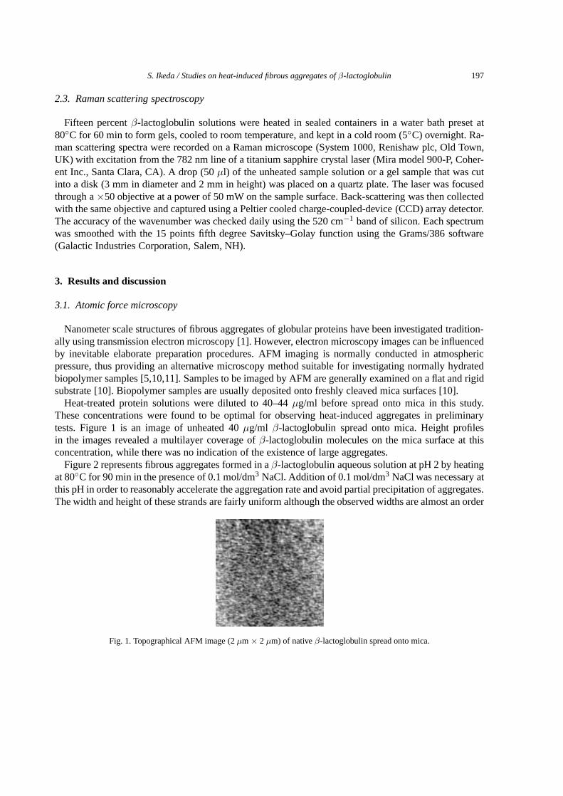
Heat-treated protein solutions were diluted to 40–44µg/ml before spread onto mica in this study.These concentrations were found to be optimal for observing heat-induced aggregates in preliminarytests. Figure 1 is an image of unheated 40µg/ml β-lactoglobulin spread onto mica. Height profilesin the images revealed a multilayer coverage ofβ-lactoglobulin molecules on the mica surface at thisconcentration, while there was no indication of the existence of large aggregates.
Figure 2 represents fibrous aggregates formed in aβ-lactoglobulin aqueous solution at pH 2 by heatingat 80◦C for 90 min in the presence of 0.1 mol/dm3 NaCl. Addition of 0.1 mol/dm3 NaCl was necessary atthis pH in order to reasonably accelerate the aggregation rate and avoid partial precipitation of aggregates.The width and height of these strands are fairly uniform although the observed widths are almost an order
Fig. 1. Topographical AFM image (2µm × 2 µm) of nativeβ-lactoglobulin spread onto mica.
198 S. Ikeda / Studies on heat-induced fibrous aggregates of β-lactoglobulin
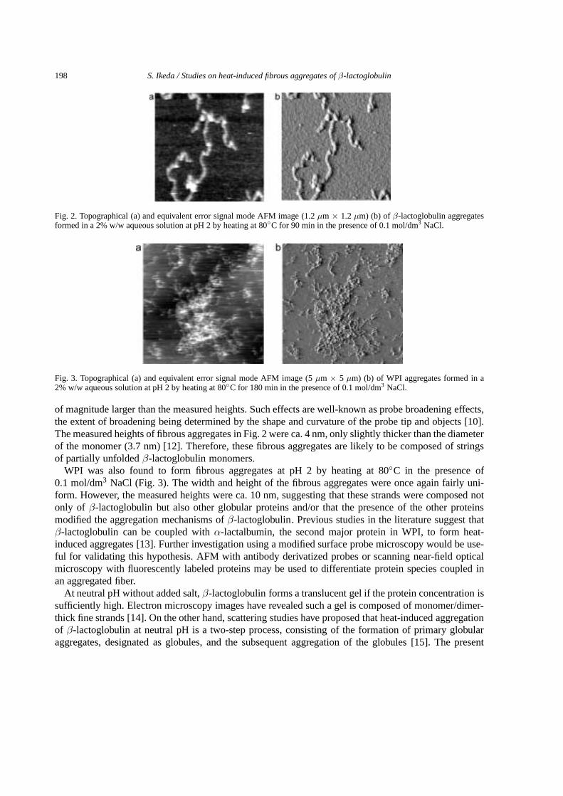
Fig. 2. Topographical (a) and equivalent error signal mode AFM image (1.2µm × 1.2µm) (b) of β-lactoglobulin aggregatesformed in a 2% w/w aqueous solution at pH 2 by heating at 80◦C for 90 min in the presence of 0.1 mol/dm3 NaCl.
Fig. 3. Topographical (a) and equivalent error signal mode AFM image (5µm × 5 µm) (b) of WPI aggregates formed in a2% w/w aqueous solution at pH 2 by heating at 80◦C for 180 min in the presence of 0.1 mol/dm3 NaCl.
of magnitude larger than the measured heights. Such effects are well-known as probe broadening effects,the extent of broadening being determined by the shape and curvature of the probe tip and objects [10].The measured heights of fibrous aggregates in Fig. 2 were ca. 4 nm, only slightly thicker than the diameterof the monomer (3.7 nm) [12]. Therefore, these fibrous aggregates are likely to be composed of stringsof partially unfoldedβ-lactoglobulin monomers.
WPI was also found to form fibrous aggregates at pH 2 by heating at 80◦C in the presence of0.1 mol/dm3 NaCl (Fig. 3). The width and height of the fibrous aggregates were once again fairly uni-form. However, the measured heights were ca. 10 nm, suggesting that these strands were composed notonly of β-lactoglobulin but also other globular proteins and/or that the presence of the other proteinsmodified the aggregation mechanisms ofβ-lactoglobulin. Previous studies in the literature suggest thatβ-lactoglobulin can be coupled withα-lactalbumin, the second major protein in WPI, to form heat-induced aggregates [13]. Further investigation using a modified surface probe microscopy would be use-ful for validating this hypothesis. AFM with antibody derivatized probes or scanning near-field opticalmicroscopy with fluorescently labeled proteins may be used to differentiate protein species coupled inan aggregated fiber.
At neutral pH without added salt,β-lactoglobulin forms a translucent gel if the protein concentration issufficiently high. Electron microscopy images have revealed such a gel is composed of monomer/dimer-thick fine strands [14]. On the other hand, scattering studies have proposed that heat-induced aggregationof β-lactoglobulin at neutral pH is a two-step process, consisting of the formation of primary globularaggregates, designated as globules, and the subsequent aggregation of the globules [15]. The present
S. Ikeda / Studies on heat-induced fibrous aggregates of β-lactoglobulin 199
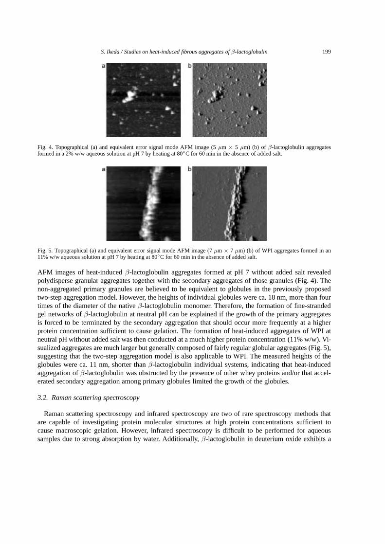
Fig. 4. Topographical (a) and equivalent error signal mode AFM image (5µm × 5 µm) (b) of β-lactoglobulin aggregatesformed in a 2% w/w aqueous solution at pH 7 by heating at 80◦C for 60 min in the absence of added salt.
Fig. 5. Topographical (a) and equivalent error signal mode AFM image (7µm × 7 µm) (b) of WPI aggregates formed in an11% w/w aqueous solution at pH 7 by heating at 80◦C for 60 min in the absence of added salt.
AFM images of heat-inducedβ-lactoglobulin aggregates formed at pH 7 without added salt revealedpolydisperse granular aggregates together with the secondary aggregates of those granules (Fig. 4). Thenon-aggregated primary granules are believed to be equivalent to globules in the previously proposedtwo-step aggregation model. However, the heights of individual globules were ca. 18 nm, more than fourtimes of the diameter of the nativeβ-lactoglobulin monomer. Therefore, the formation of fine-strandedgel networks ofβ-lactoglobulin at neutral pH can be explained if the growth of the primary aggregatesis forced to be terminated by the secondary aggregation that should occur more frequently at a higherprotein concentration sufficient to cause gelation. The formation of heat-induced aggregates of WPI atneutral pH without added salt was then conducted at a much higher protein concentration (11% w/w). Vi-sualized aggregates are much larger but generally composed of fairly regular globular aggregates (Fig. 5),suggesting that the two-step aggregation model is also applicable to WPI. The measured heights of theglobules were ca. 11 nm, shorter thanβ-lactoglobulin individual systems, indicating that heat-inducedaggregation ofβ-lactoglobulin was obstructed by the presence of other whey proteins and/or that accel-erated secondary aggregation among primary globules limited the growth of the globules.
3.2. Raman scattering spectroscopy
Raman scattering spectroscopy and infrared spectroscopy are two of rare spectroscopy methods thatare capable of investigating protein molecular structures at high protein concentrations sufficient tocause macroscopic gelation. However, infrared spectroscopy is difficult to be performed for aqueoussamples due to strong absorption by water. Additionally,β-lactoglobulin in deuterium oxide exhibits a
200 S. Ikeda / Studies on heat-induced fibrous aggregates of β-lactoglobulin
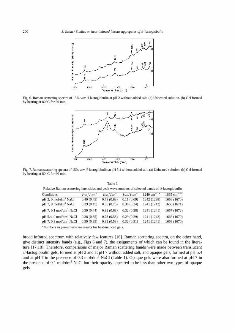
Fig. 6. Raman scattering spectra of 15% w/vβ-lactoglobulin at pH 2 without added salt. (a) Unheated solution. (b) Gel formedby heating at 80◦C for 60 min.
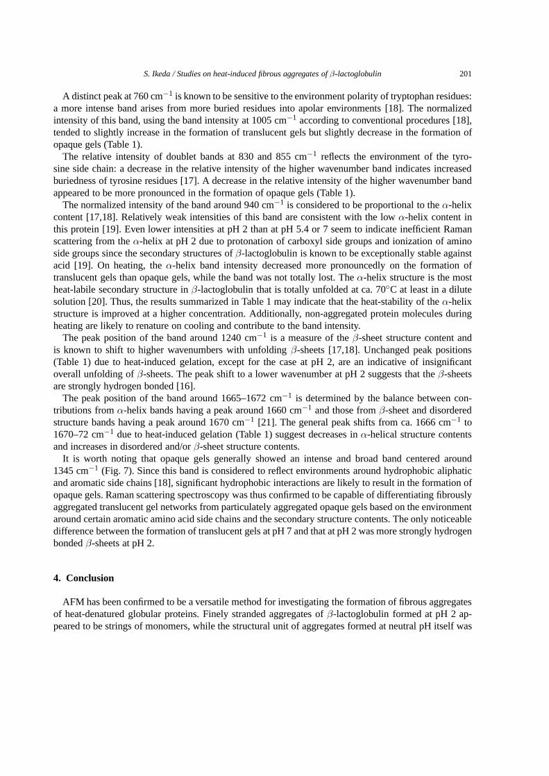
Fig. 7. Raman scattering spectra of 15% w/vβ-lactoglobulin at pH 5.4 without added salt. (a) Unheated solution. (b) Gel formedby heating at 80◦C for 60 min.
Table 1
Relative Raman scattering intensities and peak wavenumbers of selected bands ofβ-lactoglobulin
Conditions I760/I1005∗ I855/I830
∗ I940/I1005∗ 1240 cm−1∗ 1665 cm−1∗
pH 2, 0 mol/dm3 NaCl 0.40 (0.45) 0.78 (0.63) 0.11 (0.09) 1242 (1238) 1666 (1670)pH 7, 0 mol/dm3 NaCl 0.39 (0.45) 0.86 (0.73) 0.30 (0.24) 1241 (1242) 1666 (1671)
pH 7, 0.1 mol/dm3 NaCl 0.39 (0.44) 0.82 (0.63) 0.32 (0.28) 1241 (1241) 1667 (1672)
pH 5.4, 0 mol/dm3 NaCl 0.38 (0.35) 0.78 (0.58) 0.29 (0.29) 1241 (1242) 1666 (1670)pH 7, 0.3 mol/dm3 NaCl 0.39 (0.35) 0.82 (0.53) 0.32 (0.31) 1241 (1241) 1666 (1670)∗Numbers in parenthesis are results for heat-induced gels.
broad infrared spectrum with relatively few features [16]. Raman scattering spectra, on the other hand,give distinct intensity bands (e.g., Figs 6 and 7), the assignments of which can be found in the litera-ture [17,18]. Therefore, comparisons of major Raman scattering bands were made between translucentβ-lactoglobulin gels, formed at pH 2 and at pH 7 without added salt, and opaque gels, formed at pH 5.4and at pH 7 in the presence of 0.3 mol/dm3 NaCl (Table 1). Opaque gels were also formed at pH 7 inthe presence of 0.1 mol/dm3 NaCl but their opacity appeared to be less than other two types of opaquegels.
S. Ikeda / Studies on heat-induced fibrous aggregates of β-lactoglobulin 201
A distinct peak at 760 cm−1 is known to be sensitive to the environment polarity of tryptophan residues:a more intense band arises from more buried residues into apolar environments [18]. The normalizedintensity of this band, using the band intensity at 1005 cm−1 according to conventional procedures [18],tended to slightly increase in the formation of translucent gels but slightly decrease in the formation ofopaque gels (Table 1).
The relative intensity of doublet bands at 830 and 855 cm−1 reflects the environment of the tyro-sine side chain: a decrease in the relative intensity of the higher wavenumber band indicates increasedburiedness of tyrosine residues [17]. A decrease in the relative intensity of the higher wavenumber bandappeared to be more pronounced in the formation of opaque gels (Table 1).
The normalized intensity of the band around 940 cm−1 is considered to be proportional to theα-helixcontent [17,18]. Relatively weak intensities of this band are consistent with the lowα-helix content inthis protein [19]. Even lower intensities at pH 2 than at pH 5.4 or 7 seem to indicate inefficient Ramanscattering from theα-helix at pH 2 due to protonation of carboxyl side groups and ionization of aminoside groups since the secondary structures ofβ-lactoglobulin is known to be exceptionally stable againstacid [19]. On heating, theα-helix band intensity decreased more pronouncedly on the formation oftranslucent gels than opaque gels, while the band was not totally lost. Theα-helix structure is the mostheat-labile secondary structure inβ-lactoglobulin that is totally unfolded at ca. 70◦C at least in a dilutesolution [20]. Thus, the results summarized in Table 1 may indicate that the heat-stability of theα-helixstructure is improved at a higher concentration. Additionally, non-aggregated protein molecules duringheating are likely to renature on cooling and contribute to the band intensity.
The peak position of the band around 1240 cm−1 is a measure of theβ-sheet structure content andis known to shift to higher wavenumbers with unfoldingβ-sheets [17,18]. Unchanged peak positions(Table 1) due to heat-induced gelation, except for the case at pH 2, are an indicative of insignificantoverall unfolding ofβ-sheets. The peak shift to a lower wavenumber at pH 2 suggests that theβ-sheetsare strongly hydrogen bonded [16].
The peak position of the band around 1665–1672 cm−1 is determined by the balance between con-tributions fromα-helix bands having a peak around 1660 cm−1 and those fromβ-sheet and disorderedstructure bands having a peak around 1670 cm−1 [21]. The general peak shifts from ca. 1666 cm−1 to1670–72 cm−1 due to heat-induced gelation (Table 1) suggest decreases inα-helical structure contentsand increases in disordered and/orβ-sheet structure contents.
It is worth noting that opaque gels generally showed an intense and broad band centered around1345 cm−1 (Fig. 7). Since this band is considered to reflect environments around hydrophobic aliphaticand aromatic side chains [18], significant hydrophobic interactions are likely to result in the formation ofopaque gels. Raman scattering spectroscopy was thus confirmed to be capable of differentiating fibrouslyaggregated translucent gel networks from particulately aggregated opaque gels based on the environmentaround certain aromatic amino acid side chains and the secondary structure contents. The only noticeabledifference between the formation of translucent gels at pH 7 and that at pH 2 was more strongly hydrogenbondedβ-sheets at pH 2.
4. Conclusion
AFM has been confirmed to be a versatile method for investigating the formation of fibrous aggregatesof heat-denatured globular proteins. Finely stranded aggregates ofβ-lactoglobulin formed at pH 2 ap-peared to be strings of monomers, while the structural unit of aggregates formed at neutral pH itself was

202 S. Ikeda / Studies on heat-induced fibrous aggregates of β-lactoglobulin
confirmed to be granular aggregates. It was suggested that, in the presence of other species of globularproteins in milk whey,β-lactoglobulin was coupled with other proteins to form fibrous aggregates. Theformation of fibrous aggregates was found to accompany a loss ofα-helix structures generally, whilefurther investigation is needed to clarify the molecular origin of the shift from the two-step aggregationat neutral pH to the fibrous aggregation at acidic pH.
Acknowledgements
The author is grateful to Dr. V.J. Morris of Institute of Food Research, Norwich, UK for instructingatomic force microscopy, Professor E.C.Y. Li-Chan of The University of British Columbia, Vancouver,Canada for instructing Raman scattering spectroscopy, and Professor Shuryo Nakai of The University ofBritish Columbia, Vancouver, Canada for valuable discussions on biophysical properties of proteins.
References
[1] A.H. Clark, in: Functional Properties of Food Macromolecules, 2nd edn, S.E. Hill, D.A. Ledward and J.R. Mitchell, eds,Aspen Publishers, Inc., Gaithersburg, 1998, pp. 77–142.
[2] W.S. Gosal and S.B. Ross-Murphy,Curr. Opin. Colloid Interface Sci. 5 (2000), 188–194.[3] S. Ikeda, E.A. Foegeding and T. Hagiwara,Langmuir 15 (1999), 8584–8589.[4] S. Ikeda, K. Nishinari and E.A. Foegeding,Biopolymers 56 (2001), 109–119.[5] S. Ikeda and V.J. Morris,Biomacromolecules 3 (2002), 382–389.[6] J.I. Guijarro, M. Sunde, J.A. Jones, I.D. Campbell and C.M. Dobson,Proc. Natl. Acad. Sci. USA 95 (1998), 4224–4228.[7] Y. Takahashi, A. Ueno and H. Mihara,Structure 8 (2000), 915–925.[8] S. Ikeda and K. Nishinari,Biomacromolecules 1 (2000), 757–763.[9] G. Panick, R. Malessa and R. Winter,Biochem. 38 (1999), 6512–6519.
[10] V.J. Morris, A.R. Kirby and A.P. Gunning,Atomic Force Microscopy for Biologists, Imperial College Press, London, 1999.[11] S. Ikeda, V.J. Morris and K. Nishinari,Biomacromolecules 2 (2001), 1331–1337.[12] P. Aymard, T. Nicolai, D. Durand and A. Clark,Macromolecules 32 (1999), 2542–2552.[13] G.M. Kavanagh, A.H. Clark and S.B. Ross-Murphy,Macromolecules 33 (2000), 7029–7037.[14] M. Langton and A.-M. Hermansson,Food Hydrocolloids 5 (1992), 523–529.[15] P. Aymard, J.C. Nicolai and D. Durand,J. Chim. Phys. 93 (1996), 987–997.[16] T. Lefèvre and M. Subirade,Biopolymers 54 (2000), 578–586.[17] B.G. Frushour and J.L. Koenig,Biopolymers 14 (1975), 649–662.[18] E.C.Y. Li-Chan,Trends Food Sci. Technol. 7 (1996), 361–370.[19] F. Fogolari, L. Ragona, L. Zetta, S. Romagnoli, K.G. De Kruif and H. Molinari,FEBS Lett. 436 (1998), 149–154.[20] X.L. Qi, C. Holt, D. McNulty, D.T. Clarke, S. Brownlow and G.R. Jones,Biochem. J. 324 (1997), 341–346.[21] E.B. Carew, H.E. Stanley, J.C. Seidel and J. Gergely,Biophys. J. 44 (1983), 219–224.

Submit your manuscripts athttp://www.hindawi.com
Hindawi Publishing Corporationhttp://www.hindawi.com Volume 2014
Inorganic ChemistryInternational Journal of
Hindawi Publishing Corporation http://www.hindawi.com Volume 2014
International Journal ofPhotoenergy
Hindawi Publishing Corporationhttp://www.hindawi.com Volume 2014
Carbohydrate Chemistry
International Journal of
Hindawi Publishing Corporationhttp://www.hindawi.com Volume 2014
Journal of
Chemistry
Hindawi Publishing Corporationhttp://www.hindawi.com Volume 2014
Advances in
Physical Chemistry
Hindawi Publishing Corporationhttp://www.hindawi.com
Analytical Methods in Chemistry
Journal of
Volume 2014
Bioinorganic Chemistry and ApplicationsHindawi Publishing Corporationhttp://www.hindawi.com Volume 2014
SpectroscopyInternational Journal of
Hindawi Publishing Corporationhttp://www.hindawi.com Volume 2014
The Scientific World JournalHindawi Publishing Corporation http://www.hindawi.com Volume 2014
Medicinal ChemistryInternational Journal of
Hindawi Publishing Corporationhttp://www.hindawi.com Volume 2014
Chromatography Research International
Hindawi Publishing Corporationhttp://www.hindawi.com Volume 2014
Applied ChemistryJournal of
Hindawi Publishing Corporationhttp://www.hindawi.com Volume 2014
Hindawi Publishing Corporationhttp://www.hindawi.com Volume 2014
Theoretical ChemistryJournal of
Hindawi Publishing Corporationhttp://www.hindawi.com Volume 2014
Journal of
Spectroscopy
Analytical ChemistryInternational Journal of
Hindawi Publishing Corporationhttp://www.hindawi.com Volume 2014
Journal of
Hindawi Publishing Corporationhttp://www.hindawi.com Volume 2014
Quantum Chemistry
Hindawi Publishing Corporationhttp://www.hindawi.com Volume 2014
Organic Chemistry International
ElectrochemistryInternational Journal of
Hindawi Publishing Corporation http://www.hindawi.com Volume 2014
Hindawi Publishing Corporationhttp://www.hindawi.com Volume 2014
CatalystsJournal of