Assessment and Management of Patients With Vascular Disorders and Problems of Peripheral...
50
Assessment and Management of Patients With Vascular Disorders and Problems of Peripheral Circulation
-
Upload
matthew-watkins -
Category
Documents
-
view
225 -
download
2
Transcript of Assessment and Management of Patients With Vascular Disorders and Problems of Peripheral...
Assessment and Management of Patients With Vascular Disorders and Problems of Peripheral Circulation
Assessment and Management of Patients With Vascular Disorders and Problems of Peripheral Circulation
Vascular System
• Arteries and arterioles• Capillaries• Veins and venules• Lymphatic vessels• Function of the vascular system
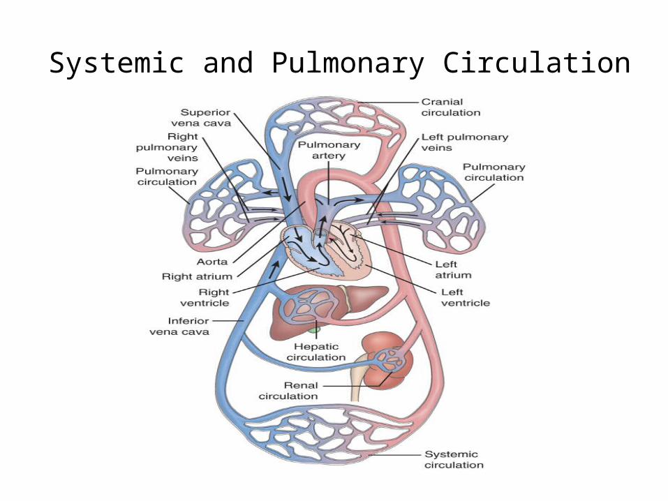
Systemic and Pulmonary Circulation
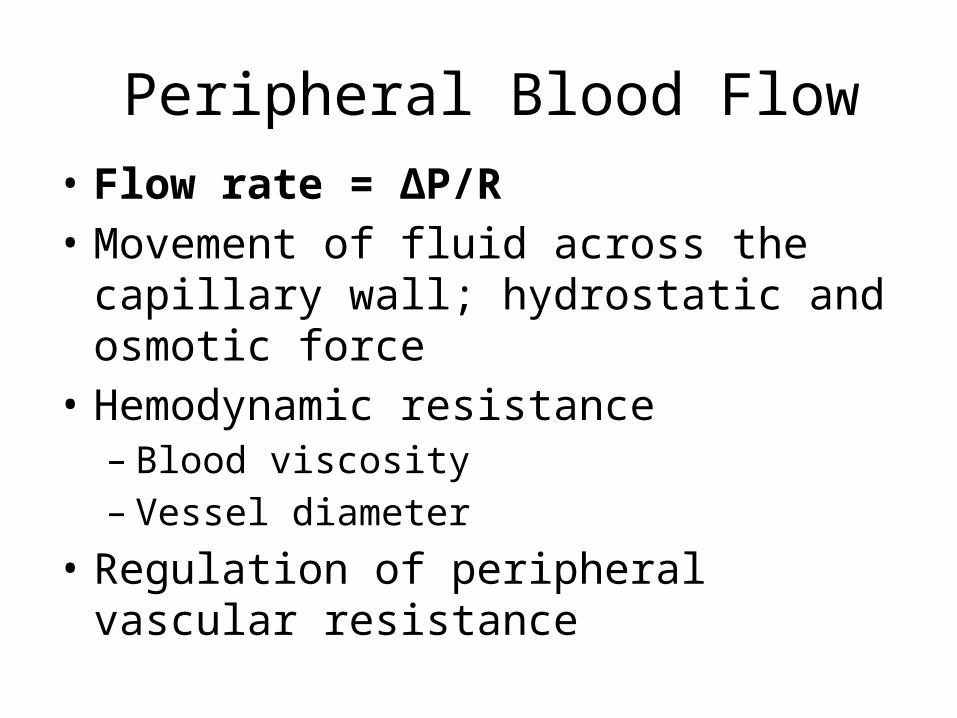
Peripheral Blood Flow• Flow rate = ΔP/R• Movement of fluid across the capillary wall;
hydrostatic and osmotic force• Hemodynamic resistance– Blood viscosity– Vessel diameter
• Regulation of peripheral vascular resistance
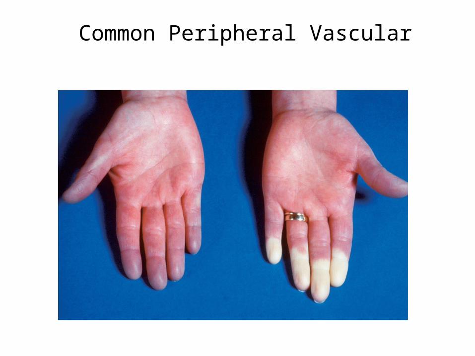
Assessment • Characteristics of arterial and venous
insufficiency• Intermittent claudication• Rest pain• Changes in skin and appearance• Pulses• Aging changes
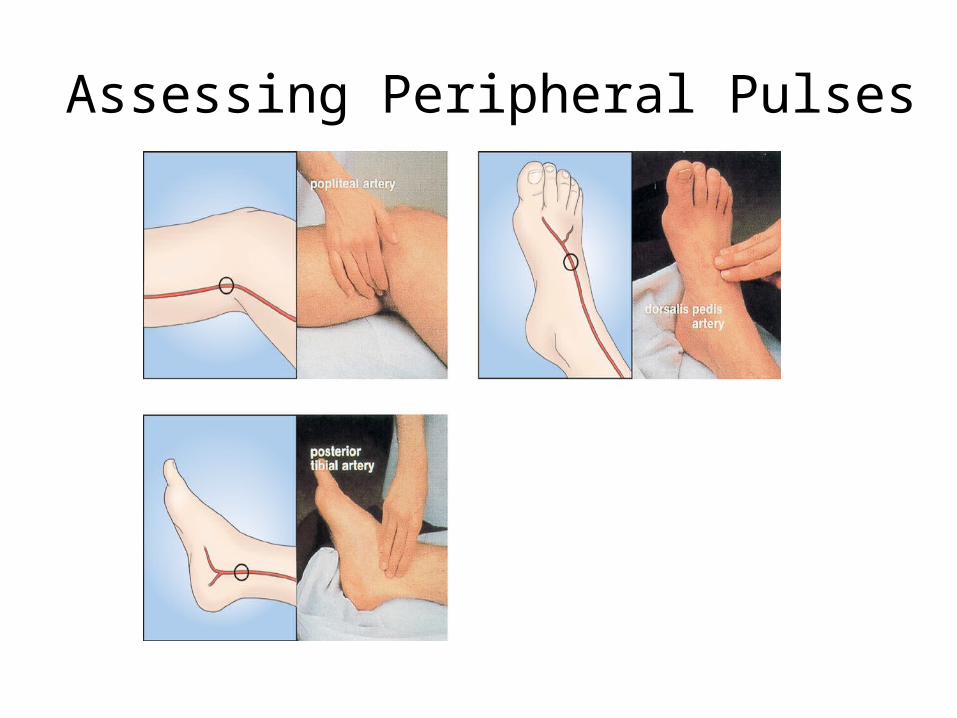
Assessing Peripheral Pulses
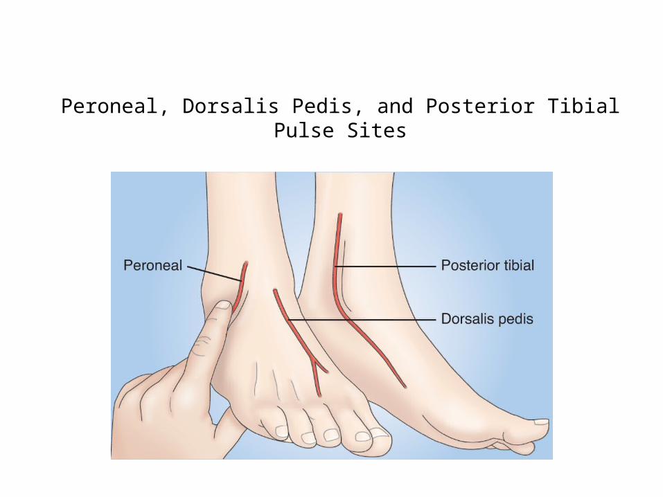
Peroneal, Dorsalis Pedis, and Posterior Tibial Pulse Sites
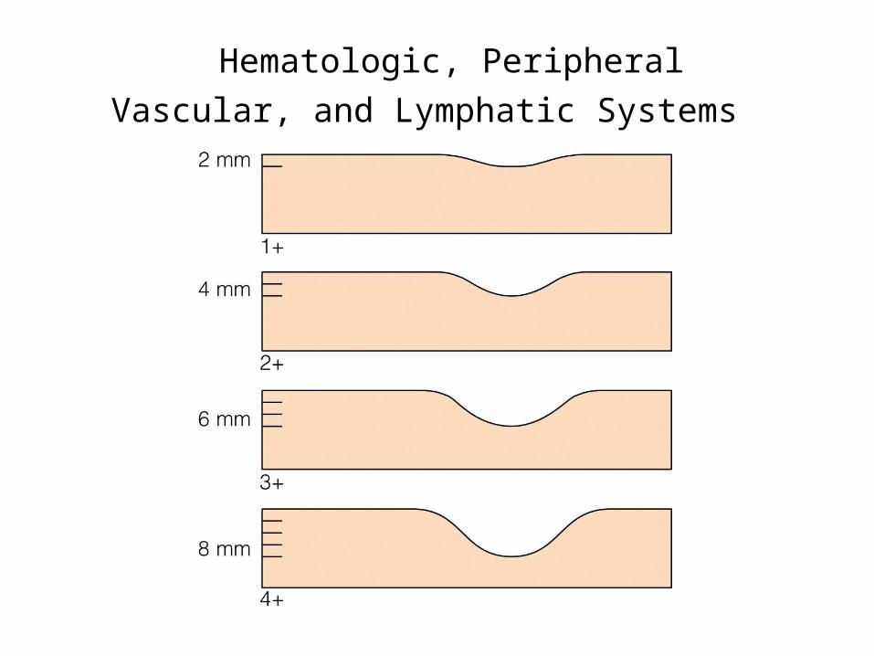
Hematologic, Peripheral Vascular, and Lymphatic Systems
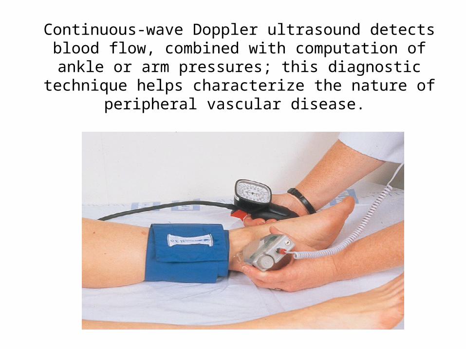
Continuous-wave Doppler ultrasound detects blood flow, combined with computation of ankle or arm pressures; this diagnostic
technique helps characterize the nature of peripheral vascular disease.
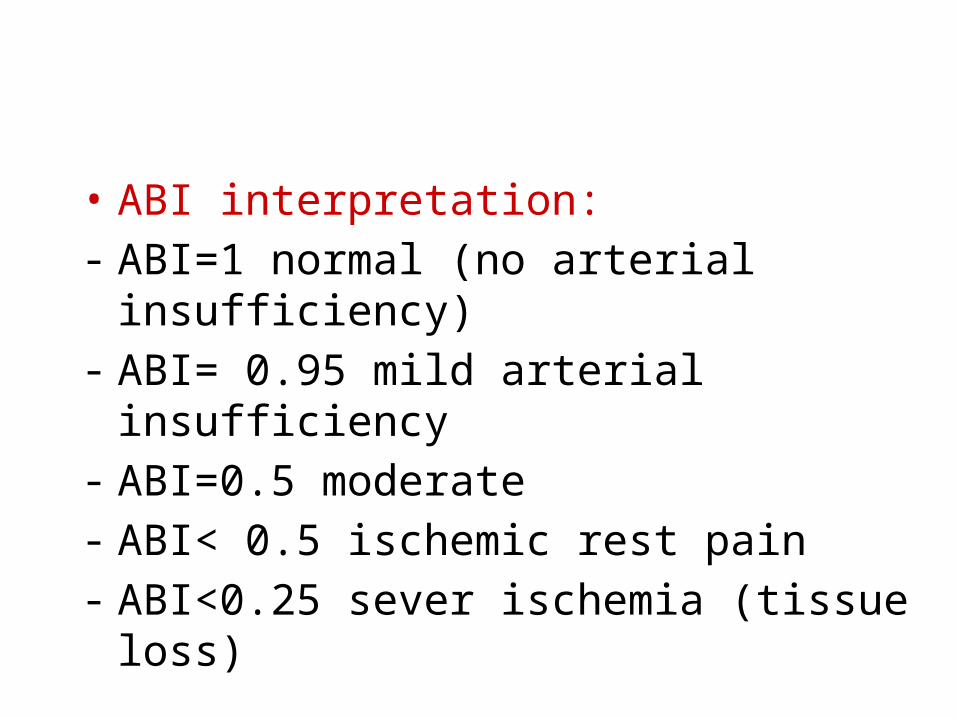
• ABI interpretation: - ABI=1 normal (no arterial insufficiency)- ABI= 0.95 mild arterial insufficiency- ABI=0.5 moderate - ABI< 0.5 ischemic rest pain- ABI<0.25 sever ischemia (tissue loss)
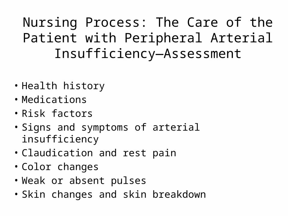
Nursing Process: The Care of the Patient with Peripheral Arterial
Insufficiency—Assessment
• Health history • Medications • Risk factors• Signs and symptoms of arterial insufficiency• Claudication and rest pain• Color changes• Weak or absent pulses• Skin changes and skin breakdown
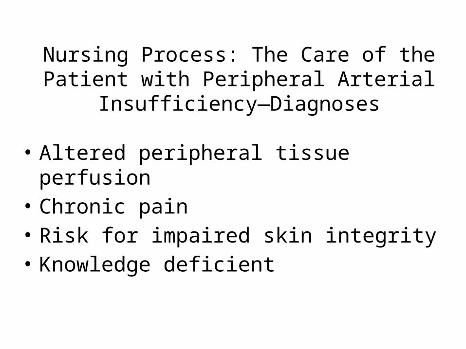
Nursing Process: The Care of the Patient with Peripheral Arterial Insufficiency—
Diagnoses• Altered peripheral tissue perfusion• Chronic pain• Risk for impaired skin integrity• Knowledge deficient
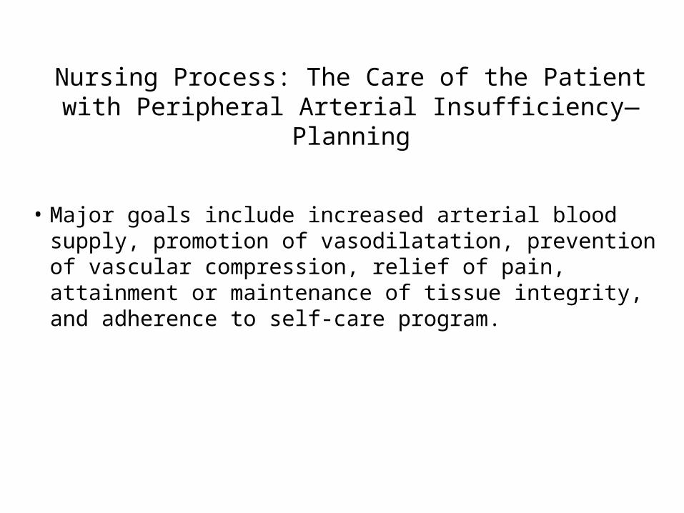
Nursing Process: The Care of the Patient with Peripheral Arterial Insufficiency—Planning
• Major goals include increased arterial blood supply, promotion of vasodilatation, prevention of vascular compression, relief of pain, attainment or maintenance of tissue integrity, and adherence to self-care program.
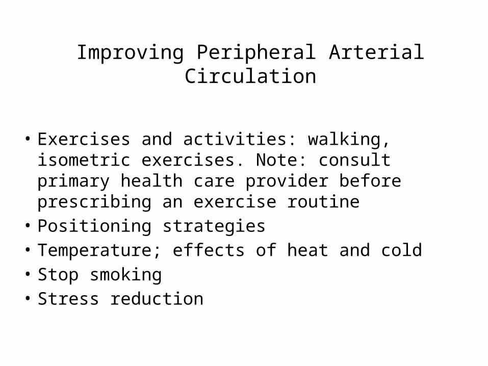
Improving Peripheral Arterial Circulation
• Exercises and activities: walking, isometric exercises. Note: consult primary health care provider before prescribing an exercise routine
• Positioning strategies• Temperature; effects of heat and cold• Stop smoking• Stress reduction
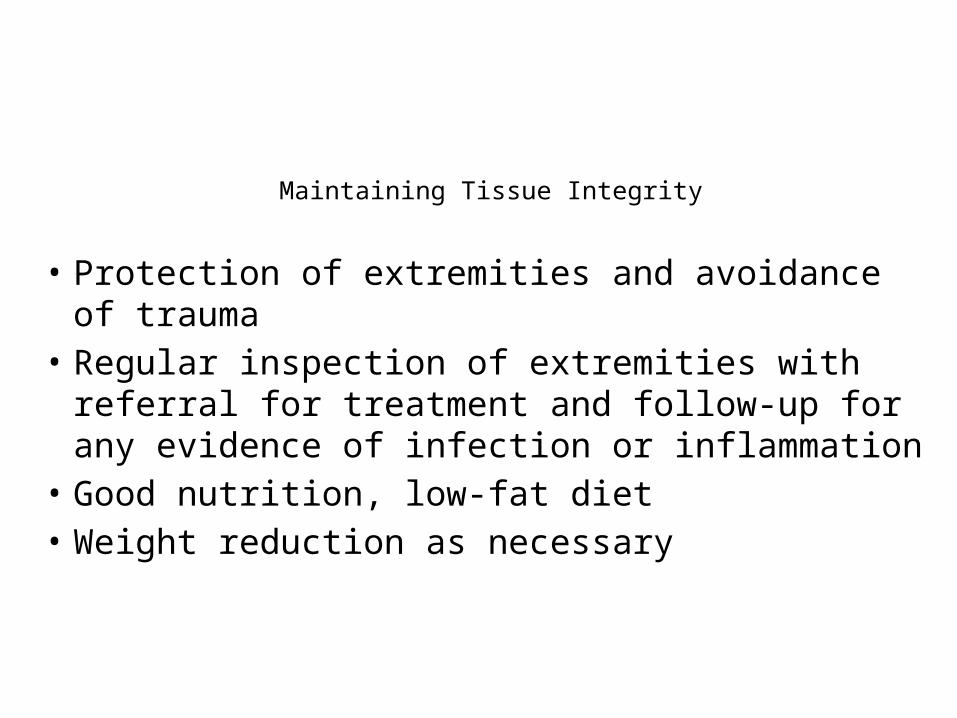
Maintaining Tissue Integrity
• Protection of extremities and avoidance of trauma• Regular inspection of extremities with referral for
treatment and follow-up for any evidence of infection or inflammation
• Good nutrition, low-fat diet• Weight reduction as necessary
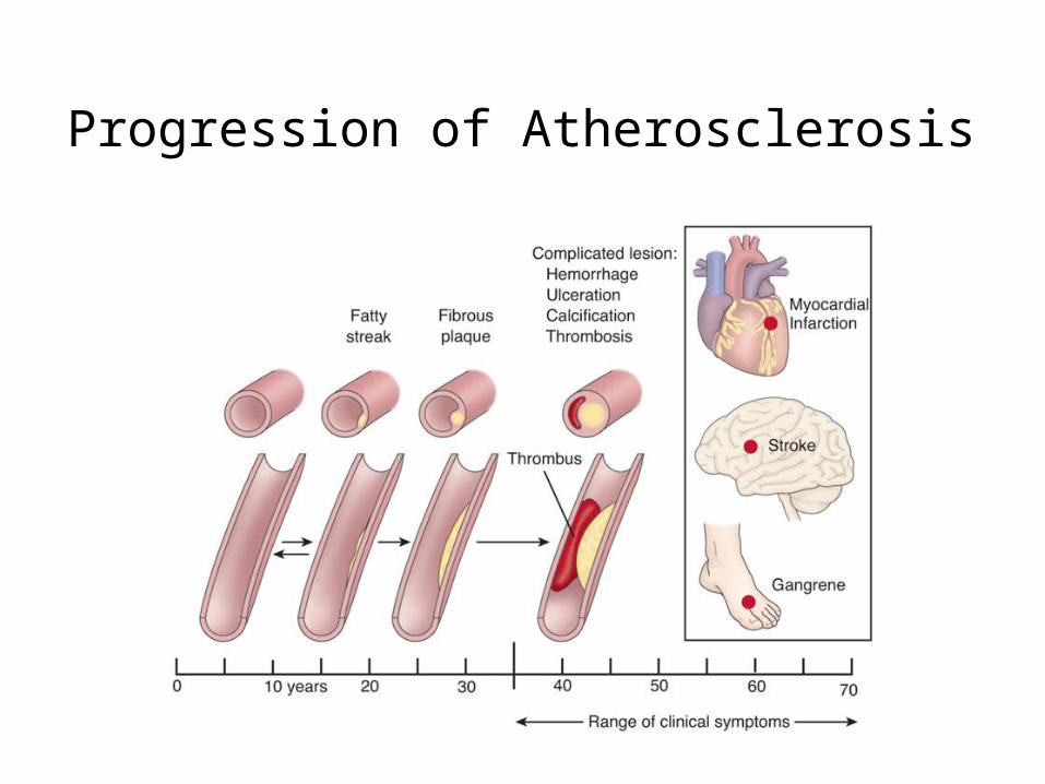
Progression of Atherosclerosis
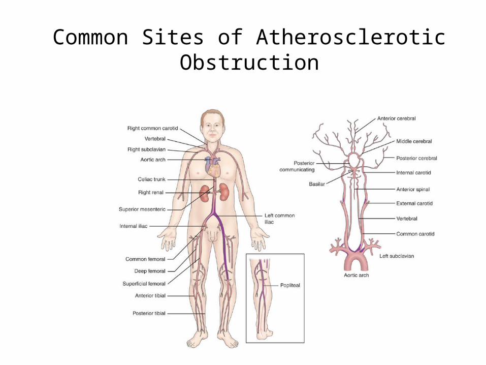
Common Sites of Atherosclerotic Obstruction
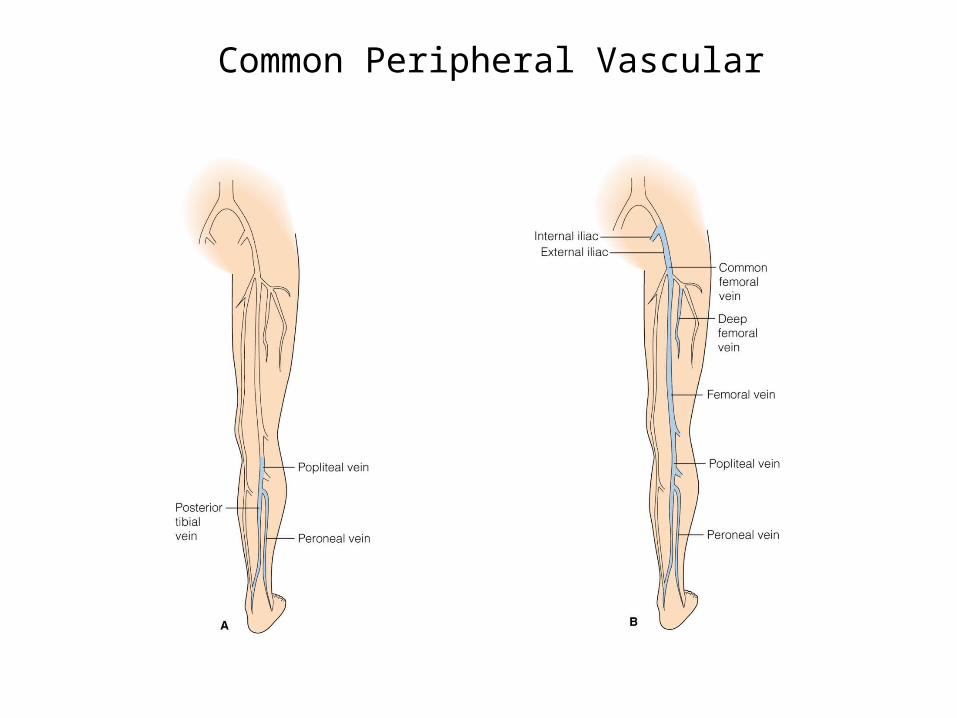
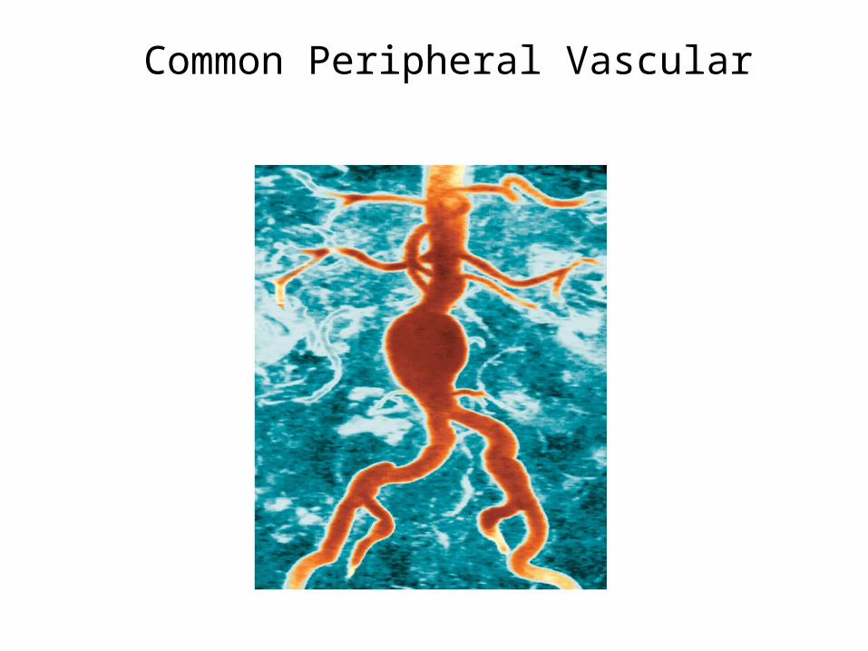
Common Peripheral Vascular
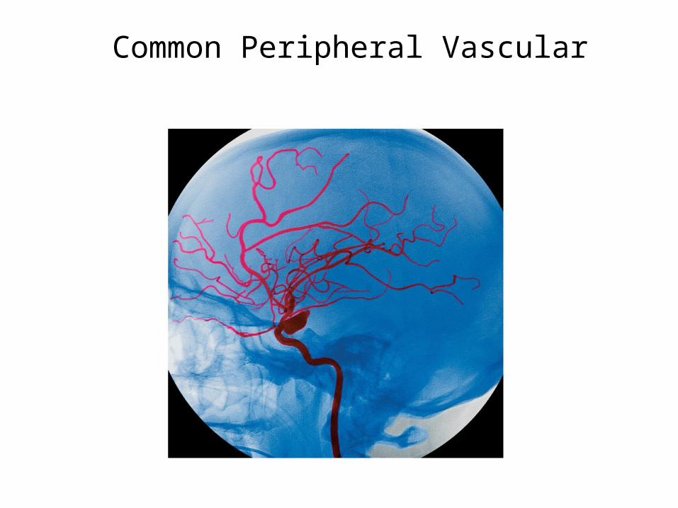
Common Peripheral Vascular
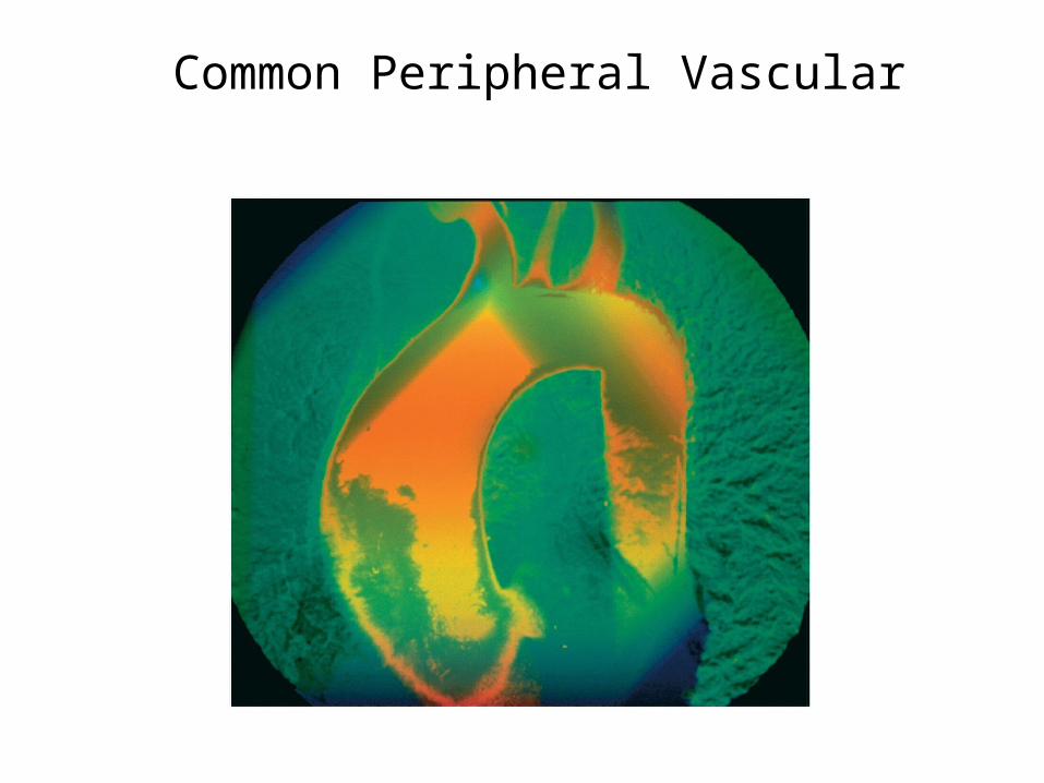
Common Peripheral Vascular
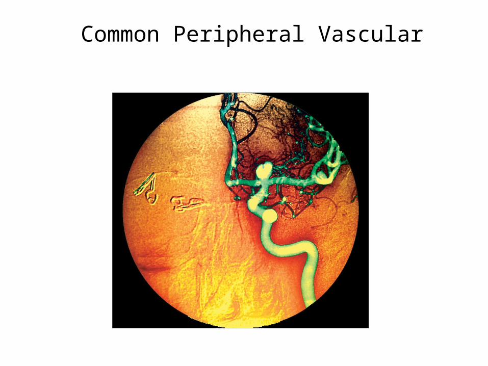
Common Peripheral Vascular
Common Peripheral Vascular
Common Peripheral Vascular
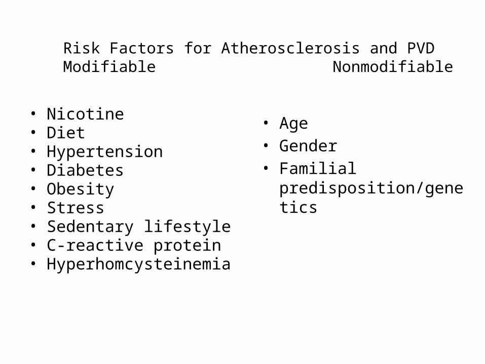
Risk Factors for Atherosclerosis and PVD Modifiable Nonmodifiable
• Nicotine• Diet• Hypertension• Diabetes• Obesity • Stress• Sedentary lifestyle• C-reactive protein• Hyperhomcysteinemia
• Age • Gender• Familial
predisposition/genetics
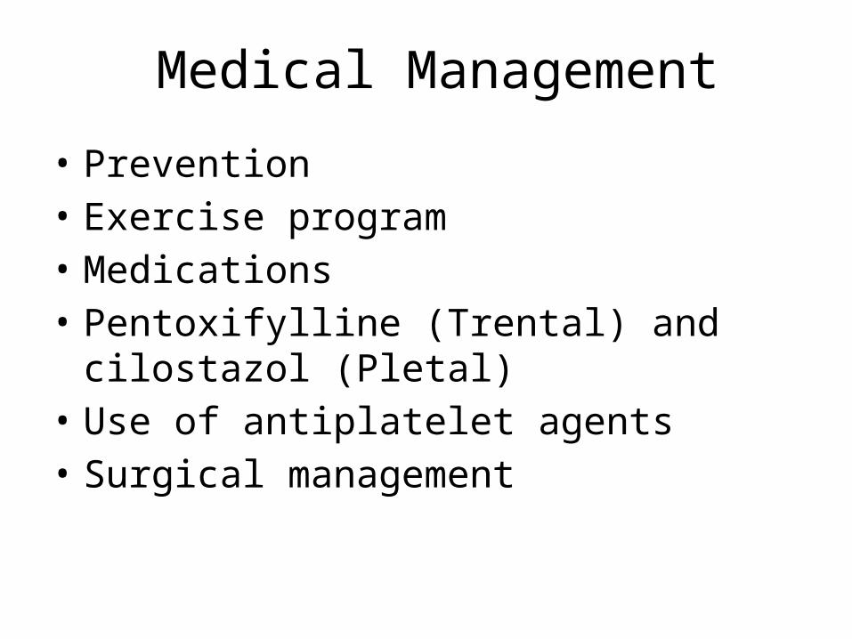
Medical Management
• Prevention• Exercise program• Medications• Pentoxifylline (Trental) and cilostazol (Pletal) • Use of antiplatelet agents• Surgical management
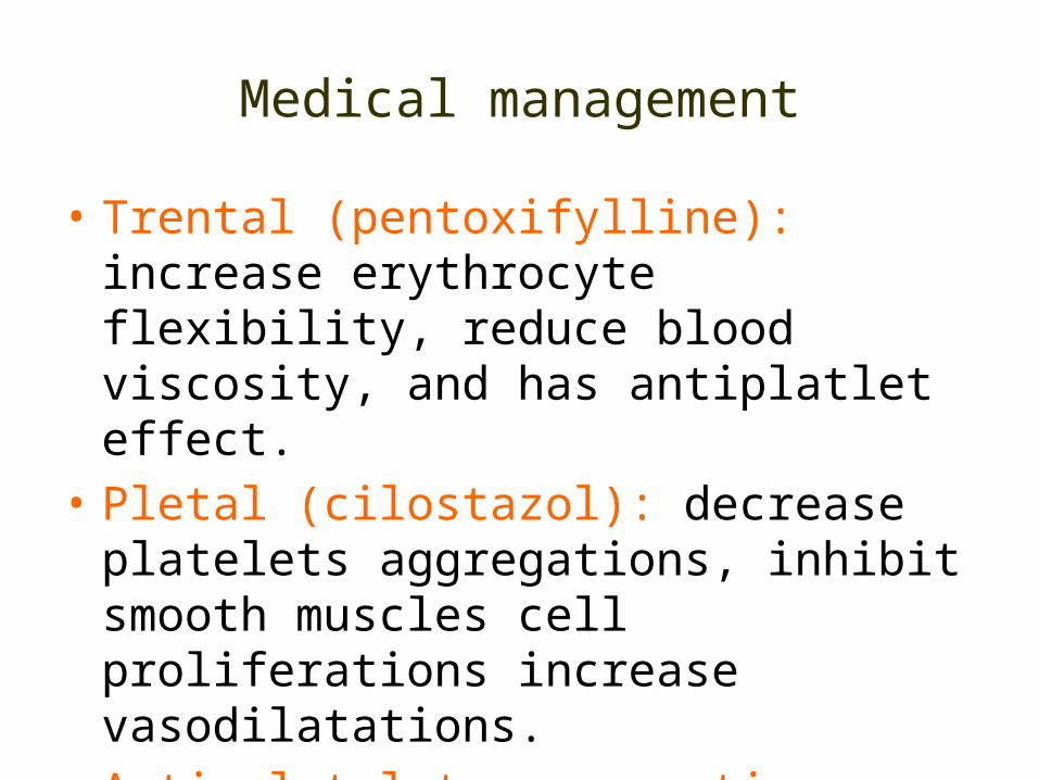
Medical management
• Trental (pentoxifylline): increase erythrocyte flexibility, reduce blood viscosity, and has antiplatlet effect.
• Pletal (cilostazol): decrease platelets aggregations, inhibit smooth muscles cell proliferations increase vasodilatations.
• Anti-platelets aggregating agents (aspirin, clopidogrel (Plavix)): prevent the formation of thromboemboli
Surgical managements
• Amputations (if occlusion is sever)• Vascular grafting (anastemosis) depends on the
degree and location of stenosis or occlusion.• Endarterectomy: thrombus that obstruct the
artery removed through incision to the artery affected.
Venous Thromboembolism
• Pathophysiology• Risk factors• Endothelial damage– Venous stasis– Altered coagulation
• Manifestations– Deep veins– Superficial veins
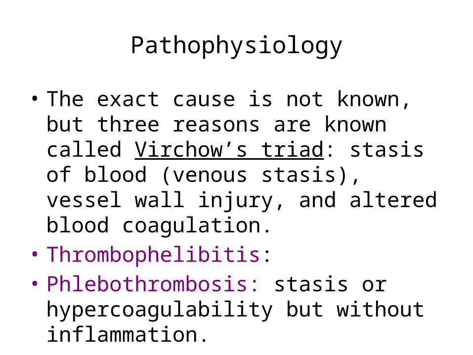
Pathophysiology
• The exact cause is not known, but three reasons are known called Virchow’s triad: stasis of blood (venous stasis), vessel wall injury, and altered blood coagulation.
• Thrombophelibitis: • Phlebothrombosis: stasis or hypercoagulability
but without inflammation.
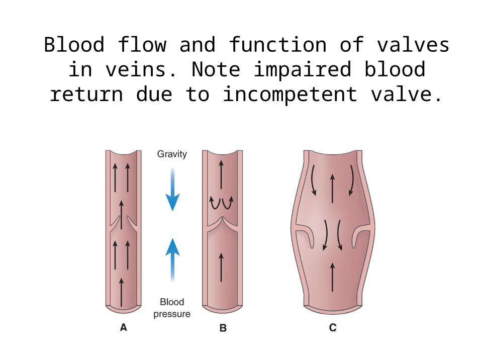
Blood flow and function of valves in veins. Note impaired blood return due to incompetent valve.
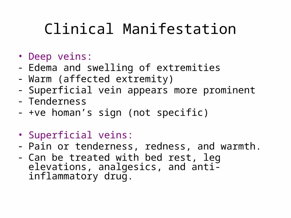
Clinical Manifestation • Deep veins:- Edema and swelling of extremities- Warm (affected extremity)- Superficial vein appears more prominent- Tenderness- +ve homan’s sign (not specific)
• Superficial veins:- Pain or tenderness, redness, and warmth. - Can be treated with bed rest, leg elevations, analgesics, and anti-
inflammatory drug.
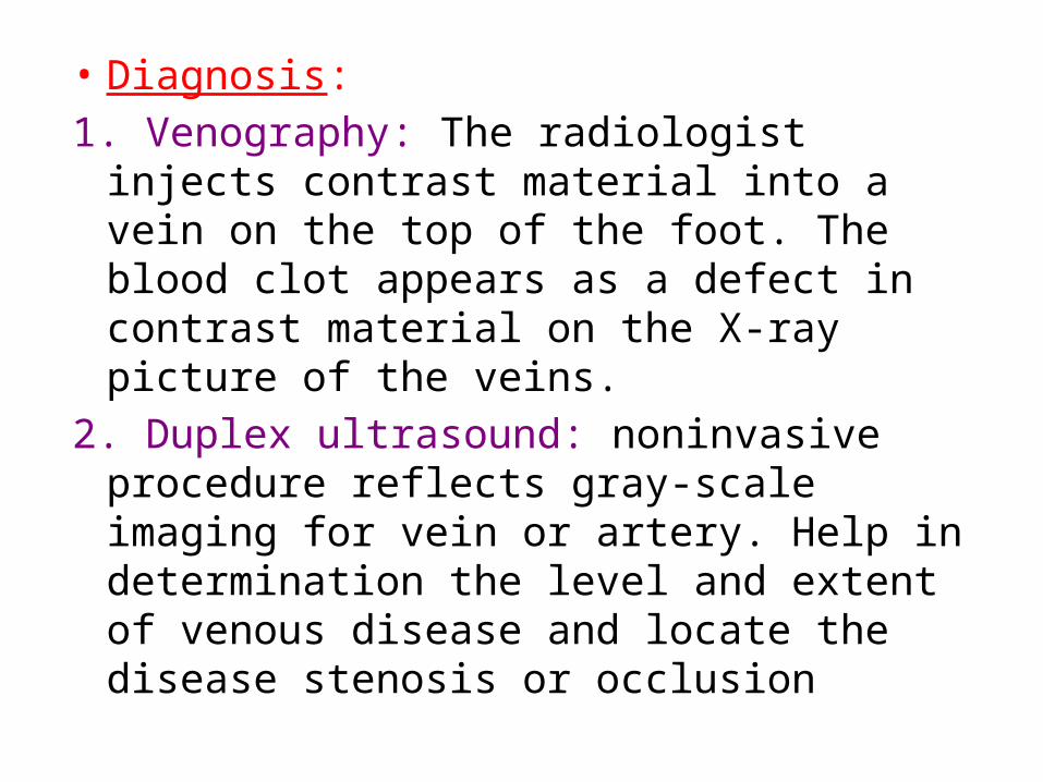
• Diagnosis:
1. Venography: The radiologist injects contrast material into a vein on the top of the foot. The blood clot appears as a defect in contrast material on the X-ray picture of the veins.
2. Duplex ultrasound: noninvasive procedure reflects gray-scale imaging for vein or artery. Help in determination the level and extent of venous disease and locate the disease stenosis or occlusion
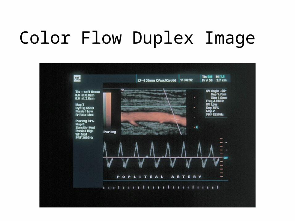
Color Flow Duplex Image
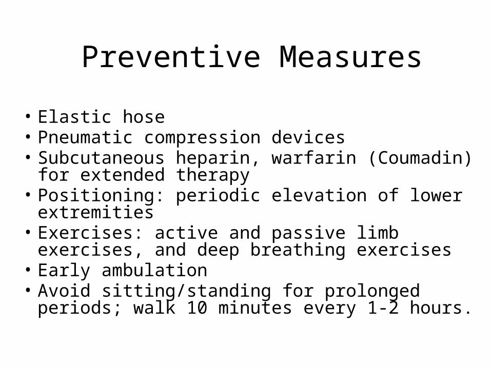
Preventive Measures
• Elastic hose• Pneumatic compression devices• Subcutaneous heparin, warfarin (Coumadin) for
extended therapy • Positioning: periodic elevation of lower extremities• Exercises: active and passive limb exercises, and
deep breathing exercises• Early ambulation• Avoid sitting/standing for prolonged periods; walk
10 minutes every 1-2 hours.
Nursing Process: The Care of the Patient with Leg Ulcers—Assessment
• History of the condition• Treatment depends upon the type of ulcer• Assess for presence of infection• Assess nutrition
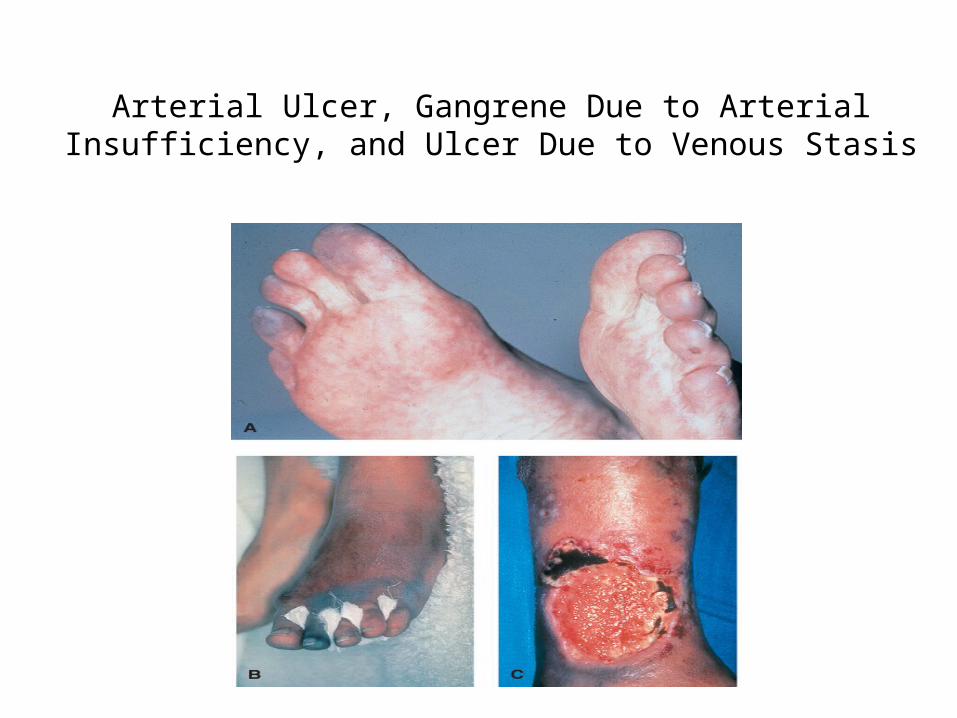
Arterial Ulcer, Gangrene Due to Arterial Insufficiency, and Ulcer Due to Venous Stasis
Medical Management
• Anti-infective therapy is dependent upon infecting agent– Oral antibiotics are usually prescribed.
• Compression therapy• Debridement of wound• Dressings• Other
Nursing Process: The Care of the Patient with Leg Ulcers- Diagnoses
• Impaired skin integrity• Impaired physical mobility• Imbalanced nutrition
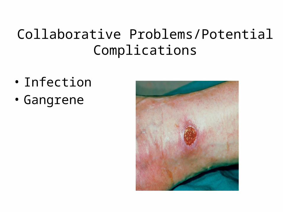
Collaborative Problems/Potential Complications
• Infection • Gangrene
Nursing Process: The Care of the Patient with Leg Ulcers—Planning
• Major goals include restoration of skin integrity, improved physical mobility, adequate nutrition, and absence of complications.
Mobility
• With leg ulcers, activity is usually initially restricted to promote healing
• Gradual progression of activity• Activity to promote blood flow; encourage patient to
move about in bed and exercise upper extremities• Diversional activities• Pain medication prior to activities
Other Interventions• Skin integrity– Skin care/hygiene and wound care– Positioning of legs to promote circulation– Avoidance of trauma
• Nutrition– Measures to ensure adequate nutrition– Adequate protein, vitamin C and A, iron, and zinc are
especially important for wound healing– Include cultural considerations and patient teaching
in the dietary plan
Varicose Veins (Varicosities)
• Are abnormally dilated, tortuous, superficial veins caused by incompetent venous valves
• Occurs in lower extremities, in the saphenous system or the lower trunk
• Correlated with ↑ age, most in women, and people with occupation required prolonged standing
• Other factors that cause VV are: hereditary, pregnancy
Pathophysiology: • Primary: without involvement of deep veins)• Secondary: resulting from obstruction of deep veins• Reflux of venous blood result in venous stasis• Clinical Manifestations:- Dull aches muscle cramps- ↑ muscle fatigue in lower legs- Ankle edema- Feeling of heaviness of the legs- If deep veins obstructed pt will have S&S of chronic
venous insufficiency (edema, pain, pigmentation, ulceration)
- Increased susceptibility to infection and injury.
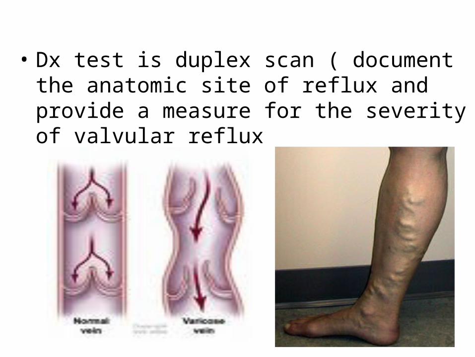
• Dx test is duplex scan ( document the anatomic site of reflux and provide a measure for the severity of valvular reflux
• Prevention:- Avoid activity that cause venous stasis as
( wearing constrictive clothing, crossing the legs, sitting or standing for long periods)
- Change position frequently- Elevating the legs- Walking 1-2 miles each day- Elastic stoking - Control wt.
Medical Management
• Ligation and stripping: is done for primary VV, deep veins should be patent. Saphenous vein ligated in the groin where the saphenous vein meets the femoral vein, then 2-3 incision is made below the knee, stripper( wire) is inserted to the point of ligation, the wire is then withdrawn and vein as it is removed.
• Thermal ablation• sclerotherapy
Nursing Management
After surgery:• Bed rest is discouraged and early ambulation is
encouraged • Instruct pt to walk Q one hour for 5-10min
while awake for the 1st 24hr, then ↑ activity as tolerated
• Wear elastic stocking continuously for 1wk• Elevate foot of bed• Standing and sitting are discouraged
• Promote comfort and understanding:- give analgesic, inspect dressing for bleeding, alert
for reported sensations of “pins and needles.” Hypersensitivity to touch in the involved extremity may indicate a temporary or permanent nerve injury resulting from surgery
- The patient is instructed to dry the incisions well with a clean towel using a patting technique, rather than rubbing
- The patient is instructed to apply sunscreen or zinc oxide to the incisional area prior to sun exposure
- If the patient underwent sclerotherapy, a burning sensation in the injected leg may be experienced for 1 or 2 days
Cellulitus and Lymphatic Disorders
• Cellulitus: infection and swelling of skin tissues • Lymphangitis: inflammation/infection of the
lymphatic channels• Lymphadenitis: inflammation/infection of the
lymph nodes • Lymphedema: tissue swelling related to
obstruction of lymphatic flow– Primary: congenital– Secondary: acquired obstruction


![Peripheral modifications of [Ψ[CH NH]Tpg4]vancomycin ...](https://static.fdocument.org/doc/165x107/6211b4c5b9a3d33a3c037f89/peripheral-modifications-of-ch-nhtpg4vancomycin-.jpg)