Assessing Native and Non-native Conformational States of a Protein by Methylene Carbene Labeling: ...
11
Assessing Native and Non-native Conformational States of a Protein by Methylene Carbene Labeling: The Case of Bacillus licheniformis -Lactamase Daniela B. Ureta, Patricio O. Craig, ‡ Gabriela E. Go ´mez, and Jose ´ M. Delfino* Departamento de Quı ´mica Biolo ´ gica-IQUIFIB (UBA-CONICET), Facultad de Farmacia y Bioquı ´mica, UniVersidad de Buenos Aires, Junı ´n 956, C1113AAD, Buenos Aires, Argentina ReceiVed June 29, 2007; ReVised Manuscript ReceiVed October 10, 2007 ABSTRACT: Much knowledge of protein folding can be derived from the examination of the nature and size of solvent-exposed surfaces along conformational transitions. We exploit here a general photochemical modification with methylene carbene of the accessible surface area (ASA) of the polypeptide chain. Labeling of Bacillus licheniformis -lactamase (BL-L) with 1 mM 3 H-diazirine yielded 8.3 × 10 -3 mol CH 2 /mol protein, in agreement with the prediction for an unspecific surface labeling phenomenon. The unfolded state U in 7 M urea was labeled 60% more than the native state N. This result lies well below the increment of ASA expected from theoretical estimates and points to the presence of residual organization in state U and/or of cavities or crevices favoring the partition of the reagent in state N. A partially folded state I was demonstrated from two sequential transitions occurring at 1.5-3.0 M and 3.5-6.5 M urea. This technique shows a close correlation with optical probes most sensitive to changes in tertiary structure, a statement supported by the fact that the largest change occurs along the N-I portion of the N-I-U transition and along the acid pH-induced N-A transition. In the latter case, state A is labeled 70% more than state N, an increment consistent with the loosening of tight interactions in the core of the protein. Fragmentation of labeled BL-L into peptides provides a sequential map of solvent accessibility. Thus, amino acid residues pertaining to the Ω-loop and to helices R5 and R6 line the major cavity of the protein, that is big enough to lodge the diazirine reagent. Methylene labeling, by introducing an original (and perhaps unique) experimental measurement of ASA, enlightens subtle aspects of complex transitions and makes possible a comparative structural characterization of the native as well as non-native states. Proteins represent essential building blocks behind cell structure and the engines supporting every metabolic reaction. The close relationship existing between the detailed confor- mation and function of proteins substantiates the current emphasis on their structural studies. In this context, the characterization of the native state, as well as the unfolded and intermediate states, constitutes an important issue in protein science. The native state is marginally stable with respect to alternative conformations. Thus, it can be readily perturbed by subtle changes of the physicochemical environ- ment. At the other end, the unfolded ensemble of states can be stabilized under strong denaturing conditions and exhibits open and highly flexible conformations characterized by extensive exposure of the polypeptide chain to the aqueous solvent. Finally, under milder denaturing conditions, inter- mediate states can indeed be populated (1). One typical example of the latter is the so-called molten globule state (2, 3). Among its distinctive features, one can mention that it adopts a globular but less compact conformation than the native state, giving rise to a slightly expanded form endowed with substantial secondary structure but lacking specific tertiary contacts. Thus, the increased mobility of hydrophobic side chains in the semiliquid core facilitates the access of the aqueous solvent to otherwise excluded regions in the native state. The importance of studying intermediate states in equilibrium cannot be underestimated because they have been postulated to share similar features to late kinetic intermediates detected along folding pathways, thus shedding light on the general process of protein folding. The knowledge of intermediate and unfolded states has been hampered by the lack of appropriate high-resolution techniques to address their conformation. Extensive fluctua- tions of the polypeptide chain in a very short time scale hinder the useful application of techniques such as X-ray crystallography or NMR. In this context, optical methods (CD, fluorescence, light scattering), chemical techniques (H/D exchange, chemical modification of side chains of amino acids), and size-exclusion chromatography, coupled to defined alteration of the amino acid sequence (site-directed mutagenesis, truncations, construction of chimaeras, etc.) have been used with profit to address specific structural aspects. Although unable to provide a thorough picture, these ² D.B.U. was a recipient of a graduate student fellowship from the FOMEC program. G.E.G. was a recipient of a graduate student fellowship from the University of Buenos Aires (UBA) and is currently an Estenssoro fellow from the YPF Foundation. P.O.C. and J.M.D. are career investigators of CONICET. This research has been supported by grants to J.M.D. from Universidad de Buenos Aires (UBA), the Consejo Nacional de Investigaciones Cientı ´ficas y Te ´cnicas (CONICET), and the Agencia Nacional de Promocio ´n Cientı ´fica y Tecnolo ´gica (ANPCyT). * Author to whom correspondence should be addressed. E-mail: [email protected], tel: 54 11 4964 8291, extension 116, fax: 54 11 4962 5457. ‡ Current address: Fundacio ´n Instituto Leloir, Av. Patricias Argen- tinas 435, C1405BWE, Buenos Aires, Argentina. 14567 Biochemistry 2007, 46, 14567-14577 10.1021/bi7012867 CCC: $37.00 © 2007 American Chemical Society Published on Web 11/17/2007
Transcript of Assessing Native and Non-native Conformational States of a Protein by Methylene Carbene Labeling: ...
Assessing Native and Non-native Conformational States of a Protein by MethyleneCarbene Labeling: The Case ofBacillus licheniformisâ-Lactamase
Daniela B. Ureta, Patricio O. Craig,‡ Gabriela E. Go´mez, and Jose´ M. Delfino*
Departamento de Quı´mica Biologica-IQUIFIB (UBA-CONICET), Facultad de Farmacia y Bioquı´mica, UniVersidad de BuenosAires, Junı´n 956, C1113AAD, Buenos Aires, Argentina
ReceiVed June 29, 2007; ReVised Manuscript ReceiVed October 10, 2007
ABSTRACT: Much knowledge of protein folding can be derived from the examination of the nature andsize of solvent-exposed surfaces along conformational transitions. We exploit here a general photochemicalmodification with methylene carbene of the accessible surface area (ASA) of the polypeptide chain. Labelingof Bacillus licheniformisâ-lactamase (BL-âL) with 1 mM 3H-diazirine yielded 8.3× 10⁻³ mol CH2/molprotein, in agreement with the prediction for an unspecific surface labeling phenomenon. The unfoldedstate U in 7 M urea was labeled 60% more than the native state N. This result lies well below the incrementof ASA expected from theoretical estimates and points to the presence of residual organization in state Uand/or of cavities or crevices favoring the partition of the reagent in state N. A partially folded state I wasdemonstrated from two sequential transitions occurring at 1.5-3.0 M and 3.5-6.5 M urea. This techniqueshows a close correlation with optical probes most sensitive to changes in tertiary structure, a statementsupported by the fact that the largest change occurs along the N-I portion of the N-I-U transition andalong the acid pH-induced N-A transition. In the latter case, state A is labeled 70% more than state N,an increment consistent with the loosening of tight interactions in the core of the protein. Fragmentationof labeled BL-âL into peptides provides a sequential map of solvent accessibility. Thus, amino acid residuespertaining to theΩ-loop and to helicesR5 andR6 line the major cavity of the protein, that is big enoughto lodge the diazirine reagent. Methylene labeling, by introducing an original (and perhaps unique)experimental measurement of ASA, enlightens subtle aspects of complex transitions and makes possiblea comparative structural characterization of the native as well as non-native states.
Proteins represent essential building blocks behind cellstructure and the engines supporting every metabolic reaction.The close relationship existing between the detailed confor-mation and function of proteins substantiates the currentemphasis on their structural studies. In this context, thecharacterization of the native state, as well as the unfoldedand intermediate states, constitutes an important issue inprotein science. The native state is marginally stable withrespect to alternative conformations. Thus, it can be readilyperturbed by subtle changes of the physicochemical environ-ment. At the other end, the unfolded ensemble of states canbe stabilized under strong denaturing conditions and exhibitsopen and highly flexible conformations characterized byextensive exposure of the polypeptide chain to the aqueoussolvent. Finally, under milder denaturing conditions, inter-
mediate states can indeed be populated (1). One typicalexample of the latter is the so-called molten globule state(2, 3). Among its distinctive features, one can mention thatit adopts a globular but less compact conformation than thenative state, giving rise to a slightly expanded form endowedwith substantial secondary structure but lacking specifictertiary contacts. Thus, the increased mobility of hydrophobicside chains in the semiliquid core facilitates the access ofthe aqueous solvent to otherwise excluded regions in thenative state. The importance of studying intermediate statesin equilibrium cannot be underestimated because they havebeen postulated to share similar features to late kineticintermediates detected along folding pathways, thus sheddinglight on the general process of protein folding.
The knowledge of intermediate and unfolded states hasbeen hampered by the lack of appropriate high-resolutiontechniques to address their conformation. Extensive fluctua-tions of the polypeptide chain in a very short time scalehinder the useful application of techniques such as X-raycrystallography or NMR. In this context, optical methods(CD, fluorescence, light scattering), chemical techniques(H/D exchange, chemical modification of side chains ofamino acids), and size-exclusion chromatography, coupledto defined alteration of the amino acid sequence (site-directedmutagenesis, truncations, construction of chimaeras, etc.)have been used with profit to address specific structuralaspects. Although unable to provide a thorough picture, these
† D.B.U. was a recipient of a graduate student fellowship from theFOMEC program. G.E.G. was a recipient of a graduate studentfellowship from the University of Buenos Aires (UBA) and is currentlyan Estenssoro fellow from the YPF Foundation. P.O.C. and J.M.D.are career investigators of CONICET. This research has been supportedby grants to J.M.D. from Universidad de Buenos Aires (UBA), theConsejo Nacional de Investigaciones Cientıficas y Tecnicas (CONICET),and the Agencia Nacional de Promocio´n Cientıfica y Tecnologica(ANPCyT).
* Author to whom correspondence should be addressed. E-mail:[email protected], tel: 54 11 4964 8291, extension 116, fax: 5411 4962 5457.
‡ Current address: Fundacio´n Instituto Leloir, Av. Patricias Argen-tinas 435, C1405BWE, Buenos Aires, Argentina.
14567Biochemistry2007,46, 14567-14577
10.1021/bi7012867 CCC: $37.00 © 2007 American Chemical SocietyPublished on Web 11/17/2007
techniques are very valuable tools to gain insights onstructure because they can be adapted to the various time-scales of the folding process (for general references see refs4-7).
In regard to the thermodynamics of folding, minimizationof the accessible surface area (ASA1) associated to thehydrophobic side chains and the amide groups of the peptidebonds has been generally recognized as an importantstructural parameter (8-10). A fundamental issue worthinvestigating is to be able to describe the nature as well asto measure the extension of the ASA at each stage of theprocess. Unfortunately, there is hardly any technique ap-propriate to address a direct experimental measurement ofASA. In this regard, the chemical reactivity of a givenfunctional group present on an amino acid side chain againsta set of reagents of different nature can provide a descriptionof the environment around this group, thus making it possibleto infer solvent accessibility to this point region. However,drawbacks of the selective chemical modification techniquesare to strictly control the extent of modification, theoccurrence of side reactions, and the frequent induction ofconformational changes and to be constrained to alter a singleor a few chemical functionalities (11). On the other hand,proton-deuteron (H/D) exchange has been widely appliedin protein folding because it addresses the accessibility ofexchangeable protons along the polypeptide chain withoutintroducing hardly any modification in chemical nature.Although one can derive a landscape of solvent accessibilitywith this technique, it is noteworthy that data is intrinsicallylimited to amide protons belonging to the backbone chain,therefore becoming strongly dependent on secondary struc-ture integrity. Besides, one main drawback arises from thelabile nature of the label, a factor that greatly restricts furtheranalytical processing of the sample. By contrast, a reagentaimed to act as ageneral probeshould define the proteinsurfaceindependentlyof its chemical nature. From a chemicalstandpoint, successful modification of the inert surfaceprovided by the hydrophobic side chains poses an absoluterequirement for a free radical, or a photogenerated nitreneor carbene. Along these lines, there have been recentdevelopments toward the general labeling of proteins. Amongthese, one can mention the use of the hydroxyl radicalchemistry (•OH) (12-18), and another technique, put forwardby Richards et al. (19) and developed in our own laboratory,takes advantage of the modification of the polypeptide chainwith singlet methylene (:CH2) (20-22).
The extreme reactivity of methylene makes it an idealreagent to achieve nonselective chemical modification ofproteins. Unlike other radical species that give rise to chainreactions, methylene inserts readily into any X-H bondyielding defined and stable methylated products. Diazirine(DZN) fulfills all the requirements to become the sourcereagent of methylene. In this regard, trifluoromethylphenyl-substituted diazirines have been widely used in hydrophobic
photolabeling of membrane proteins (e.g., see refs23, 24,and references cited therein). However, to achieve solventmimicry, one relies on the fact that unsubstituted DZN iscomparable in size to the water molecule, so DZN is expectedto probe the same surface as the aqueous solvent. In addition,DZN is a chemically inert gas at room temperature, and onlyafter photolysis at the correct wavelength (∼320 nm) doesit generate the reactive methylene species.
In this work we take advantage of the DZN labelingmethod, aided by the traceability provided by a3H label inthe molecule, to address the study of the native state,intermediate species along the folding pathway, and theunfolded ensemble of states in the paradigm protein systemrepresented byB. licheniformisâ-lactamase (BL-âL). To thisend, we employed the exo-small variant of this enzymeisolated from bacterial cultures (5). In addition to the wealthof knowledge available on this enzyme, including a high-resolution structure derived from X-ray crystallography (26,27), this protein constitutes an alternative model toR-lac-talbumin, that was studied earlier in our laboratory, bothbecause of its different folding motif and of its larger size.Most relevant to the goal of this work, intermediate statesof BL-âL can be stabilized with the aid of denaturing agents,at acidic pH or after the addition of salts (28), renderingthem amenable to study by DZN labeling. In this framework,we address here for the first time a comparative analysis ofASA, as inferred by the extent of modification with meth-ylene, among differently folded states of the single-domainprotein BL-âL.
EXPERIMENTAL PROCEDURES
Materials.3H-Formaldehyde (1-5 mCi, 90.0 mCi/mmol)was purchased from New England Nuclear, formaldehyde,37% (w/v), was from E. Merck, and formamide, urea, andguanidine hydrochloride were from Sigma Chemical Co.Urea was recrystallized from ethanol before use. TPCKtrypsin was from Worthington. Acetonitrile from E. Merckand trifluoroacetic acid (TFA) from Riedel de Hae¨n were ofHPLC grade. All other reagents and chemicals used were ofanalytical grade.
BL-âL and its CNBr and tryptic peptides were separatedon an AKTA purifier (Amersham Pharmacia Biotech) andRainin Dynamax FPLC/HPLC systems. UV measurementswere carried out on a Jasco 7850 spectrophotometer.
Circular Dichroism.Spectra were recorded on Jasco J-20and Jasco J-810 spectropolarimeters, using quartz cylindricalcuvettes of 1 or 10 mm path lengths for the far (200-250nm) and near (250-310 nm) UV regions, respectively. Inevery case, five consecutive spectra were recorded andaveraged to reduce the signal-to-noise ratio. Data wereconverted to molar ellipticity [θ]M (in units of deg cm2
dmol-1) using a mean residue weight value for BL-âL of110.49 g/mol.
Fluorescence Spectroscopy.Measurements were carriedout with an Aminco Bowman Series II spectrofluorometer,using a cuvette of 0.4 cm path length. For intrinsic tryptophanfluorescence measurements, we used excitation and emissionbandwidths of 4 nm and an excitation wavelength of 290nm to selectively excite W residues present in the protein,and the emission was collected in the range 300-450 nm.For fluorescence of the extrinsic probe ANS measurements,
1 Abbreviations: DZN, diazirine;3H-DZN, tritiated derivative ofdiazirine; :CH2, methylene carbene; ASA, accessible surface area; BL-âL, Bacillus licheniformisâ-lactamase (exo-small variant);R-LA,bovineR lactalbumin; HEWL, hen egg white lysozyme; N, native state;U, unfolded state; I, partially folded state; A, acid-stabilized moltenglobule-like state; CD, circular dichroism; ANS, 1-anilino-8-naphtha-lenesulfonic acid; RP-HPLC, reversed-phase high-performance liquidchromatography.
14568 Biochemistry, Vol. 46, No. 50, 2007 Ureta et al.
we used excitation and emission bandwidths of 4 nm andan excitation wavelength of 380 nm, and the emission wascollected in the range 450-550 nm.
Expression, Purification, and Characterization of BL-âL.BL-âL was expressed inE. coli BL21 (DE3) and isolatedfrom the bacterial cultures as described by Frate et al. (25).Transformed cells of thisE. coli strain were produced byDr. Anthony Fink’s laboratory (University of California atSanta Cruz) and were a gift to our laboratory of Dr. MarioErmacora (Universidad de Quilmes, Buenos Aires, Argen-tina). The protein was purified by a method describedpreviously (29). Its functionality was monitored by enzymaticactivity measurements according to Jansson (30), usingbenzylpenicillin (penicillin G) as substrate and measuringthe hydrolysis of theâ-lactam ring by the decrease in theabsorption at 240 nm. The identity and purity of theexpressed protein were assessed through the followingassays: SDS-PAGE electrophoresis (31), reversed phaseHPLC, UV absorption, and CD spectra. The concentrationof exo-small B. licheniformis â-lactamase (BL-âL) wasdetermined by its UV absorption at 280 nm, using a molarextinction coefficient of 23,430 M-1 cm-1 (32).
Synthesis of DZN.3H-DZN was synthesized and purifiedin our laboratory by following the method described by Craiget al. (20). 3H-DZN concentration in aqueous solution wasestimated both by measuring (i) the absorbance of thedissolved gas at 320 nm using an extinction coefficient ofε320 ) 180 M-1 cm-1 (20) and (ii) the concentration ofradioactivity of the solution after complete photolysis of3H-DZN to 3H-methanol had taken place.
Photolabeling of BL-âL. 3H-DZN (0.2-1 mCi/mmol) wasdissolved in samples of BL-âL (30 µM), with (i) 20 mMsodium phosphates buffer, pH 7.4, in the presence of differenturea concentrations (0-7 M), (ii) 90 mM AGT buffer (30mM acetate, 30 mM glycine, 30 mM Tris), pH adjusted withHCl to a given value in the range 2.0-7.0, or (iii) 90 mMAGT buffer, pH 2.0 in the presence of different ureaconcentrations (0-7 M), depending on the conformationaltransition under study: N-U, N-A, or A-U, respectively.Each sample was placed in a quartz cuvette (4 mL) of 1 cmpath length and capped with a Teflon stopper. The buffersused were degassed in advance and kept under an inertatmosphere of nitrogen gas before use. Photolysis was carriedout using a UV light source (Philips HPA 1000 Halogen/Hg lamp) placed at 12 cm from the samples. Light wasfiltered from the emission below 300 nm (with Oriel 59044long-pass filter) to prevent photolytic damage to proteinchromophores. The cuvettes were immersed in a water baththermostated at 20°C, so that even at the high luminic fluxused, no heating occurred. Typically, UV irradiation wasextended for 45 min, a time that corresponds to ap-proximately four-half-lives of the reagent in our photolysissetup (t1/2 ) 10.3 min).
After photolysis, the routine workup procedure consistsof an unfolding step of the protein in 8 M urea, followed bydialysis against 0.07 mM Na2CO3 (pH 7.5-8.0) and freezedrying. Finally, the samples were dissolved in 6 M guanidinehydrochloride in 0.05% TFA and separated from anyremaining radioactive impurity by reversed phase HPLC ona C4 column (Vydac 214TP510, 10 mm× 250 mm),developed with a linear gradient of acetonitrile:water (0 to80% in 80 min) in 0.05% TFA at a 3.0 mL/min flow rate.
The elution was monitored by UV absorption at 280 nm,and the protein was collected manually. For analyticalpurposes (see Figure 1C), samples were run on a smallerC4 column (Vydac 214TP54, 4.6 mm× 250 mm) with anidentical gradient at a 1.0 mL/min flow rate. Samples cleanedin this fashion were freeze-dried to remove HPLC solventsand redissolved in 20 mM sodium phosphates buffer, pH7.4, before the radioactivity was measured on a Pharmacia1214 Rackbeta liquid scintillation counter. The extent of3H-methylene carbene incorporation into BL-âL was expressedas the average number of moles of CH2 per mol of protein,as estimated by the radioactivity measured on a sample ofknown protein concentration, by taking into account thespecific radioactivity of the reagent.
Fragmentation of BL-âL into Peptides.After the cleanupprocedure described before, samples of BL-âL labeled with3H-methylene under native (20 mM sodium phosphatesbuffer, pH 7.4) or denaturing conditions (in the presence of8 M urea) were cleaved with CNBr following the procedurespreviously described by Kamp (33) and Fontana and Gross(34). After the separation of the resultant peptides (see sectionbelow), in some instances, a complete tryptic subdigestionof fragments was achieved with TPCK trypsin in 0.1 M NH4-CO3H, pH 8.0, after 12-18 h at 37°C, using a 2% (w/ w)enzyme/substrate ratio, following the method described byWilkinson (35).
Peptide Mapping.Separation of mixtures of peptidesobtained by CNBr treatment of BL-âL was carried out byRP-HPLC on a C4 column (Vydac 214TP510, 10 mm×250 mm), using a linear gradient of acetonitrile:water (0 to60% in 90 min) in 0.05% TFA at a 3.0 mL/min flow rate.For analytical purposes a smaller amount of the peptide
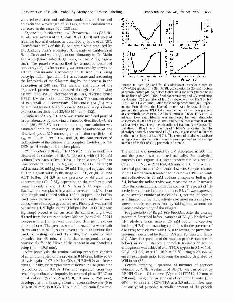
FIGURE 1: Near (A) and far (B) ultraviolet-circular dichroism(UV-CD) spectra of a 25µM BL-âL solution in 20 mM sodiumphosphate buffer, pH 7.4, before (solid lines) and after (dashed lines)the addition of DZN (3 mM final concentration) and UV irradiationfor 40 min. (C) Separation of BL-âL labeled with3H-DZN by RP-HPLC on a C4 column. After the cleanup procedure (see Experi-mental Procedures), the labeled protein sample was chromato-graphed through an HPLC C4 column eluted with a linear gradientof acetonitrile:water (0 to 80% in 80 min) in 0.05% TFA at a 1.0mL/min flow rate. Elution was monitored by both ultravioletabsorption at 280 nm (solid line) and by the measurement of theradioactivity associated to each collected fraction (gray bars). (D)Labeling of BL-âL as a function of3H-DZN concentration. Thephotolyzed samples contained BL-âL (35 µM) dissolved in 20 mMsodium phosphate buffer, pH 7.4. The extent of methylene carbeneincorporation into the protein sample was expressed as the averagenumber of moles of CH2 per mole of protein.
Conformation of BL-âL Probed by Methylene Carbene Labeling Biochemistry, Vol. 46, No. 50, 200714569
mixtures was sampled (inset to Figure 5). The elution wasmonitored by UV absorption at 215 nm, and fractions werecollected and subsequently identified as described below.
Tryptic peptides obtained from subdigestion of CNBrfragments were separated by size exclusion chromatographyon a Superdex Peptide HR 10/30 column (AmershamPharmacia Biotech) developed with buffer 40 mM Tris/HCl,4 M urea, pH 7.4, at a flow rate of 0.4 mL/min. Elution wasmonitored by UV absorption at 215 and 280 nm, and peakswere collected manually.
Peptide identification was achieved by (i) amino acidanalysis on an Applied Biosystems 420 A amino acidanalyzer, (ii) Edman sequencing (36) where every samplewas subjected to 3-5 cycles, or (iii) electrospray MS on aThermo Finnigan LCQ Duo-ion trap mass spectrometer.
Molecular Modeling.Calculations of the ASA of BL-âLand its peptides in the native state (ASA N) were based onthe crystallographic structure available for this protein (pdbcode: 4blm:24) with the program MacroModel (37) usinga probe radius of 1.4 Å. Considering DZN as an idealizedsphere of radius 2.1 Å did not produce significant differencesin the estimates of ASA. The interactive module of thisprogram was used for constructing the protein and its CNBrpeptides in extended chain conformations with the aim ofcalculating ASA extended. Additional ASA estimates of theunfolded protein and its peptides were obtained through aweb resource (http://roselab.jhu.edu/utils/unfolded.html: (38,39) that makes use of models that bracket the surface areaof the unfolded state between limiting extremes (ASA maxand ASA min). The analysis of cavities present in nativeBL-âL was based on coordinates of structure 4blm devoidof water molecules, and performed with the program MSP(40) using a probe radius of 1.4 Å. Figure 6 was renderedwith the program Grasp (41). MacroModel, MSP, and Graspwere run on SGI workstations (Indigo R4000 XS24Z andO2 R10000).
RESULTS
Photoreaction of3H-DZN with BL-âL. The unspecific3H-methylene labeling of the polypeptide chain provides a tooluseful for shedding light on structural features of the nativeas well as non-native states. In this work we address thegeneral applicability of this method to study the conforma-tional transitions experienced byBacillus licheniformisâ-lactamase (exo-small variant: BL-âL).
A critical control that enables one to extract usefulstructural information from this labeling reaction consists ofdemonstrating that exposure to the3H-DZN reagent and thesubsequent photolysis step do not perturb protein structure.This is indeed the case, as shown by the invariance of thenear and far UV CD spectra (Figure 1A and 1B) that aresensitive probes of changes in the tertiary and secondarystructure, respectively (42).
After the photolysis step, to reach a reliable estimate ofthe extent of protein labeling, one should be able toaccurately measure the fraction of the reagent that becomescovalently attached to the polypeptide chain. To achieve thisgoal, one has to ensure the efficient removal of any trace ofbyproducts. Routinely, the workup procedure developed forthis purpose consists of (i) an unfolding step in 8 M urea,followed by (ii) dialysis and (iii) RP-HPLC chromatography.
In a typical elution profile of a labeled protein sample (Figure1C), a radioactivity peak is always associated to the mass(absorption) peak. This demonstrates unambiguously thecovalent incorporation of3H-methylene carbene to theprotein. Nevertheless, the eluted radioactivity shows a smallincrease in the retention time and is somewhat broadenedwith respect to the mass peak. Both facts point to the slightlymore hydrophobic nature of the product and the intrinsicchemical heterogeneity brought about by the methylationreaction. At the very low level of methylene incorporationachieved under these experimental conditions, the slightbroadening of the elution peak arises from a collection ofmicroscopic molecular species where a single methylenegroup is expected to be attached to different sites along thepolypeptide chain. This contention is fully consistent withthe known unspecificity of the reaction, a characteristic ofthe extreme reactivity of the methylene carbene. In addition,(i) no modification occurs in the absence of photoirradiation,and (ii) negligible incorporation of radioactive label takesplace in a control sample where a3H-DZN solution was firstphotolyzed and thereafter mixed with BL-âL (data notshown). This allowed us to discard any contribution of ‘darkreactions’ to the labeled product.
On the other hand, the specific radioactivity incorporatedinto the protein shows a linear dependence on the concentra-tion of 3H-DZN (Figure 1D). Interestingly, the slope of thegraph (8.3× 10⁻³ mol CH2/mol protein/mM DZN) becomesidentical to that measured before for bovineR-lactalbumin(R-LA: 3.7‚10-3 mol CH2/mol protein/mM DZN: 20), oncethese values are normalized by the ASA of each protein.This behavior is consistent with the unspecific character ofthe photoreaction with target sites in the protein, regardlessof their chemical nature.
A main advantage of the application of the methylenelabeling technique to study conformational transitions inproteins or protein-protein interactions is that the methy-lation reaction is not influenced by the solvent environment,as attested by the invariance in the extent of modification ofthe polypeptide chain dissolved in buffers of differentchemical nature, denaturants, or various pH conditions. Tosupport this statement, under experimental conditions whereno conformational changes occur, no differences in the yieldof methylene labeling were ever observed. These are thecases of hen egg white lysozyme (HEWL) at neutral pH inphosphates buffer (22) or in the pH range 2-7 in AGTbuffers (43), R-LA in the urea concentration range 0.2-4.0M (20), and BL-âL at high urea concentration (∼7.0 M)(this work, Figure 2). However, in the last two cases aconformation-independent effect of urea (at mM concentra-tion) or pH indeed exists and likely represents their influenceon DZN binding sites present in the native state, as will bediscussed below.
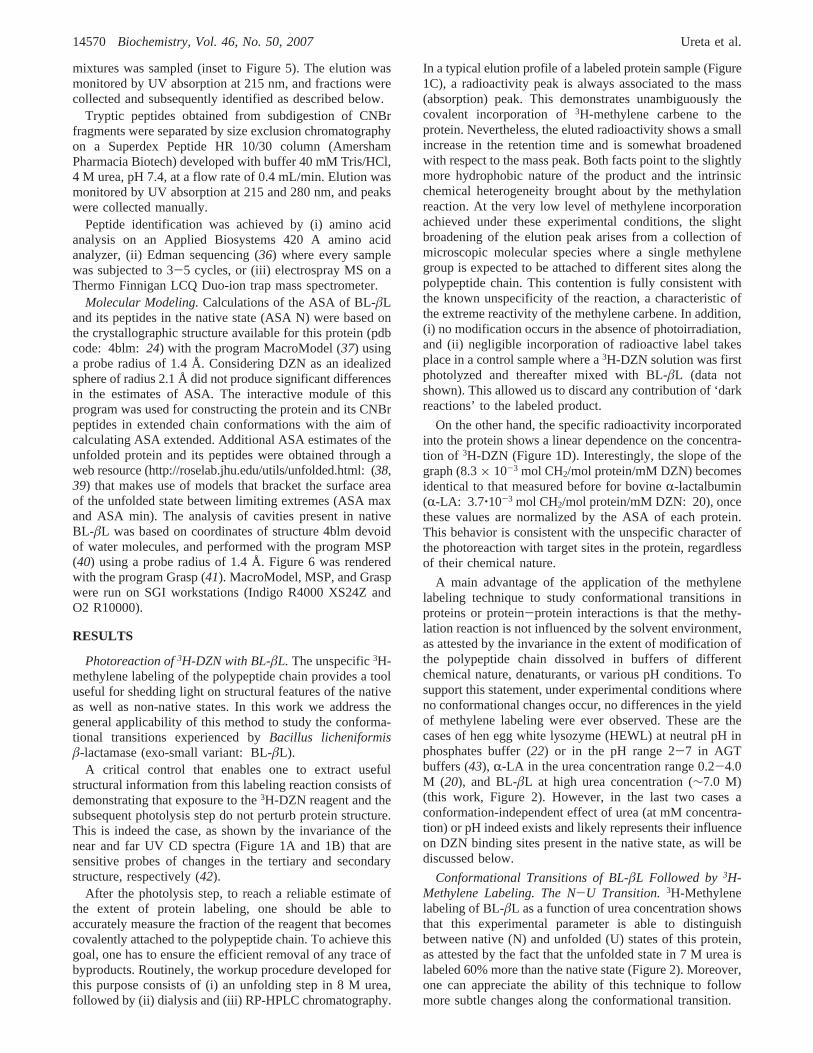
Conformational Transitions of BL-âL Followed by3H-Methylene Labeling. The N-U Transition. 3H-Methylenelabeling of BL-âL as a function of urea concentration showsthat this experimental parameter is able to distinguishbetween native (N) and unfolded (U) states of this protein,as attested by the fact that the unfolded state in 7 M urea islabeled 60% more than the native state (Figure 2). Moreover,one can appreciate the ability of this technique to followmore subtle changes along the conformational transition.
14570 Biochemistry, Vol. 46, No. 50, 2007 Ureta et al.
To prove the validity of this approach, parallel measure-ments of CD (in both UV regions) and the intensity of theintrinsic tryptophan fluorescence were carried out for the sakeof comparison. The diversity in the traces of the unfoldingcurves monitored by the spectroscopic techniques clearlysupports the existence of intermediates, as was also observedby others in this same protein system (25, 44-46) and in arelatedâL of S. aureus(47). An added dimension in thiscomplex transition is here provided by the methylene labelingdata. The presence of at least three states was inferred fromtwo sequential transitions occurring at 1.5-3.0 M and 3.5-6.5 M urea, involving a partially unfolded state I (predomi-nant between 3.0 and 3.5 M urea). Interestingly, in the firsttransition (N-I) 3H-DZN labeling shows the largest ampli-tude: 67% of the total change observed, as compared to themagnitude of changes in the CD signals: 56% for [θ]275 and29% for [θ]222. Even at the highest urea concentration assayed(8 M), 18% of the far UV-CD signal remains, indicatingthe presence of residual secondary structure in the U state.In addition, a relatively minor perturbation in the environ-ment of tryptophan would occur in state I, as judged by thesmall amplitude of the change in the fluorescence intensity(24% of the total change is observed in the first transition)and the invariance of the maximum of the spectra (∼337nm) in the range 0-3.5 M urea. By contrast, both parameterschange dramatically as the protein proceeds from state I tostate U (the maximum shifts to∼351 nm in the latter).
Remarkably, apart from the substantial increase in theextent of labeling with3H-methylene occurring upon unfold-ing, a smaller but significantdecreaseis observed at verylow urea concentration, in a range where no conformationalchanges are evident by spectroscopy (inset to Figure 2). Onelikely explanation for this phenomenon, which was alsoreported forR-LA (20) and might well represent the generalbehavior of many proteins, would be the displacement of3H-DZN by urea from binding sites present in the N state(note that 1-10 mM urea concentration is comparable tothe concentration of3H-DZN in the samples).
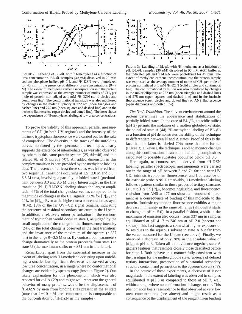
The N-A Transition.The solvent environment around theprotein determines the appearance and stabilization ofpartially folded states. In the case of BL-âL, an acidic milieu(pH 2) permits the isolation of a molten globule-like state,the so-called state A (44). 3H-methylene labeling of BL-âLas a function of pH demonstrates the ability of the techniqueto differentiate between N and A states. Proof of this is thefact that the latter is labeled 70% more than the former(Figure 3). Likewise, the technique is able to monitor changesalong this conformational transition, including complexitiesassociated to possible substates populated below pH 3.5.
Here again, to contrast results derived from3H-DZNlabeling, parallel spectroscopic measurements were carriedout in the range of pH between 2 and 7: far and near UVCD, intrinsic tryptophan fluorescence, and fluorescence ofthe extrinsic probe ANS. Remarkably,3H-methylene labelingfollows a pattern similar to those probes of tertiary structure,i.e., at pHe 3.5 [θ]275 becomes negligible, and fluorescenceemission from ANS at 477 nm shows a pronounced incre-ment as a consequence of binding of this molecule to theprotein. Intrinsic tryptophan fluorescence exhibits a majordecrease in intensity in the same pH range (although it startsto change at pHe 5.0). In a parallel fashion, a shift in themaximum of emission also occurs: from 337 nm in samplesequilibrated at pH 4-7 to 340 nm at pH 2.0 (spectra notshown). This fact suggests a somewhat higher exposure ofW residues to the aqueous solvent in state A but far fromthe value measured for the U state (see above). Finally, weobserved a decrease of only 28% in the absolute value of[θ]222 at pH e 3. Taken all this evidence together, state Agathers features that resemble closely those described beforefor state I. Both behave in a manner fully consistent withthe paradigm for the molten globule state: absence of definedtertiary interactions, preservation of substantial secondarystructure content, and permeation to the aqueous solvent (48).
In the course of these experiments, adecreaseof lessermagnitude in the extent of labeling was observed in samplesequilibrated at pH 5 as compared to those at pH 7, wellwithin a range where no conformational changes occur. Thisphenomenon bears resemblance to that observed at very lowurea concentrations (see above) and might result as aconsequence of the displacement of the reagent from binding
FIGURE 2: Labeling of BL-âL with 3H-methylene as a function ofurea concentration. BL-âL samples (30µM) dissolved in 20 mMsodium phosphate buffer, pH 7.4, and3H-DZN were photolyzedfor 45 min in the presence of different urea concentrations (0-7M). The extent of methylene carbene incorporation into the proteinsample was expressed as the average number of moles of CH2 permole of protein normalized at 1 mM3H-DZN (solid circles andcontinuous line). The conformational transition was also monitoredby changes in the molar ellipticity at 222 nm (open triangles anddashed line) and 275 nm (open squares and dashed line) and in theintrinsic fluorescence (open circles and dotted line). The inset showsthe dependence of3H-methylene labeling at low urea concentrations.
FIGURE 3: Labeling of BL-âL with 3H-methylene as a function ofpH. BL-âL samples (30µM) dissolved in 90 mM AGT buffer atthe indicated pH and3H-DZN were photolyzed for 45 min. Theextent of methylene carbene incorporation into the protein samplewas expressed as the average number of moles of CH2 per mole ofprotein normalized at 1 mM3H-DZN (solid circles and continuousline). The conformational transition was also monitored by changesin the molar ellipticity at 222 nm (open triangles and dashed line)and 275 nm (open squares and dashed line) and in the intrinsicfluorescence (open circles and dotted line) or ANS fluorescence(open diamonds and dotted line).
Conformation of BL-âL Probed by Methylene Carbene Labeling Biochemistry, Vol. 46, No. 50, 200714571
sites present in the N state. Tentatively, here protonation ofcritical amino acid side chains might bring about this effect.
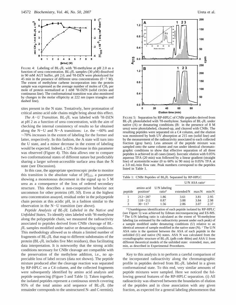
The A-U Transition.BL-âL was labeled with3H-DZNat pH 2 as a function of urea concentration, with the aim ofchecking the internal consistency of results so far obtainedalong the N-U and N-A transitions: i.e. the∼60% and∼70% increases in the extent of labeling for the former andlatter, respectively. In this fashion, the A state will turn intothe U state, and a minor decrease in the extent of labelingwould be expected. Indeed, a 12% decrease in this parameterwas observed (Figure 4). This data allows one to comparetwo conformational states of different nature but predictablysharing a larger solvent-accessible surface area than the Nstate (see Discussion).
In this case, the appropriate spectroscopic probe to monitorthis transition is the absolute value of [θ]222, a parametershowing a monotonous decrement in the signal up to 5 Murea as a consequence of the loss of ordered secondarystructure. This describes a non-cooperative behavior notuncommon for other proteins (49, 50). Even at the highesturea concentration assayed, residual order in the polypeptidechain persists at this acidic pH, in a fashion similar to theobservation in the N-U transition (see above).
Peptide Analysis of BL-âL Labeled in the NatiVe andUnfolded States.To identify sites labeled with3H-methylenealong the polypeptide chain, we measured the radioactivityassociated to peptides derived from CNBr cleavage of BL-âL samples modified under native or denaturing conditions.This methodology allowed us to obtain a limited number offragments of BL-âL that map to different subdomains of theprotein (BL-âL includes five Met residues), thus facilitatingdata interpretation. It is noteworthy that the strong acidicconditions necessary for CNBr cleavage are compatible withthe preservation of the methylene addition, i.e., no ap-preciable loss of label occurs (data not shown). The peptidemixture produced after the cleavage reaction was separatedby RP-HPLC on a C4 column, and the collected fragmentswere subsequently identified by amino acid analysis andpeptide sequencing (Figure 5 and Table 1). Taken together,the isolated peptides represent a continuous stretch covering95% of the total amino acid sequence of BL-âL (theremainder corresponds to the unstructured N- and C-termini).
Key to this analysis is to perform a careful comparison ofthe incorporated radioactivity along the chromatographicprofile in peptides derived from BL-âL labeled in a givenconformational state. To this end, very similar amounts ofpeptide mixtures were sampled. Here we noticed the fol-lowing general features of the RP-HPLC separation. (i) Allradioactivity was observed between the boundaries of elutionof the peptides and in close association with any givenfraction, as expected for a general labeling phenomenon that
FIGURE 4: Labeling of BL-âL with 3H-methylene at pH 2.0 as afunction of urea concentration. BL-âL samples (30µM) dissolvedin 90 mM AGT buffer, pH 2.0, and3H-DZN were photolyzed for45 min in the presence of different urea concentrations (0-7 M).The extent of methylene carbene incorporation into the proteinsample was expressed as the average number of moles of CH2 permole of protein normalized at 1 mM3H-DZN (solid circles andcontinuous line). The conformational transition was also monitoredby changes in the molar ellipticity at 222 nm (open triangles anddashed line).
FIGURE 5: Separation by RP-HPLC of CNBr peptides derived fromBL-âL photolabeled with3H-methylene. Samples of BL-âL undernative (A) or denaturing conditions (B: in the presence of 8 Murea) were photolabeled, cleaned-up, and cleaved with CNBr. Theresulting peptides were separated on a C4 column, and the elutionwas monitored by both UV absorption at 215 nm (solid line) andby the measurement of the radioactivity associated to each collectedfraction (gray bars). Less amount of the peptide mixture wassampled onto the same column and run under identical chromato-graphic conditions to show that effective separation of all threepeptides is achieved in all cases (inset). Isocratic elution with 0.05%aqueous TFA (20 min) was followed by a linear gradient (straightline) of acetonitrile:water (0 to 60% in 90 min) in 0.05% TFA ata 3.0 mL/min flow rate. Peak numbers correspond to the peptideslisted in Table 1.
Table 1: CNBr Peptides of BLâL Separated by RP-HPLC
U/N ASA ratioc
peptideamino acidpositiona
U/N labelingratiob extended/N max/N min/N
1 212-287 1.86 3.61 3.81 2.952 118-211 0.87 3.88 3.84 2.983 30-117 1.56 2.86 3.07 2.37
a Unambiguous identification of each peptide isolated by RP-HPLC(see Figure 5) was achieved by Edman microsequencing and ES-MS.b The U/N labeling ratio is calculated as the extent of3H-methylenelabeling (as estimated by the radioactivity present under each peak) ofa sample modified under denaturing conditions (U) relative to anidentical amount of sample modified in the native state (N).c The U/NASA ratio is the quotient between the ASA of each peptide in theunfolded (U) and native (N) states. ASA N was calculated from thecrystallographic structure of BL-âL (pdb code 4blm) and ASA U fromdifferent theoretical models of the unfolded state: extended, max, andmin, as described in Experimental Procedures.
14572 Biochemistry, Vol. 46, No. 50, 2007 Ureta et al.
causes minimal modification of the polypeptide chain. (ii)A trend exists to have more methylene label incorporationas the molecular mass and/or the hydrophobicity of thepeptides increase, a fact that agrees with the indiscriminatereactivity of the methylene carbene. (iii) Overall, an incre-ment of 65% in label incorporation was observed for the Ustate relative to the N state. This value agrees well with theincrement measured for the whole protein (see above datafor the N-U transition). (iv) Similarly, a cross comparisonwas carried outfor each isolated peptide, where results areexpressed as U/N labeling ratios (Table 1). The individualbehavior of each peptide is not uniform. Although the generaltrend points to an increase in labeling, yielding U/N ratioshigher than one, as observed for peptides 1 and 3, a differentresult is observed for peptide 2. Further enzymatic digestionwith trypsin and separation by size exclusion chromatographyof all three peptides rendered profiles from which confirma-tory evidence was derived. Thus, peptide 1 and 3 producedpatterns where the U/N ratio is consistently higher than onefor all tryptic peptides, whereas peptide 2 exhibited a clear‘inversion’ of the ratio at certain fractions (e.g., U/N ratiosas low as 0.37, data not shown).
The observation of differences in the extent of labelingalong the polypeptide chain, rather than being a peculiaraspect of BL-âL, is common to other proteins, as wasdemonstrated forR-LA (20) and HEWL (22). The mainsource of these differences does not arise from the intrinsicchemical nature of the target functional groups, but ratherfrom the conformational state of the peptide, which willobviously affect the microenvironment around a given site.The overall increment in labeling and the individual valuesmeasured for each peptide are most relevant pieces ofinformation that will be discussed later in terms of theincreased surface exposure predicted at different sites alongthe polypeptide chain.
DISCUSSION
Methylene Carbene as an Unspecific and NonintrusiVeProbe of the Polypeptide Surface.In order to attempt anychemical modification experiment aimed at addressing con-formational aspects of the protein, one should ensure thatthe reagent itself does not pose a structural perturbation onthe system. This is indeed the case, as shown in Figure 1Aand 1B, where it becomes evident that neither the codisso-lution of the parent reagent DZN in the protein sample northe ensuing photoirradiation step necessary to cause insertionof the methylene biradical affect conformation. Nevertheless,one should bear in mind that at the typical extents ofmodification achievable under our experimental conditionsonly a minor fraction of the protein molecules in the samplewill become methylated.
This work underscores the novel applicability of thisreaction to modify the polypeptide chain immersed in itsnatural aqueous environment and relates this to the conceptof accessible surface area (ASA). Because ASA is expectedto depend strongly on the folding state of the protein, onewould predict the realization of an experimental measurementof this critical parameter through DZN labeling.
The linearity observed in Figure 1D is consistent with acollisional behavior for the reaction of methylene carbenewith the protein, where no appreciable affinity of DZN needs
to be invoked, i.e., under our experimental conditions no hintof curvature suggesting a hyperbolic behavior is seen. Indeed,typically the ‘binding affinities’ measured for small organicmolecules to proteins (HEWL) range between 0.1 and 5 M-1
(51). In support of the collisional behavior, a simpletheoretical analysis that considers the mole fraction (R) ofthe ASA of the protein with respect to all components presentin the system (that includes the buffer and the water solvent)yields a reasonably good estimate of the experimentallydetermined extent of protein modification, as is discussednext. TheR value equals the ratio of the following prod-ucts: (i) the ASA of BL-âL (12 576 Å2, estimated from pdbfile 4blm, using a probe radius of 1.4 Å) times theconcentration of this protein in the sample (25µM), and (ii)the total ASA available for labeling in the system, i.e., thesum of the corresponding products calculated for the protein,the buffer, and the water solvent (essentially dominated bythat for the latter: 32.4 Å2 times 55.55 M). If the incorpora-tion of label follows a purely unspecific mode, that is, if thereagent adds to components of the system proportionally tothe available area, then one would expect a fractionR ofmethylene equal to 0.175‰ incorporated into the proteincomponent, that is an amount equivalent to 7× 10⁻³ mol ofmethylene per mole of protein, at a standard concentrationof 1 mM bulk 3H-DZN reagent. Indeed, the experimentallydetermined slope of 8.3× 10⁻³ mol CH2/mol protein/mMDZN (Figure 1D) agrees very well with the prediction. Anidentical calculation carried out forR-LA (ASA: 7236 Å2,taken from pdb file 1hfz, subunit D) yields a value of 4×10⁻³ mol CH2/mol protein/mM DZN, that is also in excelentagreement with experiment: 4.1× 10⁻³ mol CH2/molprotein/mM DZN (20). Taking these values together, onecan estimate that, on average,∼6 × 10⁻⁷ molecules CH2per Å2 at 1 mM DZN will be incorporated. Related to thispoint, an independent study aimed at measuring the ASAoccluded due to an antigen-antibody interaction betweenHEWL and the immunoglobulin IgG1D1.3 (749 Å2, mea-sured in pdb file 1vfb) yielded a value of 0.44× 10⁻³ molCH2/mol protein/mM DZN for the difference in the labelingextent between free and complexed HEWL (22). The ratiobetween these numbers equals 5.9× 10⁻⁷ molecules CH2per Å2 at 1 mM DZN, a value remarkably close to thatreported above.
Nevertheless, one should point out that this correlationfound between different proteins in their native statesnecessarily includes features of their surface that mightinfluence the final experimental value observed. For instance,the presence of cavities or crevices or hydrophobic patches,with areas of different sizes that might favor DZN partition,would tend to overestimate the measurements, as will bediscussed below. In principle, one would expect a morestraightforward correlation for the unfolded state because thelatter will lack the topographical complexities associated withmore structured conformations.
3H-Methylene Labeling Is SensitiVe to the ConformationalState of the Protein.The cross comparison between theprofile of methylene labeling and measurements derived fromother biophysical methods such as CD and fluorescencedemonstrates a close correlation with those probes mostsensitive to changes in tertiary structure. This contention issupported by the observations that (i) along the urea-inducedN-I-U transition (Figure 2) the most significant change in
Conformation of BL-âL Probed by Methylene Carbene Labeling Biochemistry, Vol. 46, No. 50, 200714573
DZN labeling occurs along the N-I portion, and (ii) alongthe acid pH-induced N-A transition (Figure 3) DZN labelingshows a large amplitude change in parallel with opticalprobes of tertiary structure (Trp fluorescence, ANS binding,and near UV CD).
Careful comparison of the extent of labeling correspondingto each state assayed allowed us to attempt inferences ontheextentandnatureof the surface exposed to the aqueoussolvent. We hypothesize that the close packing characteristicof the native state would represent the main impediment tothe penetration of the DZN probe, in a fashion similar tothat experienced by the water solvent. On the other hand,ready accessibility to the solvent governs the behavior ofthe unfolded state, where an increase in DZN labeling is thecommon observation. In between these extremes, one findsthe I (or A) states, where the increment observed with respectto the native state is consistent with (i) the proposedloosening of tight interactions in the core of the proteinallowing enhanced permeation of solvent and probe, and (ii)the concomitant appearance of a hydrophobic ‘phase’ proneto a more favorable partition of the reagent. The organizationof this putative hydrophobic phase would no longer persistin the unfolded state.
From a quantitative elaboration on the extent of labelingobserved for each conformational state alongside theoreticalestimates of the ASA parameter one can draw useful insightson features of the native and non-native states. In regard tothis point, a∼60% increment in the extent of methylenelabeling was observed along the urea-induced N-U transi-tion. An interpretation of this result requires taking intoaccount the geometry expected for the unfolded state.Because there is no consensus picture on the nature of thelatter, one should compare experimental measurements withaccurate predictions derived from different models: (i) anextended chain (phi and psi 180°) marks the upper limit ofsolvent exposure for the polypeptide (ASAextended); (ii)alternatively, one can regard a statistical ensemble offluctuating conformers simulated with a Monte Carlo tech-nique and a hard sphere potential (ASAmax,38, 39); (iii)the surface of the fragments folded as in the native staterenders the lower bound (ASAmin,38, 39). The consider-ation of a fully extended chain predicts an increment thatlies way above that observed (∼190-195%). Under theassumptions described before for ASAmax and ASAmin, thecorresponding increments in solvent accessibility would be∼199% and∼132%, respectively. Even under the mostrealistic model considered here (ASAmin), predictions stilloverestimate solvent exposure. Nevertheless, thermodynamicconsiderations on the unfolding process based on calorimetryput forward the notion that the unfolded state would exposeat most about two-thirds of the value calculated for the fullyextended chain model (52). Taking into account this result,an upper boundary of∼97% would be set to the incrementin ASA associated to the BL-âL unfolding. In addition, aresidual dichroic signal exists in the presence of 8 M urea(∼18% of that measured at 222 nm for the native state).Taken together, this evidence would point to the fact thatthe unfolded state would differ significantly from a purelyextended form. This is fully consistent with the notion thatin many cases a certain extent of remaining structure persistsin the unfolded state (53, 54). At the other end, the presenceof cavities and crevices known to exist in the native state
might cause the binding of DZN, thus incrementing theresidence time of the reagent at certain sites (see below) andconsequently augmenting the extent of methylene labeling.The combined effect of these factors will bring about areduction in the increment of labeling expected in progressingfrom the native to the unfolded state.
A different effect, albeit of lesser magnitude, is alsoobserved at very low concentration of the denaturant (insetto Figure 2). Here, no change in conformation due toweakening of hydrophobic interactions should be invoked,but rather a urea-borne ‘uncoating’ of the protein surfacefrom sites putatively occupied by DZN. This is consistentwith (i) the existence of hydrophobic spots on the surfaceand/or cavities, where the reagent would be expected toreside longer, and (ii) the micromolar affinity measured forurea binding to protein surfaces as opposed to the millimolarto molar affinities expected for gases of similar size to DZN(see above), thus presumably causing the ready displacementof the latter by the former.
The extreme reactivity of the methylene carbene speciesoffers a technical advantage over other conventional chemicalmodification reagents, bringing about the possibility ofreacting with the polypeptide chain regardless of the pH ofthe milieu. Proof of the latter is the invariance in the extentof modification that has been demonstrated for HEWL inthe pH range 2-7, where its native conformation is preserved(43). Unlike the former behavior, for BL-âL an incrementof 70% in methylene labeling is observed for the A statewith respect to the N state (Figure 3). This marks a sharptransition that runs in parallel with optical measurements and,in particular, with the binding of the fluorescent dye ANS.This probe is believed to bind to clusters of nonpolar atomsaccessible to the solvent that are absent in the denatured stateand are relatively rare in the native state (55). In this fashion,the intermediate A is consistent with a state exhibitingexposed (hydrophobic) patches, substantially altered tertiaryinteractions and only a minor loss of secondary structure.All these features characterize a molten globule state for BL-âL, as it has been described by others (28, 47, 56, 57). Theemerging picture for state A includes the conservation of ahydrophobic, although less well-packed core, exhibiting amore ready accessibility to the aqueous solvent and likewiseto the reagent.
Finally, the extent of methylene labeling decreases mini-mally (∼12%) along the non-cooperative A-U transition(Figure 4). This fact might arise as the consequence of abalance between the following phenomena: (i) the prefer-ential partition of the reagent DZN in hydrophobic regionspresent in the A state; (ii) the expected ASA increaseoccurring upon protein unfolding. This emphasizes theabove-mentioned usefulness of DZN labeling to monitorchanges of tertiary structure and, less so, of secondarystructure, as demonstrated by the gradual and large amplitudechange of secondary structure content (as shown by [θ]222).Here, it is noteworthy to state that there hardly exists muchdifference in the extent of solvent exposure between thepolypeptide chain adopting a random or extended conforma-tion and one folded as anR helix (result not shown). Thisarises mostly from the much larger weight of the side chainsof amino acids on the total surface of the polypeptide.
Methylene Carbene Labeling ReVeals Local Features ofthe Folding of BL-âL. A thorough peptide analysis of those
14574 Biochemistry, Vol. 46, No. 50, 2007 Ureta et al.
extreme cases represented by the N and U states allowed usto produce a map of local solvent accessibility (Figures 5and 6). On average, in the U state, peptide labeling amountsto 6.95 cpm/mol amino acid residue, whereas in the N state,this value is 4.20 cpm/mol amino acid residue. Therefore,an increment of 65% is observed for the former relative tothe latter, a result fully consistent with measurementsobtained for the whole protein (cf. 60% for the N-Utransition, see above). In addition, as has also been observedfor R-LA (20), the variation in the extent of labeling amongdifferent peptides is greater in the N state than in the U state,an observation pointing to the lesser influence exerted bythe environment on the yield of reaction in the latter case.
The U/N labeling ratio measured for each isolated peptideprovides a reliable experimental estimate that can best becorrelated with conformational aspects (Figure 6 and Table1). Given the shortcomings implied in each model for theunfolded state, one should not expect to find a straightforwardcorrespondence in the absolute values. All these predictionstend to overestimate the solvent exposure of the unfoldedstate. However, a positive correlation in relative terms couldbe traced, and that would include the cases of peptides 1and 3, where the experimental U/N ratio matches the morerealistic min/N theoretical estimate. This was also the casefor a larger set of tryptic peptides derived fromR-LA (20).At variance with this, peptide 2 is a clear outlier: here theU/N is inverted. To address this point, a detailed descriptionof thenatiVe stateshould offer a hint on this behavior. TheBL-âL structure can be envisioned as a close associationbetween two subdomains: one characterized by anR+âmotif and the other rich inR structure. Two crevices lie atthe interface between these subdomains, one of which formsthe active site cleft (26, 27, 58, 59). This constitutes a poorlypacked region where water molecules occur and possiblyexhibiting enhanced flexibility. In addition, the largest cavityoccurs in this neighborhood: it shows an irregular shape andis bounded by helixR2, the so-calledΩ-loop and residuespertaining to helicesR5 andR6. In this regard, the presenceof crevices or cavities that could potentially lodge the reagentDZN -thus favoring its partition into these sites- wouldpresent a preferential target for labeling in state N. It issuggestive that the ‘anomalously’ labeled peptide 2 actuallyincludes all the amino acids belonging to theΩ-loop and
those lining the major cavity in BL-âL (of 64 Å3 and limitedby Q135, L139, P145, L148, P162, R164, L169, and D179,see Figure 6B). This space is big enough to fit DZN (∼43-45 Å3), provided that water molecules could be expelled fromthe site.
CONCLUSIONS AND PERSPECTIVES
In this work we demonstrate the general usefulness of anovel technique based on the photochemically triggeredmethylene labeling of the polypeptide chain to followconformational transitions in proteins. This is exemplifiedhere by BL-âL, a protein undergoing defined changes in itsconformation as a consequence of modification of the solventenvironment. In particular, methylene labeling made possiblea comparative structural characterization of the native as wellas non-native states, including partially folded (moltenglobule) and unfolded ensembles. The accurate determinationof the extent of labeling enlightens even subtle aspects ofcomplex transitions and introduces an original (and perhapsunique) experimental measurement of ASA. The improvedresolution of this analysis at the level of peptides providesprofiles of solvent accessibility along the amino acidsequence from which three-dimensional information can bederived. Further developments along these lines would permitstudy of short-lived kinetic intermediates through the use ofvery powerful (synchrotron) light sources for the photolysisstep. At the analytical front, modern mass spectrometrymethods will be used with advantage to detect the excessmass associated to the methylated products, thus avoidingthe use of radiotracers.
ACKNOWLEDGMENT
We thank Dr. Javier Santos for his helpful comments andcritical reading of the manuscript.
REFERENCES
1. Dill, K. A., and Shortle, D. (1991) Denatured states of proteins,Annu. ReV. Biochem. 60, 795-825.
2. Ptitsyn, O. B. (1995) Molten globule and protein folding,AdV.Protein Chem. 47, 83-229.
3. Arai, M., and Kuwajima, K. (2000) Role of the molten globulestate in protein folding,AdV. Protein Chem. 53, 209-282.
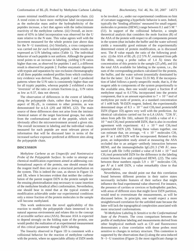
FIGURE 6: Structure of native BL-âL (pdb code 4blm:27). (A) Peptides 1-3 (see Table 1) are colored in blue, red, and green, respectively.(B) The peptide having an anomalously low labeling increment (peptide 2) is colored in red, and the remainder of the protein is shown inwhite. The major cavity (64 Å3), delimited by residues Q135, L139, P145, L148, P162, R164, L169, and D179, is illustrated as a blobcolored in green. For the sake of comparison, a DZN molecule is shown as a CPK model (43 Å3). Cavity volumes and boundaries werecalculated with MSP (40) using a probe radius of 1.4 Å. The final figure was rendered with GRASP (41).
Conformation of BL-âL Probed by Methylene Carbene Labeling Biochemistry, Vol. 46, No. 50, 200714575
4. Hammes, G. G. (2005)Spectroscopy for the biological sciences,John Wiley & Sons, Inc., Hoboken, NJ.
5. Fersht, A. (1999)Structure and mechanism in protein science: aguide to enzyme catalysis and protein folding, W. H. Freemanand Company, New York, NY.
6. Creighton, T. E., ed. (1992)Protein folding, W. H. Freeman andCompany, New York, NY.
7. Pain, R. H., ed. (1994)Mechanisms of protein folding, IRL Pressat Oxford University Press, Oxford, U. K.
8. Lee, B., and Richards, F. M. (1971) The interpretation of proteinstructures: estimation of static accessibility,J. Mol. Biol. 55, 379-400.
9. Miller, S., Janin, J., Lesk, A. M., and Chothia, C. (1987) Interiorand surface of monomeric proteins,J. Mol. Biol. 196, 641-656.
10. Pace, C. N. (2001) Polar group burial contributes more toprotein stability than nonpolar group burial,Biochemistry 40, 310-313.
11. Lundblad, R. L. (2005)Chemical reagents for protein modification,3rd ed., CRC Press, Boca Raton, FL.
12. Goshe, M. B., and Anderson, V. E. (1999) Hydroxyl radical-induced hydrogen/deuterium exchange in amino acid carbon-hydrogen bonds,Radiat. Res. 151, 50-58.
13. Goshe, M. B., Chen, Y. H., and Anderson, V. E. (2000)Identification of the sites of hydroxyl radical reaction with peptidesby hydrogen/deuterium exchange: prevalence of reactions withthe side chains,Biochemistry 39, 1761-1770.
14. Maleknia, S. D., Brenowitz, M., and Chance, M. R. (1999)Millisecond radiolytic modification of peptides by synchrotronX-rays identified by mass spectrometry,Anal. Chem. 71, 3965-3973.
15. Maleknia, S. D., Ralston, C. Y., Brenowitz, M. D., Downard, K.M., and Chance, M. R. (2001) Determination of macromolecularfolding and structure by synchrotron x-ray radiolysis techniques,Anal. Biochem. 289, 103-115.
16. Maleknia, S. D., and Downard, K. M. (2001) Unfolding ofapomyoglobin helices by synchrotron radiolysis and mass spec-trometry,Eur. J. Biochem. 268, 5578-5588.
17. Englander, S. W., and Krishna, M. M. G. (2001) Hydrogenexchange,Nat. Struct. Biol. 8, 741-742.
18. Nukuna, B. N., Goshe, M. B., and Anderson, V. E. (2001) Sitesof hydroxyl radical reaction with amino acids identified by (2)HNMR detection of induced (1)H/(2)H exchange,J. Am. Chem.Soc. 123, 1208-1214.
19. Richards, F. M., Lamed, R., Wynn, R., Patel, D., and Olack, G.(2000) Methylene as a possible universal footprinting reagent thatwill include hydrophobic surface areas: overview and feasibil-ity: properties of diazirine as a precursor,Protein Sci. 9, 2506-2517.
20. Craig, P. O., Ureta, D. B., and Delfino, J. M. (2002) Probingprotein conformation with a minimal photochemical reagent,Protein Sci. 11, 1353-1366.
21. Nuss, J. E., and Alter, G. M. (2004) Denaturation of replicationprotein A reveals an alternative conformation with intact domainstructure and oligonucleotide binding activity,Protein Sci. 13,1365-1378.
22. Gomez, G. E., Cauerhff, A., Craig, P. O., Goldbaum, F. A., andDelfino, J. M. (2006) Exploring protein interfaces with a generalphotochemical reagent,Protein Sci. 15, 744-752.
23. Delfino, J. M., Schreiber, S. L., and Richards, F. M. (1993)Design, synthesis and properties of a photoactivatable membrane-spanning phospholipidic probe,J. Am. Chem. Soc. 115, 3458-3474.
24. Weber, T., and Brunner, J. (1995) 2-(Tributylstannyl)-4-[3-(trifluoromethyl)-3H-diazirin-3-yl]benzyl Alcohol: A BuildingBlock for Photolabeling and Cross-Linking Reagents of VeryHigh Specific Radioactivity,J. Am. Chem. Soc. 117, 3084-3095.
25. Frate, M. C., Lietz, E. J., Santos, J., Rossi, J. P., Fink, A. L., andErmacora, M. R. (2000) Export and folding of signal-sequencelessBacillus licheniformis beta-lactamase in Escherichia coli,Eur. J.Biochem. 267, 3836-3847.
26. Moews, P. C., Knox, J. R., Dideberg, O., Charlier, P., and Frere,J. M. (1990) Beta-lactamase of Bacillus licheniformis 749/C at 2Å resolution.Proteins 7, 156-171.
27. Knox, J. R., and Moews, P. C. (1991) Beta-lactamase of Bacilluslicheniformis 749/C. Refinement at 2 Å resolution and analysisof hydration,J. Mol. Biol. 220, 435-455.
28. Goto, Y., Calciano, L. J., and Fink, A. L. (1990) Acid-inducedfolding of proteins,Proc. Natl. Acad. Sci. U. S. A. 87, 573-577.
29. Yanagida, N., Uozumi, T., and Beppu, T. (1986) Specific excretionof Serratia marcescens protease through the outer membrane ofEscherichia coli,J. Bacteriol. 166, 937-944.
30. Jansson, J. A. (1965) A Direct Spectrophotometric Assay ForPenicillin Beta-Lactamase (Penicillinase),Biochim. Biophys. Acta99, 171-172.
31. Schagger, H., and von Jagow, G. (1987) Tricine-sodium dodecylsulfate-polyacrylamide gel electrophoresis for the separation ofproteins in the range from 1 to 100 kDa,Anal. Biochem. 166,368-379.
32. Ellerby, L. M., Escobar, W. A., Fink, A. L., Mitchinson, C., andWells, J. A. (1990) The role of lysine-234 in b-lactamasecatalysis probed by site-directed mutagenesis,Biochemistry 29,5797-5806.
33. Kamp, R. M. (1986) inAdVanced Methods in Protein Microse-quence Analysis, pp 8-20, Springer-Verlag, Berlin.
34. Fontana, A., and Gross, E. (1987) Fragmentation of polypeptidesby chemical methods, inPractical Protein Chemistry-a Handbook,pp 67-120, John Wiley & Sons Ltd, New York NY.
35. Wilkinson, J. M. (1986) inPractical Protein Chemistry-A Hand-book, pp 121-148. John Wiley & Sons Ltd., Chichester.
36. Edman, P., and Begg, G. (1967) A protein sequenator,Eur. J.Biochem. 1, 80-91.
37. Mohamadi, F., Richards, N. G. J., Guida, W. C., Liskamp, R.,Lipton, M., Caufield, C., Chang, G., Hendrickson, T., and Still,W. C. (1990) Macromodel-An integrated software system formodeling organic and bioorganic molecules using molecularmechanics,J. Comput. Chem. 11, 440-467.
38. Creamer, T. P., Srinivasan, R., and Rose, G. D. (1995) Modelingunfolded states of peptides and proteins,Biochemistry 34, 16245-16250.
39. Creamer, T. P., Srinivasan, R., and Rose, G. D. (1997) Modelingunfolded states of proteins and peptides. II. Backbone solventaccessibility,Biochemistry 36, 2832-2835.
40. Connolly, M. L. (1993) The molecular surface package,J. Mol.Graph. 11, 139-141.
41. Nicholls, A., Sharp, K. A., and Honig, B. (1991) Protein foldingand association: insights from the interfacial and thermodynamicproperties of hydrocarbons,Proteins 11, 281-296.
42. Fasman, G. D. (1996)Circular dichroism and the conformationalanalysis of biomolecules,Plenum Press, New York, NY.
43. Craig, D.B. (2004).Diazirine: a photoreactiVe probe for theconformational study of proteins, Ph.D. Dissertation, Universityof Buenos Aires.
44. Calciano, L. J., Escobar, W. A., Millhauser, G. L., Miick, S. M.,Rubaloff, J., Todd, A. P., and Fink, A. L. (1993) Side-chainmobility of the â-lactamase A state probed by electron spinresonance spectroscopy,Biochemistry 32, 5644-5649.
45. Santos, J., Gebhard, L. G., Risso, V. A., Ferreyra, R. G., Rossi, J.P., and Erma´cora, M. R. (2004) Folding of an abridgedâ-lacta-mase,Biochemistry 43, 1715-1723.
46. Santos, J., Risso, V. A., Sica, M. P.and Erma´cora, M. R. (2007)Effects of Serine-to-Cysteine Mutations onbeta-LactamaseFolding,Biophys. J. 93, 1707-1718.
47. Uversky, V. N. (1993) Use of fast protein size-exclusionliquid chromatography to study the unfolding of proteins whichdenature through the molten globule,Biochemistry 32, 13288-13298.
48. Ptitsyn, O. B. (1992) inProtein folding(Creighton, T. E., Ed.) pp243-300, W.H. Freeman and Company, New York, NY.
49. Schulman, B. A., Kim, P. S., Dobson, C. M., and Redfield, C.(1997) A residue-specific NMR view of the non-cooperativeunfolding of a molten globule,Nat. Struct. Biol. 4, 630-634.
50. Wijesinha-Bettoni, R., Dobson, C. M., and Redfield, C. (2001)Comparison of the denaturant-induced unfolding of the bovineand human alpha-lactalbumin molten globules,J. Mol. Biol. 312,261-273.
51. Liepinsh, E., and Otting, G. (1997) Organic solvents identifyspecific ligand binding sites on protein surfaces,Nat. Biotechnol.15, 264-268.
52. Lee, B. (1991) Isoenthalpic and isoentropic temperatures and thethermodynamics of protein denaturation,Proc. Natl. Acad. Sci.U.S.A. 88, 5154-5158.
53. Baldwin, R. L., and Zimm, B. H. (2000) Are denatured proteinsever random coils?Proc. Natl. Acad. Sci. U. S. A. 97, 12391-12392.
14576 Biochemistry, Vol. 46, No. 50, 2007 Ureta et al.
54. Shortle, D., and Ackerman, M. S. (2001) Persistence of native-like topology in a denatured protein in 8 M urea,Science 293,487-489.
55. Ptitsyn, O. B., Pain, R. H., Semisotnov, G. V., Zerovnik, E., andRazgulyaev, O. I. (1990) Evidence for a molten globule state asa general intermediate in protein folding,FEBS Lett. 262, 20-24.
56. Uversky, V. N., Semisotnov, G. V., Pain, R. H., and Ptitsyn, O.B. (1992) ‘All-or-none’ mechanism of the molten globule unfold-ing, FEBS Lett. 314, 89-92.
57. Vanhove, M., Lejeune, A., Guillaume, G., Virden, R., Pain, R.H., Schmid, F. X., and Frere, J. M. (1998) A collapsed intermedi-
ate with nonnative packing of hydrophobic residues in the foldingof TEM-1 b-lactamase,Biochemistry 37, 1941-1950.
58. Herzberg, O., and Moult, J. (1987) Bacterial resistance to beta-lactam antibiotics: crystal structure of beta-lactamase fromStaphylococcus aureus PC1 at 2.5 Å resolution,Science 236, 694-701.
59. Banerjee, S., Pieper, U., Kapadia, G., Pannell, L. K., and Herzberg,O. (1998) Role of the omega-loop in the activity, substratespecificity, and structure of class A b-lactamase,Biochemistry 37,3286-3296.
BI7012867
Conformation of BL-âL Probed by Methylene Carbene Labeling Biochemistry, Vol. 46, No. 50, 200714577


![Highly Branched Poly(α-Methylene-γ-Butyrolactone) from …file.scirp.org/pdf/OJPChem_2017112914172525.pdf · 2017-12-01 · ... (3.00 g, 0.013 mol), and L-valinol [(S)-(+)-2-Amino-3-methyl-1-butanol]](https://static.fdocument.org/doc/165x107/5b1be3007f8b9a28258f0d54/highly-branched-poly-methylene-butyrolactone-from-filescirporgpdfojpchem.jpg)


![Supporting Information - Royal Society of Chemistry · Supporting Information N-Heterocyclic Carbene-Catalyzed [3+2] Annulation of Bromoenals with 3-Aminooxindoles: Highly Enantioselective](https://static.fdocument.org/doc/165x107/5f0dee5b7e708231d43cc95a/supporting-information-royal-society-of-supporting-information-n-heterocyclic.jpg)