ApplyingComputationalScoringFunctionsto ... · largely the receptor/enzyme and its small molecule...
21
Nuclear Receptor Research Vol. 3 (2016), Article ID 101202, 21 pages doi:10.11131/2016/101202 AgiAl Publishing House http://www.agialpress.com/ Review Article Applying Computational Scoring Functions to Assess Biomolecular Interactions in Food Science: Applications to the Estrogen Receptors Francesca Spyrakis 1 , Pietro Cozzini 1 , and Glen Eugene Kellogg 2 1 University of Parma, Department of Food Science, Molecular Modelling Laboratory, Parma, Italy 2 Virginia Commonwealth University, Department of Medicinal Chemistry & Institute for Structural Biology, Drug Discovery and Development Richmond, Virginia, USA Corresponding Author: Glen Eugene Kellogg; email: [email protected] Received 3 March 2016; Accepted 7 September 2016 Editor: Yong Li Copyright © 2016 Francesca Spyrakis et al. This is an open access article distributed under the Creative Commons Attribution License, which permits unrestricted use, distribution, and reproduction in any medium, provided the original work is properly cited. Abstract. During the last decade, computational methods, which were for the most part developed to study protein-ligand interactions and especially to discover, design and develop drugs by and for medicinal chemists, have been successfully applied in a variety of food science applications [1, 2]. It is now clear, in fact, that drugs and nutritional molecules behave in the same way when binding to a macromolecular target or receptor, and that many of the approaches used so extensively in medicinal chemistry can be easily transferred to the fields of food science. For instance, nuclear receptors are common targets for a number of drug molecules and could be, in the same way, affected by the interaction with food or food-like molecules. Thus, key computational medicinal chemistry methods like molecular dynamics can be used to decipher protein flexibility and to obtain stable models for docking and scoring in food-related studies, and virtual screening is increasingly being applied to identify molecules with potential to act as endocrine disruptors, food mycotoxins, and new nutraceuticals [3–5]. All of these methods and simulations are based on protein-ligand interaction phenomena, and represent the basis for any subsequent modification of the targeted receptor’s or enzyme’s physiological activity. We describe here the energetics of binding of biological complexes, providing a survey of the most common and successful algorithms used in evaluating these energetics, and we report case studies in which computational techniques have been applied to food science issues. In particular, we explore a handful of studies involving the estrogen receptors for which we have a long-term interest. Keywords: Docking; Scoring Functions; Computational Chemistry; Nuclear Receptors; Estrogen Receptors; Food Science; Drug Discovery; Endocrine Disruptors; Binding Free Energy; Force Field; HINT 1. Introduction 1.1. Basic principles and concepts. Very extensive computa- tional technology was developed by academic and industrial scientists for use in discovering, developing and validating small molecules as “drugs”, which is, of course, an eco- nomic activity that is worth many billions of dollars. These technologies have been reviewed previously with respect to their translation to food science [1], so extensive background is not necessary here. However, the key facts are, that on the molecular scale, an interaction is an interaction, whether it is a drug identifying and occupying its binding site and setting into motion a series of steps leading to a biological action, or a protein organizing itself into its compact native
Transcript of ApplyingComputationalScoringFunctionsto ... · largely the receptor/enzyme and its small molecule...
Nuclear Receptor ResearchVol. 3 (2016), Article ID 101202, 21 pagesdoi:10.11131/2016/101202
AgiAlPublishing House
http://www.agialpress.com/
Review Article
Applying Computational Scoring Functions toAssess Biomolecular Interactions in Food Science:Applications to the Estrogen Receptors
Francesca Spyrakis1, Pietro Cozzini1, and Glen Eugene Kellogg2
1University of Parma, Department of Food Science, Molecular Modelling Laboratory, Parma, Italy2Virginia Commonwealth University, Department of Medicinal Chemistry & Institute for Structural Biology, Drug Discovery andDevelopment Richmond, Virginia, USA
Corresponding Author: Glen Eugene Kellogg; email: [email protected]
Received 3 March 2016; Accepted 7 September 2016
Editor: Yong Li
Copyright © 2016 Francesca Spyrakis et al. This is an open access article distributed under the Creative Commons AttributionLicense, which permits unrestricted use, distribution, and reproduction in any medium, provided the original work is properlycited.
Abstract. During the last decade, computational methods, which were for the most part developed to study protein-ligandinteractions and especially to discover, design and develop drugs by and for medicinal chemists, have been successfully applied ina variety of food science applications [1, 2]. It is now clear, in fact, that drugs and nutritional molecules behave in the same waywhen binding to a macromolecular target or receptor, and that many of the approaches used so extensively in medicinal chemistrycan be easily transferred to the fields of food science. For instance, nuclear receptors are common targets for a number of drugmolecules and could be, in the same way, affected by the interaction with food or food-like molecules. Thus, key computationalmedicinal chemistry methods like molecular dynamics can be used to decipher protein flexibility and to obtain stable modelsfor docking and scoring in food-related studies, and virtual screening is increasingly being applied to identify molecules withpotential to act as endocrine disruptors, food mycotoxins, and new nutraceuticals [3–5]. All of these methods and simulations arebased on protein-ligand interaction phenomena, and represent the basis for any subsequent modification of the targeted receptor’sor enzyme’s physiological activity. We describe here the energetics of binding of biological complexes, providing a survey of themost common and successful algorithms used in evaluating these energetics, and we report case studies in which computationaltechniques have been applied to food science issues. In particular, we explore a handful of studies involving the estrogen receptorsfor which we have a long-term interest.
Keywords: Docking; Scoring Functions; Computational Chemistry; Nuclear Receptors; Estrogen Receptors; Food Science; DrugDiscovery; Endocrine Disruptors; Binding Free Energy; Force Field; HINT
1. Introduction
1.1. Basic principles and concepts. Very extensive computa-tional technology was developed by academic and industrialscientists for use in discovering, developing and validatingsmall molecules as “drugs”, which is, of course, an eco-nomic activity that is worth many billions of dollars. These
technologies have been reviewed previously with respect totheir translation to food science [1], so extensive backgroundis not necessary here. However, the key facts are, that onthe molecular scale, an interaction is an interaction, whetherit is a drug identifying and occupying its binding site andsetting into motion a series of steps leading to a biologicalaction, or a protein organizing itself into its compact native
2 Nuclear Receptor Research
and biologically-active form, or the poisoning of an enzymeor receptor by a toxin in a way that mimics another, andconsiderably more healthy, interaction of that enzyme orreceptor. Also, while the food industry in toto is certainly afar more potent economic force than “Big Pharma”, it ismuchmore distributed – basically from Farm to Table with a highlyvariable number of intermediaries depending on how muchprocessing the final foodstuff has endured. So, instead of thehundreds to thousands of drug companies and API (activepharmaceutical ingredient) manufacturers, there are millionsof for-profit participants in the food industry worldwide.Quality control is a much more difficult proposition in thisscenario. Finally, we don’t know what we don’t know infood science because the potential problem areas are almostinfinite in number and thus unimaginable.
In this chapter we are focusing on “scoring functions”.These are components of the computational docking/scoringparadigm, i.e., tools to predict the energetics of protein-small molecule (or sometimes protin-protein) binding andthe stability of the resulting complexes. The first part of theproblem, docking, is a geometric problem – the positioningof a “solid” structure defined by its Cartesian coordinateswithin a cavity or pocket composed of another solid. Thisis not an inherently difficult problem, it can always be solvedby exhaustive methods, but the trick and applied intelligenceis to not do that, as there are many irrelevant solutions. Thesecond part of the problem is physico-chemical: evaluatingthe energetics involved in the association of two or moremolecules resulting in binding.
There is a very rich literature describing the virtues ofparticular docking/scoring methods that inevitably comparethe predictions and/or limits of scoring functions. Successfor a docking method is usually defined as its accuracy inrecreating crystal structure “poses” by docking the separatecomponents together. A classic review by Hawkins et al.(2008) demonstrates why this is not really a completely usefulor robust exercise [6]. Also of importance to note with respectto scoring functions is that accurate estimations of bindingfree energies may be more relevant than simply recreatingcrystal structures, but there are a number of potential issueshere too [6, 7].
It is also very significant that continuous progress in“omics” coupled with advances in (and emphasis on) struc-ture determination has led to a growing database of structuralinformation for biological macromolecules and multicom-ponent complexes. Advances in computer power, or moreexactly the far reduced cost of the same, have enabled manypredictive techniques for biomacromolecular structure. Untilrecently, perhaps only over the last decade, the primary focuswas on modeling a much-simplified version of the system –largely the receptor/enzyme and its small molecule ligand.Flexibility was considered – if at all – for the small moleculeand occasionally for a few specific amino acid residues inthe protein part. Other factors such as water were generallyignored, even to the point of deleting water molecules that
Table 1: Energies of biomolecular Interactions.
Interaction type Energy (kJ mol−1)Ion-ion [8] 150
Ion-dipole [8] 40-60
Ion-induced dipole [8] 3-15
Dipole-dipole [8] 5-25
Hydrogen bond [8, 9] 10-40
Dipole-induced dipole [8] 2-10
Induced dipole-induced dipole [8] 0.05-4.0
Hydrophobic [10] 10-12
had been experimentally resolved. Clearly, however, the bulksolvent properties, discrete water molecules in and aroundthe active site, metal ions and/or cofactors are part of theblueprint of biomolecular association, and real biologicalmolecules are not rigid.
Excepting the formation of covalent bonds, the forces ofbiomolecular associations are all, in broad terms, electro-static. Charge-charge interactions, whether formal or partialand centered on atoms, are known as salt bridges, dipoles andinduced dipoles. Hydrogen bonds are simply a special typeof electrostatic interaction, and the Van der Waals (LondonForce) attractions/repulsions arise from induced dipole-dipole interactions. The hydrophobic effect is actually anemergent electrostatic property due to the attraction (largelyhydrogen bonding) between water and polar atoms or groups“forcing” hydrophobic atoms or groups together. It is usefuland intuitive, however, to explain the observed structures,especially in aqueous solvent, in terms of a “hydrophobicinteraction” beyond what is normally expected from a purelyVan der Waals perspective. Some of the above weak forcesare set out in Table 1 with typical values.
The many complexities in approximating, quantitatively,the vast number of individual steps of biochemical recogni-tion to yield an interaction explains the overarching failureto obtain a precise and rapid estimation of binding freeenergy. Such a computational tool, if universally applicable,is sometimes referred to as the “Computational Holy Grail”[11, 12]. The basic problem is that we really have anincomplete understanding of the physics and thermody-namics of biomolecular associations [13]. Concomitantly,applying a purely reductionist approach is likely to ignorethe inherent complexity, e.g., the hydrophobic effect, drivingsuch processes. Starting with the basics of thermodynamics,the formation of biological complexes should be consideredin terms of the standard Gibb’s free energy of binding (ΔG∘),which measures the equilibrium of the reaction P𝑎𝑞 + L𝑎𝑞↔ PL𝑎𝑞 in aqueous solution:
ΔG∘ = ΔH∘ − TΔS∘, (1)
where ΔH∘ is the enthalpic contribution (breaking/formationof hydrogen bonds or specific polar and non-polar contacts)
AgiAlPublishing House | http://www.agialpress.com/
Nuclear Receptor Research 3
and TΔS∘ is the entropic contribution (release of “con-strained” water to solvent and changes in conformationalmobility of molecules). ΔG∘ is also related to Ka, theassociation constant:
ΔG∘ = −RT ln Ka, (2)
where Ka is for the above reaction. In many cases, we use thereverse dissociation constant Kd, defined as [Paq,Laq]/[PLaq].
While the direct interactions between molecules are mostimportant for favorable enthalpies of binding, the ubiquitouspresence of water also contributes energetically. Prior tointeraction, each component of the reaction is solvated,requiring partial or full desolvation during the bindingprocess. Enthalpy gained is the difference between that ofthe molecular association and those desolvation enthalpiesfrom the involved molecules. The entropy gain arises fromthe release of the previously bound waters, which is, in turn,compensated by their reorganization into new and orderedhydrogen bonds with the existing waters in bulk. In addition,unfavorable entropic contributions arise from changes indegrees of freedom, both translational and rotational of themolecules. Hydrogen bonding is often considered to be themain driving force in binding energy (see Table 1), buthydrogen bonds are susceptible to disruption by water, whichis ubiquitously present in biological systems. Thus, hydrogenbonds not solvent-accessible are stronger than those on thesurface. Hydrophobic interactions, on the other hand, areenergetically much weaker when evaluated one-by-one, butcan be be significant when considered as a whole, and are farless influenced by interactions with solvent.
For evaluating interactions we must consider the totalfree energy of binding (ΔG∘
bind), which is the sum ofthe interaction energy between the interacting molecules(ΔG∘
int), the solvation energies of molecule 1, molecule2 and of the resulting complex (ΔG∘mol1
solv, ΔG∘mol2solv,
ΔG∘complexsolv, respectively), as well as entropic (TΔS∘) and
conformational (Δ𝜆) changes in energy [14]:
ΔG∘bind = ΔG∘complex
solv − ΔG∘protsolv
−Δ𝐺∘𝑙𝑖𝑔𝑠𝑜𝑙𝑣 + Δ𝐺∘
𝑖𝑛𝑡 − 𝑇 Δ𝑆∘ + Δ𝜆(3)
There is a catch, however: Dill [15] has shown thatadding up terms as above ignores the fact that biologicalinteractions are actually concerted events that cannot besimply deconstructed. Even if each of the interaction typesare individually calculated with the utmost of care, theresulting sum may not yield an accurate and reliable ΔG∘
bind.
1.2. Practical matters. The first requirement for anystructure-based computational experiment is reliable struc-tural data, preferably from experiment. This is usuallyderived from one of three experimental techniques: X-raydiffraction, neutron diffraction and Nuclear Magnetic Reso-nance (NMR). Alternatively, when an experimental structure
is not known, but significant “homology” exists between thebiomolecule of interest and one or more known structures,theoretical comparative modeling technique may be of someuse (vide infra). To date, X-ray diffraction has been themost productive experimental method because it is relativelyinexpensive and not as susceptible to limitations of othermethods. While the big advantages of neutron diffractionover X-ray diffraction are higher resolution and the abilityto directly observe hydrogen atoms, neutron diffraction ismore expensive and rare because neutron sources are onlyavailable at accelerator facilities and the technique neces-sitates somewhat larger crystals, i.e., more material. Theprimary NMR limitations are that larger (molecular weight)proteins are more difficult to assign and that solubility isoften an issue. However, NMR is a solution technique and theresulting structure is thus free of crystallization and packingeffects, while also providing a description, albeit limited, offlexibility.
There are few examples of structures derived by homologymodeling – when there are modest comparative identities –that have led to comprehensive understanding of structureor creating models as accurate targets for drug discovery,etc. The problem is that many relatively small errors anduncertainties lead, altogether, to fairly large problems over anentire structure. Thus, this approach is best used when thereis more direct experimental structural information.
There are a number of reviews [16, 17] describinglimitations of structural data for structure-based studiessuch as drug discovery; Protein crystal structures are notimmune from error: a commentary in Nature [18] discussedthis issue in important protein structures. It is clear thatthere is a correlation between input structural quality andsuccessful computational studies [6, 19]. Below, we willbriefly highlight some of these factors, while defining someimportant terms (see also Giacovazzo [20]).
Three parameters representing quality of structural dataare: resolution, R factor and temperature (B) factor. First,resolution, usually reported in Å, describes the accuracyin data collection, but not in refinement. With currentinstrumentation this effectively is a measure of the intrinsicquality of the crystal. Resolution is directly related toBragg’s law (d = 𝜆/2sin𝜃). It is important to note that,when atoms are closer together than this resolution, theycan be differentiated. This is because atoms are modeled asspheres, the entire reflection (peak) is fit, and the underlyingchemical structure of the molecule is generally known. Inother words, resolution does not indicate a standard deviationof atomic position. Resolutions under 2.0 Å are consideredgood and values close 1.0 Å are termed atomic resolution.Second, the R factor or residual factor represents how wellthe refined structure explains the observed data. A newerand increasingly used adaptation is R𝑓𝑟𝑒𝑒, which uses asmall subset of data not included in the refinement to testthe structure model [21]. Third, the B factor, also called thetemperature factor, represents a composite of uncertainties
AgiAlPublishing House | http://www.agialpress.com/

4 Nuclear Receptor Research
for an atomic position that incorporates effects such asmovements of atoms (along Cartesian axes from isotropic oranisotropic thermal motion), and static disorder in the crystal,among others. For reference, a B of around 20 suggeststhat atomic positions are indeterminate about ±0.25 Å. Notethat B factors are fit simultaneously with atomic positionsduring refinement. Thus, the B is not an independent andunbiased metric of structure quality. To ameliorate someof the problems with structural data quality, a numberof researchers have “curated” test sets: e.g., LPDB [22],PDBbind [23, 24], BindingDB [25], Binding MOAD [26],CCDC/Astex [27], AutoBind [28]. These sets can be usedas test beds for parameterizing scoring functions in dockingprograms.
Even given a good quality structure model, there are anumber of preparation issues before computational studiescan commence: 1) proper interpretation of atom “type” andhybridization from structural data files – the ubiquitous “PDBformat” does not encode multiple bonds; 2) the positions ofprotein hydrogen atoms are seldom known experimentally –thus, tautomeric states of histidine or ligands or the ionizationstates of numerous functional groups on the protein or ligandare similarly unknown. These effects, especially for residueslocated within the protein active site, can significantly impact(even reverse) ligand binding; and 3) while docking/scoringproblems appear to be two body interactions (e.g., protein-ligand, protein-protein, protein-DNA, organic host-guest,etc.), there are invariably a number of other actors on thescene, e.g., other proteins, other ligands and water (bothdiscrete and bulk), along with other cofactors, ions, etc. So,to completely estimate binding energy, we must consider allinteractions between all players, including those that maychange due to changes in ionization states.
2. Docking and Scoring
Ligand docking is the prototypical version of this technology.Docking is often applied as a computational technique forlead discovery, i.e., part of a virtual screening workflow.Algorithmically, leads are found with two basic approaches:“true” docking algorithms and de novo design methods.Docking approaches consider ligands as the whole, while inde novo design, the molecules are built sequentially froma seed that is “grown” by adding building blocks of preor-ganized organic fragments. An alternative is simultaneouslyplacing multiple fragments that are later linked [29]. Thestrategies adopted by de novo design programs are a separatesubject from this review and the interested reader is referredto the literature [30, 31]. Our interest here is on scoringalgorithms for docking and energy evaluation, particularly forligands docked to biomacromolecules. The docking of twomacromolecules together is a much more difficult problem[32], and a topic for another day. However, the evaluationof energy should be, in principle, independent of the types
of molecules if they are in the same environment, i.e., thebiological media.
At its core, docking for drug discovery is a rationalapproach with a goal of predicting the structure and thebinding energy of a complex given only the structures of thecomponent free ligand and receptor [33]. The primary aim isto correctly predict the most favorable binding conformationand modality of the ligand in the target’s active pocket. Thus,both a good positioning algorithm and a reliable scoringfunction are the necessary component prerequisites for asuccessful docking tool [34]. The search step is simplyevaluating the accessible conformational space for the inter-action between the two molecules, while balancing accuracy,redundancy and speed. The score step is the evaluation of thegenerated conformations that discriminates between native(correct) and non-native (incorrect) binding models [35]. Intheory, the highest scored complex should mimic what wasor would be observed experimentally. This is unlikely for anumber of reasons: 1) scoring functions are seldom able tofind the global minimum energy; 2) optimization algorithmscapable of identifying such a minimum are too complex andslow for docking simulations [36, 37]; and 3) the search spaceis very large with six degrees of translational and rotationalfreedom for the ligand and numerous internal conformationaldegrees of freedom for both the ligand and the protein [34].Generally, only a subregion of conformational space anda reduced set of possible binding poses can be effectivelyexplored.
Counterintuitively, in order to increase the probability offinding the global minimum, or at least to try to samplemore promising conformational space effectively, constraintstrategies can be adopted. The most severe constraints involvetreating one or both components as rigid bodies, thus explor-ing only six degrees of translational and rotational freedom.This constraint forces the assumption that the starting ligandconformation is very similar to the final experimental one,and that there are no conformational adjustments of theprotein due to the ligand’s binding [34]. Not surprisingly,there is a lot of evidence that this is a poor approximation, asinduced fit effects in adenylate kinase [38], nuclear receptors[39, 40], G-protein-coupled receptors (GPCRs) [41], amongother systems led, at a minimum, to ligand conformationalflexibility [42], and later for limited exploration of receptorflexibility [37].
The time and resource demands of rigorous scoringfunctions make them impractical for virtual screening. Again,the approach has been simplification of these functionsto more mathematically tractable algorithms, which leads,inevitably, less accurate energy estimations. This should beviewed, however, in the light of knowledge that no currentscoring function, regardless of its complexity, is completelyaccurate. The goal is thus to find the right compromisebetween computational time and binding prediction accuracy[34].
AgiAlPublishing House | http://www.agialpress.com/
Nuclear Receptor Research 5
A particularly useful innovation was the evolution fromthe representation of the target biomolecule with explicitatoms [43, 44], to molecular surfaces [45], and eventuallyto regular three-dimensional grids encoding the receptor’sphysico-chemical properties [46–49]. The efficiency arisesfrom the ability to “look-up” rather than calculate the scores(energies) of different ligand conformations generated by thedocking algorithm. Although Platzer and Scheraga reportedthe use of molecular dynamics (MD) simulations on enzyme-substrate complexes in the early 1970s [50], Kuntz’s DOCKalgorithm, which uses combinatorial sampling methods, isacknowledged as the first “docking” tool [51–53]. Many otheralgorithms and protocols have been designed and imple-mented since, and docking is considered a key technologyin computer-aided drug discovery. Considerable debts to thesuccess of docking are the rapidly increasing availability ofinteresting and important target structure and the even morerapid decrease in the cost of computing.
From the most basic to most complex, docking methodscan be classified with respect to the allowed degrees offlexibility inherent in the algorithm, i.e., from: 1) placingrigid ligands in rigid sites, i.e., the classic “lock and key”[51]; 2) placing flexible ligands in rigid sites [54]; 3) placingflexible ligands into “semiflexible” sites [55]; and 4) fullflexibility. Algorithms and methods for the latter are beingdeveloped, but numerous issues related to torsion samplingand scoring render this a difficult problem. This article willfocus on the scoring function part of the process, but we willfirst present a brief review of docking methods for reference.It also should be noted that this article is not a criticalreview of these computational technologies. There is alreadyvoluminous literature comparing docking tools and scoringfunctions, e.g., by Foreman et al. [56], Bissantz et al. [57],Bursulaya et al. [58], Kroemer et al. [59], Cole et al. [60],Warren et al. [61], Truchon et al. [62], Englebienne et al.[63], Weill et al. [64], Biesiada et al. [65], Cerqueira et al.[66], Vajda et al. [67], Wang and Lin [68], and Cheng et al.[28].
2.1. Docking algorithms. At the heart of docking algorithmsis the method by which orientations of the incoming moleculeare searched and generated for interaction with its target.Algorithms that search conformational space are of threegeneral types: systematic, stochastic and deterministic [37].Systematic or combinatorial search methods [35] calculatea grid of values for each degree of freedom designatedfor exploration. Thus, as the system complexity increases,with more degrees of freedom, the search likewise becomesmore complex – necessitating termination criteria. Dockingmethods keying on shape complementarity with point-to-point matching or distance geometry algorithms wereadequate for rigid-rigid or semirigid-rigid docking. Addingmore flexibility led to “place and join” and incrementalconstruction algorithms that avoid combinatorial explosionsin searching conformational space [35]. Notable examples of
docking programs that use point complementary matchingare: FTDOCK by Gabb, Jackson and Sternberg [69], FRED(Fast Rigid Exhaustive Docking) developed by OpenEye[70, 71], PhDOCK [72] and Ph4DOCK [73], DragHome bySchafferhans and Klebe [74], and Schneidman-Duhovny’sPatchDOCK [75]. Distance geometry-based methods adopt aform of shape matching algorithms; programs of note includethe earliest incarnations of DOCK (1982) and the Wallqvistand Covell program ADAM [76]. FRED, by using conformerpose libraries built by OMEGA [77] is also exhaustive.Prominent docking programs using incremental constructionor “place and join” include: the 1992 Leach and Kuntz (andlater) implementations of DOCK [78], Rarey et al.‘s FlexX[79], SURFLEX by Jain [80], and SLIDE by Leslie Kuhn[81, 82].
Unfortunately, energy minimization techniques do notexhaustively sample conformational space as they progresstowards the closest (usually) local minimum. Thus, otherstrategies to overcome are applied to cross high energybarriers [35]. Alternative approaches – stochastic evaluationsof conformational space – are thus often used. Monte Carlosimulations and Genetic Algorithms (GAs) search for globalminima of binding free energies evaluated with moleculardynamics or molecular mechanics [83]. Stochastic algo-rithms also have the advantage of more complete modeling ofboth ligand and receptor flexibility. However, they are intrin-sically random in nature, and issues with stochastic dockingmethods are related to trouble in reaching convergence, oftenrequiring multiple independent runs [37]. In Monte Carlo(MC) methods, poses are explored by randomly applyingbond rotations, translations, and etc. one degree of freedom ata time. Following, each conformation’s energy is evaluated.The earliest formulation of Goodsell and Olson’s AutoDock(1990) was based on MC/SA methods [84]. Other softwareusing MC methods include: DockVision by Hart and Read[85], MCDOCK by Liu and Wang [86], HADDOCK (HighAmbiguity Driven protein-protein Docking) by Dominguezand co-workers [87], PRODOCK by Trosset and Scheraga[88], and Schrödinger’s Glide docking and scoring program[89]. Also considered as stochastic search methods arethose relying on genetic algorithms that mimic evolution bymanipulating structural data parameters or “chromosomes”[90]. GOLD (Genetic Optimization for Ligand Docking) [91]is a particularly well known GA-based program, as is post-1998 AutoDock [92].
In contrast, deterministic methods rely on the architectureof the initial conformation to project the moves for generatingthe next state model. There are significant drawbacks of thesemethods in that there is a high probability of being trappedin local minima due to many high energetic barriers [37].In response, two divergent mathematical approaches havebeen applied in MD-based docking algorithms: 1) simulatedannealing that has been enhanced with “tricks” to overcomesuch barriers has been successfully applied in approachessuch as CDOCKER by Wu et al. [93], Miranker and Karplus’s
AgiAlPublishing House | http://www.agialpress.com/
6 Nuclear Receptor Research
Multiple-Copy Simultaneous Search (MCSS) [94], and by DiNola in the MDD (MD Docking) algorithm [95]; 2) heuristictabu searches are designed to ensure that unexplored space issampled during docking simulations with a variety of penaltyand reward functions. The best known docking method ofthis type is PRO_LEADS (Ligand Evaluation by AutomaticDocking Studies) by Baxter and co-workers [96].
2.2. Scoring functions for docking: overview. The primarygoal of this article is to provide the food scientist withsome background information and theory (albeit not at anextremely deep level) to evaluate the pros and cons ofusing computational docking methodology in his or herresearch. In particular, we are focusing here on the scoringfunctions used in these computational tools. This sectionwill illuminate several approaches that have been used todesign and implement scoring functions associated withdocking tools. Note that there are imprecise definitions forscoring functions because their goal is actually somewhatambiguous: the measure of a binding force, which is notuniversally defined, is manifested as a simple numeric value.These values are, in the simplest terms, rankings of oneligand pose relative to another.
There is an interesting feature of scoring functions as usedin docking that should be mentioned. In many cases, thesescoring functions are used for both scoring after docking withtheir most rigorous energetic evaluations, and scoring whileposing with less sophistication. Depending on the dockingalgorithm, many intermediate decisions may be requiredduring docking of a single pose, and this type of scoringneed not be as detailed and precise as the final comparisonof poses. Thus, completely separating discussions of dockingalgorithms from scoring functions is difficult. However, sincethere are distinct algorithmic classes for scoring functions,this separation makes sense pedagogically.
There are four primary classes of scoring algorithms: 1)those based on Newtonian molecular mechanics force fields;2) those, termed semi-empirical, obtained when molecularmechanics terms are supplemented by empirical parameterssuch as hydrophobic-contact surface area; 3) those basedon analyses of training sets that yield predictive equationwith QSAR-like descriptors are of the empirical class; and4) the knowledge-based function class that use potentialsof mean force (PMF) data to interpret known structures interms of rule sets. Finally, because scoring functions are oftenbuilt from complementary information, consensus scoringhas been used to make docking decisions.
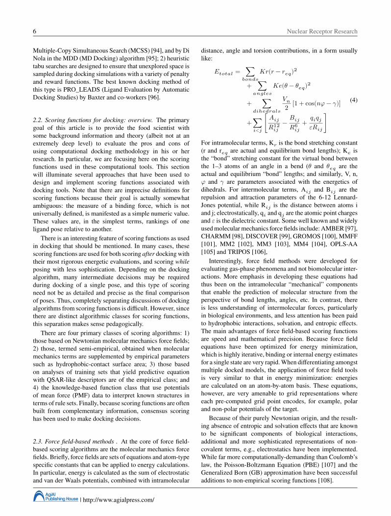
2.3. Force field-based methods . At the core of force field-based scoring algorithms are the molecular mechanics forcefields. Briefly, force fields are sets of equations and atom-typespecific constants that can be applied to energy calculations.In particular, energy is calculated as the sum of electrostaticand van der Waals potentials, combined with intramolecular
distance, angle and torsion contributions, in a form usuallylike:
𝐸𝑡𝑜𝑡𝑎𝑙 = ∑𝑏𝑜𝑛𝑑𝑠
𝐾𝑟(𝑟 − 𝑟𝑒𝑞)2
+ ∑𝑎𝑛𝑔𝑙𝑒𝑠
𝐾𝑒(𝜃 − 𝜃𝑒𝑞)2
+ ∑𝑑𝑖ℎ𝑒𝑑𝑟𝑎𝑙𝑠
𝑉𝑛2 [1 + cos(𝑛𝜑 − 𝛾)]
+ ∑𝑖<𝑗
[ 𝐴𝑖𝑗𝑅12
𝑖𝑗− 𝐵𝑖𝑗
𝑅6𝑖𝑗
+ 𝑞𝑖𝑞𝑗𝜀𝑅𝑖𝑗
]
(4)
For intramolecular terms, K𝑟 is the bond stretching constant(r and r𝑒𝑞 are actual and equilibrium bond lengths); K𝑒 isthe “bond” stretching constant for the virtual bond betweenthe 1–3 atoms of an angle in a bend (𝜃 and 𝜃𝑒𝑞 are theactual and equilibrium “bond” lengths; and similarly, V, n,𝜑 and 𝛾 are parameters associated with the energetics ofdihedrals. For intermolecular terms, A𝑖𝑗 and B𝑖𝑗 are therepulsion and attraction parameters of the 6-12 Lennard-Jones potential, while R𝑖𝑗 is the distance between atoms iand j; electrostatically, q𝑖 and q𝑗 are the atomic point chargesand 𝜀 is the dielectric constant. Some well known and widelyused molecular mechanics force fields include: AMBER [97],CHARMM [98], DISCOVER [99], GROMOS [100], MMFF[101], MM2 [102], MM3 [103], MM4 [104], OPLS-AA[105] and TRIPOS [106],
Interestingly, force field methods were developed forevaluating gas-phase phenomena and not biomolecular inter-actions. More emphasis in developing these equations hadthus been on the intramolecular “mechanical” componentsthat enable the prediction of molecular structure from theperspective of bond lengths, angles, etc. In contrast, thereis less understanding of intermolecular forces, particularlyin biological environments, and less attention has been paidto hydrophobic interactions, solvation, and entropic effects.The main advantages of force field-based scoring functionsare speed and mathematical precision. Because force fieldequations have been optimized for energy minimization,which is highly iterative, binding or internal energy estimatesfor a single state are very rapid. When differentiating amongstmultiple docked models, the application of force field toolsis very similar to that in energy minimization: energiesare calculated on an atom-by-atom basis. These equations,however, are very amenable to grid representations whereeach pre-computed grid point encodes, for example, polarand non-polar potentials of the target.
Because of their purely Newtonian origin, and the result-ing absence of entropic and solvation effects that are knownto be significant components of biological interactions,additional and more sophisticated representations of non-covalent terms, e.g., electrostatics have been implemented.While far more computationally-demanding than Coulomb’slaw, the Poisson-Boltzmann Equation (PBE) [107] and theGeneralized Born (GB) approximation have been successfuladditions to non-empirical scoring functions [108].
AgiAlPublishing House | http://www.agialpress.com/
Nuclear Receptor Research 7
The first scoring function used in DOCK [51] was anadaptation of force field parameters from AMBER, whereprotein-ligand interactions were calculated with repulsionand hydrogen bonding terms. Meng and colleagues addeda PBE-based electrostatic interaction energy function andLennard-Jones terms to DockScore [47]. Consideration ofsolvation effects in the function was added by Shoichet,Leach and Kuntz [109], by first calculating the electrostaticpotentials and van der Waals interactions with PBE of DelPhi[110] and CHEMGRID [47], respectively; later, Zap [111]electrostatics was implemented. Interaction free energieswere then corrected with HYDREN’s [112] calculations ofelectrostatic and non-polar ligand solvation energies. Withthis scoring approach’s inclusion of the HYDREN penaltyterms yielded virtual screening results of molecules withsmaller size and lower formal charges. DOCK 5 and DOCK6 include corrections for ligand conformational entropies,desolvation, Generalized Born/surface area (GB/SA) andPoisson-Boltzmann/surface area (PB/SA) solvation scoring,and receptor flexibility and implicit solvent, both withAMBER.
FLOG (Flexible Ligands Oriented on Grid) by Millerand co-workers [113] used a scoring equation that estimateddocking energy as a pairwise sum of van der Waals,electrostatic, hydrophobic and hydrogen bonding terms thatwere size-normalized to avoid scoring problems with largeand bulky molecules. The QXP approach (McMartin and co-workers) was based on a MC search protocol with AMBERenergies [114]. While rigid body docking is driven by elec-trostatic potentials and van der Waals forces, superposition isincluded as a separate term and helps identify the bioactiveconformation of flexible ligands [115]. In later versions,additional QXP terms estimated non-bonded interactions,van der Waals, electrostatic energy and contact energies ofinteractions and the count of hydrophobic contacts [116].
At this point, treating the contribution of solvent requiresmore discussion. Kollman and co-workers developed theMM/PBSA approach, which is a solvent continuum methodcombined with classic molecular mechanics. The lattercalculates the usual bond, angle, torsion, van der Waals andelectrostatic terms. To this was added Poisson-Boltzmann-based solvation free energy and solute entropy derivedfrom analyses of MD trajectories [117]. While Free EnergyPerturbation (FEP) methods are more accurate (and moreresource intensive), MM/PBSA involves MD simulations foronly the free ligand in solution and the protein-bound ligand[118]. Later, Massova et al. introduced a Generalized Born(GB) variant that further reduced computational time [119].A further enhancement was the OWFEG (One Window FreeEnergy Grid) method of Pearlman et al. [120], which is asimplified FEP that performs the analyses on a single MDtrajectory. Free energy is calculated at each point of a gridthat represents protein structure with three probes (neutral,positively charged and negatively charged). The final Gscoreis the sum of the grid score for each atom in the ligand.
Newer developments include MedusaScore, by Yin etal., a forcefield-based scoring function including van derWaals, solvation and hydrogen bond terms derived fromnewly parameterized forcefields and algorithms [121]. A keyfeature of MedusaScore is that it is not trained to fit specificexperimental data and thus possesses high transferability.
2.4. Semi-empirical and empirical methods. Semi-empiricalmethods use empirical or empirically-calibrated energeticterms to calculate or estimate interaction types that arenot generally available from molecular mechanics, whileempirical scoring functions represent free energies of bindingas sums, often empirically weighted by regression, of uncor-related terms that each account for different types of contribu-tions [122]. There are advantages and disadvantages for both:while allowing contributions for fundamental biologicalinteractions such as hydrogen bonding, solvent effects andhydrophobic interactions, these scoring functions lose someof the supposedly universal “first principles” applicability ofthe purely physics-based molecular mechanics forcefields.
Typically, semi-empirical scoring functions include elec-trostatic, hydrophobic, and entropic terms. In the ICMmethod, electrostatic contributions are represented as thesum of desolvation and Coulombic ΔGs obtained with thePoisson equation [107, 123], hydrophobic contributions frommultiplying the solvent accessible surface area (SASA) bythe surface tension, and entropy is calculated as RT ln[Ns](Ns is the count of conformational states with low-energyfound via MC simulations).[124] GOLD [125] combinesthree terms: hydrogen bonding energy, estimated with a4-8 (Lennard-Jones-like) potential, and VdW forces forprotein-ligand interactions and the internal energy of theligand are calculated with a 6-12 Lennard-Jones potential.Similarly, an AMBER-based scoring function implementedin early releases of AutoDock used a 6-12 Lennard-Jonespotential for vdW interactions, a 12-10 function for hydrogenbonding, and a Coulombic term for electrostatic interactions[126, 127]. AutoDock 3.0 [92] and later use semi-empiricalscoring based on Wesson and Eisenberg’s thermodynamiccycle [128], and ligand conformational and desolvationcontributions from Stouten’s pairwise volume-based method[129].
The summation of uncorrelated terms in empirical scoringis without physical foundation, especially with respect tothe assumed additivity of such terms [15]. In fact, thisparadigm has more similarities to QSAR (QuantitativeStructure-Activity Relationships) [130] than to any theoret-ically sound physics-based method. However, like QSAR,there are pragmatic and interpretative advantages; e.g., thevarious terms, hydrogen bonds, hydrophobic interactions,ionic contacts, entropic effects, etc., are familiar ones. Asin QSAR analyses, functions and weighting coefficients areobtained by fitting parameters to training sets of complexes.Empirical scoring functions are simple and fast, but haveseveral drawbacks largely related to the quality of data,
AgiAlPublishing House | http://www.agialpress.com/
8 Nuclear Receptor Research
i.e., experimental binding data or crystallographic structures,in the training sets. SCORE1 by Böhm was probably thefirst empirical scoring function and was implemented inhis de novo design program LUDI [131], and later wasused as the scoring function of FlexX [132]. Only termsfor hydrogen bonding and hydrophobic interactions werepart of SCORE1, but terms for losses of internal entropy,deviations from ideal hydrogen bond geometries, the lossof binding energy from restricting the internal degrees offreedom of the ligand when it binds, cation-𝜋 interactions,differentiated between neutral and ionic hydrogen bonds,and contributions of water molecules were implementedin the SCORE2, algorithm [133, 134]. The ChemScorefunction was modeled after SCORE1 with the addition ofa hydrogen bond term for water-mediated contacts, ligandflexibility and atom-atom contact estimates of hydrophobicinteractions [135]. Following Verdonk’s addition of covalentenergy terms [136] ChemScore was implemented in GOLD[137], FRED, Sybyl and PLANTS. Wang developed theSCORE algorithm by reconstituting binding affinity as fiveterms, representing vdW forces, metal-ligand interactions, H-bond energy, desolvation (a form of logP), and deformation,localized on each atom of the ligand [138]. The later SCORE3.0 [139] includes terms for entropic effects and internalligand energy.
One trend in scoring function development has been asteady increase in the size of training and test sets usedfor calibration. This makes sense on a number of levels,particularly related to statistical robustness, but outliers aremore difficult to detect and the stories they tell are often notheard. As part of the “Scoring Function Consortium” project[140], Sotriffer recently described the SCFscore algorithm,based on a training set of more than 850 protein-ligandcomplexes and more than 60 unique descriptors, includingsome usually absent, such as interactions with aromatic rings.Still missing are descriptors that account for the energeticroles of water molecules, internal strain energy and etc.
HINT (Hydropathic INTeractions) by Kellogg and Abra-ham [141] is almost completely based on the hydrophobiceffect, as measured by logP𝑜/𝑤, the partition coefficientfor 1-octanol and water [142, 143] – the free energy ofsolvent transfer. Each atom’s hydrophobic atomic constant,derived by fragmentation of experimentally determinedLogP𝑜/𝑤values, is a partial 𝛿g and directly related to theatom’s propensity of interacting with 1-octanol/water or otherhydrophobic or polar atoms. The HINT Score is correlatedwith experimental free energies of binding to create thescoring function for the dataset of interest. The HINT Scorefunction is optimized for binding free energy and has oftenbeen used in docking applications as a post-processing filter.
2.5. Knowledge-based methods. Algorithms of theknowledge-based class capture the steadily evolving“knowledge” that is available in publicly accessibledatabases, in particular the Protein Data Bank. Mügge, who
was one of the first to organize these data for docking scoringfunctions, describes potential of mean forces (PMF) scoringfunctions as statistical potentials that are derived fromstatistical mechanics [144]. The key statistical assumptionis that more commonly observed interactions contributepositively to binding, while less common interactions shouldbe marked as repulsive. Then, with the Boltzmann law,the extensive experimental information for structurallycharacterized protein-ligand complexes is converted into setsof atom-pair potentials for each pair of interacting atoms.Knowledge-based algorithms have a physical equation, butare statistical methods. However, the quality and diversityof the training databases is critical [145]; for example,more rare interactions (like 𝜋-𝜋 or 𝜋-cation) can be poorlypredicted.
Wallqvist and co-workers designed their knowledge-basedpotential function to focus on calculated buried surface areasin complexes by comparing the interface surface area by atomtype in complexed and uncomplexed structures [146], whichenabled determination of specific atom-atom and residue-residue preferences. The most notable and widely appliedPMF smoothed-grained potential was derived by Müggeand Martin from the statistical analysis of protein-ligandcomplexes found in the PDB [147] Their list of atom pairpotentials was correlated (n = 77, R2= 0.61) to bindingfree energy with a standard error of < 2 log K𝑖 units.These PMF potentials have continued to evolve, largely bydrastically increasing the sizes of the training sets [148,149]. For all knowledge-based approaches, since they arecalibrated on specific training sets, tunable parameters mayrequire extensive adjustments before these functions can beextrapolated outside their training sets’ chemical space.
2.6. Consensus scoring. The survey above of scoring func-tions included a brief look at several different classes,from Newtonian, first principles-based to semi-empirical,empirical and knowledge-based systems. While all achieveto a large extent their goals of discerning properly positionedligands in binding sites and some reasonably useful rankingof ligand poses based on estimations of binding free energies,all scoring methods to date rely on approximations, in manycases due to the desire for faster throughput, and these leadto errors. However, it needs to be reiterated that even themost sophisticated methods of free energy estimation are notwholly accurate. In the absence of a single function that isvalid for many or all targets [150], a strategy of mergingseveral diverse scoring functions to form a consensus hasemerged. This was proposed by Charifson [151] because thecombination of diverse functions could potentially compen-sate for the weaknesses of individual functions, and thusyield better overall scoring. While other strategies such aspost-docking minimization [93, 152] or topological filtering[153] The consensus scoring model has been widely accepted[154–156], There are three types of consensus methods: 1)rank by “vote”, where only conformations within the top x%
AgiAlPublishing House | http://www.agialpress.com/
Nuclear Receptor Research 9
are polled; 2) rank by rank, where the ranking from eachfunction is averaged; and 3) rank by number, where the scorevalue from each function is averaged.
Charifson’s rank by vote strategy rescored compoundsafter docking/virtual screening with thirteen algorithms; thehit lists from each function were compared, and moleculesappearing multiple times among the first several positionsof each list were selected. A significant improvement inenrichment (discrimination between active and inactive com-pounds) and fewer false positives were observed in the hitlistsfiltered in this manner. An interesting observation by Bissanzet al., after analyzing the performances of seven scoringfunctions, Chemscore, DOCK, FlexX, Fresno, GOLD, PMFand SCORE, with three different docking algorithms on thesame systems was that enrichments are largely independentof the method of docking but impacted much more by thescoring methods [57].
The CScore program used a rank by rank strategy [157](ChemScore, DockScore, GoldScore and PMF) and showedpreferences for near-native conformations. Application ofthree or more functions works better than single or doublescoring function methods. Wang et al. used a rank by numberscheme in developing the X-CScore method [158]. Lastly,Stahl and Rarey [159] appropriated terms from differentalgorithms and merged them into a single scoring functiontermed ScreenScore, in order to exploit the strengths of eachof the component methods.
3. Unresolved Issues in Docking and Scoring
The key problem with docking and scoring is that “chem-istry” is not entirely representable with mathematics. Ligandbinding is in many ways a balance between physical andchemical phenomena and the more chemical phenomena aremore difficult to represent mathematically. Briefly, we willdescribe several potential problem areas with the docking andscoring paradigm, along with some discussion of solutionsthat have been applied in order to address them. Most of whatwas described above solved reasonably well the problemof creating ligand-biomacromolecule models complexes andscoring those models in terms of their “physical” realism,which is generally based on Newtonian mechanics. However,chemical subtleties that are not accounted for may be the keyto identification of the one completely correct model.
The concept of emergent properties from systems biologyis also in play. These properties are observed from collectionsof components acting in concert, but cannot be observedby a reductionist dissection back to the components. Thehydrophobic effect is such a property, as hydrophobic speciesappear to self-associate due to the actual self-associationof polar (hydrogen bonding) species. This property cannotbe gleaned from the physical properties of a single atom.Similarly, chemical “reactions” such as changes in ionizationstates, resonance structures, or tautomerization are often notparameterized or otherwise represented in current docking
scoring functions. Another factor, which we alluded toseveral times above, is the flexibility of both the proteinand the docked molecule. This is especially relevant innuclear receptors where active sites are often quite loose.The flexibility problem can be resolved with physics, but ata high cost. Simulating flexibility in docking is a problemthat has been attracting significant attention in the pastdecade. A related issue is that the relationship betweenmolecular models of similar energy is poorly understood.The reflection data underlying crystal structure models aregenerally collected at very low temperatures – yielding asingle conformer, while at biological temperatures manymolecular conformations, ionization states, and etc. co-exist.In contrast, structural data collected with solution-phaseNMR present a family of structural models, but their effectiveresolution is relatively poor. In terms of predicting bindingmodes and for virtual screening, Carlson has suggested thatdocking to an NMR structure ensemble has advantages overdocking to a single structure for a molecular target [160].
In the next several paragraphs some of the issues aboveare discussed in more detail and solutions currently beingemployed are presented.
3.1. Hydrophobicity and entropy. Entropy clearly has asignificant effect on all chemical processes, and biologicalbinding events are no exception. Entropy is difficult tomeasure and even more difficult to model with calculations.The simple textbook definition of entropy is that it representsan increase of disorder. This is hard to envision in many casesand the consequences can even be counterintuitive. If twoliquids like water and acetone are mixed, it is obvious thata random distribution of one in the other is more disordered:it would be impossible for them to be segregated. On the otherhand, changing the mixed liquids to water and octanol wouldrequire the creation of highly ordered water cages aroundthe octanol molecules or else they couldn’t exist in water.Thus, finding the two liquids in separate layers is the moreentropically favored state.
There are a number of effects in protein structure andprotein-ligand binding attributable to entropy: 1) the dis-placement of water molecules from a binding site has theeffect of increasing entropy because such solvent moleculeshave more degrees of freedom and are less ordered; 2) proteinfolding also has an entropic driving force because morewaters are released in a compact protein structure with ahydrophobic core with more polar sidechains exposed to sol-vent. This explains the, at least partial, relationship betweenhydrophobic interactions and entropy. Also, calculations andanalyses show that the packing of X-ray protein structuremodels is more “clever” than that of NMR or decoy models,with higher sidechain entropy than even models that aresimilarly compact [161].
The thermodynamics of ligand binding also involves– in a similar way – entropy, although it is difficult toquantify. Because most scoring functions treat hydrophobic
AgiAlPublishing House | http://www.agialpress.com/
10 Nuclear Receptor Research
interactions in simple terms, like quantifying simply withcontact surface areas, teasing out the interaction energyassociated with entropy is impossible. Even HINT, based onthe partitioning, i.e., free energy, of small organic moleculesbetween two solvents is inadequate for identifying andquantifying the many, often small, effects that contributeto entropy. In contrast, free energy perturbation (FEP)methods exploit thermodynamic integration cycles – becausedifferences in free energy are independent of path – andthus estimate binding free energy. Importantly, intermediatestates on the path are chosen because of their computationalaccessiblity. Nevertheless, these are difficult and expensivecalculations, and at present not really applicable in dockingor virtual screening experiments. FEP and related methodsare also do not provide notably more accurate free energypredictions than empirical scoring functions that have beencalibrated for the data set of interest.
3.2. Modeling water and its biology. Water is always presentin the biological environment. It is a key ingredient for proteinfolding, as a solvent in biological reactions and in supportingprotein-protein, protein-DNA and other associations. Inprotein-ligand associations, water molecules in the active sitecan mediate (i.e., bridge) interactions between the ligand andprotein to change unfavorable interactions to favorable bytheir appropriate use of two hydrogen bond donors and twoacceptors. Water can be thought of as a nano-buffer [162].Water molecules shape the properties of an active site whenthey present different steric and electrostatic profile thanthe desolvated biomacromolecule. Despite several decadesof intestigation [163, 164], much about water is still poorlyunderstood. While on average there is about one watermolecule per residue in a protein, the quality or resolutionof the structure has a large effect [165] with one found perresidue at ∼2.0 Å and more (> 1.5) at 1.0 Å resolutions [166].In some cases, adding water molecules to structural modelswith computational prediction tools [46, 167–177] is a goodstrategy, especially if such a water is highly conserved andmissing for experimental reasons such as X-ray resolution,etc.
The basic question in docking is determining what watermolecules will be displaced or retained. In terms of medicinalchemistry, understanding the roles of water molecules inenzyme active sites can may assist in inhibitor design [178–184]. These roles can be evaluated in terms of a numberof criteria such as count of water-involved hydrogen bonds,the crystallographic thermal B factors, accessible surfaceareas, and, most importantly, their conservation and/ordisplacement as a result of ligand binding. Experimentalstructural data are sometimes difficult compare with simu-lations because single water molecules cannot be observedor tracked experimentally.
Consolv [176] applies a K-nearest-neighbors geneticalgorithm to identify conserved waters in biomacromolecule-ligand complexes. Its evaluation is based on geometric fea-tures that are in turn dependent on the quality of the examinedcrystallographic data. In practice, each water molecule’senvironment is characterized: geometrical features knownto correlate with water binding suggest whether particularwaters are conserved or displaced. While all conservedwaters are important, and thus should be included modelingand docking, probably only those waters that are bridging orfunctionally displaceable are essential. Similar to Consolv,WaterScore [185] is based on geometrical parameters (B-factor, solvent-accessible surface area (SASA), number ofprotein-water contacts) and an energetic term that is thetotal hydrogen bond energy. WaterScore catalogues water asbound, sterically displaced and (otherwise) displaced. Thefactors indicated by WaterScore to suggest conservation alsoseem to be correlated with restricted water mobility. We haveproposed a simpler model based on HINT score and Rank[186, 187], where the HINT score is calculated between eachwater and its site and Rank is only geometry-based, i.e., thepotential of each water for making hydrogen bonds. Thiscombination led to a mathematical model that predicts whichwater molecules will be displaced and which are “relevant”,with up to 90% accuracy. Relevant waters are potentiallydisplaceable but they should be replaced by functional groupsthat reprise or improve upon the waters’ hydrogen-bondingcapabilities.
Ideally, docking programs, combined with their scoringfunctions should be able to: 1) insert a ligand into a solvatedactive site; 2) displace the irrelevant and non-conservedwaters, but retain all others; and 3) estimate a reasonablefree energy of binding for that ligand, which includes thecontributions from the retained water molecules. This goal isnot yet achieved, but progress is being made: 1) the FlexXparticle algorithm places and continually optimizes watermolecules by maximizing the number and quality of hydro-gen bonds at the receptor-ligand interface during incrementalconstruction docking simulations [188]; 2) SLIDE uses adesolvation penalty term for displacement to predict theretention or displacement of water molecules upon ligandbinding [81, 189]; 3) GOLD [190] makes water moleculesindividually user switchable and allows them to rotate aroundtheir three main axes; 4) a new potential for displaceablewater molecules was added to AutoDock by Moitissier etal. implemented by combining water grids into a continuumgrid [191]; 5) van Dijk and Bonvin proposed a Monte Carloprocedure that applied water-mediated contact propensity toremove extraneous waters after docking completely solvatedmolecules [192]; and 6) Abel et al. described a methodcalled WaterMap that measures the contribution of thesolvent to the binding free energy of small molecules intheir associated receptors including the effects of the liganddisplacing the solvent in atomic detail [193], based onrelatively short molecular dynamics simulations. While noneof these treatments for water are ideal, they can yield
AgiAlPublishing House | http://www.agialpress.com/
Nuclear Receptor Research 11
significant improvement in binding poses and in predictingbinding free energies [81, 188, 190, 194–202].
3.3. Ionization and tautomerization. Modeling the protona-tion state of the system at the active site is another challenge.Since there are several ionizable residues with acidic or basicsidechains, potentially ionizable N- and C-termini, and theacidic or basic functional groups in ligands, there is a lotof room for ambiguity in intermolecular and intramolecularinteractions. The ionization states of nearly all functionalgroups are well understood when they are isolated, andusually their pK𝑎s are known. However, the pH properties ofthe microenvironments within or on the surface of proteinsand at protein-ligand interfaces are less understood andconsiderably more difficult to model.
Importantly, crystal structures seldom reveal direct infor-mation on the positions of their hydrogen atoms. Only for thehighest resolution X-ray structures, at ∼1.0 Å or better andfor neutron diffraction structures are hydrogens observed, andusually those sets are incomplete because the H atom is small.Thus, for ionizable groups, both for proteins and ligandsin many environments, it is usually quite difficult to assigntheir ionization states because such groups are not isolatedand influence each other’s states. It goes without sayingthat accurate docking and scoring depends on a definitiveassignment of each atoms charge, etc. The tautomerizationof specific functional groups on the ligand can also beaffected by either the solution pH or the microenvironmentpH, which can have a similar effect on the binding of aligand in the site. Adding to the complexity, the number ofmodel possibilities, i.e., protonation states for each ionizableresidue in an ensemble, grows exponentially as the number ofthese groups increases. An active site commonly has severalprotonation state models that exist in equilibrium with eachother, and a protein-protein interface may have hundreds.Water molecules present in the active site further complicatethe problem because of their ability to act as Lewis basesand/or acids. Computational studies on a binding pocket inthe engineered cavity of cytochrome c peroxidase [203],showed better docking results when a single conserved waterwas allowed to move. Crystallographers and computationalchemists have been interested in the proper placement andoptimization of polar protons for decades. The correction ofatom placement, coupled with the assignment of chemicallyand biologically reasonable protonation states for ionizableresidues in proteins is a necessary prerequisite for dockingexperiments. There are a few web-enabled applications forexamining and validating X-ray protein crystal structuressuch as MolProbity [204], which both places hydrogen atomsand corrects various other errors in structure models.
Computational ionization state evaluation and optimiza-tion is a complex problem to which quantum mechanics[205], quantum mechanics–molecular mechanics (QM/MM)[206, 207] approaches have been applied. However, solving
the Poisson-Boltzmann equation to evaluate possible proto-nation states and hydrogen positions [208–212] has generallybeen the method of choice, sometimes when combined withMD simulation [213–215], The speed of PBE solutions hasbeen a handicap for general use as a scoring function, butoptimized code such as OpenEye’s Zap, and faster computershave been narrowing the gap. The goal of PBE methods isalso somewhat orthogonal to docking – in most applicationsthe result is a set of pK𝑎s for protein residues rather than theset of energetically accessible protonation states. Only a fewdocking programs tackle the ligand tautomerization problem,e.g., eHITS and GLIDES’s LigPrep. For each group, allpossible tautomerization and ionization cases are simulatedand evaluated, with the best for each selected independentof the others, thus avoiding, rather than dealing with, theinherent combinatorial issues [216]. However, most dockingprograms require the input of all tautomers by the user.This docking problem has to date received only modestattention, and remains a problem that must be solved beforeuniversally reliable docking, scoring or virtual screening willbe available.
3.4. Flexibility. All proteins are flexible and this oftenturns out to be fundamental for their function. Significantconformational flexibility is vital in nuclear receptors for theirbiological function, as the pharmacology of their ligandsinvolves movements of short 𝛼-helix segments at carboxytermini [39, 57, 217]. While structure-based moleculardesign has been a relatively successful strategy in the pastwithout overly respecting target flexibility, future effortsare not likely to be as fruitful. The current content ofthe Protein Data Bank (PDB) [218] likely overemphasizesrigid proteins because these are easier to crystallize andanalyze. Furthermore, disorder is reduced by collectingdiffraction data at low (∼liquid N2) temperatures, which isin stark contrast to biology that generally occurs in waterat or above room temperature. Thus, even though X-raycrystallography is the best technique to obtain experimentalmodels of biomacromolecules, these models are static andoften reveal little about their flexibility –likely to be keyfor their biological activity. Nuclear Magnetic Resonance(NMR) spectroscopy, however, solves three-dimensionalprotein structures in solution and thus encodes dynamicflexibility of molecules. NMR also provides an ensemble ofconformations with low energy. Currently proteins of 80-100kD are amenable for studying with NMR [219, 220], and isespecially useful for systems that are difficult to crystallizeor when the crystals obtained are disordered.
Molecular dynamics (MD) calculations are able to usethe structural framework provided by experimental X-raycrystallography and NMR data to extrapolate at least apartial understanding of molecular motions. Thus, MD cangenerate many protein conformations for docking and/orvirtual screening studies [221–223]. Various scales areavailable from these approaches, usually dictated by the
AgiAlPublishing House | http://www.agialpress.com/
12 Nuclear Receptor Research
nature and size of the system in the study. Coarse-grainedapproaches smooth out the atomistic details in order tomake longer time-scale dynamics more accessible, and thusreveal general protein flexibility. Such dynamics can oftenmodel protein dynamics, i.e., formation or reshaping ofbinding pockets, etc., but without atom-level detail, are ofinsufficient quality for docking. In atomic-level MD, bothsmall atomic fluctuations and large protein movements canbe observed, but at higher computational cost. The cost isso large that effective sampling of conformational states isoften in question because even several ns is too brief toensure adequate sampling. In recent years, MD methods havebecome more ubiquitously accessible, but experimentallydetermined conformational data remain more desirable.
In simulating docking, integration of detailed flexibilitydefeats, in a sense, the rationale behind docking – speedand the ability to explore multiple conformations and manyputative ligands. However, true protein conformations areoften stabilized by the bound ligand, which makes thecomputational simulation a bit like the chicken and egg.In designing ligands for such targets, it cannot be knownbeforehand how the target will adapt to binding of a particularligand. In the past few years, much effort has been expendedto simulate active site flexibility in docking. The mostconservative methods incorporate flexibility with active-sitesidechains, e.g., AutoDock v4 [54, 84, 126, 224] explicitlyallows such flexibility, GOLD samples several alternativerotamers for specified sidechains and optimize hydrogen-bond interactions involving these residues [136], and SLIDErotates sidechains and substructures of ligands to removeunfavorable van der Waals interactions [82]. Simulatingsidechain flexibility appears to be most effective when thedocking site has not been preformed, e.g., into a un-ligandedstructure. To simulate significant conformational adjust-ments, an induced fit docking approach such as suggested byGohlke based on rigidity and elastic network theory [225],can be effective. Induced fit docking simulations supportthe “conformation selection” concept [226, 227], whereobserved bound conformations may be predetermined bymotions of the protein in the unbound state. Lastly, ensemblesof target structures, usually derived from MD, can be used fordocking [228]. Ensemble docking places the ligand of interestinto each structural snapshot from an MD trajectory, or into agrid that encodes the composite similarity from the ensemble.The disadvantages of ensemble docking are that the ensembleof conformers is seldom complete or even representative,and the shortfalls of scoring functions in representing freeenergy are magnified here because binding mode differencesare often dominated by entropy.
4. Example Case Studies of Estrogen ReceptorToxicology
As stated above, a good understanding of biological interac-tions (and even better a good estimation of them!), is the basis
for successful medicinal chemistry investigations, even thosedriven by experimental results. In the same way, applicationof well-calibrated and understood computational tools can bepart of the process for creating and producing safer food andfood-related products.
Many current computational investigations are focused onthe identification of endocrine-disrupting chemicals (EDCs),which are exogenous compounds capable of interfering withthe human endocrine system either by direct or indirectinteractions with nuclear receptor proteins. Common targetsare often estrogen receptors (ERs) that, once activatedor inhibited by interaction with such compounds, calledxenoestrogens, alter the normal homeostatic transcriptionand signaling pathways.
4.1. Docking and scoring. One of the first applications ofcomputational methods in food science was reported byAmadasi et al. (2009) [3], with the aim to identify possiblexenoestrogens in a set of allowed and common food additives.These were surveyed by virtually screening a food additivelist maintained by the Food and Agriculture Organization ofthe United Nations (FAO) and World Health Orgainization(WHO) that contained nearly 500 chemicals with theirchemical structures. These were each docked/scored in thealpha estrogen receptor using the GOLD algorithm and thein-built GOLD scoring functions as filters. Those compoundspassing through these filters were re-scored with HINT. Themost “suspicious” molecules, that is, those that better fit thereceptor’s binding site and had the highest HINT scores,were selected for experimental testing in an in vitro assay.Among the thirteen potential xenoestrogens thus identifiedfor testing„four were already known to exhibit estrogenicactivity. The assays of the other nine compounds determinedtheir binding affinities to the receptor and, by inference, theirresulting biological effects. In this way, propyl gallate wasfound to act as an antagonist, while 4-hexylresorcinol as apotent transactivator. Both ligands were active at nanomolarconcentrations, as predicted by the in silico analysis.
Similar techniques, i.e., GOLD and HINT scoring tools,were applied in investigating the biological effect of theknown food contaminant zearalenone and of its sulphonateand glucorinate metabolites [229, 230]. Zearalenone is aknown potent micotoxin produced by several Fusariumspecies and can be found in different cereal crops such asbarley, wheat, rice and others. Again, it affects estrogenreceptor activation by acting as an agonist and inducing genetranscription. In silico analyses predicted the molecules to beactive towards the alpha estrogen receptor, in good agreementwith experimental evidence. These findings further supportedthe fruitful application of computational technique to sup-port and illuminate experimental results, and in particularto possibly reduce the number of and/or replace animalexperiments.
The alpha and beta estrogen receptors were also the targetsof an innovative “full-dry” in silico method for evaluation of
AgiAlPublishing House | http://www.agialpress.com/
Nuclear Receptor Research 13
the potential xenoestrogenic behavior of ellagitannin-derivedmetabolites. The strategy involved structure-based virtualscreening, docking simulations followed by re-scoring, andthe results revealed valuable insights into the phase IIconjugations (glucuronidation, sulfation, and methylation,occurring in vivo) affecting the estrogenicity of these com-pounds on both 𝛼- and 𝛽-ER isoforms [231]. Structure-based docking techniques were adopted to predict the pos-sible effects of tioxhantone (TX) derivatives that had beenidentified as food contaminants, especially in infant formulas,by the European Food Safety Authority. Phase I metabolitesfor the TX class of compounds were predicted and theirbinding affinities for the AR ligand-binding pocket weredefined using local models based on available informationabout metabolism and AR activity. The results confirmed thatthe precursors can bind to the ER targets and suggested someof their metabolites to be possible new disruptors suitable forfurther in vitro and/or in vivo toxicological evaluations [232].
With a similar goal, Ng et al. combined agonist and antag-onist docking models with competitive docking, enabling theevaluation of potential ER biological functional changes dueto chemicals binding to the receptor. This also allowed theassessment of a chemical’s endocrine disrupting potential.The potential of these applications is impressive, consideringtheir applicability in risk assessment on potential EDCs,but also for the identification of potential agonists andantagonists in drug discovery projects.[233]
Alternariol and alternariol methyl ether, produced bydifferent Alternaria species, have been recognized as anotherset of emerging micotoxins. In silico docking, scoring andre-scoring strategies were applied to investigate first whetherthese molecules could generate genotoxic effects by theactivation of DNA topoisomerase I and/or II [229]. Thesecompounds, alteranariol and its derivatives, may also haveinteractions with nuclear receptors such as the alpha and betaestrogen receptors. This possibility should also be assessed.In summary, the fairly simple computational techniques ofvirtual screening, docking and/or scoring have revealed alot of experimentally difficult-to-obtain data. We are notyet at the stage where computational procedures can totallyreplace well-constructed and executed experiments, but justthe promising results presented here argue for the importanceof including them in risk assessment investigations.
4.2. Molecular dynamics. The purpose of molecular dynam-ics (MD) methods is to simulate motion, which is a veryimportant aspect for the very flexible nuclear receptors likeestrogen receptor. Many more of these studies have been pub-lhished and many more are underway, but we highlight heretwo recent examples. The best MD studies are incorporatedinto a comprehensive work that uses conformational infor-mation from the dynamics as the starting place for virtualscreening, docking, etc. Molecular dynamics techniques wereused by Li et al. to investigate the binding mechanism ofbisphenol A (BPA) to three typical nuclear receptors: ER𝛼,
ERR𝛾 and PPAR𝛾. Bisphenol A is commonly present infood packaging and is known to affect the normal function ofnuclear receptors at very low doses. The simulations providedstructural support for the capability of this xenoestrogen tobind to these receptors, where it appears to mimic the actionof the natural hormone and keeps the nuclear receptors inactive conformations [234]. These results could form thebasis for identification of safer alternatives for BPA and saferpackaging.
Dal Palú et al. recently described the development of alter-native and innovative approaches for simulation of nuclearreceptor (NR) intrinsic dynamics. The authors reported afast strategy based on logic constraint programming andapplied it to a study of the flexibility of 𝛼-ER alpha. Theyinvestigated, in particular, the routes possibly followed bythe receptor to transform from the inactive to the agonist-and antagonist-active states. Also, they presented a numberof energetically accessible conformations that could possiblyused in structure-based drug design for the identification ofnew potential binders and disruptors [4]. Their methodologyshould be reasonably and successfully applied to any othernuclear receptor.
5. Conclusions
In this last section, we make a few general comments andthen discuss in more detail the relevance of docking, and inparticular scoring functions, to receptors likely targeted byfood and feed molecules as, for instance, nuclear receptors.Docking had rather modest beginnings, and perhaps its inven-tors could never have imagined the current broad applicationsof their concept. The docking paradigm has become analmost universal tool for computational chemistry appliedto drug discovery and in probing other small molecule-biomacromolecule interactions. There are numerous successstories, many of which have been presented in this article’sreferences.
A fundamental question that might be asked is, are theremultiple “correct” solutions to a docking problem? BothNMR and MD suggest that there are more often than notmultiple, energetically indistinct, conformers. Remember,the crystal “structure” is also a model that is only specialbecause it fits the experimental electron density [6]. Thatmodel is unlikely to be unique, and other conformers,tautomers, varients of ionization states, and etc. may also fitthe electron density well. Even the wrong molecule may havebeen modeled into the density, because of a mistake or afteran in-situ reaction. Thus, believing the top scoring candidatemodel is the only one of value, while dumping others withsimilar score, may lead to erroneous results.
It is important to keep in mind that running a dockingprogram is an experiment, albeit computational. To succeed,the assumptions and conditions of the experiment shouldbe evaluated and the correct controls should be run. Wehave usually viewed any computational result with suspicion
AgiAlPublishing House | http://www.agialpress.com/
14 Nuclear Receptor Research
until another approach, preferably experimental, can beapplied to the problem. Concomitantly, because running thesame experiment the same way will yield the same result(precisely), to evaluate uncertainty in docking, differentmethods should be applied, such as changes in conditions,parameters, docking program, or especially scoring function.In “wet” experiments the observed results are not from asingle, discrete molecule interacting with another, but frommany molecules of the first species interacting with many ofthe other. Should we assume that the one-to-one reactions wemodel with docking calculations are representative of exper-iment? The only consolation is that all docking algorithmsrely on some degree of randomness to drive their calculationengines. The best rule of thumb is to not treat computationalresults as “answers” to problems, but to see them clues orhypothesis generators for designing experiments.
In our perspective, in silico experiments, such as virtualscreening, docking and scoring, and etc. represent invaluabletools to investigate the possible interactions of chemical smallmolecules or biological molecules with relevant receptors.Nuclear Receptors have become increasingly relevant targetsof study for both pharmaceuticals and food chemicals, fromhit to lead compounds, from xenostrogens, to nutraceuticals,mycotoxins, inks for food packaging, etc. However, a rapidlyemerging concern, for environmental, ethical as well asfinancial reasons, is to reduce the frequency and number ofin vitro and in vivo tests. Thus, in silico experiments havebecome mandatory for the investigation of large librariesof compounds in order to predict their possible interactionswith the mentioned targets. Follow up experiments are thenmuch more focused. Finally, as computational approaches tothese problems are beginning to enter the regulatory domain,computational practices will have to be well established andproperly applied, and the user (and regulatory officials) mustbe conscious of the pros and cons of the various tools andmethodologies, and above all avoid the use of such softwareas a “magic black box”.
Competing Interests
The authors declare that they have no competing interests.
Acknowledgments
The authors acknowledge the inspiration of Prof. Donald J.Abraham (VCU) and Prof. Andrea Mozzarelli (U. Parma),who sparked our collaboration because of their mutualinterest in structural biology and drug discovery.
References
[1] From Medicinal Chemistry to Food Science: A Transfer of InSilico Methods Applications, Nova Science Publisher, 2015.
[2] T. Hartung and S. Hoffmann, Food for thought... on in silicomethods in toxicology, Altex, 26, no. 3, 155–166, (2009).
[3] A. Amadasi, A. Mozzarelli, C. Meda, A. Maggi, and P.Cozzini, Identification of xenoestrogens in food additives by anintegrated in silico and in vitro approach, Chemical Researchin Toxicology, 22, no. 1, 52–63, (2009).
[4] A. Dal Palu, F. Spyrakis, and P. Cozzini, A new approachfor investigating protein flexibility based on Constraint LogicProgramming. The first application in the case of the estrogenreceptor, European Journal of Medicinal Chemistry, 49, 127–140, (2012).
[5] A. Vedani, M. Dobler, Z. Hu, and M. Smiesko,OpenVirtualToxLab—A platform for generating andexchanging in silico toxicity data, Toxicol Lett, 232, 519–532, (2015).
[6] P. C. D. Hawkins, G. L. Warren, A. G. Skillman, and A.Nicholls, How to do an evaluation: Pitfalls and traps, Journalof Computer-Aided Molecular Design, 22, no. 3-4, 179–190,(2008).
[7] F. Spyrakis, A. Amadasi, M. Fornabaio, D. J. Abraham, A.Mozzarelli, G. E. Kellogg, and P. Cozzini, The consequencesof scoring docked ligand conformations using free energycorrelations,European Journal ofMedicinal Chemistry, 42, no.7, 921–933, (2007).
[8] P. W. Atkins, General Chemistry, Zanichelli, Bologna, 1992.[9] G. A. Jeffrey and W. Saenger, Hydrogen bonding in biological
structures, Springer-Verlag, Berlin, 1994.[10] M. Nishio, The CH-Pi interactions: evidence, nature and
consequences, Wiley-VCH, New York, 1998.[11] H. Gohlke and G. Klebe, Approaches to the Description
and Prediction of the Binding Affinity of Small-MoleculeLigands to Macromolecular Receptors, Angewandte ChemieInternational Edition, 41, no. 15, 2644–2676, (2002).
[12] D. A. Pearlman and P. A. Kollman, The calculated free energyeffects of 5-methyl cytosine on the B to Z transition in DNA,Biopolymers, 29, no. 8-9, 1193–1209, (1990).
[13] J. R. H. Tame, Scoring functions: A view from the bench,Journal of Computer-Aided Molecular Design, 13, no. 2, 99–108, (1999).
[14] I. Muegge, Effect of Ligand Volume Correction on PMFScoring, Journal of Computational Chemistry, 22, no. 4, 418–425, (2001).
[15] K. A. Dill, Additivity Principles in Biochemistry, The Journalof Biological Chemistry, 272, 701–704, (1997).
[16] A. M. Davis, S. J. Teague, and G. J. Kleywegt, Application andLimitations of X-ray Crystallographic Data in Structure-BasedLigand and Drug Design, Angewandte Chemie InternationalEdition, 42, no. 24, 2718–2736, (2003).
[17] M. A. DePristo, P. I. W. De Bakker, and T. L. Blundell,Heterogeneity and inaccuracy in protein structures solved byX-ray crystallography, Structure, 12, no. 5, 831–838, (2004).
[18] C.-I. Bränd́en and T. Alwyn Jones, Between objectivity andsubjectivity, Nature, 343, no. 6260, 687–689, (1990).
[19] V. Mohan, A. C. Gibbs, M. D. Cummings, E. P. Jaeger, andR. L. DesJarlais, Docking: Successes and challenges, CurrentPharmaceutical Design, 11, no. 3, 323–333, (2005).
[20] C. Giacovazzo, H. L. Monaco, D. Viterbo, F. Scordari, G. Gilli,G. Zanotti, and M. Catti, Fundamentals of crystallography,Oxford University Press, New York, 1992.
[21] A. T. Brunger, Free R value: A novel statistical quantity forassessing the accuracy of crystal strucutres, Nature, 355, no.6359, 472–475, (1992).
AgiAlPublishing House | http://www.agialpress.com/
Nuclear Receptor Research 15
[22] O. Roche, R. Kiyama, and C. L. Brooks, Ligand−ProteinDataBase: Linking Protein−Ligand Complex Structures toBinding Data, Journal of Medicinal Chemistry, 44, no. 22,3592–3598, (2001).
[23] R. Wang, X. Fang, Y. Lu, and S. Wang, The PDBbindDatabase: Collection of Binding Affinities for Protein−LigandComplexes with Known Three-Dimensional Structures, Jour-nal of Medicinal Chemistry, 47, 2977–2980, (2004).
[24] R. Wang, X. Fang, Y. Lu, C. Y. Yang, and S. Wang, ThePDBbind Database: Methodologies and Updates, Journal ofMedicinal Chemistry, 48, 4111–4119, (2005).
[25] X. Chen, Y. Lin, and M. K. Gilson, The binding database:Overview and user’s guide, Biopolymers, 61, no. 2, 127–141,(2001).
[26] L. Hu, M. L. Benson, R. D. Smith, M. G. Lerner, and H. A.Carlson, Binding MOAD (Mother of All Databases), Proteins:Structure, Function and Genetics, 60, no. 3, 333–340, (2005).
[27] J. W. M. Nissink, C. Murray, M. Hartshorn, M. L. Verdonk, J.C. Cole, and R. Taylor, A new test set for validating predictionsof protein-ligand interaction, Proteins: Structure, Function andGenetics, 49, no. 4, 457–471, (2002).
[28] T. Cheng, Q. Li, Z. Zhou, Y. Wang, and S. H. Bryant, Structure-based virtual screening for drug discovery: A problem-centricreview, AAPS Journal, 14, no. 1, 133–141, (2012).
[29] H. J. Bohm, New computational approaches tp predict protein-ligand interactions, in In Structure-based ligand design, R.Mannhold, H. Kubinyi, and H. Timmerman, Eds., 6, Wiley-VCH: Weinheim, 1998.
[30] M. A. Murcko, Recent advances in ligand design methods, in InReviews in Computational Chemistry, Lipkowitz, K. a. B. DB,Ed., 11, p. 66, Wiley-VCH, John Wiley and Sons, Inc, NewYork, 1997.
[31] D. Clark, C. W. Murray, and J. Li. Current issues, in de novomolecular design, in In Reviews in Computational Chemistry,Lipkowitz, K. a. B. DB, Ed., 11, 67–125, Wiley-VCH, JohnWiley and Sons, Inc, New York, 1997.
[32] H. I. Parikh and G. E. Kellogg, Intuitive, but not simple:Including explicit water molecules in protein-protein dockingsimulations improves model quality, Proteins: Structure, Func-tion and Bioinformatics, 82, no. 6, 916–932, (2014).
[33] J. M. Blaney and J. S. Dixon, A good ligand is hard to find:Automated docking methods, Perspectives in Drug Discoveryand Design, 1, no. 2, 301–319, (1993).
[34] R. D. Taylor, P. J. Jewsbury, and J. W. Essex, A review ofprotein-small molecule docking methods, Journal of Computer-Aided Molecular Design, 16, no. 3, 151–166, (2002).
[35] C. A. Sotriffer, G. Klebe, M. Stahl, and H. J. Bohm, Dockingand Scoring Functions/Virtual Screening, in In Burger’sMedic-inal Chemistry and Drug Discovery, D. J. Abraham, Ed., 1,281–331, John Wiley Sons, Inc, 2003.
[36] I. Muegge and I. Rarey, Small molecule docking and scoring,in In Reviews Comput Chem, K. Lipkowitz and D. Boyd, Eds.,17, p. 61, Wiley-VCH, John Wiley and Sons, Inc, New York,2001.
[37] N. Brooijmans and I. D. Kuntz, Molecular Recognitionand Docking Algorithms, Annual Review of Biophysics andBiomolecular Structure, 32, 335–373, (2003).
[38] K. A. Henzler-Wildman, M. Lei, V. Thai, S. J. Kerns, M.Karplus, and D. Kern, A hierarchy of timescales in proteindynamics is linked to enzyme catalysis, Nature, 450, no. 7171,913–916, (2007).
[39] C. Hellal-Levy, J. Fagart, A. Souque, J.-M. Wurtz, D. Moras,and M.-E. Rafestin-Oblin, Crucial role of the H11-H12 loop instabilizing the active conformation of the human mineralocorti-coid receptor, Molecular Endocrinology, 14, no. 8, 1210–1221,(2000).
[40] X. Hu and M. A. Lazar, Transcriptional Repression by NuclearHormone Receptors, Trends in Endocrinology & Metabolism,11, no. 1, 6–10, (2000).
[41] A. E. Brady and L. E. Limbird, G protein-coupled receptorinteracting proteins: Emerging roles in localization and signaltransduction, Cellular Signalling, 14, no. 4, 297–309, (2002).
[42] P. Cozzini, G. E. Kellogg, F. Spyrakis, D. J. Abraham, G.Costantino, A. Emerson, F. Fanelli, H. Gohlke, L. A. Kuhn,G. M. Morris, M. Orozco, T. A. Pertinhez, M. Rizzi, and C. A.Sotriffer, Target flexibility: An emerging consideration in drugdiscovery and design, Journal of Medicinal Chemistry, 51, no.20, 6237–6255, (2008).
[43] C. Levinthal, S. J. Wodak, P. Kahn, and A. K. Dadivanian,Hemoglobin interaction in sickle cell fibers I: Theoreticalapproaches to the molecular contacts, Proceedings of theNational Academy of Sciences of the United States of America,72, no. 4, 1330–1334, (1975).
[44] S. J. Wodak, M. De Crombrugghe, and J. Janin, Computerstudies of interactions between macromolecules, Progress inBiophysics and Molecular Biology, 49, 29–63, (1987).
[45] R. Norel, S. L. Lin, H. J. Wolfson, and R. Nussinov, Shapecomplementarity at protein-protein interfaces,Biopolymers, 34,no. 7, 933–940, (1994).
[46] P. J. Goodford, Aldose reductase inhibitors: a potential newclass of agents for the pharmacological control of certaindiabetic complications, Journal of Medicinal Chemistry, 28,no. 7, 849–857, (1985).
[47] E. Meng, B. Shoichet, and I. Kuntz, Automated dockingwith grid-based energy evaluation, Journal of ComputationalChemistry, 13, no. 4, 505–524, (1992).
[48] M. Totrov and R. Abagyan, Flexible protein-ligand docking byglobal energy optimization in internal coordinates, Proteins:Structure, Function and Genetics, 29, no. 1, 215–220, (1997).
[49] J. Y. Trosset and H. A. Scheraga, Reaching the global minimumin docking simulations: A Monte Carlo energy minimizationapproach using Bezier splines, Proceedings of the NationalAcademy of Sciences of the United States of America, 95, 8011–8015, (1998).
[50] K. E. Platzer, F. A. Momany, and H. A. Scheraga, Conforma-tional energy calculations of enzyme-substrate interactions. I.Computation of preferred conformations of some substrates of-chymotrypsin, International Journal of Peptide and ProteinResearch, 4, no. 3, 187–200, (1972).
[51] I. D. Kuntz, J. M. Blaney, S. J. Oatley, R. Langridge, andT. E. Ferrin, A geometric approach to macromolecule-ligandinteractions, Journal of Molecular Biology, 161, no. 2, 269–288, (1982).
[52] D. T. Moustakas, P. T. Lang, S. Pegg, E. Pettersen, I. D. Kuntz,N. Brooijmans, and R. C. Rizzo, Development and validationof a modular, extensible docking program: DOCK 5, Journalof Computer-Aided Molecular Design, 20, no. 10-11, 601–619,(2006).
[53] C. M. Oshiro, I. D. Kuntz, and J. S. Dixon, Flexible liganddocking using a genetic algorithm, Journal of Computer-AidedMolecular Design, 9, no. 2, 113–130, (1995).
[54] G. M. Morris, D. S. Goodsell, R. S. Halliday, R. Huey, W. E.Hart, R. K. Belew, and A. J. Olson, Automated docking using
AgiAlPublishing House | http://www.agialpress.com/
16 Nuclear Receptor Research
a Lamarckian genetic algorithm and an empirical binding freeenergy function, Journal of Computational Chemistry, 19, no.14, 1639–1662, (1998).
[55] C. N. Cavasotto and R. A. Abagyan, Protein Flexibility inLigand Docking and Virtual Screening to Protein Kinases,Journal of Molecular Biology, 337, no. 1, 209–225, (2004).
[56] K. W. Foreman, A. T. Phillips, J. B. Rosen, and K. A. Dill,Comparing search strategies for finding global optima onenergy landscapes, Journal of Computational Chemistry, 20,no. 14, 1527–1532, (1999).
[57] C. Bissantz, G. Folkers, and D. Rognan, Protein-based virtualscreening of chemical databases. 1. Evaluation of differentdocking/scoring combinations, Journal of Medicinal Chem-istry, 43, no. 25, 4759–4767, (2000).
[58] B. D. Bursulaya, M. Totrov, R. Abagyan, and C. L. BrooksIII, Comparative study of several algorithms for flexible liganddocking, Journal of Computer-AidedMolecular Design, 17, no.11, 755–763, (2003).
[59] R. T. Kroemer, A. Vulpetti, J. J. McDonald, D. C. Rohrer,J.-Y. Trosset, F. Giordanetto, S. Cotesta, C. McMartin, M.Kihlén, and P. F. W. Stouten, Assessment of docking poses:Interactions-based accuracy classification (IBAC) versus crys-tal structure deviations, Journal of Chemical Information andComputer Sciences, 44, no. 3, 871–881, (2004).
[60] J. C. Cole, C. W. Murray, J. W. M. Nissink, R. D. Taylor, and R.Taylor, Comparing protein-ligand docking programs is difficult,Proteins: Structure, Function and Genetics, 60, no. 3, 325–332,(2005).
[61] G. L. Warren, C. W. Andrews, A.-M. Capelli, B. Clarke, J.LaLonde, M. H. Lambert, M. Lindvall, N. Nevins, S. F. Semus,S. Senger, G. Tedesco, I. D. Wall, J. M. Woolven, C. E. Peishoff,and M. S. Head, A critical assessment of docking programs andscoring functions, Journal of Medicinal Chemistry, 49, no. 20,5912–5931, (2006).
[62] J.-F. Truchon and C. I. Bayly, Evaluating virtual screeningmethods: Good and bad metrics for the ”early recognition”problem, Journal of Chemical Information and Modeling, 47,no. 2, 488–508, (2007).
[63] P. Englebienne, H. Fiaux, D. A. Kuntz, C. R. Corbeil, S.Gerber-Lemaire, D. R. Rose, and N. Moitessier, Evaluationof docking programs for predicting binding of Golgi α-mannosidase II inhibitors: A comparison with crystallography,Proteins: Structure, Function and Genetics, 69, no. 1, 160–176,(2007).
[64] N. Weill, E. Therrien, V. Campagna-Slater, and N. Moitessier,Methods for docking small molecules to macromolecules: Auser’s perspective. 1. The theory, Current PharmaceuticalDesign, 20, no. 20, 3338–3359, (2014).
[65] J. Biesiada, A. Porollo, P. Velayutham, M. Kouril, and J. Meller,Survey of public domain software for docking simulations andvirtual screening, Human Genomics, 5, no. 5, 497–505, (2011).
[66] N. M. F. S. A. Cerqueira, D. Gesto, E. F. Oliveira, D.Santos-Martins, N. F. Brás, S. F. Sousa, P. A. Fernandes, andM. J. Ramos, Receptor-based virtual screening protocol fordrug discovery, Archives of Biochemistry and Biophysics, 582,article no. 6986, 56–67, (2015).
[67] S. Vajda, D. R. Hall, and D. Kozakov, Sampling and scoring:A marriage made in heaven, Proteins: Structure, Function andBioinformatics, 81, no. 11, 1874–1884, (2013).
[68] J. C. Wang and J. H. Lin, Scoring Functions for Prediction ofProtein-Ligand Interactions, Curr Pharm Des, 19, 2174–2182,(2013).
[69] H. A. Gabb, R. M. Jackson, and M. J. E. Sternberg, Modellingprotein docking using shape complementarity, electrostaticsand biochemical information, Journal of Molecular Biology,272, no. 1, 106–120, (1997).
[70] M. R. McGann, H. R. Almond, A. Nicholls, J. A. Grant, and F.K. Brown, Gaussian docking functions, Biopolymers, 68, no. 1,76–90, (2003).
[71] G. B. McGaughey, R. P. Sheridan, C. I. Bayly, J. C. Culberson,C. Kreatsoulas, S. Lindsley, V. Maiorov, J.-F. Truchon, andW. D. Cornell, Comparison of topological, shape, and dockingmethods in virtual screening, Journal of Chemical Informationand Modeling, 47, no. 4, 1504–1519, (2007).
[72] D. Joseph-McCarthy, B. E. Thomas IV, M. Belmarsh, D.Moustakas, and J. C. Alvarez, Pharmacophore-based moleculardocking to account for ligand flexibility, Proteins: Structure,Function and Genetics, 51, no. 2, 172–188, (2003).
[73] J. Goto, R. Kataoka, and N. Hirayama, Ph4Dock:Pharmacophore-based protein - Ligand docking, Journalof Medicinal Chemistry, 47, no. 27, 6804–6811, (2004).
[74] A. Schafferhans and G. Klebe, Docking ligands onto bindingsite representations derived from proteins built by homologymodelling, Journal of Molecular Biology, 307, no. 1, 407–427,(2001).
[75] D. Schneidman-Duhovny, Y. Inbar, R. Nussinov, and H. J.Wolfson, PatchDock and SymmDock: Servers for rigid andsymmetric docking, Nucleic Acids Research, 33, no. 2, W363–W367, (2005).
[76] A. Wallqvist and D. G. Covell, Docking enzyme-inhibitor com-plexes using a preference-based free-energy surface, Proteins:Structure, Function and Genetics, 25, no. 4, 403–419, (1996).
[77] http://www.eyesopen.com/products/applications/omega.html.
[78] A. R. Leach and I. D. Kuntz, Conformational analysis of flexibleligands in macromolecular receptor sites, J Comput Chem, 13,730–748, (1992).
[79] M. Rarey, B. Kramer, T. Lengauer, and G. Klebe, A fast flexibledocking method using an incremental construction algorithm,Journal of Molecular Biology, 261, no. 3, 470–489, (1996).
[80] A. N. Jain, Ligand-Based Structural Hypotheses for VirtualScreening, Journal of Medicinal Chemistry, 47, 947–961,(2004).
[81] V. Schnecke and L. A. Kuhn, Virtual screening with solvationand ligand-induced complementarity, Perspectives in DrugDiscovery and Design, 20, 171–190, (2000).
[82] M. I. Zavodszky and L. A. Kuhn, Side-chain flexibilityin protein-ligand binding: The minimal rotation hypothesis,Protein Science, 14, no. 4, 1104–1114, (2005).
[83] M. Totrov and R. Abagyan, Drug-Receptor Thermodynamics:Introduction and Applications, in R. Abagyan. Drug-ReceptorThermodynamics: Introduction and Applications, p. 603, JohnWiley E Sons: Chichester, 2001.
[84] D. S. Goodsell and A. J. Olson, Automated docking of sub-strates to proteins by simulated annealing, Proteins: Structure,Function and Genetics, 8, no. 3, 195–202, (1990).
[85] T. N. Hart and R. J. Read, A multiple-start Monte Carlo dockingmethod, Proteins: Structure, Function and Genetics, 13, no. 3,206–222, (1992).
[86] M. Liu and S. Wang, MCDOCK: A Monte Carlo simula-tion approach to the molecular docking problem, Journal ofComputer-Aided Molecular Design, 13, 435–451, (1999).
AgiAlPublishing House | http://www.agialpress.com/
Nuclear Receptor Research 17
[87] C. Dominguez, R. Boelens, and A. M. J. J. Bonvin, HAD-DOCK: A protein-protein docking approach based on bio-chemical or biophysical information, Journal of the AmericanChemical Society, 125, no. 7, 1731–1737, (2003).
[88] J.-Y. Trosset and H. A. Scheraga, PRODOCK: Softwarepackage for protein modeling and docking, Journal of Compu-tational Chemistry, 20, no. 4, 412–427, (1999).
[89] S. D. Schrödinger Inc., CA 92122.[90] G. Jones, P. Willett, R. C. Glen, A. R. Leach, and R. Taylor,
Development and validation of a genetic algorithm for flexibledocking, Journal of Molecular Biology, 267, no. 3, 727–748,(1997).
[91] G. Jones, P. Willett, and R. C. Glen, A genetic algorithmfor flexible molecular overlay and pharmacophore elucidation,Journal of Computer-Aided Molecular Design, 9, no. 6, 532–549, (1995).
[92] G. M. Morris, D. S. Goodsell, R. S. Halliday, R. Huey, W. E.Hart, R. K. Belew, and A. J. Olson, Automated docking usinga Lamarckian genetic algorithm and an empirical binding freeenergy function, Journal of Computational Chemistry, 19, no.14, 1639–1662, (1998).
[93] G. Wu, D. H. Robertson, C. L. Brooks III, and M. Vieth,Detailed analysis of grid-based molecular docking: A casestudy of CDOCKER—A CHARMm-based MD docking algo-rithm, J Comput Chem, 24, no. 13, 1549–1562, (2003).
[94] A. Miranker and M. Karplus, Functionality maps of bindingsites: A multiple copy simultaneous search method, Proteins:Structure, Function and Genetics, 11, no. 1, 29–34, (1991).
[95] A. Di Nola, D. Roccatano, and H. J. C. Berendsen, Moleculardynamics simulation of the docking of substrates to proteins,Proteins: Structure, Function and Genetics, 19, no. 3, 174–182,(1994).
[96] C. A. Baxter, C. W. Murray, D. E. Clark, D. R. Westhead,and M. D. Eldridge, Flexible docking using Tabu search andan empirical estimate of binding affinity, Proteins: Structure,Function and Genetics, 33, no. 3, 367–382, (1998).
[97] D. Case, J. Berryman, R. Betz, D. Cerutti, T. Cheatham, T.Darden, R. Duke, T. Giese, H. Gohlke, A. Goetz, N. Homeyer,S. Izadi, P. Janowski, J. Kaus, A. Kovalenko, T. Lee, S.LeGrand, P. Li, T. Luchko, R. Luo, B. Madej, K. Merz, G.Monard, P. Needham, H. Nguyen, I. Omelyan, A. Onufriev,D. Roe, A. Roitberg, R. Salomon-Ferrer, C. Simmerling, W.Smith, R. Swails, R. Walker, J. Wang, R. Wolf, X. Wu, D.York, and P. A. Kollman, (2015)., University of California, SanFrancisco.
[98] B. Brooks, R. Bruccoleri, B. Olafson, D. States, S. Swami-nathan, and M. Karplus, CHARMM: A program for macro-molecular energy, minimization, and dynamics calculations, JComput Chem, 4, no. 2, 187–217, (1983).
[99] U. Dinur and A. Hagler, New Approaches to Empirical ForceFields, in In Reviews in Computational Chemistry, Lipkowitz,K. a. B. DB and VCH. , Eds., 2, 99–164, New York, 1991.
[100] J. E. H. Koehler, W. Saenger, and W. F. van Gunsteren, Amolecular dynamics simulation of crystalline α-cyclodextrinhexahydrate, European Biophysics Journal, 15, no. 4, 197–210,(1987).
[101] T. A. Halgren, Merck molecular force field. I. Basis, form,scope, parameterization, and performance of MMFF94, Jour-nal of Computational Chemistry, 17, no. 5-6, 490–519, (1996).
[102] N. L. Allinger and H. Von Voithenberg, Conformationalanalysis-131. A molecular mechanics treatment of phosphines1
1 Paper 130, N.L. Allinger, J. Am. Chem. Soc. 99, 8127 (1977).,Tetrahedron, 34, no. 6, 627–633, (1978).
[103] N. L. Allinger, Y. H. Yuh, and J.-H. Lii, Molecular mechanics.The MM3 force field for hydrocarbons. 1, Journal of theAmerican Chemical Society, 111, no. 23, 8551–8566, (1989).
[104] J. P. Bowen and N. L. Allinger, In Rev Comput Chem, in InRev Comput Chem, K. Lipkowitz and D. Boyd, Eds., 2, 81–97,VCH Publishers, New York, 1991.
[105] G. Kaminski and W. L. Jorgensen, Performance of theAMBER94, MMFF94, and OPLS-AA force fields for modelingorganic liquids, Journal of Physical Chemistry, 100, no. 46,18010–18013, (1996).
[106] , , Tripos Inc., St. Louis MO, 1995.[107] B. Honig and A. Nicholls, Classical electrostatics in biology
and chemistry, Science, 268, no. 5214, 1144–1149, (1995).[108] D. Bashford and D. A. Case, Generalized Born Models of
Marcomolecular Solvation Effects, Annu Rev Phys Chem, 51,129–152, (2000).
[109] B. K. Shoichet, A. R. Leach, and I. D. Kuntz, Ligandsolvation in molecular docking, Proteins: Structure, Functionand Genetics, 34, no. 1, 4–16, (1999).
[110] A. Nicholls and B. Honig, A rapid finite difference algorithm,utilizing successive over-relaxation to solve the Poisson–Boltzmann equation, J Comput Chem, 12, 435–445, (1991).
[111] J. A. Grant, B. T. Pickup, and A. Nicholls, A smooth per-mittivity function for Poisson–Boltzmann solvation methods,J Comput Chem, 22, no. 6, 608–640, (2001).
[112] A. A. Rashin, Hydration phenomena, classical electrostatics,and the boundary element method, Journal of Physical Chem-istry, 94, no. 5, 1725–1733, (1990).
[113] M. D. Miller, S. K. Kearsley, D. J. Underwood, and R. P.Sheridan, FLOG: A system to select ’quasi-flexible’ ligandscomplementary to a receptor of known three-dimensionalstructure, Journal of Computer-Aided Molecular Design, 8, no.2, 153–174, (1994).
[114] C. McMartin and R. S. Bohacek, QXP: Powerful, rapidcomputer algorithms for structure-based drug design, Journalof Computer-Aided Molecular Design, 11, no. 4, 333–344,(1997).
[115] C. McMartin and R. S. Bohacek, Flexible matching of testligands to a 3D pharmacophore using a molecular superpositionforce field: Comparison of predicted and experimental confor-mations of inhibitors of three enzymes, J Comput Aided MolDes, 9, 237–250, (1995).
[116] M. Mezei, Efficient Monte Carlo sampling for long molecularchains using local moves, tested on a solvated lipid bilayer,Journal of Chemical Physics, 118, no. 8, 3874–3879, (2003).
[117] P. A. Kollman, I. Massova, C. Reyes, B. Kuhn, S. Huo, L.Chong, M. Lee, T. Lee, Y. Duan, W. Wang, O. Donini,P. Cieplak, J. Srinivasan, D. A. Case, and T. E. CheathamIII, Calculating structures and free energies of complexmolecules: Combining molecular mechanics and continuummodels, Accounts of Chemical Research, 33, no. 12, 889–897,(2000).
[118] B. Kuhn and P. A. Kollman, A ligand that is predicted tobind better to avidin than biotin: Insights from computationalfluorine scanning, Journal of the American Chemical Society,122, no. 16, 3909–3916, (2000).
[119] I. Massova and P. A. Kollman, Combined molecular mechan-ical and continuum solvent approach (MM- PBSA/GBSA) topredict ligand binding, Perspectives in Drug Discovery andDesign, 18, 113–135, (2000).
AgiAlPublishing House | http://www.agialpress.com/
18 Nuclear Receptor Research
[120] D. A. Pearlman and P. S. Charifson, Improved scoring of ligand-protein interactions using OWFEG free energy grids, Journalof Medicinal Chemistry, 44, no. 4, 502–511, (2001).
[121] S. Yin, L. Biedermannova, J. Vondrasek, and N. V. Dokholyan,MedusaScore: An accurate force field-based scoring functionfor virtual drug screening, Journal of Chemical Informationand Modeling, 48, no. 8, 1656–1662, (2008).
[122] H. Bohm and M. Stahl, Medicinal Chemistry Research, 9, 445–462, (1999).
[123] M. Davis and J. A. McCammon, Electrostatics in biomolecularstructure and dynamics, Chem Rev, 90, 509–521, (1990).
[124] M. Schapira, M. Totrov, and R. Abagyan, Prediction of thebinding energy for small molecules, peptides and proteins,Journal of Molecular Recognition, 12, no. 3, 177–190, (1999).
[125] G. Jones, P. Willett, and R. C. Glen, Molecular recognition ofreceptor sites using a genetic algorithm with a description ofdesolvation, Journal of Molecular Biology, 245, no. 1, 43–53,(1995).
[126] G. M. Morris, D. S. Goodsell, R. Huey, and A. J. Olson,Distributed automated docking of flexible ligands to proteins:Parallel applications of AutoDock 2.4, Journal of Computer-Aided Molecular Design, 10, no. 4, 293–304, (1996).
[127] J. O. Hirschfelder, C. F. Curtiss, and R. B. Bird, Moleculartheory of gases and liquids, John Wiley, New York, 1964.
[128] L. Wesson and D. Eisenberg, Atomic solvation parametersapplied to molecular dynamics of proteins in solution, ProteinScience, 1, no. 2, 227–235, (1992).
[129] P. F. W. Stouten and C. Frommel, Effective solvation termbased on atomic occupancies fore use in protein simulations,Molecular Simulation, 10, no. 2-6, 97–120, (1993).
[130] S. M. Green and G. R. Marshall, 3D-QSAR: a currentperspective, Trends Pharmacol Sci, 16, 285–291, (1995).
[131] H. J. Bohm, The computer program LUDI: A new method forthe de novo design of enzyme inhibitors, J Comput Aided MolDes, 6, 61–78, (1992).
[132] M. Rarey, S. Wefing, and T. Lengauer, Placement of medium-sized molecular fragments into active sites of proteins, Journalof Computer-AidedMolecular Design, 10, no. 1, 41–54, (1996).
[133] H. J. Bohm, The development of a simple empirical scoringfunction to estimate the binding constant for a protein-ligandcomplex of known three-dimensional structure, J ComputAided Mol Des, 8, 243–256, (1994).
[134] H. J. Bohm, Prediction of binding constants of protein ligands:A fast method for the prioritization of hits obtained from denovo design or 3D database search programs, J Comput AidedMol Des, 12, 309–323, (1998).
[135] M. D. Eldridge, C. W. Murray, T. R. Auton, G. V. Paolini, andR. P. Mee, Empirical scoring functions: I. The development of afast empirical scoring function to estimate the binding affinityof ligands in receptor complexes, Journal of Computer-AidedMolecular Design, 11, no. 5, 425–445, (1997).
[136] M. L. Verdonk, J. C. Cole, M. J. Hartshorn, C. W. Murray, andR. D. Taylor, Improved protein-ligand docking using GOLD,Proteins: Structure, Function and Genetics, 52, no. 4, 609–623,(2003).
[137] C. De Graaf, C. Oostenbrink, P. H. J. Keizers, T. Van Der Wijst,A. Jongejan, and N. P. E. Vermeulen, Catalytic site predictionand virtual screening of cytochrome P450 2D6 substrates byconsideration of water and rescoring in automated docking,Journal of Medicinal Chemistry, 49, no. 8, 2417–2430, (2006).
[138] R. Wang, L. Liu, L. Lai, and Y. Tang, SCORE: A new empiricalmethod for estimating the binding affinity of a protein-ligandcomplex, Journal of Molecular Modeling, 4, no. 12, 379–394,(1998).
[139] J. Pei, Q. Wang, Z. Liu, Q. Li, K. Yang, and L. Lai, PSI-DOCK:Towards highly efficient and accurate flexible ligand docking,Proteins: Structure, Function and Genetics, 62, no. 4, 934–946,(2006).
[140] C. A. Sotriffer, P. Sanschagrin, H. Matter, and G. Klebe,SFCscore: Scoring functions for affinity prediction of protein-ligand complexes, Proteins: Structure, Function and Genetics,73, no. 2, 395–419, (2008).
[141] G. E. Kellogg, S. F. Semus, and D. J. Abraham, HINT: A newmethod of empirical hydrophobic field calculation for CoMFA,Journal of Computer-Aided Molecular Design, 5, no. 6, 545–552, (1991).
[142] C. Hansch and A. J. Leo, Substituent constants for correlationanalysis in chemistry and biology, John Wiley and Sons, Inc,New York, 1979.
[143] D. J. Abraham and A. J. Leo, Extension of the fragment methodto calculate amino acid zwitterion and side chain partitioncoefficients., Proteins, 2, no. 2, 130–152, (1987).
[144] I. Muegge and M. Rarey, Small molecule docking and scoring,Reviews in Computational Chemistry, 17, 1–60, (2001).
[145] S. Liu, C. Zhang, H. Zhou, and Y. Zhou, A physical referencestate unifies the structure-derived potential of mean force forprotein folding and binding, Proteins: Structure, Function andGenetics, 56, no. 1, 93–101, (2004).
[146] A. Wallqvist, R. L. Jernigan, and D. G. Covell, A preference-based free-energy parameterization of enzyme-inhibitor bind-ing. Applications to HIV-1-protease inhibitor design, ProteinScience, 4, no. 9, 1881–1903, (1995).
[147] I. Muegge and Y. C. Martin, A general and fast scoring functionfor protein-ligand interactions: A simplified potential approach,Journal of Medicinal Chemistry, 42, no. 5, 791–804, (1999).
[148] I. Muegge, Y. C. Martin, P. J. Hajduk, and S. W. Fesik,Evaluation of PMF scoring in docking weak ligands to theFK506 binding protein, Journal of Medicinal Chemistry, 42,no. 14, 2498–2503, (1999).
[149] I. Muegge, PMF scoring revisited, Journal of MedicinalChemistry, 49, no. 20, 5895–5902, (2006).
[150] E. Krovat, T. Steindl, and T. Langer, Current Computer-AidedDrug Design, Current Computer-Aided Drug Design, 1, 93–102, (2005).
[151] P. S. Charifson, J. J. Corkery, M. A. Murcko, and W. P. Walters,Consensus scoring: A method for obtaining improved hit ratesfrom docking databases of three-dimensional structures intoproteins, Journal of Medicinal Chemistry, 42, no. 25, 5100–5109, (1999).
[152] E. Perola, W. P. Walters, and P. S. Charifson, A detailedcomparison of current docking and scoring methods on systemsof pharmaceutical relevance, Proteins: Structure, Function andGenetics, 56, no. 2, 235–249, (2004).
[153] M. Stahl and H. J. Bohm, Development of filter functionsfor protein–ligand docking, J Mol Graph Model, 16, 121–32,(1998).
[154] S. Gruneberg, M. T. Stubbs, and G. Klebe, Successful VirtualScreening for Novel Inhibitors of Human Carbonic Anhydrase: Strategy and Experimental Confirmation, Journal of MedicinalChemistry, 45, 3588–3602, (2002).
AgiAlPublishing House | http://www.agialpress.com/
Nuclear Receptor Research 19
[155] J.-C. Mozziconacci, E. Arnoult, P. Bernard, Q. T. Do, C. Marot,and L. Morin-Allory, Optimization and validation of a docking-scoring protocol; application to virtual screening for COX-2inhibitors, Journal of Medicinal Chemistry, 48, no. 4, 1055–1068, (2005).
[156] Z. Zhou, A. K. Felts, R. A. Friesner, and R. M. Levy,Comparative performance of several flexible docking programsand scoring functions: Enrichment studies for a diverse setof pharmaceutically relevant targets, Journal of ChemicalInformation and Modeling, 47, no. 4, 1599–1608, (2007).
[157] R. D. Clark, A. Strizhev, J. M. Leonard, J. F. Blake, and J.B. Matthew, Consensus scoring for ligand/protein interactions,Journal of Molecular Graphics and Modelling, 20, no. 4, 281–295, (2002).
[158] R. Wang, L. Lai, and S. Wang, Further development andvalidation of empirical scoring functions for structure-basedbinding affinity prediction, Journal of Computer-Aided Molec-ular Design, 16, 11–26, (2002).
[159] M. Stahl and M. Rarey, Detailed analysis of scoring functionsfor virtual screening, Journal of Medicinal Chemistry, 44, no.7, 1035–1042, (2001).
[160] K. L. Damm and H. A. Carlson, Exploring ExperimentalSources of Multiple Protein Conformations in Structure-BasedDrug Design, J Am Chem Soc, 129, 8225–8235, (2007).
[161] J. Zhang and J. S. Liu, On Side-Chain Conformational Entropyof Proteins, PLoS Comput Biol, 2, p. e168, (2006).
[162] M. H. Ahmed, F. Spyrakis, P. Cozzini, P. K. Tripathi, A.Mozzarelli, J. N. Scarsdale, M. A. Safo, and G. E. Kellogg,Bound water at protein-protein interfaces: Partners, roles andhydrophobic bubbles as a conserved motif, PLoS ONE, 6, no.9, Article ID e24712, (2011).
[163] J. A. Rupley and G. Careri, Protein hydration and function,Advances in Protein Chemistry, 41, 37–172, (1991).
[164] H. Frauenfelder, P. W. Fenimore, and B. H. McMahon, Hydra-tion, slaving and protein function, Biophysical Chemistry, 98,no. 1-2, 35–48, (2002).
[165] P. A. Karplus and C. Faerman, Ordered water in macromolec-ular structure, Current Opinion in Structural Biology, 4, no. 5,770–776, (1994).
[166] O. Carugo and D. Bordo, How many water molecules can bedetected by protein crystallography? Acta Crystallogr D BiolCrystallogr, 55, 479–483, (1999).
[167] C. Barillari, J. Taylor, R. Viner, and J. W. Essex, Classificationof water molecules in protein binding sites, Journal of theAmerican Chemical Society, 129, no. 9, 2577–2587, (2007).
[168] D. N. A. Boobbyer, P. J. Goodford, P. M. McWhinnie, and R.C. Wade, New hydrogen-bond potentials for use in determiningenergetically favorable binding sites on molecules of knownstructure, Journal of Medicinal Chemistry, 32, no. 5, 1083–1094, (1989).
[169] L. Ehrlich, M. Reczko, H. Bohr, and R. C. Wade, Predictionof protein hydration sites from sequence by modular neuralnetworks, Protein Engineering, 11, no. 1, 11–19, (1998).
[170] A. T. Garcia-Sosa, R. L. Mancera, and P. M. Dean, WaterScore:a novel method for distinguishing between bound and displace-able water molecules in the crystal structure of the binding siteof protein-ligand complexes, J Mol Model, 9, 172–182, (2003).
[171] L. Jiang, B. Kuhlman, T. Kortemme, and D. Baker, A ”sol-vated rotamer” approach to modeling water-mediated hydrogenbonds at protein-protein interfaces, Proteins: Structure, Func-tion and Genetics, 58, no. 4, 893–904, (2005).
[172] M. A. Kastenholz, M. Pastor, G. Cruciani, E. E. J. Haaksma,and T. Fox, GRID/CPCA: A new computational tool to designselective ligands, Journal of Medicinal Chemistry, 43, no. 16,3033–3044, (2000).
[173] Z. Li and T. Lazaridis, Thermodynamic Contributions of theOrdered Water Molecule in HIV-1 Protease, J Am Chem Soc,125, p. 6636, (2003).
[174] D. G. Lloyd, A. T. Garcia-Sosa, I. L. Alberts, N. P. Todorov, andR. L. Manceral, The effect of tightly bound water molecules onthe structural interpretation of ligand-derived pharmacophoremodels, J Comput Aided Mol Des, 18, 89–100, (2004).
[175] W. R. Pitt, J. Murray-Rust, and J. M. Goodfellow, AQUAR-IUS2: Knowledge-based modeling of solvent sites aroundproteins, Journal of Computational Chemistry, 14, no. 9, 1007–1018, (1993).
[176] M. L. Raymer, P. C. Sanschagrin, W. F. Punch, S. Venkatara-man, E. D. Goodman, and L. A. Kuhn, Predicting conservedwater-mediated and polar ligand interactions in proteins usinga k-nearest-neighbors genetic algorithm, Journal of MolecularBiology, 265, no. 4, 445–464, (1997).
[177] S. M. Roe and M. M. Teeter, Patterns for prediction of hydrationaround polar residues in proteins, Journal of Molecular Biol-ogy, 229, no. 2, 419–427, (1993).
[178] I. L. Alberts, N. P. Todorov, and P. M. Dean, Receptor flexibilityin de novo ligand design and docking, Journal of MedicinalChemistry, 48, no. 21, 6585–6596, (2005).
[179] M. R. Arkin, M. Randal, W. L. DeLano, J. Hyde, T. N. Luong, J.D. Oslob, D. R. Raphael, L. Taylor, J. Wang, R. S. McDowell, J.A. Wells, and A. C. Braisted, Binding of small molecules to anadaptive protein-protein interface, Proceedings of the NationalAcademy of Sciences of the United States of America, 100, no.4, 1603–1608, (2003).
[180] P. Cozzini, M. Fornabaio, A. Marabotti, D. J. Abraham, G. E.Kellogs, and A. Mozzarelli, Free energy of ligand binding toprotein: Evaluation of the contribution of water molecules bycomputational methods, Current Medicinal Chemistry, 11, no.23, 3093–3118, (2004).
[181] H. Gohlke and G. Klebe, Approaches to the description andprediction of the binding affinity of small-molecule ligands tomacromolecular receptors, Angewandte Chemie - InternationalEdition, 41, no. 15, 2644–2676, (2002).
[182] C. S. Poornima and P. M. Dean, Hydration in drug design.3. Conserved water molecules at the ligand-binding sites ofhomologous proteins, J Comput Aided Mol Des, 9, 521–531,(1995).
[183] C. S. Poornima and P. M. Dean, Hydration in drug design. 2.Influence of local site surface shape on water binding, J ComputAided Mol Des, 9, 513–520, (1995).
[184] C. S. Poornima and P. M. Dean, Hydration in drug design.1. Multiple hydrogen-bonding features of water molecules inmediating protein-ligand interactions, J Comput Aided MolDes, 9, 500–512, (1995).
[185] A. T. Garcia-Sosa, R. L. Mancera, and P. M. Dean, WaterScore:a novel method for distinguishing between bound and displace-able water molecules in the crystal structure of the binding siteof protein-ligand complexes, J Mol Model, 9, 172–182, (2003).
[186] A. Amadasi, J. A. Surface, F. Spyrakis, P. Cozzini, A. Moz-zarelli, and G. E. Kellogg, Robust classification of ”relevant”water molecules in putative protein binding sites, Journal ofMedicinal Chemistry, 51, no. 4, 1063–1067, (2008).
AgiAlPublishing House | http://www.agialpress.com/

20 Nuclear Receptor Research
[187] A. Amadasi, F. Spyrakis, P. Cozzini, D. J. Abraham, G. E.Kellogg, and A. Mozzarelli, Mapping the energetics of water-protein and water-ligand interactions with the ”natural” HINTforcefield: Predictive tools for characterizing the roles of waterin biomolecules, Journal of Molecular Biology, 358, no. 1,289–309, (2006).
[188] M. Rarey, B. Kramer, and T. Lengauer, The particle concept:Placing discrete water molecules during protein- ligand dockingpredictions, Proteins: Structure, Function and Genetics, 34, no.1, 17–28, (1999).
[189] R. A. Friesner, J. L. Banks, R. B. Murphy, T. A. Halgren, J. J.Klicic, D. T. Mainz, M. P. Repasky, E. H. Knoll, M. Shelley,J. K. Perry, D. E. Shaw, P. Francis, and P. S. Shenkin, Glide:A New Approach for Rapid, Accurate Docking and Scoring.1. Method and Assessment of Docking Accuracy, Journal ofMedicinal Chemistry, 47, no. 7, 1739–1749, (2004).
[190] M. L. Verdonk, G. Chessari, J. C. Cole, M. J. Hartshorn, C.W. Murray, J. W. M. Nissink, R. D. Taylor, and R. Taylor,Modeling water molecules in protein-ligand docking usingGOLD, Journal of Medicinal Chemistry, 48, no. 20, 6504–6515, (2005).
[191] N. Moitessier, E. Westhof, and S. Hanessian, Docking ofaminoglycosides to hydrated and flexible RNA, Journal ofMedicinal Chemistry, 49, no. 3, 1023–1033, (2006).
[192] A. D. J. van Dijk and A. M. J. J. Bonvin, Solvated docking:Introducing water into the modelling of biomolecular com-plexes, Bioinformatics, 22, no. 19, 2340–2347, (2006).
[193] R. Abel, T. Young, R. Farid, B. J. Berne, and R. A. Friesner,Role of the active-site solvent in the thermodynamics of factorXa ligand binding, Journal of the American Chemical Society,130, no. 9, 2817–2831, (2008).
[194] C. De Graaf, P. Pospisil, W. Pos, G. Folkers, and N. P. E.Vermeulen, Binding mode prediction of cytochrome P450 andthymidine kinase protein-ligand complexes by considerationof water and rescoring in automated docking, Journal ofMedicinal Chemistry, 48, no. 7, 2308–2318, (2005).
[195] F. Österberg, G. M. Morris, M. F. Sanner, A. J. Olson,and D. S. Goodsell, Automated docking to multiple targetstructures: Incorporation of protein mobility and structuralwater heterogeneity in autodock, Proteins: Structure, Functionand Genetics, 46, no. 1, 34–40, (2002).
[196] P. Pospisil, T. Kuoni, L. Scapozza, and G. Folkers, Method-ology and problems of protein-ligand docking: Case study ofdihydroorotate dehydrogenase, thymidine kinase, and phospho-diesterase 4, Journal of Receptors and Signal Transduction, 22,no. 1-4, 141–154, (2002).
[197] J.-M. Yang and C.-C. Chen, GEMDOCK: A Generic Evolu-tionary Method for Molecular Docking, Proteins: Structure,Function and Genetics, 55, no. 2, 288–304, (2004).
[198] A. Afanasyeva, S. Izmailov, M. Grigoriev, and M. Petukhov,AquaBridge: A novel method for systematic search of structuralwater molecules within the protein active sites, Journal ofComputational Chemistry, 36, no. 26, 1973–1977, (2015).
[199] A. Bortolato, B. G. Tehan, M. S. Bodnarchuk, J. W. Essex,and J. S. Mason, Water network perturbation in ligand binding:Adenosine A2A antagonists as a case study, Journal ofChemical Information and Modeling, 53, no. 7, 1700–1713,(2013).
[200] K. Elokely, P. Velisetty, L. Delemotte, E. Palovcak, M. L.Klein, T. Rohacs, and V. Carnevale, Understanding TRPV1activation by ligands: Insights from the binding modes ofcapsaicin and resiniferatoxin, Proceedings of the National
Academy of Sciences of the United States of America, 113, no.2, E137–E145, (2016).
[201] P. Sharma, A. Shukla, K. Kalani, V. Dubey, S. K. Srivastava,S. Luqman, and F. Khan, Water Molecules Increases BindingAffinity of Natural PI3Kγ Inhibitors Against Cancer, CurrComput Aided Drug Des, 11, 304–320, (2016).
[202] F. Spyrakis and C. N. Cavasotto, Open challenges in structure-based virtual screening: Receptor modeling, target flexibilityconsideration and active site water molecules description, ArchBiochem Biophys, 583, 105–119, (2015).
[203] R. Brenk, S. W. Vetter, S. E. Boyce, D. B. Goodin, and B.K. Shoichet, Probing molecular docking in a charged modelbinding site, Journal of Molecular Biology, 357, no. 5, 1449–1470, (2006).
[204] I. W. Davis, L. W. Murray, J. S. Richardson, and D. C.Richardson, MolProbity: Structure validation and all-atomcontact analysis for nucleic acids and their complexes, NucleicAcids Research, 32, W615–W619, (2004).
[205] W. Zhong, J. P. Gallivan, Y. Zhang, L. Li, H. A. Lester, and D.A. Dougherty, From ab initio quantum mechanics to molecularneurobiology: A cation–π binding site in the nicotinic receptor,Proc Natl Acad Sci U S A, 95, 12088–12093, (1998).
[206] R. B. Murphy, D. M. Philipp, and R. A. Friesner, A MixedQuantum Mechanics/Molecular Mechanics (QM/MM) Methodfor Large-Scale Modeling of Chemistry in Protein Environ-ments, Journal of Computational Chemistry, 21, no. 16, 1442–1457, (2000).
[207] J. Gao and X. Xia, A priori evaluation of aqueous polarizationeffects through Monte Carlo QM-MM simulations, Science,258, no. 5082, 631–635, (1992).
[208] J. Antosiewicz, J. A. McCammon, and M. K. Gilson, Predictionof pH-dependent properties of proteins, Journal of MolecularBiology, 238, no. 3, 415–436, (1994).
[209] D. Bashford and M. Karplus, PKa’s of ionizable groups inproteins: Atomic detail from a continuum electrostatic model,Biochemistry®, 29, no. 44, 10219–10225, (1990).
[210] E. Demchuk and R. C. Wade, Improving the continuumdielectric approach to calculating pKas of ionizable groups inproteins, Journal of Physical Chemistry, 100, no. 43, 17373–17387, (1996).
[211] L. I. Krishtalik, A. M. Kuznetsov, and E. L. Mertz, Electrostat-ics of proteins: Description in terms of two dielectric constantssimultaneously,Proteins: Structure, Function andGenetics, 28,no. 2, 174–182, (1997).
[212] A.-S. Yang, M. R. Gunner, R. Sampogna, K. Sharp, and B.Honig, On the calculation of pK(a)s in proteins, Proteins:Structure, Function and Genetics, 15, no. 3, 252–265, (1993).
[213] G. J. Bartlett, C. T. Porter, N. Borkakoti, and J. M. Thornton,Analysis of catalytic residues in enzyme active sites, Journal ofMolecular Biology, 324, no. 1, 105–121, (2002).
[214] R. E. Georgescu, E. G. Alexov, and M. R. Gunner, Combiningconformational flexibility and continuum electrostatics forcalculating pKas in proteins, Biophysical Journal, 83, no. 4,1731–1748, (2002).
[215] H. W. T. Van Vlijmen, M. Schaefer, and M. Karplus, Improvingthe accuracy of protein pK(a) calculations: Conformationalaveraging versus the average structure, Proteins: Structure,Function and Genetics, 33, no. 2, 145–158, (1998).
[216] Z. Zsoldos, D. Reid, A. Simon, S. B. Sadjad, and A. P. Johnson,eHiTS: A new fast, exhaustive flexible ligand docking system,Journal of Molecular Graphics and Modelling, 26, no. 1, 198–212, (2007).
AgiAlPublishing House | http://www.agialpress.com/
Nuclear Receptor Research 21
[217] G. Krauss, Biochemistry of signal transduction and regulation,in G. Krauss. Biochemistry of signal transduction and regula-tion, p. 541, Wiley-VCH, 2003.
[218] H. M. Berman, J. Westbrook, Z. Feng, G. Gilliland, T. N. Bhat,H. Weissig, I. N. Shindyalov, and P. E. Bourne, The ProteinData Bank,Nucleic Acids Research, 28, no. 1, 235–242, (2000).
[219] R. Horst, G. Wider, J. Fiaux, E. B. Bertelsen, A. L. Hor-wich, and K. Wüthrich, Proton-proton Overhauser NMR spec-troscopy with polypeptide chains in large structures, Proceed-ings of the National Academy of Sciences of the United Statesof America, 103, no. 42, 15445–15450, (2006).
[220] A. Grishaev, V. Tugarinov, L. E. Kay, J. Trewhella, and A.Bax, Refined solution structure of the 82-kDa enzyme malatesynthase G from joint NMR and synchrotron SAXS restraints,Journal of Biomolecular NMR, 40, no. 2, 95–106, (2008).
[221] J.-H. Lin, A. L. Perryman, J. R. Schames, and J. A. McCammon,Computational drug design accommodating receptor flexibil-ity: The relaxed complex scheme, Journal of the AmericanChemical Society, 124, no. 20, 5632–5633, (2002).
[222] J. A. McCammon, Biochim Biophys Acta, 1754, 221–224,(2005).
[223] J. R. Schames, R. H. Henchman, J. S. Siegel, C. A. Sotriffer,H. Ni, and J. A. McCammon, Discovery of a Novel BindingTrench in HIV Integrase, Journal of Medicinal Chemistry, 47,no. 8, 1879–1881, (2004).
[224] R. Huey, G. M. Morris, A. J. Olson, and D. S. Goodsell,Software news and update a semiempirical free energy forcefield with charge-based desolvation, Journal of ComputationalChemistry, 28, no. 6, 1145–1152, (2007).
[225] A. Ahmed and H. Gohlke, Multiscale modeling of macromolec-ular conformational changes combining concepts from rigidityand elastic network theory, Proteins: Structure, Function andGenetics, 63, no. 4, 1038–1051, (2006).
[226] A. Macchiarulo, I. Nobeli, and J. M. Thornton, Ligandselectivity and competition between enzymes in silico, NatureBiotechnology, 22, no. 8, 1039–1045, (2004).
[227] C.-J. Tsai, S. Kumar, B. Ma, and R. Nussinov, Folding funnels,binding funnels, and protein function, Protein Science, 8, no. 6,1181–1190, (1999).
[228] M. Zentgraf, J. Fokkens, and C. A. Sotriffer, Addressing proteinflexibility and ligand selectivity by ”in situ cross-docking”,ChemMedChem, 1, no. 12, 1355–1359, (2006).
[229] L. Dellafiora, G. Galaverna, C. Dall’Asta, and P. Cozzini, Haz-ard identification of cis/trans-zearalenone through the looking-glass, Food and Chemical Toxicology, 86, 65–71, (2015).
[230] V. A. Ehrlich, L. Dellafiora, J. Mollergues, C. Dall’Asta, P.Serrant, M. Marin-Kuan, E. Lo Piparo, B. Schilter, and P.Cozzini, Hazard assessment through hybrid in vitro/in silicoapproach: The case of zearalenone, Altex, 32, no. 4, 275–286,(2015).
[231] L. Dellafiora, P. Mena, P. Cozzini, F. Brighenti, and D.Del Rio, Modelling the possible bioactivity of ellagitannin-derived metabolites. in silico tools to evaluate their potentialxenoestrogenic behavior, Food and Function, 4, no. 10, 1442–1451, (2013).
[232] T. Ginex, C. Dall’Asta, and P. Cozzini, Preliminary hazardevaluation of androgen receptor-mediated endocrine-disruptingeffects of thioxanthone metabolites through structure-basedmolecular docking, Chemical Research in Toxicology, 27, no.2, 279–289, (2014).
[233] H. W. Ng, W. Zhang, M. Shu, H. Luo, W. Ge, R. Perkins, W.Tong, and H. Hong, Competitive molecular docking approachfor predicting estrogen receptor subtype α agonists and antago-nists, BMC Bioinformatics, 15(Suppl 11), p. S4, (2014).
[234] L. Li, Q. Wang, Y. Zhang, Y. Niu, X. Yao, and H. Liu, TheMolecular Mechanism of Bisphenol A (BPA) as an EndocrineDisruptor by Interacting with Nuclear Receptors: Insights fromMolecular Dynamics (MD) Simulations, PLoS One, (2015).
AgiAlPublishing House | http://www.agialpress.com/