A novel all-trans retinoic acid derivative inhibits proliferation and ...
TH
EJ
OU
RN
AL
OF
CE
LL
BIO
LO
GY
JCB: ARTICLE
© The Rockefeller University Press $8.00The Journal of Cell Biology, Vol. 172, No. 3, January 30, 2006 409–421http://www.jcb.org/cgi/doi/10.1083/jcb.200510002 JCB 409
AP-2α: a regulator of EGF receptor signaling and proliferation in skin epidermis
Xuan Wang,1,2 Diana Bolotin,1,2 David H. Chu,1,2 Lisa Polak,1,2 Trevor Williams,3,4 and Elaine Fuchs1,2
1The Howard Hughes Medical Institute and 2Laboratory of Mammalian Cell Biology and Development, Rockefeller University, New York, NY 100213Department of Craniofacial Biology and 4Department of Cell and Developmental Biology, University of Colorado Health Sciences Center, Denver, CO 80262
AP-2 transcription factors have been implicated in epidermal biology, but their functional signifi -cance has remained elusive. Using conditional
knockout technology, we show that AP-2α is essential for governing the balance between growth and differentiation in epidermis. In vivo, epidermis lacking AP-2α exhibits elevated expression of the epidermal growth factor recep-tor (EGFR) in the differentiating layers, resulting in hyper-proliferation when the receptors are activated. Chromatin immunoprecipitation and promoter activity assays identify
EGFR as a direct target gene for AP-2α repression, and, in the absence of AP-2α, this is manifested primarily in excessive EGF-dependent phosphoinositol-3 kinase/Akt activity. Together, our fi ndings unveil a hitherto unrecog-nized repressive role for AP-2α in governing EGFR gene transcription as cells exit the basal layer and withdraw from the cell cycle. These results provide insights into why elevated AP-2α levels are often associated with terminal differentiation and why tumor cells often display reduced AP-2α and elevated EGFR proteins.
IntroductionMammalian skin epithelium is a self-renewing tissue that consti-tutes the barrier between an organism and its environment. To provide the organism with this essential function, epidermis must balance proliferation and differentiation (Niemann and Watt, 2002; Dai and Segre, 2004). Its innermost basal layer ad-heres to an underlying basement membrane rich in ECM. This layer contains proliferative keratinocytes that are typifi ed by their expression of genes encoding integrins and growth factor receptors, particularly EGF receptor (EGFR; also referred to as ErbB1), as well as the structural keratins 5 and 14 (K5 and K14; Fuchs and Raghavan, 2002; Atit et al., 2003). As basal cells move upward, they repress basally expressed genes and switch to expressing a set of differentiation-associated proteins, includ-ing keratins K1 and K10. As keratinocytes continue their trek, they further adjust their transcriptional program to culminate in the production of dead, fl attened squames that are sloughed from the skin surface as new cells moving outward replace them.
X. Wang and D. Bolotin contributed equally to this paper.Correspondence to Elaine Fuchs: [email protected]. Bolotin’s present address is Department of Molecular Genetics and Cell Biology, The University of Chicago, Chicago, IL 60637.Abbreviations used in this paper: ChIP, chromatin immunoprecipitation; cKO, conditional KO; EGFR, EGF receptor; IGF, insulin growth factor; KO, knockout; PI3K, phosphoinositol-3 kinase; TPA, 12-O-tetradecanoylphorbol-13- acetate; WT, wild type.
X. Wang and D. Bolotin contributed equally to this paper.Correspondence to Elaine Fuchs: [email protected]. Bolotin’s present address is Department of Molecular Genetics and Cell Biology, The University of Chicago, Chicago, IL 60637.Abbreviations used in this paper: ChIP, chromatin immunoprecipitation; cKO, conditional KO; EGFR, EGF receptor; IGF, insulin growth factor; KO, knockout; PI3K, phosphoinositol-3 kinase; TPA, 12-O-tetradecanoylphorbol-13- acetate; WT, wild type.
Epidermal homeostasis is under tight transcriptional regu-lation (Dai and Segre, 2004). Sequence motifs for the binding of the AP-2 family of transcription factors are found in most epi-dermal promoters and enhancers irrespective of terminal differ-entiation status (Leask et al., 1990; Byrne et al., 1994; Wang et al., 1997; Zeng et al., 1997; Maytin et al., 1999; Sinha et al., 2000; Kaufman et al., 2002; Luo et al., 2002; Vernimmen et al., 2003). Of the fi ve known murine AP-2 proteins, four are differ-entially expressed in the skin. Of these, AP-2α is most highly expressed (Byrne et al., 1994; Panteleyev et al., 2003), making it an attractive candidate transcription factor for regulating epidermal-specifi c transcription.
Although a role for AP-2 factors in epidermal gene ex-pression seems likely, a clear picture as to how they may be in-volved has not yet emerged. Do AP-2 family members promote or repress proliferation and/or differentiation? Are these effects dependent on the particular AP-2 family member expressed or the relative differentiation stage of the keratinocytes? Often, studies have led to seemingly opposing conclusions. In cultured keratinocytes, for example, AP-2α seems to repress the pro-moter activity of the basal cell keratin gene K5 (Byrne et al., 1994), but in vivo, AP-2 factors are expressed throughout the epidermis and K5 is restricted to basal cells. In hyperprolifera-tive skin, AP-2 factors are coexpressed with K5 suprabasally (Panteleyev et al., 2003).
on April 3, 2019jcb.rupress.org Downloaded from http://doi.org/10.1083/jcb.200510002Published Online: 30 January, 2006 | Supp Info:
TH
EJ
OU
RN
AL
OF
CE
LL
BIO
LO
GY
JCB • VOLUME 172 • NUMBER 3 • 2006 410
Data on the role of AP-2 proteins in other epithelial cells offer little assistance in resolving these issues, where both active and repressive roles for AP-2 proteins have been described (Johnson, 1996; Maytin et al., 1999; Braganca et al., 2003). In mammary carcinoma cell lines, for instance, 5′ regulatory sequences for the growth-promoting TGFα and the ErbB subfamily of EGFR genes seem to be positively regulated by AP-2α (Wang et al., 1997), whereas overexpression of AP-2α appears to be growth and proliferation inhibitory (Zhang et al., 2003). Similarly, in breast cancer tissue, enhanced expression of ErbB2 is a frequent occurrence, and yet diminished AP-2α expression has often been cited as a poor prognostic marker for breast cancer survival (Pellikainen et al., 2004; Friedrichs et al., 2005). These tantalizing but often contrasting results underscore the importance of resolving the possible link between AP-2α and epithelial growth.
A major diffi culty in evaluating how AP-2 family mem-bers orchestrate transcriptional regulation in skin epidermis stems from the disparate results obtained from functional stud-ies across different vertebrate species. In frog embryos, injec-tion of antisense AP-2α oligonucleotides leads to the loss of epidermal character and the gain of neural gene expression (Luo et al., 2002). In contrast, the embryonic epidermis of mice lack-ing AP-2α seems to develop normally, although early perinatal lethality has precluded analyses of postnatal mouse skin (Schorle et al., 1996; Zhang et al., 1996; Talbot et al., 1999). The physiological relevance of other AP-2 family members in
skin also remains unknown, as AP-2β–null mice do not display a skin phenotype, and targeting of AP-2γ results in lethality before epidermal and follicle development (Hilger-Eversheim et al., 2000; Auman et al., 2002).
In this study, we use conditional gene targeting of AP-2α to explore the functional signifi cance of AP-2α in postnatal skin development. We show that AP-2α functions in the epidermis by repressing EGFR gene expression as cells exit the basal layer and commit to terminally differentiate. We show that nuclear AP-2α is present normally in some basal and many suprabasal epidermal cells and that it is essential for governing the EGFR-mediated control of epidermal cell proliferation. Other AP-2 family members do not appear to compensate in the suprabasal differentiating epidermal layers, where EGFR expression fails to switch off in the absence of AP-2α. Upon growth factor sig-naling and EGFR activation, AP-2α–null epidermis displays hyperproliferation and formation of papilloma-like invagina-tions accompanied by abnormal suprabasal elevation of acti-vated Akt. Finally, we show that, mechanistically, the EGFR promoter possesses AP-2–binding sites that are occupied by AP-2α and that transcriptionally temper receptor gene expression. In vitro, loss of AP-2α elevates EGFR gene tran-scription, and regulatory circuitries for the phosphoinositol-3 kinase (PI3K), Akt, and MAPK fail to function properly. These fi ndings have major implications for understanding why reduc-tions in AP-2 expression have been associated with tumorigene-sis and cancer.
Figure 1. Targeted ablation of AP-2𝛂 gene expression in mouse epidermis. K14-Cre:AP-2αlox/lox conditional knockout (cKO) mice were generated and compared with their control wild-type (WT) littermates. (A) PCR genotype analysis. Bands were of the expected sizes. (B) Real-time PCR on P0 epidermal mRNAs. Primers are specifi c for the fi ve known AP-2 family members. Error bars represent SD. (C) Immunoblot analyses of P0 epidermal extracts probed with antibodies to AP-2α and AP-2γ, the two most abundantly expressed members. (D) Immunofl uorescence microscopy. Color coding is for secondary antibodies used in detection. Nuclei are counterstained with DAPI. Note that in WT skin, the basal epidermal layer displayed some cells with strong anti–AP-2α labeling (white arrows) and others with weaker labeling (yellow arrows). In the inner spinous layers, cells were typically strongly labeled. Anti-AP2α labeling was uniformly absent in cKO skin. De, dermis; epi, epidermis; hf, hair follicle. Dotted white lines denote dermo–epidermal boundaries. Solid white lines denote skin surface. Bars, 20 μm.
TH
EJ
OU
RN
AL
OF
CE
LL
BIO
LO
GY
AP-2α: BALANCING GROWTH AND DIFFERENTIATION • WANG ET AL. 411
Results
Conditional ablation of the AP-2𝛂 gene in mouse skin epidermisRecently, mice were genetically engineered so that essential coding sequences of the AP-2α gene were fl anked by loxP sites (AP-2αlox/lox; Brewer et al., 2004). To elucidate the role of AP-2α in postnatal skin epidermis, we bred these animals to mice harboring a K14-Cre recombinase transgene, which effi ciently expresses Cre throughout epidermis by embryonic day (E) 15.5 (Vasioukhin et al., 1999). The heterozygous K14-Cre:AP-2αlox/+ line was phenotypically indistinguishable from wild type (WT). Offspring generated from matings of K14-Cre:AP-2αlox/+ lines were produced at expected Mende-lian ratios of genotypes according to PCR analyses of genomic DNA (Fig. 1 A).
Expression of other AP-2 family members in epidermis is not affected by loss of AP-2𝛂Microarray analyses revealed the presence of AP-2α, AP-2ε, and AP-2γ mRNAs in E18.5 skin epidermis, whereas AP-2δ is primarily in dermis (unpublished data; for expression analyses, see Williams et al., 1988; Byrne et al., 1994; Panteleyev et al., 2003). We corroborated and extended these results by conduct-ing real-time PCR on mRNAs isolated from WT epidermis that was purifi ed from skin by dispase treatment. Expression of AP-2α and AP-2γ mRNAs was particularly high (Fig. 1 B). Similar analyses on K14-Cre:AP-2αlox/lox showed that the signal for AP-2α’s Mfl oxed coding exon was abolished, verifying the
effi cacy of the targeting event. Importantly, the absence of AP-2α did not appreciably affect the expression of any of the other AP-2 family members, indicating a failure to compensate at the level of gene expression (Fig. 1 B).
Immunoblot analyses confi rmed the expression of AP-2α and AP-2γ in WT skin epidermis and verifi ed that the AP-2α targeting event resulted in the loss of AP-2α protein production and also underscored the specifi city of the AP-2α antibody (Fig. 1 C). By immunofl uorescence, anti-AP2α labeled basal and su-prabasal nuclei of WT cells, whereas anti-AP2γ preferentially labeled basal nuclei (Fig. 1 D and not depicted). Consistent with our protein studies, anti-AP2α staining was absent in K14-Cre:AP-2αlox/lox skin, and anti-AP2γ staining was unchanged. Here-after, we refer to the K14-Cre:AP-2αlox/lox mice as conditional knockout (KO [cKO]).
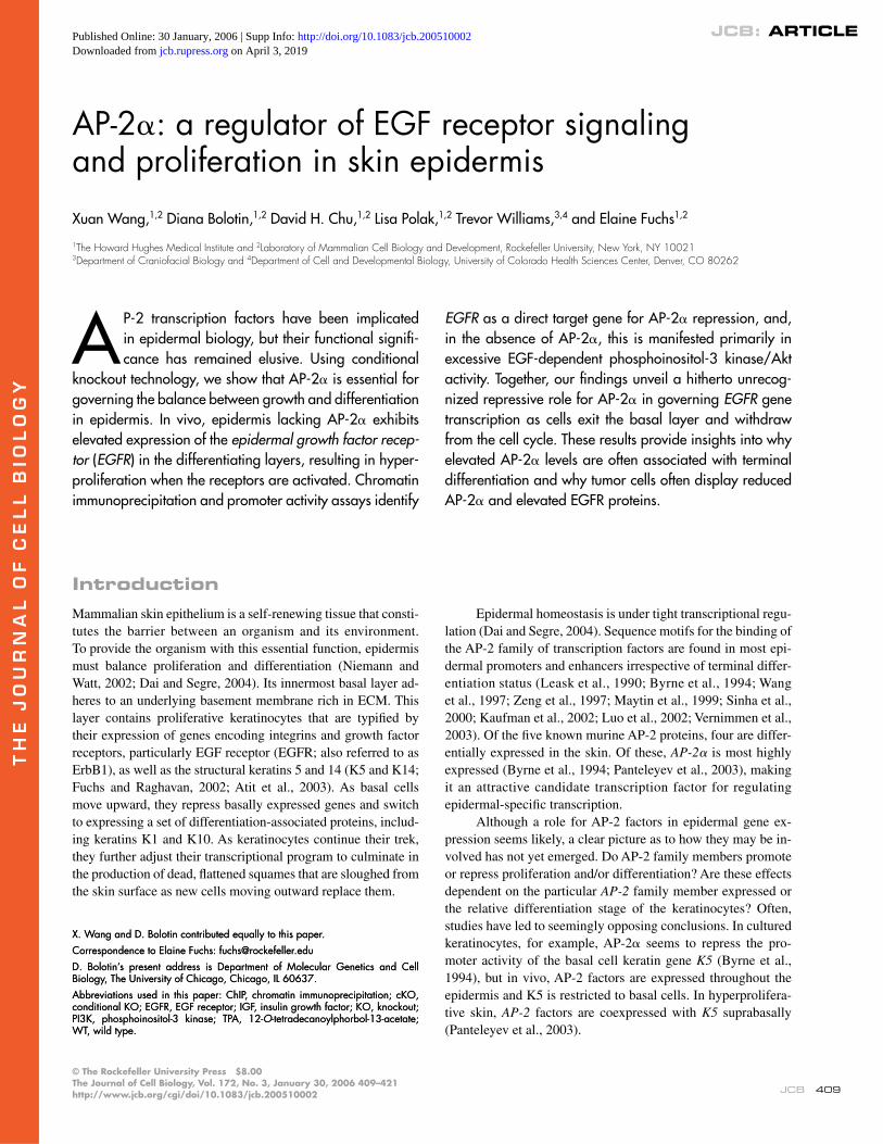
Morphological changes associated with conditional ablation of AP-2𝛂 in epidermisIn agreement with and expected from previous reports of the full KO of AP-2α in mice, the skin surface of newborn cKO pups ap-peared similar to that of their WT littermates (not depicted; Schorle et al., 1996; Zhang et al., 1996; Talbot et al., 1999). Upon histologi-cal inspection, however, cKO skin displayed a thickened interfol-licular epidermis (Fig. 2 A). Despite the complete absence of AP-2α, the thickening was not uniform across the postnatal day (P) 0–3 epidermis and began to wane altogether by P6. These aber-rations had not been noted previously in the straight AP-2α KO an-imals that died at birth. Whether this is attributable to the relatively mild nature of the defects or to strain-related differences in the mice used for the two studies (Talbot et al., 1999) was not addressed.
Figure 2. Analysis of morphological and phenotypic defects in AP-2𝛂 cKO mice. (A) 0.75-μm semi-thin sections stained with toluidine blue showing epi-dermal morphology beginning at postnatal day 1 (P1)–P3. Vertical bars represent the thickness of the epidermis. (B and C) Thoracic hair loss is associated with papilloma-like invaginations and hyperthickened epidermis in adult (6 mo old) conditionally AP-2α–null mice. (D) Thickened dorsal ear skin in condi-tionally AP-2α–null adult mice. Inset shows a high magnifi cation of the dorsal ear skin in conditionally AP-2α–null adult mice, with an arrow denoting the site of parakeratosis. C and D are H&E-stained sections (8 μM). Dotted lines denote dermo–epidermal boundaries. Hf, hair follicle.
JCB • VOLUME 172 • NUMBER 3 • 2006 412
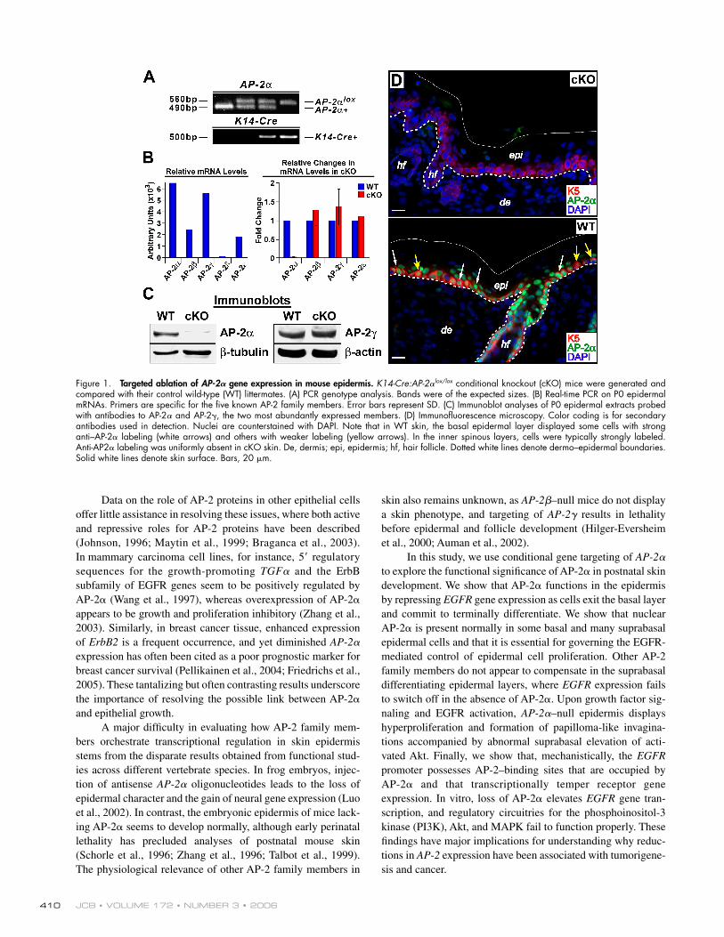
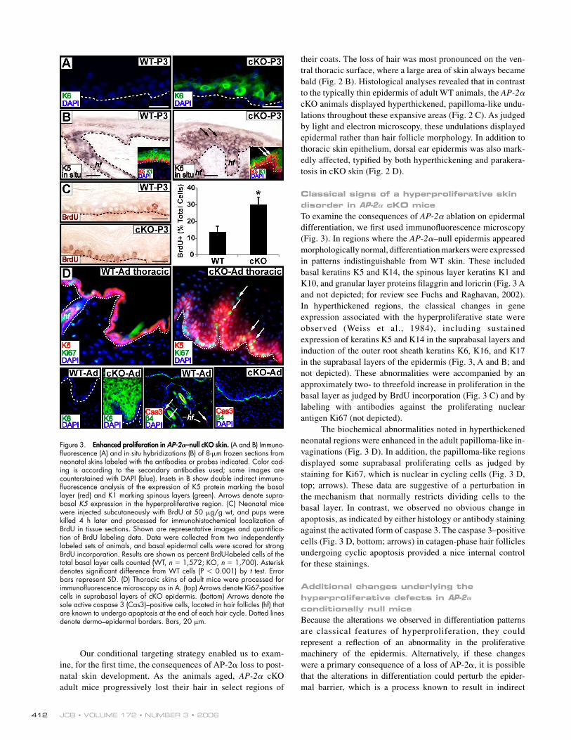
Figure 3. Enhanced proliferation in AP-2𝛂–null cKO skin. (A and B) Immuno-fl uorescence (A) and in situ hybridizations (B) of 8-μm frozen sections from neonatal skins labeled with the antibodies or probes indicated. Color cod-ing is according to the secondary antibodies used; some images are counterstained with DAPI (blue). Insets in B show double indirect immuno-fl uorescence analysis of the expression of K5 protein marking the basal layer (red) and K1 marking spinous layers (green). Arrows denote supra-basal K5 expression in the hyperproliferative region. (C) Neonatal mice were injected subcutaneously with BrdU at 50 μg/g wt, and pups were killed 4 h later and processed for immunohistochemical localization of BrdU in tissue sections. Shown are representative images and quantifi ca-tion of BrdU labeling data. Data were collected from two independently labeled sets of animals, and basal epidermal cells were scored for strong BrdU incorporation. Results are shown as percent BrdU-labeled cells of the total basal layer cells counted (WT, n = 1,572; KO, n = 1,700). Asterisk denotes signifi cant difference from WT cells (P < 0.001) by t test. Error bars represent SD. (D) Thoracic skins of adult mice were processed for immunofl uorescence microscopy as in A. (top) Arrows denote Ki67-positive cells in suprabasal layers of cKO epidermis. (bottom) Arrows denote the sole active caspase 3 (Cas3)–positive cells, located in hair follicles (hf) that are known to undergo apoptosis at the end of each hair cycle. Dotted lines denote dermo–epidermal borders. Bars, 20 μm.
Our conditional targeting strategy enabled us to exam-ine, for the fi rst time, the consequences of AP-2α loss to post-natal skin development. As the animals aged, AP-2α cKO adult mice progressively lost their hair in select regions of
their coats. The loss of hair was most pronounced on the ven-tral thoracic surface, where a large area of skin always became bald (Fig. 2 B). Histological analyses revealed that in contrast to the typically thin epidermis of adult WT animals, the AP-2α cKO animals displayed hyperthickened, papilloma-like undu-lations throughout these expansive areas (Fig. 2 C). As judged by light and electron microscopy, these undulations displayed epidermal rather than hair follicle morphology. In addition to thoracic skin epithelium, dorsal ear epidermis was also mark-edly affected, typifi ed by both hyperthickening and parakera-tosis in cKO skin (Fig. 2 D).
Classical signs of a hyperproliferative skin disorder in AP-2𝛂 cKO miceTo examine the consequences of AP-2α ablation on epidermal differentiation, we fi rst used immunofl uorescence microscopy (Fig. 3). In regions where the AP-2α–null epidermis appeared morphologically normal, differentiation markers were expressed in patterns indistinguishable from WT skin. These included basal keratins K5 and K14, the spinous layer keratins K1 and K10, and granular layer proteins fi laggrin and loricrin (Fig. 3 A and not depicted; for review see Fuchs and Raghavan, 2002). In hyperthickened regions, the classical changes in gene expression associated with the hyperproliferative state were observed (Weiss et al., 1984), including sustained expression of keratins K5 and K14 in the suprabasal layers and induction of the outer root sheath keratins K6, K16, and K17 in the suprabasal layers of the epidermis (Fig. 3, A and B; and not depicted). These abnormalities were accompanied by an approximately two- to threefold increase in proliferation in the basal layer as judged by BrdU incorporation (Fig. 3 C) and by labeling with antibodies against the proliferating nuclear antigen Ki67 (not depicted).
The biochemical abnormalities noted in hyperthickened neonatal regions were enhanced in the adult papilloma-like in-vaginations (Fig. 3 D). In addition, the papilloma-like regions displayed some suprabasal proliferating cells as judged by staining for Ki67, which is nuclear in cycling cells (Fig. 3 D, top; arrows). These data are suggestive of a perturbation in the mechanism that normally restricts dividing cells to the basal layer. In contrast, we observed no obvious change in apoptosis, as indicated by either histology or antibody staining against the activated form of caspase 3. The caspase 3– positive cells (Fig. 3 D, bottom; arrows) in catagen-phase hair follicles undergoing cyclic apoptosis provided a nice internal control for these stainings.
Additional changes underlying the hyperproliferative defects in AP-2𝛂 conditionally null miceBecause the alterations we observed in differentiation patterns are classical features of hyperproliferation, they could represent a refl ection of an abnormality in the proliferative machinery of the epidermis. Alternatively, if these changes were a primary consequence of a loss of AP-2α, it is possible that the alterations in differentiation could perturb the epider-mal barrier, which is a process known to result in indirect
AP-2α: BALANCING GROWTH AND DIFFERENTIATION • WANG ET AL. 413
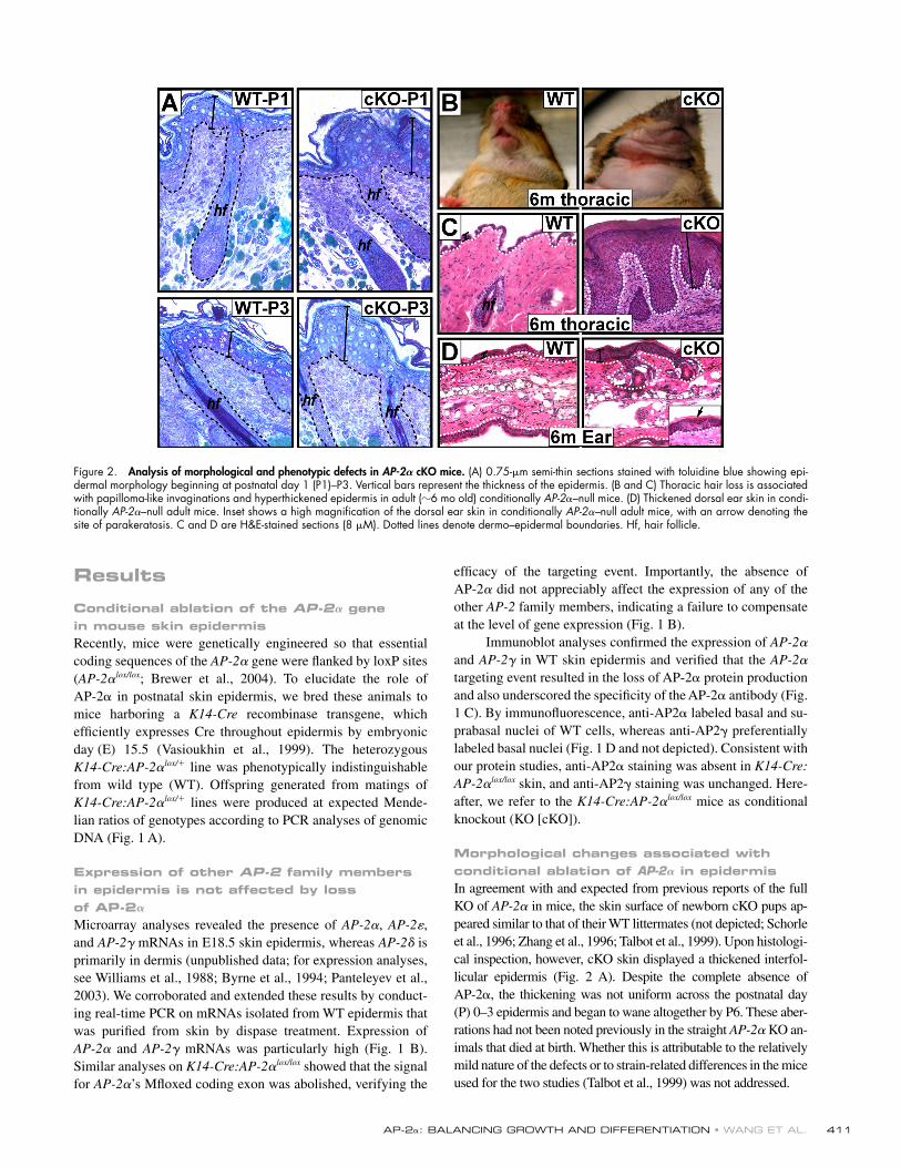
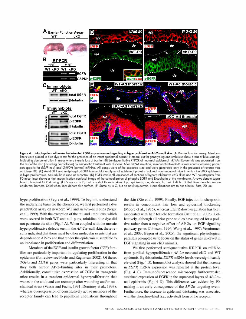
Figure 4. Intact epidermal barrier but elevated EGFR expression and signaling in hyperproliferative AP-2𝛂–null skin. (A) Barrier function assay. Newborn litters were placed in blue dye to test for the presence of an intact epidermal barrier. Note tail cut for genotyping and umbilicus show areas of blue staining, indicating dye penetration in areas where there is loss of barrier. (B) Semiquantitative RT-PCR of neonatal epidermal mRNAs. Epidermis was separated from the rest of the skin (including hair follicles) by enzymatic treatment with dispase. After mRNA isolation, semiquantitative RT-PCR was conducted using primer sets specifi c for EGFR (test) and GAPDH (control) mRNAs. All bands were of the expected size and were generated only in the presence of reverse tran-scriptase (RT). (C) Anti-EGFR and antiphospho-EGFR immunoblot analyses of epidermal proteins isolated from neonatal mice in which the cKO epidermis is hyperproliferative. Anti-tubulin is used as a control. (D) EGFR immunofl uorescence of sections of hyperproliferative cKO skins and WT counterparts from P0 mice. Inset shows a high magnifi cation confocal image of the colocalization of phospho-EGFR and E-cadherin at the membrane. Arrows denote supra-basal phospho-EGFR staining. (E) Same as in D, but on adult thoracic skins. Epi, epidermis; de, dermis; hf, hair follicle. Dotted lines denote dermo– epidermal borders. Solid white lines denote skin surface. (F) Same as in C, but on adult epidermis. Normalizations are to anti-tubulin. Bars, 20 μm.
hyperproliferation (Segre et al., 1999). To begin to understand the underlying basis for the phenotype, we fi rst performed a dye penetration assay on newborn WT and AP-2α–null pups (Segre et al., 1999). With the exception of the tail and umbilicus, which were severed in both WT and null pups, toluidine blue dye did not penetrate the skin (Fig. 4 A). When coupled with the mosaic hyperproliferative defects seen in the AP-2α–null skin, these re-sults indicated that there must be other molecular events that are dependent on AP-2α and that render the epidermis susceptible to an imbalance in proliferation and differentiation.
Members of the EGF and insulin growth factor (IGF) fam-ilies are particularly important in regulating proliferation in the epidermis (for review see Fuchs and Raghavan, 2002). Of these, TGFα and EGFR genes were particularly interesting in that they both harbor AP-2–binding sites in their promoters. Additionally, constitutive expression of TGFα in transgenic mice results in a transient epidermal hyperproliferation that wanes in the adult and can reemerge after wounding and/or me-chanical stress (Vassar and Fuchs, 1991; Dominey et al., 1993), whereas overexpression of constitutively active members of the receptor family can lead to papilloma undulations throughout
the skin (Xie et al., 1999). Finally, EGF injection in sheep skin results in concomitant hair loss and epidermal thickening (Moore et al., 1985), whereas EGFR down-regulation has been associated with hair follicle formation (Atit et al., 2003). Col-lectively, although all prior gene studies have argued for a posi-tive rather than a negative effect of AP-2α on EGF signaling pathway genes (Johnson, 1996; Wang et al., 1997; Vernimmen et al., 2003; Begon et al., 2005), the signifi cant physiological parallels prompted us to focus on the status of genes involved in EGF signaling in our cKO animals.
We fi rst performed semiquantitative RT-PCR on mRNAs from purifi ed hyperproliferative AP-2α neonatal cKO and WT epidermis. By this criteria, EGFR mRNA levels were signifi cantly elevated (Fig. 4 B). Immunoblot analysis showed that the increase in EGFR mRNA expression was refl ected at the protein level (Fig. 4 C). Immunofl uorescence microscopy furtherrevealed sustained expression of EGFR in the suprabasal layers of AP-2α–null epidermis (Fig. 4 D). This difference was evident by P0, making it an early consequence of the AP-2α targeting event. Furthermore, the increase in epidermal thickening was associated with the phosphorylated (i.e., activated) form of the receptor.
JCB • VOLUME 172 • NUMBER 3 • 2006 414
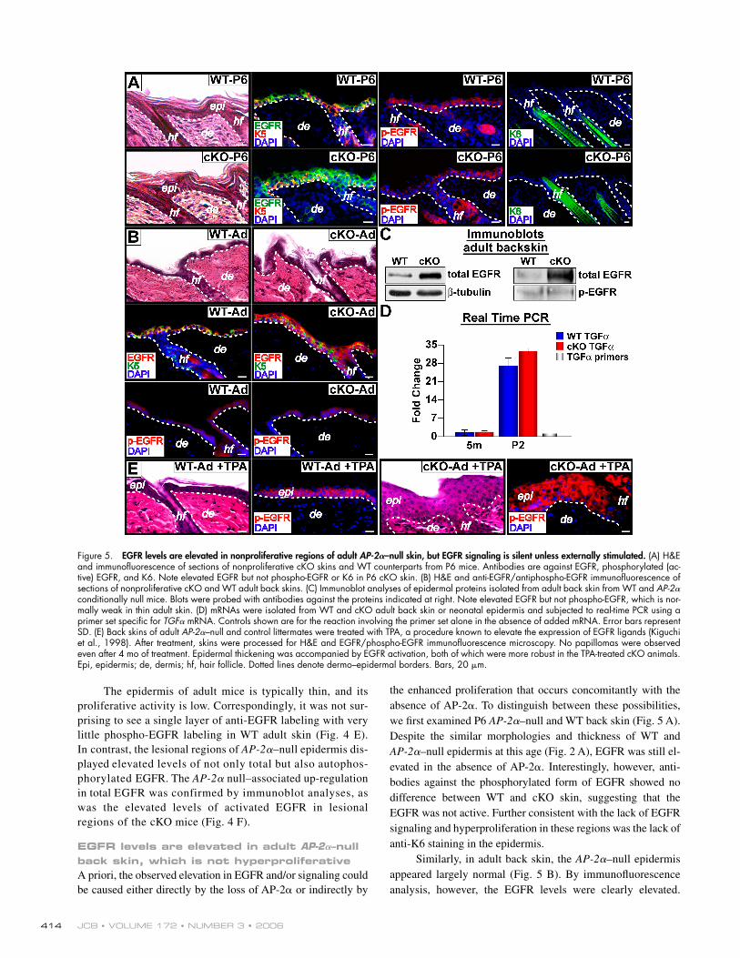
Figure 5. EGFR levels are elevated in nonproliferative regions of adult AP-2𝛂–null skin, but EGFR signaling is silent unless externally stimulated. (A) H&E and immunofl uorescence of sections of nonproliferative cKO skins and WT counterparts from P6 mice. Antibodies are against EGFR, phosphorylated (ac-tive) EGFR, and K6. Note elevated EGFR but not phospho-EGFR or K6 in P6 cKO skin. (B) H&E and anti-EGFR/antiphospho-EGFR immunofl uorescence of sections of nonproliferative cKO and WT adult back skins. (C) Immunoblot analyses of epidermal proteins isolated from adult back skin from WT and AP-2α conditionally null mice. Blots were probed with antibodies against the proteins indicated at right. Note elevated EGFR but not phospho-EGFR, which is nor-mally weak in thin adult skin. (D) mRNAs were isolated from WT and cKO adult back skin or neonatal epidermis and subjected to real-time PCR using a primer set specifi c for TGFα mRNA. Controls shown are for the reaction involving the primer set alone in the absence of added mRNA. Error bars represent SD. (E) Back skins of adult AP-2α–null and control littermates were treated with TPA, a procedure known to elevate the expression of EGFR ligands (Kiguchi et al., 1998). After treatment, skins were processed for H&E and EGFR/phospho-EGFR immunofl uorescence microscopy. No papillomas were observed even after 4 mo of treatment. Epidermal thickening was accompanied by EGFR activation, both of which were more robust in the TPA-treated cKO animals. Epi, epidermis; de, dermis; hf, hair follicle. Dotted lines denote dermo–epidermal borders. Bars, 20 μm.
The epidermis of adult mice is typically thin, and its proliferative activity is low. Correspondingly, it was not sur-prising to see a single layer of anti-EGFR labeling with very little phospho-EGFR labeling in WT adult skin (Fig. 4 E). In contrast, the lesional regions of AP-2α–null epidermis dis-played elevated levels of not only total but also autophos-phorylated EGFR. The AP-2α null–associated up-regulation in total EGFR was confirmed by immunoblot analyses, as was the elevated levels of activated EGFR in lesional regions of the cKO mice (Fig. 4 F).
EGFR levels are elevated in adult AP-2𝛂–null back skin, which is not hyperproliferativeA priori, the observed elevation in EGFR and/or signaling could be caused either directly by the loss of AP-2α or indirectly by
the enhanced proliferation that occurs concomitantly with the absence of AP-2α. To distinguish between these possibilities, we fi rst examined P6 AP-2α–null and WT back skin (Fig. 5 A). Despite the similar morphologies and thickness of WT and AP-2α–null epidermis at this age (Fig. 2 A), EGFR was still el-evated in the absence of AP-2α. Interestingly, however, anti-bodies against the phosphorylated form of EGFR showed no difference between WT and cKO skin, suggesting that the EGFR was not active. Further consistent with the lack of EGFR signaling and hyperproliferation in these regions was the lack of anti-K6 staining in the epidermis.
Similarly, in adult back skin, the AP-2α–null epidermis appeared largely normal (Fig. 5 B). By immunofl uorescence analysis, however, the EGFR levels were clearly elevated.
AP-2α: BALANCING GROWTH AND DIFFERENTIATION • WANG ET AL. 415
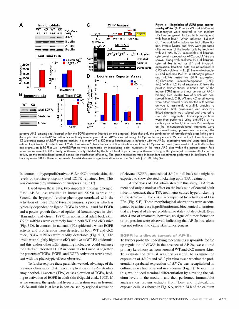
Figure 6. Regulation of EGFR gene expres-sion by AP-2𝛂. (A) Primary WT and AP-2α–null keratinocytes were cultured in rich medium (15% serum, growth factors, high density, and with feeder layer). When indicated, 1.5 mM Ca2+ was added to induce terminal differentia-tion. Protein lysates and RNA were prepared after removal of the feeder cells by treatment with 0.1 mM EDTA. Immunoblots of keratino-cyte proteins probed for AP-2α and AP-2γ are shown, along with real-time PCR of keratino-cyte mRNAs tested for K1 and involucrin expression. Real-time data are normalized to 0.05 mM calcium (= 1). (B) Immunoblot analy-sis and real-time PCR of keratinocyte protein and mRNAs tested for EGFR expression. (C) Chromatin immunoprecipitation (ChIP). (top) Within 1.2 kb of sequence 5′ from the putative transcriptional initiation site of the mouse EGFR gene are four consensus AP-2–binding sites (ovals), two of which are con-served (in red). ChIP, WT, and KO keratinocytes were either treated or not treated with formal-dehyde to transiently cross-link proteins to chromatin. Both cross-linked and noncross-linked chromatin was isolated and sheared to 400-bp fragments. Immunoprecipitations were then performed using anti-AP2α or no antibody or control IgG antisera. PCR analyses on the immunoprecipitated fragments were performed using primers encompassing the
putative AP-2–binding sites located within the EGFR promoter (marked on the diagram). Note that only the combination of formaldehyde cross-linking and the application of anti–AP-2α antibody specifi cally immunoprecipitated AP-2α site-containing EGFR promoter sequences in WT and not in KO keratinocytes. (D) Luciferase assays of EGFR promoter activity in primary WT or KO mouse keratinocytes ± infection with the AP-2α–expressing retroviral vector (see Sepa-ration of epidermis…transfections). 1.2 kb of sequence 5′ from the transcription initiation site of the EGFR promoter (see C) was used to drive fi refl y lucifer-ase expression (pEGFRpr-Luc). pMutEGFRpr-luc was engineered by introducing point mutations in the three AP-2 sites within the parent vector. Fold increases represent EGFRpr fi refl y luciferase activity divided by the basal level of p-Luc fi refl y luciferase activity, with cytomegalo virus–Renilla luciferase activity as the standardized internal control for transfection effi ciency. The graph represents three independent experiments performed in duplicate. Error bars represent SD for these experiments. Asterisk denotes a signifi cant difference from WT cells (P < 0.001) by t test.
In contrast to hyperproliferative AP-2α cKO thoracic skin, the levels of tyrosine-phosphorylated EGFR remained low. This was confi rmed by immunoblot analyses (Fig. 5 C).
Based upon these data, two important fi ndings emerged. First, AP-2α loss resulted in increased EGFR expression. Second, the hyperproliferative phenotype correlated with the activation of these EGFR tyrosine kinases, a process which is typically dependent on ligand. TGFα is both a ligand for EGFR and a potent growth factor of epidermal keratinocytes in vitro (Barrandon and Green, 1987). In nonlesional adult back skin, TGFα mRNAs were extremely low in both WT and cKO mice (Fig. 5 D). In contrast, in neonatal (P2) epidermis, where EGFR activity and proliferation were detected in both WT and cKO mice, TGFα mRNAs were readily detectable (Fig. 5 D). The levels were slightly higher in cKO relative to WT P2 epidermis, and this and/or other EGF signaling molecules could enhance the effects of elevated EGFR in neonatal cKO mice. Altogether, the patterns of TGFα, EGFR, and EGFR activation were consis-tent with the phenotypic effects observed.
To further explore these parallels, we took advantage of the previous observation that topical application of 12-O-tetradec-anoylphorbol-13-acetate (TPA) causes elevation of TGFα, lead-ing to activation of EGFR in adult skin (Kiguchi et al., 1998). If, as we surmise, the epidermal hyperproliferation seen in lesional AP-2α–null skin is at least in part caused by regional activation
of elevated EGFRs, nonlesional AP-2α–null back skin might be expected to show elevated thickening upon TPA treatment.
At the doses of TPA administered in this study, TPA treat-ment had only a modest effect on the back skin of control adult mice. In contrast, these TPA treatments caused hyperthickening of the AP-2α–null back skin accompanied by activation of EG-FRs (Fig. 5 E). These morphological aberrations were accom-panied by an increase in proliferation and biochemical alterations that are typical of a hyperproliferative state (not depicted). Even after 4 mo of treatment, however, no signs of tumor formation or progression were observed, indicating that AP-2α loss alone was not suffi cient to cause skin tumorigenesis.
EGFR is a direct target of AP-2𝛂To further probe the underlying mechanisms responsible for the up-regulation of EGFR in the absence of AP-2α, we cultured primary keratinocytes from neonatal WT and cKO mouse skins. To evaluate the data, it was fi rst essential to examine the expression of AP-2α and AP-2γ in vitro to see whether the pref-erential suprabasal expression of AP-2α was recapitulated in culture, as we had observed in epidermis (Fig. 1). To examine this, we induced terminal differentiation by elevating the cal-cium levels in the medium and then performed immunoblot analyses on protein extracts from low- and high-calcium exposed cells. As shown in Fig. 6 A, within 24 h of the calcium
JCB • VOLUME 172 • NUMBER 3 • 2006 416
shift, the expression of the spinous layer markers K1 and invo-lucrin were elevated, although appreciable involucrin mRNA was present even in the low-calcium state. Within the same time frame, AP-2α was largely unaffected by calcium treatment rela-tive to internal protein loading standards. In contrast, AP-2γ was markedly decreased upon calcium-induced differentiation. Moreover, the levels of AP-2γ were not appreciably changed in the AP-2α–null cells (Fig. 6 A). Together, these data were con-sistent with our prior in vivo data presented in Fig. 1.
Next, we conducted immunoblot studies in which we could examine EGFR levels under conditions where we could more rig-orously control for microenvironment and cell numbers. As shown in Fig. 6 B, EGFR levels were elevated in the AP-2α–null kerati-nocytes relative to their WT counterpart. Under enriched culture conditions, both cell populations displayed phosphorylated EGFR (not depicted). These changesin AP-2α–null keratinocytes were also refl ected at the mRNA level (Fig. 6 B). Real-time PCR showed a fourfold increase in EGFR mRNAs in KO versus WT cells. TGFα mRNA levels were only modestly elevated in the AP-2α–null state (not depicted).
If AP-2 acts to repress EGFR gene transcription, it should bind to the endogenous EGFR promoter. Previous studies have shown that recombinant AP-2α can bind to an AP-2 consensus motif in the human EGFR promoter (Johnson, 1996; Oyama et al., 2002). In the mouse EGFR promoter, multiple AP-2 consensus binding sites are situated within a kilobase upstream from the transcription initiation site (Fig. 6 C, ovals). Two of these sites are conserved across mammalian species (Fig. 6 C, ovals highlighted in red). To evaluate whether en dogenous AP-2α binds directly to one or more of these sites in epidermal cells, we conducted chromatin immunoprecipita-tion (ChIP) assays using the monospecifi c anti–AP-2α antibody. Anti–AP-2α antibodies specifi cally immunoprecipitated chromatin–protein complexes that contained an 500-bp DNA fragment encompassing the conserved putative AP-2α–binding sites (Fig. 6 C). In contrast, ChIP analysis of WT skin did not display PCR bands with primers corresponding to either the EGFR promoter or downstream regions that did not contain AP-2–binding motifs (not depicted). These data provided the fi rst ChIP data illustrating the binding of AP-2α to an endoge-nous gene that contains AP-2 consensus binding motifs.
To test whether EGFR gene transcription is affected by the loss of AP-2α, we transfected Ca2+ grown keratinocytes with a luciferase reporter gene driven by a 1.1-kb EGFR promoter fragment harboring the AP-2α–binding sites. By 48 h, luciferase activity was markedly elevated in AP-2α–null versus WT cells (Fig. 6 D). Point mutations in the AP-2α motifs raised EGFR promoter activity levels in WT but did not affect AP-2α–null cells. Finally, we infected WT and KO keratinocytes with a ret-roviral AP-2α expression vector and repeated the EGFR reporter assay. Exogenous expression of AP-2α resulted in a potent re-pression of luciferase activity driven by the WT EGFR promoter but not its mutant counterpart (Fig. 6 D). These data underscore the AP-2–specifi c nature of the manipulations and the repressive effects on EGFR transcription. These fi ndings further suggest that AP-2α functions in suppressing EGFR gene expression as epidermal cells commit to terminally differentiate.
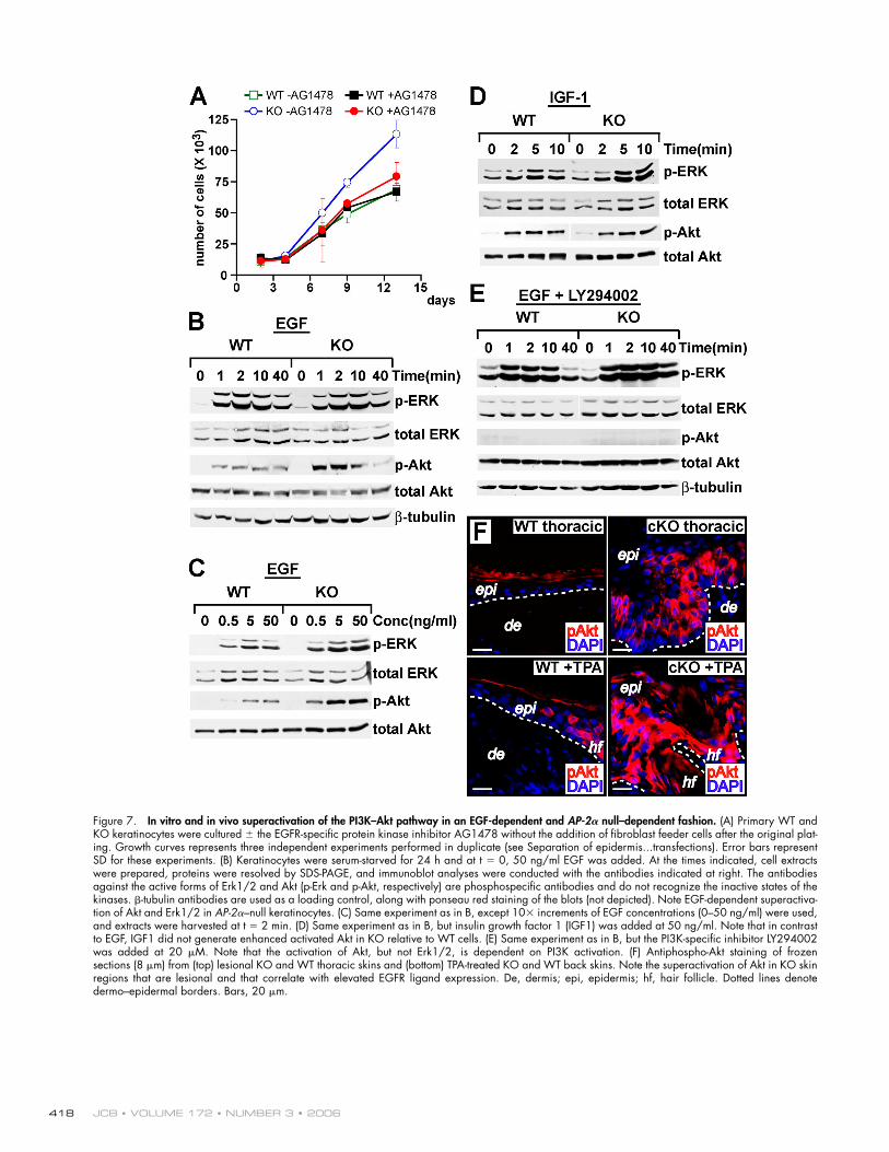
Abnormal EGFR signaling circuits in AP-2𝛂–null skinTo evaluate how downstream signaling events may be affected when EGFR levels are elevated, we fi rst examined the prolifera-tive potential of primary AP-2α–null and WT epidermal kerati-nocytes. Both populations grew equally well when plated at high density on a fi broblast feeder layer and in rich growth media (unpublished data). However, when subjected to more stringent conditions such as sparse plating with no feeder layer for long term growth (>5 d) or low serum/growth factor– supplemented medium, only KO keratinocyte cultures grew well (representative examples in Fig. 7 A). Moreover, the growth advantage of the KO keratinocytes was largely eliminated by the addition of AG1478, an EGFR-specifi c protein kinase in-hibitor, to the culture medium, suggesting that increased EGFR signaling might be responsible for the enhanced proliferative potential observed in KO keratinocytes.
EGFR stimulation typically leads to downstream activa-tion of the MAPK family members Erk1/2, but it can also lead to activation of Akt kinases. Both are required for epi-dermal growth and differentiation (Schlessinger, 2000; Sibilia et al., 2000). To determine whether the loss of AP-2α and elevated EGFR signaling results in an elevated activation of MAPK (Erk1/2) and/or Akt pathways, we stimulated 24-h serum-starved cells with EGF. Within a minute after stimula-tion, the two populations activated Erk1/2 with similar kinetics (Fig. 7 B). Relative to total Erk1/2, the phosphory-lated (i.e., activated) Erk1/2 signals were always higher in the KO keratinocytes. This was visualized over a range of EGF concentrations (Fig. 7 C). Although AP-2–null keratinocytes activated more Erk1/2 in response to EGFR ligands, the most signifi cant difference observed was in EGF-mediated Akt activation. Again, the fi rst signs of activation appeared within a minute after exposure to EGF. However, relative to equiva-lent levels of total Akt, phosphorylated (i.e., activated) Akt was nearly an order of magnitude higher in KO cells across a range of EGF concentrations.
Remarkably, as little as 0.5 ng/ml EGF was suffi cient to elicit these differences in Erk1/2 and Akt activation. Curiously, the differential effects on Akt appeared to be specifi c for EGFR signaling, whereas the differential activation of Erk1/2 was also seen with IGF-1, another potent stimulator of keratinocyte growth and survival (Fig. 7 D). The effects were obliterated by treatment with the PI3K-specifi c inhibitor LY294002, indicating that Akt activation in EGF-treated keratinocytes was mediated through PI3K activation (Fig. 7 E).
To test the physiological relevance of these fi ndings, we examined the status of activated Akt in our mice. In nonlesional regions of AP-2α–null skin where EGFRs were elevated but not activated, phospho-Akt levels remained low and comparable with WT skin (Fig. 7 F and not depicted). However, in either TPA-treated or thoracic cKO skin, activated Akt was markedly elevated. Even in WT skin, TPA treatment resulted in mildly up-regulated levels of activated Akt, which is consistent with the modest epidermal thickening we noted previously (Fig. 5 D). Based upon these criteria, the mechanisms uncovered in our in vitro analyses appeared to be operative in vivo.
AP-2α: BALANCING GROWTH AND DIFFERENTIATION • WANG ET AL. 417
DiscussionThe AP-2𝛂 and EGFR connection and its relevance to growth control in the epidermisAP-2–binding sites have been found in a myriad of genes differentially expressed in both basal and suprabasal compart-ments of mammalian epidermis (Hilger-Eversheim et al., 2000; for review see Fuchs and Raghavan, 2002). However, to date, their functional signifi cance has remained elusive. In this study, we have shown that AP-2α plays a role in the switch between epidermal cell proliferation and differentiation, and we have provided the fi rst experimental evidence to integrate this tran-scription factor into a key signaling pathway in the skin. The fi nding is especially important given that some studies have highlighted AP-2α as a tumor suppressor (Ropponen et al., 2001; Pellikainen et al., 2004; Friedrichs et al., 2005), whereas others have suggested a growth-promoting effect of the protein (Bosher et al., 1996; Vernimmen et al., 2003). Our study clar-ifi es how AP-2α governs epidermal proliferation and provides important new insights into why inverse correlations are often found between AP-2α and EGFR family members in human epithelial cancers.
Our studies place AP-2α in the EGFR signal transduction cascade. Using in vivo and in vitro approaches, we found that EGFR gene expression was elevated upon genetic ablation of AP-2α or by mutation of the AP-2–binding sites in the EGFR promoter. Correspondingly, EGFR gene expression was down-regulated when AP-2α was added back to the KO cells through retroviral transgene expression. Although AP-2α is likely to have many other target genes in epidermis, several of the pheno-typic abnormalities in the AP-2α conditionally null mice can be explained by EGFR misregulation.
EGFRs have long been known to play an important role in regulating the development of the epidermis and its appendages (Luetteke et al., 1994; Miettinen et al., 1995; Sibilia and Wagner, 1995; Threadgill et al., 1995). In mammals and birds, over-expression or injection of EGF can arrest epidermal appendage development and promote epidermal thickening, and, conversely, activation of EGFRs is typically diminished in areas of follicle formation (Moore et al., 1985; Kashiwagi et al., 1997; Atit et al., 2003). Postnatally, EGFRs are predominantly expressed in the basal epidermal layer and down-regulated as cells commit to terminally differentiate (King et al., 1990).
Once activated, EGFR has the ability to activate Ras–MAPK signaling, which is often linked to proliferation, as well as PI3K–Akt signaling, which is more typically associated with cell survival (Rodeck et al., 1997; Schlessinger, 2000). Relevant to the data presented here, tumorigenesis can arise from super-activated Ras–MAPK signaling in transgenic mouse skin but only in conjunction with EGFR signaling to activate PI3–Akt (Sibilia et al., 2000). A key feature of EGFR signaling is that its activation is dependent on ligand stimulation. Made and se-creted by salivary glands, EGF can enter the bloodstream and reach epidermal tissue, whereas TGFα is the major autocrine growth factor of the epidermis. TGFα is down-regulated post-natally but can be up-regulated upon injury or TPA treatment
(Kiguchi et al., 1998). That postnatal skin is limiting for EGFR ligands is graphically illustrated by the papilloma-like undula-tions that develop in skin of mice engineered to overexpress an EGFR relative (ErbB2) that lacks the requirement for ligand stimulation (Xie et al., 1999; Kiguchi et al., 2000).
When placed in the context of these prior studies, we can ac-count for many of the features of the seemingly complex phenotype of our AP-2α cKO mice. The hyperproliferation that occurs in cKO newborn epidermis despite elevated EGFR levels suggests that the ligand pool for these receptors must be quite large at this age. The levels appear to be saturating for WT but not cKO EGFR levels, and this is refl ected by the observed enhancement of phosphorylation and activation of the EGFRs in newborn cKO skin. Soon after birth, hyperproliferation and phosphorylation of EGFRs returns to nor-mal in cKO despite sustained elevated EGFR levels in cKO skin. After embryogenesis, the ligand pool is known to be attenuated along with other growth-promoting signals, and our results indicate that these pool levels have dipped below saturation even for WT EGFRs. Finally, the markedly hyperproliferative epidermis and phosphorylation of EGFRs in lesional but not nonlesional regions of adult cKO thoracic and ear skin and TPA-treated cKO back skin is refl ective of the elevated ligand pools that are known to arise upon mechanical irritation and TPA treatment. Additionally, when animals lick their wounds, the EGF-rich saliva could further con-tribute to the robust proliferative response in the ventral chin area.
An additional point worthy of mention is that the AP-2α–null papilloma-like undulations were not as severe as the spon-taneous skin tumors that arise from chemical or transgenic mutations in epidermally expressed Ha-Ras. We surmise that the underlying reason for this may be rooted in the normal feed-back regulatory loops that are operative in dampening the dele-terious effects of overly active EGFR signaling. Indeed, for sustained epidermal hyperproliferation and tumorigenesis to occur on a background of Ha-Ras mutations, sustained EGFR signaling must still be permitted to activate and sustain elevated PI3K–Akt signaling (Sibilia et al., 2000).
Our data shed additional insight onto this signaling cir-cuitry by demonstrating that the EGFR response to Akt activa-tion is different than that of IGF receptor in epidermal cells and that in situations in which EGFR signaling is hyperactive, Akt activation can be very high and suprabasal proliferation can occur. Although Akt activation is typically associated with cell survival, several examples that link PI3K–Akt activation to pro-liferation may be relevant to the fi ndings we report. Sustained PI3K–Akt activation also occurs in liver tumors, where it post-translationally silences the C/EBPα transcriptional repressor that controls hepatocyte proliferation (Wang et al., 2004). Intriguingly, C/EBPα has also been implicated in epidermal dif-ferentiation and is repressed in some skin cancers (Shim et al., 2005). Although beyond the scope of this study, such a mecha-nism could explain why AP-2α null–mediated hyperactivation of the EGFR–PI3K–Akt pathway leads to an increase in prolif-eration and epidermal thickening in vivo.
AP-2 and transcriptional regulation in skinAfter 15 yr since the original implication of AP-2 in transcrip-tional regulation in the epidermis (Leask et al., 1990; Snape
JCB • VOLUME 172 • NUMBER 3 • 2006 418
Figure 7. In vitro and in vivo superactivation of the PI3K–Akt pathway in an EGF-dependent and AP-2𝛂 null–dependent fashion. (A) Primary WT and KO keratinocytes were cultured ± the EGFR-specifi c protein kinase inhibitor AG1478 without the addition of fi broblast feeder cells after the original plat-ing. Growth curves represents three independent experiments performed in duplicate (see Separation of epidermis...transfections). Error bars represent SD for these experiments. (B) Keratinocytes were serum-starved for 24 h and at t = 0, 50 ng/ml EGF was added. At the times indicated, cell extracts were prepared, proteins were resolved by SDS-PAGE, and immunoblot analyses were conducted with the antibodies indicated at right. The antibodies against the active forms of Erk1/2 and Akt (p-Erk and p-Akt, respectively) are phosphospecifi c antibodies and do not recognize the inactive states of the kinases. β-tubulin antibodies are used as a loading control, along with ponseau red staining of the blots (not depicted). Note EGF-dependent superactiva-tion of Akt and Erk1/2 in AP-2α–null keratinocytes. (C) Same experiment as in B, except 10× increments of EGF concentrations (0–50 ng/ml) were used, and extracts were harvested at t = 2 min. (D) Same experiment as in B, but insulin growth factor 1 (IGF1) was added at 50 ng/ml. Note that in contrast to EGF, IGF1 did not generate enhanced activated Akt in KO relative to WT cells. (E) Same experiment as in B, but the PI3K-specifi c inhibitor LY294002 was added at 20 μM. Note that the activation of Akt, but not Erk1/2, is dependent on PI3K activation. (F) Antiphospho-Akt staining of frozen sections (8 μm) from (top) lesional KO and WT thoracic skins and (bottom) TPA-treated KO and WT back skins. Note the superactivation of Akt in KO skin regions that are lesional and that correlate with elevated EGFR ligand expression. De, dermis; epi, epidermis; hf, hair follicle. Dotted lines denote dermo–epidermal borders. Bars, 20 μm.
AP-2α: BALANCING GROWTH AND DIFFERENTIATION • WANG ET AL. 419
et al., 1991), the functional importance of these proteins in epi-dermis is now emerging. Recent knockdown experiments underscore a role for Xenopus laevis AP-2α in embryonic skin development (Luo et al., 2002), and our studies now reveal an essential role for mammalian AP-2α in orchestrating the bal-ance between epidermal proliferation and differentiation. This process can be largely explained by AP-2α’s repressive effects on EGFR gene transcription, but given the repertoire of epider-mally expressed genes with AP-2–binding sites, there are likely to be many additional key genes controlled by AP-2α. Why have these other target genes not surfaced in our analyses?
The answer seems, at least in part, to reside in functional redundancy among AP-2 family members, four of which are expressed in the epidermis. This is substantiated by the fact that both in vivo and in vitro, the consequences of AP-2α ablation ap-peared to be more dramatic in the differentiating cells where AP-2γ expression is down-regulated. Additionally, AP-2α ap-pears to be more highly expressed in those basal cells that are not actively cycling, leading us to speculate that AP-2’s role may be most critical at the juncture between proliferation and differentia-tion in the epidermis. Evaluating the degree of AP-2 functional redundancy in the epidermis must await functional studies on the other members of the AP-2 family that are expressed in skin.
Cell type and differentiation-specifi c cofactors are likely to further impact the complexities of when and how AP-2 pro-teins act in the epidermis. Studies in other systems have already indicated that interactive partners for AP-2 proteins can infl u-ence whether AP-2 proteins act as transcriptional repressors or activators (Pfi sterer et al., 2002). Differences in AP-2 recogni-tion motifs are also likely to infl uence target gene specifi city, and the differential expression of AP-2 family members and possibly putative AP-2 cofactors could further magnify differ-ences in the spatial and temporal behavior of putative AP-2 tar-get genes within a tissue. Such differences are also likely to contribute to our understanding of why the loss of AP-2β results in massive apoptosis in the kidney (Hilger-Eversheim et al., 2000) and why the loss of AP-2α in neural crest impaired cra-niofacial development and pigmentation (Brewer et al., 2004). These issues are also likely to underlie the seemingly opposing fi ndings that the addition of recombinant AP-2α to a nuclear extract from a human squamous cell carcinoma line in vitro led to an increase in EGFR transcription (Johnson, 1996), whereas AP-2α in mouse epidermis and in cultured epidermal keratino-cytes had an inhibitory effect on EGFR promoter activity (our study). The fi nding that AP-2 proteins positively regulate cell proliferation in some cells and inhibit growth in others may help in the future to explain the seemingly disparate results obtained concerning the roles for AP-2 family members in human can-cers (Bosher et al., 1996; Ropponen et al., 2001; Vernimmen et al., 2003; Pellikainen et al., 2004; Friedrichs et al., 2005).
Materials and methodsGeneration of cKO miceAP-2α fl oxed mice (AP-2αlox) and K14-Cre transgenic mice were generated as described previously (Vasioukhin et al., 1999; Brewer et al., 2004). Genotyping was conducted by PCR of tail skin DNAs.
Histology, immunofl uorescence, and in situ hybridizationFor light or fl uorescence microscopy, tissues were embedded in optimal cutting temperature compound and frozen on dry ice. For semi-thin sec-tions and transmission EM, tissues were fi xed and processed as previously described (Vasioukhin et al., 2001). For indirect immunofl uorescence, 10-μm sections were permeabilized with 0.1% Triton X-100 and washed. For mouse monoclonal antibodies, we used the MOM kit (Vector Labora-tories); for antibodies from other species, we used 2.5% normal donkey serum, 2.5% normal goat serum, 1% BSA, 2% gelatin, 0.1% Triton X-100 as block, and antibody diluent.
Primary antibodies used are as follows: (1) mouse: AP-2α (1:5; T. Williams) and phospho-EGFR (1:200; Upstate Biotechnology); (2) rabbit: K6 (1:200; Fuchs laboratory), K1 (1:250; Fuchs laboratory), Ki67 (1:1,000; Novocastra laboratories), active caspase 3 (1:500; R&D Systems), K17 (1:1,000; a gift from P. Coulombe, The Johns Hopkins University School of Medicine, Baltimore, MD), EGFR (1:100; Upstate Biotechnology), and phospho-Akt (1:250, Cell Signaling); (3) guinea pig: K5 (1:200; Fuchs laboratory); (4) rat: BrdU (1:150, AbCam) and β4 inte-grin (1:250, BD Biosciences). For phospho-EGFR staining, sections were fi xed in cold 100% methanol, primary antibody incubations were per-formed as above, and the signal was amplifi ed using the ABC kit (Vector Laboratories) and visualized using the tyramide signal amplifi cation Plus Fluorescence detection kit (PerkinElmer). In situ hybridizations of K5 transcripts were performed as described previously (Byrne et al., 1994). Probe synthesis was performed according to the manufacturer’s instructions (Roche).
Image acquisition and manipulationThe histology, immunofl uorescent, and in situ hybridization images were taken by a mot plus microscope (Axioskop 2; Carl Zeiss MicroImaging, Inc.). The objectives used were 20× NA 0.5 plan Neofl uar ∞/0.17 and 40× NA 1.3 oil plan Neofl uar ∞/0.17 (Carl Zeiss MicroImaging, Inc.). The images were taken at room temperature in antifade as an imaging medium for immunofl uorescent images and 80% glycerol for hematoxylin and eosin (H&E) images. The fl uorochromes used were FITC, Texas red/rhodamine red X, and DAPI. A slider camera (SPOT RT; Diagnostic Instruments) and MetaMorph 6 (Molecular Devices) software were used to acquire the pictures. Adobe Photoshop 6.0 software was used for con-trast and brightness adjustment. The immunoblot and PCR gel images were acquired with an Alphaimager (Innotech), and Quantity One software (Bio-Rad Laboratories) was used for contrast and brightness adjustment.
Separation of epidermis, keratinocyte isolation, culture, and transfectionsEnzymatic separation of epidermis from skins and primary newborn mouse epidermal cell cultures were performed as described previously (Blanpain et al., 2004). To induce keratinocyte differentiation in culture, the calcium concentration of the media was raised from 0.05 to 1.5 mM for 24 h, after which protein and RNA were extracted.
For growth comparisons, 2.5 × 10⁴ of freshly isolated cells were plated with feeder layers in 24-well dishes. For stringent growth conditions, serum was adjusted to 2%, and/or growth factor supplements and the addition of fi broblast feeder cells were omitted. For growth comparisons with the presence of AG1478, 200 nM of the inhibitor (Sigma-Aldrich) was added to the serum daily after plating. Keratinocyte numbers were determined with a coulter counter.
Nucleotides −1,180–29 (transcription initiation site = 0) of the mouse EGFR promoter were cloned upstream of the fi refl y luciferase gene in the pGL2 basic plasmid (Promega). Cells were transferred into 12-well dishes and grown to 30–40% confl uency before Fugene 6 (Roche) reagent-assisted transfections of 2 ng cytomegalo virus–Renilla luciferase DNA (control) and 100 ng of either WT or mutant EGFR promoter fi refl y luciferase constructs (pEGFRpr-Luc or pMut-EGFRpr-Luc) or empty vector (p-Luc; Promega). 48 h after transfection, luciferase assays were performed as described previously (Sinha et al., 2000; Kaufman et al., 2002). Trans-fection effi ciency was 2–3%. A retroviral vector containing the full-length mouse AP-2α cDNA was engineered and infected for AP-2α rescue studies (70% effi ciency).
TPA treatmentMice were anesthetized, and their backs were shaved. Each mouse received 2.5 μg TPA (dissolved in acetone) on the right half of the back twice per week for 4 mo. As a control, the left half was treated with acetone.
ChIPIn vivo ChIP was performed as described previously (Jamora et al., 2003). The presence of AP-2α sites was confi rmed by rVista analysis of 5′ upstream
JCB • VOLUME 172 • NUMBER 3 • 2006 420
sequences as defi ned by the ECR Browser and Ensemble software (Euro-pean Bioinformatics Institute and Sanger Institute). AP-2 sites were chosen for ChIP analysis based on the conservation and alignment between mouse and at least one other species, including human, canine, and rat, and clus-tering of sites when applicable. As a control, PCR was also performed using primers that recognize other sites within the same promoter or down-stream portions of the same gene to demonstrate the specifi city of the pull-down.
AP-2𝛂, AP-2𝛄, EGFR, MAPK (Erk1/2), and Akt activity measurementsSDS-PAGE and immunoblot analyses were used to identify the AP-2α, AP-2γ, EGFR, MAPK (Erk1/2), and Akt proteins. AP-2α and AP-2γ proteins were identifi ed with monoclonal antibodies against AP-2α and AP-2γ (Santa Cruz Biotechnology, Inc.), respectively. The active (i.e., phosphory-lated) forms of the EGFR, MAPK, and Akt proteins were identifi ed with phos-phospecifi c antibodies against EGFR (Tyr1173; Upstate Biotechnology), MAPK (Thr183 and Tyr185 in Erk2; Sigma-Aldrich), and Akt (Ser473; Cell Signaling), respectively. The total levels of these proteins were identifi ed with panel antibodies against EGFR (Upstate Biotechnology), MAPK (Cell Signaling), and Akt (Cell Signaling), respectively.
We thank Drs. V. Horsley, H. Nguyen, and C. Blanpain (Fuchs laboratory) for their critical reading of the manuscript and meaningful discussions. We thank Dr. J. Huang (Williams laboratory) and P. Romero (Fuchs laboratory) for assistance with K14-Cre and AP-2αlox/lox mice. We thank Dr. H.A. Pasolli for processing and analyzing semi-thin tissue sections.
D. Bolotin is supported by the Medical Scientist National Research Service Award, National Institutes of Health (NIH)/National Institute of Gen-eral Medical Sciences grant 5-T32-GM07281, and The University of Chi-cago Division of Biological Sciences Naomi Ragins Goldsmith Fund. X. Wang is a Postdoctoral Fellow supported by the Women and Science Organization at The Rockefeller University. D.H. Chu is a Marion Sulzberger Research Fellow supported by the NIH training grant 5-T3-AR07190, an American Skin Associ-ation Research grant, and an American Cancer Society postdoctoral fellowship. E. Fuchs is a Howard Hughes Medical Institute Investigator. This work is sup-ported by NIH grant R01-AR31737 to E. Fuchs, grant R01-CA77833 to T. Williams, and grant R01-DE12728 to T. Williams.
Submitted: 3 October 2005Accepted: 22 December 2005
ReferencesAtit, R., R.A. Conlon, and L. Niswander. 2003. EGF signaling patterns the
feather array by promoting the interbud fate. Dev. Cell. 4:231–240.
Auman, H.J., T. Nottoli, O. Lakiza, Q. Winger, S. Donaldson, and T. Williams. 2002. Transcription factor AP-2gamma is essential in the extra-embry-onic lineages for early postimplantation development. Development. 129:2733–2747.
Barrandon, Y., and H. Green. 1987. Three clonal types of keratinocyte with different capacities for multiplication. Proc. Natl. Acad. Sci. USA. 84:2302–2306.
Begon, D.Y., L. Delacroix, D. Vernimmen, P. Jackers, and R. Winkler. 2005. Yin Yang 1 cooperates with activator protein 2 to stimulate ERBB2 gene expression in mammary cancer cells. J. Biol. Chem. 280:24428–24434.
Blanpain, C., W.E. Lowry, A. Geoghegan, L. Polak, and E. Fuchs. 2004. Self-renewal, multipotency, and the existence of two cell populations within an epithelial stem cell niche. Cell. 118:635–648.
Bosher, J.M., N.F. Totty, J.J. Hsuan, T. Williams, and H.C. Hurst. 1996. A family of AP-2 proteins regulates c-erbB-2 expression in mammary carcinoma. Oncogene. 13:1701–1707.
Braganca, J., J.J. Eloranta, S.D. Bamforth, J.C. Ibbitt, H.C. Hurst, and S. Bhattacharya. 2003. Physical and functional interactions among AP-2 transcription factors, p300/CREB-binding protein, and CITED2. J. Biol. Chem. 278:16021–16029.
Brewer, S., W. Feng, J. Huang, S. Sullivan, and T. Williams. 2004. Wnt1-Cre- mediated deletion of AP-2alpha causes multiple neural crest-related defects. Dev. Biol. 267:135–152.
Byrne, C., M. Tainsky, and E. Fuchs. 1994. Programming gene expression in developing epidermis. Development. 120:2369–2383.
Dai, X., and J.A. Segre. 2004. Transcriptional control of epidermal specifi cation and differentiation. Curr. Opin. Genet. Dev. 14:485–491.
Dominey, A.M., X.J. Wang, L.E. King Jr., L.B. Nanney, T.A. Gagne, K. Sellheyer, D.S. Bundman, M.A. Longley, J.A. Rothnagel, D.A. Greenhalgh, et al. 1993. Targeted overexpression of transforming growth factor alpha in
the epidermis of transgenic mice elicits hyperplasia, hyperkeratosis, and spontaneous, squamous papillomas. Cell Growth Differ. 4:1071–1082.
Friedrichs, N., R. Jager, E. Paggen, C. Rudlowski, S. Merkelbach-Bruse, H. Schorle, and R. Buettner. 2005. Distinct spatial expression patterns of AP-2alpha and AP-2gamma in non-neoplastic human breast and breast cancer. Mod. Pathol. 18:431–438.
Fuchs, E., and S. Raghavan. 2002. Getting under the skin of epidermal morphogenesis. Nat. Rev. Genet. 3:199–209.
Hilger-Eversheim, K., M. Moser, H. Schorle, and R. Buettner. 2000. Regulatory roles of AP-2 transcription factors in vertebrate development, apoptosis and cell-cycle control. Gene. 260:1–12.
Jamora, C., R. DasGupta, P. Kocieniewski, and E. Fuchs. 2003. Links be-tween signal transduction, transcription and adhesion in epithelial bud development. Nature. 422:317–322.
Johnson, A. 1996. Activation of epidermal growth factor receptor gene tran-scription by phorbol 12-myristate 13-acetate is mediated by activator protein 2. J. Biol. Chem. 271:3033–3038.
Kashiwagi, M., T. Kuroki, and N. Huh. 1997. Specifi c inhibition of hair follicle formation by epidermal growth factor in an organ culture of developing mouse skin. Dev. Biol. 189:22–32.
Kaufman, C.K., S. Sinha, D. Bolotin, J. Fan, and E. Fuchs. 2002. Dissection of a complex enhancer element: maintenance of keratinocyte specificity but loss of differentiation specificity. Mol. Cell. Biol. 22:4293–4308.
Kiguchi, K., L. Beltran, T. Rupp, and J. DiGiovanni. 1998. Altered expression of epidermal growth factor receptor ligands in tumor promoter-treated mouse epidermis and in primary mouse skin tumors induced by an initiation-promotion protocol. Mol. Carcinog. 22:73–83.
Kiguchi, K., D. Bol, S. Carbajal, L. Beltran, S. Moats, K. Chan, J. Jorcano, and J. DiGiovanni. 2000. Constitutive expression of erbB2 in epidermis of transgenic mice results in epidermal hyperproliferation and spontaneous skin tumor development. Oncogene. 19:4243–4254.
King, L.E., Jr., R.E. Gates, C.M. Stoscheck, and L.B. Nanney. 1990. The EGF/TGF alpha receptor in skin. J. Invest. Dermatol. 94:164S–170S.
Leask, A., M. Rosenberg, R. Vassar, and E. Fuchs. 1990. Regulation of a human epidermal keratin gene: sequences and nuclear factors involved in keratinocyte-specifi c transcription. Genes Dev. 4:1985–1998.
Luetteke, N.C., H.K. Phillips, T.H. Qiu, N.G. Copeland, H.S. Earp, N.A. Jenkins, and D.C. Lee. 1994. The mouse waved-2 phenotype results from a point mutation in the EGF receptor tyrosine kinase. Genes Dev. 8:399–413.
Luo, T., M. Matsuo-Takasaki, M.L. Thomas, D.L. Weeks, and T.D. Sargent. 2002. Transcription factor AP-2 is an essential and direct regulator of epidermal development in Xenopus. Dev. Biol. 245:136–144.
Maytin, E.V., J.C. Lin, R. Krishnamurthy, N. Batchvarova, D. Ron, P.J. Mitchell, and J.F. Habener. 1999. Keratin 10 gene expression during differentiation of mouse epidermis requires transcription factors C/EBP and AP-2. Dev. Biol. 216:164–181.
Miettinen, P.J., J.E. Berger, J. Meneses, Y. Phung, R.A. Pedersen, Z. Werb, and R. Derynck. 1995. Epithelial immaturity and multiorgan failure in mice lacking epidermal growth factor receptor. Nature. 376:337–341.
Moore, G.P.M., B.A. Panaretto, and N.B. Carter. 1985. Epidermal hyperplasia and wool follicle regression in sheep infused with epidermal growth- factor. J. Invest. Dermatol. 84:172–175.
Niemann, C., and F.M. Watt. 2002. Designer skin: lineage commitment in postnatal epidermis. Trends Cell Biol. 12:185–192.
Oyama, N., H. Takahashi, M. Tojo, K. Iwatsuki, H. Iizuka, K. Nakamura, Y. Homma, and F. Kaneko. 2002. Different properties of three isoforms (alpha, beta, and gamma) of transcription factor AP-2 in the expression of human keratinocyte genes. Arch. Dermatol. Res. 294:273–280.
Panteleyev, A.A., P.J. Mitchell, R. Paus, and A.M. Christiano. 2003. Expression patterns of the transcription factor AP-2alpha during hair follicle morphogenesis and cycling. J. Invest. Dermatol. 121:13–19.
Pellikainen, J.M., K.M. Ropponen, V.V. Kataja, J.K. Kellokoski, M.J. Eskelinen, and V.M. Kosma. 2004. Expression of matrix metalloproteinase (MMP)-2 and MMP-9 in breast cancer with a special reference to activa-tor protein-2, HER2, and prognosis. Clin. Cancer Res. 10:7621–7628.
Pfi sterer, P., J. Ehlermann, M. Hegen, and H. Schorle. 2002. A subtractive gene expression screen suggests a role of transcription factor AP-2 alpha in con-trol of proliferation and differentiation. J. Biol. Chem. 277:6637–6644.
Rodeck, U., M. Jost, C. Kari, D.T. Shih, R.M. Lavker, D.L. Ewert, and P.J. Jensen. 1997. EGF-R dependent regulation of keratinocyte survival. J. Cell Sci. 110:113–121.
Ropponen, K.M., J.K. Kellokoski, R.T. Pirinen, K.I. Moisio, M.J. Eskelinen, E.M. Alhava, and V.M. Kosma. 2001. Expression of transcription factor AP-2 in colorectal adenomas and adenocarcinomas; comparison of immunohistochemistry and in situ hybridisation. J. Clin. Pathol. 54:533–538.
AP-2α: BALANCING GROWTH AND DIFFERENTIATION • WANG ET AL. 421
Schlessinger, J. 2000. Cell signaling by receptor tyrosine kinases. Cell. 103:211–225.
Schorle, H., P. Meier, M. Buchert, R. Jaenisch, and P.J. Mitchell. 1996. Transcription factor AP-2 essential for cranial closure and craniofacial development. Nature. 381:235–238.
Segre, J.A., C. Bauer, and E. Fuchs. 1999. Klf4 is a transcription factor required for establishing the barrier function of the skin. Nat. Genet. 22:356–360.
Shim, M., K.L. Powers, S.J. Ewing, S. Zhu, and R.C. Smart. 2005. Diminished expression of C/EBPalpha in skin carcinomas is linked to onco-genic Ras and reexpression of C/EBPalpha in carcinoma cells inhibits proliferation. Cancer Res. 65:861–867.
Sibilia, M., and E.F. Wagner. 1995. Strain-dependent epithelial defects in mice lacking the EGF receptor. Science. 269:234–238.
Sibilia, M., A. Fleischmann, A. Behrens, L. Stingl, J. Carroll, F.M. Watt, J. Schlessinger, and E.F. Wagner. 2000. The EGF receptor provides an essential survival signal for SOS-dependent skin tumor development. Cell. 102:211–220.
Sinha, S., L. Degenstein, C. Copenhaver, and E. Fuchs. 2000. Defi ning the regulatory factors required for epidermal gene expression. Mol. Cell. Biol. 20:2543–2555.
Snape, A.M., R.S. Winning, and T.D. Sargent. 1991. Transcription factor AP-2 is tissue-specifi c in Xenopus and is closely related or identical to keratin transcription factor 1 (KTF-1). Development. 113:283–293.
Talbot, D., J. Loring, and H. Schorle. 1999. Spatiotemporal expression pattern of keratins in skin of AP-2alpha-defi cient mice. J. Invest. Dermatol. 113:816–820.
Threadgill, D.W., A.A. Dlugosz, L.A. Hansen, T. Tennenbaum, U. Lichti, D. Yee, C. LaMantia, T. Mourton, K. Herrup, R.C. Harris, et al. 1995. Targeted disruption of mouse EGF receptor: effect of genetic background on mu-tant phenotype. Science. 269:230–234.
Vasioukhin, V., L. Degenstein, B. Wise, and E. Fuchs. 1999. The magical touch: genome targeting in epidermal stem cells induced by tamoxifen application to mouse skin. Proc. Natl. Acad. Sci. USA. 96:8551–8556.
Vasioukhin, V., C. Bauer, L. Degenstein, B. Wise, and E. Fuchs. 2001. Hyperproliferation and defects in epithelial polarity upon conditional ablation of alpha-catenin in skin. Cell. 104:605–617.
Vassar, R., and E. Fuchs. 1991. Transgenic mice provide new insights into the role of TGF-alpha during epidermal development and differentiation. Genes Dev. 5:714–727.
Vernimmen, D., D. Begon, C. Salvador, S. Goffl ot, M. Grooteclaes, and R. Winkler. 2003. Identifi cation of HTF (HER2 transcription factor) as an AP-2 (activator protein-2) transcription factor and contribution of the HTF binding site to ERBB2 gene overexpression. Biochem. J. 370:323–329.
Wang, D., T.H. Shin, and J.E. Kudlow. 1997. Transcription factor AP-2 con-trols transcription of the human transforming growth factor-alpha gene. J. Biol. Chem. 272:14244–14250.
Wang, G.L., P. Iakova, M. Wilde, S. Awad, and N.A. Timchenko. 2004. Liver tu-mors escape negative control of proliferation via PI3K/Akt- mediated block of C/EBP alpha growth inhibitory activity. Genes Dev. 18:912–925.
Weiss, R.A., R. Eichner, and T.T. Sun. 1984. Monoclonal antibody analysis of keratin expression in epidermal diseases: a 48- and 56-kdalton keratin as molecular markers for hyperproliferative keratinocytes. J. Cell Biol. 98:1397–1406.
Williams, T., A. Admon, B. Luscher, and R. Tjian. 1988. Cloning and expression of AP-2, a cell-type-specifi c transcription factor that activates inducible enhancer elements. Genes Dev. 2:1557–1569.
Xie, W., L.T. Chow, A.J. Paterson, E. Chin, and J.E. Kudlow. 1999. Conditional expression of the ErbB2 oncogene elicits reversible hyperplasia in strati-fi ed epithelia and up-regulation of TGFalpha expression in transgenic mice. Oncogene. 18:3593–3607.
Zeng, Y.X., K. Somasundaram, and W.S. el-Deiry. 1997. AP2 inhibits cancer cell growth and activates p21WAF1/CIP1 expression. Nat. Genet. 15:78–82.
Zhang, J., S. Hagopian-Donaldson, G. Serbedzija, J. Elsemore, D. Plehn-Dujowich, A.P. McMahon, R.A. Flavell, and T. Williams. 1996. Neural tube, skeletal and body wall defects in mice lacking transcription factor AP-2. Nature. 381:238–241.
Zhang, J., S. Brewer, J. Huang, and T. Williams. 2003. Overexpression of transcription factor AP-2alpha suppresses mammary gland growth and morphogenesis. Dev. Biol. 256:127–145.