Antioxidant and α-Glucosidase Inhibitory Activities Guided ...
15
Research Article Antioxidant and α-Glucosidase Inhibitory Activities Guided Isolation and Identification of Components from Mango Seed Kernel Dan Yang, Xuee Chen, Xida Liu, Na Han, Zhihui Liu , Sikai Li, Jianxiu Zhai, and Jun Yin Department of Pharmacognosy and Utilization Key Laboratory of Northeast Plant Materials, School of Traditional Chinese Medicine, Shenyang Pharmaceutical University, Shenyang 110016, China Correspondence should be addressed to Zhihui Liu; [email protected] and Jun Yin; [email protected] Received 21 August 2020; Accepted 28 November 2020; Published 29 December 2020 Academic Editor: Gianna Ferretti Copyright © 2020 Dan Yang et al. This is an open access article distributed under the Creative Commons Attribution License, which permits unrestricted use, distribution, and reproduction in any medium, provided the original work is properly cited. In the present study, petroleum ether, dichloromethane, ethyl acetate, and n-butanol fractions of mango seed kernel exhibited different degrees of antioxidant and α-glucosidase inhibitory activity. Thus, quantitative and qualitative analysis of the petroleum ether fraction was conducted by GC-MS. Among identified components, four unsaturated fatty acids had never been reported in natural products before, together with 19 known components. In addition, 17 compounds were isolated and elucidated from other active fractions. Compounds 2, 9, 15, and 17 were isolated for the first time from Mangifera genus. Compounds 1 and 2 exhibited prominent DPPH radical scavenging and α-glucosidase inhibitory effects. In order to further explore their mechanism of α-glucosidase inhibition, their enzyme kinetics and in silico modeling experiments were performed. The results indicated that 1 inhibited α-glucosidase in a noncompetitive manner, whereas 2 acted in a competitive manner. In molecular docking, the stability of binding was enhanced by π-π T-shaped, π-alkyl, π-π stacked, hydrogen bond, and electrostatic interactions. Thus, compounds 1 and 2 were determined to be new potent antioxidant and α-glucosidase inhibitors for preventing food oxidation and enhancing hypoglycemic activity. 1. Introduction Type 2 diabetes is a chronic and complex metabolic disease characterized mainly by hyperglycemia, insulin resistance, and insufficient insulin secretion [1]. When carbohydrates are swallowed, dietary polysaccharides are absorbed by the hydrolysis effects of enteral α-glucosidase to obtain free monosaccharides [2]. Free monosaccharides assimilated in the blood can lead to hyperglycemia caused by type 2 diabetes [3]. One of the diabetes treatments is to inhibit the enzyme activity of α-glucosidase to minimize the formation of blood glucose. α-Glucosidase inhibitors extend the overall carbohy- drate digestion time and decrease the absorption of glucose, which slow postprandial glucose elevation [4]. Several drugs, α-glucosidase inhibitors, such as voglibose, acarbose, and miglitol, are now available to treat the patients who suffer from postprandial hyperglycemia. However, a long-term use of these inhibitors has been associated with severe side effects, such as diarrhea and vomiting [5]. Hyperglycemia- induced oxidative stress is implicated in the onset and pro- gression of diabetes and, if left untreated, can lead to severe complications [6]. Free radicals and reactive oxygen species are products of normal cellular metabolism and are extremely reactive and potentially damaging transient chemical species [7]. Numerous studies showed that over generation of free radicals and reactive oxygen species could be harmful to human health and trigger many diseases, such as cancer, arteriosclerosis, inflammatory disorders, and aging processes [7]. Therefore, the development of high- efficiency and low-toxicity antioxidant and α-glucosidase inhibitors from natural resources has become one research hotspot. Hindawi Oxidative Medicine and Cellular Longevity Volume 2020, Article ID 8858578, 15 pages https://doi.org/10.1155/2020/8858578
Transcript of Antioxidant and α-Glucosidase Inhibitory Activities Guided ...
Research ArticleAntioxidant and α-Glucosidase Inhibitory Activities GuidedIsolation and Identification of Components from MangoSeed Kernel
Dan Yang, Xuee Chen, Xida Liu, Na Han, Zhihui Liu , Sikai Li, Jianxiu Zhai, and Jun Yin
Department of Pharmacognosy and Utilization Key Laboratory of Northeast Plant Materials, School of TraditionalChinese Medicine, Shenyang Pharmaceutical University, Shenyang 110016, China
Correspondence should be addressed to Zhihui Liu; [email protected] and Jun Yin; [email protected]
Received 21 August 2020; Accepted 28 November 2020; Published 29 December 2020
Academic Editor: Gianna Ferretti
Copyright © 2020 Dan Yang et al. This is an open access article distributed under the Creative Commons Attribution License,which permits unrestricted use, distribution, and reproduction in any medium, provided the original work is properly cited.
In the present study, petroleum ether, dichloromethane, ethyl acetate, and n-butanol fractions of mango seed kernel exhibiteddifferent degrees of antioxidant and α-glucosidase inhibitory activity. Thus, quantitative and qualitative analysis of thepetroleum ether fraction was conducted by GC-MS. Among identified components, four unsaturated fatty acids had never beenreported in natural products before, together with 19 known components. In addition, 17 compounds were isolated andelucidated from other active fractions. Compounds 2, 9, 15, and 17 were isolated for the first time from Mangifera genus.Compounds 1 and 2 exhibited prominent DPPH radical scavenging and α-glucosidase inhibitory effects. In order to furtherexplore their mechanism of α-glucosidase inhibition, their enzyme kinetics and in silico modeling experiments were performed.The results indicated that 1 inhibited α-glucosidase in a noncompetitive manner, whereas 2 acted in a competitive manner. Inmolecular docking, the stability of binding was enhanced by π-π T-shaped, π-alkyl, π-π stacked, hydrogen bond, andelectrostatic interactions. Thus, compounds 1 and 2 were determined to be new potent antioxidant and α-glucosidase inhibitorsfor preventing food oxidation and enhancing hypoglycemic activity.
1. Introduction
Type 2 diabetes is a chronic and complex metabolic diseasecharacterized mainly by hyperglycemia, insulin resistance,and insufficient insulin secretion [1]. When carbohydratesare swallowed, dietary polysaccharides are absorbed by thehydrolysis effects of enteral α-glucosidase to obtain freemonosaccharides [2]. Free monosaccharides assimilated inthe blood can lead to hyperglycemia caused by type 2 diabetes[3]. One of the diabetes treatments is to inhibit the enzymeactivity of α-glucosidase to minimize the formation of bloodglucose. α-Glucosidase inhibitors extend the overall carbohy-drate digestion time and decrease the absorption of glucose,which slow postprandial glucose elevation [4]. Several drugs,α-glucosidase inhibitors, such as voglibose, acarbose, andmiglitol, are now available to treat the patients who suffer
from postprandial hyperglycemia. However, a long-termuse of these inhibitors has been associated with severe sideeffects, such as diarrhea and vomiting [5]. Hyperglycemia-induced oxidative stress is implicated in the onset and pro-gression of diabetes and, if left untreated, can lead to severecomplications [6]. Free radicals and reactive oxygen speciesare products of normal cellular metabolism and areextremely reactive and potentially damaging transientchemical species [7]. Numerous studies showed that overgeneration of free radicals and reactive oxygen speciescould be harmful to human health and trigger many diseases,such as cancer, arteriosclerosis, inflammatory disorders, andaging processes [7]. Therefore, the development of high-efficiency and low-toxicity antioxidant and α-glucosidaseinhibitors from natural resources has become one researchhotspot.
HindawiOxidative Medicine and Cellular LongevityVolume 2020, Article ID 8858578, 15 pageshttps://doi.org/10.1155/2020/8858578
Mango (Mangifera indica L.) is one of the most popularfruit worldwide, which belongs to the Anacardiaceae andgrown in many places around the world, particularly intropical and subtropical countries. This fruit is availableas a dietary supplement due to their high nutritional value,such as providing dietary fiber, fats, proteins, carbohy-drates, and phenolic compounds, which are vital to humangrowth, development, and health [8]. Mango seed kernel, atraditional Chinese medicine, is widely available and usedin China, and its bioactivities had been recorded. Chaianunand Praphan [9] indicated that the 95% ethanol extractsfrom two different kinds of mango seed kernel, namely,Kaew and Choke-Anan, both had good α-glucosidase inhib-itory activity with IC50 values of 163:19 ± 2:33 and 113:51± 5:85 μg/mL, respectively. Emmanuel, Ganiyu, Afolabi,Aline, and Margareth [10] had demonstrated that themethanol extract of mango seed kernel could significantlyinhibit the activity of α-glucosidase. Although it was usedfor centuries as a traditional folk prescription in China,there are few studies on chemical components in mangoseed kernel and bioactivities of its monomeric compounds.Meanwhile, no investigations on the composition and activ-ity of its petroleum ether fraction have been previouslyreported.
Consequently, a bioassay-guided fractionation methodwas used by us to research the bioactive components inmango seed kernel. Its crude extract was successively parti-tioned with petroleum ether, dichloromethane, ethyl acetate,and n-butanol, respectively. The ethyl acetate and n-butanolfractions showed significant DPPH radical scavenging activ-ity. Petroleum ether, dichloromethane, and ethyl acetate frac-tions revealed moderate α-glucosidase inhibitory activity.These finding prompted us to perform a detailed chemicalevaluation, leading to the identification of 23 componentsfrom the petroleum ether fraction by LC-MS and the isola-tion and purification of 17 compounds from other activefractions of mango seed kernel. Antioxidant and α-glucosi-dase inhibitory activities of the identified components fromthe petroleum ether fraction had been reported in many ref-erences. Bioactivities of 13 isolated compounds from otheractive fractions were tested, and compounds 1 and 2 werefound to be active. The enzyme kinetic parameters of activecompounds were also determined. The results of the molecu-lar docking were used to visualize how they bind to α-gluco-sidase, providing insights into α-glucosidase inhibitionmechanism of the active compounds.
2. Materials and Methods
2.1. Chemicals and Reagents. 1D NMR spectra was measuredon a bruker ARX-600 spectrometer using tetramethylsilaneas an internal standard. Absorbance data were recorded onKHB-ST-360 ELIASA (Shanghai Kehua Bio-engineeringCo., Ltd.). Preparative chromatography was performed on aCHEETAH Flash MP200, while semipreparative HPLC wasperformed on a Shimadzu LC-10A spectrophotometer, usinga Welch XB-C18 semipreparative column (5μm, 10 × 250mm). Column chromatographies (CC) were carried out oversilica gel (200–300 mesh, Qingdao Haiyang Chemical Co.,
Shandong, China), ODS (50–100 mesh, YMC, Co., Ltd.,Japan), and Sephadex LH-20 (Pharmacia, USA). Gas chro-matography mass spectrometry (GC-MS) spectra wererecorded on a Shimadzu (TQ-8040) series GC-MS system(n). α-Glucosidase from Baker’s yeast was purchased fromShanghai Yuanye Biotechnology Co., Ltd. Acarbose was pur-chased from Hangzhou Zhongmei Huadong PharmaceuticalCo., Ltd. p-NPG (p-nitrophenyl-α-D-glucopyranoside) waspurchased from Shanghai McLean Biochemical TechnologyCo., Ltd. DPPH (1,1-diphenyl-2-picrylhydrazyl) and anhy-drous Na2CO3 were obtained from Sigma Aldrich (St. Louis,MO, USA). Deuterium generation reagents were obtainedfrom Cambridge Isotope Laboratories, Inc. Dipotassiumphosphate was obtained from Junsei Chemical Co., Ltd.(Tokyo, Japan). Monopotassium phosphate was purchasedfrom Yakuri Pure Chemicals Co., Ltd. (Osaka, Japan), andall other chemicals and solvents of analytically pure andHPLC grade were purchased from Shenyang Lebo ReagentCo. Ltd (Shenyang, China).
2.2. Plant Material. The seeds of Mangifera indica L. wereobtained from Shenyang Guoda Pharmacy Co., Ltd. (Liao-ning Province, China, in 2017). The botanical identificationwas made by Prof. Jun Yin of Shenyang Pharmaceutical Uni-versity. A voucher specimen (No. 20170920) was depositedin the Research Department of Traditional Chinese Medi-cine, Shenyang Pharmaceutical University, Shenyang, Liao-ning, China.
2.3. Antioxidant and α-Glucosidase Inhibitory ActivitiesEvaluation of the Extract and Fractions from Mango SeedKernel. The seeds ofMangifera indica L. (1 kg) were removedfrom the shell, which homogenized with a homogenizer.Dried powders (600 g) of mango seed kernel were exhaus-tively extracted with 60% ethanol for 1 h (20 L × 2 times) atroom temperature and were concentrated under reducedpressure. The concentrated solution was successively parti-tioned with petroleum ether, dichloromethane, ethyl acetate,and n-butanol.
2.3.1. DPPH Assay. The DPPH radical scavenging activityassay was determined according to the reported method withminor modifications [11]. Briefly, extract and four fractionswere dissolved in methyl alcohol at concentrations rangingfrom 62.5 to 1000μg/mL, which were prepared to mix witha methyl alcohol solution of DPPH (0.2mM, 100μL) on a96-well plate. After that, shaken gently and stood at roomtemperature for 30min, the optical density (OD) 517nm ineach well were determined by a microplate reader. Triplicatesof each sample were run, and the mean values were calcu-lated. Ascorbic acid was chosen as positive control. The per-centage inhibition (%) for each sample was calculated by thefollowing formula:
Inhibition %ð Þ = 1 − Abssamples −Abscontrol� �
Absblank
" #× 100%:
ð1Þ
2 Oxidative Medicine and Cellular Longevity
DPPH solution was replaced by a methanol solution,which was used as control, and samples were replaced bythe methanol solution as the black. All the results wereexpressed as means ± standard deviation (SD). IC50 valuesdenoted the concentration of the sample required to scavenge50% of the DPPH radical.
2.3.2. α-Glucosidase Inhibitory Assay. The α-glucosidaseinhibitory assay was performed on a 96-well plate by usinga microplate reader according to the published method [12]with minor modifications. Extract and four fractions wererequired to dissolve in 5% DMSO at the concentrations rang-ing from 62.5 to 1000μg/mL. In brief, 20μL of α-glucosidase(1.3U/mL) and 20μL sample were added to 20μL of 0.1Mpotassium phosphate buffer (pH6.8). After 5min incubationat 37°C, 20μL of 4-pNPG (p-nitrophenyl-α-D-glucopyrano-side) (2.5mM) was added and incubated for 15min; then,80μL of Na2CO3 (0.2M) was added to stop the reaction.The optical density (OD) values were determined using amicroplate reader at 405nm. Triplicates of each sample wererun, and the mean values were calculated. Acarbose was usedas positive control. The percentage inhibition (%) was calcu-lated as the following formula:
Inhibition %ð Þ = 1 − Aa − Abð ÞAc − Ad
� �× 100%:
Aa : absorbance of the sample groupwith enzyme:Ab : absorbance of the sample control groupwithout enzyme:Ac : absorbance of the control groupwithout samples:Ad : absorbance of the blank control groupwithout samples and enzyme:
ð2Þ
The IC50 was calculated by using SPSS software.
2.4. GC-MS Analysis of the Bioactive Fraction (PetroleumEther Fraction) from Mango Seed Kernel. The GC-MSanalysis of constitutes in the bioactive fraction (Petroleumether fraction) of mango seed kernel was carried out on aShimadzu (TQ-8040) series GC-MS system (n) equippedwith an AOC-20i autosampler [13]. The operational processof GC-MS analysis was as follows: rising from 40°C to 130°Cat a rate of 4°C/min and holding for 2min, rising from 130°Cto 150°C at a rate of 2°C/min and holding for 3min, risingfrom 150°C to 180°C at a rate of 2°C/min and holding for3min, and finally enhanced to 210°C and held isothermallyfor 5min. Chemical constituents were then pointed out basedon the mass spectra and retention times with already knowncompounds in the NIST14 and NIST14s (National Instituteof Standards and Technologies, Mass Spectra Libraries,Gaithersburg, MD, USA) [13].
2.5. Isolation and Purification of Compounds in ActiveFractions from Mango Seed Kernel. The dichloromethanefraction (4.2 g) was subjected to a silica gel column (200-300 mesh) in gradient elution of mixture solvent composedof petroleum ether-acetone (from 100 : 10 to 100 : 100, v/v)and led to four subfractions (Fr.1-1-Fr.1-4). Compounds 10(22.2mg) and 7 (2.4mg) were yielded through recrystalliza-
tion (MeOH) from Fr.1-2, and the remaining fraction wasfurther purified by semipreparative HPLC (MeOH-H2O,45 : 55) to obtain compound 13 (2.1mg). Fr.1.3 was furtherseparated repeatedly with Sephadex LH-20 (MeOH) toobtain two subfractions (Fr.1.3.1- Fr.1.3.2). Compound 6(100.1mg) was obtained through recrystallization (MeOH)from Fr.1.3.1. Fr.1.3.2 was subjected to preparative TLC(CH2Cl2-Acetone, 10 : 8) to obtain compound 8 (2.0mg).Fr.1.4 was further separated repeatedly with Sephadex LH-20 (MeOH) to obtain compound 16 (2.3mg).
The ethyl acetate fraction (19.6 g) was subjected to a silicagel column chromatography (200-300 mesh) and was elutedin a gradient manner with CH2Cl2/MeOH (from 50 : 1 to 3 : 1,v/v) to yield three subfractions (Fr.1-Fr.3). Fr.2 was separatedby Sephadex LH-20 (MeOH) to obtain two subfractions(Fr.2-1-Fr.2-2). Fr.2-1 was isolated by preparative TLC(EtOAC-MeOH-H2O, 5 : 1 : 0.9) to obtain compound 14(2.1mg). Fr.2-2 was further purified by semipreparativeHPLC (MeOH-H2O, 35 : 65) to obtain 15 (2.0mg). Fr.3 waspartly separated by Sephadex LH-20 (MeOH) to obtain somesubfractions, compounds 1 (420.0mg) and 5 (10.2mg). Oneof the subfractions was subjected to preparative TLC(EtOAC-MeOH-H2O, 5 : 1 : 0.9) to obtain compounds 11(2.1mg) and 12 (2.2mg). Another part of Fr.3 was chro-matographed on a reversed-phase ODS by gradient elutionwith MeOH/H2O (10⟶ 70%, v/v) to obtain five subfrac-tions (Fr.3.1-Fr.3.5). Fr.3.3 was separated repeatedly withSephadex LH-20 (MeOH) to obtain two subfractions(Fr.3.3.1-Fr.3.3.2). Fr.3.3.1 was further purified throughpreparative TLC (EtOAc-MeOH-H2O, 5 : 1 : 0.9) to obtaincompound 4 (8.3mg). Fr.3.3.2 was chromatographed oversemipreparative HPLC (MeOH-H2O, 35 : 65) to obtain com-pound 3 (2.3mg). Fr.3.4 was separated by semipreparativeHPLC (MeOH-H2O, 40 : 60) to obtain compound 2 (4.4mg).
The n-butanol fraction (25.3 g) was subjected to areversed-phase ODS, and elution with a gradient of MeOH(5⟶ 50%, v/v) in water to obtain four subfractions (Fr.1-Fr.4). Compounds 9 (6.2mg) and 17 (5.2mg) were obtainedfrom Fr.1 by preparative TLC (EtOAc-MeOH-H2O, 5 : 1 : 0.9).
1, 2, 3, 4, 6-Penta-O-galloyl-β-D-glucoside (1): maplepowder; 1H and 13C NMR data matched literature values [14].
1, 2, 3, 4, 6-Penta-O-galloyl-α-D-glucoside (2): maplepowder; 1H and 13C NMR data were in agreement well withpublished data [15].
1, 2, 3, 6-Tetra-O-galloyl-β-D-glucoside (3): white pow-der; 1H and 13C NMR data were similar to the literaturevalues of 1, 2, 3, 6-Tetra-O-galloyl-β-D-glucoside [16].
1-O-(3, 4, 5-Trihydroxybenzoyl)-β-D-glucoside (4):white powder; 1H and 13C NMR data were in agreement withthe literature values of 1-O-(3, 4, 5-Trihydroxybenzoyl)-β-D-glucose [17].
Methyl galate (5): yellow Amorphous powder; 1H and13C NMR data agreed well with literature values [18].
Gallic acid (6): white needle crystal; 1H and 13C NMRdata matched literature values (Ni, Liang, Huang, Chen,Zou, Li, et al., 2019).
3,4-Dihydroxybenzoic acid (7): colorless needle crystal;1H and 13C NMR data were identical to those recorded byChunhakant and Chaicharoenpong [19].
3Oxidative Medicine and Cellular Longevity
4-Hydroxybenzoic acid (8): colorless needle crystal; 1Hand 13C NMR data were identical to those reported [20].
1-Glycerol gallate (9): pale yellow powder; 1H and 13CNMR data were in agreement with the literature values [21].
Ethyl gallate (10): colorless powder; 1H and 13C NMRdata were similar to those recorded by Chen et al. [22].
Ferulic acid (11): white powder; 1H and 13C NMR datamatched literature values [23].
Caffeic acid (12): white needle crystal; 1H and 13CNMR data were in agreement with those dealt with in theliterature [24].
Quercetin (13): yellow powder; 1H and 13C NMR datawere identical to those reported [25].
Genistein (14): pale yellow powder; 1H and 13C NMRdata matched literature values [26].
Gallocatechin (15): yellow amorphous powder; 1H and13C NMR data were identical to those recorded by Foo, Lu,Molan, Woodfield, and McNabb [27].
Epicatechin (16): white powder; 1H and 13C NMR datawere in agreement well with published data (M. [28]).
Hexyl-β-D-glucoside (17): pale yellow powder; 1H and13C NMR data agreed well with literature values [29].
2.6. Determination of Antioxidant and α-GlucosidaseInhibitory Activities of the Isolated Compounds. The antioxi-dant and α-glucosidase inhibitory activities of each isolatedcompound at concentrations ranging from 15.625 to500μM (DPPH) and from 125 to 2000μM (α-glucosidaseinhibitory), respectively, were determined using the methodsdescribed in Section 2.3.
2.7. Inhibitory Kinetic Analysis. The kinetic mechanisms ofactive compounds 1 and 2 towards α-glucosidase were deter-mined by the graphical views of Dixon and Lineweaver-Burkplots [30]. The concentration ranges for the substrates were0.8-5.0mM. The concentration of the enzyme was kept con-stant at 1.3U/mL. Lineweaver-Burk plots were established toevaluate the type of inhibition. Dixon plots were used to cal-culate inhibitory constant (K i) values of the tested com-pounds. Each kinetic analysis was implemented in triplicatefrom which mean K i values.
2.8. Molecular Docking Calculation. To investigate the inter-action between active compounds and α-glucosidase, molec-ular simulations were generated by using AutoDock Vina(version 1.1.2). The crystal structure of isomaltase from Sac-charomyces cerevisiae (PDB ID: 3A4A) was diffusely used inthe molecular docking analysis [31]. The 3D structure ofcompounds 1-2 and acarbose was built by ChemBioDrawUltra 14.0 and energy minimized by ChemBio3D Ultra 14.0software. The ligands were modified for docking by incorpo-rating nonpolar hydrogen and setting revolvable bonds. Asfor receptor, all the water molecules were removed and nec-essary hydrogen atoms were added to the protein. Com-pounds 1 and 2 and acarbose were considered fully flexible.The grid for docking was formed using X-, Y-, and Z-axes.The docking runs were performed using the Lamarckiangenetic algorithm (LGA). In addition, acquiescent argumentswere used unless otherwise indicated. The docked formation
with the lowest energy value between α-glucosidase andactive compounds was obtained and visually evaluated usingPyMoL 1.7.6 software (http://www.pymol.org/).
2.9. Statistical Analysis. The statistical analysis was con-ducted by the Statistical Program for Social Sciences (SPSS;Chicago, IL) version 13.0 for Windows. All the data wereexpressed asmean values ± standard deviation (n = 3). Statis-tical significance between groups was assessed by using one-way analysis of variance followed by the LSD test in thecondition of variance homogeneity and Dunnett’s T3 test inthe condition of variance heterogeneity. Differences wereconsidered significant at p < 0:05.
3. Results and Discussion
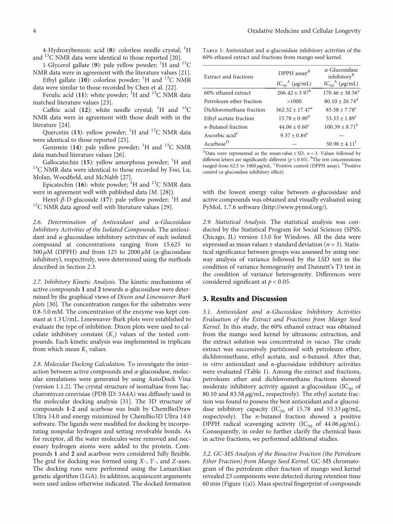
3.1. Antioxidant and α-Glucosidase Inhibitory ActivitiesEvaluation of the Extract and Fractions from Mango SeedKernel. In this study, the 60% ethanol extract was obtainedfrom the mango seed kernel by ultrasonic extraction, andthe extract solution was concentrated in vacuo. The crudeextract was successively partitioned with petroleum ether,dichloromethane, ethyl acetate, and n-butanol. After that,in vitro antioxidant and α-glucosidase inhibitory activitieswere evaluated (Table 1). Among the extract and fractions,petroleum ether and dichloromethane fractions showedmoderate inhibitory activity against α-glucosidase (IC50 of80.10 and 83.58μg/mL, respectively). The ethyl acetate frac-tion was found to possess the best antioxidant and α-glucosi-dase inhibitory capacity (IC50 of 15.78 and 53.33μg/mL,respectively). The n-butanol fraction showed a positiveDPPH radical scavenging activity (IC50 of 44.06μg/mL).Consequently, in order to further clarify the chemical basisin active fractions, we performed additional studies.
3.2. GC-MS Analysis of the Bioactive Fraction (the PetroleumEther Fraction) from Mango Seed Kernel. GC-MS chromato-gram of the petroleum ether fraction of mango seed kernelrevealed 23 components were detected during retention time60min (Figure 1(a)). Mass spectral fingerprint of compounds
Table 1: Antioxidant and α-glucosidase inhibitory activities of the60% ethanol extract and fractions from mango seed kernel.
Extract and fractionsDPPH assayB
α-GlucosidaseinhibitoryB
IC50A (μg/mL) IC50
A (μg/mL)
60% ethanol extract 206:42 ± 3:97b 170:46 ± 38:56a
Petroleum ether fraction >1000 80:10 ± 20:74d
Dichloromethane fraction 362:32 ± 17:47a 83:58 ± 7:78c
Ethyl acetate fraction 15:78 ± 0:90d 53:33 ± 1:89e
n-Butanol fraction 44:06 ± 0:60c 100:39 ± 8:71b
Ascorbic acidC 9:37 ± 0:84e —
AcarboseD — 50:90 ± 4:11fAData were represented as the mean value ± SD, n = 3. Values followed bydifferent letters are significantly different (p ≤ 0:05). BThe test concentrationsranged from 62.5 to 1000μg/mL. CPositive control (DPPH assay). DPositivecontrol (α-glucosidase inhibitory effect).
4 Oxidative Medicine and Cellular Longevity
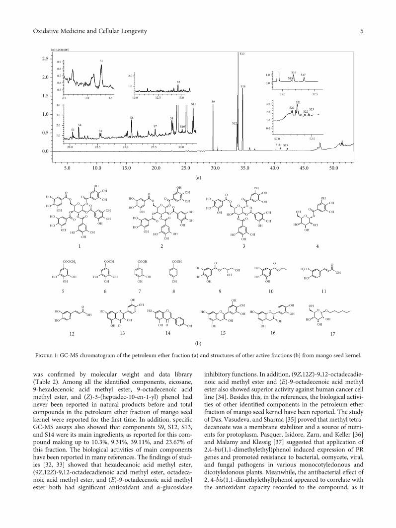
was confirmed by molecular weight and data library(Table 2). Among all the identified components, eicosane,9-hexadecenoic acid methyl ester, 9-octadecenoic acidmethyl ester, and (Z)-3-(heptadec-10-en-1-yl) phenol hadnever been reported in natural products before and totalcompounds in the petroleum ether fraction of mango seedkernel were reported for the first time. In addition, specificGC-MS assays also showed that components S9, S12, S13,and S14 were its main ingredients, as reported for this com-pound making up to 10.3%, 9.31%, 39.11%, and 23.67% ofthis fraction. The biological activities of main componentshave been reported in many references. The findings of stud-ies [32, 33] showed that hexadecanoic acid methyl ester,(9Z,12Z)-9,12-octadecadienoic acid methyl ester, octadeca-noic acid methyl ester, and (E)-9-octadecenoic acid methylester both had significant antioxidant and α-glucosidase
inhibitory functions. In addition, (9Z,12Z)-9,12-octadecadie-noic acid methyl ester and (E)-9-octadecenoic acid methylester also showed superior activity against human cancer cellline [34]. Besides this, in the references, the biological activi-ties of other identified components in the petroleum etherfraction of mango seed kernel have been reported. The studyof Das, Vasudeva, and Sharma [35] proved that methyl tetra-decanoate was a membrane stabilizer and a source of nutri-ents for protoplasm. Pasquer, Isidore, Zarn, and Keller [36]and Malamy and Klessig [37] suggested that application of2,4-bis(1,1-dimethylethyl)phenol induced expression of PRgenes and promoted resistance to bacterial, oomycete, viral,and fungal pathogens in various monocotyledonous anddicotyledonous plants. Meanwhile, the antibacterial effect of2, 4-bis(1,1-dimethylethyl)phenol appeared to correlate withthe antioxidant capacity recorded to the compound, as it
5.0
( 10,000,000)
0.0
0.5
1.0
1.50.5
2.5 3.0 3.5
0.6
0.7
0.8
0.9
2.0
2.5
10.0 15.0 20.0 25.0 30.0 35.0 40.0 45.0 50.0
10.0 12.5 15.0
82
S1
0.0
1.0
35.0 37.5
0.0
1.0
2.0
3.0
50.0 52.5
1.0
2.0
20.0
S3S4
S5
S6
S7
S17S16
S20S21
S18 S19
S22S23
S15
S8
S10
S11S9
S14
S13
S12
1.0
2.0
3.0
4.0
22.5 25.0 27.5 30.0
(a)
HOO
OO
OHOH
OH
HO
HO
OH
HOO
O
HOOH
HOOOO
OOO HO
OH
OH
OH
HO OH
COOCH3
OHHO OH
COOH
OH
OHOH
OH
OHHO
HOO
O
OH
OHOH
OH
COOH
OH
COOH
OH
HOO
OO
OHOH
OH
HO
HO
OH HO HOOOO
OOO HO
OH
OH
OH
OH
HO
O O
HOHO
OHO
OHOH
OH
HO
OO
HOHO
HOO
OO
OHOH
OH
HO
HO
OH
HOO
O
HOOH
HOOOO
OOO HO
OH
1 2 3 4
5 6 7 8 9 10 11
12 13 14 15 16 17
OH
OH
HO
HOO
O
OHHO
H3COOH
O
HO
HO HOOH
OH O
OO
OH
OHOH
OHHO
OH
OHO
OH O
O
OH
OH
OHHO
OH
O
(b)
Figure 1: GC-MS chromatogram of the petroleum ether fraction (a) and structures of other active fractions (b) from mango seed kernel.
5Oxidative Medicine and Cellular Longevity
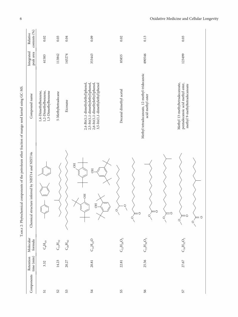
Table2:Phytochem
icalcompo
nentsof
thepetroleum
etherfraction
ofmango
seed
kernelusingGC-M
S.
Com
ponents
Retention
time(m
in)
Molecular
form
ula
Chemicalstructureinferred
byNIST14
andNIST14s
Com
poun
dname
Integrated
peak
area
Relative
contents(%
)
S13.32
C8H
10
1,4-Dim
ethylbenzene,
1,2-Dim
ethylbenzene,
1,3-Dim
ethylbenzene
61583
0.02
S214.23
C15H
325-Methyltetradecane
113842
0.03
S320.27
C20H
42Eicosane
145274
0.04
S420.81
C14H
22O
OH
OH
OH
OH
2,4-Bis(1,1-dim
ethylethyl)pheno
l,2,5-bis(1,1-dimethylethyl)pheno
l,2,6-bis(1,1-dimethylethyl)pheno
l,3,5-bis(1,1-dimethylethyl)pheno
l
353443
0.09
S522.81
C12H
26O2
O
ODecanaldimethylacetal
85855
0.02
S625.56
C15H
30O2
O
O O
O
Methyltetradecano
ate,12-m
ethyl-tridecanoic
acid
methylester
490546
0.13
S727.67
C16H
32O2
O
O O
OO
O
Methyl13-methyltetradecanoate,
pentadecanoicacid
methylester,
methyl9-m
ethyltetradecano
ate
123499
0.03
6 Oxidative Medicine and Cellular Longevity
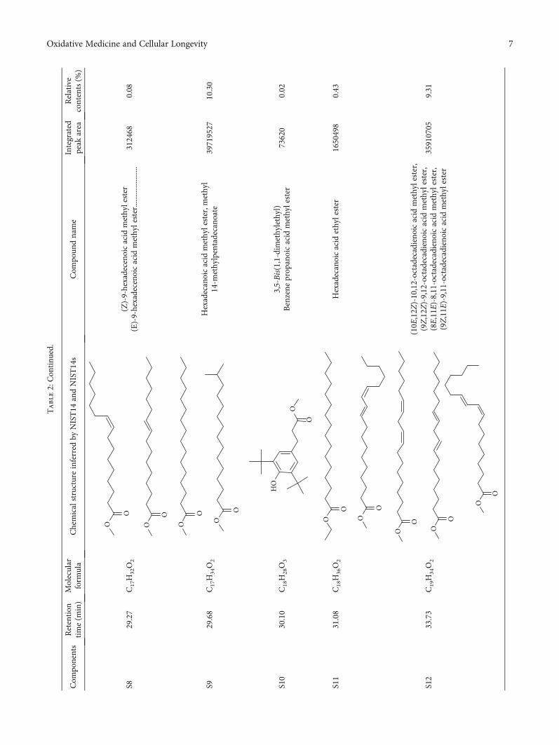
Table2:Con
tinu
ed.
Com
ponents
Retention
time(m
in)
Molecular
form
ula
Chemicalstructureinferred
byNIST14
andNIST14s
Com
poun
dname
Integrated
peak
area
Relative
contents(%
)
S829.27
C17H
32O2
O O
OO
(Z)-9-hexadeceno
icacid
methylester
(E)-9-hexadeceno
icacid
methylester......................
312468
0.08
S929.68
C17H
34O2
O
O O
O
Hexadecanoicacid
methylester,m
ethyl
14-m
ethylpentadecano
ate
39719527
10.30
S10
30.10
C18H
28O3
O
O
HO
3,5-Bis(1,1-dim
ethylethyl)
Benzene
prop
anoicacid
methylester
73620
0.02
S11
31.08
C18H
36O2
O
OHexadecanoicacid
ethylester
1650498
0.43
S12
33.73
C19H
34O2
O
O
O
O O
O
O
O
(10E
,12Z
)-10,12-octadecadienoicacid
methylester,
(9Z,12Z
)-9,12-octadecadieno
icacid
methylester,
(8E,11E)-8,11-octadecadienoicacid
methylester,
(9Z,11E
)-9,11-octadecadieno
icacid
methylester
35910705
9.31
7Oxidative Medicine and Cellular Longevity
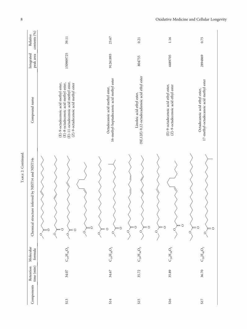
Table2:Con
tinu
ed.
Com
ponents
Retention
time(m
in)
Molecular
form
ula
Chemicalstructureinferred
byNIST14
andNIST14s
Com
poun
dname
Integrated
peak
area
Relative
contents(%
)
S13
34.07
C19H
36O2
O O O
O
OOOO
(E)-9-octadeceno
icacid
methylester,
(E)-8-octadeceno
icacid
methylester,
(E)-11-octadecenoicacid
methylester,
(Z)-9-octadeceno
icacid
methylester
150909725
39.11
S14
34.67
C19H
38O2
O
O O
O
Octadecanoicacid
methylester,
16-m
ethyl-heptadecanoicacid
methylester
91261893
23.67
S15
35.72
C20H
36O2
O O
OO
Lino
leicacid
ethylester,
(9E,12E
)-9,12-octadecadieno
icacid
ethylester
804755
0.21
S16
35.89
C20H
38O2
O
O O
O
(E)-9-octadeceno
icacid
ethylester,
(Z)-9-octadeceno
icacid
ethylester
4489705
1.16
S17
36.70
C20H
40O2
O O
OO
Octadecanoicacid
ethylester,
17-m
ethyl-octadecano
icacid
methylester
2894869
0.75
8 Oxidative Medicine and Cellular Longevity
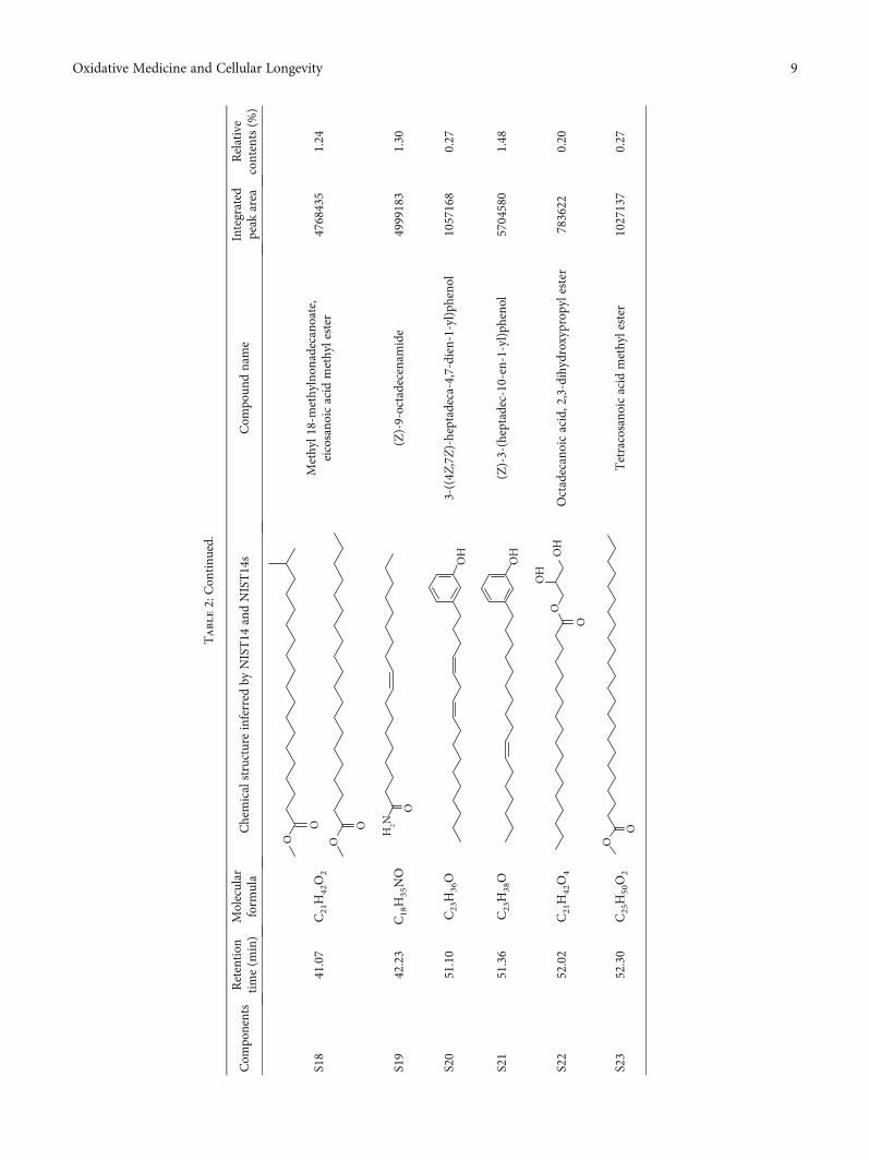
Table2:Con
tinu
ed.
Com
ponents
Retention
time(m
in)
Molecular
form
ula
Chemicalstructureinferred
byNIST14
andNIST14s
Com
poun
dname
Integrated
peak
area
Relative
contents(%
)
S18
41.07
C21H
42O2
O O
OO
Methyl18-methylnon
adecanoate,
eicosano
icacid
methylester
4768435
1.24
S19
42.23
C18H
35NO
O
H2N
(Z)-9-octadecenamide
4999183
1.30
S20
51.10
C23H
36O
OH
3-((4Z
,7Z)-heptadeca-4,7-dien-1-yl)ph
enol
1057168
0.27
S21
51.36
C23H
38O
OH
(Z)-3-(heptadec-10-en-1-yl)pheno
l5704580
1.48
S22
52.02
C21H
42O4
OH
OH
O
OOctadecanoicacid,2,3-dihydroxyprop
ylester
783622
0.20
S23
52.30
C25H
50O2
O
OTetracosano
icacid
methylester
1027137
0.27
9Oxidative Medicine and Cellular Longevity
restrained the production of ROS in both Phytophthora andAspergillus cinnamomi [38].
3.3. Isolation and Purification of Compounds in ActiveFractions from Mango Seed Kernel. Based on the activityresults of extract and all fractions, dichloromethane, ethylacetate, and n-butanol fractions were applied to further isola-tion and purification by chromotography on Sephadex LH-20 column, silica gel column, ODS, and preparative TLC, aswell as semipreparative HPLC, et al. Finally, 17 compoundswere obtained. Their structures were identified as 1, 2, 3, 4,6-penta-O-galloyl-β-D-glucoside (1) [14], 1, 2, 3, 4, 6-penta-O-galloyl-α-D-glucoside (2) [15], 1, 2, 3, 6-tetra-O-galloyl-β-D-glucoside (3) [16], 1-O-(3, 4, 5-trihydroxyben-zoyl)-β-D-glucoside (4) [17], methyl galate (5) [18], gallicacid (6) [39], 3,4-dihydroxybenzoic acid (7) [19], 4-hydroxybenzoic acid (8) [20], 1-glycerol gallate (9) [21], ethylgallate (10) (H. [22]), ferulic acid (11) [23], caffeic acid (12)[24], quercetin (13) [25], genistein (14) [26], gallocatechin(15) [27], epicatechin (16) (M. [28]), and hexyl-β-D-gluco-side (17) [29] by comparing their NMR spectroscopic datawith data reported in the above references. Compounds 2,9, 15, and 17 were isolated from the genus Mangifera forthe first time.
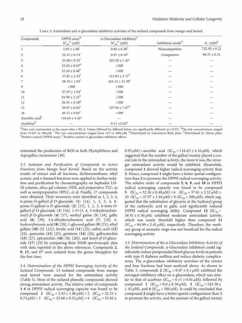
3.4. Determination of the DPPH Scavenging Activity of theIsolated Compounds. 13 isolated compounds from mangoseed kernel were assayed for the antioxidant activity(Table 3). Most of the isolated phenolic compounds showedstrong antioxidant activity. The relative order of compounds1-4 in DPPH radical scavenging capacity was found to becompound 1 (IC50 = 2:93 ± 1:06μM)> 2 (IC50 = 22:31 ±0:74 μM)> 3 (IC50 = 23:68 ± 0:52μM)> 4 (IC50 = 52:82 ±
0:93μM)> ascorbic acid (IC50 = 116:63 ± 4:16 μM), whichsuggested that the number of the galloyl moiety played a cru-cial role in the antioxidant activity, the more it was, the stron-ger antioxidant activity would be exhibited. Meanwhile,compound 1 showed higher radical scavenging activity than2. Hence, compound 1might have a better spatial configura-tion than 2 to promote the DPPH radical scavenging activity.The relative order of compounds 5, 6, 9, and 10 in DPPHradical scavenging capacity was found to be compound5 (IC50 = 32:26 ± 0:48μM)> 6 (IC50 = 37:81 ± 2:52 μM)>10 (IC50 = 37:97 ± 1:94μM)>9 (IC50 > 500 μM), which sug-gested that the substitution of glycerin at the hydroxyl groupof the carboxylic acid in gallic acid significantly reducedDPPH radical scavenging ability. Compound 12 (IC50 =36:91 ± 0:30μM) exhibited moderate antioxidant activity,which was nearly threefold higher than compound 11(IC50 = 94:99 ± 2:41μM), respectively. Therefore, the meth-oxy group at aromatic rings was not beneficial for the radicalscavenging activity.
3.5. Determination of the α-Glucosidase Inhibitory Activity ofthe Isolated Compounds. α-Glucosidase inhibitors could sig-nificantly reduce postprandial blood glucose levels in patientswith type II diabetes mellitus and reduce diabetic complica-tions. The α-glucosidase inhibitory activities of the extractand four fractions had been analyzed above. As shown inTable 3, compounds 2 (IC50 = 0:07 ± 0:1 μM) exhibited thestrongest inhibitory effect on α-glucosidase, which was simi-lar to that of acarbose (IC50 = 0:11 ± 0:01 μM), followed bycompound 1 (IC50 = 0:6 ± 0:36μM), 3 (IC50 = 243:30 ±1:43μM), and 4 (IC50 > 500 μM). It could be concluded thatcompound 2might have a better spatial configuration than 1to promote the activity, and the amount of the galloyl moiety
Table 3: Antioxidant and α-glucosidase inhibitory activities of the isolated compounds from mango seed kernel.
Compounds DPPH assayB α-Glucosidase inhibitoryC
IC50A (μM) IC50
A (μM) Inhibition modeD K i (nM)E
1 2:93 ± 1:06j 0:60 ± 0:36e Noncompetitive 732:95 ± 0:222 22:31 ± 0:74i 0:07 ± 0:10e Competitive 98:37 ± 0:313 23:68 ± 0:52i 243:30 ± 1:43c — —
4 52:82 ± 0:93d >500 — —
5 32:26 ± 0:48h >500 — —
6 37:81 ± 2:52f 313:03 ± 3:71b — —
7 38:19 ± 1:03f 425:12 ± 31:50a — —
9 >500 >500 — —
10 37:97 ± 1:94f >500 — —
11 94:99 ± 2:41b >500 — —
12 36:91 ± 0:30g >500 — —
13 59:97 ± 0:82c 137:94 ± 7:07d — —
16 40:15 ± 0:82e >500 — —
Ascorbic acidF 116:63 ± 4:16a — — —
AcarboseG — 0:11 ± 0:01e — —AData were represented as the mean value ± SD, 6. Values followed by different letters are significantly different (p ≤ 0:05). BThe test concentrations rangedfrom 15.625 to 500 μM. CThe test concentrations ranged from 125 to 2000 μM. DDetermined by Lineweaver-Burk plots. EDetermined by Dixon plots.FPositive control (DPPH assay). GPositive control (α-glucosidase inhibitory effect).
10 Oxidative Medicine and Cellular Longevity
played a crucial role in α-glucosidase inhibition. Compound6 (IC50 = 313:03 ± 3:71μM) showed low α-glucosidase inhib-itory activity. Compounds 5, 9, and 10 were reported noinhibitory activity. These results indicated that the substitu-tion of methoxyl, ethyoxyl, and glyceryl at the hydroxylgroup on the carboxylic acid of gallic acid could reduce theinhibition of α-glucosidase. Meanwhile, compound 6 exhib-ited higher α-glucosidase inhibitory potential than 7(IC50 = 425:12 ± 31:50μM). From this study, it indicated thatincreasing the number of hydroxyl group on the aromaticring could enhance the inhibitory activity of α-glucosidase.
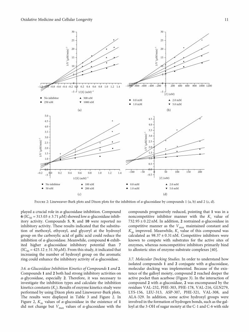
3.6. α-Glucosidase Inhibition Kinetics of Compounds 1 and 2.Compounds 1 and 2 both had strong inhibitory activities onα-glycosidase, especially 2. Therefore, it was necessary toinvestigate the inhibition types and calculate the inhibitionkinetics constants (K i). Results of enzyme kinetics study wereperformed by using Dixon plots and Lineweaver-Burk plots.The results were displayed in Table 3 and Figure 2. InFigure 2, Km values of α-glucosidase in the existence of 1did not change but Vmax values of α-glucosidase with the
compounds progressively reduced, pointing that 1 was in anoncompetitive inhibitor manner with the K i value of732:95 ± 0:22 nM. In addition, 2 restrained α-glucosidase incompetitive manner as the Vmax maintained constant andKm improved. Meanwhile, K i value of this compound wascalculated as 98:37 ± 0:31 nM. Competitive inhibitors wereknown to compete with substrates for the active sites ofenzymes, whereas noncompetitive inhibitors primarily bindto allosteric sites of enzyme-substrate complexes [40].
3.7. Molecular Docking Studies. In order to understand howisolated compounds 1 and 2 conjugate with α-glucosidase,molecular docking was implemented. Because of the exis-tence of the galloyl moiety, compound 2 reached deeper theactive pocket than acarbose (Figure 3). In the interaction ofcompound 2 with α-glucosidase, 2 was encompassed by theresidues VAL-232, PHE-303, PHE-178, VAL-216, GLN279,LYS-156, LEU-313, ASP-307, PHE-321, VAL-308, andALA-329. In addition, some active hydroxyl groups wereinvolved in the formation of hydrogen bonds, such as the gal-loyl at the 3-OH of sugar moiety at the C-1 and C-6 with side
–1.2 –1.0 –0.8 –0.6 –0.4 –0.2
–5
0
5
10
15
20
No inhibitor
25
30
0.0 0.2 0.4 0.6 0.8
1/[S] (mM)–1
1/V
(𝜇M
/min
)–1
1.0 1.2 1.4
250 nM500 nM1000 nM
(a)
–5
–1000 –800 –600 –400 –200 0 200 400 600 800 1000 12000
5
10
15
20
0.8 mM
25
30
[I] (nM)
1/V
(𝜇M
/min
)–1
1.0 mM2.0 mM5.0 mM
(b)
–0.2–5
No inhibitor
0.20.0 0.4 0.6 0.8
1/[S] (mM)–1
0.0
0.5
1.0
1.5
2.0
2.5
3.0
3.5
4.0
4.5
5.0
1.0 1.2 1.4
50 nM100 nM200 nM
1/V
(𝜇M
/min
)–1
(c)
1/V
(𝜇M
/min
)–1
0.8 mM1.0 mM
2.0 mM5.0 mM
–100 –50 0 50 100 150 200
[I] (nM)–0.5
0.0
0.5
1.0
1.5
2.0
2.5
3.0
3.5
4.0
4.5
(d)
Figure 2: Lineweaver-Burk plots and Dixon plots for the inhibition of α-glucosidase by compounds 1 (a, b) and 2 (c, d).
11Oxidative Medicine and Cellular Longevity
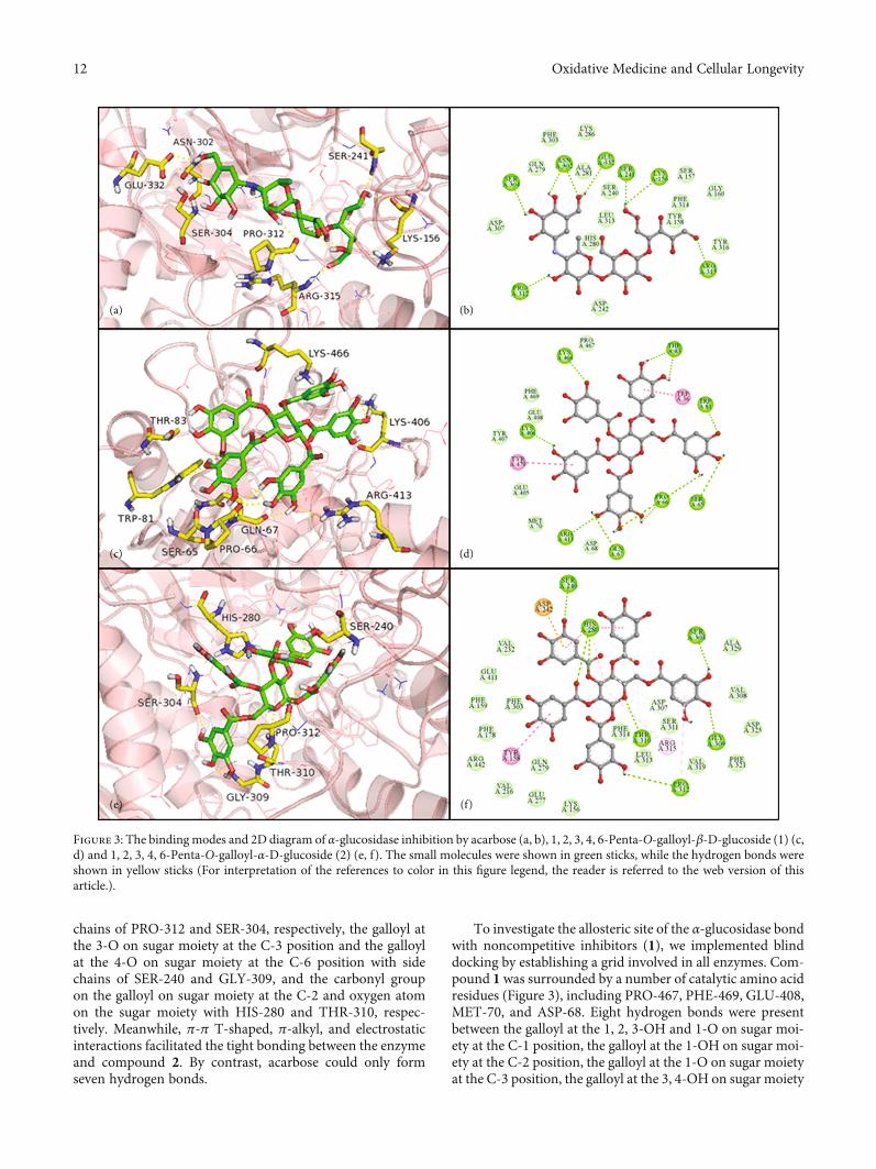
chains of PRO-312 and SER-304, respectively, the galloyl atthe 3-O on sugar moiety at the C-3 position and the galloylat the 4-O on sugar moiety at the C-6 position with sidechains of SER-240 and GLY-309, and the carbonyl groupon the galloyl on sugar moiety at the C-2 and oxygen atomon the sugar moiety with HIS-280 and THR-310, respec-tively. Meanwhile, π-π T-shaped, π-alkyl, and electrostaticinteractions facilitated the tight bonding between the enzymeand compound 2. By contrast, acarbose could only formseven hydrogen bonds.
To investigate the allosteric site of the α-glucosidase bondwith noncompetitive inhibitors (1), we implemented blinddocking by establishing a grid involved in all enzymes. Com-pound 1 was surrounded by a number of catalytic amino acidresidues (Figure 3), including PRO-467, PHE-469, GLU-408,MET-70, and ASP-68. Eight hydrogen bonds were presentbetween the galloyl at the 1, 2, 3-OH and 1-O on sugar moi-ety at the C-1 position, the galloyl at the 1-OH on sugar moi-ety at the C-2 position, the galloyl at the 1-O on sugar moietyat the C-3 position, the galloyl at the 3, 4-OH on sugar moiety
(a) (b)
(c) (d)
(e) (f)
Figure 3: The bindingmodes and 2D diagram of α-glucosidase inhibition by acarbose (a, b), 1, 2, 3, 4, 6-Penta-O-galloyl-β-D-glucoside (1) (c,d) and 1, 2, 3, 4, 6-Penta-O-galloyl-α-D-glucoside (2) (e, f). The small molecules were shown in green sticks, while the hydrogen bonds wereshown in yellow sticks (For interpretation of the references to color in this figure legend, the reader is referred to the web version of thisarticle.).
12 Oxidative Medicine and Cellular Longevity
at the C-4 position and the galloyl at the 3, 4-OH, and 5-O onsugar moiety at the C-6 position of compound 1 with GLN-67, PRO-66, ARG-413, LYS-406, LYS-466, THR-83, TRP-81, and SER-65, respectively. The aromatic rings of thegalloyl on sugar moiety at the C-4 and C-2 positions of com-pound 1 formed π-π T-shaped and π-π stacked interactionswith TRP-36 and TYR-470, respectively.
In recent years, molecular docking simulations havebecome an important tool for understanding the interactionmode and the structure-activity relationships of ligands withreceptors. The in silico research results of compounds 1 and2 were in agreement with the IC50 data and the enzymekinetic study in vitro. The integration of enzyme activity,kinetics, and molecular docking studies provided principleinsights into the molecular basis underlying ligand bindingaffinity and α-glucosidase inhibition.
4. Conclusions
In summary, antioxidant and α-glucosidase inhibitory activ-ities of the extract, fractions, and isolated compounds frommango seed kernel were first documented. The petroleumether, dichloromethane, ethyl acetate, and n-butanol frac-tions exhibited different degrees of antioxidant and α-gluco-sidase inhibitory activity. In order to further clarify thechemical basis, a total of 23 ingredients were identified byefficient and fast GC-MS analysis from the petroleum etherfraction. As far as we know, it was the first report on thedetailed compounds of the petroleum ether fraction ofmango seed kernel. Among all the identified components,(Z)-3-(Heptadec-10-en-1-yl) phenol, eicosane, 9-octadecenoicacid methyl ester, and 9-hexadecenoic acid methyl esterhad never been reported before. In addition, 17 compoundswere isolated and identified from other active fractions ofmango seed kernel. 1, 2, 3, 4, 6-penta-O-galloyl-α-D-gluco-side (2), 1-glycerol gallate (9), gallocatechin (15), andhexyl-β-D-glucoside (17) were isolated from Mangifera forthe first time. Among isolated components, 1, 2, 3, 4, 6-penta-O-galloyl-β-D-glucoside (1) and 1, 2, 3, 4, 6-penta-O-galloyl-α-D-glucoside (2) showed stronger DPPH radicalscavenging and α-glucosidase inhibitory capacities thanothers. The structure-activity relationship has been discussed.According to the kinetic research, 2 inhibited α-glucosidase ina competitive manner, whereas 1 acted in a noncompetitivemanner. Molecular docking studies showed that compound1 accommodated well in the allosteric site of the α-glucosidaseinteracting with a number of crucial amino residues. Com-pound 2 reached deeper the active site pocket of α-glucosidasethan acarbose. The stability of binding was enhanced by π-πT-shaped, π-alkyl, hydrogen bond, and electrostatic interac-tions. These results might be useful to investigate the pharma-cological activity in this fruit. Meanwhile, our study provided achemical basis for the further development and utilization ofmango seed kernel in the food and medicine fields.
Data Availability
The underlying data supporting our findings can be foundand generated during the study.
Conflicts of Interest
The authors declare no conflict of interest, financial orotherwise.
Authors’ Contributions
Dan Yang and Xuee Chen are joint the first authors. Thestudy conceptualization was done by Jun Yin and ZhihuiLiu. Isolation and purification of compounds were conductedby Xuee Chen and Xida Liu. The data analysis was done byDan Yang, Janxiu Zhai, and Sikai Li. Dan Yang and XueeChen wrote and prepared the original draft. Zhihui Liu, JunYin, and Na Han wrote, reviewed, and edited the manuscript.All authors have read and agreed to the published version ofthe manuscript.
Supplementary Materials
Figure S1: GC-MS chromatogram of the petroleum etherfraction from mango kernel. Figure S2 to Figure S24: GC-MS spectrum of component 1 to 23. Table S1: molecularinteractions of the α-glucosidase (3A4A) active site withcompounds 1 and 2. (Supplementary Materials)
References
[1] B. Lorenzati, C. Zucco, S. Miglietta, F. Lamberti, and G. Bruno,“Oral hypoglycemic drugs: pathophysiological basis of theirmechanism of actionoral hypoglycemic drugs: pathophysio-logical basis of their mechanism of action,” Pharmaceuticals,vol. 3, no. 9, pp. 3005–3020, 2010.
[2] S. H. Jo, K. S. Ha, K. S. Moon, O. H. Lee, H. D. Jang, and Y. I.Kwon, “In vitro and in vivo antihyperglycemic effects of Omija(Schizandra chinensis) fruit,” International journal of molecu-lar sciences, vol. 12, no. 2, pp. 1359–1370, 2011.
[3] S. Imran, M. Taha, N. H. Ismail et al., “Synthesis of novelflavone hydrazones: in-vitro evaluation of α-glucosidase inhi-bition, QSAR analysis and docking studies,” European Journalof Medicinal Chemistry, vol. 105, pp. 156–170, 2015.
[4] M. R. Bhandari, N. Jong-Anurakkun, G. Hong, andJ. Kawabata, “α-Glucosidase and α-amylase inhibitory activ-ities of Nepalese medicinal herb Pakhanbhed (Bergeniaciliata, Haw.),” Food Chemistry, vol. 106, no. 1, pp. 247–252, 2008.
[5] M. A. T. Phan, J. Wang, J. Tang, Y. Z. Lee, and K. Ng, “Evalu-ation of α-glucosidase inhibition potential of some flavonoidsfrom Epimedium brevicornum,” LWT - Food Science andTechnology, vol. 53, no. 2, pp. 492–498, 2013.
[6] J. S. Johansen, A. K. Harris, D. J. Rychly, and A. Ergul, “Oxida-tive stress and the use of antioxidants in diabetes: linking basicscience to clinical practice,” Cardiovascular Diabetology, vol. 4,no. 1, p. 5, 2005.
[7] J. P. Kehrer, T. E. Tipple, J. D. Robertson, and C. V. Smith,“Free radicals and reactive oxygen species,” in Reference Mod-ule in Biomedical Sciences, Elsevier, 2015.
[8] M. sDar, P. Oak, H. Chidley, A. Deshpande, A. Giri, andV. Gupta, “Nutrient and flavor content of mango (Mangiferaindica L.) cultivars,” Nutritional Composition of Fruit Culti-vars, pp. 445–467, 2016.
13Oxidative Medicine and Cellular Longevity
[9] C. Namngam and P. Pinsirodom, “Antioxidant properties,selected enzyme inhibition capacities, and a cosmetic creamformulation of Thai mango seed kernel extracts,” TropicalJournal of Pharmaceutical Research, vol. 16, no. 1, p. 9,2017.
[10] E. A. Irondi, G. Oboh, A. A. Akindahunsi, A. A. Boligon, andM. L. Athayde, “Phenolic composition and inhibitory activityof Mangifera indica and Mucuna urens seeds extracts againstkey enzymes linked to the pathology and complications of type2 diabetes,” Asian Pacific Journal of Tropical Biomedicine,vol. 4, no. 11, pp. 903–910, 2014.
[11] J. Pan, X. M. Yi, S. J. Zhang et al., “Bioactive phenolics frommango leaves (Mangifera indica L.),” Industrial Crops andProducts, vol. 111, pp. 400–406, 2018.
[12] M. B. Hlila, H. Mosbah, K. Majouli et al., “α-Glucosidase inhi-bition by Tunisian Scabiosa arenaria Forssk. extracts,” Interna-tional Journal of Biological Macromolecules, vol. 77, pp. 383–389, 2015.
[13] X. Gao, J. Wei, L. Hong, S. Fan, G. Hu, and J. Jia, “Comparativeanalysis of chemical composition, anti-inflammatory activityand antitumor activity in essential oils from Siegesbeckiaorientalis, S. glabrescens and S. pubescens with an ITSsequence analysis,” Molecules, vol. 23, no. 9, p. 2185, 2018.
[14] R. T. D. Santos, L. L. Hiramoto, J. H. G. Lago, P. Sartorelli,A. G. Tempone, and E. G. Pinto, “Anti-trypanosomal activityof 1,2,3,4,6-penta-O-galloyl-β-D-glucose isolated from Plec-tranthus barbatus Andrews (Lamiaceae),” Quimica Nova,vol. 35, no. 11, pp. 2229–2332, 2012.
[15] M. Nishizawa, T. Yamagishi, G. Nonaka, I. Nishioka, andH. Bando, “Novel hydrolyzable tannins from Nuphar Japoni-cum DC,” Chemical and Pharmaceutical Bulletin, vol. 30,no. 3, pp. 1094–1097, 1982.
[16] T. Okuda, T. Yoshida, and T. Hatano, “New methods of ana-lyzing tannins,” Journal of Natural Products, vol. 52, no. 1,pp. 1–31, 1989.
[17] Y. Zhang, D. L. DeWitt, S. Murugesan, and M. G. Nair, “Novellipid-peroxidation- and cyclooxygenase-inhibitory tanninsfrom Picrorhiza kurroa seeds,” Chemistry and biodiversity,vol. 1, no. 3, pp. 426–441, 2004.
[18] D. J. Kwon and Y. S. Bae, “Chemical constituents from thestem bark of Acer barbinerve,” Chemistry of Natural Com-pounds, vol. 47, no. 4, pp. 636–638, 2011.
[19] S. Chunhakant and C. Chaicharoenpong, “Antityrosinase,antioxidant, and cytotoxic activities of phytochemical constit-uents from Manilkara zapota L. bark,” Molecules, vol. 24,no. 15, p. 2798, 2019.
[20] S. Swarnalatha, A. Umamaheswari, and A. Puratchikody,“Immunomodulatory activity of kaempferol 5-O-β-d-gluco-pyranoside from Indigofera aspalathoides Vahl ex DC. (Papi-lionaceae),” Medicinal Chemistry Research, vol. 24, no. 7,pp. 2889–2897, 2015.
[21] A. F. Artamonov, F. S. Nigmatullina, M. T. Aldabergenova,and B. Z. Dzhiembaev, “Synthesis of α-monoglycerides of aro-matic acids,” Chemistry of Natural Compounds, vol. 35, no. 4,pp. 404–408, 1999.
[22] H. Chen, M. Li, C. Zhang et al., “Isolation and identificationof the anti-oxidant constituents from Loropetalum chinense(R. Brown) oliv. based on UHPLC-Q-TOF-MS/MS,” Mole-cules, vol. 23, no. 7, p. 1720, 2018.
[23] S. Aynur and K. Zeynep, “Phytochemical investigations onchemical constituents of Taraxacum bessarabicum (Hornem.)
Hand.-Mazz. subsp. bessarabicum (Hornem.) Hand.-Mazz,”Iranian Journal of Pharmaceutical Research, vol. 18, pp. 400–405, 2019.
[24] L. Zhou, D. Li, J. Wang, Y. Liu, and J. Wu, “Antibacterialphenolic compounds from the spines of Gleditsia sinensisLam,” Natural Product Research, vol. 21, no. 4, pp. 283–291, 2007.
[25] B. B. Gao, G. M. She, and D. M. She, “Chemical constituentsand biological activities of plants from the genus Ligustrum,”Chemistry and biodiversity, vol. 10, no. 1, pp. 96–128, 2013.
[26] J. S. Yoon, S. H. Sung, J. H. Park, and Y. C. Kim, “Flavonoidsfrom Spatholobus suberectus,” Archives of pharmacal research,vol. 27, no. 6, pp. 589–592, 2004.
[27] L. Y. Foo, Y. Lu, A. L. Molan, D. R. Woodfield, and W. C.McNabb, “The phenols and prodelphinidins of white cloverflowers,” Phytochemistry, vol. 54, no. 5, pp. 539–548, 2000.
[28] M. Chen and S. Yu, “Characterization of lipophilized mono-meric and oligomeric grape seed flavan-3-ol derivatives,” Jour-nal of Agricultural and Food Chemistry, vol. 65, no. 40,pp. 8875–8883, 2017.
[29] L. Shuai, K. Hai-xue, Y. Okada, and O. Toru, “Chemical con-stituents of Bidens bipinnata (II),” Chinese Traditional andHerbal Drugs, vol. 35, no. 9, pp. 972–975, 2004.
[30] G. Renda, S. Sari, B. Barut et al., “α-Glucosidase inhibitoryeffects of polyphenols from Geranium asphodeloides: inhibi-tion kinetics and mechanistic insights through in vitro and insilico studies,” Bioorganic Chemistry, vol. 81, pp. 545–552,2018.
[31] L. Xu, W. Li, Z. Chen et al., “Inhibitory effect of epigallocate-chin-3-O-gallate on α-glucosidase and its hypoglycemic effectvia targeting PI3K/AKT signaling pathway in L6 skeletal mus-cle cells,” International Journal of Biological Macromolecules,vol. 125, pp. 605–611, 2019.
[32] A. G. Cruz, A. I. Mtz-Enríquez, L. Díaz-Jiménez et al., “Produc-tion of fatty acid methyl esters and bioactive compounds fromcitrus wax,” Waste Management, vol. 102, pp. 48–55, 2020.
[33] R. Agada, W. A. Usman, S. Shehu, and D. Thagariki, “In vitroand in vivo inhibitory effects of Carica papaya seed on α-amy-lase and α-glucosidase enzymes,” Heliyon, vol. 6, no. 3,p. e03618, 2020.
[34] F. R. Yu, X. Z. Lian, H. Y. Guo et al., “Isolation and character-ization of methyl esters and derivatives from Euphorbia kansui(Euphorbiaceae) and their inhibitory effects on the humanSGC-7901 cells,” Journal of pharmacy and pharmaceutical sci-ences, vol. 8, no. 3, pp. 528–535, 2005.
[35] S. Das, N. Vasudeva, and S. Sharma, “Chemical composition ofethanol extract of Macrotyloma uniflorum (Lam.) Verdc.using GC-MS spectroscopy,” Organic and Medicinal Chemis-try Letters, vol. 4, no. 1, 2014.
[36] F. Pasquer, E. Isidore, J. Zarn, and B. Keller, “Specific patternsof changes in wheat gene expression after treatment with threeantifungal compounds,” Plant molecular biology, vol. 57, no. 5,pp. 693–707, 2005.
[37] J. Malamy and D. F. Klessig, “Salicylic acid and plant diseaseresistance,” Plant journal, vol. 2, no. 5, pp. 643–654, 1992.
[38] R. C. María Teresa, V. G. Rosaura, C. M. Elda, and G. P.Ernesto, “The avocado defense compound phenol-2,4-bis(1,1-dimethylethyl) is induced by arachidonic acid and actsvia the inhibition of hydrogen peroxide production by patho-gens,” Physiological and Molecular Plant Pathology, vol. 87,pp. 32–41, 2014.
14 Oxidative Medicine and Cellular Longevity
[39] L. Ni, W. Liang, W. Huang et al., “Chemical constituents ofEuscaphis konishii and their inhibitory activities,” Chemistryof Natural Compounds, vol. 55, no. 5, pp. 832–834, 2019.
[40] D. Şöhretoğlu, S. Sari, B. Barut, and A. Özel, “Discovery ofpotent α-glucosidase inhibitor flavonols: insights into mecha-nism of action through inhibition kinetics and docking simu-lations,” Bioorganic Chemistry, vol. 79, pp. 257–264, 2018.
15Oxidative Medicine and Cellular Longevity